a novel type of dual-modality molecular probe for mr and ... · a novel type of dual-modality...
TRANSCRIPT

A Novel Type of Dual-Modality Molecular Probe for MRand Nuclear Imaging of Tumor: Preparation,
Characterization and in Vivo Application
Shujie Liu,†,‡ Bing Jia,‡,§ Ruirui Qiao,†,‡ Zhi Yang,| Zilin Yu,§ Zhaofei Liu,§
Kan Liu,⊥ Jiyun Shi,§ Han Ouyang,⊥ Fan Wang,*,§ and Mingyuan Gao*,†
Institute of Chemistry, CAS, Bei Yi Jie 2, Beijing 100190, China, Medical IsotopesResearch Center, Peking UniVersity, Xueyuan Road 38, Beijing 100191, China, Cancer
Hospital of Chinese Academy of Medical Sciences, Pan Jia Yuan Nan Li 17, Beijing100021, China, and Oncology School of Peking UniVersity, Fu Cheng Lu 52,
100036, China
Received October 30, 2008; Accepted June 15, 2009
Abstract: A novel dual-modality molecular probe composed of biocompatible Fe3O4 nanocrystal,monoclonal antibody and radionuclide was designed and prepared. All functional componentsin the dual-modality molecular probe, i.e., Fe3O4, PEG, mAb 3H11 and 125I, were chemicallybonded together for forming a stable molecular probe. Systematic in vitro experiments werecarried out for evaluating the biological activity of the antibody in the targeting probe. A seriesof in vivo experiments were performed based on the dual-modality imaging probe for detectingxenografted tumors in nude mice by MRI and γ-imaging techniques. The pharmacokinetics ofthe dual-modality molecular probe in tumor-bearing nude mice was studied.
Keywords: Dual-modality molecular probe; MRI; nuclear imaging; Fe3O4 nanocrystals; tumor
IntroductionMolecular imaging has become a booming area as it will
surely offer revolutionary tools not only for fundamentaldiagnostic studies but also for clinical applications.1-3 Overthe past decade, different types of molecular imaging probes
have been developed for fluorescence imaging,4,5 magneticresonance imaging (MRI),2,6-10 nuclear imaging,11,12 etc.Among them, nanoparticle-based molecular probes havebecome more and more attractive. First, the size-dependentphysical properties of nanoparticles such as fluorescentquantum dots and magnetic iron oxide nanoparticles arepotentially useful for acquiring detailed physiological infor-mation at the cellular level in Vitro and even in ViVo.5,13
Second, in comparison with small molecular imaging agents,nanoparticles injected into the bloodstream present veryspecial size- and surface-dependent in ViVo behaviors gov-erned by the uptake effects of reticuloendothelial system(RES), thus offering new possibilities for disease diagnosisvia different passive targeting modes.14,15 Third, the nano-particles can provide multiple surface binding sites for furtherattaching various types of bioactive molecules, consequently
* To whom correspondence should be addressed. M.G.: Mailingaddress, Institute of Chemistry, CAS, Bei Yi Jie 2, ZhongGuan Cun, Beijing 100190, China. Fax: 86-10-82613214. Tel:86-10-82613214. E-mail: [email protected]. F.W.: Mailingaddress, Medical Isotopes Research Center, Peking University,Xueyuan Road 38, Beijing 100191, China. Fax and tel: 86-10-82801145. E-mail: [email protected].
† Institute of Chemistry, CAS.‡ These authors contributed equally to this work.§ Medical Isotopes Research Center, Peking University.| Oncology School of Peking University.⊥ Cancer Hospital of Chinese Academy of Medical Sciences.(1) Weissleder, R.; Ntziachristos, V. Shedding light onto live mo-
lecular targets. Nat. Med. 2003, 9, 123–128.(2) Lee, J. H.; Huh, Y. M.; Jun, Y. W.; Seo, J. W.; Jang, J. T.; Song,
H. T.; Kim, S.; Cho, E. J.; Yoon, H. G.; Suh, J. S.; Cheon, J.Artificially engineered magnetic nanoparticles for ultra-sensitivemolecular imaging. Nat. Med. 2007, 13, 95–99.
(3) Weissleder, R.; Pittet, M. J. Imaging in the era of molecularoncology. Nature 2008, 452, 580–589.
(4) (a) Bruchez, M. J.; Moronne, M.; Gin, P.; Weiss, S.; Alivisatos,A. P. Semiconductor nanocrystals as fluorescent biological labels.Science 1998, 281, 2013–2016. (b) Chan, W. C.; Nie, S. Quantumdot bioconjugates for ultrasensitive nonisotopic detection. Science1998, 281, 2016–2018.
articles
1074 MOLECULAR PHARMACEUTICS VOL. 6, NO. 4, 1074–1082 10.1021/mp900143a CCC: $40.75 2009 American Chemical SocietyPublished on Web 06/15/2009
Dow
nloa
ded
by I
NST
ITU
TE
OF
CH
EM
IST
RY
on
Aug
ust 4
, 200
9Pu
blis
hed
on J
une
15, 2
009
on h
ttp://
pubs
.acs
.org
| do
i: 10
.102
1/m
p900
143a

enabling the establishment of nanoparticle-based molecularimaging probes for targeting specific disease sites throughmolecular recognition.
MRI, as a noninvasive tool, has been widely used in theclinic worldwide as it can produce images with extraordinar-ily high temporal and spatial resolution. It has also been
demonstrated that magnetic iron oxide nanoparticles are ableto effectively enhance the contrast between normal andpathological tissues. Pioneered by Weissleder, both smallparticles of iron oxide (SPIO) and ultrasmall particles of ironoxide (USPIO) have received intensive investigations in earlytumor in ViVo detections by either passive or active targetingmode.2,8-10,16 In the latter mode, various types of targetingmolecules are required to conjugate to iron oxide nanopar-ticles for specifically targeting the receptors expressed onthe tumor cell surface. This concept has been proven to befeasible by different groups, consequently raising greatresearch interests of using USPIO-enhanced MRI in earlytumor detection in ViVo.2,8,9 Yet, the chemical synthesis ofbiocompatible iron oxide nanoparticles with satisfactoryblood circulation time and particle surface functionalizabilityremain far from optimal. Furthermore, the in ViVo behaviorsof nanoparticles with different surface structures are neces-sarily subjected to systematic investigations.17
In our previous investigations, we have established aninnovative synthetic route for achieving water-soluble andbiocompatible magnetite nanocrystals via a “one-pot” reac-tion.18-20 PEG (poly(ethylene glycol))-coated Fe3O4 nano-crystals, as the direct product of the “one-pot” reaction, weredemonstrated to be useful in disease detection via eitherpassive mode or active mode.9,21 Following on from ourprevious investigations, we have recently been working onthe synthesis and in ViVo applications of a novel type of dual-modality molecular probe designed for tumor detection byboth MRI and single-photon emission computed tomography(SPECT). For a molecular imaging tool to be useful, highenough sensitivity, sufficiently high spatial resolution and ahigh degree of specificity for the target of interest areessentially required. MRI is powerful for its high spatialresolution and tomographic capabilities, but limited by itslow signal sensitivities. By contrast, nuclear imaging modali-ties such as positron emission tomography (PET) and SPECTare characterized by very high sensitivity, up to 10-12 mol/L, but with relatively low imaging resolution. Therefore, itis meaningful to design a dual-modality molecular imaging
(5) Gao, X.; Cui, Y.; Levenson, R. M.; Chung, L. W.; Nie, S. InViVo cancer targeting and imaging with semiconductor quantumdots. Nat. Biotechnol. 2004, 22, 969–976.
(6) Wisniewski, T.; Pankiewicz, J.; Scholtzova, H.; Fernando, G.;Chabalgoity, J. A.; Ji, Y.; Wadghiri, Y. Z.; Gan, W. B.; Tang,C. Y.; Turnbull, D. H.; Mathis, C. A.; Kascsak, R.; Klunk, W. E.;Carp, R. I.; Frangione, B.; Sigurdsson, E. M.; Sadowski, M.; Carp,I. Imaging and therapeutic approaches for [beta]-sheet structuresin prion and Alzheimer’s diseases. Neurobiol. Aging 2004, 25,S30–S31.
(7) De, V. I.; Lesterhuis, W. J.; Barentsz, J. O.; Verdijk, P.; van,K. J.; Boerman, O. C.; Oyen, W. J.; Bonenkamp, J. J.; Boezeman,J. B.; Adema, G. J.; Bulte, J. W.; Scheenen, T. W.; Punt, C. J.;Heerschap, A.; Figdor, C. G. Magnetic resonance tracking ofdendritic cells in melanoma patients for monitoring of cellulartherapy. Nat. Biotechnol. 2005, 23, 1407–1413.
(8) Huh, Y. M.; Jun, Y. W.; Song, H. T.; Kim, S.; Choi, J. S.; Lee,J. H.; Yoon, S.; Kim, K. S.; Shin, J. S.; Suh, J. S.; Cheon, J. InViVo magnetic resonance detection of cancer by using multifunc-tional magnetic nanocrystals. J. Am. Chem. Soc. 2005, 127,12387–12391.
(9) Hu, F. Q.; Wei, L.; Zhou, Z.; Ran, Y. L.; Li, Z.; Gao, M. Y.Preparation of Biocompatible Magnetite Nanocrystals for In ViVoMagnetic Resonance Detection of Cancer. AdV. Mater. 2006, 18,2553–2556.
(10) Sun, C.; Veiseh, O.; Gunn, J.; Fang, C.; Hansen, S.; Lee, D.; Sze,R.; Ellenbogen, R. G.; Olson, J.; Zhang, M. In ViVo MRI detectionof gliomas by chlorotoxin-conjugated superparamagnetic nano-probes. Small 2008, 4, 372–379.
(11) Czernin, J.; Phelps, M. E. Positron emission tomography scanning:current and future applications. Annu. ReV. Med. 2002, 53, 89–112.
(12) (a) Wu, Y.; Zhang, X.; Xiong, Z.; Cheng, Z.; Fisher, D. R.; Liu,S.; Gambhir, S. S.; Chen, X. microPET imaging of glioma integrin{alpha}v{beta}3 expression using 64Cu-labeled tetrameric RGDpeptide. J. Nucl. Med. 2005, 46, 1707–1718. (b) Jia, B.; Shi, J. Y.;Yang, Z.; Xu, B.; Liu, Z. F.; Zhao, H. Y.; Liu, S.; Wang, F. 99mTc-labeled cyclic RGDfK dimer: Initial evaluation for SPECTimaging of glioma integrin {alpha}v{beta}3 expression. Biocon-jugate Chem. 2006, 17, 1069–1076.
(13) Wang, Y. X.; Hussain, S. M.; Krestin, G. P. Superparamagneticiron oxide contrast agents: physicochemical characteristics andapplications in MR imaging. Eur. Radiol. 2001, 11, 2319–2331.
(14) (a) Anzai, Y.; Blackwell, K. E.; Hirschowitz, S. L.; Rogers, J. W.;Sato, Y.; Yuh, W. T.; Runge, V. M.; Morris, M. R.; McLachlan,S. J.; Lufkin, R. B. Initial clinical experience with dextran-coatedsuperparamagnetic iron oxide for detection of lymph nodemetastases in patients with head and neck cancer. Radiology 1994,192, 709–715. (b) Ros, P. R.; Freeny, P. C.; Harms, S. E.; Seltzer,S. E.; Davis, P. L.; Chan, T. W.; Stillman, A. E.; Muroff, L. R.;Runge, V. M.; Nissenbaum, M. A.; et al. Hepatic MR imagingwith ferumoxides: a multicenter clinical trial of the safety andefficacy in the detection of focal hepatic lesions. Radiology 1995,196, 481–488.
(15) Corot, C.; Robert, P.; Idee, J. M.; Port, M. Recent advances iniron oxide nanocrystal technology for medical imaging. AdV. DrugDeliVery ReV. 2006, 58, 1471–1504.
(16) Harisinghani, M. G.; Barentsz, J.; Hahn, P. F.; Deserno, W. M.;Tabatabaei, S.; van, K. C.; de, R. J.; Weissleder, R. Noninvasivedetection of clinically occult lymph-node metastases in prostatecancer. N. Engl. J. Med 2003, 348, 2491–2499.
(17) Qiao, R. R.; Yang, C. H.; Gao, M. Y. Superparamagnetic ironoxide nanoparticles: from preparations to in vivo MRI applications.J. Mater. Chem. 2009, DOI: 10.1039/b902394a.
(18) Li, Z.; Chen, H.; Bao, H. B.; Gao, M. Y. One-pot reaction tosynthesize water-soluble magnetite nanocrystals. Chem. Mater.2004, 16, 1391–1393.
(19) Hu, F.; Li, Z.; Tu, C.; Gao, M. Y. Preparation of magnetitenanocrystals with surface reactive moieties by one-pot reaction.J. Colloid Interface Sci. 2007, 311, 469–474.
(20) Li, Z.; Wei, L.; Gao, M. Y.; Lei, H. One-pot reaction to synthesizebiocompatible magnetite nanoparticles. AdV. Mater. 2005, 17,1001–1005.
(21) Wei, L.; Zhou, G.; Li, Z.; He, L.; Gao, M. Y.; Tan, J.; Lei, H.Detection of toxoplasmic lesions in mouse brain by USPIO-enhanced magnetic resonance imaging. Magn. Reson. Imaging2007, 25, 1442–1448.
Fe3O4 Nanoparticle-Based Dual-Modality Imaging Probe articles
VOL. 6, NO. 4 MOLECULAR PHARMACEUTICS 1075
Dow
nloa
ded
by I
NST
ITU
TE
OF
CH
EM
IST
RY
on
Aug
ust 4
, 200
9Pu
blis
hed
on J
une
15, 2
009
on h
ttp://
pubs
.acs
.org
| do
i: 10
.102
1/m
p900
143a

probe capable of being detected by these complementaryimaging techniques for further increasing the diagnosticaccuracy.22,23
Herein, we report a new dual-modality molecular probecomposed of biocompatible Fe3O4 nanocrystal, monoclonalantibody and radionuclide. The biocompatible Fe3O4 nano-crystals were prepared by modifying our “one-pot” syntheticroute established previously.9,18-20 In similarity, R,ω-dicar-boxyl-terminated PEG (HOOC-PEG-COOH) was adoptedas the particle surface capping agent for providing thenanocrystal biocompatibility, meanwhile offering free sur-face carboxylic acid groups for further covalently bindingwith a targeting molecule,9,19 i.e., antigastric cancer mono-clonal antibody 3H11 (mAb 3H11) in the current investiga-tion. The latter was labeled with radionuclide 125I for SPECTimaging. All the functional components in the dual-modalitymolecular probe, i.e., Fe3O4, PEG, mAb 3H11 and 125I, werechemically bonded together for forming a stable molecularprobe. Systematic in Vitro experiments were carried out forevaluating the biological activity of the antibody in thetargeting probe. A series of in ViVo experiments wereperformed for detecting xenografted tumors in nude miceby MRI and γ-imaging techniques. The pharmacokineticsof the molecular probe in tumor-bearing nude mice wasstudied by γ-counter analyses.
Experimental SectionChemicals. Iron(III) acetylacetonate (Fe(acac)3) was pur-
chased from Aldrich (14024-18-1) and used after 2 timesrecrystallizations. Analytical grade chemicals such as ethanol,ether, and diphenyl oxide were purchased from SinopharmChemical Reagent Beijing, Co., Ltd. Diphenyl oxide wasused after further purification by reduced pressure distillation.EDC (1-ethyl-3-(3-dimethylaminopropyl carbodiimide), 39391)and sulfo-NHS (N-hydroxysulfosuccinimide sodium salt,56485) from Fluka and SDS (sodium dodecyl sulfate, L5750)from Sigma were used as received. Iodogen (1,3,4,6-tet-rachloro-3R,6R-diphenylglycouril, T0656) was purchasedfrom Sigma-Aldrich and used without further purification.Iodine-125 carrier free radionuclide (Na125I in 0.1 M NaOH)used in this study was purchased from Perkin-Elmer (NEZ033,Perkin-Elmer) at a radioactive concentration of 350 mCi/mL. Antigastric cancer monoclonal antibody 3H11 andhuman gastric cancer cell line BGC823 were obtained as
gifts from the Oncology School of Peking University.24
Murine immunoglobulin G (mIgG) was purchased fromSigma (I5381). HOOC-PEG-COOH was synthesized ac-cording to literature.20
Preparation of Biocompatible Fe3O4 Nanocrystals. Astock solution of diphenyl oxide (15 mL) containingFe(acac)3 (0.06 M), HOOC-PEG-COOH (0.12 M, Mn )2000) and oleylamine (0.24 M) was prepared. After beingpurged with nitrogen, the reaction mixture was quickly heatedup and kept refluxing for 24 h. Then, ether was introducedinto the reaction mixture to precipitate the resultant Fe3O4
nanocrystals, which were subsequently collected by cen-trifugation. After that, the precipitate was redissolved inethanol followed by addition of ether as precipitant. Typi-cally, this purifying procedure was repeated for three cycles.The PEG-coated Fe3O4 nanocrystals finally obtained weredissolved in either Milli-Q water or PBS (phosphate buffersaline) for further experiments. The Fe3O4 nanocrystalsstabilized by HOOC-PEG-COOH have surface carboxylicacid groups available for further covalently conjugating theFe3O4 nanocrystals to molecules bearing amine groups viathe EDC/NHS mediated amidation reaction, as demonstratedbefore.9,19
Colloidal Stability of the Biocompatible Fe3O4 Nanocryst-als. The colloidal stability of the PEG-coated Fe3O4 nano-crystals under physiological conditions was studied by thedynamic light scattering (DLS) method. The hydrodynamicsizes of Fe3O4 nanocrystals in both physiological saline andfetal bovine serum (FBS, Invitrogen) were monitored andcompared with those recorded from Fe3O4 nanocrystalsprepared in 2-pyrrolidone rather than diphenyl oxide ac-cording to a previous report.9
Radiolabeling. mAb 3H11 was labeled with 125I using theIodogen method as previously reported.25 Briefly, 450 µgof mAb 3H11 (225 µL, 2.0 mg/mL) in phosphate buffer (0.2M, pH 7.4) and 1.05 mCi of Na125I (3 µL, 350 mCi/mL)were added into a glass vial coated with 20 µg of Iodogen.After 5 min of reaction at room temperature, the resultant3H11-125I was purified by a PD-10 Sephadex G-25 column(17-0851-01, GE Healthcare) equilibrated with phosphatebuffer (0.2 M, pH 7.4) to remove unreacted radioiodide. Theradiolabeling yield was 70-85% and radiochemical puritywas greater than 98% after purification, resulting in a specificactivity of 1.6-2.0 mCi/mg. By a similar recipe, the mIgG-125I was also prepared and purified.
(22) Catana, C.; Wu, Y.; Judenhofer, M. S.; Qi, J.; Pichler, B. J.;Cherry, S. R. Simultaneous acquisition of multislice PET and MRimages: initial results with a MR-compatible PET scanner. J. Nucl.Med. 2006, 47, 1968–1976.
(23) DeNardo, S. J.; DeNardo, G. L.; Natarajan, A.; Miers, L. A.;Foreman, A. R.; Gruettner, C.; Adamson, G. N.; Ivkov, R. Thermaldosimetry predictive of efficacy of 111In-ChL6 nanoparticle AMF-induced thermoablative therapy for human breast cancer in mice.J. Nucl. Med. 2007, 48, 437–444.
(24) (a) Guo, J.; Jin, G.; Meng, L.; Ma, H.; Nie, D.; Wu, J.; Yuan, L.;Shou, C. Subcellullar localization of tumor-associated antigen3H11Ag. Biochem. Biophys. Res. Commun. 2004, 324, 922–930.(b) Chen, D.; Shou, C. Molecular cloning of a tumor-associatedantigen recognized by monoclonal antibody 3H11. Biochem.Biophys. Res. Commun. 2001, 280, 99–103.
(25) Salacinski, P. R.; McLean, C.; Sykes, J. E.; Clement-Jones, V. V.;Lowry, P. J. Iodination of proteins, glycoproteins, and peptidesusing a solid-phase oxidizing agent, 1,3,4,6-tetrachloro-3 alpha,6alpha-diphenyl glycoluril (Iodogen). Anal. Biochem. 1981, 117,136–146.
articles Liu et al.
1076 MOLECULAR PHARMACEUTICS VOL. 6, NO. 4
Dow
nloa
ded
by I
NST
ITU
TE
OF
CH
EM
IST
RY
on
Aug
ust 4
, 200
9Pu
blis
hed
on J
une
15, 2
009
on h
ttp://
pubs
.acs
.org
| do
i: 10
.102
1/m
p900
143a

Fe3O4-3H11 Conjugates. The conjugation reaction be-tween Fe3O4 nanocrystals and mAb 3H11 was performed at4 °C. Typically, EDC (2.50 µmol) and sulfo-NHS (6.25µmol) were dissolved in 830 µL of 0.01 M PBS buffersolution containing 2.63 mg of Fe3O4 nanocrystals. Afterapproximately 15 min, 380 µL of 0.01 M PBS buffer solutioncontaining 1.27 mg of mAb 3H11 was introduced. Typically,the reaction lasted for 10 h. The conjugate of Fe3O4-mIgGwas prepared in a similar way and used as a controlthroughout the following in Vitro and in ViVo experiments.By replacing mAb 3H11 with 3H11-125I or replacing mIgGwith mIgG-125I, the Fe3O4-3H11-125I and Fe3O4-mIgG-125Iprobes were prepared, respectively.
Cell Culture and Animal Models. BGC823 Cells weregrown in RPMI-1640 medium supplemented with 10% (v/v) FBS at 37 °C in an atmosphere containing 5% CO2. TheBGC823 tumor model was generated by subcutaneousinjection of ∼5 × 10⁶ cells into proximal thigh and rightupper flank regions of female BALB/c nude mice (4-5weeks old). The mice were used for in ViVo studies whenthe tumor volume reached about 200-300 mm3 (∼10 daysafter inoculation). All the animal experiments were performedaccording to a protocol approved by the Peking UniversityInstitutional Animal Care and Use Committee.
Competitive Binding Assay. BGC823 cells were seededin the wells of 24-well plates (1 × 10⁵ cells/well) andincubated overnight at 37 °C to allow a firm adherence. Afterbeing rinsed twice with PBS buffer, the cells were incubatedwith 3H11-125I (0.15 µCi) at 4 °C for 2 h in the presence ofeither Fe3O4-3H11 or mAb 3H11 with an increasingconcentration (0-1000 nM) in binding buffer (0.01 M PBS,pH 7.4, 1% bovine serum albumin). The total incubationvolume was adjusted to 200 µL. After that, the cells wererinsed three times with cold PBS buffer and then lysedwith 2 M NaOH at room temperature. The cell-associatedradioactivity was determined using a γ-counter (Wallac 1470-002, Perkin-Elmer). The half maximal inhibitory concentra-tions (IC50) of mAb 3H11 and Fe3O4-3H11 to BGC823 cellsagainst 3H11-125I were determined by fitting the data withnonlinear regression using GraphPad Prism (version 4.0,GraphPad Software, Inc.). All experiments were repeatedtwice with triplicate samples.
Binding Specificity. The BGC823 cells were harvested,rinsed three times with PBS, and resuspended in the bindingbuffer. Then, the cells were divided into three groups (1 ×10⁷ cells per group). In the first group, the cells wereincubated with Fe3O4-3H11-125I probe (0.02 µCi), while thecells in the second group were incubated with Fe3O4-3H11-125I probe (0.02 µCi) in the presence of excessive mAb 3H11(1000 folds of the probe), and the tumor cells in the lastgroup were incubated with equivalent amount of Fe3O4-mIgG-125I (0.02 µCi) in the absence of pure mAb 3H11. The totalincubation volume was 1 mL. After being gently rotatedovernight at 4 °C, the cells were washed and the cell-associated radioactivity was determined by γ-counter. Ex-periments were performed with triplicate samples per group.
In Vitro MRI. For the in Vitro MRI experiments, 6 ×10⁷ BGC823 cells were trypsinized with 0.25% of trypsinsolution for 3 min at 37 °C, and washed twice with PBS,then resuspended in binding buffer (2 × 10⁶ cells/mL).Afterward they were incubated in triplicate with Fe3O4-3H11-125I, Fe3O4-mIgG-125I, or Fe3O4 (80 µg of Fe3O4/group)overnight at 4 °C, respectively. After being thoroughlywashed with cold PBS buffer, the cells were lysed by 300µL of 10% SDS. MRI measurements were carried out on a3 T clinical MRI instrument (GE signa 3.0T HD, Milwaukee,WI). The parameters were set as follows: field of view (FOV)) 10 × 10 cm2; matrix size ) 192 × 192; slice thickness )1 mm; echo time (TE) ) 25, 50, 109 ms; repetition time(TR) ) 2100 ms; number of excitations (NEX) ) 8.
In ViWo MRI. Nude mice bearing BGC823 tumor xe-nografts implanted at their proximal thigh and right upperflank regions were anesthetized and then injected via tail veinwith Fe3O4-3H11-125I or Fe3O4-mIgG-125I [(16.8 mg of Fe)/(kg of body weight), n ) 3/group]. At 10 min, 2 h, 4 h, 8 h,12 h, 16 h, 24 h, 48 and 72 h postinjection, the mice weresubjected to the in ViVo MR imaging. For image acquisition,a 1 in. animal quadrature coil, developed by MR Modalityof GE HealthCare, China, was used. The detailed imagingparameters were set as follows: FOV ) 8 × 8 cm2; matrixsize ) 160 × 128; slice thickness ) 1 mm; TE ) 120 ms;TR ) 2500 ms; and NEX ) 4.
γ-Imaging. γ-Imaging studies were performed as previ-ously reported.12b Briefly, the nude mice bearing BGC823tumor xenografts were anesthetized and then injected via tailvein with either Fe3O4-3H11-125I or Fe3O4-mIgG-125I [10mCi/(kg of body weight), corresponding to injection doseof (16.8 mg of Fe)/(kg of body weight); n ) 3/group]. Themice were imaged by using a two-head γ-camera (SI-EMENS, E. CAM) equipped with a low energy high res-olution (LEHR) parallel hole collimator. At 10 min, 4 h, 14 h,24 h, 48 h, 72 and 96 h postinjection, planar images wereacquired and stored digitally in a 128 × 128 matrix. Theacquisition count limits were set at 100 k. For each image,regions of interest (ROI) were drawn over the tumors as wellas the whole body of each mouse. The radio counts wereobtained by summing up the counts from all pixels withinROI. For more quantitative analysis, 1% of injected dose ofFe3O4-3H11-125I was used as an internal reference andimaged simultaneously at each image acquisition time point.
Biodistribution. For the biodistribution study, animalswere anesthetized by intraperitoneal injection of sodiumpentobarbital at a dose of 45.0 mg/kg. Then, the Fe3O4-3H11-125I or 3H11-125I (10 µCi per mouse) was intravenouslyinjected into BGC823 tumor-bearing mice. Animals weresacrificed at 10 min, 14 h, 24 h, 48 and 72 h postinjection,respectively (n ) 4/group). Tumor tissues and other organsof interest (such as blood, heart, liver, spleen, kidney, andstomach) were harvested and measured for radioactivity ina γ-counter. The organ uptake was calculated and expressedin percentage injected dose per organ (% ID/organ).
Fe3O4 Nanoparticle-Based Dual-Modality Imaging Probe articles
VOL. 6, NO. 4 MOLECULAR PHARMACEUTICS 1077
Dow
nloa
ded
by I
NST
ITU
TE
OF
CH
EM
IST
RY
on
Aug
ust 4
, 200
9Pu
blis
hed
on J
une
15, 2
009
on h
ttp://
pubs
.acs
.org
| do
i: 10
.102
1/m
p900
143a

Results and discussionIn our previous investigations, we have demonstrated that
biocompatible Fe3O4 nanocrystals can be prepared via thepyrolysis of Fe(acac)3 in 2-pyrrolidone in the presence ofvarious types of carboxyl-terminated PEG.9,20 In thosepreparations, 2-pyrrolidone not only offers a high enoughreaction temperature for pyrolyzing Fe(acac)3 but also servesas a coordinating solvent.18,19 Nevertheless, due to the smallmolecular size it does not offer strong enough hindranceeffect to the resultant nanocrystals dispersed in solution.17
Therefore, to further enhance the colloidal stability of theresultant nanocrystals in physiological saline and serum, thesynthetic approach was modified in the current investigationby replacing the strong polar solvent 2-pyrrolidone withnonpolar diphenyl oxide. In addition, oleylamine was alsoused to costabilize the resultant nanocrystals together withR,ω-dicarboxyl-terminated PEG (Mn ) 2000). More detailson the chemical synthesis of these new types of particleswill be published elsewhere.
The resultant PEG-coated Fe3O4 nanocrystals were char-acterized by transmission electron microscopy (TEM) andelectron diffraction (data not shown). A representative TEMimage given in Figure 1a shows the general morphology ofthe resultant nanocrystals. Their size distribution is depictedby the histogram shown in Figure 1b. The number-averageparticle size was determined to be of 8.1 nm by measuringmore than 300 particles. Electron diffraction experimentssuggested that the particles shown in Figure 1a are magnetitenanocrystals. The room-temperature magnetization curveshown in Figure 1c further demonstrates that the PEG-coatedFe3O4 nanocrystals are superparamagnetic with a saturationmagnetization of 40.4 emu/g, corresponding to 55.8 emu/(mass of Fe).
Satisfying colloidal stability of the Fe3O4 nanoparticles isgreatly important with respect to in ViVo applications of Fe3O4
nanoparticles as MRI contrast agents. Poor colloidal stabilitywill surely lead to short blood half-time, which is thereforenot suitable for constructing molecular probes.17 In the worst
case, the agglomeration of colloidal particles in blood canalso cause vascular embolism as colloidal particles tend toaggregate in salt environment. To show the enhancedcolloidal stability of the Fe3O4 nanoparticle sample presentedin Figure 1, another PEG-coated Fe3O4 nanoparticle withcomparable size was prepared in 2-pyrrolidone instead ofdiphenyl oxide according to one of our previous publica-tions.9 The colloidal stability of these two nanocrystalsamples in physiological saline (0.9% NaCl) and fetal bovineserum (FBS) was investigated by the DLS method. Thetemporal evolutions of the hydrodynamic size of these twosamples are shown in Figure 2. In physiological saline, theFe3O4 nanocrystals prepared in 2-pyrrolidone quickly floc-culate with hydrodynamic size increasing to >300 nm withintwo hours, and obvious precipitation was practically observedin several hours. In contrast, the Fe3O4 nanocrystals preparedin diphenyl oxide presented an excellent stability with thehydrodynamic size being nearly unchanged for more than 2weeks. In FBS, the nanoparticles prepared in 2-pyrrolidonepresent an increased stability but start to flocculate after 4days, while the nanoparticles shown in Figure 1 keep stablefor more than 2 weeks. All these observations stronglydemonstrate that current synthetic protocol offers biocom-patible Fe3O4 nanoparticles with greatly improved colloidalstability both in physiological saline and in FBS.
The Fe3O4 nanoparticle-based dual-modality molecularprobe was prepared by two different routes. In the first route,mAb 3H11 was first conjugated to Fe3O4 nanocrystal throughR,ω-dicarboxyl-terminated PEG chemically bonded on theparticle surface,19,20 then labeled with 125I by the Iodogen
Figure 1. (a) TEM image, (b) histogram, and (c) room-temperature magnetization curve of the as-prepared PEG-coated Fe3O4 nanocrystals.
Figure 2. Temporal evolutions of the hydrodynamicsizes of the PEG-coated Fe3O4 nanocrystals synthe-sized in 2-pyrrolidone (2-Py) and diphenyl oxide (DO),respectively.
articles Liu et al.
1078 MOLECULAR PHARMACEUTICS VOL. 6, NO. 4
Dow
nloa
ded
by I
NST
ITU
TE
OF
CH
EM
IST
RY
on
Aug
ust 4
, 200
9Pu
blis
hed
on J
une
15, 2
009
on h
ttp://
pubs
.acs
.org
| do
i: 10
.102
1/m
p900
143a
method. In the second route, mAb 3H11 was first labeledwith 125I, then conjugated to the PEG-coated Fe3O4 nano-crystals as mentioned in the Experimental Section. Inpractice, both of these two synthetic routes had nearly thesame effectiveness except that, by the first synthetic route,a purification process prior to radiolabeling was required toremove impurities, introduced by the conjugation reactionmediated by EDC and sulfo-NHS, which interfered with thefollowing radiolabeling reaction. Therefore, the latter syn-thetic route was adopted for preparing Fe3O4-3H11-125I andFe3O4-mIgG-125I probes.
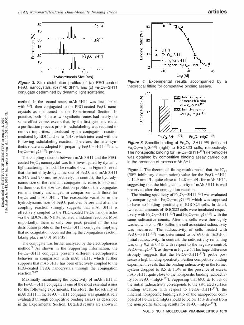
The coupling reaction between mAb 3H11 and the PEG-coated Fe3O4 nanocrystal was first investigated by dynamiclight scattering method. The results shown in Figure 3 revealthat the initial hydrodynamic size of Fe3O4 and mAb 3H11is 24.9 and 9.0 nm, respectively. In contrast, the hydrody-namic size of the resultant conjugate increases to 33.5 nm.Furthermore, the size distribution profile of the conjugatesremains nearly unchanged in comparison with those forFe3O4 and mAb 3H11. The reasonable variation in thehydrodynamic size of Fe3O4 particles before and after thecoupling reaction strongly suggests that mAb 3H11 iseffectively coupled to the PEG-coated Fe3O4 nanoparticlesvia the EDC/sulfo-NHS-mediated amidation reaction. Mostimportantly, there is only one peak present in the sizedistribution profile of the Fe3O4-3H11 conjugate, implyingthat no coagulation occurred during the conjugation reactiontaking place in 0.01 M PBS.
The conjugate was further analyzed by the electrophoresismethod.9 As shown in the Supporting Information, theFe3O4-3H11 conjugate presents different electrophoreticbehavior in comparison with mAb 3H11, which furthersupports that mAb 3H11 has been effectively coupled to thePEG-coated Fe3O4 nanocrystals through the conjugationreaction.9,19
Maximally maintaining the bioactivity of mAb 3H11 inthe Fe3O4-3H11 conjugate is one of the most essential issuesfor the following experiments. Therefore, the bioactivity ofmAb 3H11 in the Fe3O4-3H11 conjugate was quantitativelyevaluated through competitive binding assays as describedin the Experimental Section. Detailed results are shown in
Figure 4. The theoretical fitting results reveal that the IC50
(50% inhibitory concentration) value for the Fe3O4-3H11is 14.9 nmol/L, quite close to 14.8 nmol/L for mAb 3H11,suggesting that the biological activity of mAb 3H11 is wellpreserved after the conjugation reaction.
The binding specificity of Fe3O4-3H11-125I was evaluatedby comparing with Fe3O4-mIgG-125I which was supposedto have no binding specificity to BGC823 cells. In detail,two equal amounts of BGC823 cells were incubated respec-tively with Fe3O4-3H11-125I and Fe3O4-mIgG-125I with thesame radioactive counts. After the cells were thoroughlywashed with cold PBS buffer, the cell-associated radioactivitywas measured. The radioactivity of cells treated withFe3O4-3H11-125I was determined to be 69.0 ( 16.3% ofinitial radioactivity. In contrast, the radioactivity remainingwas only 9.5 ( 0.4% with respect to the negative control,Fe3O4-mIgG-125I, as shown in Figure 5. This huge differencestrongly suggests that the Fe3O4-3H11-125I probe pos-sesses a high binding specificity. Further competitive bindingexperiment reveals that the binding radioactivity in the formersystem dropped to 8.5 ( 1.3% in the presence of excessmAb 3H11, quite close to the nonspecific binding radioactiv-ity for Fe3O4-mIgG-125I. Supposing that 69.0 ( 16.3% ofthe initial radioactivity corresponds to the saturated surfacebinding situation with respect to Fe3O4-3H11-125I, theinherent nonspecific binding ratio of the nanoprobes com-posed of Fe3O4 and mIgG should be below 15% derived fromthe nonspecific binding results for Fe3O4-mIgG-125I.
Figure 3. Size distribution profiles of (a) PEG-coatedFe3O4 nanocystals, (b) mAb 3H11, and (c) Fe3O4-3H11conjugate determined by dynamic light scattering.
Figure 4. Experimental results accompanied by atheoretical fitting for competitive binding assays.
Figure 5. Specific binding of Fe3O4-3H11-125I (left) andFe3O4-mIgG-125I (right) to BGC823 cells, respectively.The nonspecific binding for Fe3O4-3H11-125I (left-middle)was obtained by competitive binding assay carried outin the presence of excess mAb 3H11.
Fe3O4 Nanoparticle-Based Dual-Modality Imaging Probe articles
VOL. 6, NO. 4 MOLECULAR PHARMACEUTICS 1079
Dow
nloa
ded
by I
NST
ITU
TE
OF
CH
EM
IST
RY
on
Aug
ust 4
, 200
9Pu
blis
hed
on J
une
15, 2
009
on h
ttp://
pubs
.acs
.org
| do
i: 10
.102
1/m
p900
143a

The above-mentioned results allowed further in Vitro MRIexperiments to assess the targeting ability of the dual-modality molecular probe to human gastric cancer cell line(BGC823). For comparison, Fe3O4-mIgG-125I was used asa negative control. Four equal portions of a BGC823 cellsolution were prepared, and three of them were used toincubate with Fe3O4-3H11-125I, Fe3O4-mIgG-125I, and thePEG-coated Fe3O4 nanocrystals, respectively. The fourth cellsample was also subjected to the same incubation conditionssimply for comparison. After being thoroughly washed withcold PBS buffer to remove unbound Fe3O4 particles, the cellsamples were lysed and subjected to MRI experiments.Figure 6 presents spin-spin relaxation time (T2)-weightedMR images of these four samples acquired by different echotime. It is quite evident that only the cell sample treated withFe3O4-3H11-125I presents a strongly enhanced MRI signal,suggesting that the Fe3O4-3H11-125I probe can specificallytarget BGC823 cells. From the signal intensity measured atdifferent echo times, the longitudinal relaxivity was calcu-lated to be 14.62 s-1.
On the basis of the aforementioned systematic characteriza-tions as well as successful in Vitro experiments, Fe3O4-3H11-125I was used in the following experiments for in ViVodetecting tumors in BALB/c nude mice bearing BGC823human gastric cancer xenografts implanted at their proximalthigh and right upper flank regions. Fe3O4-mIgG-125I servedas a negative control. T2-weighted MR images acquiredbefore and at different time points after injection ofFe3O4-3H11-125I or Fe3O4-mIgG-125I are shown in Figure7a. More quantitative results on the temporal variations ofT2 values are given in Figure 7b. In general, the T2 valueof tumor starts to decrease 4 h after the injection ofFe3O4-3H11-125I, reaching a minimum at 24 h by a decreaseof ∼30%, and then increases. However, the hypointensiveT2 signal remains throughout the whole inspected timewindow. With respect to the control experiment, a decreasedT2 value of the tumor also appears but with a much lowerT2 decrease (<10%), which is probably caused by thenonspecific binding of Fe3O4-mIgG-125I to tumor cells asdemonstrated by in Vitro experiments. However, the T2 valueof the tumor completely recovers 60 h after the administrationof Fe3O4-mIgG-125I. The significant difference between T2
enhancing effects caused by Fe3O4-3H11-125I and Fe3O4-mIgG-125I, respectively, clearly demonstrates that the dual-modalitymolecular probe possesses specific targeting ability to tumorvia the interaction between the targeting molecule and thereceptor expressed on tumor cells. Even though Fe3O4-mIgG-125I also gives rise to a weaker hypointensive T2 signal,which is a subject of our ongoing study, the latter part ofthe time window for T2 signal enhanced by Fe3O4-3H11-125I remains available for unambiguously identifying tumor.In comparison with our previous investigations,9 the generalvariation of T2 value presents a rather similar behavior duringthe first 24 h postinjection. But the maximal variation of T2value in the current case reaches ∼30%, much higher thanthat (∼10%) observed in our previous investigations.9 Thisimproved MR enhancing effect is undoubtedly attributed tothe new synthetic route which produces nanoparticles withmuch improved biocompatibility. For example, by dopingiron oxide particles with different types of transition metalssuch as Mn2+, Fe2+, Co2+, and Ni2+, Suh and Cheon haveinvented a series of “magnetism-engineered” iron oxide(MEIO) particles. Among them, MnFe2O4 particles presentthe highest magnetic susceptibility. Nevertheless, the R2(relativity value being equal to 1/T2) enhancement factorbased on MnFe2O4 particles is only 31% in in ViVo tumordetection.2 By converting the maximum T2 enhancementfactor of the current dual-modality probe to R2 enhancementfactor, ∆R2 reaches ∼42%. Most importantly, the hy-pointensive T2 signal observed in the current investigationkeeps increasing until 24 h postinjection, suggesting that thePEG-coated Fe3O4 nanocrystals prepared by the currentsynthetic route possess greatly improved circulating behaviorin blood.
Since nuclear imaging techniques including PET andSPECT offer much higher sensitivity than MRI, therefore,it is practically meaningful to develop nuclear-MR dual-modality molecular imaging probes for more unambiguouslydetecting early stage tumors as well as other types ofdiseases. We chose 125I as radionuclide in the current in-vestigations due to the following reasons: (1) 125I can be usedfor SPECT imaging for animal experiments; (2) it also offersa sensitive approach for tracing the in ViVo behaviors of thenanocrystals with different size and surface structure, whichwill provide important feedback for optimizing the nano-particle-based imaging probes; (3) it is relatively safer tohandle; (4) the established synthetic protocol can easily beextended to other iodine isotopes, such as 123I, 124I, and 131I,which are useful not only for SPECT and PET imaging butalso for radiotherapy. With respect to the current investiga-tions, to crosscheck the in ViVo tumor imaging results basedon MRI experiments, another group of tumor-bearing nudemice was chosen for γ-imaging upon injections ofFe3O4-3H11-125I with Fe3O4-mIgG-125I being used ascontrol. Representative images acquired at 10 min, 14 h, 24 h,48 h, and 72 h postinjection were selected and are shown inFigure 7c. 1% of the injected dose of Fe3O4-3H11-125I wasplaced in the top middle of each image as a reference foranalyzing the imaging results. Quantitative data extracted
Figure 6. T2-weighted MR images of lysed BGC823cells treated with Fe3O4-3H11-125I, Fe3O4-mIgG-125I,and Fe3O4, respectively; the sample on the bottom isthe untreated cell sample for comparison.
articles Liu et al.
1080 MOLECULAR PHARMACEUTICS VOL. 6, NO. 4
Dow
nloa
ded
by I
NST
ITU
TE
OF
CH
EM
IST
RY
on
Aug
ust 4
, 200
9Pu
blis
hed
on J
une
15, 2
009
on h
ttp://
pubs
.acs
.org
| do
i: 10
.102
1/m
p900
143a

from the images are shown in Figure 7d. It is quite obviousthat the dual-modality molecular probe can effectively targetthe tumors implanted at both proximal thigh and right upperflank regions. The γ-signals of tumors in the experimentalgroup are much more intensive than that in the control group.Moreover, the general targeting behavior of Fe3O4-3H11-125I is well in consistence with that obtained by MRI.
Apart from providing the signal from tumor sites, theγ-imaging experiments also offer the possibility to monitorthe biodistribution and in ViVo characteristics of the nano-probes, which is difficult to achieve solely by MRI. Theresults shown in Figure 7c as well as more parallel controlstudies not shown here reveal that the Fe3O4-based probesare quickly distributed in liver within the first 10 min afterinjection. Then, the nanoparticle probes start to be washedout of liver and give rise to detectable accumulation at tumorsites approximately 14 h postinjection. Eventually, themolecular probe distributed more in tumor than in liver. Asliver is a blood-rich organ, the initial quick distribution inliver suggests that there are almost no nanoparticle probesstacked in the bloodstream due to the possible coagulationcaused by their interactions with serum proteins and bloodcells. The following decay of the liver signal together withthe signal increase in tumors implies that the current Fe3O4
nanocrystal possesses excellent stealth nature, which isessential for in ViVo molecular imaging upon the use ofnanoparticle-based molecular probes. It is also worthy ofmentioning that the whole body γ-counts decrease againsttime, suggesting that at least 125I is being eliminated againsttime. It is interesting to ask how these nanoprobes aremetabolized. Note that in the γ-images shown in Figure 7as well as those not being presented, the bladder starts toshow γ-signal shortly after the injection of the probes.Therefore, the urine was collected after the injection of the
probes and then subjected to TEM measurements. It wasconfirmed that Fe3O4 nanoparticles were present in urine.
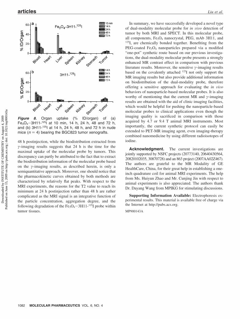
To provide more accurate biodistribution information onthe Fe3O4-3H11-125I probe, tumor tissues and other organsof interest, including blood, heart, liver, spleen, kidney, andstomach, were harvested from BGC823 tumor-bearing miceat different time points postinjection, and then the radioactiv-ity of the tumor tissues as well as the aforementioned organswas determined by γ-counter. The results shown in Figure8a demonstrate that the Fe3O4-3H11-125I probe is heavilydistributed in liver within 10 min (by 38.4 ( 3.6%) apartfrom being in blood (by 51.2 ( 6.8%). From then on, theliver uptake of the probe keeps decreasing within theinspected time window. Meanwhile, the tumor uptake ofthe probe quickly increases within 24 h postinjection to 4.1( 0.6% and reaches a maximum of 4.7 ( 0.7% at 48 hpostinjection. Eventually, the molecular probe is distributedmore in tumor than in liver. For comparison, the biodistri-bution behavior of 3H11-125I was also investigated. Theresults shown in Figure 8b suggest that the general behaviorof the Fe3O4-3H11-125I probe follows that of 3H11-125Iexcept that the former one presents slightly heavier distribu-tion in liver, which is probably caused by the bigger size ofthe Fe3O4-3H11-125I probe. It is worthy of mentioning thatthere is a huge percentage of the molecular probe remainingin blood at the third day postinjection, suggesting that thecurrent dual-modality molecular probe possesses excellentcirculating behavior in blood.
The results shown in Figure 8 strongly support the generalpharmacokinetics behavior of the Fe3O4-3H11-125I probeextracted from both MRI (Figure 7b) and γ-imaging mea-surements (Figure 7d). The only discrepancy is that theresults shown in Figure 8 suggest that the tumor uptake ofthe dual-modality molecular probe reaches its maximum at
Figure 7. (a) T2-weighted MR images of tumor-bearing nude mice acquired before and at different time points afterintravenous injections of Fe3O4-3H11-125I and Fe3O4-mIgG-125I, respectively. (b) Variations of T2 values of tumorsafter the injections of Fe3O4-3H11-125I (solid line) and Fe3O4-mIgG-125I (dotted line), respectively. (c) γ-Images oftumor-bearing nude mice captured at different times postinjection. (d) The normalized γ-counts extracted afterinjection of Fe3O4-3H11-125I from the whole body (dotted line), the tumor at upper flank region (solid line), and thetumor at proximal thigh region (dashed line) of the mouse at the left-hand side in each image, by using 1% injectiondose (top middle) as internal reference. Both MR and γ-images are color-coded for better showing the tumors.
Fe3O4 Nanoparticle-Based Dual-Modality Imaging Probe articles
VOL. 6, NO. 4 MOLECULAR PHARMACEUTICS 1081
Dow
nloa
ded
by I
NST
ITU
TE
OF
CH
EM
IST
RY
on
Aug
ust 4
, 200
9Pu
blis
hed
on J
une
15, 2
009
on h
ttp://
pubs
.acs
.org
| do
i: 10
.102
1/m
p900
143a
48 h postinjection, while the biodistribution extracted fromγ-imaging results suggests that 24 h is the time for themaximal uptake of the molecular probe by tumors. Thisdiscrepancy can partly be attributed to the fact that to extractthe biodistribution information of the molecular probe basedon the γ-imaging results, as described herein, is only asemiquantitative approach. Moreover, one should notice thatthe pharmacokinetic curves obtained by both methods arecharacterized by relatively flat peaks. With respect to theMRI experiments, the reasons for the T2 value to reach itsminimum at 24 h postinjection rather than 48 h are rathercomplicated as the MRI signal is an integrative function ofthe particle concentration, aggregation degree, and thefollowing degradation of the Fe3O4-3H11-125I probe withintumor tissues.
In summary, we have successfully developed a novel typeof dual-modality molecular probe for in ViVo detection oftumor by both MRI and SPECT. In this molecular probe,all components, Fe3O4 nanocrystal, PEG, mAb 3H11, and125I, are chemically bonded together. Benefiting from thePEG-coated Fe3O4 nanoparticles prepared via a modified“one-pot” synthetic route based on our previous investiga-tions, the dual-modality molecular probe presents a stronglyenhanced MR contrast effect in comparison with previousliterature results. Moreover, the sensitive γ-imaging resultsbased on the covalently attached 125I not only support theMR imaging results but also provide additional informationon biodistribution of the dual-modality probe, thereforeoffering a sensitive approach for evaluating the in ViVobehaviors of nanoparticle-based molecular probes. It is alsoworthy of mentioning that the current MR and γ-imagingresults are obtained with the aid of clinic imaging facilities,which would be helpful for pushing the nanoparticle-basedmolecular probes to clinical applications even though theimaging quality is sacrificed in comparison with thoseacquired by 4.7 or 9.4 T animal MRI instruments. Mostimportantly, the current synthetic protocol can easily beextended to PET-MR imaging agent, even imaging-therapycombined nanomedicine by using different radioisotopes ofiodine.
Acknowledgment. The current investigations arejointly supported by NSFC projects (20773140, 20640430564,20820102035, 30870728) and an 863 project (2007AA02Z467).The authors are grateful to the MR Modality of GEHealthCare, China, for their great help in establishing a one-inch quadrature coil for animal MRI experiments. The helpfrom Ms. Huiyun Zhao and Mr. Cunjing Jin with respect toanimal experiments is also appreciated. The authors thankDr. Dayang Wang from MPIKG for stimulating discussions.
Supporting Information Available: Electrophoresis ex-perimental results. This material is available free of charge viathe Internet at http://pubs.acs.org.
MP900143A
Figure 8. Organ uptake (% ID/organ) of (a)Fe3O4-3H11-125I at 10 min, 14 h, 24 h, 48 and 72 h,and (b) 3H11-125I at 14 h, 24 h, 48 h, and 72 h in nudemice (n ) 4) bearing the BGC823 tumor xenografts.
articles Liu et al.
1082 MOLECULAR PHARMACEUTICS VOL. 6, NO. 4
Dow
nloa
ded
by I
NST
ITU
TE
OF
CH
EM
IST
RY
on
Aug
ust 4
, 200
9Pu
blis
hed
on J
une
15, 2
009
on h
ttp://
pubs
.acs
.org
| do
i: 10
.102
1/m
p900
143a