a novel dynamic random-dot stereopsis assessment to
TRANSCRIPT

Page 1 of 9
© Annals of Translational Medicine. All rights reserved. Ann Transl Med 2021;9(4):308 | http://dx.doi.org/10.21037/atm-20-3896
A novel dynamic random-dot stereopsis assessment to measure stereopsis in intermittent exotropia
Jing Zhong, Daming Deng, Zidong Chen, Jinrong Li, Junpeng Yuan, Lei Feng, Minbin Yu
State Key Laboratory of Ophthalmology, Zhongshan Ophthalmic Center, Sun Yat-sen University, Guangzhou, China
Contributions: (I) Conception and design: J Zhong, Z Chen, D Deng, M Yu; (II) Administrative support: J Zhong, Z Chen, D Deng, M Yu; (III)
Provision of study materials or patients: J Zhong, Z Chen, D Deng, M Yu; (IV) Collection and assembly of data: J Zhong, J Li, J Yuan; (V) Data
analysis and interpretation: J Zhong, J Li, J Yuan; (VI) Manuscript writing: All authors; (VII) Final approval of manuscript: All authors.
Correspondence to: Minbin Yu. State Key Laboratory of Ophthalmology, Zhongshan Ophthalmic Center, Zhongshan School of Medicine, Sun Yat-sen
University, Guangzhou 510064, China. Email: [email protected].
Background: To investigate dynamic stereopsis in intermittent exotropia [X(T)] patients using a novel dynamic random-dot stereopsis assessment method.Methods: A novel dynamic random-dot stereopsis test was performed using novel self-programmed software, which consisted of red and green dots and four blocks on the screen. The test included motion + disparity (MD), motion (M), and disparity (D), where the D cues ranged from 200 to 1,200 arc-seconds. The characteristics of preoperative dynamic stereopsis in 83 X(T) patients and associations with clinical features were analysed, and the prognosis was followed up on the 1st day and at the 2nd, 6th and 12th months postoperatively.Results: Preoperatively, the mean reciprocal values of near and far stereopsis were 0.013±0.0016 and 0.0011±0.0005 arc-seconds in static stereopsis patients, respectively, and the MD, M, and D values were 0.002±0.0002, 0.0018±0.0001, and 0.0012±0.0002 arc-seconds in dynamic stereopsis, respectively. The MD value was negatively correlated with the Newcastle control score, disease course, and prism deviations (P<0.05), M was correlated with disease course and the Newcastle control score (P<0.05), and D was not correlated with any clinical features. Postoperative dynamic stereopsis improved from the 1st day and gradually peaked at the 6th month, while static stereopsis showed a decreased tendency in near but not far stereopsis.Conclusions: The dynamic stereopsis quantitative evaluation method based on random dots is a feasible test and shows that destruction of X(T) patients’ dynamic stereopsis is affected by age at surgery, disease course, strabismus controllability and the strabismus degree.
Keywords: Disparity; dynamic stereopsis; intermittent exotropia [X(T)]; motion + disparity; motion
Submitted May 12, 2020. Accepted for publication Nov 15, 2020.
doi: 10.21037/atm-20-3896
View this article at: http://dx.doi.org/10.21037/atm-20-3896
Introduction
Intermittent exotropia [X(T)] is the most prevalent type of childhood exotropia, affecting almost 1% of the general population (1). The non-parallelism of the binocular visual axes can cause a series of binocular visual dysfunctions, such as diplopia, visual confusion, amblyopia, cross gaze, paracentral gaze, visual suppression and abnormal retinal correspondence, which is one of the most important causes
of binocular visual function abnormalities (2).Stereopsis refers to an advanced visual function based
on the simultaneous vision and fusion function of both eyes and the depth perception of 3D structures (3). Some studies have shown that patients with X(T) have different degrees of destruction to their stereopsis function (4), while others have suggested that patients with X(T) may retain good near stereoacuity (5). The inconsistency of
308
Original Article

Zhong et al. A novel dynamic stereopsis test for X(T)
© Annals of Translational Medicine. All rights reserved. Ann Transl Med 2021;9(4):308 | http://dx.doi.org/10.21037/atm-20-3896
Page 2 of 9
vergence deviations increases the difficulty of detecting true binocular function via the traditional static stereopsis test in X(T) patients in everyday life (6) because the stimulus’s frontoparallel location and its dot pattern are constant and fixed, and retinal images are always in motion (M) in a dynamic world (7). Currently, the most commonly used classification in clinical examination is designed for static stereopsis, including Titmus stereopsis, TNO stereopsis, and Frisby stereopsis (8,9), which mainly focus on static disparity (D) information. Therefore, a comprehensive evaluation of binocular function that utilizes a dynamic stereopsis test including M and D is warranted.
Many detection methods have recently been updated for the detection of dynamic stereopsis, and some interaction between reduced binocular stereopsis and M cues has been reported in X(T) patients, indicating that patients with diminished or no binocular stereopsis can still perceive M in depth (10,11); examples include Watanabe’s method for calculating depth perception by calculating the depth of rotation and oscillation published in 2008 (10); Hess’s method published in 2009, which calculates stereopsis by similar square wave modulation over time (11); and the change-based parallax (CD) and binocular speed difference (IOVD) methods published in 2015 by Allen et al. (12) Moreover, our previous study focused on a computer program that generates dynamic random-dot stereograms, which is a useful method for measuring dynamic stereopsis, and demonstrated that dynamic stereopsis is preserved in certain X(T) patients diagnosed with stereo-blindness via traditional tests and provides good references clinically (13,14). However, the condition and prognosis of dynamic stereopsis in X(T) patients remain unclear. Thus, an optimized version of the software and a further analysis of the dynamic stereopsis of X(T) patients would be useful and meaningful.
The aim of this study was to analyse the dynamic stereopsis of patients with X(T) with a novel and optimized self-programmed dynamic stereopsis test and to further study the preoperative and postoperative patterns of dynamic stereopsis, as well as its relationship with related clinical features. We present the following article in accordance with the STROBE reporting checklist (available at http://dx.doi.org/10.21037/atm-20-3896).
Methods
Participants
The subjects were recruited between February 2017 and
June 2018 at the Zhongshan Ophthalmic Center. All patients with X(T) who met the inclusion criteria were included in the study after being informed of the study content and providing written informed consent. A total of 83 cases X(T) patients and 50 normal subjects were enrolled and assessed by Daming Deng, a professor with rich clinical experience of more than 20 years in the field of strabismus, according to the criteria listed below in the study. The research was performed according to the tenets of the Declaration of Helsinki (as revised in 2013), and the protocol was approved by the Zhongshan Ophthalmic Ethical Committee (ID: 2016MEKY032).
All subjects provided informed consent after receiving both written and verbal explanations of the nature and intent of the study. The inclusion criteria for the X(T) groups were as follows: (I) near and distance exodeviation angles greater than 5 prism degrees (PD) and alternate cover tests and X(T) diagnosis performed by an experienced doctor; (II) age between 5 and 30 years; (III) 0.0 or better best-corrected visual acuity (BCVA; logMAR, log of the minimum angle of resolution); and (IV) the absence of other ocular disorders or other systemic diseases. The inclusion criteria for the normal group were as follows: (I) age between 5 and 30 years; (II) 0.0 or better logMAR BCVA; (III) near and distance exodeviation angles less than 5 PD and alternate cover tests; and (IV) no history of any ocular pathology or systemic disease.
The characteristics and related factors of dynamic stereopsis, such as the age at first detection, Newcastle control score, disease course, and prism deviation, were analysed in the X(T) group preoperatively. Then, the prognosis of dynamic stereopsis was followed up on the 1st day and at the 2nd, 6th and 12th months postoperatively.
The static stereopsis test
The Titmus stereotest (Stereo Fly Test, Stereo Optical Company, Inc., Chicago, IL, USA) was performed at 40 cm, while the distance Randot stereotest (Stereo Optical Company) was performed at 3 m. The Titmus stereotest detected stereoacuity varying from 40, 50, 60, 80, 100, 140, 200, 400 to 800 arc-seconds by the circle figures. The distance Randot stereotest detected stereoacuity varying from 60, 100, 200 to 400 arc-seconds. Each measurement was repeated twice.
The dynamic stereopsis test
The dynamic stereopsis test was performed using novel

Annals of Translational Medicine, Vol 9, No 4 February 2021 Page 3 of 9
© Annals of Translational Medicine. All rights reserved. Ann Transl Med 2021;9(4):308 | http://dx.doi.org/10.21037/atm-20-3896
self-programmed software in which stimuli were displayed on a 13.3-inch colour monitor with a 1.6-GHz dual-core Intel Core i5, 256 GB (Apple, MacBook Air®, CA, USA) using JAVA programming language. The whole screen was 31.0 cm × 22.0 cm with 320×200 pixels, which were all composed of red and green dots and four blocks on the screen, each consisting of 60×50 pixels (6° × 5° arc). Three patterns were involved: (I) the depth + M pattern: three blocks at the same depth executed the same horizontal displacement movement (red square), and the fourth performed a Z-shaped block M; the depth was not the same (yellow square). The subject judged which box was different from the other boxes (Figure 1A). (II) The depth-only pattern: four blocks executed the same Z-shaped M, where three blocks had the same depth (red square), while the fourth seemed further (yellow square). The subject judged which box was different (Figure 1B). (III) The M-only pattern: the depth of the four blocks was the same, where three blocks performed the same horizontal displacement M (red square), while the fourth box performed a Z-shaped
M (yellow square). The subject judged which one was the Z-shaped box (Figure 1C).
The specific procedure is as follows: the patient sits 58.5 cm in front of the monitor, wears red and blue glasses, and starts the software; the patient clicks “Information Management”, adds user information and then checks motion + disparity (MD), M, or D. Next, he or she clicks “check” and double clicks on the software area to display the full screen; he or she then presses 0 to start the automatic check and presses the 2, 4, 6, or 8 button to select the corresponding result; the patient finally clicks “inspection result” to view the user’s examination result after finishing all the tests. Each measurement was repeated twice to avoid any learning effects (the program has been uploaded on the website “https://pan.baidu.com/s/1i54bkVc4MJPacdoEHo0Rqg”, please visit it for further details).
Statistical analysis
This study used SPSS24.0 software (SPSS, Inc., Chicago,
Figure 1 The dynamic stereopsis test includes three patterns: the depth + motion pattern (A), motion-only pattern (B) and depth-only pattern (C) (the differences among the three patterns could be observed only with a pair of red and blue glasses. The program has been uploaded on the website “https://pan.baidu.com/s/1i54bkVc4MJPacdoEHo0Rqg”; please visit this website for further details). (D,E,F) Bland-Altman plot for the dynamic stereopsis test: the differences between the scores for the first and second measurements for depth + motion pattern (D), motion-only pattern (E) and depth-only pattern (F) tests are plotted against the average score of two measurements. The upper and lower lines represent the 95% limits of agreement.
A
D
B
E
C
F

Zhong et al. A novel dynamic stereopsis test for X(T)
© Annals of Translational Medicine. All rights reserved. Ann Transl Med 2021;9(4):308 | http://dx.doi.org/10.21037/atm-20-3896
Page 4 of 9
Figure 2 Dynamic and static stereopsis in the intermittent exotropia group. A statistics summary for dynamic and static stereopsis in the intermittent exotropia group. (A) The distribution of different forms of dynamic stereopsis; (B) the mean reciprocal values for motion and disparity (MD), motion only (M), and disparity only (D); (C) the distribution of near and far dynamic stereopsis; (D) the mean reciprocal values for near and far dynamic stereopsis. ***, P<0.001. NS, not significant.
A B
C D
Num
ber
of p
atie
nts
1/S
tere
opsi
s
Num
ber
of p
atie
nts
1/S
tere
opsi
s
IL, USA) to perform all statistical analyses of the data. The repeatability analysis of the normal group and X(T) group was performed using Bland-Altman plots to show the limits of agreement (LOA) between tests of dynamic stereopsis. A comparison of dynamic stereopsis between the preoperative measure and the different postoperative measures among the X(T) group was performed using a paired t-test. Pearson correlation coefficients between dynamic stereopsis and clinical features in X(T) patients were calculated. A two-sided P<0.05 was considered statistically significant.
Results
The repeatability of the new dynamic stereopsis test
Eighty-three patients with X(T) were enrolled in this study, including 42 males (50.6%) and 41 females (49.40%) aged 15.9±7.6 years, with a range of 5–34 years old; 30 normal healthy volunteers were included, including 16 males (53.33%) and 14 females (46.67%) aged 14.6±2.5 years, with a range of 7–20 years. Figure 1D,E,F present the LOA
between the first and second mean values, where the LOA of MD, M, and D were −0.0007±0.0005, −0.0015±0.0015, and −0.0005±0.0045 arc-seconds, respectively.
The preoperative characteristics of dynamic and static stereopsis in X(T)
Preoperatively, 53 (63.86%), 56 (67.47%) and 30 (36.14%) patients passed the MD, M and D examinations for dynamic stereopsis, respectively. The mean reciprocal values of MD, M, and D were 0.002±0.0002, 0.0018±0.0001, 0.0012±0.0002 arc-seconds, respectively, while no significant difference was observed between groups (Figure 2A,B). For near static stereopsis, 35 X(T) cases (42.17%) did not have near static stereopsis, and 48 cases (57.83%) had near static stereopsis. Seventy-four cases (89.16%) did not have far static stereopsis, and 9 cases (10.84%) had far static stereopsis. The mean reciprocal value of near stereopsis was 0.013±0.0016 arc-seconds, and the mean reciprocal value of far stereopsis was 0.0011±0.0005 arc-second, which is significantly higher
Annals of Translational Medicine, Vol 9, No 4 February 2021 Page 5 of 9
© Annals of Translational Medicine. All rights reserved. Ann Transl Med 2021;9(4):308 | http://dx.doi.org/10.21037/atm-20-3896
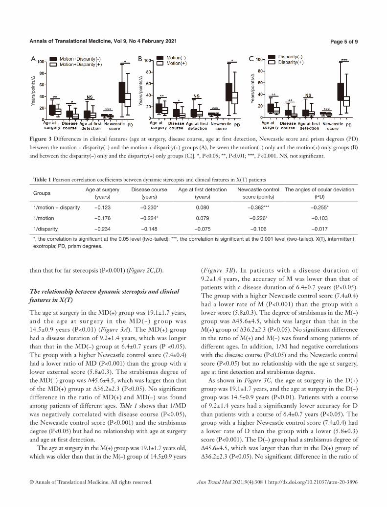
Figure 3 Differences in clinical features (age at surgery, disease course, age at first detection, Newcastle score and prism degrees (PD) between the motion + disparity(−) and the motion + disparity(+) groups (A), between the motion(−) only and the motion(+) only groups (B) and between the disparity(−) only and the disparity(+) only groups (C)]. *, P<0.05; **, P<0.01; ***, P<0.001. NS, not significant.
than that for far stereopsis (P<0.001) (Figure 2C,D).
The relationship between dynamic stereopsis and clinical features in X(T)
The age at surgery in the MD(+) group was 19.1±1.7 years, and the age at surgery in the MD(−) group was 14.5±0.9 years (P<0.01) (Figure 3A). The MD(+) group had a disease duration of 9.2±1.4 years, which was longer than that in the MD(−) group at 6.4±0.7 years (P <0.05). The group with a higher Newcastle control score (7.4±0.4) had a lower ratio of MD (P<0.001) than the group with a lower external score (5.8±0.3). The strabismus degree of the MD(−) group was Δ45.6±4.5, which was larger than that of the MD(+) group at Δ36.2±2.3 (P<0.05). No significant difference in the ratio of MD(+) and MD(−) was found among patients of different ages. Table 1 shows that 1/MD was negatively correlated with disease course (P<0.05), the Newcastle control score (P<0.001) and the strabismus degree (P<0.05) but had no relationship with age at surgery and age at first detection.
The age at surgery in the M(+) group was 19.1±1.7 years old, which was older than that in the M(−) group of 14.5±0.9 years
(Figure 3B). In patients with a disease duration of 9.2±1.4 years, the accuracy of M was lower than that of patients with a disease duration of 6.4±0.7 years (P<0.05). The group with a higher Newcastle control score (7.4±0.4) had a lower rate of M (P<0.001) than the group with a lower score (5.8±0.3). The degree of strabismus in the M(−) group was Δ45.6±4.5, which was larger than that in the M(+) group of Δ36.2±2.3 (P<0.05). No significant difference in the ratio of M(+) and M(−) was found among patients of different ages. In addition, 1/M had negative correlations with the disease course (P<0.05) and the Newcastle control score (P<0.05) but no relationship with the age at surgery, age at first detection and strabismus degree.
As shown in Figure 3C, the age at surgery in the D(+) group was 19.1±1.7 years, and the age at surgery in the D(−) group was 14.5±0.9 years (P<0.01). Patients with a course of 9.2±1.4 years had a significantly lower accuracy for D than patients with a course of 6.4±0.7 years (P<0.05). The group with a higher Newcastle control score (7.4±0.4) had a lower rate of D than the group with a lower (5.8±0.3) score (P<0.001). The D(−) group had a strabismus degree of Δ45.6±4.5, which was larger than that in the D(+) group of Δ36.2±2.3 (P<0.05). No significant difference in the ratio of
Table 1 Pearson correlation coefficients between dynamic stereopsis and clinical features in X(T) patients
GroupsAge at surgery
(years)Disease course
(years)Age at first detection
(years)Newcastle control
score (points)The angles of ocular deviation
(PD)
1/motion + disparity −0.123 −0.230* 0.080 −0.362*** −0.255*
1/motion −0.176 −0.224* 0.079 −0.226* −0.103
1/disparity −0.234 −0.148 −0.075 −0.106 −0.017
*, the correlation is significant at the 0.05 level (two-tailed); ***, the correlation is significant at the 0.001 level (two-tailed). X(T), intermittent exotropia; PD, prism degrees.
A B C
Year
s/po
ints
/Δ
Year
s/po
ints
/Δ
Year
s/po
ints
/Δ

Zhong et al. A novel dynamic stereopsis test for X(T)
© Annals of Translational Medicine. All rights reserved. Ann Transl Med 2021;9(4):308 | http://dx.doi.org/10.21037/atm-20-3896
Page 6 of 9
Fig
ure
4 T
he p
rogn
osis
of
dyna
mic
and
sta
tic s
tere
opsi
s in
the
int
erm
itten
t ex
otro
pia
grou
p. (
A,B
,C)
The
mot
ion
and
disp
arity
, mot
ion
only
, and
dis
pari
ty o
nly
grou
ps
amon
g th
e 1st
day
(1D
) and
2nd
, 6th
and
12th
mon
ths
(2M
, 6M
and
1Y
) pos
tope
rativ
ely.
(D) T
he p
rogn
ostic
feat
ures
of d
ynam
ic s
tere
opsi
s fr
om t
he 1
st d
ay t
o th
e 12
th m
onth
po
stop
erat
ivel
y. (E
,F) N
ear
and
far
ster
eops
is a
mon
g th
e 1st
day
and
2nd
, 6th
and
12th
mon
ths
post
oper
ativ
ely.
(G) T
he p
rogn
ostic
feat
ures
of s
tatic
ste
reop
sis
from
the
1st d
ay
to th
e 12
th m
onth
pos
tope
rativ
ely.
***
, P<0
.001
. NS,
not
sig
nific
ant.
A E
B F
C
G
D

Annals of Translational Medicine, Vol 9, No 4 February 2021 Page 7 of 9
© Annals of Translational Medicine. All rights reserved. Ann Transl Med 2021;9(4):308 | http://dx.doi.org/10.21037/atm-20-3896
D(+) and D(−) was found among patients with different ages at first detection. 1/D was not associated with the disease duration, age at first detection, degree of strabismus, or Newcastle control score (P>0.05).
The prognosis of dynamic and static stereopsis in X(T) postoperatively
In the dynamic stereopsis of MD, postoperative stereopsis improved after the 1st day, the 2nd and 6th months and the 1st year compared with preoperative stereopsis (P<0.05). No significant difference was noted between the 2nd and 6th months (P>0.05). 1/MD is the reciprocal value of MD, which increased gradually from preoperatively to the 1st day and 2nd and 6th months postoperatively, while in the 1st year postoperatively, a slight decrease was observed (Figure 4A). For M, postoperative stereopsis improved after the 1st day, 2nd and 6th months and 1st year compared with preoperative stereopsis (P<0.05); at the 1st year after surgery, M notably decreased (Figure 4B). For D, postoperative stereopsis improved after 6 months (P<0.05), but no significant difference was found between the 1st day and 2nd months postoperatively and preoperative stereopsis (P>0.05) (Figure 4C). The positive rates of MD, M, and D showed gradual decreases from the preoperative period to the 6th month postoperatively, with a rebound in the 12th month postoperatively (Figure 4D).
Static stereopsis improved gradually in the 2nd and 6th months and the 1st year compared with the preoperative period (P<0.05) in near static stereopsis (Figure 4E), while in far static stereopsis, no significant improvement was observed at each timepoint postoperatively (P>0.05) (Figure 4F), with a decreasing tendency for near stereopsis but not far stereopsis (Figure 4G).
Discussion
In this study, the new dynamic stereopsis inspection method is based on the optimization of our previous program (13), in which the stereoacuity detection range was expanded from 600'' to 1,200'' to 200'' and the former program is manually recorded and cannot be automatically read, and the new dynamic stereopsis program can be operated easily and used for stereopsis detection in young children aged 5 years and older. The program can also perform dynamic stereopsis screening in patients with anisometropia, ametropia, amblyopia, and strabismus. X(T) is the most common type of exotropia-type eye disease, accounting for 80% of all kinds
of exotropia (15), and is a transitional strabismus between external strabismus and constant exotropia that is thought to be caused by insufficient convergence function and low fusion. In our study, we used X(T) with the highest clinical incidence as the research object.
The new dynamic stereopsis test showed good reproducibility, and we found that X(T) results in different degrees of damage in dynamic stereopsis, such as MD, M and D impairments, with D corresponding to the worst depth perception, but no significant difference was found between the groups, indicating no significant difference in the degree of motor sensation and depth perception before strabismus surgery. This conclusion is presumed to be related to the mechanism of stereopsis formation. The formation of stereopsis is based on the V1 region and occurs step by step along the dorsal and ventral pathways, projecting onto the visual cortex to transmit binocular parallax information to both eyes (16); thus, we suspect that the regions and manners in which the MD, M, and D dominated in the cerebral cortex are similar. This speculation is consistent with Andrew’s finding where the selection of the M and D directions in the cerebral cortex was similar by functional magnetic resonance imaging (fMRI) (17).
Interestingly, patients who were older at surgery or had a longer disease course, higher Newcastle score and larger strabismus degree had worse dynamic stereopsis, and we therefore inferred that these factors may have a destructive effect on stereopsis; however, no significant differences in dynamic stereopsis were found among patients with different ages at first detection. Lee showed the associations between static stereopsis and clinical features, where patients with a younger age at surgery or at first detection had poor static stereopsis (18), while the age at first detection is not a factor in determining dynamic stereopsis. Moreover, the value of D in dynamic stereopsis is not associated with any clinical features, which did not show a gradual destructive process; thus, we suspect that destruction of D is an “on-off” process, indicating that it becomes worse without any rules once it occurs but is not a gradual process associated with any clinical features. The value of M is related to disease course and the Newcastle control score, indicating that its destruction is more regular and occurs relatively earlier but is less severe compared with D.
Dynamic stereopsis gradually improved after 2 and 6 months and peaked at the 6th month postoperatively, indicating that strabismus surgery can significantly improve the dynamic stereopsis of X(T) patients, and that the 6th month might be a key timepoint to recover dynamic

Zhong et al. A novel dynamic stereopsis test for X(T)
© Annals of Translational Medicine. All rights reserved. Ann Transl Med 2021;9(4):308 | http://dx.doi.org/10.21037/atm-20-3896
Page 8 of 9
stereopsis. We deduce that after the eyes are maintained in a stable ortho position postoperatively, the binocular parallax will always stimulate the visual cortex, and activation of the visual cortex reaches its peak at the 6th month after the operation. Static stereopsis was restored to the peak in near stereopsis after 6 months, but no obvious regularity was observed in far stereopsis. The concept of postoperative stereopsis improvement is consistent with the rule from the close to the long distance, and the recovery of far stereopsis usually takes longer. However, in static stereopsis, a significant improvement trend was observed until 6 months after surgery; thus, the sensitivity to detection was lower than that of dynamic stereopsis. We determined that the decreasing tendency or rebound of both static and dynamic stereopsis at the 12th month postoperatively may be related to the recurrence of strabismus or an increase in the strabismus degree, and that an intervention for binocular function or stereopsis training in the 6th month helps improve or stabilize binocular function and even reduces recurrence rates (19,20), which deserves further study.
Some limitations existed in this study. Firstly, the operation interface was not sufficiently smart and automatic. Secondly, the number of clinical samples was relatively small, and large samples as well as other kinds of strabismus, such as constant exotropia or esotropia, are necessary to confirm the clinical significance of the dynamic stereopsis test. Lastly, the drop-out rate during the follow-up would be controlled to avoid biased results.
Overall, the novel dynamic stereopsis quantitative evaluation method based on random dots is a feasible test and shows that destruction of X(T) patients’ dynamic stereopsis is affected by age at surgery, disease course, strabismus controllability and the strabismus degree. Strabismus surgery can significantly improve dynamic stereopsis, and the 6th month is a key timepoint for recovery. Our data provide a useful basis or reference for detecting or improving stereopsis clinically.
Acknowledgments
The authors thank all patients and family members for their participation.Funding: This work was supported by grants from the Natural Science Foundation Team Project of Guangdong Province (No. 2015A030312016).
Footnote
Reporting Checklist: The authors have completed the STROBE reporting checklist. Available at http://dx.doi.org/10.21037/atm-20-3896
Data Sharing Statement: Available at http://dx.doi.org/10.21037/atm-20-3896
Conflicts of Interest: All authors have completed the ICMJE uniform disclosure form (available at http://dx.doi.org/10.21037/atm-20-3896). All authors report that they have a patent Patents 2997545 licensed.
Ethical Statement: The authors are accountable for all aspects of the work in ensuring that questions related to the accuracy or integrity of any part of the work are appropriately investigated and resolved. The research was performed according to the tenets of the Declaration of Helsinki (as revised in 2013), and the protocol was approved by the Zhongshan Ophthalmic Ethical Committee (ID: 2016MEKY032). All patients with X(T) who met the inclusion criteria were included in the study after being informed of the study content and providing written informed.
Open Access Statement: This is an Open Access article distributed in accordance with the Creative Commons Attribution-NonCommercial-NoDerivs 4.0 International License (CC BY-NC-ND 4.0), which permits the non-commercial replication and distribution of the article with the strict proviso that no changes or edits are made and the original work is properly cited (including links to both the formal publication through the relevant DOI and the license). See: https://creativecommons.org/licenses/by-nc-nd/4.0/.
References
1. Feng X, Zhang X, Jia Y. Improvement in fusion and stereopsis following surgery for intermittent exotropia. J Pediatr Ophthalmol Strabismus 2015;52:52-7.
2. Hess BJM. On the role of ocular torsion in binocular visual matching. Sci Rep 2018;8:10666.
3. Vishwanath D. Toward a new theory of stereopsis. Psychol Rev 2014;121:151-78.

Annals of Translational Medicine, Vol 9, No 4 February 2021 Page 9 of 9
© Annals of Translational Medicine. All rights reserved. Ann Transl Med 2021;9(4):308 | http://dx.doi.org/10.21037/atm-20-3896
Cite this article as: Zhong J, Deng D, Chen Z, Li J, Yuan J, Feng L, Yu M. A novel dynamic random-dot stereopsis assessment to measure stereopsis in intermittent exotropia. Ann Transl Med 2021;9(4):308. doi: 10.21037/atm-20-3896
4. Patil Chhablani P, Tibrewal S, Haque MN, et al. Ocular torsion among patients with intermittent exotropia: relationships with disease severity factors. Am J Ophthalmol 2013;156:411-2.
5. Jiang JJ, Wu Q. Results of conservative management for consecutive esotropia after intermittent exotropia surgery. Eye (Lond) 2015;29:1626.
6. Serrano-Pedraza I, Clarke MP, Read JC. Single vision during ocular deviation in intermittent exotropia. Ophthalmic Physiol Opt 2011;31:45-55.
7. Matsuo T, Negayama R, Sakata H, et al. Correlation between depth perception by three-rods test and stereoacuity by distance randot stereotest. Strabismus 2014;22:133-7.
8. Jang T, Lee K. A novel registration method for computer-assisted total knee arthroplasty using a patient-specific registration guide. Surg Innov 2014;21:80-9.
9. Hatt SR, Mohney BG, Leske DA, et al. Variability of stereoacuity in intermittent exotropia. Am J Ophthalmol 2008;145:556-61.
10. Watanabe Y, Kezuka T, Harasawa K, et al. A new method for assessing motion-in-depth perception in strabismic patients. Br J Ophthalmol 2008;92:47-50.
11. Hess RF, Mansouri B, Thompson B, et al. Latent stereopsis for motion in depth in strabismic amblyopia. Invest Ophthalmol Vis Sci 2009;50:5006-16.
12. Allen B, Haun AM, Hanley T, et al. Optimal combination of the binocular cues to 3D motion. Invest Ophthalmol
Vis Sci 2015;56:7589-96.13. Maeda M, Sato M, Ohmura T, et al. Binocular depth-
from-motion in infantile and late-onset esotropia patients with poor stereopsis. Invest Ophthalmol Vis Sci 1999;40:3031-6.
14. Zhong J, Deng DM, Chen ZD, et al. Evaluation of dynamic stereopsis in intermittent exotropia patients. Int J Ophthalmol 2019;12:83-8.
15. Lavrich JB. Intermittent exotropia: continued controversies and current management. Curr Opin Ophthalmol 2015;26:375-81.
16. Samonds JM, Tyler CW, Lee TS. Evidence of stereoscopic surface disambiguation in the responses of V1 neurons. Cereb Cortex 2017;27:2260-75.
17. Guclu H, Gurlu VP, Ozal SA, et al. Prognostic factors for stereopsis in refractive accommodative esotropia. Pak J Med Sci 2015;31:807-11.
18. Lee DS, Kim SJ, Yu YS. The relationship between preoperative and postoperative near stereoacuities and surgical outcomes in intermittent exotropia. Br J Ophthalmol 2014;98:1398-403.
19. Andalib D, Nabie R, Poormohammad B. Factors affecting improvement of stereopsis following successful surgical correction of childhood strabismus in adults. Strabismus 2015;23:80-4.
20. Portela-Camino JA, Martin-Gonzalez S, Ruiz-Alcocer J, et al. A random dot computer video game improves stereopsis. Optom Vis Sci 2018;95:523-35.