a new taeniolabidoid multituberculate (mammalia) from the
TRANSCRIPT

University of Nebraska - LincolnDigitalCommons@University of Nebraska - Lincoln
Papers in the Earth and Atmospheric Sciences Earth and Atmospheric Sciences, Department of
10-2015
A new taeniolabidoid multituberculate(Mammalia) from the middle Puercan of theNacimiento Formation, New Mexico, and arevision of taeniolabidoid systematics andphylogenyThomas E. WilliamsonNew Mexico Museum of Natural History and Science, [email protected]
Stephen L. BrusatteUniversity of Edinburgh, [email protected]
Ross SecordUniversity of Nebraska-Lincoln, [email protected]
Sarah ShelleyUniversity of Edinburgh, [email protected]
Follow this and additional works at: http://digitalcommons.unl.edu/geosciencefacpub
Part of the Paleobiology Commons, and the Paleontology Commons
This Article is brought to you for free and open access by the Earth and Atmospheric Sciences, Department of at DigitalCommons@University ofNebraska - Lincoln. It has been accepted for inclusion in Papers in the Earth and Atmospheric Sciences by an authorized administrator ofDigitalCommons@University of Nebraska - Lincoln.
Williamson, Thomas E.; Brusatte, Stephen L.; Secord, Ross; and Shelley, Sarah, "A new taeniolabidoid multituberculate (Mammalia)from the middle Puercan of the Nacimiento Formation, New Mexico, and a revision of taeniolabidoid systematics and phylogeny"(2015). Papers in the Earth and Atmospheric Sciences. 445.http://digitalcommons.unl.edu/geosciencefacpub/445

Introduction
Multituberculates were a diverse group of mammals that thrived alongside dinosaurs during much of the Mesozoic, survived the end-Cretaceous extinction, radiated yet again in the post-extinction world of the Paleocene, and then rap-
idly dropped in diversity in the late Paleocene and early Eocene as more modern groups of mammals (particularly the anatomically and ecologically similar rodents) spread across the globe (Krause, 1986; Kielan-Jaworowska, Cife-lli & Luo, 2004; Weil & Krause, 2008). Multituberculates reached their peak in species diversity, body size, and mor-
Published in Zoological Journal of the Linnean Society, 2015; doi: 10.1111/zoj.12336 Copyright © 2015 The Linnean Society of London. Used by permission. Submitted March 11, 2015; revised July 24, 2015; accepted July 31, 2015; version of record
published online October 5, 2015.
A new taeniolabidoid multituberculate (Mammalia) from the middle Puercan of the Nacimiento Formation,
New Mexico, and a revision of taeniolabidoid systematics and phylogeny
Thomas E. Williamson,1 Stephen L. Brusatte,2 Ross Secord,3 and Sarah Shelley2
1 New Mexico Museum of Natural History and Science, 1801 Mountain Road, NW, Albuquerque, NM 87104-1375, USA
2 School of GeoSciences, University of Edinburgh, Grant Institute, James Hutton Road, Edinburgh EH9 3FE, UK
3 University of Nebraska–Lincoln, Department of Earth and Atmospheric Sciences, 200 Bessey Hall, Lincoln, NE 68588-0340, USA
Corresponding author — T.E. Williamson, email [email protected]
AbstractMultituberculates were amongst the most abundant and taxonomically diverse mammals of the late Mesozoic and the Paleocene, reaching their zenith in diversity and body size in the Paleocene. Taeniolabidoidea, the topic of this paper, includes the largest known multituberculates, which possess highly complex cheek teeth adapted for her-bivory. A new specimen from the early Paleocene (middle Puercan; biochron Pu2) of the Nacimiento Formation, New Mexico represents a new large-bodied taeniolabidoid genus and species, Kimbetopsalis simmonsae. A phy-logenetic analysis to examine the relationships within Taeniolabidoidea that includes new information from Kim-betopsalis gen. et sp. nov. and gen. nov. and from new specimens of Catopsalis fissidens, first described here, and data from all other described North American and Asian taeniolabidoids. This analysis indicates that Catop-salis is nonmonophyletic and justifies our transfer of the basal-most taeniolabidoid ‘Catopsalis’ joyneri to a new genus, Valenopsalis. Kimbetopsalis and Taeniolabis form a clade (Taeniolabididae), as do the Asian Lambdop-salis, Sphenopsalis, and possibly also Prionessus (Lambdopsalidae). Taeniolabidoids underwent a modest taxo-nomic radiation during the early Paleocene of North America and underwent a dramatic increase in body size, with Taeniolabis taoensis possibly exceeding 100 kg. Taeniolabidoids appear to have gone extinct in North Amer-ica by the late Paleocene but the appearance of lambdopsalids in the late Paleocene of Asia suggests that they dis-persed from North America in the early to middle Paleocene.
Keywords: body size, dispersal, ecological recovery, mammalian radiation, multituberculata, paleobiogeography, Paleocene, San Juan Basin, Taeniolabididae, Taeniolabidoidea.
1
digitalcommons.unl.edudigitalcommons.unl.edu
2 w i l l i a m s o n e t a l . i n z o o l o g i c a l j o u r n a l o f t h e l i n n e a n s o c i e t y (2015)
phological disparity in the early Paleocene, within a few million years of the non-avian dinosaur extinction (Weil & Krause, 2008; Wilson et al., 2012). Some of the most dis-tinctive multituberculates flourishing during this time were the taeniolabidoids. Taeniolabidoids attained the largest sizes of any multituberculates and were characterized by a short rostrum, reduced premolars, a pair of gliriform in-cisors separated from cheek teeth by a long diastema, and enormous molars with a high number of cusps, all of which permitted an unusual herbivorous diet. These taeniolabi-doids – including such familiar taxa as Taeniolabis and Ca-topsalis – were amongst the most aberrant and specialized mammals of the early Paleocene, a time when terrestrial ecosystems were being dramatically reshaped and mam-mals were beginning their march to dominance.
The first taeniolabidoids were discovered during the geological surveys of western North America during the 1880s, around the same time that multituberculates were recognized as a distinct group of extinct mammals (Cope, 1884, 1888a; Marsh, 1889a, b). Taeniolabidoids are com-mon components of Paleocene faunas of the western USA and are now also known from Asia. Some species are im-portant index taxa in defining the mammalian biochrono-logical timescale (Lofgren et al., 2004; Ting et al., 2011) and taeniolabidoids have played a key role in understand-ing patterns of extinction and survivorship, and changes in mammalian body size and dietary habits across the Cre-taceous–Paleogene boundary (e.g. Archibald, 1983; Wil-son et al., 2012; Wilson, 2013, 2014).
Although taeniolabidoids have been known for some 130 years, their phylogenetic relationships are still poorly understood and their taxonomy is in need of revision. It is widely accepted that genera such as Taeniolabis, Catopsa-lis, and Sphenopsalis form an anatomically and paleobio-logically distinctive taeniolabidoid clade (e.g. Rougier, No-vacek & Dashzeveg, 1997; Kielan-Jaworowska et al., 2004; Mao, Wang & Meng, 2015), but the number of valid species in this clade and their detailed ingroup relationships are the subject of debate. A pioneering phylogenetic analysis by Simmons & Miao (1986) found the characteristic North American genus Catopsalis to be a nonmonophyletic array of several diagnostic species, some constituting a paraphy-letic grade of basal taeniolabidoids, and others forming a polytomy with Taeniolabis and a clade of Asian taxa. Over the following three decades, several new taeniolabidoid specimens have been discovered, which promise to provide insight into the evolution of large body size and high den-tal complexity in this clade (e.g. Simmons, 1987; Buckley, 1995; Lucas, Williamson & Middleton, 1997; Meng, Zhai & Wyss, 1998; Mao, Wang & Meng, 2015). These have yet to be synthesized into a comprehensive phylogenetic anal-ysis and systematic revision of the group.
We here describe a peculiar new taeniolabidoid from the early Paleocene of the San Juan Basin of New Mex-
ico, USA. This is the first new taeniolabidoid taxon to be described in nearly 20 years, and the first to be described from New Mexico – one of the world’s premier localities for early Paleocene mammal fossils – since 1884. We establish the validity of this species, compare it with other taeniola-bidoids, and use this as a jumping-off point for a system-atic revision of the group, based on a species-level phylo-genetic analysis. We then use the results of this analysis to clarify the taxonomy of Taeniolabidoidea and ingroup clades, provide an updated list of all valid species, desig-nate a new genus name for a problematic species of ‘Catop-salis’ that is now strongly supported as a basal taeniola-bidoid, and discuss evolutionary trends in diversification, body size, and biogeography across taeniolabidoid history.
Material and Methods
INSTITUTIONAL ABBREVIATIONS
AMNH, American Museum of Natural History, New York, USA; NMMNH, New Mexico Museum of Natural History and Science, Albuquerque, USA; SDSM, South Dakota School of Mines, Rapid City, South Dakota, USA; SPSM, St. Paul Science Museum, Minnesota, USA; UCM, Uni-versity of Colorado, Boulder, USA; UM, University of Michigan, Ann Arbor, USA; UMVP, University of Minne-sota Vertebrate Paleontology, Minneapolis, USA; USNM, United States National Museum, Washington D.C., USA;UW, University of Wyoming, Laramie, USA.
ANATOMICAL ABBREVIATIONS
I2, second upper incisor; L, length; M, upper molar; m, lower molar; P, upper premolar; W, width; cusp formula following the pattern established by Simpson (1929), counting the buccal row first and the lingual row last.
Systematic Paleontology
MAMMALIA LINNAEUS, 1758 MULTITUBERCULATA COPE, 1884 TAENIOLABIDOIDEA SLOAN & VAN VALEN, 1965 TAENIOLABIDIDAE GRANGER & SIMPSON, 1929 KIMBETOPSALIS SIMMONSAE GEN. ET SP. NOV. (FIGS 1, 2, TABLES 1 AND 2) ( http://zoobank.org/urn:lsid:zoobank.org:pub:9E9F07C3-D042-4E8F-862A-279072E04035 )
Holotype NMMNH P-69902 from locality L-9181.
Type locality and horizon The specimen was discovered in the lower Paleocene part of the Nacimiento Formation of the San Juan Basin of

r e v i s i o n o f t a e n i o l a b i d o i d s y s t e m a t i c s a n d p h y l o g e n y 3
northwestern New Mexico, in the west flank of Kimbeto Wash, at locality 11 of Williamson (1996: fig. 18). It is from Fossil Horizon A and within the Hemithlaeus kowa-levskianus – Taeniolabis taoensis Biozone (H-T Zone) (Wil-liamson, 1996). The vertebrate fauna from this horizon is considered part of the type faunas of the middle Puercan Interval Zone (Pu2) (Archibald et al., 1987; Williamson, 1996; Lofgren et al., 2004).
The west flank of Kimbeto Wash has yielded numer-ous taxa that are restricted to the H-T Zone, including Hemithlaeus kowalevskianus and Conacodon entoconus. These taxa are particularly abundant in H-T Zone fau-nas of the Nacimiento Formation, but are absent from the overlying Fossil Horizon B that yields the type faunas of the late Puercan Interval Zone (Pu3) (Williamson, 1996). Furthermore, no specimens of T. taoensis have been re-covered from the west flank of Kimbeto Wash. This is im-
portant because the first occurrence of Taeniolabis defines the beginning of the Pu3 Interval Zone (Archibald et al., 1987; Lofgren et al., 2004). Although it does not in itself support a Pu2 age for the locality, the absence of Taenio-labis is further evidence that the west flank of Kimbeto Wash is not Pu3 in age (a time when other large taeniola-bidids are known from the Nacimiento Formation). Speci-men NMMNH P-69902 was found fragmented, but in close association, weathering from a silty mudstone in an area of low relief. There is no possibility that the specimen is float from a higher horizon and therefore we are confident that it is a member of the H-T Zone fauna, and thus is Pu2 in age.
Etymology Kimbeto, for Kimbeto Wash; psalis, ‘cutting shears’ (Greek). Simmonsae, after Nancy Simmons, in recogni-tion of her work on taeniolabidoid multituberculates.
Figure 1. Holotype of Kimbetopsalis simmonsae gen. et sp. nov., NMMNH P-69902. A, dorsal view of partial skull roof; B–C, dorsal view of portion of braincase and base of right zygomatic arch in dorsal (B) and ventral (C) views.

4 w i l l i a m s o n e t a l . i n z o o l o g i c a l j o u r n a l o f t h e l i n n e a n s o c i e t y (2015)
Figure 2. Holotype of Kimbetopsalis simmonsae gen. et sp. nov., NMMNH P-69902, A, right I2 in labial view; B, left pre-maxillary fragment with I2 and roots of I3 in labial view; C–E, right partial maxilla with P4–M2 in occlusal (C, stereopair), lingual (D), and buccal (E) views; F–H, left partial maxilla with partial M1–M2 in occlusal (F, stereopair), lingual (G), and buccal (H) views. I2, second upper incisor; M, upper molar; P, upper premolar.

r e v i s i o n o f t a e n i o l a b i d o i d s y s t e m a t i c s a n d p h y l o g e n y 5
Diagnosis Taeniolabidid taeniolabidoid multituberculate that is smaller than Taeniolabis (4% smaller than Taeniolabis lamberti; ~ 21% smaller than T. taoensis), has fewer M1 cusps (eight cusps in the buccal and median rows of M1, compared with nine or more cusps in the buccal and me-dial rows in Taeniolabis; Simmons, 1987), and has a larger P4/M1 ratio (0.47, compared with 0.40 in Taeniolabis). It has a greater number of M2 cusps (1:4:5) than any species of taeniolabidoid other than Taeniolabis (outside of Tae-niolabis, no taeniolabidoid has more than three cusps in the median cusp row of M2).
Possible additional specimens Sloan (1981) referred an edentulous left dentary preserv-ing two prominent alveoli from the Puercan of the San
Juan Basin (AMNH 3030) to Catopsalis foliatus. This spec-imen was originally referred by Cope (1888b) to the eucos-modontid multituberculate Eucosmodon molestus, an iden-tification that Granger & Simpson (1929: 651) considered to be incorrect because it was much larger and more robust than the ‘neotype’ of Eu. molestus. McKenna (1960) also found the affinities of this specimen ‘puzzling’ because he considered that the two preserved alveoli would not have housed a large, elongate, double-rooted premolar, nor were they likely to have housed a small, single-rooted premolar and the anterior root of the m1. Sloan (1981) argued that the size and proportions of the ramus were consistent with the size and proportions of Ca. foliatus. However, Lucas et al. (1997) argued that the dentary was much larger than that of Catopsalis fissidens, which is significantly larger than Ca. foliatus, and with an incisor width of 8.3 mm, based on alveolar diameter, was within the size range of T. taoensis. An alternative interpretation is that AMNH 3030 is a mandibular ramus of Ki. simmonsae, which is expected to be smaller than T. taoensis, but larger than Ca. fissidens, the next largest taeniolabidoid that has been previously documented from the Nacimiento Formation. Unfortunately, the dentary and dentition is unknown for Kimbetopsalis, making direct comparison between Kimbe-topsalis and AMNH 3030 currently impossible. We hope that additional discoveries will clarify the systematic po-sition of AMNH 3030.
Sloan (1981) referred a left lower incisor from fossil ho-rizon B of De-na-zin Wash (USNM 23273) to Catopsalis utahensis (here considered a junior synonym of Ca. foli-atus; see below). He considered it much too large (maxi-mum height = 8.0 mm; maximum width = 5.7 mm; ventral radius of curvature approximately 51 mm; Sloan, 1981) to belong to Ca. foliatus. It may be that this specimen belongs to Ki. simmonsae, a taxon unknown at the time of Sloan’s description, but because it lacks longitudinal fluting it probably does not represent a lower incisor of that taxon.
ANATOMICAL DESCRIPTION
NMMNH P-69902 consists of associated cranial fragments and dentition with right I2, a partial left premaxilla pre-serving the I2 and roots of I3, and most of both maxillae with associated right P4–M2 and left M1–M2. The speci-men is mostly encrusted with a concretionary covering, a preservation style common to fossils from this part of the Nacimiento Formation. However, the teeth are largely in-tact and unobscured. Reassembled parts of the skull are readily identified as a portion of the skull roof (Fig. 1A) and a portion of the zygomatic arch with the glenoid fossa (Fig. 1B, C).
Cranium One preserved portion of the skull roof includes a nearly flat to slightly dorsally convex intraorbital area bordered
Table 1. Measurements of the holotype of Kimbetopsalis simmon-sae gen. et sp. nov., NMMNH P-69902. Measurements are in mm
Tooth L W Cusp formula
I2 9.64* P4 8.6 5.16 M1 18.23 9.69 8:8–9:9* M2 12.37 10.69 1:4:5
I2, second upper incisor; L, length; M, upper molar; P, upper pre-molar; W, width. *estimated.
Table 2. Measurements of Catopsalis fissidens. Measurements are in mm
Tooth L W Cusp formula
I2 NMMNH P-63938 5.36 M1 NMMNH P-63938 13.84 7.24 8:9:11* M2 NMMNH P-63938 7.86 9.26 2:3:4 I NMMNH P-62373 4.90 NMMNH P-63938 4.44 p4 NMMNH P-62373 4.93* 2.21 m1 NMMNH P-62373 11.64 6.10 6:5 NMMNH P-63938 12.39 6.33 7:5 m2 NMMNH P-62373 6.79 6.75 2:4 NMMNH P-63938 7.03 6.79 2:4
I2, second upper incisor; L, length; M, upper molar; m, lower mo-lar; P, upper premolar; p, lower premolar; W, width. *estimated.

6 w i l l i a m s o n e t a l . i n z o o l o g i c a l j o u r n a l o f t h e l i n n e a n s o c i e t y (2015)
by temporal crests that converge posteriorly to the sagit-tal crest (Fig. 1A). The sides of the braincase lateral to the sagittal crest slope steeply laterally. No sutural contacts are visible. This skull roof portion closely resembles the in-terorbital region of the skull of T. taoensis (AMNH 16321) as illustrated in Broom (1914: fig. 6), Granger & Simpson (1929: fig. 5a), and Matthew (1937: fig. 72a).
An additional skull fragment (Fig. 1B, C) is interpreted to represent the posterolateral side of the braincase, pre-serving a portion of the right lambdoidal crest and the root of the right zygomatic arch. The glenoid fossa is preserved on the ventral surface of the fragment, but the surface is distorted and obscured by a concretionary crust.
The partial maxillae (Fig. 2C–H) preserve a portion of the vaulted palate and the roots of the zygomatic arches. The latter structures project laterally from the body of the maxilla, with the posterior margin of the arch bases po-sitioned approximately between the P4 and M1 as in T. taoensis. Laterally directed zygomatic arches, which proj-ect approximately 90° transversely relative to the sagit-tal plane in dorsal view, are a synapomorphy of Taeniola-bidoidea (e.g. Kielan-Jaworowska et al., 2004). They act to demarcate the short and wide rostrum as a distinct re-gion relative to the remainder of the skull.
Dentition Incisors: Both left and right I2s are nearly complete, but are missing the tips and portions of the crowns (Fig. 2A, B). The roots appear to be intact, but are largely cov-ered with a concretionary crust. In places, portions of the enamel are missing or crushed and fractured. The par-tial left premaxillary fragment is mostly obscured with concretion (Fig. 2B). However, a break in the specimen posterior to the I2 exposes the root of the I3 immedi-ately posterior to the I2, indicating that the I3 is in the same position on the premaxilla as in other taeniolabi-doids (e.g. Simmons & Miao, 1986; Simmons, 1993; Rou-gier et al., 1997).
The incisors closely resemble those of T. taoensis; they are large, mesiodistally broad, curved, taper towards their apices, and bear pronounced longitudinal fluting and transverse growth bands. The longitudinal fluting tends to be regularly spaced mesiodistally across the la-bial enamel band and of uniform width. At least eight flutes are present near the base of the left I2. This flut-ing also occurs on both upper and lower incisors of T. taoensis. On upper incisors of T. taoensis, the flutes ra-diate basally and individual ‘flutes’ anastomose basally to create additional flutes. Flutes are truncated mesially and distally where they intersect the margins of the la-bial enamel band. Over ten flutes are present on I2s of T. taoensis (e.g. NMMNH P-2937, 12387). In both T. taoen-sis and Kimbetopsalis the fluting of the enamel band is
similar in size and spacing and also anastomoses basally. Although the upper incisors of Kimbetopsalis are very similar to those of T. taoensis, they differ from the flut-ing exhibited by Ca. fissidens, which outside of T. taoensis (incisors are unknown for T. lamberti) and Kimbetopsa-lis, has the most distinctive fluting amongst taeniolabi-doids. In Ca. fissidens the individual flutes are narrower, fewer in number, more variable in size, less distinct, and do not anastomose. Some fluting is also evident in Catop-salis alexanderi (UCM 39553), but the I2 of this specimen possesses only three broad longitudinal ridges, which are separated by broad and shallow grooves. In addition, the entire enamel surface is also marked by fine irregular wrinkles that are orientated longitudinally. These are not present in Kimbetopsalis.
P4: An in-situ P4 is preserved on the right maxilla (Fig. 2C–E), and although the partial left maxilla (Fig. 2D–H) is missing P4 it preserves two corresponding alveoli of subequal size that are filled with matrix. These alve-oli clearly show that the P4 has two roots; the mesial root projects mesiodorsally into the maxilla and the distal root appears to have been directed dorsally. The right P4 is pear-shaped in occlusal view, being mesiodistally elon-gate and expanded distally, and distinctly bulbous. Por-tions of the enamel surface are missing from the mesi-olingual edge and the distobuccal face of the tooth. The right P4 bears a medial row of four cusps. The mesial-most, or first, cusp is about half the height of the other three cusps and rises from the steeply sloping mesial face of the tooth. The middle two cusps have worn apices, but it appears that the second of the row is the largest in di-ameter. The third cusp has the highest preserved height. Creases on the lingual and buccal faces of the tooth de-limit each cusp. The distal face of P4 is broadly rounded and bears at least three basolingual cusps. A deep cleft separates the cuspate area from the distal-most cusp of the medial row of cusps.
M1: Both the left and right M1s are present. They are both incomplete, but together they preserve most of the important anatomical features, and allow for an accurate determination of a complete cusp formula. The left M1 (Fig. 2F–H) is missing most of the lingual row of cusps but is otherwise well preserved. The right M1 (Fig. 2C–E) is more complete, missing a portion of one of the cusps of the buccal row, and several of the lingual row.
The M1 is approximately rectangular in occlusal view (Fig. 2C, F), with three rows of cusps extending mesio-distally down the entire length of the tooth, which is a synapomorphy of Taeniolabidoidea (e.g. Simmons & Miao, 1986; Kielan-Jaworowska et al., 2004). The cusps are pyramidal in shape. Several of the cusps, especially

r e v i s i o n o f t a e n i o l a b i d o i d s y s t e m a t i c s a n d p h y l o g e n y 7
the two most distal cusps of the medial and lingual rows, exhibit weak crenulations. Although the lingual cusp row is incomplete on both specimens, the speci-mens together indicate that M1 had nine lingual cusps when complete. The medial row contains eight cusps, as does the buccal row. The resulting cusp formula is 8:8:9. The cusps of the lingual row decrease in size mesially and the mesial-most cusp is very small in comparison to the mesial-most cusp of the medial row. The mesial-most cusp of the buccal row is mesiodistally compressed. There are no accessory cuspules or basal cingula on any of the cusps of any of the rows. The buccal face of the tooth shows weak, transverse apicobasal wrinkling ex-tending vertically, from the base of the crown towards the apex of each cusp.
M2: Both the left and right M2 are complete. They are approximately heart-shaped in occlusal view (Fig. 2C, F), with a concave mesial margin and a rounded distal mar-gin, and they taper distally in buccolingual width. The most distinctive features of the M2 are its large, swollen, pyramidal cusps, which form three longitudinal rows as in the M1. The lingual row contains five cusps, but the distal-most cusp is small and might be considered a cus-pule. The medial row consists of four cusps, the mesial-most of which is mesiodistally compressed. The buccal row consists of a single large cusp, located at the mesiobuccal corner of the tooth when seen in occlusal view. Several of the cusps have undulatory margins in occlusal view owing to weak folding of the enamel. The margins of the tooth also show transverse apicobasal wrinkling, particularly on the buccal side.
DISCUSSION
Comparisons with other taeniolabidoids Kimbetopsalis simmonsae is clearly referable to Taenio-labidoidea by the possession of diagnostic characters of the clade not seen in other multituberculates, including transversely orientated zygomatic arches, a rectangular M1 with three longitudinal rows of cusps that extend along the entire length of the tooth, an M2 with four or more cusps in the lingual and medial rows, the very large size of I2 and M2, which are bigger in taeniolabidoids than in any other multituberculates, and small size of the P4 rel-ative to the M1 (Table 3; e.g. Simmons, 1987; Kielan-Ja-worowska et al., 2004; Mao et al., 2015).
Kimbetopsalis simmonsae is similar to species of Tae-niolabis in many features. For example, it possesses some (but not all) features considered diagnostic for Taeniola-bis by Simmons (1987), in her revision of Taeniolabis sys-tematics. These include large I2 and M2 (which are larger in Taeniolabis and Kimbetopsalis compared with other
taeniolabidoids), nine or more cusps in the lingual row of M1, and four or more cusps in the lingual row of M2. However, Ki. simmonsae can be distinguished from both species of Taeniolabis [T. taoensis (Cope, 1882) and T. lamberti Simmons, 1987] by being smaller in size, based on M1 measurements. Kimbetopsalis simmonsae has an M1 length of 18.2 mm (Table 1), falling well outside of the range of M1 length (21.9–24.4 mm with a mean of 23.0 mm) reported by Simmons (1987: table 2) for T. taoensis. The M1 length of Ki. simmonsae, therefore, is 79% of the mean M1 length (N = 22) of T. taoensis. It is also smaller than, but closer in size to, the M1 referred by Simmons (1987) to T. lamberti, which is 18.9 mm in length (about 4% larger than in Ki. simmonsae). The M1 width of Ki. simmonsae (9.7 mm) is also smaller than in T. taoensis (10.8– 12.1 mm range, 11.6 mm mean) and T. lamberti (10.0 mm) (Simmons, 1987).
Kimbetopsalis simmonsae can also be distinguished from Taeniolabis by its lower number of cusps in the buccal cusp row of M1, based on the diagnosis of Sim-mons (1987). The cusp number for the M1 of Kimbetop-salis is 8:8:9, lower than for T. taoensis and for T. lam-berti. Simmons (1987) gave a range in cusp number for M1 of 8–11:8–10:7–11 for the genus Taeniolabis and 8–11:9–10:9–11 for the species T. taoensis (the upper range of T. taoensis is actually slightly higher, as AMNH 16313 has 12 lingual cusps). Taeniolabis lamberti has a cusp formula of 9:10:10 based on the single referred M1, UCMP 110452 (Simmons, 1987). However, the cusp formula of Kimbe-topsalis is similar to that of some specimens referred to Catopsalis, including Catopsalis calgariensis (UW 6407), which has a cusp formula of 8:8:10 (see Middleton, 1982: plate 1, fig. 8) and Ca. fissidens (NMMNH P-63938), which has a cusp formula of 8:9:10? (Fig. 3).
The M1 cusp formula for Kimbetopsalis is the same as that of an isolated M1, UCMP 128280, from the Garbani Quarry, Montana, that Simmons (1987: fig. 6.1) referred to Taeniolabis sp. However, that specimen is notably dif-ferent from Kimbetopsalis and all other M1 specimens re-ferred to Taeniolabis in having a relatively short lingual cusp row. In UCMP 128280 the lingual cusp row extends anteriorly only as far as the sulcus between cusps 2 and 3 of the medial cusp row, whereas in the M1 of Kimbe-topsalis and specimens referred to either T. taoensis or T. lamberti, it extends medially to a point lingual to the first cusp of the medial row (Kimbetopsalis), or at least as far as the sulcus between medial cusps 1 and 2 (Taeniola-bis). UCMP 128280 is also larger than the M1 of Kimbe-topsalis (M1L = 22.0; Simmons, 1987: table 4), and within the lower size range of T. taoensis. Simmons (1987) sug-gested that this one unusual specimen may be primitive compared with T. taoensis and T. lamberti. Regardless of its precise affinities, it is clear that this specimen does not belong to Kimbetopsalis.

8 w i l l i a m s o n e t a l . i n z o o l o g i c a l j o u r n a l o f t h e l i n n e a n s o c i e t y (2015)
Tabl
e 3.
Com
pila
tion
of d
enta
l mea
sure
men
ts fo
r tae
niol
abid
ids a
nd o
ther
taxa
from
a v
arie
ty o
f sou
rces
: Arc
hiba
ld (1
982)
, Buc
kley
(199
5), C
lem
ens (
1964
), Fo
x (2
005)
, Luc
as e
t al.
(199
7), M
ao e
t al.
(201
5), M
atth
ew e
t al.
(192
8), M
idd
leto
n (1
982)
, Sim
mon
s (19
87),
Sim
mon
s & M
iao
(198
6), S
loan
& V
an V
alen
(196
5),
Wils
on (1
987)
, and
Cho
u &
Qi (
1978
)
Taxa
p4
m
1 m
1 m
1 cu
sp
m2
m2
m2
cusp
P4
P4
P4
cus
p M
1 M
1 M
1 cu
sp
M2
M2
M2
cusp
L
L W
fo
rmul
a L
W
form
ula
L W
fo
rmul
a L
W
form
ula
L W
fo
rmul
a
Mic
roco
smod
on c
onus
1.
6 3.
0 1.
1 7:
4.5
1.3
1.2
3:2
1.7
0.7
3:4:
0 2.
5 1.
3 7:
7:5
1.4
1.3
1:3:
4
Esso
nod
on b
row
ni
4.2
7.1
1.1
3–4:
7:6
2.5
3.6
3:2
3.3
2.1
1:5:
0?
7.5
3.9
8:7:
7 2.
7 2.
8 0-
R:2:
3–4
Cim
exom
ys ju
dith
ae
3.2
2.0
3.8
6:4
1.2
1.1
3:2
2.2
0.6
3:5:
0 2.
3 1.
1 5:
6:1
1.3
1.3
1:3:
3
Cim
olom
ys g
raci
lis
4.5
4.8
0.9
7–8:
5–7
3.7
2.6
4–6:
2–3
2.8
1.5
1–2:
5–6:
2–3
4.8
2.7
5–7:
7–10
:4–6
3.
6 3.
1 2–
3:3–
4:4–
7
Men
iscoe
ssus
robu
stus
4.
0 9.
4 2.
1 5–
6:4
7.3
4.7
4–5:
2 4.
1 2.
8 1:
4:?
9.1
5.2
5–6:
7:6–
7 6.
3 6.
0 3–
5:3–
4:4–
5
Bubo
den
s mag
nus
– 12
.8
4.4
10:8
–
– –
– –
– –
– –
– –
–
“Cat
opsa
lis”
joyn
eri
3.5
6.4
3.3
5:4
5.5
– 3:
2 3.
2 2.
3 1:
5:1
8.1
4.4
8:7:
8 5.
2 –
1:3:
3
Cat
opsa
lis w
add
leae
–
16.4
9.
3 5:
4 –
– –
– –
– –
– –
12.9
11
.8
1:3:
4
Cat
opsa
lis fo
liatu
s 4.
07*
10.5
5.
1*
6:4
6.7*
5.
9 4:
2 –
– –
– –
– –
– –
Cat
opsa
lis fi
ssid
ens
4.8
12.6
6.
4 5–
7:4–
6 7.
5 7.
1 4:
2 –
– –
14.9
7.
3 8:
9:11
–12
8.2
8.4
1–2:
3:4
Cat
opsa
lis a
lexa
nder
i 4.
3 8.
7 4.
8 5–
9:4
6.5
5.0
3–4:
2 4.
3 3.
3 0–
1:4:
1 10
.3
5.6
7–8:
7–8:
9 6.
5 5.
9 1–
2:3:
3–4
Taen
iola
bis t
aoen
sis
6.6
19.8
10
.4
7–8:
6–7
14.1
11
.5
4–6:
4–6
7.1
– 0:
4:1
23.0
11
.6 8
–11:
9–10
:9–1
2 14
.4
12.0
1:
4–5:
4–6
Taen
iola
bis l
ambe
rti
5.7
16.0
8.
0 8:
6 15
.4
10.7
6:
4 5.
8 3.
9 0:
4:1
18.9
10
.0
9:10
:10
15.7
13
.5
2:4:
7
Kim
beto
psal
is sim
mon
sae
– –
– –
– –
– 8.
6 5.
2 0:
4:1*
18
.2
9.7
8:8:
9*
12.4
10
.7
1:4:
5
Lam
bdop
salis
bul
la
– 7.
8 3.
2 5:
4 7.
5 5.
3 4:
2 –
– 0:
1:0
10.0
5.
0 6–
7:7–
8:7
– –
1:2:
3
Prio
ness
us lu
cife
r 1.
6 5.
6*
2.4
5:4
3.1
2.9
3:2
– –
0:1:
0 5.
6 3.
3 7:
6:5
3.7
3.5
1:3:
3
Sphe
nops
alis
nobi
lis
4.6
13.6
7.
3 5:
4 13
.8
9.9
3:2
2.9
3.0*
–
16.4
8.
3 7:
7:8
13.3
12
.2
1:2:
4
*est
imat
ed; I
2, se
cond
upp
er in
ciso
r; L,
leng
th; M
, upp
er m
olar
; m, l
ower
mol
ar; P
, upp
er p
rem
olar
; p, l
ower
pre
mol
ar; W
, wid
th.

r e v i s i o n o f t a e n i o l a b i d o i d s y s t e m a t i c s a n d p h y l o g e n y 9
Figure 3. New specimens of Catopsalis fissidens. NMMNH P-63938. A, right partial premaxilla with I2 in labial view; B, left M1–M2 in occlusal view (stereopair), left partial dentary with roots of p4, m1–m2 in occlusal (C; stereopair), buccal (D), and lingual (E) views. NMMNH P-62373, right p4–m2 in occlusal (F; stereopair), buccal (G), and lingual (H) views. I2, second up-per incisor; M, upper molar; m, lower molar; p, lower premolar.

10 w i l l i a m s o n e t a l . i n z o o l o g i c a l j o u r n a l o f t h e l i n n e a n s o c i e t y (2015)
The M2 of Kimbetopsalis is similar in size to that of T. taoensis, falling within the size range for length (12.37 mm, compared with 12.3–16.0 mm range of 11 teeth of T. taoensis: Simmons, 1987: table 2). The cusp formula of the Kimbetopsalis M2 also overlaps with that of T. taoensis (1:4–5:4–6; Simmons, 1987: table 1). However, the width of the Kimbetopsalis M2 (10.69 mm) is smaller than in T. taoensis, falling outside of the latter’s size range (11.2–12.8 mm: Simmons, 1987: table 2). Com-pared with species of Catopsalis, the M2 of Kimbetopsa-lis is larger (about 20 per cent longer than the M2 of Ca. calgariensis and 50 per cent larger than that of Ca. fis-sidens) and has a higher cusp number than any known Catopsalis specimen.
Comparisons between the M2 morphology of Kimbe-topsalis and T. lamberti are less straightforward, owing to uncertainty over whether some M2 specimens can be confidently referred to T. lamberti. Simmons (1987) re-frained from referring an M2 to T. lamberti. However, she described an isolated M2, UCMP 128291, from the same locality as the holotype of T. lamberti (locality Garbani 13 of the Tullock Formation), but referred it to Taenio-labis sp. because of its large size, which was reported to be wider, with an estimated width of 11.1 mm, than any M2 referred to T. taoensis. However, she considered that it was possible that the tooth would prove to be referable to T. lamberti if discovery of more specimens indicated a larger range of variation in this species. The tooth dif-fers from the M2 of T. taoensis, Ki. simmonsae, and all other taeniolabidoids in having a higher cusp formula of 2:4:7. A second distinctive specimen from the Tullock Formation (UMVP 1455) consists of a partial M2, also possessing two cusps in the buccal cusp row, supporting the idea that the Tullock taeniolabidoid has a distinc-tive M2 cusp formula. We suggest that it is most likely that there is only a single large taeniolabidoid present from the Garbani Quarry of the Tullock Formation, be-cause in other faunas that contain taeniolabidoids, only a single large species is present. Therefore, we suggest that it is most parsimonious to consider that these speci-mens from Garbani 13 are all referable to a single taxon, T. lamberti (known from other definitive material from Garbani 13), rather than a different, but unknown coex-isting species of Taeniolabis.
Tooth proportions also differ between Kimbetopsalis and Taeniolabis. The ratio of P4 length to M1 length (P4 L/M1 L) in Kimbetopsalis (0.47) is outside the up-per range of this ratio in Taeniolabis (0.31–0.33; Sim-mons, 1987), but similar to the value found in ‘Catopsa-lis’ joyneri and Ca. alexanderi. In addition, the ratio of M1 length to M2 length (M1 L/M2 L) in Ki. simmonsae (1.47) is below the range observed in Taeniolabis (1.59– 1.79; Simmons, 1987), but similar to the values found in
‘Ca.’ joyneri (M1 L/M2 L = 1.49) and Ca. fissidens (1.50). Together, these ratios indicate that the M1 in Kimbe-topsalis and Catopsalis is smaller relative to the other cheek teeth than in Taeniolabis. Large M1 size in Tae-niolabis, therefore, is probably a derived feature amongst taeniolabidoids.
Differences in P4 morphology also distinguish Kimbe-topsalis from Taeniolabis. Most notably, Kimbetopsalis has multiple distobuccal basal cusps on the P4, rather than the single basal cusp of T. lamberti. It also differs from the P4 of Catopsalis, where known, in which cusps tend to be arranged in two or more parallel cuspal rows (‘Ca.’ joyneri, Ca. alexanderi). The P4 is unknown for Ca. folia-tus and Ca. fissidens. Only a partial P4 is known for Ca. calgariensis and this preserves only cusps of the medial cuspal row. It is not known if this taxon possessed disto-buccal basal cusp(s).
One potential difference in P4 morphology used to dif-ferentiate taeniolabidoid taxa by previous authors does not hold up to scrutiny. Kimbetopsalis clearly possesses a double-rooted P4, as two alveoli for this tooth are pres-ent on the right maxilla. This differs from the supposed presence of a single-rooted P4 in some other taeniolabi-doids, including Taeniolabis and Catopsalis. Simmons & Miao (1986) reported that the P4 of Taeniolabis is single-rooted and scored it as such in their phylogenetic data set. They also reported that both Ca. alexanderi and Ca. calgariensis possess a single-rooted P4. These character states have been perpetuated in subsequent phylogenetic analyses (e.g. Simmons, 1993; Rougier et al., 1997; Weil, 1999). However, a review of the literature and collections demonstrates that neither Taeniolabis nor Catopsalis possess a single-rooted P4. This tooth is double-rooted in Ca. alexanderi (Middleton, 1982). Catopsalis fissidens is also double-rooted based on a new specimen, NMMNH P-63938, which includes a fragment of maxilla that con-tains the alveoli and broken roots of P4 (see below). Mid-dleton (1982) reported that Ca. calgariensis is double-rooted based on an isolated P4 (UW 6387), although we have not been able to independently verify this. Taeniola-bis taoensis is double-rooted based on several specimens, including AMNH 16313, NMMNH P-8621, 47470, and 47660; the latter three specimens each preserve two alve-oli separated by a distinct septum for a double-rooted P4, identical to the condition in Ki. simmonsae. The condition in T. lamberti is unclear, as Simmons (1987) referred a single P4 to T. lamberti (UCMP 128281) and described it as not preserving a root. The only taeniolabidoids that can be confidently said to possess a single-rooted P4 ap-pear to be the Asian lambdopsalid taxa Sphenopsalis no-bilis and Lambdopsalis bulla. Prionessus lucifer includes specimens with both single-rooted and double-rooted P4s (Meng, Zhai & Wyss, 1998; Mao et al., 2015).

r e v i s i o n o f t a e n i o l a b i d o i d s y s t e m a t i c s a n d p h y l o g e n y 11
PHYLOGENETIC ANALYSIS
The first study using modern phylogenetic methods for tae-niolabidoids was conducted by Simmons & Miao (1986). This pioneering study was amongst the first to use phy-logenetic systematics to study extinct mammal phylog-eny. They concluded that Catopsalis, as then conceived, was a paraphyletic taxon that gave rise to Taeniolabis and an Asian clade composed of Lambdopsalis, Priones-sus, and Sphenopsalis. This study emerged as an impor-tant work on the phylogeny of Cretaceous and Paleocene mammals, and was held to have broader implications for understanding species turnover after the end-Cretaceous mass extinction. Archibald (1993), for example, included a slightly modified version of the Simmons & Miao (1986) cladogram to illustrate examples of ‘metaspecies’ and to show the impact of pseudoextinction on interpretations of early Paleocene species turnover.
Subsequently, very little work has been conducted on taeniolabidoid phylogeny and systematics, despite the importance of members of this group as Paleocene index taxa and their role as some of the first mammals to evolve mid-to-large body size after the end-Cretaceous extinc-tion. Hypothetical inter-relationships of the Multituber-culata, including a handful of taeniolabidids, were pro-posed by Kielan-Jaworowska & Hurum (2001: text-fig. 2) and reproduced in Kielan-Jaworowska et al. (2004: fig. 8.45), but these were not based on a numerical analysis. Some taeniolabidoids were included in higher-level phy-logenetic analyses of Multituberculata conducted by Sim-mons (1993), Rougier et al. (1997), Weil (1999), and Yuan et al. (2013). None of these analyses yielded well-resolved or strongly supported results. In the most recent higher-level analysis, Mao et al. (2015) included more taeniola-bidoid taxa than previous studies, but scored Taeniola-bis and ‘Catopsalis’ as single composite taxa, as the aim of their study was not to untangle the detailed inter-rela-tionships of taeniolabidoids but to test the monophyly of the group and place it on the larger multituberculate tree. The use of such composite taxa, although understandable in a higher-level analysis, has obscured the ingroup rela-tions of taeniolabidoids. Particularly troublesome is scor-ing ‘Catopsalis’ as a single composite, which in the study of Mao et al. (2015) was based on four species (‘Ca.’ joy-neri, Ca. foliatus, Ca. fissidens, and Ca. alexanderi), be-cause previous analyses have shown this genus to be non-monophyletic (Simmons & Miao, 1986).
We present a new phylogenetic analysis of Taeniolabi-doidea, including all Asian and North American taxa re-ferable to this clade. Several new taeniolabidoids have been recognized and described since the Simmons & Miao (1986) analysis, including T. lamberti, Catopsalis wad-dleae, and Ki. simmonsae. Additionally, new data are available for Pr. lucifer (Meng et al., 1998), S. nobilis (Mao
et al., 2015), and Ca. fissidens (Lucas et al., 1997, and de-scribed below). A wealth of new data is also now available on potential close outgroups of Taeniolabidoidea, includ-ing Cimexomys, Meniscoessus, and Microcosmodon (Mon-tellano, Weil & Clemens, 2000; Fox, 2005).
Taxa not included in our analysis are three Asian taxa that Simmons & Miao (1986) included in Taeniolabidi-dae [Kamptobaatar, Djadochtatherium, and Catopsbaatar ( = ‘Catopsalis’) catopsaloides after Kielan-Jaworowska (1994)], which have subsequently been shown to be para-phyletic and remote from Taeniolabidoidae (sensu stricto; Simmons, 1993) and to be part of a monophyletic group of Asian taxa (Rougier et al., 1997) that were relegated to the suborder Djadochtatheria by Kielan-Jaworowska & Hurum (1997). Hence, all remaining taeniolabidoids are Paleocene in age, with the possible exception of Bubodens magnus (Wilson, 1987), a poorly known taxon, represented only by an isolated m1 from the Upper Cretaceous Fox Hills Formation of South Dakota. Bubodens was tenta-tively referred to Taeniolabididae (= Taeniolabidoidea in our usage) by Kielan-Jaworowska et al. (2004; see also Weil & Krause, 2008).
We used the results of our analysis to revise the sys-tematics of taeniolabidoids, erect a new genus-level taxon (whose distinctiveness follows from the phylogenetic anal-ysis), and comment on the evolutionary history of the group.
TAXON SAMPLING FOR PHYLOGENETIC ANALYSIS
‘Catopsalis’ joyneri Sloan & Van Valen, 1965: ‘Catopsa-lis’ joyneri was illustrated and briefly described by Sloan & Van Valen (1965) from the Bug Creek Anthills local-ity of eastern Montana. Sloan & Van Valen (1965) con-sidered this site to be Cretaceous in age, but it has since been shown to be a mixed assemblage of earliest Paleo-cene and reworked Cretaceous fossils (Lofgren, 1995). The ‘Catopsalis’ specimens are now considered to be early Pa-leocene (early Puercan; Pu1) in age (Lofgren et al., 2004; Wilson, 2013) [note that Wilson (2014) listed only Catop-salis foliatus in Pu1 faunas of Garfield County, which is a mistake (G. P. Wilson, pers. comm., 2014)]. Scoring of this taxon was based on descriptions and illustrations in Sloan & Van Valen (1965), Archibald (1982), Lofgren (1995), and Fox (2005).
Catopsalis alexanderi Middleton, 1982: Middleton (1982) described numerous specimens of Ca. alexanderi from the early Paleocene (Pu1) of the Denver Basin. He distin-guished Ca. alexanderi from ‘Ca.’ joyneri and Ca. foliatus by its intermediate size. Specimens from the early Paleo-cene (Pu1) of the Tullock or Hell Creek formations were tentatively referred to Ca. cf. Ca. foliatus by Archibald

12 w i l l i a m s o n e t a l . i n z o o l o g i c a l j o u r n a l o f t h e l i n n e a n s o c i e t y (2015)
(1982), but were subsequently referred to Ca. alexanderi (Middleton, 1982). Sloan (1981) referred several specimens from the early Puercan Mantua Quarry of the Fort Union Formation in northern Wyoming to Ca. foliatus and these were also subsequently referred by Middleton to Ca. alex-anderi. We based our scoring of this taxon on descriptions and illustrations in Middleton (1982) and on casts of UCM 14550 (left P4), 16647 (right M1), 16648 (right M2), 16649 (left m1), and 16650 (left m2), 34146 (right M1), 34186 (left partial dentary with p4–m1), 34979 (right partial dentary with I, p4–m2; the holotype of Ca. alexanderi), 39553 (left I2, right I2–3, left and right P4–M2), YPM PU 18196, (I2), 14550 (left P4), 16647 (right M1), 16648 (right M2), 16649 (left m1), 16650 (left m2).
Catopsalis waddleae Buckley, 1995: Buckley (1995) de-scribed three isolated teeth, one fragmentary, from the Simpson Quarry of Puercan age, Bear Formation, Crazy Mountain Basin of Montana, as a new taxon, Ca. wad-dleae. Our scorings are based on descriptions and illustra-tions from Buckley (1995) and casts of UM 90242 (right m1, not left m1 as reported by Buckley, 1995; the holo-type of Ca. waddleae), 90212 (partial right m1), and 90215 (left M2).
Catopsalis foliatus Cope, 1882: The precise provenance of the lectotype of Ca. foliatus, AMNH 3035, a partial den-tary preserving p4–m2, remains uncertain. It is proba-bly Puercan or Torrejonian in age (see Lucas et al., 1997). Lucas et al. (1997) suggested that a second specimen, AMNH 16325, a partial skeleton not preserving a den-tition (see Granger & Simpson, 1929), may be referable to this taxon.
We consider another species of Catopsalis, Catopsalis johnstoni Fox, 1989, to be synonymous with Ca. foliatus. Catopsalis johnstoni was based on three isolated teeth from the lower Paleocene Porcupine Hills Formation of southern Alberta: a left I2, a lower incisor, and a left m1. Lucas et al. (1997) tentatively synonymized Ca. johnstoni with Ca. foliatus, outlining that Fox (1989) had stated only that Ca. johnstoni was intermediate in size (m1 L = 9.7; W = 4.65; Johnston & Fox, 1984: table 18) between ‘Ca.’ joy-neri or Ca. alexanderi and Ca. calgariensis, and therefore did not distinguish the species from Ca. foliatus or Ca. fis-sidens. Johnston & Fox (1984) described this taxon (‘Ca-topsalis n. sp.’) as having a higher m1 cusp formula (7:4) than ‘Ca.’ joyneri, Ca. foliatus, or Ca. alexanderi. How-ever, we found that the type of Ca. foliatus (m1 L = 10.3; W = 5.15) has an m1 cusp formula of 6:4 (not 5:4 as re-ported by Johnston & Fox, 1984: p. 209), with a small cusp on the distal end of the buccal cusp row. A similar small sixth cusp is present on at least some specimens referred to Ca. alexanderi, but the distinction of this cusp can be
erased with wear (Middleton, 1982) and so the m1 cusp formula is probably variable (4–5:4). Lucas et al. (1997) also found the m1 cusp formula of Ca. fissidens to be vari-able (5–7:4–6; Lucas et al., 1997) based on a small sample (N = 8) from the San Juan Basin. Two new m1s of Ca. fis-sidens (described below) fall within this range of cusp for-mula, with formulas of 6–7:5.
Johnston & Fox (1984) and Fox (1989, 1990) considered specimens referred to Ca. johnstoni from the Long Fall ho-rizon (the Wounded Knee Local Fauna; Fox, 1989) of the Ravenscrag Formation of southcentral Saskatchewan to be latest Cretaceous in age, based partly on the presence of typical latest Cretaceous dinosaur teeth and mamma-lian taxa (Alphadon sp. and Pediomys elegans) character-istic of the Lancian Land Mammal Age (Johnston & Fox, 1984; Cifelli et al., 2004). Other workers considered the Wounded Knee Local Fauna to be early Paleocene in age (Lerbekmo, 1985; Sloan, 1987; Archibald & Lofgren, 1990; Cifelli et al., 2004; Lofgren et al., 2004). We consider it pos-sible that the Wounded Knee Local Fauna represents a time-averaged assemblage (Rogers, 1993) that is a mix of latest Cretaceous and early Paleocene faunas, similar to the Bug Creek Anthills in Montana. The fossils are from the Medicine Hat Brick and Tile Quarry and the deposits that yielded the fossils have since been removed by min-ing operations.
We scored this taxon based on casts of AMNH 3035, the lectotype of Ca. foliatus.
Catopsalis fissidens Cope 1884: The lectotype of Ca. fis-sidens, AMNH 3044, a partial dentary with m1– m2 from the Chico Springs locale (Williamson & Lucas, 1997) of Gallegos Canyon, Nacimiento Formation, San Juan Ba-sin, New Mexico, remained the only known specimen of this taxon for many decades. Several additional specimens were referred to Ca. fissidens by Lucas et al. (1997). Lu-cas et al. (1997) concluded that Ca. utahensis Gazin, 1939, described from specimens from the Dragon local fauna of central Utah, is a junior synonym of Ca. fissidens, a con-clusion that we follow here.
Two new specimens of Ca. fissidens from the Chico Springs locale (see Williamson & Lucas, 1997), which we describe and figure here for the first time, represent topo-typic specimens of this species (Fig. 3). The first of these specimens is NMMNH P-63938, which includes a fragment of the right premaxilla with the complete and little-worn right I2 (Fig. 3A); the partial left premaxilla containing the root and crown base of the left I2; left? maxilla frag-ment containing the roots of P4; left maxilla fragment with M1–2 (Fig. 3B); left partial dentary with base of i, roots of p4, and m1–2; and right partial dentary with base of i, al-veoli for p4, and m1–m2 (Fig. 3C–E). This represents the most complete specimen of Ca. fissidens currently known.
r e v i s i o n o f t a e n i o l a b i d o i d s y s t e m a t i c s a n d p h y l o g e n y 13
The second specimen, NMMNH P-62373, consists of a con-creted left partial dentary with i, roots of p4, alveoli for m1, and fragments of the left m1 and m2; and a concreted right partial dentary with the base and fragments of the first lower incisor and p4–m2 (Fig. 3F– H). This specimen includes the first complete p4 for Ca. fissidens. Although both specimens are at least partially concreted, the teeth are mostly in good condition and unobscured. Specimens of Ca. fissidens from the San Juan Basin are rare and the new specimens provide important new morphological in-formation for this taxon. Therefore, they warrant a brief description here.
The complete I2 of NMMNH P-63938 (Fig. 3A) is curved, with the enamel restricted to the labial surface. The tip is worn to a chisel point. The enamel of the labial surface bears longitudinal fluting, with five prominent par-allel ridges extending nearly the entire length of the tooth. Growth bands are present and most prominent near the base of the enamel.
The maxilla of NMMNH P-63938 contains two roots of P4. The mesial and distal roots are subequal in transverse diameter, but the anterior root is nearly circular in cross-section and the distal root is mesiodistally compressed to give an ovoid cross-section.
The M1 of NMMNH P-63938 (Fig. 3B) is nearly com-plete, but missing a portion of the lingual cusp row, a small portion of the buccal tooth row near the anterior end of the tooth, and a small fragment at the distal mar-gin of the tooth at the distal end of the medial cusp row. The M1 is nearly rectangular in occlusal view. The cusps of the lingual row decrease in size anteriorly and reach nearly to the mesial margin of the tooth, terminating lin-gual to the midpoint of the second cusp of the medial row. We estimate that three cusps are missing from the lin-gual tooth row and the apex of one cusp is missing from the buccal tooth row, giving a reconstructed cusp formula of 8:9:11, which is somewhat different from the cusp for-mula in the single previously reported well-preserved M1 of this taxon (NMMNH P-8608; Lucas et al., 1997: fig. 2.11–12), which has a cusp formula of 8:9:12. Similar vari-ability in cusp number is found in other taeniolabidoids such as Ca. alexanderi (Middleton, 1982) and Ta. taoen-sis (Simmons, 1987).
The M2 of NMMNH P-63938 (Fig. 3B) is notable in pos-sessing two cusps in the buccal cusp row: a mesial mesio-distally compressed cusp and a larger distal cusp. The cusps are conjoined near their bases. There are three cusps in the medial row, and four in the lingual row. Three rela-tively large accessory cusps are present on the distal mar-gin of the tooth, the largest of which is situated distal to the distal-most cusp of the medial cusp row. Lucas et al. (1997) briefly described, but did not illustrate, the M2 of Ca. fissidens based on three specimens, all of which
have a cusp formula of 1:3:4. The new specimen NMMNH P-63938 shows that Ca. fissidens has a M2 cusp formula with a variable number of cusps in the buccal tooth row (one or two), similar to what is found in Ca. alexanderi (Middleton, 1982).
Both the left and right lower first incisors of NMMNH P-63938 are incomplete, but the portions preserved show a relatively narrow labial band of enamel that is rugose with longitudinally aligned, irregular wrinkles, rather than the strong fluting preserved on the I2 of the same specimen, or on the upper and lower incisors of T. taoensis. The left lower first incisor of NMMNH P-62373 is nearly complete, missing only the tip, and only the enamel on the labial face of the tooth is unobscured by concretion. It possesses a smoother enamel surface, with a more muted corrugated texture than does NMMNH P-63938, but there are two rel-atively distinct longitudinal ridges, one near the lingual margin of the labial face and one at about the midpoint of the labial surface, separated by a shallow furrow. Two similar, but less distinct, flutes occupy the distal half of the labial surface.
The p4 of NMMNH P-62373 is well preserved, but the base is partially obscured by concretion (Fig. 3F– H). It is nearly triangular in profile, but canted distally, with a sin-gle cusp at the apex, unlike the p4 of ‘Ca.’ joyneri, which bears two to three rows of multiple cusps, or Ca. alexan-deri, and Taeniolabis, which have multiple cusps in one row. The p4 is unknown for Ca. calgariensis and Ca. wad-dleae. Prionessus and Sphenopsalis have smaller, trans-versely compressed crowns that are not canted distally. The p4 of the holotype and only known specimen of Ca. foliatus is too poorly preserved to discern whether or not multiple cusps are present. The p4 of NMMNH P-62373 also differs from the p4 of ‘Ca.’ joyneri, Ca. alexanderi, and Taeniolabis in lacking a distinct basal cusp or cingulid at the distobuccal margin of the tooth, but there is a swell-ing in the same area of the tooth.
The m1 of both NMMNH P-63938 (Fig. 3C–E) and 62373 (Fig. 3F–H) resemble those previously described for Ca. fissidens by Lucas et al. (1997). The m1 of NMMNH P-62373 (Fig. 3F–H) shows only a small amount of wear. The distobuccal margin of the right m1 is damaged, but the corresponding portion is preserved on a fragment of the left m1. The m1 is rugose with apicobasally aligned wrinkles. The cusps are pyramidal in shape. A distobuc-cal cingulid is present and connects the distal-most three cusps on the buccal cusp row. The cusps of the lingual row are generally larger, but the dorsal-most cusp is small. No accessory cusps are present, unlike some other specimens referred to this taxon (Lucas et al., 1997). The m1 cusp formula for this tooth is 6:5. The m1 of NMMNH P-63938 (Fig. 3C, D) is more heavily worn than in NMMNH P-62373 and the enamel surface is smoother, with less

14 w i l l i a m s o n e t a l . i n z o o l o g i c a l j o u r n a l o f t h e l i n n e a n s o c i e t y (2015)
pronounced apicobasal wrinkling. A basal cingulid is pres-ent at the mesial margin of the tooth before the first cusp of the lingual cusp row and the distobuccal cingulid is bet-ter-developed and connects the last four cusps of the buc-cal cusp row. It has a cusp formula of 7:5.
The m2 of both NMMNH P-62373 and 63938 are simi-lar to those previously described for Ca. fissidens by Lucas et al. (1997). They have a cusp formula of 2:4. The cusps of the buccal tooth row become progressively more mesio-distally compressed and crenulated from front to back. A distal cuspidate cingulum is situated at the distal margin of the tooth connecting the lingual margin of the lingual cusp row with a ridge descending from the apex of the dis-tal cusp of the buccal cusp row. A similar morphology is present in the type of Ca. foliatus and Ca. calgariensis, but the m2 of both specimens are heavily worn. However, this is different than in ‘Ca.’ joyneri and Ca. alexanderi in which the cusps of the buccal cusp row are subequal in size and fewer in number.
Catopsalis calgariensis Russell, 1926: The holotype of Ca. calgariensis was collected from the Porcupine Hills For-mation of Alberta (Russell, 1926; Fox, 1990; Scott, Spi-vak & Sweet, 2013) and Middleton (1982) referred addi-tional specimens to this taxon, all consisting of isolated teeth, from the Shotgun fauna (Twin Buttes) of the Fort Union Formation of Wyoming (UW localities V-600014 and V-60016; region ‘VIIIG’ of Lofgren et al., 2004, re-garded by them to be Ti1 in age). Catopsalis calgarien-sis has also been reported from the Torrejonian/Tiffanian ‘Overlap Zone’ of the Hanna Basin, southern Wyoming (Higgins, 2003). Catopsalis calgariensis occurs in To3 to Ti1 age faunas and is the youngest reported species of Ca-topsalis (which has a generic range of Pu1 through to Ti4, according to Lofgren et al., 2004, although the report from Ti4 appears to be erroneous). Our scorings for this taxon are based on specimens described and illustrated by Rus-sell (1926), which are from the Porcupine Hills Formation of Alberta. We also utilized the additional teeth referred to Ca. calgariensis by Middleton (1982) from the Shotgun Member of the Fort Union Formation of Wyoming. We also examined casts of specimens AMNH 11324 (right m2; plastotype of Ca. calgariensis), UW 6387 (partial right P4), 6388 (left m2), 6407 (right M1), 14058 (right M2), 14063 (I2), and 14068 (I2), and NMMNH C-62711 (right M1–2; right m1, left m2).
Taeniolabis taoensis Cope, 1882: Taeniolabis taoensis is known from numerous specimens including partial skulls, dentaries, and teeth from the late Puercan (Pu3) of the Nacimiento Formation. We based our scorings of T. taoensis on descriptions and illustrations in Granger & Simpson (1929), Matthew (1937), and Simmons (1987)
as well as on personal examination of numerous speci-mens housed at the NMMNH (see above) and casts of AMNH 725 (left partial m1–m2; holotype of T. taoensis), 16313 (partial I2, left P4–partial M1, M2, right M1–2), and 27745 (right M1).
Taeniolabis lamberti Simmons, 1987: Taeniolabis lamberti is known from several specimens, including the holotype, a partial lower jaw preserving p4–m2, and two isolated teeth from the Garbani Quarry, Tullock Formation of eastern Montana (see Clemens, 2013 for a recent discussion of the geological setting of the Garbani Quarry). This taxon was scored based on descriptions and illustrations in Simmons (1987). We used the isolated M2 (UCMP 128291) from Gar-bani Quarry that Simmons (1987) referred to Taeniolabis sp. to score this tooth position for T. lamberti.
Lambdopsalis bulla Chou & Qi, 1978: Lambdopsalis bulla is represented by numerous specimens including skulls and dentaries from the Gashatan Asian Land Mammal Age (ALMA; Ting, 1998), from the upper Paleocene Naran Bulak Formation of China (Meng et al., 2005; Van Itter-beeck et al., 2007). We scored L. bulla based on descrip-tions and illustrations in Chow & Qi (1978), Simmons & Miao (1986), Miao (1988), and Van Itterbeeck et al. (2007), and on casts (NMMNH C-1035: left M1–2, right p4–m2) of unnumbered specimens.
Sphenopsalis nobilis Matthew, Granger & Simpson, 1928: Sphenopsalis nobilis is represented by a few isolated teeth and tooth fragments from the Gashato Formation of the Gashatan ALMA, Mongolia, that were described by Mat-thew et al. (1928) and Granger & Simpson (1929), and from additional material, including some postcranial bones, from the upper Paleocene Nomogen Formation, Erlian Basin, Inner Mongolia, China, described and il-lustrated by Mao et al. (2015). We based our scoring for this taxon on descriptions and illustrations in Matthew et al. (1928), Granger & Simpson (1929), Simmons & Miao (1986), and Mao et al. (2015). We also examined casts of AMNH 21713 (partial left m2), 21715 (partial left m1), 21719 (partial left m1), and 21736 (left M2).
Prionessus lucifer Matthew & Granger, 1925: Prionessus lucifer is known with certainty only from a few specimens of the Gashatan ALMA, from the upper Paleocene Nomo-gen and Bayan Ulan formations and the Naran and Zhig-den members of the Naran Bulak Formation of Mongolia (Meng et al., 2005; Van Itterbeeck et al., 2007). Descrip-tions and illustrations in Matthew et al. (1928), Granger & Simpson (1929), Simmons & Miao (1986), and Meng et al. (1998) formed the basis of our scorings for this taxon. We also examined casts of AMNH 21710 (right partial

r e v i s i o n o f t a e n i o l a b i d o i d s y s t e m a t i c s a n d p h y l o g e n y 15
dentary with partial m1, m2), 21717 (left alveolus for P4, left and right M1– M2), 21724 (partial left M1, right M2), and 21731 (left partial dentary with p4–m2).
Bubodens magnus Wilson, 1987: Bubodens magnus is rep-resented only by a single isolated m1, SDSM 13033, from the Lancian North American Land Mammal Age (NA-LMA), Fox Hills Formation of South Dakota, which was named and described by Wilson (1987). We used this pa-per to score this taxon.
OUTGROUP TAXA
The choice of an outgroup taxon for a taeniolabidoid in-group analysis is difficult. Ideally, the closest relatives of Taeniolabidoidea, as found in higher-level multituber-culate phylogenetic analyses, could be chosen. It is not clear, however, what these closest relatives may be. In their higher-level phylogenies, Simmons (1993) and Rou-gier et al. (1997) found Meniscoessus as the closest sister to Taeniolabididae (sensu Simmons, 1993). Weil (1999) found little resolution amongst higher-level multituber-culate relationships, but recovered Essonodon browni, followed by Meniscoessus robustus, and then followed by a clade composed of Meniscoessus major and Cimolomys gracilis, as the progressive sister taxa to Taeniolabidi-dae (= Taeniolabidoidea in our usage). However, she in-cluded only three taeniolabidoid taxa in her analysis: Ca. joyneri, Ca. alexanderi, and T. taoensis. Weil (1999) con-cluded that based on her analyses there was support for a paraphyletic Cimolomyidae that gave rise to Taeniolabi-didae. Yuan et al. (2013) and Mao et al. (2015) also found low resolution in their strict consensus trees and neither analysis included Essonodon or Cimolomys, taxa that have been found to be close to Taeniolabidoidae (sensu Sim-mons, 1993) in previous studies.
Owing to the uncertainty regarding the closest relatives of Taeniolabidoidea, we experimented with different out-groups: Microcosmodon conus, Men. robustus, Cimo. grac-ilis, Cimexomys judithae, and Es. browni. As only a sin-gle outgroup can be employed in the phylogenetic program that we used (TNT), we ran multiple analyses in which each of these taxa was considered the outgroup. Each of these outgroups requires further discussion.
Microcosmodon conus Jepsen, 1930: Microcosmodon conus is from the Tiffanian NALMA of the Silver Coulee beds of the Bighorn Basin, Wyoming (Jepsen, 1930; Krishtalka, Black & Riedel, 1975; Rose, 1981;Wilf et al., 1998; Sec-ord, 2008), and the Ravenscrag Formation of Saskatche-wan (Krause, 1977; Fox, 1990). Descriptions and illustra-tions used to score this taxon are from Jepsen (1930), Weil (1998), Fox (2005), and Secord (2008). Meniscoessus robus-
tus Marsh, 1889a: Meniscoessus robustus is from the lat-est Cretaceous of Alberta, Saskatchewan, Montana, Wy-oming, the Dakotas, and Colorado (Kielan-Jaworowska et al., 2004; Wilson, Dechesne & Anderson, 2010). Descrip-tions and illustrations used to score Men. robustus are from Archibald (1982). We also examined casts AMNH 57400 (left partial dentary with p4–m1 and lower incisor fragments), 57961 (left m2), 57469 (left P4), 57422 (right M1), and 57445 (right M2), and UCMP 48109 (left M1), 51999 (left m1), and 48154 (left m2). Although Weil (1999) argued that Men. robustus may be derived relative to other members of its genus, and therefore not a good exemplar species to use as an outgroup for a taeniolabidoid phyloge-netic analysis, we used this species because it is substan-tially more complete than other species of Meniscoessus, and is the only member of this genus that can be scored for cranial characters.
Cimolomys gracilis Marsh, 1889a: Cimolomys gracilis is from the Lancian NALMA, late Maastrichtian of Alberta, Saskatchewan, Montana, South Dakota, and Wyoming. Descriptions and illustrations used to score Cimo. graci-lis are from Archibald (1982) and Clemens (1964).
Cimexomys judithae Sahni, 1972: Cimexomys judithae is reported from the late Campanian, Late Cretaceous of Wy-oming, Montana, and New Mexico (Kielan-Jaworowska et al., 2004). We based our scoring of Cime. judithae on de-scriptions and illustrations from Montellano (1992) and Montellano et al. (2000), and of casts AMNH 77100 (right p4; holotype of Cime. judithae).
Essonodon browni Simpson, 1927: Essonodon browni is a latest Cretaceous taxon reported from the Lance Forma-tion of Wyoming (Clemens, 1964), the Hell Creek Forma-tion of eastern Montana (Archibald, 1982), and the Naas-hoibito Member of north-western New Mexico (Lehman, 1984) The scoring of this taxon is based on the descriptions and illustrations in Archibald (1982) and Lehman (1984). Archibald (1982: 85) suggested that P4s tentatively re-ferred to Men. robustus of ‘atypical morphology’ might per-tain to Es. browni. These teeth possess four medial cusps and lack a mesioexternal cusp and distal basal cusps. They are used to score Es. browni for characters of the P4. We also examined specimens NMMNH P- 32570 (left M2), 32771 (right? m1), 32773 (left m2), and cast AMNH 14410 (m2; holotype of Es. browni).
CHARACTER DATA SET
We scored the ingroup and outgroup taxa for 42 charac-ters of the dentition and cranium. This data set was com-piled by combining characters from previous anatomical

16 w i l l i a m s o n e t a l . i n z o o l o g i c a l j o u r n a l o f t h e l i n n e a n s o c i e t y (2015)
descriptions and phylogenetic analyses (Archibald, 1982; Simmons & Miao, 1986; Simmons, 1993; Rougier et al., 1997; Weil, 1999; Kielan-Jaworowska & Hurum, 2001) with novel characters revealed during our comparative study of taeniolabidoids, which are used here for the first time. Published source data sets were scrutinized so that characters were not duplicated in our data set, and all characters were written in a standardized language. The character list is presented in Appendix S1, with previous usage of the characters cited. Character scores for the taxa are presented in Appendix S2.
ANALYTICAL PROTOCOLS
We analyzed our data set in TNT v. 1.1 (Goloboff et al, 2008). We first analyzed the data set (Appendix S2) un-der the ‘New Technology’ search option, using sectorial search, ratchet, drift, and tree fuse options with default settings. The minimum length tree was found in ten rep-licates, a procedure that aims to sample a large swathe
of tree space by jumping between as many tree islands as possible. The recovered trees were then analyzed under traditional tree bisection reconnection branch swapping, which more meticulously explores each tree island. This analytical procedure was conducted five separate times, as a different analysis was run using each of the five out-groups (see above).
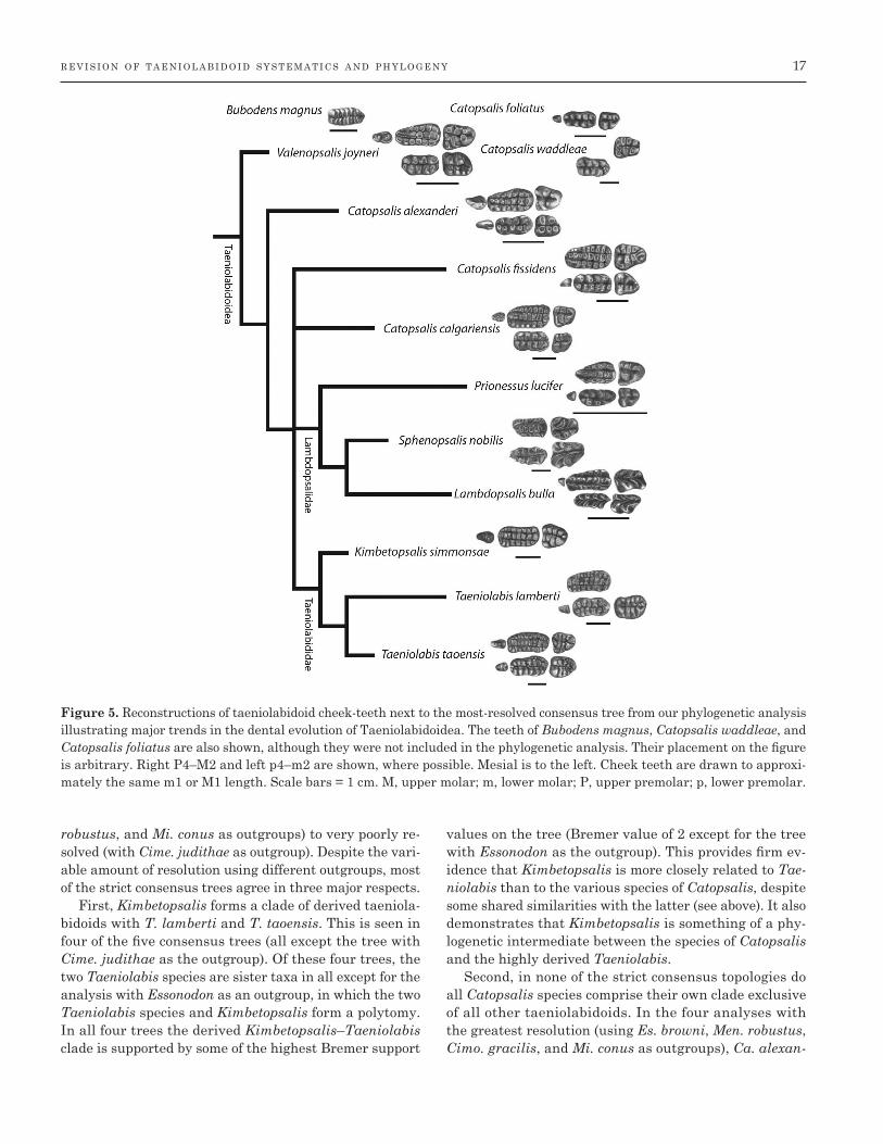
Results
Strict consensus trees for each of the five analyses with a different outgroup are shown in Figure 4. Reconstruc-tions of taeniolabidoid cheek teeth are shown next to the most resolved consensus tree (with Cimo. gracilis or Men. robustus as outgroups) in Figure 5. Excluded a posteriori from these trees were three fragmentary taxa that act as wildcards and decrease resolution (Ca. foliatus, Ca. wad-dleae, and Bubodens). The topologies of these trees range from nearly completely resolved (with Cimo. gracilis, Men.
Figure 4. Cladograms resulting from phylogenetic analyses. Data matrix for each analysis includes 11 taxa and 46 charac-ters. Letters refer to nodes (see Appendix S3 for list of common synapomorphies for each node). Numbers above each node are Bremer support values. A, Essonodon browni as outgroup, 51 steps, nine trees, consistency index (CI) = 0.647, retention in-dex (RI) = 0.600; B, Meniscoessus robustus as outgroup, 62 steps, two trees, CI = 0.677, RI = 0.615; C, Cimolomys gracilis as outgroup, 56 steps; two trees; CI = 0.661; RI = 0.604; D, Cimexomys judithae as outgroup, 64 steps; 25 trees; CI = 0.703; RI = 0.620; E, Microcosmodon conus as outgroup, 60 steps; two trees; CI = 0.733; RI = 0.673.
r e v i s i o n o f t a e n i o l a b i d o i d s y s t e m a t i c s a n d p h y l o g e n y 17
robustus, and Mi. conus as outgroups) to very poorly re-solved (with Cime. judithae as outgroup). Despite the vari-able amount of resolution using different outgroups, most of the strict consensus trees agree in three major respects.
First, Kimbetopsalis forms a clade of derived taeniola-bidoids with T. lamberti and T. taoensis. This is seen in four of the five consensus trees (all except the tree with Cime. judithae as the outgroup). Of these four trees, the two Taeniolabis species are sister taxa in all except for the analysis with Essonodon as an outgroup, in which the two Taeniolabis species and Kimbetopsalis form a polytomy. In all four trees the derived Kimbetopsalis–Taeniolabis clade is supported by some of the highest Bremer support
values on the tree (Bremer value of 2 except for the tree with Essonodon as the outgroup). This provides firm ev-idence that Kimbetopsalis is more closely related to Tae-niolabis than to the various species of Catopsalis, despite some shared similarities with the latter (see above). It also demonstrates that Kimbetopsalis is something of a phy-logenetic intermediate between the species of Catopsalis and the highly derived Taeniolabis.
Second, in none of the strict consensus topologies do all Catopsalis species comprise their own clade exclusive of all other taeniolabidoids. In the four analyses with the greatest resolution (using Es. browni, Men. robustus, Cimo. gracilis, and Mi. conus as outgroups), Ca. alexan-
Figure 5. Reconstructions of taeniolabidoid cheek-teeth next to the most-resolved consensus tree from our phylogenetic analysis illustrating major trends in the dental evolution of Taeniolabidoidea. The teeth of Bubodens magnus, Catopsalis waddleae, and Catopsalis foliatus are also shown, although they were not included in the phylogenetic analysis. Their placement on the figure is arbitrary. Right P4–M2 and left p4–m2 are shown, where possible. Mesial is to the left. Cheek teeth are drawn to approxi-mately the same m1 or M1 length. Scale bars = 1 cm. M, upper molar; m, lower molar; P, upper premolar; p, lower premolar.

18 w i l l i a m s o n e t a l . i n z o o l o g i c a l j o u r n a l o f t h e l i n n e a n s o c i e t y (2015)
deri, Ca. fissidens, and Ca. calgariensis are more closely related to the clade of Taeniolabis + Kimbetopsalis (tae-niolabidids, see below) and/or lambdopsalids (Prionessus and Sphenopsalis + Lambdopsalis) than is ‘Ca.’ joyneri, which is consistently recovered as the basal-most taenio-labidoid. Furthermore, in three of these trees ‘Ca.’ joy-neri and Ca. alexanderi comprise a series of successive outgroups on the line toward a clade of Ca. fissidens, Ca. calgariensis, and the derived taeniolabidoids. These re-sults support the idea, first proposed by Simmons & Miao (1986), that Catopsalis is a paraphyletic array of spe-cies rather than a single monophyletic taxon. Depending upon which outgroup is used and how polytomies are re-solved, the degree of paraphyly ranges from moderate to total (i.e. a stepwise array of taxa on the line to the de-rived taeniolabidoids).
Third, in all of the strict consensus topologies L. bulla and S. nobilis form a sister-taxon pair. In three of the trees, Pr. lucifer is the outgroup to this pair, with these three Asian taxa forming their own clade. This agrees with the results of Mao et al. (2015), which recovered Lambdop-salis and Sphenopsalis in their own subgroup, and goes a step beyond that analysis in finding moderately strong support for Prionessus as a member of this group. Mao et al. (2015) considered Prionessus as incertae sedis (not eas-ily assigned to any particular taeniolabidoid subgroup), both based on the results of their phylogenetic analysis and their intuition that its molar morphology is ‘primi-tive’ and more similar to that of Late Cretaceous multitu-berculates than other taeniolabidoids.
Systematic revision of Taeniolabidoidea Our phylogenetic analysis prompts a systematic revision of Taeniolabidoidea. This includes a clarified usage of the names Taeniolabidoidea and Taeniolabididae (which have been used variably in the literature), new phylogenetic definitions for important clades, and the designation of a new genus name for ‘Ca.’ joyneri.
Phylogenetic definitions delineate clades based on ancestry instead of possession of ‘key’ characters of the anatomy or a simple list of a priori included species (e.g. de Queiroz & Gauthier, 1990; de Queiroz & Gauthier, 1992; Sereno, 2005). Such genealogically based defini-tions are held to be more stable, clear, and objective than other approaches (de Queiroz & Gauthier, 1990, 1992), and they have been employed commonly by mammalian paleontologists Rowe, 1987; Rowe, 1988; Novacek et al., 1997; Luo, Kielan-Jaworowska & Cifelli, 2002; Sereno, 2006; Williamson et al. 2012). However, to our knowl-edge, such definitions have yet to be applied to taeniola-bidoids or ingroup clades.
We here define Taeniolabidoidea as the most inclu-sive clade containing T. taoensis Cope, 1882, but not Es.
browni Simpson, 1927, Men. robustus Marsh, 1889a, Cimo. gracilis Marsh, 1889a, Cime. judithae Sahni, 1972, Ca-topsbaatar catopsaloides (Kielan-Jaworowska, 1974); Eu. molestus Cope, 1886; Mesodma formosa Marsh, 1889b; Pti-lodus montanus Douglass, 1908; Krauseia clemensi (Sloan, 1981), Mi. conus Jepsen, 1930, Buginbaatar transaltaien-sis Kielan-Jaworowska & Sochava, 1969, Kogaionon un-gureanui Rădulescu & Samson (1996), or Boffius splendi-dus Vianey-Liaud, 1979.
Given that a clade of taeniolabidoids including Kimbe-topsalis and the Taeniolabis species is recovered in all of our phylogenetic analyses, and is supported by relatively high Bremer values, we feel that this important group of derived, fairly large-bodied taeniolabidoids should have a clear name. The commonly used Taeniolabididae is well suited for such a group. We here define Taeniolabididae to be the most inclusive clade containing T. taoensis (Cope, 1882), but not L. bulla Chow & Qi, 1978.
We also recovered a clade of taeniolabidoids that in-cludes Lambdopsalis and Sphenopsalis, and possibly Pri-onessus. Lambdopsalidae, a family-level taxon erected by Chow & Qi (1978) and resurrected by Mao et al. (2015), is a well-suited name for this group. We here define Lamb-dopsalidae to be the most inclusive clade containing L. bulla Chow & Qi, 1978, and not T. taoensis (Cope, 1882).
Our phylogenetic results strongly support the hypoth-esis that the various named species of Catopsalis do not form a single monophyletic group, but rather a paraphy-letic array. Therefore, if one considers a genus to be a clade, as is standard practice in modern vertebrate pa-leontology, then the genus name Catopsalis should not be applied to all of the species that currently bear this name. The most extreme taxonomic revision could give a new genus name to all Catopsalis species except for the type species. However, because the degree of paraphyly of Catopsalis differs depending upon the outgroup chosen and the resolution of polytomies, we feel that this is too extreme of a step to take at this point in time, while the evidence is still equivocal. With that said, our analyses strongly support the hypothesis that one species of Ca-topsalis, ‘Ca.’ joyneri, is a basal-most taeniolabidoid that does not closely group with other Catopsalis species (at least some of which do belong to a restricted subclade of multiple Catopsalis species in some of our most parsimo-nious topologies). For this reason, we here erect a new ge-nus name for ‘Ca.’ joyneri.
VALENOPSALIS GEN. NOV.
Etymology: Named after the late Leigh Van Valen, one of the 20th century’s great mammalian paleontologists, who studied Cretaceous–Paleogene multituberculates (includ-

r e v i s i o n o f t a e n i o l a b i d o i d s y s t e m a t i c s a n d p h y l o g e n y 19
ing ‘Ca.’ joyneri) and was a colorful inspiration to T. E. W. (who fondly remembers Leigh’s visit to the NMMNH collections when he was a graduate student) and S. L. B. (when he was an undergraduate student in Chicago).
Type species: Catopsalis joyneri Sloan & Van Valen, 1965.
Included species: Type species only.
Diagnosis: As for the type species.
Distribution Early Puercan (Pu1) of eastern Montana.
VALENOPSALIS JOYNERI (SLOAN & VAN VALEN, 1965)
Holotype: UMVP 1494, a right maxilla with M1, roots of P4.
Type locality and horizon: Bug Creek Anthills and Bug Creek West, Montana, USA.
Diagnosis: Differs from all other taeniolabidoids in having a p4 with two roots that are subequal in size, p4 length relatively greater compared with m1 length (p4L/m1L > 0.4), P3 present, the M1 internal cusp row length being be-tween 50 and 80% of the length of the tooth, and the mo-lar crown not being ornamented.
THE EVOLUTION AND BIOGEOGRAPHY OF TAENIOLABIDOIDEA
Multituberculates were amongst the most abundant and taxonomically diverse groups of mammals throughout the Cretaceous (Kielan-Jaworowska et al., 2004). They orig-inated by the earliest part of the Late Jurassic (c. 160 Mya, Yuan et al., 2013) and experienced a rapid increase in taxonomic diversity, dental complexity and disparity, and maximum body size beginning about 20 000 000 years before the end of the Cretaceous, coincident with the eco-logical rise of angiosperms (Wilson et al., 2012).
Amongst the latest Cretaceous multituberculates is a poorly known taxon from western North America, Bub. magnus Wilson, 1987, represented by a single isolated m1. Bubodens was amongst the largest mammals of the day: it attained a body size of about 5 kg, based on es-timates derived from tooth size (Table 4; see Legendre, 1986; Wilson et al., 2012). Additionally, it had bulbous cusps that show apical cusp wear, similar to that of tae-niolabidoids (Kielan-Jaworowska et al., 2004; Mao et al., 2015). It may represent the oldest, and only, Cretaceous
example of Taeniolabidoidea. This remains to be ade-quately tested by a higher-level multituberculate phylo-genetic analysis that includes an array of taeniolabidoids and other multituberculates.
Multituberculates and other mammals with relatively large body size and more specialist diets (such as strict carnivory and herbivory) were decimated at the end-Cre-taceous mass extinction, approximately 66 Mya (Wilson, 2013, 2014). Following the mass extinction, multituber-culates underwent an additional burst in taxonomic and morphological diversification and the earliest undisputed taeniolabidoids are present in earliest Paleocene assem-blages of western North America (Fig. 6). These taxa – Va-lenopsalis joyneri and Ca. alexanderi – exhibit the hall-mark dental adaptations found in other taeniolabidoids, including large, chisel-shaped incisors with enamel re-stricted to the labial surface, a large diastema separating incisors from cheek teeth, relatively reduced P4s, large molars with long rows of many cusps, and large body size. At an estimated size of about 2 kg, both exceed the size of any known Mesozoic multituberculate with the exception of Bub. magnus (Table 4; Wilson et al., 2012). It may be noteworthy that the earliest Paleocene taeniolabidoids are smaller than Bubodens (Fig. 6), a probable latest Creta-ceous ancestor or close relative, as this would fit with the general pattern of body size selectivity across the Creta-ceous–Paleogene boundary (Wilson, 2013, 2014).
Although Clemens (2010) recognized Bubodens to be a Cretaceous member of Taeniolabidoidea, he considered V. joyneri and Ca. alexanderi to be immigrant taxa, presum-ably because he did not consider Bubodens to be directly an-cestral to these taxa. We are unable to corroborate or refute this based on our phylogenetic analysis. However, we agree with his conclusion that there is no compelling evidence that taeniolabidoids originated in the Cretaceous of Asia and dispersed into North America near the beginning of the Paleocene. We currently consider the origins and ances-tral geographical range of taeniolabidoids to be uncertain.
Within a geologically brief time, during the first 2,000,000 years of the Paleocene, even larger taeniola-bidoids appear in the North American fossil record (Fig. 6). These taxa include Ca. waddleae, Ki. simmonsae, and species of Taeniolabis, all of which exceeded 10 kg in body size. The largest and most aberrant of these, T. taoensis, may have reached masses of 100 kg (Wilson et al., 2012), exceeding the size of the extant beaver (Castor canadensis) (Weil & Krause, 2008). Taeniolabis taoensis is the largest multituberculate that has yet been found and may repre-sent something of a maximum size bound for this long-lived and ecologically diverse group of mammals. Trends in body size evolution are not currently clear, owing to un-certainties in the phylogenetic relationships of taeniolabi-doids, but it is possible that there was a general trend of

20 w i l l i a m s o n e t a l . i n z o o l o g i c a l j o u r n a l o f t h e l i n n e a n s o c i e t y (2015)
size increase after the end- Cretaceous extinction, or alter-natively that different taeniolabidoids may have evolved large size independently of each other (Fig. 6).
North American taeniolabidoids underwent a modest taxonomic diversification early in the Paleocene. At least seven species are present in the Western Interior of North America within the first c. 1 500 000 years of the begin-ning of the epoch. A single taeniolabidid is present in nu-merous early Paleocene North American faunas, but only one fauna contains more than one taxon: the middle or late Puercan of the San Juan Basin of New Mexico. This fauna contains the very rare (represented by only a sin-gle specimen) Ca. foliatus, which either co-occurs with the equally rare middle Puercan Kimbetopsalis, or with the relatively more abundant late Puercan T. taoensis. Whereas some taxa are very rare and represented by at most only a few specimens, all from a single fauna (e.g. Ca. foliatus, Ca. waddleae, Kimbetopsalis, T. lamberti), several taxa are represented by numerous specimens and from multiple faunas from relatively widely separated lo-cales (e.g. Ca. alexanderi, Ca. fissidens, Ca. calgariensis, T. taoensis). Taeniolabidoids survived into the later part
of the early Paleocene (Ca. fissidens) and the early part of the late Paleocene (Ca. calgariensis) before apparently go-ing extinct in western North America. Catopsalis calgar-iensis had a body mass exceeding 10 kg, suggesting that taeniolabidoids remained fairly large in North America before disappearing.
One subgroup of taeniolabidoids, Lambdopsalidae, is a uniquely Asian clade that represents the last surviving radiation of Taeniolabidoidea. This clade includes Lamb-dopsalis and Sphenopsalis at a minimum, and probably also Prionessus. Long the subject of debate, they appear to co-occur and are now each represented by several spec-imens from multiple localities in the late Paleocene, c. 57 Mya (see Mao et al., 2015). The appearance of taeniolabi-doids in the late Paleocene of Asia, without any obvious close Asian relatives, strongly indicates that taeniolabi-doids dispersed into Asia from North America, probably at some point during the early–middle Paleocene. Several clades of therian mammals occurred in the Paleocene of both Asia and North America (e.g. Dinocerata, Carpolest-idae, Carnivoramorpha, Arctostylopidae, Mesonychia, Til-lodontia, Pantodonta), indicating that one or more disper-
Table 4. Mass estimates for taeniolabidoids and the outgroup taxa used in the phylogenetic analyses. For taxa for which m1s are not known, m1 area [length (L) × width] was predicted using a regression formula derived from the m1 vs. M1 area for other taeniolabidoids. Mass estimates were estimated using Wilson et al.’s (2012) formula from skull L, from m1 area using Legendre’s (1986) formula for all mammals and also for only rodents, and Wilson et al.’s (2012) formula from skull L–m1
Mass estimates (kg)
M1 Skull From From m1 area – From m1 area – From skull area length skull Legendre (1986, Legendre (1986, L–m1 – WilsonTaxon (mm2) (mm) length all mammals) rodents) et al. (2012)
Microcosmodon conus 3.11 0.05 0.07 0.05Essonodon browni 27.27 2.56 3.02 1.18Cimexomys judithae 1.73 0.02 0.02 0.02Cimolomys gracilis 10.08 0.42 0.52 0.28Meniscoessus robustus 41.36 69.0 1.208 5.49 6.31 2.15Bubodens magnus 76.80 17.01 18.82 5.25Valenopsalis joyneri 21.45 1.65 1.98 0.83Catopsalis waddleae 152.52 59.57 63.27 14.13Catopsalis foliatus 53.55 8.80 9.95 3.12Catopsalis fissidens 80.64 18.59 20.52 5.63Catopsalis alexanderi 41.76 5.59 6.41 2.18Catopsalis calgariensis 139.73 50.76 54.2 12.45Kimbetopsalis simmonsae 126.16 42.12 45.25 10.74Taeniolabis taoensis 205.92 160.0 22.697 103.09 107.55 21.79Taeniolabis lamberti 128.00 43.29 46.42 10.97Lambdopsalis bulla 24.96 60.8 0.776 2.18 2.58 1.04Prionessus lucifer 13.44 0.70 0.87 0.42Sphenopsalis nobilis 99.28 27.19 29.63 7.60
M, upper molar; m, lower molar.

r e v i s i o n o f t a e n i o l a b i d o i d s y s t e m a t i c s a n d p h y l o g e n y 21
sive episodes occurred between the two continents, but the number and timing of such episodes is not well resolved. The lambdopsalids were, on the whole, smaller than most Paleocene North American taeniolabidoids in body size (Table 4). This could represent a phylogenetic reduction in size that characterizes the Asian clade. As the last-sur-viving North American taeniolabidoids were quite large, and because phylogenetic relationships are still fairly un-certain, it is not clear that the small size of the late-surviv-ing Asian taxa is the culmination of any long-term trend in body size reduction in Taeniolabidoidea.
THE PALAEOBIOLOGY OF TAENIOLABIDOIDEA
Taeniolabidoids had an unusual diet amongst multitu-berculates, as indicated by their dental morphology. The dentition and dentaries of multituberculates superficially resemble those of rodents, but modern rodents are an im-precise functional analogue for Cretaceous–Paleocene
multituberculates (Jenkins & Krause, 1983; Wilson et al., 2012). Unlike rodents, or any extant mammals, multitu-berculates chewed with a palinal (front-to-back) chewing motion. Taeniolabidoids probably evolved from a multitu-berculate with large, laterally compressed and blade-like P4s, which were probably used to chew in a two-phase, slicing– crushing masticatory cycle (Krause, 1982). Most multituberculates that possessed these dental adapta-tions were probably omnivorous (Weil & Krause, 2008; Wilson et al., 2012). Taeniolabidoids, however, differ from this ancestral pattern in having greatly reduced P4s rel-ative to molar size, which is related to the reduction or elimination of the slicing–crushing chewing phase and an increased emphasis on the grinding phase of the mas-tication cycle. The small P4s are seen in all taeniolabi-doids, regardless of stratigraphical age or body size, and is a diagnostic feature of the group. Therefore, a grind-ing-focused chewing stroke evolved early in the history of the group and set taeniolabidoids apart from most other multituberculates.
Figure 6. Time-calibrated phylogeny based on the most-resolved consensus tree from our phylogenetic analyses. Each taxon is accompanied by a silhouette that illustrates the relative sizes resulting from our mass estimates (Table 4; skull length – m1 estimate). The area of the silhouette is proportionate to the mass [ln(area) = 0.6667*ln(volume) + 0.231]. The time scale follows Ogg (2012). The placement of the Puercan faunas of the Nacimiento Formation is after Williamson et al. (2014). Asian Paleocene mammal biochronology is after Ting et al. (2011). Fm., Formation.

22 w i l l i a m s o n e t a l . i n z o o l o g i c a l j o u r n a l o f t h e l i n n e a n s o c i e t y (2015)
Additional evidence for the aberrant diets of taeniolabi-doids comes from their distinctive molar morphology. All taeniolabidoids have complex molars with three rows of prominent cusps, which is another diagnostic feature of the group. The molars of early taeniolabidoids are large, with many cusps. These cusps are bulbous and in some taxa (e.g. Taeniolabis), highly crenulated. This results in high orientation patch count values, a measure of dental complexity, which reaches its peak amongst multituber-culates in T. taoensis (Wilson et al., 2012). These adapta-tions are related to herbivory, as described by Wilson et al. (2012). This shift to herbivory in taeniolabidoids is also accompanied by an increase in body size, as is the case in many terrestrial tetrapods. Senescent T. taoensis speci-mens exhibit extreme molar and incisor wear (Granger & Simpson, 1929), and tooth wear may have been a factor in an individual’s longevity.
The large incisors of taeniolabidoids are gliriform, with an enamel band that provides a sharp, chisel-like cutting apex, and were probably used for snipping vegetation. In some taeniolabidoids (e.g. Ca. fissidens, Ki. simmon-sae, T. taoensis), the incisors bear longitudinal striations that probably strengthened the tooth against compressive forces applied to their tips.
There is some evidence that the Asian lambdopsalids may have diverged from their North American relatives and developed a somewhat different diet. Sphenopsalis and Lambdopsalis have uniquely high-crowned molars with crescentic cusps (Mao et al., 2015). The high, narrow, and sharp, v-shaped cusp crests form opposing, triangu-lar-shaped cutting blades that would have formed an effec-tive shearing mechanism. This probably indicates a more folivorous diet than in other taeniolabidoids, particularly the North American Taeniolabis-like taxa.
Little is known about the locomotor habits of taenio-labidoids, because substantial postcrania are known for only two taxa: the lambdopsalids Sphenopsalis (Mao et al., 2015) and probably Lambdopsalis (Kielan-Jaworowska & Qi, 1990; Mao et al., 2015). Amongst these specimens are partial humeri, which are very robust, suggesting that these taxa were fossorial (Miao, 1988; Kielan-Jaworowska & Qi, 1990; Mao et al., 2015).
Conclusions
The recovery of a new genus and species of large taenio-labidoid multituberculate, Ki. simmonsae, from the early Paleocene (middle Puercan; Pu2) of the Nacimiento For-mation prompted a revision of Taeniolabidoidea and an evaluation of their phylogeny and evolution. Our phyloge-netic analysis of Taeniolabidoidea included all Asian and North American species referable to this clade. Owing to
uncertainties over the choice of an appropriate outgroup we ran analyses using five different outgroups and found that trees were highly resolved using three (Cimo. grac-ilis, Men. robustus, and Mi. conus) of the five outgroups. In the highly resolved trees, species of the largest-bodied North American forms, Kimbetopsalis and Taeniolabis, consistently form a monophyletic clade as do species of the Asian Lambdopsalis, Sphenopsalis, and Prionessus. We here define Kimbetopsalis and Taeniolabis as the ba-sis of Taeniolabididae and Lambdopsalis and Sphenopsa-lis as the basis of Lambdopsalidae.
This study underscores the extreme rapidity of develop-ment of large body size and the increase in dental complex-ity within taeniolabidoids within the first 800 Kyr of the end-Cretaceous mass extinction (Wilson et al., 2012; Wil-liamson et al., 2014). Taeniolabidoids evolved extremely unusual and highly specialized, large, chisel-shaped in-cisors and massive, multicusped cheek teeth for grind-ing vegetation and attained large body masses, exceeding 20 kg. The shifts to larger body sizes and increased cusp complexity strongly suggest a shift toward herbivory, and possibly folivory.
Bubodens magnus, the largest multituberculate and largest mammal of the latest Cretaceous, probably rep-resents the sole Cretaceous representative of Taeniolabi-doidea. Taeniolabidoids of the earliest Paleocene faunas of North America include V. joyneri, which our phylogenetic analyses found to be the basalmost taeniolabidoid. We find that Kimbetopsalis simmonsae is the basal-most member of Taeniolabididae and it provides a plausible progenitor for T. taoensis, which first appeared in the San Juan Ba-sin within the next 200 Kyr.
Although taeniolabidoids disappeared from North America several million years before the end of the Pa-leocene, they dispersed to Asia where they underwent a subsequent modest radiation towards the end of the Pa-leocene, becoming extinct near the Paleocene–Eocene boundary.
Acknowledgments — We thank C. Raymond for discov-ery of the holotype of Ki. simmonsae and A. Patterson and E. Davidson for assistance in the field. We thank B. Clem-ens for providing helpful comments and two anonymous re-viewers for giving constructive criticism. The United States Bureau of Land Management provided the permit (NM 14-09S) and some support for T. E. W., B. L. S., S. S., and R. S. Grants from the National Science Foundation provided additional support for T. E. W. (EAR 0207750), TEW and B. L. S. (EAR 1325544), and R. S. (EAR 1325612). B. L. S. is also funded by a Marie Curie Career Integration Grant (EC 630652) and the University of Edinburgh. S. S. was supported by a Natural Environment Research Council stu-dentship during the course of this project.

r e v i s i o n o f t a e n i o l a b i d o i d s y s t e m a t i c s a n d p h y l o g e n y 23
References
Archibald JD. 1982. A study of Mammalia and geology across the Cretaceous-Tertiary boundary in Garfield County, Montana. University of California Publications in Geological Sciences 122: 1–286.
Archibald JD. 1983. Structure of the K-T Mammal Radia-tion in North America: speculations on turnover rates and trophic structure. Acta Palaeontologica Polonica 28: 7–17.
Archibald JD. 1993. The importance of phylogenetic anal-ysis for the assessment of species turnover; a case his-tory of Paleocene mammals in North America. Paleobiol-ogy 19: 1–27.
Archibald JD, Clemens WA, Gingerich PD, Krause DW, Lindsay EH, Rose KD. 1987. First North American land mammal ages of the Cenozoic era. In: Woodburne MO, ed. Cenozoic mammals of North America. Berkeley: Univer-sity of California Press, 24–76.
Archibald JD, Lofgren DL. 1990. Mammalian zonation near the Cretaceous-Tertiary boundary. In: Bown TM, Rose KD, eds. The dawn of the Age of Mammals in the northern part of the Rocky Mountain Interior, North Amer-ica. Boulder, Colorado: Geological Society of America, 243: 31–50.
Broom R. 1914. On the structure and affinities of the Mul-tituberculata. Bulletin of the American Museum of Natu-ral History 33: 115–134.
Buckley GA. 1995. The multituberculate Catopsalis from the early Paleocene of the Crazy Mountains Basin in Montana. Acta Palaeontologica Polonica 40: 389–398.
Chow M, Qi T. 1978. Paleocene mammalian fossils from the Nomogen Formation of Inner Mongolia. Vertebrata Pala-siatica 16: 77–85.
Cifelli RL, Eberle JJ, Lofgren DL, Lillegraven JA, Cle-mens WA. 2004. Mammalian biochronology of the latest Cretaceous. In: Woodburne MO, ed. Mammalian biochro-nology of the latest Cretaceous. New York: Columbia Uni-versity Press, 21–42.
Clemens WA. 1964. Fossil mammals of the Type Lance For-mation, Wyoming, part I. introduction and Multitubercu-lata. University of California Publications in Geological Sciences 48: 1–105.
Clemens WA. 2010. Were immigrants a significant part of the earliest Paleocene mammalian fauna of the North American western interior? Vertebrata Palasiatica 48: 285–307.
Clemens WA. 2013. cf. Wortmania from the early Paleocene of Montana and an evaluation of the fossil record of the ini-tial diversification of the Taeniodonta (Mammalia). Cana-dian Journal of Earth Science 50: 341–354.
Cope ED. 1882. A second genus of Eocene Plagiaulacidae. American Naturalist 16: 416–417.
Cope ED. 1884. The tertiary Marsupialia. American Natu-ralist 18: 686–697.
Cope ED. 1886. The Plagiaulacidae of the Puerco epoch. American Naturalist 20: 451.
Cope ED. 1888a. The mechanical causes of the origin of the dentition of the Rodentia. American Naturalist 25: 80–88.
Cope ED. 1888b. Synopsis of the vertebrate fauna of the Puerco series. Transactions of the American Philosophi-cal Society 16: 298–361.
Douglass E. 1908. Vertebrate fossils from the Fort Union beds. Annals of the Carnegie Museum 5: 11–26.
Fox RC. 1989. The Wounded Knee local fauna and mam-malian evolution near the Cretaceous-Tertiary boundary, Saskatchewan, Canada. Palaeontographica Abteilung A 208: 11–59.
Fox RC. 1990. The succession of Paleocene mammals in west-ern Canada. In: Bown TM, Rose KD, eds. Dawn of the Age of Mammals in the northern part of the Rocky Mountain Interior, North America. Boulder, Colorado: Geological So-ciety of America, 51–70.
Fox RC. 2005. Microcosmodontid multituberculates (Alloth-eria, Mammalia) from the Paleocene and Late Cretaceous of western Canada. Palaeontographica 23: 1–109.
Gazin CL. 1939. A Paleocene mammalian fauna from cen-tral Utah. Journal of the Washington Academy of Sciences 28: 271–277.
Goloboff PA, Farris JS, Nixon KC. 2008. TNT, a free pro-gram for phylogenetic analysis. Cladistics: The Interna-tional Journal of the Willi Hennig Society 24: 774–786.
Granger W, Simpson GG. 1929. A revision of the Tertiary Multituberculata. Bulletin of the American Museum of Nat-ural History 56: 601–679.
Higgins P. 2003. A Wyoming succession of Paleocene mam-mal-bearing localities bracketing the boundary between the Torrejonian and Tiffanian North American land mam-mal ‘ages’. Rocky Mountain Geology 38: 247–280.
Jenkins FA, Krause DW. 1983. Adaptations for climbing in North American multituberculates (Mammalia). Sci-ence 220: 712–715.
Jepsen GL. 1930. Stratigraphy and paleontology of the Pa-leocene of northeastern Park County, Wyoming. Proceed-ings of the American Philosophical Society 83: 463–528.
Johnston PA, Fox RC. 1984. Paleocene and Late Creta-ceous mammals from Saskatchewan, Canada. Palaeonto-graphica Abteilung A: Palaeozoologie Stratigraphie 186: 1–6.
Kielan-Jaworowska Z. 1974. Multituberculate succession in the Late Cretaceous of the Gobi Desert (Mongolia). Pa-laeontologia Polonica 30: 23–44.
Kielan-Jaworowska Z. 1994. A new generic name for the multituberculate mammal ‘Djadochtatherium’ catopsaloi-des. Acta Palaeontologica Polonica 39: 134–136.
Kielan-Jaworowska Z, Cifelli RL, Luo Z-X. 2004. Mam-mals from the Age of Dinosaurs: origins, evolution, and structure. New York: Columbia University Press.

24 w i l l i a m s o n e t a l . i n z o o l o g i c a l j o u r n a l o f t h e l i n n e a n s o c i e t y (2015)
Kielan-Jaworowska Z, Hurum JH. 1997. Djadochtathe-ria – a new suborder of multituberculate mammals. Acta Palaeontologica Polonica 42: 201–242.
Kielan-Jaworowska Z, Hurum JH. 2001. Phylogeny and systematics of multituberculate mammals. Palaeontology 44: 389– 429.
Kielan-Jaworowska Z, Qi T. 1990. Fossorial adaptations of a taeniolabidoid multituberculate mammal from the Eo-cene of China. Vertebrata Palasiatica 28: 81–94.
Kielan-Jaworowska Z, Sochava AV. 1969. The first mul-tituberculate from the uppermost Cretaceous of the Gobi Desert (Mongolia). Acta Palaeontologica Polonica 14: 355– 371.
Krause DW. 1977. Paleocene multituberculates (Mamma-lia) of the Roche Percée Local Fauna, Ravenscrag Forma-tion, Saskatchewan, Canada. Palaeontographica Abteilung A 159: 1–36.
Krause DW. 1982. Jaw movement, dental function, and diet in the Paleocene multituberculate Ptilodus. Paleobiology 8: 265–281.
Krause DW. 1986. Competitive exclusion and taxonomic dis-placement in the fossil record: the case of rodents and mul-tituberculates in North America. In: Flanagan KM, Lille-graven JA, eds. Vertebrates, phylogeny, and philosophy. Laramie: University of Wyoming, 95–117.
Krishtalka L, Black CC, Riedel DW. 1975. Paleontology and geology of the Badwater Creek area, central Wyoming, Part 10. A late Paleocene mammal fauna from the Shotgun Member of the Fort Union Formation. Annals of Carnegie Museum 45: 179–212.
Legendre S. 1986. Analysis of mammalian communities from the late Eocene and Oligocene of southern France. Palaeovertebrata 16: 191–212.
Lehman TM. 1984. The multituberculate Essonodon browni from the Upper Cretaceous Naashoibito Member of the Kirtland Shale, San Juan Basin, New Mexico. Journal of Vertebrate Paleontology 4: 602–603.
Lerbekmo JF. 1985. Magnetostratigraphic and biostrati-graphic correlations of Maastrichtian to Early Paleocene strata between South-Central Alberta and Southwestern Saskatchewan. Bulletin of Canadian Petroleum Geology 33: 213– 226.
Linnaeus C. 1758. Systema naturae per regna tria naturae, secundum classes, ordines, genera, species, cum characteri-bus, differentiis, synonymis, locis. Vol. 1: regnum animale. Stockholm: Laurentii Salvii.
Lofgren DL. 1995. The Bug Creek problem and the Creta-ceous- Tertiary transition at McGuire Creek, Montana. University of California Publications in the Geological Sci-ences 140: 1–185.
Lofgren DL, Lillegraven JA, Clemens WA, Gingerich PD, Williamson TE. 2004. Paleocene biochronology; the Puercan through Clarkforkian land mammal ages. In: Woodburne MO, ed. Late Cretaceous and Cenozoic mam-
mals of North America. New York: Columbia University Press, 43–105.
Lucas SG, Williamson TE, Middleton MD. 1997. Catop-salis (Mammalia: Multituberculata) from the Paleocene of New Mexico and Utah: taxonomy and biochronological sig-nificance. Journal of Paleontology 71: 484–493.
Luo Z-X, Kielan-Jaworowska Z, Cifelli RL. 2002. In quest for a phylogeny of Mesozoic mammals. Acta Palaeontolog-ica Polonica 47: 1–78.
Mao F-Y, Wang Y-Q, Meng J. 2015. New specimens of the multituberculate mammalian Sphenopsalis from the Paleocene of Inner Mongolia, China: implications for phylogeny and biology of taeniolabidoid multituber-culates. Acta Palaeontologica Polonica. doi: 10.4202/app.00117.2014
Marsh OC. 1889a. Discovery of Cretaceous Mammalia. American Journal of Science 3: 81–92.
Marsh OC. 1889b. Discovery of Cretaceous Mammalia. Part II. American Journal of Science 38: 177–180.
Matthew WD. 1937. Paleocene faunas of the San Juan Ba-sin, New Mexico [edited by Walter Granger, William King Gregory, and Edwin Harris Colbert]. Transactions of the American Philosophical Society 30: 1–510.
Matthew WD, Granger W. 1925. Fauna and correlation of the Gashato Formation of Mongolia. American Museum Novitates 189: 1–12.
Matthew WD, Granger W, Simpson GG. 1928. Paleocene multituberculates from Mongolia. American Museum No-vitates 331: 1–4.
McKenna MC. 1960. A continental Paleocene vertebrate fauna from California. American Museum Novitates 2024: 1–20.
Meng J, Wyss A, Hu YM, Wang Y, Bowen GJ, Koch PI. 2005. Glires (Mammalia) from the Late Paleocene Bayan Ulan locality of Inner Mongolia. American Museum Novi-tates 3473: 1–25.
Meng J, Zhai R, Wyss AR. 1998. The late Paleocene Bayan Ulan fauna of Inner Mongolia, China. In: Beard KC, Daw-son MR, eds. Dawn of the Age of Mammals in Asia. Pitts-burgh: Bulletin of the Carnegie Museum of Natural His-tory, 148– 185.
Miao D. 1988. Skull Morphology of Lambdopsalis bulla (Mammalia, Multituberculata) and its implications to mammalian evolution. University of Wyoming Special Pa-per 4: 1–104.
Middleton MD. 1982. A new species and additional mate-rial of Catopsalis (Mammalia: Multituberculata) from the Western Interior of North America. Journal of Paleontol-ogy 56: 1197–1206.
Montellano M. 1992. Mammalian Fauna of the Judith River Formation (Late Cretaceous, Judithian), Northcen-tral Montana. University of California Publications in Geo-logical Sciences 136: 1–115.

r e v i s i o n o f t a e n i o l a b i d o i d s y s t e m a t i c s a n d p h y l o g e n y 25
Montellano M, Weil A, Clemens WA. 2000. An exceptional specimen of Cimexomys judithae (Mammalia: Multituber-culata) from the Campanian Two Medicine Formation of Montana, and the phylogenetic status of Cimexomys. Jour-nal of Vertebrate Paleontology 20: 333–340.
Novacek MJ, Rougier GW, Wible JR, McKenna MC, Dashzeveg D, Horovitz I. 1997. Epipubic bones in eu-therian mammals from the Late Cretaceous of Mongolia. Nature 389: 483–486.
Ogg JG. 2012. Geomagnetic polarity time scale. In: Grad-stein FM, Ogg JG, Schmitz MD, Ogg G, eds. The geologic time scale 2012. Oxford: Elsevier, 85–113.
de Queiroz K, Gauthier J. 1990. Phylogeny as a central principle in taxonomy: phylogenetic definitions of taxon names. Systematic Zoology 39: 307–322.
de Queiroz K, Gauthier J. 1992. Phylogenetic taxonomy. Annual Review of Ecology and Systematics 23: 449– 480.
Rădulescu R, Samson P. 1996. The first multituberculate skull from the Late Cretaceous (Maastrichtian) of Europe (Haţeg Basin, Romania). Anuarul Institututlui de Geolo-gie al României 69 (Suppl. 1): 177–178.
Rogers RR. 1993. Systematic patterns of time averaging in the terrestrial vertebrate record: a Cretaceous case study. In: Kidwell SM, Behrensmeyer AK, eds. Taphonomic ap-proaches in time resolution in fossil assemblages. Short courses in paleontology. Knoxville: Paleontological Soci-ety, 228– 249.
Rose KD. 1981. The Clarkforkian land-mammal age and mammalian faunal composition across the Paleocene-Eo-cene boundary. University of Michigan Papers on Paleon-tology 26: 1–197.
Rougier GW, Novacek MJ, Dashzeveg D. 1997. A new multituberculate from the Late Cretaceous locality Ukhaa Tolgod, Mongolia. Considerations on multituberculate in-terrelationships. American Museum Novitates 3191: 1–26.
Rowe T. 1987. Definition and diagnosis in the phylogenetic system. Systematic Zoology 36: 208–211.
Rowe T. 1988. Definition, diagnosis, and origin of Mamma-lia. Journal of Vertebrate Paleontology 8: 241–264.
Russell LS. 1926. A new genus of the species Catopsalis from the Paskapoo Formation of Alberta. American Journal of Science 12: 230–234.
Sahni A. 1972. The vertebrate fauna of the Judith River For-mation, Montana. Bulletin of the American Museum of Nat-ural History 147: 321–412.
Scott CS, Spivak DN, Sweet AR. 2013. First mammals from the Paleocene Porcupine Hills Formation of south-western Alberta, Canada. Canadian Journal of Earth Sci-ences 50: 355–378.
Secord R. 2008. The Tiffanian land-mammal age (middle and late Paleocene) in the northern Bighorn Basin, Wyo-ming. University of Michigan Papers on Paleontology 35: 1–192.
Sereno PC. 2005. The logical basis of phylogenetic taxon-omy. Systematic Biology 54: 595–619.
Sereno PC. 2006. Shoulder girdle and forelimb in multitu-berculates: evolution of parasagittal forelimb posture in mammals. In: Carrano MT, Gaudin TJ, Blob RW, Wible JR, eds. Amniote paleobiology: perspectives on the evolu-tion of mammals, birds, and reptiles. Chicago: University of Chicago, 315–366.
Simmons NB. 1987. A revision of Taeniolabis (Mammalia: Multituberculata), with a new species from the Puercan of Eastern Montana. Journal of Paleontology 61: 794–808.
Simmons NB. 1993. Phylogeny of Multituberculata. In: Szalay FS, Novacek MJ, McKenna MC, eds. Mammal phylogeny: Mesozoic differentiation, multituberculates, monotremes, early therians, and marsupials. New York: Springer-Verlag, 146– 164.
Simmons NB, Miao D. 1986. Paraphyly in Catopsalis (Mam-malia: Multituberculata) and its biogeographic implica-tions. In: Flanagan KM, Lillegraven JA, eds. Vertebrates, phylogeny, and philosophy, contributions to Geology, Uni-versity of Wyoming, special paper 3. Laramie: University of Wyoming, 87–94.
Simpson GG. 1927. Mammalian fauna of the Hell Creek For-mation of Montana. American Museum Novitates 267: 1–7.
Simpson GG. 1929. American Mesozoic Mammalia. New Haven: Yale University Press.
Sloan RE. 1981. Systematics of Paleocene multitubercu-lates from the San Juan Basin, New Mexico. In: Lucas SG, Rigby JK Jr, Kues BS, eds. Advances in San Juan Ba-sin Paleontology. Albuquerque: University of New Mex-ico, 127– 160.
Sloan RE. 1987. Paleocene and latest Cretaceous mammal ages, biozones, magnetozones, rates of sedimentation, and evolution. In: Fassett JE, Rigby JK Jr, eds. The Cretaceous- Tertiary boundary in the San Juan and Raton Basins, New Mexico and Colorado. Boulder, Colorado: Geological Soci-ety of America, 165–200.
Sloan RE, Van Valen L. 1965. Cretaceous mammals from Montana. Science 148: 220–227.
Ting S. 1998. Paleocene and early Eocene land mammal ages of Asia. Bulletin of Carnegie Museum of Natural History 34: 124–147.
Ting S-Y, Tong Y-S, Clyde WC, Koch PI, Meng J, Wang Y-Q, Bowen GJ, Qian L, Snell KE. 2011. Asian early Paleogene chronology and mammalian faunal turnover events. Vertebrata Palasiatica 49: 1–28.
Van Itterbeeck J, Missiaen P, Folie A, Markevich VS, Van Damme D, Guo D-Y, Smith T. 2007. Woodland in a fluviolacustrine environment on the dry Mongolian Pla-teau during the late Paleocene; evidence from the mam-mal bearing Subeng section (Inner Mongolia, P.R. China). Palaeogeography, Palaeoclimatology, Palaeoecology 243: 55–78.

26 w i l l i a m s o n e t a l . i n z o o l o g i c a l j o u r n a l o f t h e l i n n e a n s o c i e t y (2015)
Vianey-Liaud M. 1979. Les mammiferes Montiens de Hainin (Paleocene moyen de Belgique); Part I, Multitubercules. Palaeovertebrata 9: 117–131.
Weil A. 1998. A new species of Microcosmodon (Mammalia: Multituberculata) from the Paleocene Tullock Formation of Montana, and an argument for the Microcosmodontinae. Paleobios 18: 1–15.
Weil A 1999. Multituberculate phylogeny and mammalian biogeography in the Late Cretaceous and earliest Paleo-cene Western Interior of North America. PhD Thesis, Uni-versity of California, Berkeley.
Weil A, Krause DW. 2008. Multituberculata. In: Janis CM, Gunnell GF, Uhen MD, eds. Evolution of Tertiary mam-mals of North America: small mammals, xenarthrans, and marine mammals. Cambridge: Cambridge Univer-sity Press, 19–38.
Wilf PK, Beard C, Davies-Vollum KS, Norejko JW. 1998. Portrait of a late Paleocene (early Clarkforkian) terres-trial ecosystem; Big Multi Quarry and associated strata, Washakie Basin, southwestern Wyoming. Palaios 13: 514–532.
Williamson TE. 1996. The beginning of the age of mammals in the San Juan Basin, New Mexico; biostratigraphy and evolution of Paleocene mammals of the Nacimiento For-mation. New Mexico Museum of Natural History and Sci-ence Bulletin 8: 1–141.
Williamson TE, Brusatte SL, Carr TD, Weil A, Stand-hardt BR. 2012. The phylogeny and evolution of Creta-ceous- Paleogene metatherians: new cladistic analysis and description of new early Paleocene specimens from the Nacimiento Formation, New Mexico. Journal of Sys-tematic Palaeontology 10: 625–651.
Williamson TE, Lucas SG. 1997. The Chico Springs local-ity, Nacimiento Formation, San Juan Basin, New Mexico. New Mexico Geological Society Guidebook 48: 259–265.
Williamson TE, Peppe DJ, Heizler MT, Brusatte SL, Se-cord R. 2014. Chronostratigraphy of the Cretaceous-Pa-
leogene transition in the San Juan Basin, northwestern New Mexico. Journal of Vertebrate Paleontology, Program and Abstracts 2014: 255–256.
Wilson GP. 2013. Mammals across the K/Pg boundary in northeastern Montana, U.S.A.: dental morphology and body-size patterns reveal extinction selectivity and im-migrant-fueled ecospace filling. Paleobiology 39: 429–469.
Wilson GP. 2014. Mammalian extinction, survival, and re-covery dynamics across the Cretaceous-Paleogene bound-ary in northeastern Montana. In: Wilson G, Clemens WA, Horner J, Hartman JE, eds. Through the end of the Cre-taceous in the type locality of the Hell Creek Formation in Montana and adjacent areas. Boulder, CO: Geological So-ciety of America special paper no. 503, 365–392.
Wilson GP, Dechesne M, Anderson IR. 2010. New latest Cretaceous mammals from northeastern Colorado with biochronologic and biogeographic implications. Journal of Vertebrate Paleontology 30: 499–520.
Wilson GP, Evans AR, Corfe IJ, Smits PD, Fortelius M, Jernvall J. 2012. Adaptive radiation of multituber-culate mammals before the extinction of dinosaurs. Na-ture 483: 457– 460.
Wilson RW. 1987. Late Cretaceous (Fox Hills) multituber-culates from the Red Owl Local Fauna of western South Dakota. Dakoterra 3: 118–122.
Yuan C-X, Ji Q, Meng Q-J, Tabrum AR, Luo Z-X. 2013. Earliest evolution of multituberculate mammals revealed by a new Jurassic fossil. Science 341: 779–783.
SUPPORTING INFORMATION
Appendix S1. Character list.
Appendix S2. Character data matrix (text file).
Appendix S3. Common synapomorphies for each of the phylogenetic analyses.
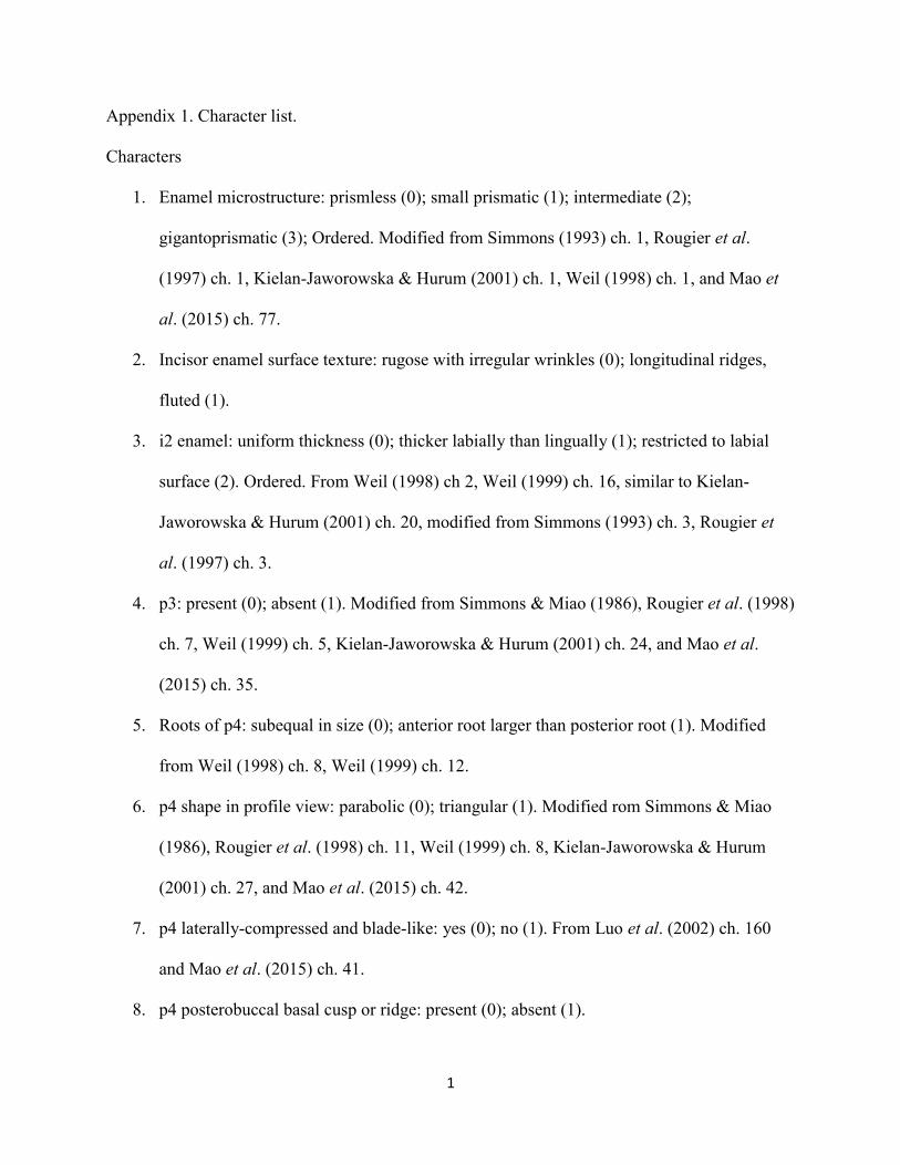
1
Appendix 1. Character list.
Characters
1. Enamel microstructure: prismless (0); small prismatic (1); intermediate (2);
gigantoprismatic (3); Ordered. Modified from Simmons (1993) ch. 1, Rougier et al.
(1997) ch. 1, Kielan-Jaworowska & Hurum (2001) ch. 1, Weil (1998) ch. 1, and Mao et
al. (2015) ch. 77.
2. Incisor enamel surface texture: rugose with irregular wrinkles (0); longitudinal ridges,
fluted (1).
3. i2 enamel: uniform thickness (0); thicker labially than lingually (1); restricted to labial
surface (2). Ordered. From Weil (1998) ch 2, Weil (1999) ch. 16, similar to Kielan-
Jaworowska & Hurum (2001) ch. 20, modified from Simmons (1993) ch. 3, Rougier et
al. (1997) ch. 3.
4. p3: present (0); absent (1). Modified from Simmons & Miao (1986), Rougier et al. (1998)
ch. 7, Weil (1999) ch. 5, Kielan-Jaworowska & Hurum (2001) ch. 24, and Mao et al.
(2015) ch. 35.
5. Roots of p4: subequal in size (0); anterior root larger than posterior root (1). Modified
from Weil (1998) ch. 8, Weil (1999) ch. 12.
6. p4 shape in profile view: parabolic (0); triangular (1). Modified rom Simmons & Miao
(1986), Rougier et al. (1998) ch. 11, Weil (1999) ch. 8, Kielan-Jaworowska & Hurum
(2001) ch. 27, and Mao et al. (2015) ch. 42.
7. p4 laterally-compressed and blade-like: yes (0); no (1). From Luo et al. (2002) ch. 160
and Mao et al. (2015) ch. 41.
8. p4 posterobuccal basal cusp or ridge: present (0); absent (1).

2
9. p4 number of cusps in median cuspal row: > 1 (0); 1 (1).
10. m1 cusp formula: ≤ 4:3 (0); ≥ 4:3 and < 5:4 (1); ≥ 5:4 and < 7:6 (2); ≥ 7:6 (3) . Ordered.
Modified from Simmons & Miao (1986), similar to Kielan-Jaworowska & Hurum (2001)
ch. 36.
11. Posterobuccal cingulid on m1: absent (0); present (1).
12. m2 cusp formula: ≥ 3:2 and < 4:4 (0); ≥ 4:4 (1). Modified from Simmons & Miao (1986).
Similar to Kielan-Jaworowska & Hurum (2001) ch. 37.
13. p4L/m1L: < 0.30 (0); ≥ 0.30 and < 0.40 (1); ≥ 0.40 and < 0.45 (2); ≥ 0.45 and < 0.65 (3);
> 0.65 (4). Ordered. Modified from Weil (1998) ch. 31, Weil (1999) ch. 34, Kielan-
Jaworowska & Hurum (2001) ch. 31, and Mao et al. (2015) ch. 48.
14. Molar crown ornamentation: smooth (0); grooved or ridged (1). Modified from Weil
(1999) ch. 33, Kielan-Jaworowska & Hurum (2001) ch. 39, Mao et al. (2015) ch. 76.
15. I2 enamel: uniform thickness (0); thicker labially than lingually (1); restricted to labial
surface (2). Ordered. Weil (1999) ch. 16, similar to Kielan-Jaworowska & Hurum (2001)
ch. 20, modified from Rougier et al. (1997) ch. 20.
16. I2 morphology: peg-like or single-cusped (0); two-cusped or more (1). From Rougier et
al. (1998) ch. 19, Kielan-Jaworowska & Hurum (2001) ch. 3, Mao et al. (2015) ch. 19.
17. I2 cusp count: 4 (0); 3 or less (1). From Simmons (1993) ch. 19, Rougier et al. (1997) ch.
19, Weil (1999) ch. 15, Kielan-Jaworowska & Hurum (2001) ch. 3.
18. Diastema between I2 and I3: absent (0); present (1). Modified from Yuan et al. (2013) ch.
18 and Mao et al. (2015) ch. 18.
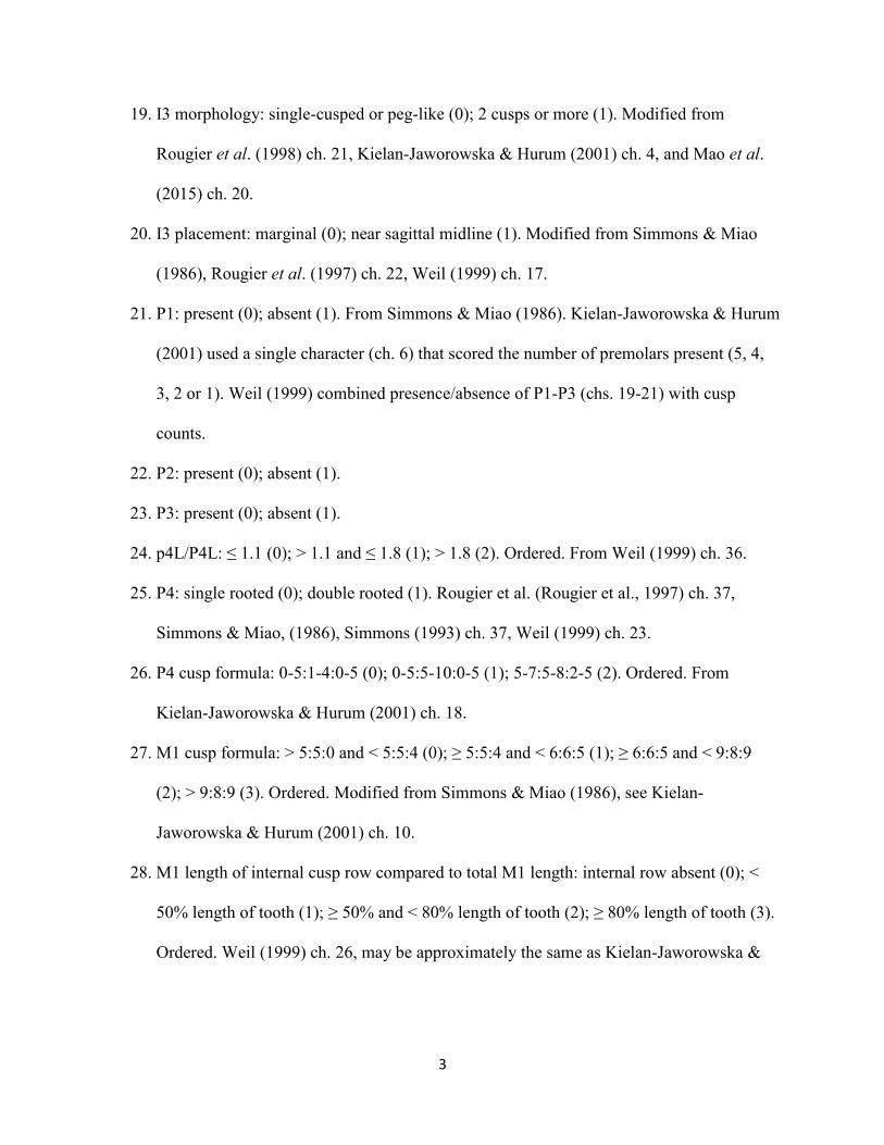
3
19. I3 morphology: single-cusped or peg-like (0); 2 cusps or more (1). Modified from
Rougier et al. (1998) ch. 21, Kielan-Jaworowska & Hurum (2001) ch. 4, and Mao et al.
(2015) ch. 20.
20. I3 placement: marginal (0); near sagittal midline (1). Modified from Simmons & Miao
(1986), Rougier et al. (1997) ch. 22, Weil (1999) ch. 17.
21. P1: present (0); absent (1). From Simmons & Miao (1986). Kielan-Jaworowska & Hurum
(2001) used a single character (ch. 6) that scored the number of premolars present (5, 4,
3, 2 or 1). Weil (1999) combined presence/absence of P1-P3 (chs. 19-21) with cusp
counts.
22. P2: present (0); absent (1).
23. P3: present (0); absent (1).
24. p4L/P4L: ≤ 1.1 (0); > 1.1 and ≤ 1.8 (1); > 1.8 (2). Ordered. From Weil (1999) ch. 36.
25. P4: single rooted (0); double rooted (1). Rougier et al. (Rougier et al., 1997) ch. 37,
Simmons & Miao, (1986), Simmons (1993) ch. 37, Weil (1999) ch. 23.
26. P4 cusp formula: 0-5:1-4:0-5 (0); 0-5:5-10:0-5 (1); 5-7:5-8:2-5 (2). Ordered. From
Kielan-Jaworowska & Hurum (2001) ch. 18.
27. M1 cusp formula: > 5:5:0 and < 5:5:4 (0); ≥ 5:5:4 and < 6:6:5 (1); ≥ 6:6:5 and < 9:8:9
(2); > 9:8:9 (3). Ordered. Modified from Simmons & Miao (1986), see Kielan-
Jaworowska & Hurum (2001) ch. 10.
28. M1 length of internal cusp row compared to total M1 length: internal row absent (0); <
50% length of tooth (1); ≥ 50% and < 80% length of tooth (2); ≥ 80% length of tooth (3).
Ordered. Weil (1999) ch. 26, may be approximately the same as Kielan-Jaworowska &

4
Hurum (2001) ch. 11. “M1 posterolingual wing to M1 length, wing absent (0); below 0.2
(1); between 0.2 and 0.3 (2); above 0.3 (3).”
29. M1 length: < 5.5 mm (0); ≥ 5.5 mm and < 9.5 mm (2); ≥ 9.5 and < 13.5 (3); ≥ 13.5 mm
and < 17.5 mm (3); ≥ 17.5 mm (4). Ordered. Modified from Simmons & Miao (1986),
Weil (1999) ch. 24.
30. M1/m1 multiple accessory roots: absent (0); present (1). Weil (1999) ch. 29.
31. P4L/M1L: < 0.5 (0); P4L/M1L ≥ 0.5 (1). Ordered. Modified from Simmons and Miao
(1986), Weil (1999) ch. 34. Weil (1999) used different divisions; 1, 1.75 for p4/m1 and
1.75, 0.8 for P4/M1. Similar to Kielan-Jaworowska & Hurum (2001) ch.8 and Mao et al.
(2015) ch. 29.
32. P4W/M1W: > 0.9 (0); ≤ 0.9 and > 0.6 (1); ≤ 0.6 and > 0.45 (2); ≤ 0.45 and > 0.2 (3).
Ordered. Modified from Kielan-Jaworowska & Hurum (2001) ch. 19 and Mao et al.
(2015) ch. 32.
33. M2 cusp formula: 1-4:2-3:3-4 (0); 1-2:4:5-7 (1).
34. M1L/M2L: ≤ 1.5 (0); > 1.5 and ≤ 2.25 (1); > 2.25 and ≤ 2.5 (2); > 2.5 (3). Ordered. From
Weil (1999) ch. 35; similar to character from Simmons & Miao (1986).
35. Cusp shape: conical or quadrangular (0); crescentic (1). Modified from Simmons & Miao
(1986), Kielan-Jaworowska & Hurum (2001), ch. 38.
36. Premaxilla with facial process: height > anteroposterior length (0); height ≤ length (1).
From Simmons (1993) ch. 42, Rougier et al. (1997) ch. 42.
37. Infraorbital foramen: double (0), single (1). From Kielan-Jaworowska & Hurum (2001)
ch. 16 and Mao et al. (2015) ch. 80; modified from Simmons (1993) ch. 43 and Rougier
et al. (1997) ch. 43.

5
38. Palatal vacuities absent (0), single (1), double (2). Ordered. From Simmons (1993) ch.
58, Rougier et al. (1997) ch. 58, Kielan-Jaworowska & Hurum (2001) ch. 17, Mao et al.
(2015) ch. 81.
39. Ridge between the palate and lateral walls of premaxilla: absent (0), present (1). From
Kielan-Jaworowska & Hurum (2001) ch. 40 and Mao et al. (2015) ch. 82.
40. Shape of the snout in dorsal view: incurved in front of the zygomatic arches with anterior
part directed posterolaterally (0); incurved with anterior part of zygomatic arches directed
transversely (1); trapezoid, not incurved in front of the zygomatic arches (2).From
Kielan-Jaworowska & Hurum (2001) ch. 41, similar to Simmons (1993) ch. 48 and
Rougier et al. (1997) ch. 48.
41. Number of pairs of vascular foramina on nasal: 1 (0); 2 (1); more than 2 (2). Ordered.
From Kielan-Jaworowska & Hurum (2001) ch. 42 and Mao et al. (2015) ch. 84.
42. Infraorbital foramen position: dorsal to P1 (0), dorsal to P2 (1), dorsal to P3 or P4 (2).
Ordered. Modified from Simmons (1993) ch. 45, Rougier et al. (1997) ch. 45, from
Kielan-Jaworowska & Hurum (2001) ch. 43, Mao et al. (2015) ch. 85.
43. Base of zygomatic arch as marked by posterior edge: dorsal to P4 or more anterior (0);
dorsal or posterior to P5/M1 or P4/M1 embrasure (1). Modified from Simmons (1993)
ch. 49 and Rougier et al. (1997) ch. 49, from Kielan-Jaworowska & Hurum (2001) ch. 44
and Mao et al. (2015) ch. 86.
44. Incisive foramen: situated within premaxilla (0); limited posteriorly by maxilla (1). From
Kielan-Jaworowska & Hurum (2001) ch. 52 and Mao et al. (2015) ch. 95; similar to
Simmon (1993) ch. 57 and Rougier et al. (1997) ch. 57.

6
45. Posterior border of palate: medial or posterior to posterior edge of M2 (0); medial to
middle of M2 (1); medial to M1-M2 embrasure (2). Ordered. Modified from Simmons
(1993) ch. 59, Rougier et al. (1997) ch. 59.
46. Coronoid process: parallel to the rest of the outer wall of the dentary (0); flared laterally
(1). From Kielan-Jaworowska & Hurum (2001) ch. 58.

xread
46 18
Bubodens_magnus ?????????30??1????????????????????0???????????
Meniscoessus_robustus 30101001020141?1011110011013110200010100?210??
Microcosmodon_conus 20200000020041111111000010220112010?1100?20???
Essonodon_browni 3????????3?0??????????0?10231102031???????????
Cimolomys_gracilis 30?00000030131?????????11113011?01[0 1]???????????
Cimexomys_judithae 300010?1020140??????00011112001201011?1?220???
Catopsalis_waddleae ?????????20??0???????????????1??0?????????????
Catopsalis_joyneri ?02?01?002003021??????0010221102010???????????
Catopsalis_calgariensis ?02?11???210102?1????????02341??010???????????
Catopsalis_foliatus ????11?1120010????????????????????0???????????
Catopsalis_fissidens ?12111?11210112??1????????2331??010???????????
Catopsalis_alexanderi ?02111?0?210?121?100???010232?02000???????????
Taeniolabis_taoensis 312111?003110121010011101033410211001001?21?00
Taeniolabis_lamberti ???1?1?0030101??????????00334?03??0???????????
Kimbetopsalis_simmonsae ?1???????????12????0??1?10234?03100????1??1???
Lambdopsalis_bulla 3021-1?1120100201000111?00232?0310111000121121
Sphenopsalis_nobilis ?021-1?????00020?00011110?233?03001??01???11??
Prionessus_lucifer ??21-1?1120000??????????[0 1]?231?03010??01???1???
;
cnames
{0 Enamel_microstructure: prismless. small_prismatic. "intermediate. gigantoprismatic.;
{1 Incisor_enamel_surface_texture: rugose_with_irregular_wrinkles. longitudinal_ridges,_fluted.;
{2 i2_enamel uniform_thickness. thicker_labially_than_lingually. restricted_to_labial_surface.;
{3 p3: present. absent.;
{4 Roots_of_p4 subequal_in_size. anterior_root_larger_than_posterior_root.;
{5 p4_shape_in_profile_view: parabolic. triangular.;
{6 p4_laterally-compressed_and_blade-like: yes. no.;
{7 p4_posterobuccal_basal_cusp_or_ridge: present. absent.;
{8 p4_number_of_cusps_in_median_cusp_row: >_1. 1.;
{9 m1_cusp_formula: "?<_4:3. "?>_or_=_4:3_and_<_5:4. "?>_or_=_5:4_and_<_7:6. "?>_or_=_7:6.;
{10 Posterobuccal_cingulid_on_m1 absent. present.;

{11 m2_cusp_formula: "?>_or_=_3:2_and_<_4:4. "?>_or_=_4:4.;
{12 p4L/m1L: <_0.30. "?>_or_=_0.30_and_<_0.40. "?>_or_=_0.40_and_<_0.45._
"?>_or_=_0.45_and_<_0.65._ >_or_=_0.65.;
{13 Molar_crown_ornamentation: smooth. grooved_or_ridged.;
{14 I2_enamel: uniform_thickness. thicker_labially_than_lingually_or_medially.
restricted_to_labial_surface.;
{15 I2_morphology: Peg-like_or_single_cusp. Two-cusped_or_more.;
{16 I2_cusp_count: 4. 3_or_less.;
{17 Diastema_betwee_I2_and_I3: absent. present.;
{18 I3_morphology: Single-cusped_or_peg-like. 2_cusps_or_more.;
{19 I3_placement: marginal. near_sagittal_midline.;
{20 P1: present. absent.;
{21 P2: present. absent.;
{22 P3: present. absent.;
{23 p4L/P4L: <_or_=_1.1. >_1.1_and_<_or_=_1.8. >_1.8.;
{24 P4: single_rooted. double_rooted.;
{25 P4_cusp_formula: 0-5:1-4:0-5. 0-5:5-10:0-5. 5-7:5-8:2-5.;
{26 M1_cusp_formula: >_5:5:0_and_<_5:5:4. >_or_=_5:5:4_and_<_6:6:5. >_or_=_6:6:5_and_<_9:8:9.
>_9:8:9.;
{27 M1_length_of_internal_cusp_row_compared_to_total_M1_length:_ internal_row_absent.
<_50%_length_of_tooth. >_or_=_50%_and_<_80%_length_of_tooth. >_or_=_80%_length_of_tooth.;
{28 M1_Length: <_5.5_mm. >_or_=_5.5_mm_and_<_9.5_mm. >_or_=_9.5_mm_and_<_13.5_mm.
>_or_=_13.5_mm_and_<_17.5_mm. >_or_=_17.5_mm.;
{29 M1/m1_multiple_accessory_roots: absent. present.;
{30 P4L/M1L: <_0.5. >_or_=_0.5.;
{31 P4W/M1W: >_0.9. <_or_=_0.9_and_>_0.6._ <_or_=_0.6_and_>_0.45. <_or_=_0.45_and_>_0.2.;
{32 M2_cusp_formula: 1-4:2-3:3-4. 1:4:5.;
{33 M1L/M2L: <_or_=_1.50. "?>_1.5_and_<_or_=_2.25. >_2.25_and_<_or_=_2.5. "?>_2.5.;
{34 Cusp_shape: conical_or_quadrangular. crescentic.;
{35 Premaxilla_with_facial_process height_>_anteroposterior_length. height_>_or_=_length.;
{36 Infraorbital_foramen: double. single.;
{37 Palatal_vacuities: absent. single. double.;
{38 Ridge_between_the_palate_and_lateral_walls_of_premaxilla: absent. present.;

{39 Shape_of_the_snout_in_dorsal_view:
incurved_in_front_of_the_zygomatic_arches_with_anterior_part_directed_posterolaterally.
incurved_with_anterior_part_of_zygomatic_arches_directed_transversely.
trapezoid,_not_incurved_in_front_of_the_zygomatic_arches.;
{40 Number_of_pairs_of_vascular_foramina_on_nasal: 1. 2. >_2.;
{41 Infraorbital_foramen_position: dorsal_to_P1. dorsal_to_P2. dorsal_to_P3_or_P4.;
{42 Base_of_zygomatic_arch_as_marked_by_posterior_edge:_ dorsal_to_P4_or_more_anterior.
dorsal_or_posterior_to_P5/M1_or_P4/M1_embrasure.;
{43 Incisive_foramen: situated_within_premaxilla. limited_posteriorly_by_maxilla.;
{44 Posterior_border_of_palate medial_or_posterior_to_posterior_edge_of_M2.
medial_to_middle_of_M2. medial_to_M1-M2_embrasure.;
{45 Coronoid_process:_ parallel_to_the_rest_of_the_outer_wall_of_the_dentary. flared_laterally.;
;
ccode + 0 2 9 12 14 23 25.28 30 31 33 37 40 41 *;
proc /;
comments 0
;

Appendix S3. Common synapomorphies for each of the phylogenetic analyses.
Appendix 2. Common synapomorphies.
Essonodon browni as Outgroup.
Synapomorphies common to 9 trees
(Node numbers refer to nodes in consensus, Fig. 3A)
Node A:
All trees:
No synapomorphies
Node B:
Some trees:
Char. 22: 0 1
Char. 28: 1 2
Node C:
All trees:
Char. 12: 1 0
Some trees:
Char. 7: 0 1
Char. 10: 1 0
Char. 31: 2 3
Node D:
All trees:
Char. 33: 1 0
Char. 34: 0 1
Node 5 :
All trees:
Char. 32: 0 1
Some trees:
Char. 1: 0 1
Char. 9: 2 3
Char. 11: 0 1
Char. 12: 1 0
Char. 28: 3 4

Appendix 3. Meniscoessus robustus as Outgroup.
Synapomorphies common to 5 trees
(Node numbers refer to nodes in consensus, Fig. 3B)
Node A:
All trees:
No synapomorphies
Node B:
All trees:
Char. 28: 1 2
Some trees:
Char. 10: 0 1
Node C:
All trees:
Char. 31: 2 3
Some trees:
Char. 28: 2 3
Node D:
All trees:
Char. 28: 23 4
Char. 32: 0 1
Char. 39: 0 1
Some trees:
Char. 1: 0 1
Node E:
All trees:
Char. 26: 2 3
Node F:
All trees:
Char. 7: 0 1
Char. 8: 0 1
Some trees:
Char. 13: 1 0
Char. 16: 0 1
Node G:
All trees:
Char. 34: 0 1
Some trees:
Char. 17: 1 0
Char. 33: 1 0
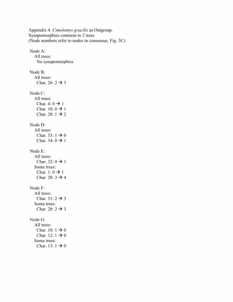
Appendix 4. Cimolomys gracilis as Outgroup.
Synapomorphies common to 2 trees
(Node numbers refer to nodes in consensus, Fig. 3C)
Node A:
All trees:
No synapomorphies
Node B:
All trees:
Char. 26: 2 3
Node C:
All trees:
Char. 4: 0 1
Char. 10: 0 1
Char. 28: 1 2
Node D:
All trees:
Char. 33: 1 0
Char. 34: 0 1
Node E:
All trees:
Char. 32: 0 1
Some trees:
Char. 1: 0 1
Char. 28: 3 4
Node F:
All trees:
Char. 31: 2 3
Some trees:
Char. 28: 2 3
Node G:
All trees:
Char. 10: 1 0
Char. 12: 1 0
Some trees:
Char. 13: 1 0

Appendix 5. Cimexomys judithae as Outgroup.
Synapomorphies common to 25 trees
(Node numbers refer to nodes in consensus, Fig. 3D)
Node A:
All trees:
No synapomorphies
Node B:
All trees:
Char. 27: 2 3
Some trees:
Char. 8: 0 1
Char. 10: 0 1
Char. 12: 3 01
Char. 13: 0 01
Char. 22: 0 1
Char. 28: 1 23
Char. 31: 2 3
Node C:
All trees:
Char. 34: 0 1
Some trees:
Char. 15: 1 0
Char. 17: 1 0
Char. 24: 1 0
Char. 33: 1 0
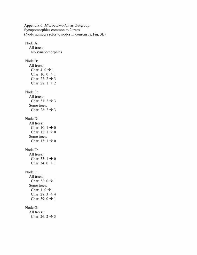
Appendix 6. Microcosmodon as Outgroup.
Synapomorphies common to 2 trees
(Node numbers refer to nodes in consensus, Fig. 3E)
Node A:
All trees:
No synapomorphies
Node B:
All trees:
Char. 4: 0 1
Char. 10: 0 1
Char. 27: 2 3
Char. 28: 1 2
Node C:
All trees:
Char. 31: 2 3
Some trees:
Char. 28: 2 3
Node D:
All trees:
Char. 10: 1 0
Char. 12: 1 0
Some trees:
Char. 13: 1 0
Node E:
All trees:
Char. 33: 1 0
Char. 34: 0 1
Node F:
All trees:
Char. 32: 0 1
Some trees:
Char. 1: 0 1
Char. 28: 3 4
Char. 39: 0 1
Node G:
All trees:
Char. 26: 2 3