a new species of the primitive dinosaur thecodontosaurus (saurischia:...
TRANSCRIPT

This article was downloaded by: [University of Connecticut]On: 12 October 2014, At: 05:52Publisher: Taylor & FrancisInforma Ltd Registered in England and Wales Registered Number: 1072954 Registered office: MortimerHouse, 37-41 Mortimer Street, London W1T 3JH, UK
Journal of Systematic PalaeontologyPublication details, including instructions for authors and subscription information:http://www.tandfonline.com/loi/tjsp20
A new species of the primitive dinosaurThecodontosaurus (Saurischia: Sauropodomorpha)and its implications for the systematics of earlydinosaursAdam M. Yates aa Department of Earth Sciences , University of Bristol , Bristol, BS8 1RJ, UKPublished online: 09 Mar 2010.
To cite this article: Adam M. Yates (2003) A new species of the primitive dinosaur Thecodontosaurus (Saurischia:Sauropodomorpha) and its implications for the systematics of early dinosaurs, Journal of Systematic Palaeontology, 1:1,1-42, DOI: 10.1017/S1477201903001007
To link to this article: http://dx.doi.org/10.1017/S1477201903001007
PLEASE SCROLL DOWN FOR ARTICLE
Taylor & Francis makes every effort to ensure the accuracy of all the information (the “Content”) containedin the publications on our platform. However, Taylor & Francis, our agents, and our licensors make norepresentations or warranties whatsoever as to the accuracy, completeness, or suitability for any purpose ofthe Content. Any opinions and views expressed in this publication are the opinions and views of the authors,and are not the views of or endorsed by Taylor & Francis. The accuracy of the Content should not be reliedupon and should be independently verified with primary sources of information. Taylor and Francis shallnot be liable for any losses, actions, claims, proceedings, demands, costs, expenses, damages, and otherliabilities whatsoever or howsoever caused arising directly or indirectly in connection with, in relation to orarising out of the use of the Content.
This article may be used for research, teaching, and private study purposes. Any substantial or systematicreproduction, redistribution, reselling, loan, sub-licensing, systematic supply, or distribution in anyform to anyone is expressly forbidden. Terms & Conditions of access and use can be found at http://www.tandfonline.com/page/terms-and-conditions

Journal of Systematic Palaeontology 1 (1): 1-42DOI: 10.1017/S1477201903001007 Printed in the United Kingdom
Issued 23 April 2003© The Natural History Museum
A new species of the primitiveDINOSAUR Thecodontosaurus(SAURISCHIA: SAUROPODOMORPHA)
AND i ts i m p l i c a t i o n s f o r t h e
SYSTEMATICS o f e a r l y d i n o s a u r s
Adam M. YatesDepartment of Earth Sciences, University of Bristol, Bristol, BS8 1RJ, UK
SYNOPSIS Juvenile sauropodomorph specimens from a Late Triassic/Early Jurassic fissure fill inPant-y-ffynnon Quarry, South Wales are redescribed and named as a new species, Thecodontosauruscaducus. T. caducus can be diagnosed by the presence of pleurocoel-like pits on the neurocentralsutures of the sixth, seventh and eighth cervical vertebrae. It is further distinguished from the typespecies of the genus, T. antiquus, by the primitive shape of its proximal humerus and ilium. Data fromthis specimen are incorporated into a cladistic analysis of basal sauropodomorph relationships. It isfound that Thecodontosaurus is basal to all other sauropodomorphs, with the exception of Saturnaliafrom the late Carnian of Brazil. As such Thecodontosaurus is a key taxon, with a novel combinationof characters that has important implications for early dinosaur phylogenetics. Thecodontosaurusprovides evidence that 'prosauropods' are paraphyletic with respect to Sauropoda and that Herrera-sauridae lies outside the clade containing Sauropodomorpha + Theropoda.
KEY WORDS Thecodontosaurus, T. caducus, Cladistic analysis, Sauropodomorpha, Prosauropoda,Paraphyletic
Contents
Introduction
Systematic palaeontologySAURISCHIA Seeley, 1888SAUROPODOMORPHA von Huene, 1932Genus THECODONTOSAURUS Riley and Stutchbury, 1836Thecodontosaurus caducus sp. nov.
DescriptionSkull roofPalateBraincaseMandibleVertebral columnForelimbHindlimb
Skeletal reconstruction
Cladistic analysisMethodsResults
Prosauropod monophyly
Acknowledgements
References
Appendix 1: list of anatomical abbreviations
Appendix 2: list of characters
Appendix 3: list of unambiguous synapomorphies
Appendix 4: character-taxon matrix
2
22222
45
10
11
151620
21
21
22
26
313133344041
Dow
nloa
ded
by [
Uni
vers
ity o
f C
onne
ctic
ut]
at 0
5:52
12
Oct
ober
201
4

A. M. YATES
INTRODUCTION
The remains of several juvenile sauropodomorphs werefound in Pant-y-ffynnon Quarry in South Wales by ProfessorK. Kermack and DrP. Robinson in 1952. The material is partof an Upper Triassic assemblage found in fine-grained sand-stone that filled a fissure in the Carboniferous Limestone ofthe Quarry. The sauropodomorph specimens include a disar-ticulated skull with associated forelimb elements and cervicalseries, isolated skull elements and several postcranial bones,including a partial hind limb. The skull was reconstructed anddescribed as Thecodontosaurus sp. by D. Kermack (1984).The postcranial remains have been featured in skeletal re-constructions (Kermack 1984; Galton 1990; Upchurch 1997;Benton et al. 2000) but remain undescribed.
Thecodontosaurus was the first sauropodomorph dino-saur to be scientifically described (Riley & Stutchbury 1836),but its anatomy has remained poorly known relative to othersauropodomorph dinosaurs. What is known is that, with anadult length of no more than 3 m (Benton etal. 2000), it is oneof the smallest and most gracile members of the Sauropodo-morpha. Some authors claim that it retained an obligatorybipedal posture during locomotion (Kermack 1984; Galton1990, 2000; Benton et al. 2000) but it certainly did not re-tain a predatory lifestyle. It had the typical sauropodomorphdental specialisations that suggest it included a high propor-tion of vegetable matter in its diet (Galton 1985a; Crompton& Attridge 1986).
The type species, T. antiquus Morris, 1843, was basedon largely disarticulated bones found in a Late Triassic fissurefill deposit from Bristol in south-west England. Many of thesebones were lost during the Second World War but hundredsstill survive and these have been redescribed recently (Bentonet al. 2000). More bones from another locality in south-westEngland are known (Whiteside 1983) and these are currentlybeing prepared and studied.
The importance of Thecodontosaurus lies in its basalposition within sauropodomorph phylogeny. It has eitherbeen thought of as the sister group of all other sauropodo-morphs (Gauthier 1986) or as the basal member of a mono-phyletic Prosauropoda (Galton 1990). The only computer-based, cladistic analysis to include Thecodontosaurus foundit to be part of a monophyletic prosauropod group but wasunable to resolve the position of the genus within this clade(Benton et al. 2000).
In this paper, the Pant-y-ffynon prosauropod specimensare fully described and illustrated and their relationship toT. antiquus and other early sauropodomorphs is investigatedusing cladistic analysis. The implications of this analysis forearly dinosaur systematics are discussed. In particular thecase for prosauropod monophyly is examined in detail.
The abbreviations for the various institutions wherematerial discussed in this paper is held are as follows:
AM = Amherst College Museum, Massachusetts,USA.
BMNH = Natural History Museum, London, UK.BRSUG = Department of Earth Sciences, University of
Bristol, UK.GPIT = Institut und Museum fur Geologie und
Palaontologie der Universit at T¨ubingen,Germany.
HMN = Museum fur Naturkunde der Humboldt Uni-versitat, Berlin, Germany.
PVL = Fundacion 'Miguel Lillo', Tucum´an, Argentina.SMNS = Staatliches Museum fur Naturkunde, Stuttgart,
Germany.
SYSTEMATIC PALAEONTOLOGY
SAURISCHIASeeley,1888
SAUROPODOMORPHAvon Huene,1932
Genus THECODONTOSAURUS Riley andStutchbury, 1836
TYPE SPECIES. Thecodontosaurus antiquus Morris, 1843;Late Triassic, Bristol, England.DIAGNOSIS. Small, gracile sauropodomorphs with the fol-lowing derived character states.
1. The basipterygoid processes are elongate and slender,with the length of the process, measured from its tip to thedorsal margin of the parabasisphenoid, being equal to theheight of the braincase, measured from the dorsal marginof the parabasisphenoid to the top of the supraoccipital(convergent in 'Efraasia diagnostica').
2. The dentary is short and deep, occupying less than 40%of the total mandibular length, and with a maximum dor-soventral depth that is greater than 20% of its length (con-vergent in Saturnalia tupiniquim).
3. The epipophyses of the cranial cervicals are flat plates thatoverhang the caudal margins of the postzygapophysealfacets but do not form raised ridges on the dorsal surfaceof the postzygapophysis.
4. The proximal and mid-caudal neural spines are positionedat the extreme caudal end of their neural arches, filling theinterpostzygapophyseal space (convergent in 'Efraasiadiagnostica').
5. The ventral furrowing of the caudal centra is reduced sothat it is only weakly present in the proximal caudals andis absent altogether from the mid and distal caudals.
REMARKS. The first two characters of the diagnosis are fromBenton et al. (2000) while the last three are novel. The thirdcharacter listed in Benton et al. (2000), 'caudal process ofthe iliac blade subquadratic' is also present in the basalsaurischian Guiabasaurus candelariensis (Bonaparte et al.1999), Neotheropoda (e.g. Dilophosaurus wetherilli Welles,1984) and 'Efraasia diagnostica' (pers. obs. SMNS 12354,12667). Consequently it is interpreted as a plesiomorphiccharacteristic of Thecodontosaurus.
Thecodontosaurus caducus sp. nov.
ETYMOLOGY. Latin, caducus, fallen. Refers to the fact thatthe holotype is an articulated specimen preserved in a fissurefill, indicating that the animal may have fallen into the fissureand died there.HOLOTYPE. BMNH P24, a nearly complete but disarticulatedskull, both mandibular rami, a complete series of cervicalvertebrae, the proximal ends of both humeri, a proximal rightscapula and both coracoids from one individual (Fig. 1).
2D
ownl
oade
d by
[U
nive
rsity
of
Con
nect
icut
] at
05:
52 1
2 O
ctob
er 2
014
DESCRIPTION OF A NEW SPECIES OF THE PRIMIT IVE DINOSAUR THECODONTOSAURUS
Figure 1 Thecodontosaurus caducus sp. nov., holotype, BMNH P24; skull and partial postcranial skeleton. 1A, photograph; 1B, interpretiveline drawing. Solid black bones represent unrelated crocodylomorph and lepidosaur bones. For abbreviations, see Appendix 1.Scale bars = 20 mm.
PARATYPES. BMNH P24/3, a right ischium; BMNH P39/2, aleft coracoid; BMNH P59/5, a right quadrate; BMNH P64/1,a series of eight proximal-mid caudals; BMNH P65/21, aright ectopterygoid; BMNH P77/1, a series of distal caudalvertebrae, the right ilium, femur, tibia, fibula and pes; BMNHP126/1, a ?proximal pubis; BMNH P141/1, a basioccipital.
TYPE HORIZON AND LOCALITY. Late Triassic fissure deposits,Pant-y-ffynnon Quarry, near Bonvilston, South Wales. Theage of the Mesozoic fissure deposits is difficult to determine.Given the faunal similarities between Pant-y-ffynnon andthe Thecodontosaurus-bearing fissure fills from south-westEngland, they are likely to be of a similar age. The presence
3D
ownl
oade
d by
[U
nive
rsity
of
Con
nect
icut
] at
05:
52 1
2 O
ctob
er 2
014

A. M. YATES
SQPRF
N
MX
PMX
A
ANG
PRF
par.p'EX-OP o!c SO
D B
Figure 2 Thecodontosaurus caducus sp. nov., holotype, BMNH P24; reconstruction of skull. 2A, lateral view; 2B, ventral view;2C, dorsal view; 2D, ventral view of mandibular ramus. For abbreviations see Appendix 1. Scale bar = 10 mm.
of phytosaurs in the English fissure fills constrains their ageto the Late Triassic (Benton etal. 2000) while palynomorphsmay indicate a Rhaetian age (Whiteside 1983). A fuller ac-count is given in Benton & Spencer (1995).DIAGNOSIS. A species of Thecodontosaurus with the follow-ing autapomorphy: pleurocoel-like pits on the neurocentralsutures of the sixth, seventh and eighth cervical vertebrae.T. caducus can be further distinguished from T. antiquus byexhibiting the plesiomorphic state for the autapomorphies ofthat species. These include a medial tubercle of the prox-
imal humerus that does not project strongly (versus stronglyprojecting in T. antiquus) and a preacetabular process of theilium that projects cranially (versus a downcurved preacetab-ular process in T. antiquus).
DESCRIPTION
The most complete specimen in the collection is the holotype,BMNH P24. Except where mentioned, the description of
4D
ownl
oade
d by
[U
nive
rsity
of
Con
nect
icut
] at
05:
52 1
2 O
ctob
er 2
014

DESCRIPTION OF A NEW SPECIES OF THE PRIMIT IVE DINOSAUR THECODONTOSAURUS
the skull, mandible and cervical vertebrae is based on thisspecimen.
The dorsal skull roof of this specimen is situated at thecranial end of the cervical series, while the elements fromthe left temporal region, mandible, palate and braincase arescattered for some distance to the left of the dorsal skull roof.All of the braincase elements have separated, indicating thatsuturing had not begun at the time of death, one of the manyjuvenile characteristics that can be seen in these specimens.The reconstruction of the skull presented here (Fig. 2) differssomewhat from that of Kermack (1984). This is partly be-cause some bones have been re-identified and partly becausemissing, or damaged, parts were restored using shapes moresimilar to those known in other early saurischians.
Skull roof
PremaxillaThe medial surface of the right premaxilla is exposed. Itssurface is damaged and the nasal process is missing. Themain body of the premaxilla is quite short (8 mm long and6 mm high) and bears four teeth. Like juvenile Massospon-dylus carinatus (Cooper 1981) and Mussaurus patagonicus(Bonaparte & Vince 1979), the height of the premaxillaryteeth exceeds the height of the maxillary teeth. The crownsare simple subcylindrical spikes that bear a few weak serra-tions on the caudal carina (there are six such serrations onthe second tooth). The first crown is the largest at 4.5 mm inheight.
Maxilla (Fig. 3)Only a fragment of the left maxilla is available for study.It is quite poorly preserved so details are hard to discern.The preserved portion, which is 25 mm long, consists of the
B
Figure 3 Thecodontosaurus caducus sp. nov., holotype, BMNHP24; left maxilla in lateral aspect. 3A, photograph; 3B, interpretivedrawing. nv.c = neurovascular canal, nv.f = neurovascular foramen.Scale bar = 5 mm.
caudal ramus and what is probably the base of the ascendingramus. The ascending ramus itself and the rostral ramus aremissing. The specimen is 4 mm high at the rostral end andtapers to a point at its caudal end. The number of alveoli can-not be determined but there are six teeth present and spacefor four more. On the lateral surface there appear to be fiverelatively large, neurovascular foramina. The caudal-mostforamen is smaller than the more rostral foramina. In mostother sauropodomorphs (e.g. Riojasaurus incertus: Bona-parte & Pumares 1995; Plateosaurus engelhardti: pers. obs.of HMN MB. 1927.19.1, GPIT Skelett 1) the foramen at thecaudal end of the row is distinctly larger than the rest ofthe maxillary foramina. This includes sauropods, where thecaudal maxillary foramen is so enlarged it has been termedthe pre-antorbital fenestra (Wilson & Sereno 1998).
A sharp edge (the ventral rim of the external antor-bital fenestra) delimits the lateral surface of the caudal ra-mus from the dorsal surface. The dorsal surface bears ashort, longitudinal groove that extends from near the mid-point to a point above the fourth preserved tooth. Thisgroove lies on the floor of the antorbital fossa and wouldhave housed the maxillary nerve and associated vascu-lature (Witmer 1997). A similar groove has been repor-ted in Plateosaurus engelhardti (Witmer 1997), Sellosaurusgracilis? (Galton 1985b) and Massospondylus carinatus(Gow et al. 1990). The groove becomes closed over bythe jugal and the lacrimal to form a canal in ornithischians(Witmer 1997), whereas a foramen, or a series of foramina,pierce the dorsal or medial surface of the maxilla in sauro-pods and non-avian theropods (Witmer 1997). Therefore, adorsally open canal for the maxillary nerve on the ventralsurface of the antorbital fossa is potentially a synapomorphyuniting the traditional Prosauropoda into a monophyleticgroup.
The maxillary teeth are poorly preserved and those de-tails that are present appear to be similar to the dentary teeth.The largest crown is 3 mm high.
NasalBoth nasals have been fractured and distorted by compressionagainst the underlying bones. Consequently many details arelost. However, the natural edge of the caudal margin is pre-served and indicates that the suture with the frontals was con-cave, as it is in primitive dinosaurs such as Coelophysis bauri(Colbert 1989) and Lesothosaurus diagnosticus (Sereno1991a). Some derived sauropodomorphs such as Plateo-saurus engelhardti, have a caudally convex naso-frontalsuture. There is no indication that there was a mediannasal depression as is present in some specimens of Plateo-saurus engelhardti (Galton 1984a) and 'Efraasia diagnost-ica' (Galton 1985b as Sellosaurus gracilis). The rostroventralprocess is missing from the main body of each nasal.
P re frontalThe dorsal surface of the main body of the left prefrontal isexposed. It is a small, dorsally facing, elliptical plate, 11 mmlong and 6 mm wide, that would have formed part of the skullroof behind the lacrimal. It is not enlarged, as it is in otherbasal sauropodomorphs such as Massospondylus carinatus(Cooper 1981) and Plateosaurus engelhardti (Galton 1984a).The right prefrontal is still articulated with the lacrimal. Mostof the main body is missing, but a short, thin descending
5D
ownl
oade
d by
[U
nive
rsity
of
Con
nect
icut
] at
05:
52 1
2 O
ctob
er 2
014

A. M. YATES
a.prf
med.r.
p.r
H
Figure 4 Thecodontosaurus caducus sp. nov., holotype, BMNH P24; elements of the skull. 4A, pair of frontals in dorsal aspect; 4B, leftlacrimal in lateral aspect (dorsal outline restored from right lacrimal); 4C, right ectopterygoid in dorsal aspect; 4D, parabasisphenoid complex inventral aspect; 4E, basioccipital in ventral aspect; 4F, basioccipital in dorsal aspect; 4G, right exoccipital-opisthotic complex in occipital view;4H, left parietal in dorsal aspect. For abbreviations see Appendix 1. Scale bars = 5 mm.
process arises from the caudolateral margin and extendsabout halfway down the medial side of the ventral ramus ofthe lacrimal. The dorsal exposure of the prefrontal is muchgreater than that of the lacrimal.
Frontal (Fig. 4A)Both frontals are well preserved and are visible in dorsalview. Each frontal is longer than it is wide, with the maximumwidth developed at the caudal end. A deep, rostromediallyinclined slot, for the reception of the frontal ramus of thepostorbital, is incised into the tip of the caudolateral cornerof the right frontal (this region is damaged in the left frontal).There is a faintly raised area medial to this slot, presenton both frontals. The supratemporal fossa extends onto thefrontals, producing a sharply defined, crescentic depression
on the caudal margin of each frontal. The midsection of thefrontals, which forms the roof over the orbits, is constrictedtransversely. The rostral end is expanded transversely, but notas greatly as the caudal. A facet for the articulation of the pre-frontal occupies the rostral third of the lateral margin. Thus,the prefrontal does not restrict the frontal contribution to theorbital margin as it does in more derived sauropodomorphssuch as Plateosaurus engelhardti (Galton 1984a) and Lufen-gosaurus huenei (Young 1941a). A row of foramina occurson each side of the median frontal symphysis.
Parietal (Fig. 4H)The parietals had separated before burial, indicating that theywere not fused or tightly sutured together, which is a signof immaturity. As in Plateosaurus engelhardti the parietalscomprise a rectangular rostral portion that forms the caudal
6D
ownl
oade
d by
[U
nive
rsity
of
Con
nect
icut
] at
05:
52 1
2 O
ctob
er 2
014

DESCRIPTION OF A NEW SPECIES OF THE PRIMIT IVE DINOSAUR THECODONTOSAURUS
end of the dorsal skull roof and a caudolateral wing thatsutures with the squamosal. The rostral end of the parietalsforms a straight suture with the frontals in dorsal view. Faintridges, on either side of the midline, mark the medial mar-gins of the supratemporal fossae. Each ridge is confluentwith the sharper ridge that bounds the rostral margin of thesupratemporal fossa on the frontal. Medial to the ridges,the parietals form a flat, horizontal surface. Lateral to eachridge the parietal curves ventrally to meet the lateral walls ofthe braincase. The caudolateral wings are more steeply in-clined than the lateral sides of the rostral end of the parietal. Ascar marking the articulation with the squamosal occupies thedistal half of the lateral surface of this wing. In lateral viewthe caudolateral wing curves ventrally so that the squamosaland, consequently, the quadrate head, would have been heldbelow the level of the dorsal skull roof.
Lacrimal (Fig. 4B)The bone identified by Kermack (1984) as the rightsquamosal is re-interpreted here as the right prefrontal andright lacrimal, exposed on their medial side. The lacrimal isan approximately L-shaped bone with a long ventral ramus(26 mm) and a short rostral ramus (8 mm). A narrow strip ofthe rostral ramus is exposed dorsolaterally on the skull roof,rostral to the prefrontal and lateral to the nasal. A sulcusextends up the dorsal half of the caudal face of the ven-tral ramus, medial to the ventral process of the prefrontal. Asingle lacrimal foramen is situated at the dorsal end of thesulcus. The rostral opening of the lacrimal foramen cannotbe seen. The short rostral ramus sutures with the ascendingramus of the maxilla. The ventral ramus is quite narrow at itsmidpoint (1 mm) but flares rostrocaudally at its ventral end(6 mm). Dislocation has made it impossible to determine ifthe ventral end of the lacrimal contacted the caudal ramus ofthe maxilla.
As in other basal sauropodomorphs (e.g. Plateosaurusengelhardti: Galton 1984a) and the basal saurischian, Her-rerasaurus ischigualastensis (Sereno & Novas 1993), thelacrimal formed a lateral wall over the caudodorsal corner ofthe antorbital sinus, whereas the caudoventral corner of thesinus extended over the lateral surface of the lacrimal, to forma laterally facing fossa. In most basal sauropodomorphs (e.g.Plateosaurus engelhardti: Galton 1984a) this fossa is smalland restricted to the rostroventral corner of the main vent-ral ramus of the lacrimal. In T. caducus, however, the fossaextends at least half way up the ventral ramus. This condi-tion is probably the plesiomorphic one, since it is also seenin neotheropods and the basal saurischians Herrerasaurusischigualastensis (Sereno & Novas 1993) and Eoraptorlunensis (Sereno et al. 1993).
JugalBoth jugals are obscured below overlying bones. Only partsof the lateral side of the left jugal are visible. It is estimatedto be about 30 mm in length. The suborbital portion of thejugal is a slender, medio-laterally compressed bar that is30 mm deep in the mid-orbital region. In keeping with therelatively large size of the orbit of this juvenile, the postorbitalramus is placed far back along the jugal (the rostral end ofits base is approximately 23 mm from the rostral end of thejugal). The postorbital process is triangular with a relativelybroad base. The ventral margin of the jugal is gently arched
upwards. No details of the maxillary, lacrimal, postorbitaland quadratojugal articulations are visible.
PostorbitalThe left postorbital is visible in lateral view, while the medialside of the right postorbital is exposed. The postorbital is tri-radiate with long jugal and frontal rami and a short squamosalramus. The frontal ramus is steeply inclined anterodorsallyfrom its junction with the other rami to the frontal. The ra-mus also curves medially to articulate with the frontal butthis curvature is not as strong as in Plateosaurus engelhardti(Galton 1984a). The frontal ramus becomes broader towardsits rostral end, which is slightly forked. The supratemporalfossa extends onto the dorsomedial surface of this ramus asit does in other basal saurischians such as Herrerasaurusischigualastensis (Sereno & Novas 1993) and Plateosaurusengelhardti (Galton 1984a). The squamosal ramus was ashort, slender and pointed process that was probably hori-zontally oriented. The jugal ramus is an elongate strap thatis mediolaterally compressed and gently bowed caudally.Bones overlying both of the postorbitals obscure the articu-lation facets for the jugals.
QuadratojugalNeither of the two quadratojugals can be positively identified;however, a small plate of bone protruding from beneath theright quadrate head is likely to be the main body of the rightquadratojugal.
Quadrate (Fig. 5)Both quadrates of BMNH P24 and an isolated right quad-rate, BMNH P59/5, can be viewed in their medial and caudalaspect. The main body of the quadrate consists of two lam-inae set at right angles to each other. Where the two lam-inae meet along the caudal edge, a sharp keel is formed.This keel extends dorsally to the small knob-like quadratehead. A large semi-circular lamina extends rostromedially
Figure 5 Thecodontosaurus caducus sp. nov., BMNH P59/5; right
quadrate. 5A, medial, 5B, caudal; 5C, mandibular condyle. Scale
b a r = 5 mm.
7D
ownl
oade
d by
[U
nive
rsity
of
Con
nect
icut
] at
05:
52 1
2 O
ctob
er 2
014

A. M. YATES
and forms the pterygoid wing, while the narrower, rostro-laterally directed lamina thickens ventrally to form the shaftthat bears the quadrate condyles. The base of the pterygoidwing is long, occupying more than 70% of the length ofthe quadrate. This is a primitive character state that is alsoseen in 'Efraasia diagnostica' (Galton 1985b as Sellosaurusgracilis) but not in more derived sauropodomorphs such asPlateosaurus engelhardti (pers. obs. of SMNS 12950), Col-oradisaurus brevis (from photographs of PVL 3967) or mostsauropods (e.g. Camarasaurus lentus: Madsen et al. 1995).Unlike most other early saurischians (e.g. Herrerasaurusischigualastensis: Sereno&Novas 1993;Liliensternuslilien-sterni: pers. obs. of HMN MB.R.2175.7.4; Sinraptor dongi:Currie & Zhao 1993; 'Efraasia diagnostica': pers. obs. ofSMNS 12668; Plateosaurus engelhardti: pers. obs. of GPITSkelett 1) the quadrate foramen is not deeply incised into thelateral margin of the rostrolateral lamina. If a quadrate fora-men was present, it would have been a narrow gap betweenthe quadrate and quadratojugal such as in Lesothosaurusdiagnosticus (Sereno 1991a) and Heterodontosaurus tucki(Weishampel & Witmer 1990). The articular surface is nar-rowly triangular in ventral view with the long axis orientedtransversely and the apex pointing laterally. An oblique sul-cus running antero-medially divides the articular surface intotwo condyles, of which the more medial is taller.
SquamosalThe bone lying under the caudal end of the basisphenoid andthe paroccipital process of the right exoccipital-opisthoticcomplex appears to be the left squamosal exposed in dor-somedial aspect (identified as part of the ?opisthotic byKermack 1984). The squamosal head is subrectangular indorsal view unlike the triangular shape that is usual amongstdinosaurs, including other basal sauropodomorphs such asPlateosaurus engelhardti (Galton 1984a). Like other dino-saurian squamosals the complete bone would have consistedof four rami, however, only two of these can be seen inthis specimen. The rostromedially directed parietal ramus isshort, slender and distinctly raised above the dorsal surface ofthe squamosal head. The rostrolaterally directed postorbitalramus has broken away. Caudal to the base of the parietalramus, the base of a slender, caudoventrally directed, quad-rate ramus can be seen. The overlying paroccipital process ofthe right exoccipital-opisthotic complex obscures the caudalramus of the squamosal.
Palate
PterygoidMost of the right pterygoid is exposed in ventral and me-dial views, while only a fragment of the transverse flangeremains of the left pterygoid. The pterygoid is a complexbone, consisting of three main projections: the rostral ramus,the quadrate ramus and the transverse flange. The rostralramus was the longest of these, measuring 25.5 mm longas preserved. It is an elongate triangular plate that, in life,would have faced ventromedially and formed a large partof the palate. Its medial margin is almost straight, with therostral end forming a median symphyseal surface. The me-dial margin is flared upwards in this region so that whenthe two pterygoids were in contact a low, dorsally project-ing, median crest was formed. The base of the transverseflange forms the caudolateral margin of the rostral ramus.
The pterygoid is bent downwards sharply along this line sothat the transverse flange faces more or less rostroventrally.The flange itself is a short, subrectangular process that is dir-ected laterally in ventral view. Judging from the shape of thecross-section of the left pterygoid fragment, the transverseflange was gently curved about the transverse axis so thatthe caudodorsal surface was concave. It is 11 mm long alongits caudal margin. The quadrate ramus is a short, vertical,triangular plate that is 7 mm long and is directed dorsallyand laterally. It flares distally from its narrow, waist-likejunction with the rest of the pterygoid, at the caudomedialcorner of the rostral ramus and the transverse flange. Perhapsthe most significant feature of the pterygoid is the absenceof a caudomedial flange that hooks around the basiptery-goid process to contact its counterpart medially. Most dino-saurs have such a flange (e.g. Plateosaurus engelhardti: pers.obs. of HMN 24; Lesothosaurus diagnosticus: Sereno 1991a;Sinraptor dongi: Currie & Zhao 1993) although it is reducedto a small dorsomedially oriented hook, or is absent alto-gether, in eusauropods (Wilson & Sereno 1998).
Ectopterygoid (Figs 4C, 6)The right ectopterygoid is visible in dorsal view, while theventral view can be seen in an isolated right ectopterygoid(BMNH P65/21). The main body is twice as long as it is wide(10 mm long) with a sinuous medial margin. A deep concav-ity occupies the ventral surface and closely resembles theventral pneumatic fossa of neotheropods. The jugal processis slender and strongly recurved. In this respect the ecto-pterygoid resembles that of a neotheropod more than anyother sauropodomorph.
PalatineA flat, roughly quadrangular sheet of bone exposedbetween the right pterygoid, the left ectopterygoid and thesupraoccipital is probably the left palatine, exposed latero-
7 I
Figure 6 Thecodontosaurus caducus sp. nov., BMNH P65/21; rightectopterygoid in ventral aspect. j.p = jugal process, v.f = ventralfossa. Scale bar = 5 mm.
8D
ownl
oade
d by
[U
nive
rsity
of
Con
nect
icut
] at
05:
52 1
2 O
ctob
er 2
014

DESCRIPTION OF A NEW SPECIES OF THE PRIMIT IVE DINOSAUR THECODONTOSAURUS
Figure 7 Thecodontosaurus caducus sp. nov., holotype, BMNH P24.7A, right prootic in lateral aspect; 7B, right prootic in medial aspect;7C, supraoccipital in cranial aspect. For abbreviations see Appendix 1. Scale bars = 5 mm.
dorsally. The ventro-lateral margin bears a deep, narrow sul-cus for the reception of the medial side of the maxilla. Littleelse can be said except that the caudal palatine margin (therostral border of the palatine fenestra) is quite straight and notas strongly emarginate as it is in Plateosaurus engelhardti(Galton 1984a).
Braincase
Supraoccipital (Fig. 7C)The inner surface of the crescent-shaped supraoccipital isexposed. Like Anchisaurus polyzelus (Galton 1976) and'Efraasia diagnostica' (Galton 1985b), but unlike most otherbasal sauropodomorphs, the bone is much wider than it ishigh. The ventral margin is concave and would have formedthe dorsal margin of the foramen magnum. The dorsal mar-gin is evenly arched and is not peaked at the midline as itis in Plateosaurus engelhardti (Galton 1984a). The caudalend of the endocranial cavity forms a deep dorsoventrallyorientated median sulcus running up the midline of the in-ner face. This sulcus is flanked by two pairs of facets, thedorsal pair of which face rostrally and would have articu-lated with the prootics while the ventral pair face rostrolat-erally and would have contacted the opisthotic-exoccipitalcomplexes. On each side, a deep, narrow channel extends lat-erally between these two facets, from the endocranial cavityto the dorsolateral margin of the supraoccipital. This channelmay have allowed the passage of the vena capitis dorsalis.
Exoccipital-opisthotic complex (Fig. 4G)The occipital face of the right complex is exposed, with lim-ited lateral exposure. A tongue-shaped paroccipital processprojects laterally and slightly dorsally. A conspicuous fora-men exits from the middle of the occipital surface of thebone, at the base of the paroccipital process. This foramenis not present in the braincase of T. antiquus, neither is itpresent in Plateosaurus engelhardti (Galton 1984a), thus theposition of this foramen on the paroccipital process may be ajuvenile characteristic or a specific autapomorphy. Below thebase of the paroccipital process a vertical sheet of bone des-cends to articulate with the basioccipital. The caudoventralcorner of this sheet projects to form the dorsolateral corner
of the occipital condyle. This sheet forms the lateral mar-gin of the foramen magnum. Its lateral surface is pierced bytwo foramina for rostral and caudal rami of the hypoglossalnerve (cranial nerve XII). A deep narrow sulcus, the metoticfissure, extends caudodorsally from a point rostral to theseforamina. This fissure is bordered rostrodorsally by a thincrista interfenestralis.
Prootic (Figs 7A, B)Both prootics are preserved, the right one of which has beenfreed from the matrix. Each bone is roughly rectangular inlateral view. The ventral margin formed a relatively straightcontact with the dorsal margin of the lateral wall of theparabasisphenoid complex, in front of the fenestra ovalis.The rostral margin bears a deep notch midway along itslength. This is an incompletely closed foramen for the exit ofthe trigeminal nerve (cranial nerve V). Exiting though thecentre of the prootic is a foramen for the facial nerve (cranialnerve VII). The facial nerve foramen lies against a crescenticridge that extends from the ventral margin to a point halfwayup the caudal margin and separates a depressed caudoventralregion from the rest of the prootic. Most of the caudal mar-gin of the prootic forms the rostral rim of the fenestra ovalis.Above this rim the caudodorsal corner was produced intoa short, caudally projecting, triangular process that wouldhave overlapped the rostral face of the opisthotic-exoccipitalcomplex. The dorsal margin, where it would have contactedthe skull roof, forms a dorsally and laterally concave saddle.Medially, a tall process, standing 6 mm from the medial sur-face, arises from the centre of the bone. This process curvescaudally to enclose a roughly pyramidal space that housed theinner ear. This cavity is open caudally, immediately adjacentto the rostral rim of the fenestra ovalis.
Basioccipital (Figs 4E, F)There are two basioccipitals in the sample, one from BMNHP24, while BMNH P141/1 is an isolated specimen. Bothhave been freed from the matrix, permitting all aspects tobe observed. The occipital condyle still bears a small noto-chordal pit, another indication of juvenility. In ventral viewthe parabasisphenoid contact forms a raised transverse ridge,which is the basioccipital contribution to the basal tubera.
9D
ownl
oade
d by
[U
nive
rsity
of
Con
nect
icut
] at
05:
52 1
2 O
ctob
er 2
014

10 A. M. YATES
* '
med-p
\_*_ J
B
w i - - • . * -
Figure 8 Thecodontosaurus caducus sp. nov., holotype, BMNH P24; mandibular elements. 8A, left articular in ventro-medial aspect; 8B, leftdentary in lateral aspect. med.p = medial process. Scale bars = 5 mm.
Unlike many sauropodomorphs, such as Plateosaurus engel-hardti (pers. obs. of HMN MB. 1927.19.1), Massospondyluscarinatus (pers. obs. of a cast of SAM 1314) and Camara-saurus lentus (Madsen et al. 1995), this raised area is un-divided by a median excavation. Dorsally there is a broadmidline sulcus that forms the caudal floor of the endocra-nial cavity. Two short perilymphatic grooves extend laterallyfrom either side of the endocranial floor, above the basaltubera. A low but sharp median ridge on the braincase floorextends from between the perilymphatic grooves to the con-tact with the basispenoid. Similar, although weaker, ridgescan also be seen in Thecodontosaurus antiquus (pers. obs. ofuncatalogued BRSUG material), Plateosaurus engelhardti(pers. obs. of SMNS 6014) and Massospondylus carinatus(Gow 1990).
Basisphenoid-parasphenoid complex (Fig. 4D)The parabasisphenoid complex is exposed in ventral aspect.In ventral view the main body comprises a flat central areafrom which the basipterygoid processes project rostrallyand the basal tubera project caudally. The elongate, peg-like basipterygoid processes extend ventrolaterally as wellas rostrally. Unlike many other sauropodomorphs, such as'Efraasia diagnostica' (pers. obs. of SMNS 12667), Plateo-saurus engelhardti (Galton 1984a), Coloradisaurus brevis(from photographs of PVL 3967), Brachiosaurus brancai(Janensch 1935-36) and Camarasaurus lentus (Madsen etal.1995), there is no interbasipterygoid web of bone. There is,however, a rostrally open fossa at the base of the cultriformprocess (the 'blind pocket' of Gow 1990) that is borderedcaudally by a scarp-like wall that spans the interbasiptery-goid space. It is from this feature that the interbasipterygoidweb of more derived taxa almost certainly evolved. The cul-triform process is a slender, laterally compressed, blade-likestructure.
On the lateral surface there is a small elliptical fora-men for the internal carotid artery set in a deep fossa locatedbetween the basal tubera and the base of the basipterygoidprocess. Dorsal to this fossa the ventral margin of the fen-estra ovale forms a semicircular embayment in the dorsolat-eral margin of the bone. Compared to the adult braincase ofT. antiquus (Benton et al. 2000), the fenestra ovale was relat-ively larger, which is almost certainly related to the juvenilenature of the specimen.
Figure 9 Thecodontosaurus caducus sp. nov., holotype, BMNHP24; eighth and ninth teeth from the left dentary. Scale bar = 1 mm.
Mandible
Dentary (Fig. 8B)The labial surface of the left dentary is exposed, while thelingual side of the right is partially exposed. The left dentaryclearly bears 12 alveoli, all of which, except the eleventh,bear teeth (Fig. 9). The first alveolus is inset a short distance,less than the width of an alveolus, from the rostral tip. Thedentary is short relative to the reconstructed length of themandible, with the dentigerous portion occupying no morethan 43% of the mandibular length (27 mm). Correlated withits brevity, the dentary is deeper, relative to its length, thanin other basal sauropodomorphs. This feature is also foundin the dentaries of T. antiquus (Benton et al. 2000). Thelabial surface of the dentary is flat and is not marked by astrong ridge below the caudal end of the tooth row, as it is inother early sauropodomorphs such as Riojasaurus incertus(Bonaparte & Pumares 1995), Anchisaurus polyzelus (Galton1976) and Plateosaurus engelhardti (Galton 1984a). A rowof neurovascular foramina exits from the lateral side of thedentary below the dentigerous margin. In lateral view theventral margin is straight while the dentigerous margin is
Dow
nloa
ded
by [
Uni
vers
ity o
f C
onne
ctic
ut]
at 0
5:52
12
Oct
ober
201
4

DESCRIPTION OF A NEW SPECIES OF THE PRIMIT IVE DINOSAUR THECODONTOSAURUS 11
gently curved ventrally at its rostral end. However, sincethe ventral margin is not concave, the dentary tip cannotbe regarded as ventrally curved as it is in Coloradisaurusbrevis (from photographs of PVL 3967) and Plateosaurusengelhardti (Galton 1984a).
SurangularBoth surangulars are exposed medially. The left surangularis the more completely exposed of the two. It is a sheet-like bone that is 35 mm long and 6 mm deep at its deepestpoint. The thickened and medially inflected dorsal marginforms a gently convex surface in lateral view that was not de-veloped into a strong coronoid peak as it is in Coloradisaurusbrevis (from photographs of PVL 3967), Plateosaurus engel-hardti (Galton 1984a) andmacronarian sauropods (Wilson &Sereno 1998). A short, medial projection from the dorsal mar-gin braced the rostral end of the articular. Behind the medialprocess there is a slender caudally projecting process thatwould have covered the ventrolateral surface of the retro-articular process. The anteroventral margin is too poorly pre-served to judge the size of the external mandibular fenestra.
Table1 Dimensions of the cervical vertebrae (in mm).
Axis
CE3
CE4
CE5
CE6
CE7
CE8
CE9
CE10
CE =
Lengthof neuralarch
16.320.6
24.2
24.4
23.1
22.0
18.716.9
16.5
cervical vertebra.
Length ofcentrum
10.7
14.5
15.8
16.9
15.2
14.9
14.1
14.412.7
Width ofanteriorcranial face6.2
6.8
8.0-
---
8.4
8.5
Height ofanteriorcranial face
5.15.2
6.96.2
6.6-
6.8
8.0
8.4
Splenial and coronoidNo splenial or coronoid can be confidently identified, al-though they are probably included amongst a number ofsimple flat bones, that are poorly exposed and remain uniden-tified.
AngularOnly a small section of what is probably the right angular canbe seen under the right prearticular and it does not offer anydetails for description other than that it appears to be quitenarrow relative to the surangular.
PrearticularThe lateral (internal) surface of the right prearticular (identi-fied as the right angular by Kermack 1984) is exposed. It is athin, elongate, sheet-like bone that is slightly curved laterallyalong its ventral margin. It is deeper caudally in the region ofthe glenoid socket, where it is 5 mm deep. Rostrally it formsa long, dorsoventrally shallow process that is at its narrowestat the midpoint, where it formed the ventral border of theinternal mandibular fenestra.
Articular (Fig. 8A)The ventral side of the left articular is exposed. It has twoflattened surfaces, one facing ventro-laterally and the otherventro-medially, that meet along the ventral midline to forma sharp keel. The ventro-lateral surface is narrower than theventro-medial surface, although it becomes broader at itscaudal end. A weakly defined, shallow fossa occupies theexpanded rostral end of the ventro-medial face. The me-dial edge of the glenoid fossa forms a deep semicircularnotch along the dorsal margin of the articular in medialview. The retro-articular process is quite primitive when com-pared to other basal sauropodomorphs such as 'Efraasia dia-gnostica' (Galton 1985b), Plateosaurus engelhardti (Galton1984a), Coloradisaurus brevis (Bonaparte 1978), Lufengo-saurus huenei (Young 1941a) and Massospondylus carinatus(Gow etal. 1990). Unlike these taxa, which have a long, lowprong-like retro-articular process, that of T. caducus is short,deep and bears a pointed medial process. The medial pro-cess is a primitive feature that can be seen in Herrerasaurusischigualastensis (Sereno & Novas 1993) and manyneotheropods (e.g. Allosaurus fragilis: Madsen 1976; Ty-rannosaurus rex: Carr 1999).
Vertebral column
Cervical vertebrae (Table 1)Ten cervical (CE) vertebrae are preserved and, given that thetenth is quite like a dorsal vertebra in its morphology, thiswas almost certainly the last cervical. Although no dorsalvertebrae are preserved with which to compare the cervicals,it can be determined that the neck was elongated as it is inother saurischians. The centra of CE3-9 are all longer thanthe axial centrum, a condition seen in other sauropodomorphsand neotheropods.
Thecodontosaurus caducus differs from other sauro-podomorphs, except Riojasaurus incertus, in not having mid-cervical centra that are at least three times as long as wide.The cervical vertebrae show strong indications of immaturity.These are the lack of fusion between any of the individual ele-ments of the atlas-axis complex and the presence of plainlyvisible neurocentral sutures on the postaxial cervicals. In-deed the neural arches have parted from their centra in CE3,6 and 7. Lack of sutural closure in the cervical vertebraewas found to be characteristic of immature crocodilians byBrochu (1996) and is almost certainly indicative of immatur-ity in dinosaurs as well.
The cervical ribs are poorly preserved, but it is clear that,like other saurischians, they are longer than their respectivevertebrae, and that in life they lay parallel to the cervicalcolumn.
Atlas (Fig. 10C)All elements of the atlas-axis complex are incompletely os-sified, remain separate from each other, and have becomescattered from their original positions. Thus it is difficultto distinguish the atlantal intercentrum from the axial in-tercentrum and the odontoid. The element identified byKermack (1984) as the atlantal intercentrum is here thoughtto be too rounded and not transversely wide enough to be thatelement, and is re-interpreted as the odontoid. The elementidentified as the axial intercentrum is re-interpreted here asthe atlantal intercentrum. It is a low, broadly U-shaped bone
Dow
nloa
ded
by [
Uni
vers
ity o
f C
onne
ctic
ut]
at 0
5:52
12
Oct
ober
201
4
12 A. M. YATES
Figure 10 Thecodontosaurus caducus sp. nov., holotype, BMNHP24; elements of the atlas-axis complex. IOA, axial intercentrum incranial aspect; IOB, axial intercentrum in left lateral aspect; IOC, rightneurapophysis in medial aspect; IOD, axial neural arch in dorsalaspect. For abbreviations see Appendix 1. Scale bars = 5 mm.
lantal epipophyses extend as far back as the cranial marginof the axial postzygapophyses. The odontoid is small androunded. The dorsal surface is flattened, while the ventraland cranial surfaces are strongly convex. The lateral surfacebears a small, round depression.
Axis (Figs 10A, B, D)The probable axial intercentrum is a small, crescentic ele-ment with a strongly concave dorsal margin in cranial viewand a pointed cranially directed process, developed on themidpoint of the ventral margin. The element is craniocaud-ally flattened and is only 2.9 mm long at its thickest point. Theaxial centrum is a simple spool-shaped element that is 11 mmlong and 5 mm wide. A probable juvenile characteristic isthe lack of any form of parapophysis at the cranial end ofthe centrum. Like other sauropodomorphs (e.g. Riojasaurusincertus: Bonaparte & Pumares 1995; Plateosaurus engel-hardti: pers. obs. of GPIT Skelett 1; Camarasaurus lentus:Madsen et al. 1995), but in contrast to other basal dinosaurs,the axis is not ventrally keeled. The axial neural arch covers awide neural canal that is 75% the width of the cranial face ofthe axial centrum. The size of the foramen magnum relativeto the size of the animal decreases through ontogeny (Dodson1975) and we can expect the width of the axial neural canal tobe strongly correlated with that of the foramen magnum. Thusthe relatively wide axial neural canal is probably a juvenilecharacteristic. The prezygapohyses are small, dorsolaterallyfacing facets mounted on tab-shaped processes that projectfrom the cranial margin of the neural arch, similar to theaxial prezygapophyses of neotheropods, but unlike those ofHerrerasaurus ischigualastensis (Sereno & Novas 1993) andother sauropodomorphs (e.g. Camarasaurus lentus: Madsenet al. 1995). In these taxa the prezygapophyses are simpleraised areas that do not project cranially. Below the prezyga-pophyses at the antero-ventral corners of the arch there areweakly developed tubercles that represent the diapophyses.In dorsal view, the lateral margins of the neural arch flare ab-ruptly outwards at the level of the postzygapophyses. Thus,like other saurischians, the postzygapophyses are set widerfrom the midline than the prezygapophyses. The axial neuralspine is damaged dorsally but it appears to be a long, lowrectangular process that extends for the full length of theneural arch. Stout epipophyses project a short distance fromthe caudal margin of the postzygapophyses.
in cranial view. In ventral view the cranial margin is convex,while the caudal margin is straight. The right neurapophysiscan be viewed medially, rostrally and dorsally. The boneconsists of two subrectangular processes, the pedicel and theprezygapophysis, and an elongate prong-like postzygapo-physis. The vertically oriented pedicel forms the lateral wallof the neural canal. The prezygapophysis joins the dorsaledge of the pedicel at a roughly right angle, to from a roofover the top of the neural canal. The slender postzygapo-physis extends caudally from the junction of the pedicel andthe prezygapophysis. A thin, pointed epipophysis continuescaudal to the postzygopophyseal facet, but it is not as elong-ated as it is in some basal sauropodomorphs such as Plateo-saurus engelhardti (von Huene 1926) and Coloradisaurusbrevis (Bonaparte & Pumares 1995). In these taxa, the at-
Cervicals3-5 (Figs 11 A, B, C)The first three postaxial cervical vertebrae are similar toone another. Their neural arches are low, flat-sided and flat-topped structures. Cranially projecting prezgapophyses over-hang the cranial face of the centrum by as much as a thirdof the length of the centrum. The prezygapophyses meetcaudally to form a U-shaped space, of which the caudalhalf is floored by a thin interprezygapophyseal lamina. Thusa cranially open, U-shaped fossa is developed at the cra-nial end of the dorsal surface of the neural arch. A longneural spine extends from the vertex of this fossa to thecaudal margin of the arch. The spines have broken off at theirbases, so their height cannot be determined. Wide, tongue-shaped postzygapophyses project posterolaterally from pos-terodorsal corners of the neural arch and overhang the caudalface of the centrum by a few millimeters. Their dorsal
Dow
nloa
ded
by [
Uni
vers
ity o
f C
onne
ctic
ut]
at 0
5:52
12
Oct
ober
201
4

DESCRIPTION OF A NEW SPECIES OF THE PRIMIT IVE DINOSAUR THECODONTOSAURUS
G
Figure 11 Thecodontosaurus caducus sp. nov., holotype, BMNH P24; cervical vertebrae. 11A, centrum of CE3 in right lateral aspect;
11B, centrum of CE3 in ventral aspect; 11C, CE4 in left lateral aspect; 11D, CE10 in dorsal aspect; 11E, CE10 in ventral aspect; 11F, CE10 in right
lateral aspect; 11G, CE10 in caudal aspect; 11H, CE10 in cranial aspect. For abbreviations see Appendix 1. Scale bars = 10 mm.
surfaces remain flat and horizontal along their lengthas they do in T. antiquus (Benton et al. 2000). Thecaudal edge projects a short distance beyond the caudaledge of the ventrally-facing articulation facet, producing astubby, caudally-projecting epipophysis. Such overhanging,postaxial epipophyses have been thought to diagnose theTheropoda (Sereno & Novas 1993), but they are also presentin Plateosaurus engelhardti (pers. obs. of GPIT Skelett 1),which suggests that they diagnose the Saurischia and have
been subsequently lost in later sauropodomorphs. There isno development of any lamina on these neural arches. Thediapophysis is not visible on CE3 while it is borne on asmall tubercle on the anteroventral corner of the arch inCE4 and CE5. The neurocentral articulation was weakly su-tured in CE3 so that the two elements separated prior toburial. The centra of these vertebrae are elongate, amphi-coelous spools that increase in length from CE3 to CE5. Thecaudal face is set distinctly lower than the cranial face in
Dow
nloa
ded
by [
Uni
vers
ity o
f C
onne
ctic
ut]
at 0
5:52
12
Oct
ober
201
4

A. M. YATES
CE6 CE7
poz
B
Figure 12 Thecodontosaurus caducus sp. nov., holotype, BMNH P24.12A, cervical vertebrae 6, 7 and 8 in left ventrolateral aspect;12B, interpretive line drawing of 12A, showing the pseudopleurocoels (arrowed). Cross-hatching = exposed sutural surfaces, horizontalhatching = surfaces of broken bone. For abbreviations see Appendix 1. Scale bar = 10 mm.
CE4 and CE5 producing an upward bend in this region of theneck. Poorly developed parapophyses occur on the antero-dorsal corners of the centra in CE4 and CE5, just below thediapophyses. The ventral surfaces of the centra are roundedtransversely without any trace of the longitudinal keel thatis commonly present in early dinosaurs (e.g. Herrerasaurusischigualastensis: Sereno & Novas 1993).
Cervicals 6-7 (Fig. 12)These vertebrae are similar to CE4 and CE5, differing mainlyin that the diapophyses are now borne on short, slender and
pointed processes. These processes, which arise from nearthe anteroventral corners of each neural arch, are stronglypendent as well as being directed slightly forwards. Thecentra are shorter than that of CE5 and show signs of hav-ing borne small simple pleurocoel-like pits developed on theneurocentral suture just caudal to the diapophyses. The largeelliptical spaces that are present below the neural arches areartefacts caused by the separation of the neural arches fromthe centra and lateral rotation of the latter relative to theformer. Nevertheless, a distinct sharp-edged depression de-veloped on the contact surfaces of the centra would haveformed small elliptical pits just caudal to the diapophyses
Dow
nloa
ded
by [
Uni
vers
ity o
f C
onne
ctic
ut]
at 0
5:52
12
Oct
ober
201
4

DESCRIPTION OF A NEW SPECIES OF THE PRIMIT IVE DINOSAUR THECODONTOSAURUS
when the elements were correctly articulated. Stout epipo-physes with planar dorsal surfaces are also present on thesevertebrae, but unlike those of more cranial vertebrae, they donot overhang the postzgapophyseal facet.
Cervical 8 (Fig. 12)The centrum of the eighth cervical is noticeably shorterthan the centra of CE3-7 but it is still longer than the axialcentrum. A small elliptical pleurocoel-like pit is located onthe neurocentral suture below the transverse process. The cra-nial projection of the prezygapophysis is less marked than inthe previous postaxials, while it is angled dorsally, indicatingthat the neck had an upward bend at this point. The transverseprocess, with its terminally placed diapophysis is centrallylocated and is directed laterally, unlike the vertebrae cranialto it. Three laminae radiate from the transverse process: theprezygapophyseal, the cranial centro-diapophyseal and thecaudal centro-diapophyseal. The diapo-postzygapophyseallamina is not expressed. 'Efraasia diagnostica' (pers. obs. ofSMNS 12667), Plateosaurus engelhardti (pers. obs. of GPITSkelett 1) and Massospondylus carinatus (Cooper 1981) alsolack a diapo-postzygapophyseal lamina, so this conditionmight diagnose the Sauropodomorpha. Sauropods that dopossess a diapo-postzygapophyseal lamina in all of their cer-vical vertebrae (Wilson 1999), have apparently reverted tothe primitive condition.
Cervicals 9 and 10 (Figs 11D-H)These are the last two cervical vertebrae. Like CE8, the centraof these two are shorter than those of CE3-7, but are longerthan the axial centrum. The centrum of CE10 is the first tobear a sharp ventral keel. The parapophysis forms an ovaltubercle halfway up the cranial margin of the centrum in lat-eral view. As they do not reach the neurocentral suture in thelast cervical, it can be deduced that the parapophyses werelocated on the centrum of the cranial dorsals, as they arein Plateosaurus engelhardit (von Huene 1926). Both sets ofzygapophyses are angled upwards in lateral view. The diapo-physis is borne on a laterally projecting, elongate, pendenttransverse process. A diapo-postzygapophyseal lamina nowconnects the base of the transverse process with the post-zygapophysis, thus creating a posterior semiconical fossa, orchonos (Welles 1984). The postzygapophy seal facet is curvedat its ventral end so that there is a narrow laterally-facingledge that is connected with its counterpart by an interpost-zygapophyseal lamina. This structure is a weakly developedhyposphene. The tenth cervical has a space, the hypantrum,to receive the hyposphene of CE9. The neural spine of eachof these vertebrae is placed caudally, so much so that it pro-jects into the interpostzygapophyseal space in dorsal view.
Caudal vertebrae (Fig. 13)There is a series of eight proximal-mid caudals (BMNHP64/1) and 13 distal caudals (BMNHP77/1). Although thesevertebrae are not very big (they probably come from asimilar-sized individual as the holotype if not from the holo-type itself) the neurocentral sutures are completely closed.This indicates that, like crocodilians, the closure of the neuro-central sutures proceded from caudal to cranial (Brochu1996). The more proximate vertebrae of the first series haveshort laterally projecting transverse processes, while theyare reduced to mere longitudinal ridges in the eighth. Bycomparison with other basal sauropododomorphs (Plateo-
prz pqz
Figure 13 Thecodontosaurus caducus sp. nov., BMNH P77/1; caudalvertebrae. 13A, mid caudal (CA20?) and associated chevron in leftlateral aspect; 13B, distal caudal in left lateral aspect with associatedchevron in proximal aspect. For abbreviations see Appendix 1.Scale bar = 5 mm.
saurus engelhardti: pers. obs. of GPIT Skelett 1; Lufen-gosaurus huenei: Young 1941a), in which the most distaltransverse process occurs in caudal 27, it is probable thatBMNH P64/1 represents caudals 20-28. The ventral sur-faces of the centra are flattened and the longitudinal sulcusbordered by two ridges, which is usually present in sauro-podomorphs, is absent. This is a derived condition sharedwith T. antiquus. Caudal 20 has a centrum that is 12 mmlong and 6 mm high at its proximal end. Such an elongateCA20 is a primitive character state, the caudal centra of allother sauropodomorphs do not develop such proportions un-til CA27-30 (Young 1941a; pers. obs. of GPIT Skelett 1).The neural spines are proximo-distally short and placed farback on the neural arches, between the postzygapophyses.Thus, the U-shaped interpostzygapophyseal space, which ispresent in most dinosaurs, is filled by the base of the neuralspine. This is a derived condition that T. caducus shares withT. antiquus. The length of the associated chevrons is equal tothe height of their respective vertebrae. The proximal endsof the chevrons are bridged-over, while the distal ends areslightly expanded in the proximo-distal plane.
The distal series of caudals consists of elongate centrawith reduced neural arches that lack transverse processes andneural spines. The prezygapophyses are small tongue-shapedprocesses that do not project far from the centra, unlike thoseof herrerasaurids (Novas 1993) and neotheropods (Chiappeet al. 1996). The postzygapophyses are similar in size andshape.
Forelimb
ScapulaOnly a fragment from the proximal ventral corner of the rightscapula of the holotype remains. It does not differ signific-antly from those of other early dinosaurs.
CoracoidThe medial surface of the right coracoid of the holotypeis exposed, while the holotype's left coracoid and anotherisolated left coracoid (BMNH P39) have been freed from thematrix. The coracoid is an elongately oval plate that is 25 mm
Dow
nloa
ded
by [
Uni
vers
ity o
f C
onne
ctic
ut]
at 0
5:52
12
Oct
ober
201
4

16 A. M. YATES
\
• >4 *•
B
Figure 14 Thecodontosaurus caducus sp. nov. 14A, proximal left humerus of BMNH P24, holotype, in caudal aspect; 14B, incomplete leftcoracoid of BMNH P39/2; lateral view and 14C, medial aspect. For abbreviations see Appendix 1. Scale bars = 10 mm.
long and 15 mm high in P24. The long axis of the coracoid isparallel to its suture with the scapula. The glenoid region isgreatly thickened compared to the cranial and caudal marginsof the bone. The ventral margin is rounded and lacks a notchseparating the glenoid from the pointed cranioventral cornerthat can be seen in some early sauropodomorphs, such asLufengosaurus huenei (Young 1941a). There is, however, alaterally projecting tubercle developed at the cranioventralextremity of the bone.
Humerus (Fig. 14)The left humerus (BMNH P19/7) could not be located, so thisdescription is based entirely on the two proximal humeralfragments preserved in the holotype. The proximal humerusis a craniocaudally flattened structure capped by a narrowhead (19 mm wide) that is gently convex in the medio-lateralplane. The medial corner of the head does not project asstrongly as it does in T. antiquus. In that species the strongmedial projection of the humeral head causes the margin ofthe humerus, underneath the medial tuberosity, to be greatlyarched, as it is in crurotarsans (Sereno 1991b). An elongatedeltopectoral crest extends 30 mm down the lateral margin,from the proximolateral corner. Distal to the deltopectoralcrest, the shaft narrows sharply to a cylindrical structure thatis 7 mm in diameter.
Hindlimb
Except where mentioned, all of the hindlimb and pelvic ele-ments described here come from a single, partially articulatedspecimen (BMNH P77/1).
Ilium (Fig. 15)The ilium is closer in shape to those of other basal sauro-podomorphs than it is to the ilium of T. antiquus. In the latterspecies the ilium is low and elongate, especially caudally,whereas in T. caducus it is tall, short and has a rhomboidalshape. The preacetabular blade is a pointed structure that isdirected cranially without any ventral curvature as it is in allother basal sauropodomorphs, except T. antiquus. The elong-ate pubic peduncle is craniocaudally flattened so that thetransverse width of the articular facet for the pubis (5.5 mm)is greater than its craniocaudal length (3 mm). This articularfacet is as long as it is wide in T. antiquus and most othersauropodomorphs. The lateral acetabular margin of the pu-bic peduncle forms a sharp ridge that is confluent with thesupra-acetabular crest. The supra-acetabular crest appears toreach its widest point at the base of the public peduncle, aderived condition amongst archosaurs, but the closely ad-pressed femur may have crushed the supra-acetabular crestbetween the pubic and ischial peduncles. Nevertheless thesupra-acetabular crest is widest over the pubic peduncle of T.antiquus and other sauropomorphs that are more derived thanSaturnalia tupiniquim, so it is simplest to infer that the supra-acetabular crest of BMNH P77/1 is not damaged. The medialwall of the acetabulum is extensive, with a gently concaveventral margin. Such an incompletely perforate acetabulumis a primitive character that is rare amongst dinosaurs butis seen in Herrerasaurus ischigualastensis (Novas 1993),Guaibasaurus candelariensis (Bonaparte et al. 1999) andSaturnalia tupiniquim (Langer etal. 1999). The ischial ped-uncle is a short rounded process. The postacetabular bladeis short and, like T. antiquus, 'Efraasia diagnostica' andneotheropods, it has a subrectangular caudal margin in lat-eral view. The medial shelf that joins to the last sacral rib
Dow
nloa
ded
by [
Uni
vers
ity o
f C
onne
ctic
ut]
at 0
5:52
12
Oct
ober
201
4
DESCRIPTION OF A NEW SPECIES OF THE PRIMIT IVE DINOSAUR THECODONTOSAURUS 17
po.p
p.ped
pr.p
acet p.ped
B
Figure 15 Thecodontosaurus caducus sp. nov., BMNH P77/1; rightilium. 15A, lateral aspect; 15B, ventral aspect. For abbreviations seeAppendix 1. Scale bar = 10 mm.
and forms the medial margin of the brevis fossa is absent,as is the brevis fossa itself. Other basal sauropodomorphs,such as Saturnalia tupiniquim, T. antiquus and 'Efraasiadiagnostica’, have large brevis fossae. It is probable that asT. caducus matured and the connection between the sacralribs and the ilium began to suture firmly, the medial shelf ofthe postacetabular blade would have ossified. For this reasonit is assumed that the absence of a brevis shelf may be morean indication of immaturity than a diagnostic character of thespecies.
PubisThere is only one fragment of a possible pubis that may be-long to T. caducus (BMNH P126/1). However, the referral isdubious because the specimen comes from an individual thatwas distinctly larger than the other specimens. The fragmentincludes what might be the iliac peduncle and some of theobturator plate, including part of the margin of an obturatorforamen. No other details can be gleaned from this specimen.
Ischium (Fig. 16)Kermack (1984) misidentified an isolated, right ischium(BMNH P24/3), as the distal end of the scapula of the holo-type. It is, however, too expanded at its distal end to be ascapula. It is nearly complete and measures 40 mm along itsgreatest dimension. The proximal half forms a plate that, inlife, would have been angled dorsolaterally from the sym-physis along its ventral margin. A thin obturator plate ex-pands ventrally at the proximal end. A short longitudinalsulcus is developed on the dorso-lateral margin where it
Figure 16 Thecodontosaurus caducus sp. nov., BMNH P24/3; rightischium. 16A& 16B, lateral aspect; 16C, dorsal aspect. Forabbreviations see Appendix 1. Scale bar = 10 mm.
curves upwards to form the iliac peduncle. Such a sulcusmay diagnose Neotheropoda + Sauropodomorpha, since it isseen in other sauropodomorphs (e.g. Saturnalia tupiniquim:pers. obs. of MCP 3844 - PV; Plateosaurus engelhardti:pers. obs. of GPIT Skelett 1; Dicraeosaurus hansemani: pers.obs. of HMN material) and neotheropods (e.g. Lilliensternuslilliensterni: pers. obs. of HMN MB.R.2175.7.4). The distalhalf forms a shaft that is triangular in cross-section with akeeled ventral edge and a flat dorsal face. This is diagnosticof Saurischia (see discussion below). The distal end is ex-panded both mediolaterally and dorsoventrally. In distal viewthe conjoined ischial expansions would have been as wide ashigh, unlike Plateosaurus engelhardti, where the conjoinedexpansions are higher than they are wide (von Huene 1926).
Femur (Fig. 17)The single known femur (from BMNH P77/1) is incomplete.The proximal end, from the middle of the fourth trochanter, ismissing. Assuming that the position of the fourth trochanteralong the femoral shaft remained constant throughout onto-geny and the femoral proportions were similar to T. antiquus,the total length of the femur is estimated to have been 72 mm.The steep distal margin of the fourth trochanter indicates thatthe profile was asymmetrical, like most other early sauro-podomorphs except Melanorosaurus readi (Van Heerden &Galton 1997). The distal shaft is strongly bowed craniallywhen viewed laterally and slightly bowed medially whenviewed cranially. The sinuous nature of the femoral shaft is aplesiomorphic feature found in most early sauropodomorphs(Galton 1990). The space between the distal condyles is dis-tincty hollowed out, suggesting incomplete ossification. Abroad but shallow popliteal fossa is developed at the distalend of the caudal surface while the cranial surface remainsconvex without any trace of an extensor groove. The tibiofib-ular crest on the caudolateral surface of the distal end is lowand is only weakly separated from the fibular condyle by apoorly impressed fibular trochlea.
Dow
nloa
ded
by [
Uni
vers
ity o
f C
onne
ctic
ut]
at 0
5:52
12
Oct
ober
201
4
18 A. M. YATES
fib-con
B
Figure 17 Thecodontosaurus caducus sp. nov., BMNH P77/1; distalright femur. 17A, medial aspect; 17B, caudal aspect. For abbreviationssee Appendix 1. Scale bar = 10 mm.
Tibia (Fig. 18)If the estimate of the length of the femur is accurate, then thetibia is only slightly shorter than the femur. It is 70 mm long,
which is 97% of the estimated length of the femur. This is incontrast to other sauropodomorphs where the tibia is muchshorter than the femur (e.g. 89% in Anchisaurus polyzelus:Galton 1976; 65% in Lufengosaurus huenei: Young 1941a;62% in Apatosaurus louisae: Gilmore 1936). The relativelyelongate tibia may be due to the small size, and juvenilenature, of the specimen, or it may be a plesiomorphic fea-ture of the species. The proximal head is similar to that ofT. antiquus (e.g. BRSMG C4531, note that BMNH 49884,the holotype of Agrosaurus mcgillivrayi, is aberrant and un-like all other tibias assigned to T. antiquus). The triangular,proximal surface is flat and slopes both mediodistally andcaudodistally. The low and simple cnemial crest projectscranially from the medial margin of the cranial face, at theproximal end. The fibular condyle forms a low, rounded,lateral projection from the centre of the lateral surface at itsproximal end. The tibial shaft is straight, slender and roundedin cross-section. The distal end is only slightly expanded, andis not flared transversely so that the distal surface is square-shaped. The lateral surface of the distal end is gently concave.This concavity is confluent with the notch that separates thecaudodistal flange from the facet for the ascending process ofthe astragulus. Although damaged distally, it is clear that thecaudodistal flange was quite low and did not project muchfurther laterally than the craniolateral corner of the distalend.
Fibula (Fig. 19)The fibula is a slender, rod-like bone that is 65 mm long.The proximal end is mediolaterally compressed but cranio-caudally expanded. The caudal proximal corner forms astout pointed process in lateral view, while the cranial prox-imal corner is rounded. The proximal tibial facet forms aplanar surface. The shaft is narrow (3.5 mm wide at itsmidpoint) and has an oval cross-section, with the long axis
fib. con —- _ t^- en
B D
Figure 18 Thecodontosaurus caducus sp. nov., BMNH P77/1; right tibia. 18A, cranial aspect; 18B, lateral aspect; 18C, caudal aspect;18D, medial aspect. fib. con = fibular condyle, cn = cnemial crest. Scale bar = 10 mm.
Dow
nloa
ded
by [
Uni
vers
ity o
f C
onne
ctic
ut]
at 0
5:52
12
Oct
ober
201
4

DESCRIPTION OF A NEW SPECIES OF THE PRIMIT IVE DINOSAUR THECODONTOSAURUS
mfl
B
mlV
Figure 19 Thecodontosaurus caducus sp. nov., BMNH P77/1; rightfibula. 19A, lateral aspect; 19B, caudal aspect; 19C, medial aspect.Scale bar = 20 mm.
oriented craniocaudally. There is no trace of a tubercle forthe tibiofibularis ligament on the cranial margin of the shaft;this is probably another feature of immaturity. Similarly theexpansion at the distal end was not ossified and the fibula isshorter than the tibia.
Pes (Fig. 20)The right pes is articulated and almost complete. It is exposedon its plantar surface, although the proximal surface of themetatarsus and the lateral and medial sides of some elementscan be observed as well. It is a slender foot, when comparedto other basal sauropodomorphs such as Plateosaurus en-gelhardti (von Huene 1926) and Massospondylus carinatus(Cooper 1981), but this is almost certainly a correlate of thespecimen's small size and juvenile nature.
Metatarsal I (Table 2)The first is the shortest digit-bearing metatarsal. It is aflattened element that is less than 60% of the length of meta-tarsal in, the longest of the metatarsals. It is gently twistedabout its long axis so that the dorsal face of the compressedproximal head faces dorsomedially while the transverse axisthrough the distal articular end is oriented mediolaterally. Theproximal head is strongly compressed and has a narrowly el-liptical head that fits against the dorsomedial articular facetof metatarsal II. The lateral side remains in contact withmetatarsal II for its entire length. In plantar view the distalarticular surface is set at an angle so that the medial side ishigher than the lateral. This would have enabled the hallux toseparate from the rest of the digits of the foot during exten-
B
Figure 20 Thecodontosaurus caducus sp. nov., BMNH P77/1; rightpes. 20A, plantar aspect; 20B, proximal aspect. For abbreviations seeAppendix 1. Scale bars = 20 mm.
sion. This could be correlated with the use of the hallux as aweapon, as is suggested by the enlarged size of the ungual inthis digit. A small, weakly developed ligament pit occurs onthe lateral side of the distal end, while a weak extensor pitoccupies its dorsal face.
Metatarsal IIThis metatarsal is shorter and more robust than metatarsalsIII and IV. The proximal articular surface is parallelogram-shaped, with the transverse width being less than thedorsoplantar depth. The dorsal and plantar faces are straight
Table 2 Dimensions of the metatarsals (in mm).
MtIMtIIft/It IIIMtIVMtV
Mt =
Length
20.129.0
~3533.314.8
= Metatarsal
Distalwidth
5.76.35.54.3-
Maximumproximaldimension
5.6
7.37.86.85.8
Width ofdorsalproximal face3.24.1
3554.2-
Dow
nloa
ded
by [
Uni
vers
ity o
f C
onne
ctic
ut]
at 0
5:52
12
Oct
ober
201
4
20 A. M. YATES
while the dorsomedial and plantolateral faces (which receivemetatarsals I and III, respectively) are slightly concave. Closecontact with metatarsal in is maintained in the proximal thirdof the element. The distal end is slightly widened transverselyand has a rectangular shape in distal view. It bears ligamentpits on each side. Ossification was not complete, as the distalsurface is slightly concave rather than being convex to fit thesocket on the proximal surface of the first phalanx.
Metatarsal IIIThis is the longest metatarsal of the foot. The proximal articu-lar surface is subrectangular, with the dorso-plantar depth ex-ceeding the mediolateral width. Its dorsal margin is straight,while the plantar margin is rounded. The flat proximolateralfacet for the reception of metatarsal IV is angled to face theplantar surface. The poorly ossified distal articular surfaceforms a transversely elongate rectangle.
Metatarsal IVMost dinosaurs have a fourth metatarsal that is subequal tothe second in length but, at 33 mm this specimen is 15%longer than the second metatarsal. In this respect T. caducusis similar to the non-dinosaur Marasuchus lilloensis (Sereno& Arcucci 1994) but unlike that species, the fourth metatarsalis still shorter than the third. As in other basal sauropodo-morphs (e.g. Massospondylus carinatus: Cooper 1981), theproximal head is flattened in the dorsoplantar dimension sothat the proximal articular surface is three times wider thanit is deep. As the facet for the fourth metatarsal on the thirdmetatarsal faces the plantar side, the former would have ex-tensively underlapped the latter. The distal end is transverselycompressed compared to that of metatarsal III, so that thedistal articular surface is square-shaped.
Metatarsal VThe fifth metatarsal is a short splint-like element, that is15 mm long, which is less than half the length of the fourth.Its distal end tapers to a point that did not bear a phalanx,unlike some basal sauropodomorphs, e.g. Plateosaurus en-gelhardti (von Huene 1926) and Massospondylus carinatus(Cooper 1981). The proximal end is transversely expanded,with the transverse width being 40% of the length. However,unlike many sauropodomorphs (e.g. Lufengosaurus huenei:Young, 1941a; Vulcanodon karibaensis: Cooper 1984), themetatarsal is smoothly tapered and the proximal expansionis not marked by a distinct change in the slope of the lateraland medial margins of the bone.
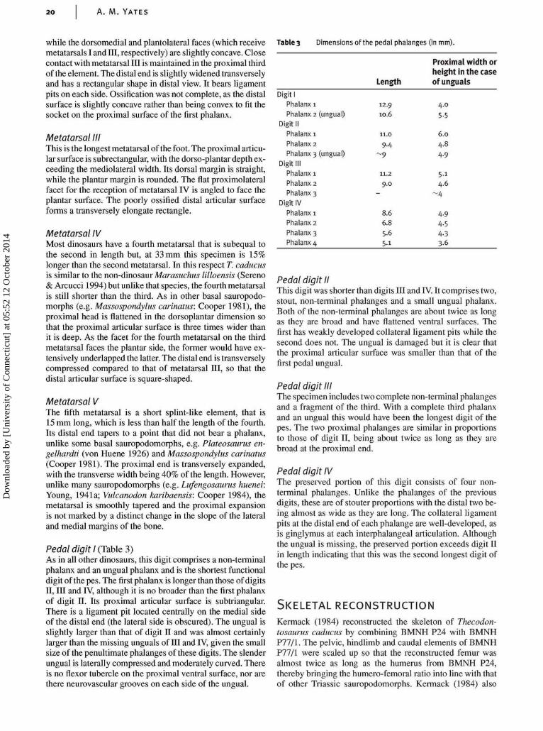
Pedal digit I (Table 3)As in all other dinosaurs, this digit comprises a non-terminalphalanx and an ungual phalanx and is the shortest functionaldigit of the pes. The first phalanx is longer than those of digitsII, in and IV, although it is no broader than the first phalanxof digit II. Its proximal articular surface is subtriangular.There is a ligament pit located centrally on the medial sideof the distal end (the lateral side is obscured). The ungual isslightly larger than that of digit II and was almost certainlylarger than the missing unguals of III and IV, given the smallsize of the penultimate phalanges of these digits. The slenderungual is laterally compressed and moderately curved. Thereis no flexor tubercle on the proximal ventral surface, nor arethere neurovascular grooves on each side of the ungual.
Table 3 Dimensions of the pedal phalanges (in mm).
Digit IPhalanx I
Phalanx 2 (ungual)
Digit II
Phalanx 1
Phalanx 2
Phalanx 3 (ungual)
Digit IIIPhalanx 1
Phalanx 2
Phalanx 3
Digit IV
Phalanx 1
Phalanx 2
Phalanx 3
Phalanx 4
Length
12.9
10.6
11.0
9.4
. 9
11.2
9.0-
8.66.85.6
5.1
Proximal width orheight in the caseof unguals
4.0
5.5
6.0
4.8
94.9
5.14.6
~4
4.94.54.33.6
Pedal digit IIThis digit was shorter than digits in and IV. It comprises two,stout, non-terminal phalanges and a small ungual phalanx.Both of the non-terminal phalanges are about twice as longas they are broad and have flattened ventral surfaces. Thefirst has weakly developed collateral ligament pits while thesecond does not. The ungual is damaged but it is clear thatthe proximal articular surface was smaller than that of thefirst pedal ungual.
Pedal digit IIIThe specimen includes two complete non-terminal phalangesand a fragment of the third. With a complete third phalanxand an ungual this would have been the longest digit of thepes. The two proximal phalanges are similar in proportionsto those of digit II, being about twice as long as they arebroad at the proximal end.
Pedal digit IVThe preserved portion of this digit consists of four non-terminal phalanges. Unlike the phalanges of the previousdigits, these are of stouter proportions with the distal two be-ing almost as wide as they are long. The collateral ligamentpits at the distal end of each phalange are well-developed, asis ginglymus at each interphalangeal articulation. Althoughthe ungual is missing, the preserved portion exceeds digit IIin length indicating that this was the second longest digit ofthe pes.
SKELETAL RECONSTRUCTION
Kermack (1984) reconstructed the skeleton of Thecodon-tosaurus caducus by combining BMNH P24 with BMNHP77/1. The pelvic, hindlimb and caudal elements of BMNHP77/1 were scaled up so that the reconstructed femur wasalmost twice as long as the humerus from BMNH P24,thereby bringing the humero-femoral ratio into line with thatof other Triassic sauropodomorphs. Kermack (1984) also
Dow
nloa
ded
by [
Uni
vers
ity o
f C
onne
ctic
ut]
at 0
5:52
12
Oct
ober
201
4

DESCRIPTION OF A NEW SPECIES OF THE PRIMIT IVE DINOSAUR THECODONTOSAURUS 21
Figure 21 Thecodontosaurus caducus sp. nov., skeletal reconstruction based on BMNH P24, P64/1 and P77/1. Scale bar = 100 mm.
thought that if BMNH P77/1 were not scaled up relative toBMNH P24 then the ilium would appear unrealistically smallin comparison to the skull. Two observations are pertinent atthis point. Firstly, in a large sample of postcranial bones ofT. antiquus from Tytherington (uncatalogued BRSUGmaterial) the largest humeri are of similar length to thelargest femora (approximately 200 mm) suggesting that, atleast in this species, the humerus-to-femur ratio was muchhigher than in other basal sauropodomorphs. Secondly, theskull of the juvenile basal sauropodomorph, Mussauruspatagonicus, was almost certainly longer and taller than itsilium (Bonaparte & Vince 1979) so we could expect a simi-lar ratio in the present juvenile specimen.
For an alternative reconstruction I have assumed thatBMNH P24, P64/1 and P77/1 represent different parts of thesame specimen and have restored the specimen accordingly(Fig. 21). There is circumstantial evidence to support theunity of these specimens. All specimens were found in thesame spoil heap on the same day (Kermack 1984) and containno overlapping parts. A small, displaced, ischium (BMNHP24/3) that would fit with the ilium of P77/1 was found in thesame slab that contains the skull, neck and pectoral skeleton(P24).
In the new reconstruction the forearms are relativelylonger than those in Kermack's (1984) reconstruction andthe hindlimbs were not as elongate relative to the trunklength. If these proportions are accurate it is possible thatThecodontosaurus was not an obligate biped as has usuallybeen assumed (e.g. Kermack 1984; Galton 1990, 2000; VanHeerden 1997), but could have progressed using quadru-pedal locomotion some of the time. Given that Saturnaliatupiniquim has proportions that are indicative of facultative,rather than obligate, bipedalism (Langer et al. 1999), it ispossible that some degree of quadrupedalism is the basalcondition for Sauropodomorpha.
Clad is t ic ana lys is
Methods
Thecodontosaurus caducus was included in a cladistic ana-lysis including 21 ingroup taxa from the Saurischia as wellas three outgroup taxa: Crurotarsi, Marasuchus and Ornith-ischia (Table 4). Amongst the ingroup there are a numberof poorly diagnosed taxa based on poor type material. Some
explanation as to what material was considered for this ana-lysis is given here. Data for T. antiquus was based on twolarge collections of disassociated material, one from the typelocality, another from a new site some 10 km to the north.The material was treated as belonging to one species becauseneither sample showed evidence that more than one type ofsauropodomorph was present. The humeri, ilia and proximalcaudal vertebrae from both localities display autapomorph-ies (unpublished results), which indicate that they belong toa single diagnosable species.
All the diagnostic sauropodomorph material fromthe Newark Supergroup of the Connecticut Valley, NorthAmerica are treated as a single species united by several auta-pomorphic features, as assumed by Sereno (1999). These area narrow, elongate preacetabular blade of the ilium, a narrowpubic apron that is half as wide as the width at the prox-imal end of the pubes and flattened coplanar ischial shafts.Anchisaurus Marsh, 1885 is used as the oldest availablegeneric name for the material. Sereno used AmmosaurusMarsh, 1891, presumably believing the type species ofAnchisaurus, A. polyzelus (Hitchock 1865), to be a nomendubium. However, the type specimen of A. polyzelus (AM41/109) clearly shows the diagnostic dorsoventrally flattened,coplanar ischial shafts (Galton 1976), so it is an adequate typespecimen. The material from the western United States re-ferred to as Ammosaurus by Galton (1976) is not included inthis taxon.
Sellosaurus gracilis from the Early Norian Stubensand-stein of Germany was not used as a terminal taxon becausethe hypodigm of this species is heterogeneous (Galton 1999;pers. obs. of SMNS and GPIT material). Furthermore, thetype specimen of Sellosaurus gracilis is indistinguishablefrom a small specimen of Plateosaurus. The sample fromthe Stubensandstein of Pfaffenhoffen Quarry appears to rep-resent a taxon that is distinct from Plateosaurus and it isused as a terminal taxon here. 'Efraasia diagnostica' is anavailable name for this taxon but it is used in quotation marksuntil a taxonomic restudy of the Stubensandstein prosauro-pods confirms that this is the correct name for the taxon.
Although Plateosaurus is a very familiar name, the typespecies, Plateosaurus engelhardti Meyer, 1837, is foundedon incomplete remains. The well-known, easily-diagnosed,taxon that is based on many complete skeletons from Europeand Greenland may not be the same species, or indeedgenus, as the type of Plateosaurus engelhardti (Galton 2000).Nevertheless it is the former taxon that has been called
Dow
nloa
ded
by [
Uni
vers
ity o
f C
onne
ctic
ut]
at 0
5:52
12
Oct
ober
201
4
22 A. M. YATES
Table 4 Operational Taxonomic Units (OTU) used in the phylogenetic analysis and the sources
from which anatomical information was obtained.
OTU References
Crurotarsi
Marasuchus
Ornithischia1
Herrerasauridae2
Neotheropoda1
Saturnalia tupiniquim1
Thecodontosaurus antiquus1
Anchisaurus polyzelus
Riojasaurus incertus2
'Efraasia diagnostica'1
Plateosaurus engelhardti1
Massospondylus carinatus2
Lufengosaurus huenei
Coloradisaurus brevis2
Yunnanosaurus huangi
'Euskelosaurus browni'
Melanorosaurus readi
Blikanasaurus cromptoni
Vulcanodon karibaensis
Eusauropoda1
Walker 1964; Sill 1974; Chatterjee 1978
Sereno & Arcucci 1994
Thulborn 1972; Santa Luca 1980; Sereno 1991a
Colbert 1970; Novas 1993; Sereno 1993; Sereno & Novas 1993
Gilmore 1920; Madsen 1976; Welles 1984; Currie & Zhao 1993
Langer et al. 1999
Benton et al. 2000
Galton 1976
Bonaparte 1972; Bonaparte & Pumares 1995
Galton 1973,1984b, 1985b; Galton & Bakker 1985
von Huene 1926; Galton 1984a, 1985c
van Hoepen 1920a,b; Cooper 1981; Gow 1990; Gow et al. 1990
Young 1941a,b
Bonaparte 1978
Young 1942
Van Heerden 1979
Van Heerden 1979; Van Heerden & Galton 1997
Galton & Van Heerden 1998
Raath 1972; Cooper 1984
Janensch 1935-36; Gilmore 1936; Bonaparte 1986; He et al. 1988;
Zhang 1988; Madsen et al. 1995
1Taxa that were examined first hand by the author.2Taxa forwhich supplementary information was obtained from casts or from unpublished, detailed photographs.
Plateosaurus for more than 70 years. To invent a new namefor this well-known taxon would be confusing and destabil-ising. In this paper, I provisionally use the name Plateosaurusengelhardti, while recognising that this usage will need to beofficially fixed by an appeal to the International Commissionfor Zoological Nomenclature (ICZN) to conserve the name.
Benton et al. (2000) chose to omit the Chinese taxaLufengosaurus huenei and Yunnanosaurus huangi from theiranalysis for the reason that it was impossible to tell whichspecimens from the Lower Lufeng Formation belonged towhich taxon. Here both taxa are included but almost all datais taken from the type specimens alone, thereby eliminatingthe problem of taxonomic mixing. Some data for Lufengo-saurus huenei were taken from the syntypes of 'Gyposaurus'sinensis. The 'Gyposaurus' sinensis specimens do not differfrom the type specimen of Lufengosaurus huenei in any ofthe characters coded in this analysis. They also share withit the autapomorphies of a strongly arched dorsal margin ofthe ilium and an exceptionally large manual digit I relativeto the other manual digits.
The type specimen of Euskelosaurus browni is veryfragmentary (Van Heerden 1979). All the data in this analysiswere taken from the collection of bones from the LowerElliott Formation of Kromme Spruit, South Africa that weredescribed by Van Heerden (1979). Since the type specimenof Euskelosaurus browni might be undiagnostic, the name isused for the Kromme Spruit sauropodomorphs with quotationmarks, pending taxonomic revision of the sauropodomorphsfrom the Lower Elliott Formation.
The outgroup taxa were included in the analysis as un-constrained terminal taxa. The final trees were then rootedto these taxa after the analysis. Characters that resolve therelationships between the outgroup taxa, but add nothing tothe resolution of ingroup relationships, were not included.
Osteological characters from Galton (1985b, 1990),Gauthier (1986), Novas (1993), Sereno (1993, 1999),Sereno & Novas (1993), Wilson & Sereno (1998), Bentonet al. (2000) and Rauhut (2000) were reviewed, and all char-acters that were found to be variable within the ingroupwere included, with modifications where necessary. Mostmodifications involved more precise definitions of charac-ter states where they had been previously defined usingsubjective terms such as 'broad', 'elongated' or 'reduced'.Ordered transformation series were split into discrete char-acters with binary coding so that all characters in the matrixcould be treated as unordered. For example, the progressiveenlargement of pedal ungual I is treated as two characters. Alltaxa where the first pedal ungual is as large as, or larger than,the second pedal ungual are coded as derived in the first ofthese characters (character 158). Only those in which the firstpedal ungual exceeds the second in size are coded as derivedin the second of the two characters (character 159). Severalnovel characters were also included, bringing the total to 164characters (see Appendix 2).
Results
Analysis of the matrix using PAUP 4.0, with the branch-and-bound search option, produced five most parsimonious trees(Tree Length = 351, Consistency Index = 0.516, HomoplasyIndex = 0.527, Retention Index = 0.668). The basal topo-logy of the strict consensus of these trees (Fig. 22) is fullyresolved. The Saurischia is monophyletic, with Herrerasaur-idae and Neotheropoda forming serially closer outgroups tothe Sauropodomorpha. The inclusion of Herrerasauridae inthe Theropoda (all taxa more closely related to Neothero-poda than to Sauropodomorpha) is contradicted by the ana-lysis. Within Sauropodomorpha, Saturnalia tupiniquim and
Dow
nloa
ded
by [
Uni
vers
ity o
f C
onne
ctic
ut]
at 0
5:52
12
Oct
ober
201
4
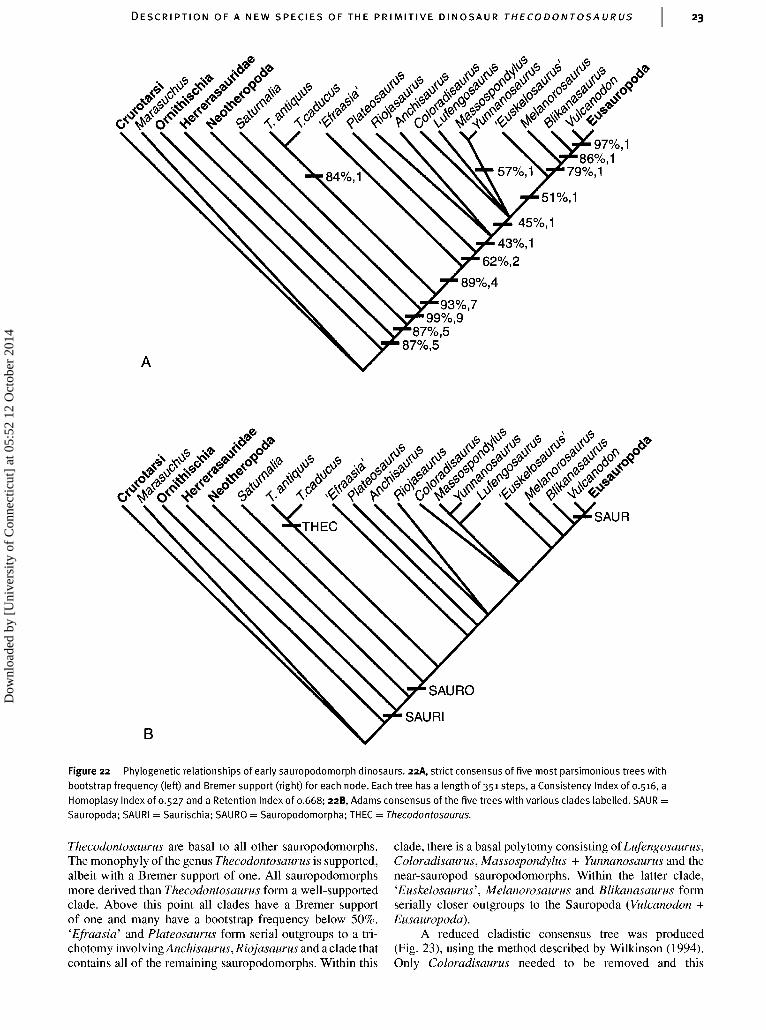
DESCRIPTION OF A NEW SPECIES OF THE PRIMIT IVE DINOSAUR THECODONTOSAURUS 23
45%, 143%, 1
62%,289%,4
87%,5
SAURO
SAURIB
Figure 22 Phylogenetic relationships of early sauropodomorph dinosaurs. 22A, strict consensus of five most parsimonious trees withbootstrap frequency (left) and Bremer support (right) for each node. Each tree has a length of 351 steps, a Consistency Index of 0.516, aHomoplasy Index of 0.527 and a Retention Index of 0.668; 22B, Adams consensus of the five trees with various clades labelled. SAUR =Sauropoda; SAURI = Saurischia; SAURO = Sauropodomorpha; THEC = Thecodontosaurus.
Thecodontosaurus are basal to all other sauropodomorphs.The monophyly of the genus Thecodontosaurus is supported,albeit with a Bremer support of one. All sauropodomorphsmore derived than Thecodontosaurus form a well-supportedclade. Above this point all clades have a Bremer supportof one and many have a bootstrap frequency below 50%.'Efraasia' and Plateosaurus form serial outgroups to a tri-chotomy involving Anchisaurus, Riojasaurus and a clade thatcontains all of the remaining sauropodomorphs. Within this
clade, there is a basal polytomy consisting of Lufengosaurus,Coloradisaurus, Massospondylus + Yunnanosaurus and thenear-sauropod sauropodomorphs. Within the latter clade,'Euskelosaurus', Melanorosaurus and Blikanasaurus formserially closer outgroups to the Sauropoda (Vulcanodon +Eusauropoda).
A reduced cladistic consensus tree was produced(Fig. 23), using the method described by Wilkinson (1994).Only Coloradisaurus needed to be removed and this
Dow
nloa
ded
by [
Uni
vers
ity o
f C
onne
ctic
ut]
at 0
5:52
12
Oct
ober
201
4
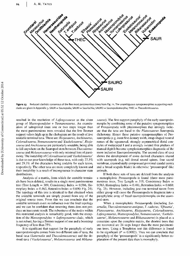
A. M. YATES
*WVto//////-wy/s/MfflM/s
SAUR
SAURO
SAURI
Figure 23 Reduced cladistic consensus of the five most parsimonious trees from Fig. 21. The unambiguous synapomorphies supporting each
clade are given in Appendix 3. SAUR = Sauropoda; SAURI = Saurischia; SAURO = Sauropodomorpha; THEC = Thecodontosaurus.
resulted in the resolution of Lufengosaurus as the sistergroup of Massospondylus + Yunnanosaurus. An examin-ation of suboptimal trees one or two steps longer thanthe most parsimonious trees revealed that the low Bremersupport values high up in the cladogram are the result of fiveunstable terminal taxa. These are: Riojasaurus, Anchisaurus,Coloradisaurus, Yunnanosaurus and 'Euskelosaurus'.Rioja-saurus and Anchisaurus are particularly unstable, being ableto fall anywhere on the Sauropod stem between Thecodonto-saurus and Melanorosaurus with only minimal loss of parsi-mony. The instability of Coloradisaurus and 'Euskelosaurus'is due to our poor knowledge of these taxa, with only 27.5%and 29.3% of the characters being codable for each taxon,respectively. The other taxa are more completely known andtheir instability is a result of incongruence in character statedistributions.
Analysis of a matrix, from which the unstable termin-als have been deleted, results in a single most parsimonioustree (Tree Length = 309, Consistency Index = 0.586, Ho-moplasy Index = 0.463, Retention Index = 0.690: Fig. 24).The topology of this tree is identical to that obtained whenthe unstable terminals are simply pruned from any of theoriginal source trees. From this we can conclude that theunstable terminals exert no influence over the final topologyand we can be confident that removing them does not pro-duce an inaccurate result. The robustness of the nodes withinthis restricted analysis is remarkably good, with the excep-tion of the Massospondylus + Lufengosaurus clade, whichis not robust, having a Bremer support of one and a bootstrapfrequency of less than 75%.
It is significant that support for the paraphyly of earlysauropodomorphs comes from two different sets of taxa, thebasal taxa (Saturnalia and Thecodontosaurus) and the de-rived taxa ('Euskelosaurus', Melanorosaurus and Blikana-
saurus). The first support paraphyly of the early sauropodo-morphs by combining some of the putative synapomorphiesof Prosauropoda with plesiomorphies that strongly indic-ate that the taxa are basal to the Plateosaurus-Sauropodadichotomy. Hence these putative synapomorphies of Pro-sauropoda (e.g. inset first dentary tooth, strap-shaped ventralramus of the squamosal, strongly asymmetrical distal con-dyles of metacarpal I and a strongly twisted first phalanx ofmanual digit I) become symplesiomorphies diagnostic of themore inclusive Sauropodomorpha. The second class of taxashows the development of some derived characters sharedwith sauropods (e.g. tall dorsal neural spines, four sacralvertebrae, craniodistally compressed proximal caudal centraand a broad scapula blade) in otherwise 'prosauropod'-likeanimals.
If both these sets of taxa are deleted from the analysisa monophyletic Prosauropoda is found (three most parsi-monious trees, Tree Length = 318, Consistency Index =0.563, Homoplasy Index = 0.481, Retention Index = 0.660:Fig. 25). However, including just one terminal taxon fromeither group will cause the Prosauropoda to fragment into aparaphyletic array of basal sauropodomorphs on the sauro-pod stem.
When a monophyletic Prosauropoda (including Sat-urnalia, Thecodontosaurus antiquus, T. caducus, 'Efraasia',Plateosaurus, Anchisaurus, Riojasaurus, Coloradisaurus,Lufengosaurus, Massospondylus, Yunnanosaurus, 'Euskelo-saurus', Melanorosaurus and Blikanasaurus) is placed as aconstraint upon the complete matrix, the shortest trees thatare recovered are 24 steps longer than the most parsimoni-ous trees. Using a Templeton test this difference is foundto be significant (P = 0.0082). Thus we can conclude thatparaphyly of the 'prosauropods' is a significantly better ex-planation of the present data than is monophyly.
Dow
nloa
ded
by [
Uni
vers
ity o
f C
onne
ctic
ut]
at 0
5:52
12
Oct
ober
201
4

DESCRIPTION OF A NEW SPECIES OF THE PRIMIT IVE DINOSAUR THECODONTOSAURUS 25
98%,490%,2
98%,4
91%,992%,7
92%,9
93%,799%,9
88%,587%,5
Figure 24 Single most parsimonious tree obtained from a restricted analysis that omits taxa that are unstable at short distances from themost parsimonious trees. Bootstrap frequency (left) and Bremer support (right) are given with each node. The tree has a length of 309, aConsistency Index of 0.586, a Homoplasmy Index of 0.463 and a Retention Index of 0.690.
PROS
SAURO
SAURI
Figure 25 Strict consensus of three most parsimonious trees obtained when basal sauropodomorphs (Saturnalia and Thecodontosaurus)and near-sauropod sauropodomorphs ('Euskelosaurus', Melanorosaurus and Blikanasaurus) are excluded from the analysis. Each tree has alength of 318 steps, a Consistency Index of 0.563, a Homoplasy Index of 0.481 and a Retention Index of 0.660. SAUR = Sauropoda; SAURI =Saurischia; SAURO = Sauropodomorpha; PROS = Prosauropoda.
Dow
nloa
ded
by [
Uni
vers
ity o
f C
onne
ctic
ut]
at 0
5:52
12
Oct
ober
201
4
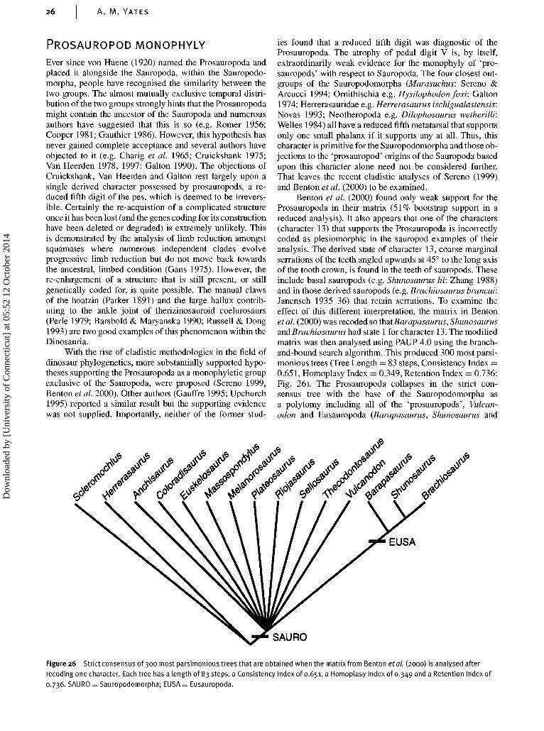
26 A. M. YATES
PROSAUROPOD MONOPHYLY
Ever since von Huene (1920) named the Prosauropoda andplaced it alongside the Sauropoda, within the Sauropodo-morpha, people have recognised the similarity between thetwo groups. The almost mutually exclusive temporal distri-bution of the two groups strongly hints that the Prosauropodamight contain the ancestor of the Sauropoda and numerousauthors have suggested that this is so (e.g. Romer 1956;Cooper 1981; Gauthier 1986). However, this hypothesis hasnever gained complete acceptance and several authors haveobjected to it (e.g. Charig et al. 1965; Cruickshank 1975;Van Heerden 1978, 1997; Galton 1990). The objections ofCruickshank, Van Heerden and Galton rest largely upon asingle derived character possessed by prosauropods, a re-duced fifth digit of the pes, which is deemed to be irrevers-ible. Certainly the re-acquistion of a complicated structureonce it has been lost (and the genes coding for its constructionhave been deleted or degraded) is extremely unlikely. Thisis demonstrated by the analysis of limb reduction amongstsquamates where numerous independent clades evolveprogressive limb reduction but do not move back towardsthe ancestral, limbed condition (Gans 1975). However, there-enlargement of a structure that is still present, or stillgenetically coded for, is quite possible. The manual clawsof the hoatzin (Parker 1891) and the large hallux contrib-uting to the ankle joint of therizinosauroid coelurosaurs(Perle 1979; Barsbold & Maryanska 1990; Russell & Dong1993) are two good examples of this phenomenon within theDinosauria.
With the rise of cladistic methodologies in the field ofdinosaur phylogenetics, more substantially supported hypo-theses supporting the Prosauropoda as a monophyletic groupexclusive of the Sauropoda, were proposed (Sereno 1999,Benton etal. 2000). Other authors (Gauffre 1995; Upchurch1995) reported a similar result but the supporting evidencewas not supplied. Importantly, neither of the former stud-
ies found that a reduced fifth digit was diagnostic of theProsauropoda. The atrophy of pedal digit V is, by itself,extraordinarily weak evidence for the monophyly of 'pro-sauropods' with respect to Sauropoda. The four closest out-groups of the Sauropodomorpha (Marasuchus: Sereno &Arcucci 1994; Ornithischia e.g. Hysilophodon foxi: Galton1974; Herrerasauridae e.g. Herrerasaurus ischigualastensis:Novas 1993; Neotheropoda e.g. Dilophosaurus wetherilli:Welles 1984) all have a reduced fifth metatarsal that supportsonly one small phalanx if it supports any at all. Thus, thischaracter is primitive for the Sauropodomorpha and those ob-jections to the 'prosauropod' origins of the Sauropoda basedupon this character alone need not be considered further.That leaves the recent cladistic analyses of Sereno (1999)and Benton et al. (2000) to be examined.
Benton et al. (2000) found only weak support for theProsauropoda in their matrix (51% bootstrap support in areduced analysis). It also appears that one of the characters(character 13) that supports the Prosauropoda is incorrectlycoded as plesiomorphic in the sauropod examples of theiranalysis. The derived state of character 13, coarse marginalserrations of the teeth angled upwards at 45• to the long axisof the tooth crown, is found in the teeth of sauropods. Theseinclude basal sauropods (e.g. Shunosaurus lii: Zhang 1988)and in those derived sauropods (e.g. Brachiosaurus brancai:Janensch 1935-36) that retain serrations. To examine theeffect of this different interpretation, the matrix in Bentonetal. (2000) was recoded so thatBarapasaurus, Shunosaurusand Brachiosaurus had state 1 for character 13. The modifiedmatrix was then analysed using PAUP 4.0 using the branch-and-bound search algorithm. This produced 300 most parsi-monious trees (Tree Length = 83 steps, Consistency Index =0.651, Homoplasy Index = 0.349, Retention Index = 0.736:Fig. 26). The Prosauropoda collapses in the strict con-sensus tree with the base of the Sauropodomorpha asa polytomy including all of the 'prosauropods', Vulcan-odon and Eusauropoda (Barapasaurus, Shunosaurus and
EUSA
SAURO
Figure 26 Strict consensus of 300 most parsimonious trees that are obtained when the matrix from Benton et al. (2000) is analysed afterrecoding one character. Each tree has a length of 83 steps, a Consistency Index of 0.651, a Homoplasy Index of 0.349 a n d a Retention Index of0.736. SAURO = Sauropodomorpha; EUSA = Eusauropoda.
Dow
nloa
ded
by [
Uni
vers
ity o
f C
onne
ctic
ut]
at 0
5:52
12
Oct
ober
201
4
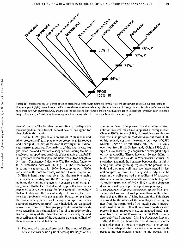
DESCRIPTION OF A NEW SPECIES OF THE PRIMIT IVE DINOSAUR THECODONTOSAURUS
/ / / / / / / / / / /
55%, 1
PROS100%, 10
Figure 27 Strict consensus of 8 trees obtained after analysing the data matrix presented in Sereno (1999) with bootstrap support (left) andBremer support (right) for each node. In this paper 'Gyposaurus' sinensis is regarded as a juvenile of Lufengosaurus, Anchisaurus is taken to bethe senior synonym of Ammosaurus, and most of the specimens in the hypodigm of Sellosaurus are taken to belong to 'Efraasia'. Each tree has alength of 34 steps, a Consistency Index of 0.971, a Homoplasy Index of 0.029 and a Retention Index of 0.947.
Brachiosaurus). The fact that one recoding can collapse theProsauropoda is indicative of the weakness of the support forthat clade in this matrix.
Sereno (1999) presented a matrix of 32 characters andnine 'prosauropod' taxa plus two outgroup taxa, Sauropodaand Theropoda, as part of his overall investigation of dino-saur interrelationships. The analysis of this matrix was notpresented, beyond a reduced cladogram containing the morestable prosauropod taxa. Analysis of this matrix using PAUP4.0 produces seven most parsimonious trees (Tree Length =34 steps, Consistency Index = 0.971, Homoplasy Index =0.029, Retention Index = 0.947: Fig. 27). The Prosauropodais strongly supported with 100% bootstrap support (1000replicates in the bootstrap analysis) and a Bremer support of10. This is hardly suprising given that the matrix contains19 characters that diagnose the Prosauropoda with virtuallyno homoplasy and no characters to suggest a conflicting ar-rangement. On the face of it, it would appear that Sereno haspresented a very strong case for 'prosauropod' monophylythat is at odds with the present analysis. However, there aretwo main problems with this analysis. Firstly, no taxa fromthe two crucial groups (basal sauropodomorphs and near-sauropod sauropodomorphs) were included. As discussedabove, taxa from these two groups provide important evid-ence regarding the relationships of basal sauropodomorphs.Secondly, many of the characters are not precisely definedor described and many of the codings are debatable. Each ofthem is examined in detail below.
1. Presence of a premaxillary beak. The snout of Rioja-saurus incertus bears a pair of parasagittal ridges on the
anterior surface of the premaxillae that define a raisedanterior area and may have supported a rhamphotheca(Sereno 1997). Sereno (1997) claimed that a subtle ver-sion was also present in Plateosaurus, but most skullsof this taxon do not show the feature (pers. obs. of GPITSkelett 1, SMNS 12950, HMN mb.1927.19.1). Onlyone snout from Frick, Switzerland, (Galton 1986: pl. 3,figs 2,3,4) shows easily recognisable parasagittal ridgeson the premaxilla. These, however, do not delimit araised platform as they do in Riojasaurus incertus, in-stead they just mark the boundary between the rostrally-facing and laterally-facing regions of the premaxillarybody and they may well have been accentuated by lat-eral compression. No trace of any sort of ridges can beseen on the well-preserved premaxillae of Massospon-dylus carinatus and an unnamed taxon from the KayentaFormation (pers. obs. of MCZ 8893), so this characterdoes not stand up as a prosauropod synapomorphy.
2. L-shapedpremaxilla-maxilla external suture. Most pro-sauropods have an external premaxilla-maxilla suturethat is shaped like an 'L' lying horizontally. This shapeis caused by the offset of the maxillary ascending ra-mus from the rostral end of the maxilla and a square-ended rostral ramus. Both of these derived conditions arepresent in most sauropods (e.g. an unnamed basal saur-opod from the Lufeng Formation: Barrett 1999; Patago-saurus fariasi: Bonaparte 1986; Brachiosaurus brancai,HMN M.B.2181) although the rostral ramus is shorterand higher than in most 'prosauropods'. The appear-ance of an L-shaped suture is less apparent in sauropodsbecause the caudolateral process of the premaxilla is
Dow
nloa
ded
by [
Uni
vers
ity o
f C
onne
ctic
ut]
at 0
5:52
12
Oct
ober
201
4
28 A. M. YATES
shortened relative to that of most 'prosauropods'. Thecharacter is best interpreted as a single derived char-acter describing the shape of the rostral ramus of themaxilla that is diagnostic of the Sauropodomorpha as awhole.
3. Development of a secondary internal wall of the ant-orbital fossa. As in most other dinosaurs, the externalantorbital fenestra of 'prosauropods' is larger than itsinternal counterpart (Witmer 1997). This results in alaterally open antorbital fossa that is bounded me-dially by an internal wall. Such an internal wall ispresent in all 'prosauropods', ornithischians, herrera-saurids, neotheropods and Marasuchus, so it is certainlyprimitive for the Dinosauria. Only sauropods lack theinternal wall of the antorbital fossa and this must be re-garded as a synapomorphy of the clade (Wilson & Sereno1998; Upchurch 1998). There is no evidence that the in-ternal wall of 'prosauropods' is secondarily developed,so this character cannot be used as a prosauropod syna-pomorphy.
4. Regular maxillary vascular foramina with one largecaudally directed foramen and five to six anteriorlydirected foramina. Primitively dinosaurs have a row ofsmall irregularly spaced neurovascular foramina placedabove the ventral margin. At the caudal end of the row in'prosauropods' (except Thecodontosaurus caducus, un-known in T. antiquus and Saturnalia tupiniquim) thereis a distinctly enlarged foramen that exceeds all othersin size. Rostrally there are no more than six smaller fo-ramina. Thus the arrangement seen in 'prosauropods'would appear to be derived and would diagnose thegroup, above Thecodontosaurus. However, sauropodscannot be said to have the primitive condition. There issome variation but most seem to have an irregular groupof 3-10 vascular foramina rostral to an enlarged foramen(e.g. Patagosaurus fariasi. Bonaparte 1986; Diplodocuslongus: Witmer 1997; Camarasaurus lentus: Madsenet al. 1995). In neosauropods, the caudal foramen isexceptionally enlarged and has been termed the 'prean-torbital fenestra' by Wilson & Sereno (1998). Witmer(1997), Wilson & Sereno (1998) and Upchurch (1998)have all considered that this fenestra is a pneumatic fea-ture that was produced by outpocketing from, or sub-division of, the antorbital pneumatic sinus. However,an internal canal leads forward from the preantorbitalfenestra of Brachiosaurus brancai (pers. obs. of HMNMB .2181) and probably connects with the anterior max-illary neurovascular foramina. Thus the preantorbitalfenestra is likely to be of neurovascular origin, althoughit may have been enlarged by invasion of a pneumaticoutpocket of the antorbital fenestra. There are no otherneurovascular foramina behind the preantorbital fenes-tra, which suggests this feature and the enlarged caudalneurovascular foramen of prosauropods are homolog-ous. Some basal sauropods (e.g. Shunosaurus lii: Zhanget al. 1988; Omeisaurus tianfuensis: He et al. 1988;Euhelopus zdanskyi: Wiman 1929), lack an enlargedcaudal maxillary foramen. However, this is best in-terpreted as further evidence that they belong to aclade, the Euhelopodidae (Upchurch 1998), and thatthey do not represent the basal sauropod condition. Inmany respects, the arrangement of vascular foramina isquite similar between sauropods and 'prosauropods' and
could be coded as a synapomorphy of the two. In anycase, if the arrangement seen in 'prosauropods' is treatedas a single derived character, the state for Sauropodacannot be coded as primitive; therefore the charactercan only be interpreted as an ambiguous synapomorphyof the Prosauropoda, at best.
5. Strap-shaped ventral process of the squamosal. A long,narrow, parallel-sided ventral process of the squamosal(more than four times longer than the width at itsmidlength) with a pointed end would appear to unam-biguously support the monophyly of Prosauropoda.
6. First dentary tooth inset from the anterior margin of thedentary. This character also appears to unambiguouslysupport the monophyly of Prosauropoda.
7. Axial postzygapophyses flush with the caudal centrumface. Primitively in dinosaurs the postzygapophysisoverhangs the caudal face of the centrum, so that at least50% of the length of the zygapophyseal facet lies caudalto the line that subtends the dorsal and ventral edges ofthe caudal face of the centrum. Most sauropods, includ-ing the basal Patagosaurus fariasi (Bonaparte 1986),display the primitive condition. In contrast, many 'pro-sauropods' (e.g. Plateosaurus engelhardti: von Huene1926) have cranially-set postzygapophyses that barelyoverhang the caudal face of the centrum, if at all. Con-fusing the picture somewhat is the occurrence of thederived state in Euhelopodidae (e.g. Shunosaurus lii:Zhang 1988; Mamenchisaurus sinocanadorum: Rus-sell & Zheng 1993) and Haplocanthosaurus delfsi(McIntosh & Williams 1988). More data from basal saur-opods will be needed to be sure that the plesiomorphicstate was ancestral for the group. If it is found that thederived state was basal for the Sauropoda this charac-ter may support the paraphyly of 'prosauropods' withrespect to sauropods.
8. Deltopectoral crest more than 50% of the length ofthe humerus. The length of the deltopectoral crest ofMarasuchus, ornithischians, herrerasaurids, neothero-pods and sauropods is usually below 45% of the lengthof the humerus, and is certainly so in the basal membersof each of these clades. In contrast, many 'prosauropods'have a deltopectoral crest that either approaches, or ex-ceeds, 50% of the length of the humerus (e.g. Plateo-saurus engelhardti: von Huene 1926). However, thedeltopectoral crests of Saturnalia tupiniquim (pers. obs.of MCP 3844-PV), Thecodontosaurus antiquus (Bentonet al. 2000), Anchisaurus polyzelus (Galton 1976) and'Euskelosaurus browni’ (Van Heerden 1979) are short. IfSaturnalia and Thecodontosaurus are accepted as basalto all other 'prosauropods', then this character cannotdiagnose the Prosauropoda, although it may diagnose aless inclusive clade within it.
9. Deltopectoral crest deflected 90• from the transverseaxis of the distal condyles. Since the angle of deflec-tion of the deltopectoral crest from the transverse axisof the distal condyles increases with distance from thebase of the crest, it is likely that this angle is correlatedwith the size of the deltopectoral crest (character 8 inthis list). Since an extremely low deltopectoral crest isa synapomorphy of Sauropoda it is unlikely that it ispossible for them to display a crest that is set at 90• tothe distal condyles even if that was the basal conditionof the clade. Given these concerns and the sensitivity
Dow
nloa
ded
by [
Uni
vers
ity o
f C
onne
ctic
ut]
at 0
5:52
12
Oct
ober
201
4

DESCRIPTION OF A NEW SPECIES OF THE PRIMIT IVE DINOSAUR THECODONTOSAURUS 29
of the measurement to postmortem distortion by sedi-ment compaction, I prefer not to use this character forphylogenetic analysis of dinosaurs.
10. Distal carpal I is large. Sereno did not specify what hemeant by 'large' but when the distal carpal I of 'prosaur-opods' is compared with distal carpal II it can be seenthat the transverse width of the former is at least 20%greater than that of the latter. Distal carpal proportionsare unknown in non-dinosaurian dinosauromorphs, butdistal carpal I is less than 10% wider than distal carpal nin Heterodontosaurus tucki (Santa Luca 1980), the onlyornithischian with ossified distal carpals. Distal carpal Iis actually smaller than distal carpal II in Herrerasaurusischigualastensis (Sereno 1993). Thus, a small first distalcarpal that is no more than 10% wider than distal carpalII is taken to be the primitive condition for the Dino-sauria. The state of this character in neotheropods isdifficult to determine because the two carpals are fusedas an apparent synapomorphy of the clade. Neverthe-less the fusion is reversed in Alxasaurus elesitaiensis(Russell & Dong 1993) and possibly Allosaurus fra-gilis (Madsen 1976) and these show that distal carpal Iwas enlarged relative to distal carpal II. If the carpalelements of sauropods are distal carpals, which seemsto be the most reasonable interpretation, then they alsohave an enlarged distal carpal I (e.g. Shunosaurus lii:Zhang 1988). Thus, the character is an ambiguous syn-apomorphy of Saurischia, or an unambiguous synapo-morphy of Sauropodomorpha + Theropoda, exclusiveof Herrerasauridae. In either case it does not support amonophyletic Prosauropoda.
11. Metacarpal I inset into the carpus. The derived char-acter is a correlate of an extremely large distal carpal Ithat overlies distal carpal II. This arrangement createsa space in the carpus, under distal carpal I and medialto distal carpal II. This space is filled by an extensionof metacarpal I into the carpus. Among 'prosauropods',Massospondylus carinatus (Cooper 1981), Plateosaurusengelhardti (pers. obs. of SMNS 13200), Lufengosaurushuenei (Young 1941a) and possibly 'Efraasia diagnost-ica' (pers. obs. of SMNS 12667) have the derived state.In other known 'prosauropod' hands (Thecodontosaurusantiquus: Benton et al. 2000; Riojasaurus incertus:Bonaparte 1972; Anchisaurus polyzelus: Galton 1976),the distal carpals do not overlap each other and the prox-imal end of the first metacarpal remains flush with theother metacarpals. This character could support a lessinclusive clade within the 'prosauropods' but it does notsupport the monophyly of 'prosauropods'.
12. Proximal width of metacarpal I more than 65% of itsmaximum length. The proximal width of the first meta-carpal of most ornithischians, herrerasaurids and mostneotheropods is no more than 55% of its length. Incontrast, Riojasaurus incertus, Anchisaurus polyzelus,'Efraasia diagnostica', Plateosaurus engelhardti, Mass-ospondylus carinatus, Lufengosaurus huenei and Yun-nanosaurus huangi have stout first metacarpals in whichthe proximal width is greater than 65% of the length.Thecodontosaurus antiquus retains a primitively elong-ated first metacarpal (pers. obs. of BRSUG 23629).Neosauropods and their closest outgroups (e.g. Jobariatiguidensis: Sereno et al. 1999) have elongate first meta-carpals (proximal width less than 55% of the length).
However, the manus of these dinosaurs is highly mod-ified to form an elongate, tubular, weight-supportingdevice consisting of a colonnade of structurally similarmetacarpals. Thus, it is unclear whether neosauropodsprimitively retain the plesiomorphic state or whether itis secondarily derived. The only basal sauropods withwell-preserved forefeet are those of euhelopodids, mostof which have stout, 'prosauropod'-like first metacarpals(e.g. Hudiesaurus sinojapanorum: Dong 1997; Shuno-saurus lii: Zhang 1988). Thus, the primitive conditionfor the Sauropoda is ambiguous. If we accept that thestate for the Neosauropoda and its allies should be codedas uncertain due to transformation, we are left with ac-cepting that stout metacarpals, present in euhelopodids,are basal for Sauropoda. In this case, the character be-comes a synapomorphy of all sauropodomorphs moreadvanced than Thecodontosaurus.
13. Manual phalanx I of digit I with a proximal heel. Theproximal articular surface of the first phalanx of thefirst digit of crurotarsans and ornithischians is a mildlyconcave structure of which the palmar lip does notprotrude beyond the dorsal lip. This can be regardedas the ancestral condition for the Dinosauria. Sauro-pods also posses a relatively flat proximal articular sur-face without any marked protrusion of the palmar lip.In contrast, the palmar lip of herrerasaurids, neothero-pods and 'prosauropods' protrudes further back than thedorsal lip. This protrusion, or heel, is especially largeand asymmetrical in some of the larger derived 'pro-sauropods' (e.g. Plateosaurus engelhardti: von Huene1926; Massospondylus carinatus: Cooper 1981). How-ever, the ventral protrusion of other 'prosauropods' suchas Thecodontosaurus antiquus and 'Efraasia diagnost-ica' is no greater than in Herrerasaurus ischigualasten-sis (Sereno 1993) or neotheropods such as Dilopho-saurus wetherilli (Welles 1984) and Allosaurus fragilis(Madsen 1976). Given that the difference between a largeand a small protrusion is slight, I prefer not to draw a dis-tinction and simply regard the presence of the protrusionas a derived character that is probably diagnostic of theSaurischia.
14. First phalanx of manual digit I rotated at least 45•ventrolaterally. This character apparently supports themonophyly of Prosauropoda. However the lack of twist-ing in the first phalanx of sauropods may be due to themodification of the hand into a weight-bearing structure,in which case the basal character state for Sauropodawould have to be coded as uncertain. If this is donethis character becomes an apomorphy of Sauropodo-morpha.
15. Subtriangular preacetabular process of the ilium. Inbasal dinosaurs (e.g. Herrerasaurus ischigualastensis:Novas 1993) and basal dinosauriforms (e.g. Marasuchuslilloensis: Sereno & Arcucci 1994) the preacetabularprocess of the ilium is short and subrectangular in pro-file. In 'prosauropods' this process is drawn out into acranially projecting subtriangular blade. The preacetab-ular process of basal sauropods (e.g. Shunosaurus lii:Zhang 1988; Barapasaurus tagorei: Jain et al. 1977) isnot like those of primitive dinosauriforms. It, like the'prosauropods', bears a pointed termination that pro-jects cranially. They differ from those of 'prosauropods'in projecting well forward of the pubic peduncle and
Dow
nloa
ded
by [
Uni
vers
ity o
f C
onne
ctic
ut]
at 0
5:52
12
Oct
ober
201
4

30 A. M. YATES
having a more strongly arched dorsal margin. How-ever, these are derived characters that represent furthermodifications of the sauropod ilium. Even if one con-siders the preacetabular process of 'prosauropods' andsauropods to differ significantly, sauropods cannot becoded as having the primitive state. Thus, at best, thischaracter offers ambiguous support for the monophylyof Prosauropoda, although it is here considered morelikely to be a synapomorphy of Sauropodomorpha as awhole.
16. Preacetabular process of the ilium with a scar. Thepreacetabular process of the ilium of basal dinosaur-iforms, including basal saurischians, probably bore acartilaginous extension upon its anterodorsal margin. Astrip of rugose bone along the anterior dorsal rim of theilium in these taxa almost certainly marks the site atwhich the cartilaginous 'cap' was attached. Such a scarof rugose bone can be seen in Marasuchus lilloensis(Sereno & Arcucci 1994), Herrerasaurus ischiguala-stensis (Novas 1993), 'prosauropods' (e.g. 'Efraasiadiagnostica': pers. obs. of SMNS 12354; Plateosaurusengelhardti. pers. obs. of GPIT Skelett 1; Massospon-dylus carinatus: Cooper 1981) and sauropods (e.g.Apatosaurus louisae: Gilmore 1936). Ornithischians andneotheropods appear to have independently lost the car-tilaginous cap. The presence of a catilaginous cap and itsassociated scar in 'prosauropods' are primitive featuresand do not support monophyly of the group.
17. Distal ischial shaft with a subtriangular cross-section.Apart from 'prosauropods' a subtriangular cross-sectionof the distal ischial shaft can also be seen in non-macronarian sauropods (e.g. Vulcanodon karibaensis:Cooper 1984; Dicraeosaurus hansemmani: Janensch1961) and herrerasaurids (e.g. Herrerasaurus is-chigualastensis: Novas 1993). Thus this character is bestinterpreted as a saurischian synapomorphy. The ovatecross-section of the distal ischial shaft of neotheropodsrepresents a further modification and a synapomorphyof the group. It is interesting to note that the basal saur-ischian, Guiabasaurus, which has distal ischia with atriangular cross-section (Bonaparte et al. 1999), maybe the plesiomorphic sister-group of the Neotheropoda(M. Langer, pers. comm. 2000).
18. Biconcave proximal articular surface of metatarsal II.The typical biconcave shape of the proximal articularsurface of metatarsal II of most prosauropods is cre-ated by concave facets on either side for the recep-tion of metatarsals I and in, respectively. A rectangularproximal articular surface, with flat contact surfaces oneither side, is the basal dinosaur condition. This is seenin non-dinosaurian dinosauromorphs (e.g. Lagerpetonchanariensis: Sereno & Arcucci 1993; Marasuchuslilloensis: Sereno & Arcucci 1994) and in Herrera-saurus ischigualastensis (Novas 1993). Like prosauro-pods, ornithischians have a concave facet for metatarsal I(in those taxa in which metatarsal I is not lost e.g.Hypsilophodonfoxi: Galton 1974; Tenontosaurus tilletti:Forster 1990; Camptosaurus prestwichii: Galton &Powell 1980). A concave facet for metatarsal IIIis present in non-iguanodontian ornithischians (e.g.Hypsilophodonfoxi: Galton 1974; Stegosaurus armatus:Ostrom & McIntosh, 1966). In neotheropods metatarsal Idoes not contact the ankle, so there is no facet for it
on the medial side of the proximal end of metatarsal II.Furthermore the facet for metatarsal III is conspicuouslyconvex. Thus neotheropods can be considered too trans-formed to be regarded as plesiomorphic for this charac-ter. Sauropods retain a flat contact with metatarsal III,while the contact with metatarsal I in basal forms isas concave as it is in any 'prosauropod'. Furthermore,there is ingroup variation with Plateosaurus engelhardtiexhibiting a plesiomorphic straight contact with meta-tarsal III (pers. obs. of GPIT Skelett 1, HMN Skelett45). These distributions suggest that the two contactsshould be considered separately in a cladistic analysis.If this approach is taken, a concave facet on metatarsalII for metatarsal I is an ambiguous synapomorphy ofSauropodomorpha, while a concave facet for metatarsalin is an ambiguous synapomorphy of a monophyleticProsauropoda.
19. Proximal articular surface of metatarsal IV three timesbroader than dorsoplantar depth. The methods by whichthese dimensions were measured were not specified. Imeasure the width as the distance between the medial-most point on the plantar surface to the lateral-mostpoint of the bone and the depth as the maximum di-mension orthogonal to this line. Using these measure-ments dinosaur outgroups and ornithischians are foundto have rather squat proximal articular surfaces that areless than twice as wide as they are deep (e.g. 1.79 inSaurosuchus galilei: Sill 1974, table 5; 1.17 in Hypsilo-phodonfoxi: Galton 1974). Eusauropods and neothero-pods share similar proportions, whereas Herrerasaurusischigualastensis (Novas 1993), prosauropods and Vul-canodon karibaensis (Cooper 1984) have proxim-ally widened fourth metatarsals that are more thantwice as wide as deep. Among sauropodomorphs, onlyMassospondylus carinatus reaches the proportion thatSereno regarded as derived (Cooper 1981). A proximalfourth metatarsal that is more than twice as wide asdeep is a derived character that has an ambiguous dis-tribution. Since the basal condition for the Sauropodais ambiguous, the character cannot be used to support'prosauropod' monophyly.
Thus we can see that of the 19 characters, only three(characters 5, 6 and 14) unambiguously support 'prosaur-opod' monophyly. Other characters cannot be coded forthe immediate 'prosauropod' outgroups (sauropods or thero-pods) either due to polymorphism or morphological trans-formation (characters 4, 7, 12, 15, 18 and 19). Thus, thesecharacters can only offer ambiguous support for the mono-phyly of Prosauropoda. Other characters seem to diagnoseclades either less or more inclusive than the proposed Pro-sauropoda (characters 2, 8,10,11,13,16 and 17). The threeremaining characters (1,3 and 9) are not deemed to be usefulfor phylogenetic analysis of basal sauropodomorph relation-ships.
In conclusion, the case for the monophyly of Prosaur-opoda is not strong and, when the data are incorporated ina more inclusive analysis, such as the one presented here,the Prosauropoda breaks into a paraphyletic array of basalsauropodomorphs. The paraphyly of 'prosauropods' not onlysolves the problem of the absence of sauropods from all butthe latest Triassic (Buffetaut et al. 2000), but also suggeststhat an organism very like Plateosaurus or Massospondylus
Dow
nloa
ded
by [
Uni
vers
ity o
f C
onne
ctic
ut]
at 0
5:52
12
Oct
ober
201
4
DESCRIPTION OF A NEW SPECIES OF THE PRIMIT IVE DINOSAUR THECODONTOSAURUS
was directly ancestral to the largest terrestrial animals inEarth's history.
ACKNOWLEDGEMENTS
I thank Angela Milner and Sandra Chapman for the loan ofthe material used in this study. Simon Powell kindly assistedwith the photography of the specimens. Mike Benton re-viewed an earlier version of the paper. Max Langer providedexcellent photographs of many of the South American andAfrican taxa used in the phylogenetic analysis and livelydiscussions on the subject of early dinosaur evolution. I alsowish to thank Rainer Schoch and Oliver Rauhut for their hos-pitality while visiting their respective institutions and MaxLanger, Rupert Wild and David Unwin for allowing me toexamine specimens in their care. This work was supportedby the Leverhulme Trust (Grant No. F/182/BK).
REFERENCESBarrett, P. M. 1999. A sauropod dinosaur from the Lower Lufeng Form-
ation (Lower Jurassic) of Yunnan Province, Peoples's Republic ofChina. Journal of Vertebrate Paleontology 19: 785-787.
Barsbold, R. & Maryanska, T. 1990. Segnosauria. Pp. 408-415 inWeishampel, D. B., Dodson, P. & Osmolska, H. (eds) The Dinosauria.University of California Press, Berkeley.
Benton, M. J. & Spencer, P. S. 1995. Fossil reptiles of Great Britain.Chapman and Hall, London, 389 pp.
—, Juul, L., Storrs, G. W. & Galton, P. M. 2000. Anatomy and system-atics of the prosauropod dinosaur Thecodontosaurus antiquus from theUpper Triassic of Southwest England. Journal of Vertebrate Paleon-tology 20: 77-108.
Bonaparte, J. F. 1972. Los tetrapodos del sector Superior de la FormacionLos Colorados, La Rioja, Argentina (Triasico Superior). I Parte. OperaLilloana 22: 1-183.
— 1978. Coloradia brevis n. g. et n. sp. (Saurischia Prosauropoda), dino-saurio Plateosauridae de la Formacion Los Colorados, Triasico Super-ior de la Rioja, Argentina. Ameghiniana 15: 327-332.
—1986. Les Dinosaures (Carnosaures, Allosauridés, Sauropodes,Cétiosauridés) du Jurassique moyen de Cerro Condor (Chubut, Ar-gentine). Annales de Paléontologie 72: 325-386.
— & Pumares, J. A. 1995. Notas sobre el primer craneo de Riojasaurusincertus (Dinosauria, Prosauropoda, Melanorosauridae) del TriasicoSuperior de La Rioja, Argentina. Ameghiniana 32: 341-349.
— & Vince, M. 1979. El hallazgo del primer nido de dinosaurios Tri-asicos (Saurischia, Prosauropoda), Triasico Superior de Patagonia,Argentina. Ameghiniana 16: 173-182.
—, Ferigolog, J. & Ribeiro, A. M. 1999. A new early Late Triassicsaurischian dinosaur from Rio Grand do Sul State, Brazil. NationalScience Museum Monographs 15: 89-109.
Brochu, C. A. 1996. Closure of neurocentral sutures during crocodilianontogeny: implications for maturity assessment in fossil archosaurs.Journal of Vertebrate Paleontology 16: 49-62.
Buffetaut, E., Suteethorn, V., Cuny, G., Tong, H., Loeuff, J. L.,Khansubha, S. & Jongautchariyakul, S. 2000. The earliest knownsauropod dinosaur. Nature 407: 72-74.
Carr, T. D. 1999. Craniofacial ontogeny of Tyrannosauridae(Dinosauria, Coelurosauria). Journal of Vertebrate Paleontology 19:497-520.
Charig, A. J., Attridge, J. & Crompton, A. W. 1965. On the origin ofthe sauropods and the classification of the Saurischia. Proceedings ofthe Linnean Society of London 176: 197-220.
Chatterjee, S. 1978. A primitive parasuchid (phytosaur) reptile fromthe upper Triassic Maleri Formation of India. Palaeontology 21:83-127.
Chiappe, L. M., Norell, M. A. & Clark, J. M. 1996. Phylogenetic posi-tion of Mononykus (Aves: Alvarezsauridae) from the Late Cretaceousof the Gobi desert. Memoirs of the Queensland Museum 39: 557-582.
Colbert, E. H. 1970. A saurischian dinosaur from the Triassic of Brazil.American Museum Novitates 2405: 1-39.
— 1989. The Triassic dinosaur, Coelophysis. Museum of Northern ArizonaBulletin 57: 1-160.
Cooper, M. R. 1981. The prosauropod dinosaur Massospondyluscarinatus Owen from Zimbabwe: its biology, mode of life and phylo-genetic significance. Occasional Papers of the National Museums andMonuments of Rhodesia B, Natural Sciences 6: 689-840.
— 1984. A reassessment of Vulcanodon karibaensis Raath (Dinosauria:Saurischia) and the origin of the Sauropoda. Palaeontologia Africana25: 203-231.
Crompton, A. W. & Attridge, J. 1986. Masticatory apparatus of thelarger herbivores during Late Triassic and Early Jurassic times.Pp. 223-236 in Padian, K. (ed.) The beginning of the age of dino-saurs. Cambridge University Press, Cambridge.
Cruickshank, A. R. 1975. The origin of sauropod dinosaurs. SouthAfrican Journal of Science 71: 89-90.
Currie, P. J. & Zhao, X. J. 1993. A new carnosaur (Dinosauria,Theropoda) from the Jurassic of Xinjiang, People's Republic of China.Canadian Journal of Earth Sciences 30: 2037-2081.
Dodson, P. 1975. Taxonomic implications of relative growth in lambeo-saurine hadrosaurs. Systematic Zoology 24: 37-54.
Dong, Z. 1997. A gigantic sauropod (Hudiesaurus sinojapanorum gen. etsp. nov.) from the Turpan Basin, China. Pp. 102-110 in Dong, Z. (ed.),Sino-Japanese Silk Road Dinosaur Expedition. China Ocean Press,Beijing.
Forster, C. A. 1990. The postcranial skeleton of the ornithopod dinosaurTenontosaurus tilletti. Journal of Vertebrate Paleontology, Chicago10: 273-294.
Galton, P. M. 1973. On the anatomy and relationships of Efraasiadiagnostica (V. Huene) n. gen., a prosauropod dinosaur (Reptilia:Saurischia) from the Upper Triassic of Germany. PalaontologischeZeitschrift 47: 229-255.
— 1974. The ornithischian dinosaur Hypsilophodon from the Wealden ofthe Isle of Wight. Bulletin of the British Museum (Natural History)Geology 25: 1-152.
— 1976. Prosauropod dinosaurs (Reptilia: Saurischia) of North America.Postilla 169: 2-98.
— 1984a. Cranial anatomy of the prosauropod dinosaur Plateosaurusfrom the Knollenmergel (Middle Keuper, Upper Triassic) of Germany.I. Two complete skulls from Trossingen/Wurtt. with comments on thediet. Geologica et Palaeontologica 18: 139-171.
— 1984b. An early prosauropod dinosaur from the upper Trias-sic of Nordwürttemberg, West Germany. Stuttgarter Beiträge zurNaturkunde, Serie B 106: 1-25.
— 1985a. Diet of prosauropod dinosaurs from the late Triassic and earlyJurassic. Lethaia 18: 105-123.
— 1985b. Cranial anatomy of the prosauropod dinosaur Sello-saurus gracilis from the Middle Stubensandstein (Upper Trias-sic) of Nordwurttemberg, West Germany. ¨Stuttgarter Beiträge zurNaturkunde, Serie B 116: 1-29.
— 1985c. Cranial anatomy of the prosauropod dinosaur Plateosaurusfrom the Knollenmergel (Middle Keuper, Upper Triassic) of Germany.II. All the cranial material and details of soft-part anatomy. Geologicaet Palaeontologica 19: 119-159.
— 1986. Prosauropod dinosaur Plateosaurus (=Gresslyosaurus) (Saur-ischia: Sauropodomorpha) from the Upper Triassic of Switzerland.Geologica et Palaeontologica 20: 167-183.
— 1990. Basal Sauropodomorpha - Prosauropoda. Pp. 320-344 inWeishampel, D. B., Dodson, P. & Osmolska, H. (eds) The Dinosauria.University of California Press, Berkeley.
— 1999. Sex, sacra and Sellosaurus gracilis (Saurischia, Sauropodo-morpha, Upper Triassic, Germany) - or why the character "two sacralvertebrae" is plesiomorphic for Dinosauria. Neues Jahrbuch für Geo-logie und Paläontologie, Abhandlungen 213: 19-55.
Dow
nloa
ded
by [
Uni
vers
ity o
f C
onne
ctic
ut]
at 0
5:52
12
Oct
ober
201
4

32 A. M. YATES
— 2000. The prosauropod dinosaur Plateosaurus Meyer, 1837 (Sauris-chia: Sauropodomorpha). 1. The syntypes of P. engelhardti Meyer,1937 (Upper Triassic, Germany), with notes on other European pro-sauropods with "distally straight" femora. Neues Jahrbuch für Geolo-gie und Paläontologie, Abhandlungen 216: 237-275.
— & Bakker, R. T. 1985. The cranial anatomy of the prosauropod di-nosaur "Efraasia diagnostica", a juvenile individual of Sellosaurusgracilis from the Upper Triassic of Nordwürttemberg, West Germany.Stuttgarter Beiträge zur Naturkunde, Serie B 117: 1-15.
— & Powell, H. P. 1980. The ornithischian dinosaur Camptosaurusprestwichii from the Upper Jurassic of England. Palaeontology 23:411-443.
— & Van Heerden, J. 1998. Anatomy of the prosauropod dinosaur Blik-anasaurus cromptoni (Upper Triassic, South Africa), with notes onthe other tetrapods from the lower Elliot Formation. PalaontologischeZeitschrift 72: 163-177.
Gans, C. 1975. Tetrapod limblessness: evolution and functional corollar-ies. American Zoologist 15: 455-467.
Gauffre, F.-X. 1995. Phylogeny of prosauropod dinosaurs. Journal ofVertebrate Paleontology 15 (supplement): 31A. (Abstract)
Gauthier, J. A. 1986. Saurischian monophyly and the origins of birds.Memoir of the Californian Academy of Sciences 8: 1-55.
Gilmore, C. W. 1920. Osteology of the carnivorous Dinosauria in theUnited States National Museum, with special reference to the generaAntrodemus (Allosaurus) and Ceratosaurus. Bulletin of the UnitedStates National Museum 110: 1-154.
— 1936. Osteology of Apatosaurus with special reference to specimensin the Carnegie Museum. Memoirs of the Carnegie Museum 11: 175-300.
Gow, C. E. 1990. Morphology and growth of the Massospondylus brain-case (Dinosauria Prosauropoda). Palaeontologia Africana 35: 59-75.
—, Kitching, J. W. & Raath, M. A. 1990. Skulls of the prosauropoddinosaur Massospondylus carinatus Owen in the collections of theBernard Price Institute for Palaeontological Research. PalaeontologiaAfricana 27: 45-58.
He, X-L., Li, C. & Cai, K. J. 1988. The Middle Jurassic dinosaur faunafrom Dashanpu, Zigong, Sichuan: sauropod dinosaurs (2) Omeisaurustianfuensis. Sichuan Publishing House of Science and Technology,Chengdu, 143 pp.
Hitchcock, E. 1865. A supplement to the ichnology of New England.Wright and Potter, Boston, 96 pp.
Hoepen, E. C. N., van 1920a. Contributions to the knowledge of thereptiles of the Karoo Formation. 5. A new dinosaur from the StormbergBeds. Annals of Transvaal Museum 7: 77-92.
— 1920b. Contributions to the knowledge of the reptiles of the KarooFormation. 6. Further dinosaurian material in the Transvaal Museum.Annals of Transvaal Museum 7: 93-140.
Huene, F., von 1920. Bemerkungen zur Systematik und Stammes-geschichte einiger Reptilien. Zeitschrift für Indukt. Abstammungslehreund Vererbungslehre 24: 162-166.
—1926. Vollständige Osteologie eines Plateosauriden aus demschwäbischen Keuper. Geologische und Paläontologische Abhandlun-gen. Neue Folge 15: 129-179.
— 1932. Die fossile Reptil-Ordnung Saurischia, ihre Entwicklung undGeschichte. Monographien zur Geologie und Paläontologie. Series 14: 1-361.
Jain, S. L., Kutty, T. S., Roychowdhury, T. & Chatterjee, S. 1979. Somecharacteristics of Barapasaurus tagorei, a sauropod dinosaur from theLower Jurassic of Deccan, India. Proceedings of the IV InternationalGondwana Symposium 1: 204-216.
Janensch, W. 1935-36. Die Schädel der Sauropoden Brachiosaurus,Barosaurus und Dicraeosaurus aus den Tendaguruschichten DeutschOstafrikas. Palaeontographica (Supplement 7) 2: 147-298.
— 1961. Die Gleidmassen und Gleidmaszengürtel der Sauropoden derTendaguru-Schichten. Palaeontographica (Supplement 7) 3: 177-235.
Kermack, D. 1984. New prosauropod material from South Wales. Zoolo-gical Journal of the Linnean Society of London 82: 101-117.
Langer, M. C., Abdala, F., Richter, M. & Benton, M. J. 1999. A saur-opodomorph dinosaur from the Upper Triassic (Carnian) of SouthernBrazil. Comptes Rendus de l'Académie des Sciences, Paris, Sciencesde la Terre et des Planètes 329: 511-517.
Madsen, J. H., Jr 1976. Allosaurus fragilis: a revised osteology. UtahGeological and Mineral Survey Bulletin 109: 1-163.
—, McIntosh, J. S. & Berman, D. S. 1995. Skull and atlas-axis com-plex of the Upper Jurassic sauropod Camarasaurus Cope (Reptilia,Saurischia). Bulletin of the Carnegie Museum of Natural History 31:1-115.
Marsh, O. C. 1885. Names of extinct reptiles. American Journal of Sci-ence (series 3) 29: 169.
— 1891. Notice of new vertebrate fossils. American Journal of Science(series 3) 42: 265-269.
McIntosh, J. S. & Williams, M. E. 1998. A new species of sauropoddinosaur, Haplocanthosaurus delfsi sp. nov., from the Upper JurassicMorrison Fm. of Colorado. Kirtlandia 43: 3-26.
Meyer, H., von 1837. Mittheilungen, an Professor Bronn gerichtet(Plateosaurus engelhardti). Neues Jahrbuch für Geologie undPalaontologie, Abhandlungen 1837: 817.
Morris, J. 1843. A catalogue of British fossils. British Museum,London, 222 pp.
Novas, F. E. 1993. New information on the systematics and postcranialskeleton of Herrerasaurus ischigualstensis (Theropoda: Hererrasaur-idae) from the Ischigualasto Formation (Upper Triassic) of Argentina.Journal of Vertebrate Paleontology 13: 400–423.
Ostrom, J. H. & McIntosh, J. S. 1966. Marsh's dinosaurs: the collectionsfrom Como Bluff. Yale University Press, New Haven, 388 pp.
Parker, W. K. 1891. On the morphology of a reptilian bird Opisthocomuscristatus. Transactions of the Zoological Society of London 13: 43-85.
Perle, A. 1979. Segnosauridae - a new family of theropods from theLate Cretaceous of Mongolia. Transactions of the Soviet-MongolianPalaeontological Expedition 8: 45-55. [In Russian].
Raath, M. A. 1972. Fossil vertebrate studies in Rhodesia: a new dino-saur (Reptilia: Saurischia) from near the Triassic-Jurassic boundary.Arnoldia 5: 1-37.
Rauhut, O. W. M. 2000. The Interrelationships and Evolution of BasalTheropods (Dinosauria, Saurischia). Unpublished PhD thesis. Univer-sity of Bristol, Bristol, UK.
Riley, H. & Stutchbury, S. 1836. A description of various fossilremains of three distinct saurian animals discovered in the au-tumn of 1834, in the Magnesian Conglomerate on Durdham Down,near Bristol. Proceedings of the Geological Society of London 2:397-399.
Romer, A. S. 1956. Osteology of the reptiles. University of Chicago Press,Chicago, 772 pp.
Russell, D. A. & Dong, Z. 1993. The affinities of a new theropod from theAlxa Desert, Inner Mongolia, People's Republic of China. CanadianJournal of Earth Sciences 30: 2107-2127.
— & Zheng, Z. 1993. A large mamenchisaurid from the Junggar Basin,Xinjiang, People's Republic of China. Canadian Journal of EarthSciences 30: 2082-2095.
Santa Luca, A. P. 1980. The postcranial skeleton of Heterodontosaurustucki from the Stormberg of Southern Africa. Annals of the SouthAfrican Museum 79: 159-211.
Seeley, H. G. 1888. On the classification of the fossil animals commonlynamed Dinosauria. Proceedings of the Royal Society of London 43:165-171.
Sereno, P. C. 1986. Phylogeny of the bird-hipped dinosaurs (OrderOrnithischia). National Geographic Society Research 2:234-256.
— 1991a. Lesothosaurus, "fabrosaurids," and the early evolution ofOrnithischia. Journal of Vertebrate Paleontology 11: 168-197.
— 1991b. Basal archosaurs: phylogenetic relationships and functionalimplications. Society of Vertebrate Paleontology Memoir 2: 1-53.[Supplement to Journal of Vertebrate Paleontology 11 (4).]
— 1993. The pectoral girdle and forelimb of the basal theropod Her-rerasaurus ischigualastensis. Journal of Vertebrate Paleontology 13:425-450.
Dow
nloa
ded
by [
Uni
vers
ity o
f C
onne
ctic
ut]
at 0
5:52
12
Oct
ober
201
4
DESCRIPTION OF A NEW SPECIES OF THE PRIMIT IVE DINOSAUR THECODONTOSAURUS 33
— 1997. The origin and evolution of dinosaurs. Annual Review of Earthand Planetary Sciences 25: 435-489.
— 1999. The evolution of dinosaurs. Science 284: 2137-2147.— & Arcucci, A. B. 1993. Dinosaurian precursors from the Middle Tri-
assic of Argentina: Lagerpeton chanarensis. Journal of VertebratePaleontology 13: 385-399.
— & — 1994. Dinosaurian precursors from the Middle Triassic of Argen-tina: Marasuchus lilloensis, gen. nov, Journal of Vertebrate Paleonto-logy 14: 53-73.
— & Novas, F. E. 1993. The skull and neck of the basal theropod Her-rerasaurus ischigualastensis. Journal of Vertebrate Paleontology 13:451-476.
—, Beck, A. L., Dutheil, D. B., Larsson, H. C. E., Lyon, G. H., Moussa,B., Sadlier, R. W., Sidor, C. A., Varricchio, D. J., Wilson, G. P.& Wilson, J. A. 1999. Cretaceous sauropods from the Sahara andthe uneven rate of skeletal evolution among dinosaurs. Science 286:1342-1347.
—, Forster, C. A., Rogers, R. R. & Monetta, A. M. 1993. Primitive di-nosaur skeleton from Argentina and the early evolution of Dinosauria.Nature 361: 64-66.
—, Wilson, J. A., Larsson, H. C. E., Dutheil, D. B. & Sues, H.-D.1994. Early Cretaceous dinosaurs from the Sahara. Science 265: 267-271.
Sill, W. D. 1974. The anatomy of Saurosuchus galilei and the relationshipsof the rauisuchid thecodonts. Bulletin of the Museum of ComparativeZoology 146: 317-362.
Thulborn, R. A. 1972. The post-cranial skeleton of the Triassic ornithis-chian dinosaur Fabrosaurus australis. Palaeontology 15: 29-60.
Upchurch, P. 1995. The evolutionary history of sauropod dinosaurs.Philosophical Transactions of the Royal Society of London B 349:365-390.
— 1997. Prosauropoda. Pp. 599-606 in Currie, P. J. & Padian, K. (eds)Encyclopaedia of Dinosaurs. Academic Press, San Diego.
—1998. The phylogenetic relationships of sauropod dinosaurs. ZoologicalJournal of the Linnean Society 124: 43-103.
Van Heerden, J. 1978. Herrerasaurus and the origin of sauropod dino-saurs. South African Journal of Science 74: 187-189.
— 1979. The morphology and taxonomy of Euskelosaurus (Reptilia:Saurischia; Late Triassic) from South Africa. Navorsinge van dieNasionale Museum 4: 21-84.
— 1997. Prosauropods. Pp. 242-263 in Farlow, J. O. & Brett-Surman,M. K. (eds) The Complete Dinosaur. Indiana University Press,Bloomington.
— & Galton, P. M. 1997. The affinities of Melanorosaurus - a LateTriassic prosauropod dinosaur from South Africa. Neues Jahrbuch fürGeologie und Paläontologie, Monatsheft 1997: 39-55.
Walker, A. D. 1964. Triassic reptiles from the Elgin area: Ornithosuchusand the origin of carnosaurs. Philosophical Transactions of the RoyalSociety of London B 248: 53-135.
Welles, S. P. 1984. Dilophosaurus wetherilli (Dinosauria, Thero-poda): osteology and comparisons. Palaeontographica, Abt. A 185:85-180.
Weishampel, D. B. & Witmer, L. M. 1990. Heterodontosauridae.Pp. 320-344 in Weishampel, D. B., Dodson, P. & Osmólska, H. (eds)The Dinosauria. University of California Press, Berkeley.
Whiteside, D. I. 1983. A Fissure Fauna from Avon. Unpublished Ph.D.thesis. University of Bristol, Bristol, UK.
Wilkinson, M. 1994. Common cladistic information and its consensusrepresentation: reduced Adams and cladistic consensus trees and pro-files. Systematic Biology 43: 343-368.
Wilson, J. A. 1999. A nomenclature for vertebral laminae in sauropodsand other saurischian dinosaurs. Journal of Vertebrate Paleontology19: 639-653.
— & Sereno, P. C. 1998. Early evolution and higher-level phylogenyof sauropod dinosaurs. Society of Vertebrate Paleontology Memoir 5:1-68. [Supplement to Journal of Vertebrate Paleontology. 18 (2)].
Wiman C. 1929. Die Kreide-Dinosaurier aus Shantung. PalaeontologiaSinica, Series C 6: 1-67.
Witmer, L. M. 1997. The evolution of the antorbital cavity of archosaurs: astudy in soft-tissue reconstruction in the fossil record with an analysisof the function of pneumaticity. Journal of Vertebrate PaleontologyMemoir 3: 1-73. [Supplement to Journal of Vertebrate Paleontology17 (1).]
Young, C. C. 1941a. A complete osteology of Lufengosaurus hueneiYoung (gen. et sp. nov.) from Lufeng, Yunnan, China. PalaeontologiaSinica (Series C) 7: 1-53.
— 1941b. Gyposaurus sinensis Young (sp. nov.) a new Prosauropoda fromthe Upper Triassic beds at Lufeng, Yunnan. Bulletin of the GeologicalSociety of China 21: 205-253.
— 1942. Yunnanosaurus huangii Young (gen. et sp. nov), a new Prosaur-opoda from the red beds at Lufeng, Yunnan. Bulletin of the GeologicalSociety of China 22: 63-105.
Zhang, Y. 1988. The Middle Jurassic dinosaur fauna fromDashanpu, Zigong, Sichuan: sauropod dinosaurs (1) Shuno-saurus. Sichuan Publishing House of Science and Technology,Chengdu, 89 pp.
APPENDIX 1: LIST OF ANATOMICAL ABBREVIATIONS
III-XIAa.c.d.la.prfa.poacetax.cax.nANGao.fat.inb.sb.tbptCc.fc.rCE1-9cnD
= cranial nerves= articular= anterior centro-diapophyseal lamina= articulation for prefrontal= articulation for postorbital= acetabulum= axial centrum= axial neural arch= angular= antorbital fenestra= atlantal intercentrum= brevis shelf= basal tubera= basipterygoid processes= coracoid= coracoid foramen= cervical rib= cervical vertebrae= cnemial crest= dentary
d.cd.i.ed.poz.ld.prz.ld.l.sdpdp.ce.m.f.ECTen.cepEX-OPext.nFf?f.ovf.trFEfib.confib.t
: distal caudal vertebra: distal ischial expansion: diapo-postzygapohyseal lamina; diapo-prezygapophyseal lamina; dorsal lateral sulcus; diapophysis: deltopectoral crest: external mandibular fenestra: ectopterygoid; endocranial cavity; epipophysis; exoccipital-opisthotic complex: external naris; frontal; unidentified foramen; fenestra ovalis; fourth trochanter; femur; fibular condyle; fibular trochlea
Dow
nloa
ded
by [
Uni
vers
ity o
f C
onne
ctic
ut]
at 0
5:52
12
Oct
ober
201
4
34 A. M. YATES
gl = glenoidH = humerush = headhs = hyposphenehy = hypantrumis.ped = ischiadic peduncleJ = jugalj.p = jugal processk = keelL = lacrimalL.(X) = left (bone name)l.f = lacrimal foramenl.t.f = lateral temporal fenestramed.r = median ridgemtI-V = metatarsalsMX = maxillaN = nasaln.s = neural spinenc.s = neurocentral suturene = neurapophysisnv.c = neurovascular canalnv.f = neurovascular forameno = orbito.p = obturator plateo.c = occipital condyleod = odontoidot.c = otic capsuleP = parietalp.c.d.l = posterior centro-diapophyseal laminap.f = popliteal fossap.ped = pubic pedunclep.r = parasphenoid rostrum
PA = palatinepa.p = preacetabular processpar.p = paroccipital processpl.g = perilymphatic groovePMX = premaxillaPO = postorbitalpo.p = postacetabular processpoz = postzygapophysispp = parapophysispr.p = preacetabular processPRF = prefrontalPRO = prooticprz = prezygapophysisPS-BS = parasphenoid-basisphenoid complexPT = pterygoidQ = quadrateQJ = quadratojugalr = ribr.(x) = right (bone name)SA = surangularSO = supraoccipitalSQ = squamosalst.f = supratemporal fossasu.a = supra-acetabular crestsym = symphyseal facett = tubercletf.c = tibiofibular cresttib.con = tibial condyleu.t.f = upper temporal fenestraun = ungualv.f = ventral fossa
APPENDIX 2: LIST OF CHARACTERS
1. Skull length exceeds 50% of the length of the femur =0. Skull length is less than 50% of the length of thefemur = 1 (Benton et al. 2000).
2. Dorsal process of the premaxilla parallel-sided ordistally tapered = 0. Dorsal process is expanded trans-versely at its distal end = 1 (Sereno 1999).
3. Premaxilla and nasal form a suture below the externalnaris = 0. Premaxilla-nasal in point contact, or not incontact at all, suture absent = 1 (Gauthier 1986).
4. Premaxilla and nasal in contact below the naris, oralmost so, allowing no more than a point contributionfrom the maxilla to the external narial margin = 0.Premaxilla and nasal widely separated below the naris,allowing the maxilla to contribute broadly to the narialmargin = 1 (Benton et al. 2000).
5. Maximum diameter of the external naris is less than50% of the maximum orbital diameter = 0. Maximumnarial diameter greater than 70% of the maximum or-bital diameter = 1 (Galton 1990).
6. Rostrocaudal length of antorbital fossa exceeds thatof the orbit = 0. Rostrocaudal length of the antorbitalfossa is less than that of the orbit = 1.
7. Subnarial foramen small (no bigger than the maxillaryand premaxillary vascular foramina) with the internalopening just above the tooth row, or absent altogether =0. Large subnarial foramen (distinctly larger than the
maxillary and premaxillary vascular foramina) on thepremaxilla-maxilla suture that opens into the palate,well above the tooth row = 1 (modified from Sereno &Novas 1993).
8. Rostral margin of the ascending ramus of the max-illa forms a gently sloping surface that is conflu-ent with the rostral margin of the maxillary body =0. Ascending ramus of the maxilla is displaced caud-ally so that a discrete rostral ramus of the maxilla isformed = 1 (Rauhut 2000).
9. Rostral ramus of the maxilla is short (dorsoventralheight exceeds rostrocaudal length) or absent = 0.Rostral ramus of the maxilla is elongate (rostrocaudallength exceeds dorsoventral height) = 1 (Serenoet al. 1994).
10. Caudal-most maxillary vascular foramen is the samesize, or smaller than, all other maxillary vascular fo-ramina = 0. Caudal-most maxillary vascular foramenis larger than all other maxillary vascular foramina =1.
11. Rostral, medial wall of the antorbital fossa is anarrow and cresentic sheet of the maxilla, with adeeply emarginate caudal margin = 0. Rostral, me-dial wall of the antorbital fossa is a broad sheet ofthe maxilla with an almost straight medial margin = 1(Galton 1985b).
Dow
nloa
ded
by [
Uni
vers
ity o
f C
onne
ctic
ut]
at 0
5:52
12
Oct
ober
201
4

DESCRIPTION OF A NEW SPECIES OF THE PRIMIT IVE DINOSAUR THECODONTOSAURUS 35
12. Medial wall of the antorbital fossa extends for the fulllength of the maxilla to form a narrow medial caudalventral wall of the antorbital fossa = 0. Medial wallof the antorbital fossa terminates about half way alongthe length of the caudal ramus of the maxilla so thatthere is no caudal ventral wall of the antorbital fossa =1 (Sereno 1999).
13. Neurovascular bundle entering the maxilla througha foramen on the medial or dorsal margin of themaxilla = 0. Neurovascular bundle entering the max-illa after passing through a dorsally open canal on thecaudal floor of the antorbital fossa = 1.
14. Nasal excluded from the antorbital fossa by an externaldorsal contact between the lacrimal and the maxilla =0. Nasal contributes to the dorsal margin of the antor-bital fossa = 1 (Sereno 1999).
15. Caudal margin of the conjoined nasals is concave or'W'-shaped in dorsal view = 0. Caudal margin of theconjoined nasals is convex in dorsal view = 1.
16. Lateral margin of the nasal without a posterolateral pro-cess = 0. A caudally projecting, posterolateral processof the nasal overlaps the lacrimal = 1.
17. Block-shaped or triangular, rostrodorsally sloping lac-rimal, with a short ventral ramus = 0. Tall, erect lac-rimal shaped like an inverted 'L' with an elongate vent-ral ramus = 1 (Rauhut 2000).
18. Lacrimal only exposed on the lateral side of the skull =0. Lacrimal exposed on the dorsal skull roof = 1(Gauthier 1986).
19. Long rostral ramus of the lacrimal forming the dorsalborder of a rhomboidal antorbital fossa = 0. Short toabsent rostral ramus of the lacrimal with a triangularantorbital fossa = 1.
20. Length of the prefrontal is much less than the rostro-caudal diameter of the orbit = 0. Caudal process of pre-frontal is enlarged so that the length of the prefrontalis equal to the rostrocaudal diameter of the orbit =1 (modified from Galton 1990).
21. Jugal contributes to the caudal ventral margin of the ant-orbital fenestra = 0. Jugal excluded from the antorbitalfenestra by a lacrimal-maxilla contact = 1 (modifiedfrom Rauhut 2000).
22. Rostral dorsal process of the jugal contributes to theanterior orbital margin = 0. Rostral end of the jugalwithout a dorsal process contributing to the anteriororbital margin = 1.
23. Lower temporal fenestra lies completely behind theorbit = 0. Lower temporal fenestra partly underlies theorbit = 1 (Upchurch 1995).
24. Length of quadratojugal ramus of the squamosal isless than four times the rostrocaudal width of themidshaft of the ramus = 0. Length of the quadrato-jugal ramus is more than four times the rostrocaudalwidth of the midshaft = 1 (modified from Sereno1999).
25. Angle between the jugal and squamosal rami of thequadratojugal is close to 90• = 0. Jugal and squamosalrami of the quadratojugal lie subparallel to each otheror are set at an acute angle (less than 45•) = 1.
26. Quadrate foramen is an elliptical foramen on thequadrate-quadratojugal suture = 0. Quadrate foramenis deeply incised into, and at least partly enclosed by,the quadrate = 1 (modified from Rauhut 2000).
27. Pterygoid flange of the quadrate occupies more than70% of the length of the quadrate = 0. Pterygoid flangeof the quadrate occupies less than 70% of the length ofthe quadrate = 1.
28. Jugal process of the ectopterygoid projects laterally,with only a weak caudal curvature = 0. Jugal process ofthe ectopterygoid strongly curved caudally, producinga hook-like structure = 1.
29. Ventral side of the main body of the ectopterygoid isa simple planar or gently concave surface = 0. Ventralsurface of the ectopterygoid bears a deep, sharp-edgedand medially open fossa = 1 (Gauthier 1986).
30. Basipterygoid process articulates to the dorsal surfaceof the pterygoid, near its medial edge, adjacent to thebase of the quadrate ramus = 0. Basipterygoid pro-cess fits in a notch, developed on the caudal margin ofthe pterygoid with a medial pterygoid process wrap-ping around the anterior and medial surfaces of thebasipterygoid process = 1 (modified from Wilson &Sereno 1998).
31. Parasphenoid rostrum is a laterally compressed, blade-like structure that is much deeper than it is wide = 0.Parasphenoid rostrum is as wide as it is deep = 1.
32. No web of bone spanning the space between the ba-sipterygoid processes = 0. A web of bones spans theinterbasipterygoid space = 1 (Sereno 1999).
33. The paroccipital processes project laterally or dorsolat-erally in caudal view = 0. The paroccipital processesproject ventrolaterally = 1.
34. Parasphenoid rostrum, basal tubera and occipital con-dyle roughly in line with one another in lateral view =0. Parasphenoid rostrum set distinctly lower than thebasal tubera and the occipital condyle = 1 (Galton1990).
35. Caudal end of the parabasisphenoid forms a flat surfacebetween the basal tubera = 0. Caudal end of the para-basisphenoid with a median fossa between the basaltubera = 1.
36. Stout basipterygoid processes of the parabasisphenoid,where the distance from the top of the parabasisphen-oid to the tip of the basipterygoid process is less thanthe height of the braincase = 0. Basipterygoid pro-cesses are elongate, with the distance from the topof the parabasisphenoid to the tip of the process ex-ceeding the height of the braincase = 1 (Benton et al.2000).
37. Basioccipital contribution to the floor of the endocra-nial cavity is smooth = 0. A low median crest on thebasioccipital portion of the floor of the endocranialcavity = 1.
38. Post-temporal fenestra bordered by the parietal andthe supraoccipital = 0. Post-temporal fenestra fullyenclosed by the supraoccipital = 1.
39. Jaw joint level with the alveolar margin of the dentary =0. Jaw joint depressed well below the alveolar marginof the dentary = 1 (Sereno 1999).
40. No splenial foramen = 0. Foramen near the ventralmargin of the splenial = 1 (Rauhut 2000).
41. Lateral surface of the dentary flat or gently convexdorsoventrally, with no buccal emargination = 0. Thicklongitudinal ridge on the lateral surface of the dentary,at its caudal end, demarcating a buccal emargination =1 (Galton 1990).
Dow
nloa
ded
by [
Uni
vers
ity o
f C
onne
ctic
ut]
at 0
5:52
12
Oct
ober
201
4
A. M. YATES
42. Dentary tip not down-turned so that the ventral marginof the dentary is straight or bowed ventrally in lateralview = 0. Dentary tip down-turned so that the vent-ral margin of the dentary is arched dorsally in lateralview = 1 (Sereno 1999).
43. First dentary tooth placed at the anterior apex of thedentary = 0. First dentary tooth inset for the dis-tance of one alveolus from the dentary tip = 1 (Sereno1999).
44. Dentary occupies more than 40% of the mandibularlength and is five times or more as long as its max-imum dorsoventral depth = 0. Dentary is short and deepoccupying less than 40% of the mandibular length isless than five times its maximum depth = 1 (Bentonetal. 2000).
45. Articular with a stout, pointed process projecting me-dially behind the glenoid = 0. Medial process of thearticular absent = 1.
46. Retroarticular process of the articular is no longer thanthe anterocaudal length of the glenoid = 0. Elongatedretroarticular process that exceeds the length of theglenoid = 1.
47. Maxillary and dentary tooth crowns tapering from theirbase to their tip = 0. Maxillary and dentary toothcrowns expanded above their base = 1 (Sereno 1986).
48. Teeth with fine serrations set at right angles to themargin of the tooth = 0. Teeth with coarse serrationsangled upwards at 45• from the margin of the tooth =1 (Sereno 1986).
49. Maxillary and dentary tooth crowns curved caudally =0. Maxillary and dentary tooth crowns not curved caud-ally = 1 (Sereno 1986).
50. Four premaxillary alveoli = 0. Five premaxillaryalveoli = 1 (Sereno 1999).
51. Serrations of the maxillary and dentary teeth occuralong most of the length of the crowns = 0. Serrationsof the maxillary and dentary teeth restricted to the upperhalf of the crowns = 1.
52. Atlantal epipophyses short, not reaching the level ofthe axial postzygapophyes = 0. Atlantal epipophyseselongated, extending caudally to the level of the an-terior edge of the axial postzygapophyses = 1.
53. Length of the atlantal intercentrum is greater than thatof the axial intercentrum = 0. Length of the axial in-tercentrum is greater than that of the atlantal inter-centrum = 1.
54. Axial prezygapophyses and postzygapophysesequidistant from the midline of the axis = 0. Axialpostzygapophyses set wider than the prezygapo-physes = 1 (Gauthier 1986).
55. Axial postzygapophysis overhangs the caudal face ofthe axial centrum by at least half of the length of thepostzygapophysis = 0. No more than 10% of the lengthof the axial postzygapophysis overhangs the caudalface of the axial centrum = 1 (modified from Sereno1999).
56. Lengths of the centra of cervical vertebrae 3-6 are lessthan that of the axial centrum = 0. Lengths of thecentra of cervical vertebrae 3-6 are greater than that ofthe axial centrum = 1.
57. Lengths of the centra of cervical vertebrae 7-9 areequal to, or less than, the length of the axial centrum =0. Lengths of the centra of cervical vertebrae 7-9 are
greater than the length of the axial centrum = 1 (mod-ified from Gauthier 1986).
58. Length of cervical centrum 3 is less than 2.5 timesthe height of its caudal face = 0. Length of cervicalcentrum 3 is greater than 2.5 times the height of itscaudal face = 1.
59. Strong median ventral keels on the axis and mostpostaxial cervicals (CE3-8) = 0. Median ventral keelsabsent from axis and anterior cervical vertebrae = 1.
60. Diapo-postzygapophyseal lamina and associatedcaudal chonos present from cervical 4 through to thesacrum = 0. Diapo-postzygopohyseal laminae andtheir associated caudal chonae are absent from cer-vical 4 through to cervical eight = 1. This characterdescribes a variation in the distribution of the diapo-postzygapophyseal lamina along the vertebral columnof taxa that have vertebral lamination. This characteris not applicable to saurischian outgroups where ver-tebral lamination is not developed, consequently theoutgroup taxa are coded as unknown in the matrix.
61. Atlantal prezygapophyses are flat articular surfaces oneach side of the anterior atlantal neural arch = 0. At-lantal prezygapophyses are mounted on short anteriorlyprojecting pedicels = 1.
62. Cervical ribs are no longer than the length of theirrespective centra and are angled posteroventrally withrespect to the long axis of its vertebra = 0. Cervicalribs are more than twice the length of their respectivecentra and have their shafts lying parallel to the longaxis of their vertebrae = 1 (Sereno 1999).
63. Epipophyses on the cervical vertebrae form longtitud-inal ridges or crests atop the postzygapophyses = 0.Epipophyses on the cervical vertebrae form horizontalplanar sheets above the postzygapophyses = 1.
64. Presacral vertebrae without extra intervertebral artic-ulations = 0. Presacral vertebrae with hyposphene-hypantra intervertebral articulations supplementingthe zygapophyseal intervertebral articulations = 1(Gauthier 1986).
65. Presacral neural arches not laminated = 0. Presacralneural arches impressed with deep semi-conical fossae(chonae of Welles 1984) separated by thin laminae =1.
66. Dorsoventral height of all dorsal neural spines is nogreater than their craniocaudal length = 0. Dorso-ventral height of the middle, and caudal, dorsal neuralspines is greater than their craniocaudal length = 1.
67. Diapo-prezygapophyseal lamina and associated chonosare present on all dorsal neural arches = 0. Diapo-prezygapophyseal and associated anterior chonos arenot present in middle dorsal neural arches (presacral18—23) = 1. As in character 60 this character is notapplicable to taxa that lack vertebral lamination.
68. Two sacral vertebrae = 0. Three or more sacral verteb-rae = 1 (Sereno 1999).
69. Three or less sacral vertebrae = 0. Four or more sacralvertebrae = 1 (Galton 1990).
70. First caudal centra at least as long as its maximumheight = 1. First caudal centrum is higher than it islong = 1 (Bentonetal. 2000).
71. Neural spines of proximal caudal vertebrae are morethan 30% of the length of the neural arch withthe caudal margin anterior to the postzygapophyses,
36D
ownl
oade
d by
[U
nive
rsity
of
Con
nect
icut
] at
05:
52 1
2 O
ctob
er 2
014
DESCRIPTION OF A NEW SPECIES OF THE PRIMIT IVE DINOSAUR THECODONTOSAURUS 37
creating a 'U'-shaped interpostzygapophyseal space =0. Neural spines of the proximal caudal vertebrae areless than 30% of the length of the neural arch andare caudally placed, filling the interpostzygapophysealspace = 1.
72. Distal caudal prezygapophyses not projecting morethan 20% of the length of the preceding vertebra =0. Distal caudal vertebra projecting more than 25%of the length of the preceding vertebra = 1 (polarityreversed from Chiappe et al. 1996).
73. Middle caudal centra (caudals 15-25) are at least twiceas long as their maximum height = 0. Middle caudalvertebrae are less than twice as long as their maximumheight = 1.
74. Well developed ventral median furrow on the caudalcentra of at least the proximal two thirds of the tail =0. Weak median ventral furrow present only on theproximal quarter of the tail = 1.
75. Length of the longest chevron is less than twice thelength of the centrum preceding it = 0. Length ofthe longest chevron is more than twice the lengthof the centrum preceding it = 1.
76. Width of the scapula at the narrowest point of the bladeis less than 20% of the length of the blade and is lessthan the length of the scapula-coracoid suture = 0.Width of the scapula blade at its narrowest point ismore than 20% of its length and is close to, if not great-er than, the length of the scapula-coracoid suture = 1.
77. The shoulder of the acromial process arises at a lowangle (less than 60•) from the long axis of the scapulablade in lateral view = 0. The shoulder of the acromialprocess arises at a steep angle (greater than 60•) fromthe long axis of the scapula = 1.
78. Forelimb less than 50% of the length of the hindlimb =0. Forelimb 50%, or more, of the length of the hindlimb= 1 (modified from Galton 1990).
79. Distance between the humeral head and the distal endof the deltopectoral crest is less than half the length ofthe humerus = 0. Distance between the humeral headand the distal end of the deltopectoral crest is half, ormore, of the length of the humerus = 1 (Sereno 1999).
80. Deltopectoral crest of the humerus is a tall, craniallyprojecting flange = 0. Deltopectoral crest is a lowrugose ridge = 1 (Wilson & Sereno 1998).
81. Humerus more than 60% of the length of the femur = 0.Humerus less than 60% of the length of the femur = 1.
82. Long axis of the deltopectoral crest of the humerusmore or less parallel with that of the humeral shaft sothat the leading edge of the crest forms a straight line inanterior view = 0. Proximal summit of the deltopect-oral crest is medially inflected resulting in a sinuousleading edge of the crest in anterior view = 1.
83. Transverse width of the distal humerus less than 33%of the length of the humerus = 0. Transverse width ofthe humerus is greater than 33% of the length of thehumerus = 1.
84. Contact for the radius on the medial surface of theproximal end of the ulna is flat or gently concave =0. Deep radial fossa is present on the medial side ofthe proximal end of the ulna = 1 (Wilson & Sereno1998).
85. Ulna with a bony projection, the olecranon process,extending proximal to the humeral articulation = 0.
Olecranon process of the ulna is absent = 1 (Wilson &Sereno 1998).
86. Radius is more than 80% of the length of the humerus= 0. Radius is less than 80% of the length of thehumerus = 1.
87. Ulnare and radiale larger than any of the distal carpals =0. Ulnare and radiale smaller than distal carpalsI-III = 1 (modified from Sereno 1999).
88. Transverse width of distal carpal I less than 110% ofthe width of distal carpal II = 0. Transverse width ofdistal carpal I greater than 120% of the width of distalcarpal n = 1 (modified from Sereno 1999).
89. Distal carpal I lies adjacent to, but does not overlap,distal carpal II = 0. Distal carpal I overlapping theproximal surface of distal carpal II = 1.
90. Proximal end of metacarpal I is flush with metacarpal IIand the medial side of distal carpal II abutts distal carpal1 = 0. Proximal end of metacarpal I is inset into thecarpus and abutts the medial side of distal carpal n =1 (Sereno 1999).
91. Maximum dimension of distal carpal II is at least 33%of that of distal carpal I and covers the entire proximalsurface of metacarpal II = 0. Maximum dimension ofdistal carpal n is less than 33% of distal carpal I andfails to cover the entire proximal surface of metacarpalH = 1 (modified from Benton et al. 2000).
92. Total length of the manus is less than 45% of the lengthof the humerus plus the radius = 0. Total length of themanus exceeds 45% of the length of the humerus plusthe radius = 1 (Gauthier 1986).
93. Proximal width of metacarpal I is equal to, or less than,that of metacarpal II = 0. Proximal width of metacarpalI is greater than that of metacarpal II = 1 (modified fromGalton 1990).
94. Proximal width of metacarpal I is less than 65% of itsmaximum length = 0. Proximal width of metacarpal Iis greater than 65% of its maximum length = 1.
95. Proximal width of metacarpal I is less than its max-imum length = 0. Proximal width of metacarpal I isequal to, or greater than, its maximum length = 1.
96. Distal condyles of metacarpal I are symmetrical ornearly so = 0. Distal condyles of metacarpal I arestrongly assymetrical with the lateral condyle being lar-ger and shifted towards the extensor side of the manus,relative to the medial condyle = 1 (modified fromGauthier 1986).
97. Proximal width of metacarpal V is less than 60% of itslength with a planar proximal surface = 0. Proximalwidth of metacarpal V is greater than 60% of its lengthwith a strongly convex proximal surface = 1.
98. Distal ends of metacarpals I-III without deep ligamentpits on their extensor surface = 0. Extensor surface ofthe distal ends of metacarpals I-III with deep ligamentpits = 1 (Novas 1993).
99. Transverse axis through the distal end of the firstphalanx of digit I is untwisted, or slightly twistedventromedially, relative to the transverse axis proximalend = 0. Transverse axis through the distal end of thefirst phalanx of digit I is distinctly twisted ventrolat-erally relative to the transverse axis of the proximalend = 1 (modified from Sereno 1999).
100. Ventrolateral twisting of transverse axis through thedistal end of first phalanx of digit I is less than 50•
Dow
nloa
ded
by [
Uni
vers
ity o
f C
onne
ctic
ut]
at 0
5:52
12
Oct
ober
201
4

A. M. YATES
from the transverse axis through the proximal end = 115.0. Ventrolateral twisting of transverse axis through thedistal end of first phalanx of digit I is greater than 60•from the transverse axis through the proximal end = 1 116.(modified from Sereno 1999).
101. Digit I shorter than either digit II or in = 0. Digit I isthe longest digit of the manus = 1.
102. First phalanx of digit I is shorter than metacarpal I = 0. 117.First phalanx of digit I is longer than metacarpal I = 1(Sereno 1999).
103. Ungual of digit II is the longest ungual of the manus =0. Ungual of digit I is at least equal in length tothe ungual of digit II = 1 (modified from Gauthier1986).
104. First phalanges of manual digits II and in are thelongest non-terminal phalanges of those digits = 0.Penultimate phalanges of manual digits n and in arethe longest non-terminal phalanges of those digits = 1(modified from Gauthier 1986).
105. Non-terminal phalanges of manual digits II and O areat least 20% longer than their proximal width = 0.Non-terminal phalanges of manual digits II and III arealmost, if not, as proximally wide as they are long = 1.
106. Manual digit III is longer than digit II = 0. Manual digitII is longer than digit III = 1 (modified from Gauthier1986).
107. Combined lengths of the non-terminal phalanges ofmanual digit in exceed those of digit II = 0. Combinedlengths of the non-terminal phalanges of manual digit Hexceed those of digit III = 1.
108. Ungual of manual digit II is greater than 75% of theungual of digit I in all linear dimensions = 0. Ungualof manual digit II is less than 75% of the ungual of digitI in all linear dimensions = 1 (modified from Bentonet al. 2000).
109. Ungual of manual digit II is greater than 50% of theungual of digit I in all linear dimensions = 0. Ungual ofmanual digit II is less than 50% of the ungual of digit Iin all linear dimensions = 1.
110. Manual digits IV and V with at least three and twophalanges, respectively = 0. Phalangeal formula ofmanual digits IV and V is reduced to two and zero,respectively = 1 (Sereno 1993).
111. Cranial tip of the preacetabular blade of the ilium doesnot reach the level of the cranial edge of the pubicpeduncle = 0. Cranial edge of the preacetabular bladeis located further forward than the cranial edge of thepubic peduncle = 1.
112. Preacetabular process of the ilium is not supportedby a buttress = 0. A prominent buttress extends fromthe supra-acetabular crest to the ventral margin of thepreacetabular process = 1.
113. Preacetabular process has a rounded, rectangular, bluntprofile = 0. Preacetabular blade is triangular and poin-ted in profile = 1. Preacetabular blade expands dorso-ventrally towards its anterior end producing ahatchet-shaped profile = 2 (modified from Sereno1999).
114. Acetabulum at least partly closed with a convex togently concave ventral margin of the internal acetabu-lar wall = 0. Acetabulaum fully open with a stronglyarched ventral margin of the internal acetabular wall =1 (modified from Galton 1990).
118.
119.
120.
121.
122.
123.
124.
125.
126.
127.
128.
129.
130.
131.
Acetabular fossa of the ilium longer than it is tall = 0.Acetabular fossa of the ilium is taller than it is long =1 (Benton et al. 2000).Brevis fossa below postacetabular process of the iliumis short, narrow and vaguely defined = 0. Brevisfossa large and sharply defined by strong ridges = 1(Gauthier 1986).Pubic peduncle of the ilium less than twice the length ofits distal articular surface = 0. Pubic peduncle greaterthan twice the length of its distal articular surface = 1(Sereno 1999).Ischial peduncle of the ilium present = 0. Ischial ar-ticular surface not borne on a protruding peduncle = 1(Wilson & Sereno 1998).Supra-acetabular crest of the ilium is at its widesthalfway between the pubic and ischial contacts = 0.Supra-acetabular crest is widest above the base of thepubic peduncle = 1.Caudal margin of the postacetabular blade is rounded inlateral view = 0. Caudal margin of the postacetabularblade is square-ended in lateral view = 1.Ventral plate-like flange extending along the full lengthof the ischium = 0. Plate-like ventral flange of theischium is restricted to the proximal end where it formsan obturator plate = 1.Pubis as long as, or longer than, the ischium = 0.Ischium longer than the pubis = 1 (Wilson & Sereno1998).Distal end of the ischium terminates simply, withoutan expansion = 0. Distal end of the ischium with anexpanded, knob-like termination = 1 (modified fromSereno 1999).Ischial midshaft with a flat rectangular cross-section =0. Ischial midshaft with a triangular cross-section = 1(Sereno 1999).Transverse width of the conjoined distal ischial expan-sions is equal to, or greater than, the length of theirsymphysis = 0. Transverse width of the conjoineddistal ischial expansions is less than the length of theirsymphysis = 1.Ischium without any sulcus developed on the dorsolat-eral surface of the proximal ischial shaft = 0. Dor-solateral surface of the proximal ischial shaft with alongitudinal sulcus = 1.Distal end of the proximal ischial obturator plate is ab-rupt and proximal to the midpoint of the ischium =0. Proximal ischial obturator plate with an elongate,gradually tapering distal end that reaches the midpointof the ischium = 1. This character is not applicable tothose taxa that have the plesiomorphic state for char-acter 123.Pubis with a knob-like ambiens process projecting lat-erally from the proximal pubic shaft = 0. Ambiensprocess of the pubis is absent = 1.Pubic apron is straight-sided or distally tapering in an-terior view = 0. Concave lateral margins of the pubesproducing a waisted pubic apron in anterior view =1.Distal ends of the pubes craniocaudally flat = 0. Distalend of pubes with a craniocaudal expansion, or pubicboot = 1 (Sereno etal. 1993).Craniocaudal width of the distal end of the pubes is lessthan 20% of the length of the pubes = 0. Craniocaudal
38D
ownl
oade
d by
[U
nive
rsity
of
Con
nect
icut
] at
05:
52 1
2 O
ctob
er 2
014

DESCRIPTION OF A NEW SPECIES OF THE PRIMIT IVE DINOSAUR THECODONTOSAURUS 39
width of the pubic boot is greater than 20% of the lengthof the pubes = 1.
132. Length of the hind limb is greater than that of thetrunk = 0. Length of the hind limb is less than that ofthe trunk = 1 (Galton 1990).
133. Distal two-thirds of the femur bowed medially in an-terior view = 0. Femur straight in anterior view = 1(Galton 1990).
134. Femur is gently sigmoid in lateral view with the prox-imal end bowed caudally and the distal end bowedcranially = 0. Femur is columnar and straight in lateralview = 1 (Wilson & Sereno 1998).
135. Femoral midshaft is subcircular in cross-section withthe craniocaudal diameter being equal to the mediolat-eral diameter = 0. Femoral midshaft with an elong-ate oval cross-section, with the mediolateral diameterexceeding the craniocaudal diameter = 1 (Wilson &Sereno 1998).
136. Lesser trochanter is a small, rounded, low tubercle = 0.Lesser trochanter is a proximodistally oriented ridge =1. Lesser trochanter is absent = 2 (modified fromGalton 1990).
137. Femoral head partially inturned with the long axis ofhead set at an angle to the transverse axis of the distalcondyles of the femur = 0. Femoral head fully inturnedwith the long axis of the head parallel to the transverseaxis of the distal femoral condyles = 1.
138. No transverse muscle attachment extending laterallyfrom the lesser trochanter = 0. Transverse muscleattachment (a sharp ridge, a low rounded ridge or awell-developed scar) extending laterally from the lessertrochanter = 1.
139. Fourth trochanter is located entirely in the proximalhalf of the femur = 0. Fourth trochanter is centrallylocated so that its distal end occurs in the distal half ofthe femur = 1 (Benton et al. 2000).
140. Dorsal and ventral sides of the fourth trochanter aresubsymmetrical in lateral view with the summit of thetrochanteric crest at its midpoint = 0. Fourth trochanterasymmetrical in lateral view with the summit of thecrest placed distally and the distal margin with a steeperslope than the proximal margin = 1.
141. In caudal view the fourth trochanter is equidistant fromthe medial and lateral edges of the shaft = 0. The fourthtrochanter is placed on the medial edge of the femoralshaft in caudal view = 1 (Galton 1990).
142. Tibia is longer than the femur = 0. Tibia is equal to,or shorter than, the femur = 1 (modified from Bentonetal. 2000).
143. Length of the tibia is greater than 60% of the lengthof the femur = 0. Length of the tibia is 60%, or less,of the length of the femur = 1 (modified from Bentonetal. 2000).
144. Cranial surface of the distal femur without a longit-udinal extensor depression = 0. Cranial surface ofthe distal femur with a longitudinal extensor dep-ression = 1.
145. Transverse width of the distal tibiais equal to its cranio-caudal length = 0. Transverse width of the distal tibiais greater than its craniocaudal length = 1 (modifiedfrom Sereno 1999).
146. Ossified distal tarsals present = 0. Ossified distal tarsalsabsent = 1 (Benton et al. 2000).
147. In cranial view the ascending process of the astragalusarises from the medial end of the bone and slopes con-tinuously upwards to its peak at the lateral end of theprocess = 0. In cranial view the ascending processarises abruptly from the centre of the bone and formsa plateau or rounded summit, medial to its lateral edge= 1.
148. Craniocaudal width of the ascending process of theastragalus in proximal view remains constant along itslength = 0. In proximal view the craniocaudal widthof the ascending process of the astragalus increasestoward the lateral end of the process = 1.
149. Caudal margin of the astragalus is straight, or almost so,in proximal view = 0. Caudal margin of the astragalusstrongly convex in dorsal view = 1.
150. Lateral side of the calcaneum without a horizontalgroove = 0. Calcaneum with a lateral, horizontalgroove = 1.
151. Transverse width of the calcaneum is greater than 30%of the transverse width of the astragalus = 0. Trans-verse width of the calcaneum is less than 30% of thetransverse width of the astragalus = 1 (modified fromBenton et al. 2000).
152. Transverse width of metatarsal I is less than thatof metatarsal n = 0. Transverse width of meta-tarsal I equal to, or greater than, the transversewidth of metatarsal n = 1 (Wilson & Sereno1998).
153. Proximal face of metatarsal II with a straight medialmargin = 0. Proximal face of metatarsal II with a con-cave medial margin for the reception of metatarsal I =1 (modified from Sereno 1999).
154. Proximal face of metatarsal II with a straight lateralmargin = 0. Proximal face of metatarsal II with a con-cave lateral margin for the reception of metatarsal III =1 (modified from Sereno 1999).
155. Transverse width of the proximal face of metatarsal IVis less than twice its depth, from the extensor to flexorsurfaces = 0. Transverse width of the proximal face ofmetatarsal IV is greater than twice its depth, from theextensor to flexor surfaces = 1 (modified from Sereno1999).
156. Transverse width of the proximal end of metatarsalV is less than 25% of its length = 0. Transversewidth of the proximal end of metatarsal V is greaterthan 30% of its length = 1 (modified from Sereno1999).
157. Metatarsal V more than twice as long as its maximumwidth = 0. Metatarsal V no more than twice as long asits maximum width = 1.
158. Ungual of pedal digit I shorter than the ungual of pedaldigit II = 0. Ungual of pedal digit I is at least aslong as the ungual of pedal digit II = 1 (Benton et al.2000).
159. Ungual of pedal digit I no longer than the ungual ofpedal digit II = 0. Ungual of pedal digit I longer thanthe ungual of pedal digit II = 1 (modified from Sereno1999).
160. Pedal ungual I is shallow and continuously tapering inlateral view, with a broad flat ventral surface = 0. Pedalungual I is deep and abruptly tapering near the distalend with a narrow, rounded ventral surface = 1 (Wilson& Sereno 1998).
Dow
nloa
ded
by [
Uni
vers
ity o
f C
onne
ctic
ut]
at 0
5:52
12
Oct
ober
201
4

A. M. YATES
161. All linear dimensions of pedal ungual III greater than90% of those of pedal ungual H = 0. Pedal ungual IIIless than 90% of pedal ungual II in all linear dimen-sions = 1.
162. Pedal digit IV with five phalanges = 0. Pedal digit IVwith three, or fewer, phalanges = 1 (modified fromBenton et al. 2000).
163. Phalanges of pedal digit V absent = 0. Pedal digit IVwith at least one phalanx = 1 (polarity reversed fromGauthier 1986).
164. Pedal digit V rudimentary, not weight-bearing = 0.Weight-bearing pedal digit V = 1.
APPENDIX 3: LIST OF UNAMBIGUOUS SYNAPOMORPHIES
This list of synapomorphies applies to the reduced cladistic consensus shown in Fig. 23.
Saurischia
Lacrimal exposed on the dorsal skull roof; axial postzy-gapophyses are set further from the midline than the axialprezygapophyses. CE3-6 longer than the axis. Hyposphene-hypantra articulations in the presacral vertebrae. Laminateddorsal neural arches. Manus more than 45% of humerus +radius. First phalanx of manual digit I longer than meta-carpal I. Phalangeal formula of manual digits IV and V re-duced to 2 and 1, respectively. Prominent supra-acetabular-preacetabular buttress on the ilium. Ischium with a proximalobturator plate.
Neotheropoda + Sauropodomorpha
Loss of premaxilla-nasal suture below naris. Large subnarialforamen within, or on the border of, the narial fossa. Erect,inverted 'L' shaped lacrimal. CE7-9 longer than the axis.Radius less than 80% of the humerus. Distal carpal I at least120% wider than distal carpal II. Asymmetric distal condylesof metacarpal I. Ungual of manual digit I at least as long asungual of manual digit II. Manual digit II longer than digit III.Caudal margin of postacetabular blade of the ilium is square-shaped in lateral view. Large distal ischial expansion. Dor-solateral groove on the proximal ischial shaft.
Sauropodomorpha
Skull less than 50% of the length of the femur. Nar-row, strap-shaped quadratojugal ramus of the squamosal.Teeth with a constriction between the root and the crown.Diapo-postzygaspophyseal lamina absent in CE3-8. Diapo-prezygapophyseal lamina absent in cranial and middle dorsalvertebrae. Forelimb at least 50% of the length of the hind-limb. Distal humerus is more than three times the width ofthe humeral midshaft. Hindlimb no longer than the trunk.Tibia no longer than the femur.
Thecodontosaurus + more derivedsauropodomorphs
First dentary tooth inset from the dentary tip. Maxillary anddentary teeth with coarse serrations angled at 45• from themargins of the tooth. All tooth crowns lack recurvature. Lossof ventral keels in cranial cervical vertebrae. Reversal to theabsence of a supra-acetabular-preacetabular buttress. Pubicpeduncle of the ilium is greater than twice the length of thepubic articular surface. Supra-acetabular crest is at its widestabove the base of the pubic peduncle. Proximal metatarsal
II with a concave medial margin for the reception of meta-tarsal I. Proximal end of metatarsal V transversely flared.
Thecodontosaurus
Plate-like epipophyses on the cervical vertebrae with planardorsal surfaces.
'Efraasia' + (Plateosaurus + (Anchisaurus,Riojasaurus + (Lufengosaurus +(Massospondylus + Yunnanosaurus)) +(Euskelosaurus + (Melanorosaurus +(Blikanasaurus + Sauropoda)))))
Enlarged caudal vascular foramen on the maxilla. Jaw jointbelow alveolar margin of the dentary. Caudolateral shelf onthe dentary. Pointed medial process of the articular absent.Retroarticular process exceeds glenoid length. Centrum ofCE3 is 2.5 times longer than high. Three sacral vertebrae.Centra of middle caudal vertebrae less than twice as longas high. Proximal width of metacarpal I more than 65% ofits length. Reversal to three and two phalanges on manualdigits IV and V, respectively. Reversal to the absence of atrochanteric shelf and associated muscle scar on the femur.
Plateosaurus + (Anchisaurus, Riojasaurus +((Lufengosaurus + Massospondylus) +(Euskelosasurus + (Melanorosaurus +(Blikanasaurus + Sauropoda)))))
Deeply cleft basal tubera. Five premaxillary alveoli. Reversalto a first phalanx of manual digit I that is shorter than meta-carpal I. Manual ungual II is less than 75% of manual ungual Iin all linear dimensions. Fully open acetabulum. Extensor de-pression on the distal femur.
Anchisaurus, Riojasaurus + ((Lufengosaurus +Massospondylus) + (Euskelosaurus +(Melanorosaurus + (Blikanasaurus +Sauropoda))))
Reversal to a manus that is less than 45% of the length ofthe humerus + radius. Loss of a lateral projecting ambiensprocess on the proximal pubis. Femur straight in cranial view.Lesser trochanter is a proximodistally elongated ridge. Longaxis of the femoral head parallel with the transverse axis ofthe distal femoral condyles. Fourth trochanter located on themedial margin of the femoral shaft in caudal view.
40D
ownl
oade
d by
[U
nive
rsity
of
Con
nect
icut
] at
05:
52 1
2 O
ctob
er 2
014

DESCRIPTION OF A NEW SPECIES OF THE PRIMIT IVE DINOSAUR THECODONTOSAURUS
Lufengosaurus + ((Massospondylus +Yunnanosaurus) + (Euskelosaurus +(Melanorosaurus + (Blikanasaurus +Sauropoda))))
Reversal to the loss of exposure of the lacrimal in the dorsalskull roof. Caudorostrally short lacrimal head with a trian-gular antorbital fossa. Jugal excluded from the margin of theantorbital fenestra by a lacrimal-maxilla contact. Reversal tofour premaxillary alveoli. Serrations restricted to the upperhalf of the crowns of all teeth. Digit I is the longest digit in themanus. Non-terminal phalanges of manual digits II and III areas wide as they are long, or wider. Craniocaudal expansion ofthe distal pubes. Pedal ungual I longer than pedal ungual II.
Lufengosaurus + (Massospondylus +Yunnanosaurus)
Enlarged caudal process of the prefrontal. Distal carpal Ioverlaps distal carpal II. Metacarpal I is inset into the manus.Distal carpal II is less than one-third the width of distal carpalI and fails to cover all of the proximal surface of metacarpalII. Metacarpal I is a wide as it is long. Transverse axis throughdistal condyles of first phalanx of manual digit I is twisted 60•ventrolaterally relative to the transverse axis of the proximalfacet.
Massospondylus + Yunnanosaurus
Distally tapering obturator plate of the ischium reaching 50%of the length of the ischium. Reversal to a femur that is is
sinuous in cranial view. Reversal to a fourth trochanter thatis inset from the medial margin of the femoral shaft in caudalview.
Euskelosaurus + (Melanorosaurus +(Blikanasaurus + Sauropoda))
Height of the caudal dorsal neural spines is greater than theircraniocaudal length. First caudal centrum is higher than it islong. Reversal to a shallow, narrow and poorly defined brevisshelf.
Melanorosaurus + (Blikanasaurus + Sauropoda)
Low ridge-like deltopectoral crest. Medial radial fossa onthe proximal ulna. Olecranon process of the ulna is absent.Craniocaudally flattened femoral shaft. Distal end of thefourth trochanter is located in the distal half of the femur.
Blikanasaurus + Sauropoda
Laterally widening ascending process of the astragalus.Caudal margin of astragalus is convex in dorsal view.
Sauropoda
Distal tarsals not ossified. Proximal end of metatarsal Ibroader than metatarsal II. Reversal to the absence of aconcave lateral margin to the proximal end of metatarsalII. Deep, narrow, pedal unguals. Pedal digit IV withthree or fewer phalanges. Weight bearing pedal digit V.
APPENDIX 4: CHARACTER-TAXON MATRIXQuestion marks represent missing data due to incomplete preservation or inapplicable characters. 'P' represents polymorph-ism (0 and 1 in all cases) in the Operational Taxonomic Units(OTU).
OTU 10 20 35 50 55
CrurotarsiMarasuchusOrnithischiaHerrerasauridaeNeotheropodaSaturnaliaT. antiquusT. caducusAnchisaurusRiojasaurus'Efraasia'PlateosaurusLufengosaurusColoradisaurusMassospondylusYunnanosaurus'Euskelosaurus'MelanorosaurusBlikanosaurusVulcanodonEusauropoda
0 o o o P11111o o o o oo o o o o0 0 1 1 01 ? ? ? ?I ? ? ? ?
I ? ? ? II ? I ? I1 1 1 0 11 1 1 0 11 ? 1 ? 1? 1 1 1 11 1 1 1 11 ? 1 1 1
ooooo 00000? ? ? ? ? ? ? ? ? ?
ooooo 00000
????? 00??00?
1000P0 00000 00000 01000
OOOOO 0 1 0 0 00100 OOOOO
00000 00110 00100 01011100
11111 0 1 ? ? ? ? 1 ? O ? ? ? ? 1 ?
o o o o o? ? ? ? ?
0 0 0 0 10 O ? ? 0
o o o o on ? n ? ?
o 0 0 P0
P O O O 01 ? 0 0 0 00 0 0 1 1?0?? ??
? ? ? ? 0
0? 1 1 ?
1110 1
? ? 111
0 1111
1 ? 1 1 1
1 ? 1 1 1
1110 1
1 ? 10?
? 1 1 ? 00 1 ? ?? 1 ? 0?0 ? ? ? ?1 1 1 1 1
?1??0 01010
01100 01010
? 1 1 ? 0 ? 1 ? ? ?
1 1 1 0 0 1 0 1
0110? ? 1 1 1 ? ? 0 1 1
11 0 01??1
1 1 1 10
1 1 1 0 0
11 ? ? 10 1 1 1 10 1 ? ? ?
? 1 0 ? ?110 11
0 0 1 1 0
? o ? ? ?
00?1010??
10 0??10101 0 1
? ? 0? 1
? 1 ? ? ?
? ? ? ? 1
o o o o o0 0 0 0 1
0 0 1 1 0 0 1
0 O ? 1 0
o o o o oo o o o o0 0 1 0 1? 0 0 ? 1
0 10 100 10 110 0 0 111 1 1 1 010 11? ? 1 ? ?
0 0 0 10
1 ? 00 ?
0000 1
0 ? ? ? ?
1 1 0 ? ?
1 1 0 0001?
1?01??
0 ? 1010 ?
1 ?0 1 ?
0 10111
0? ? 1 1
0 1 1 ?
00 ? 1 ?
0 0 00 1
ooooo
ooooo
0 0 0 1?
0 0 110?
0 0 110
10 10 1
10 10?
1 1?0? 1
1110 1
1 ? ? O 1
1110 1
10 1 1
? 0 ? 0 1
o o o o o
? o o o ?
1 1 1 1 1
ooooo
ooooo
? 1 O O ?
? 1 1 1 ?
0 1110
1 ? ? 11
? O 1 1 1
11110
11111
11110
11110
11110
11110
ooooo
0 ? O O 0
ooooo
00110
00110
0 ? ? ? ?
0 ? ? ? ?
0 0 0 1?
? 0 1 ?
0 O ? 1 1
?? ? ? 10 Po i i
1 ? ?0 ?
0 1 ? ? 1
1 1 ? ? ?
? ? ? ? 1
11111? ? ? ? ?
11111? ? ? ? ?
11111 11111 11111? ? ? ? ? ? ? ? ? ? ? ? ? ? ?
11111? ? ? ? ?
11111? ? ? ? ?
11111? ? ? ? ?
11111 11111 11111 11111 111111 0 1 1 1 1 P 0 0 1 1 0 0 0 0 1 0 1 1 1 0
1111111111110 0 1
11111111110 0101
11111
11111
Po o i i
11111? ? ? ? ?
11111 11111? ? ? ? ? ? ? ? ? ?
11111 11111 1111100001 01110 1001P
Dow
nloa
ded
by [
Uni
vers
ity o
f C
onne
ctic
ut]
at 0
5:52
12
Oct
ober
201
4
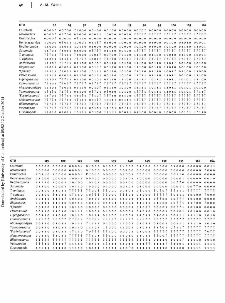
A. M. YATES
OTU 60 75 80 85 90 95 1 0 0 105 110
CrurotarsiMarasuchusOrnithischiaHerrerasauridaeNeotheropodaSaturnaliaT. antiquusT. caducusAnchisaurusRiojasaurus'Efraasia'PlateosaurusLufengosaurusColoradisaurusMassospondylusYunnanosaurus'Euskelosaurus'MelanorosaurusBlikanosaurusVulcanodonEusauropoda
0000?
0000?
0000?
10000
11000
1 1 ? 0 1
? ? ? 11
11011
1110?
11011
11111
11111
11101
1 1 10 1
11101
1 ? 1 ? 0
1 1 ? 0 1
1 1 1 1 1
0 o ? o 0
0 ? ? 00
0000 0
0 ? o 1 1
1 1 0 11
? 1 0 1 1
11111
1 1 1 1 1
1 1 1 1 10 ? o 1 1
? 1 o 1 1
0 10 11
1011 11? ? n ? ?
? ? 0 0 0
0 ? 000
0 ? 1 1 0
0000 1
1 110
0 10 0 0
1 1 000
111110 100
0 110 0
0 110 0
0 1100
0 1100
0 0 10 0
000 ? 1
000 00
011??0 10 0 0
0 ? ? ? ?
1001?
1001?
00 ? o ?
0 0 1??10 011
00 1 ? 1
010111
n ? ? ? ?
0 0 10 0
10000
00000
0 000010 0 0 0 0
0 0010
00 ? 0
? ? ? ? n
0 0 0 0 0
0 0?0? ? 0
10000
10 000
10 0 1 0
0 0 10 0
? 0 1 00
? n ? ? ?
00??0 00001 00001 00000
? ? ? ? ? ? ? ? ? ? ? ? ? ? ? ? ? ? 0 ?
0000 0
000 ?0
10 11 0
1 ? ? ? ?
1110 0
00000 00 000
01000 00100
00000 00100
0000 0 0?110
01010 00001
01110 11001
01100 10010 01100????? ????? ?????
1000 1
0010100
0 0 110
0 0 10
010110
0 1110
10 100
0 11 0
10 11 0
11 100
11100
1 1 ? o 0
1110 0
? 1 1 1 0
1 1 ? 1 1
101111
00110 1101?
00110 110
011010 1 0 0 ?
01?10 011
10110 000101
10100
0 0 10 0
0 110 0
0011100
10 1 1
10 100
10 11 0
110 0 0
11100
11100
? 1 o 1 1
1 111 1
111 11?1
? ? ? 11
0 1100 110 0
1 1 1 ? 1
1 ? 1 1 1
0 0 10?
0 ? ? o 1
? ? 10?
001??
01110 0010?0
0??010 ?01010
???10 01100
10111 00111
111000 10?01 110101
1???1 ?0111 11011
1 1 1 1 1 11111 11111
10101 10100
10101 ??11?
1 1 1 1 1 11111 11111 11111 11111? ? ? ? ? ? ? ? ? ? ? ? ? ? ? ? ? ? ? ? ? ? ? ? ?
11100101 11?0100?11? ?????11010 0 1011 10111
00101 11?01 0 0 ? 1 1
0 0 1 0 0 lllPl 0 0 0 1 1
11111 11111 11111 11111 111110 1 1 0 0 0 0 0 P 0 1 0 0 0 0 1 0 1 ? 1 ? ? 1 1 0
OTU
CrurotarsiMarasuchusOrnithischiaHerrerasauridaeNeotheropodaSaturnaliaT. antiquusT. caducusAnchisaurusRiojasaurus'Efraasia'PlateosaurusLufengosaurusColoradisaurusMassospondylusYunnanosaurus'Euskelosaurus'MelanorosaurusBlikanosaurusVulcanodon
Eusauropoda
0
0
I
0
I
0
oo0
0
0
0
07
0
ooo
7
1
0
0
0
1
1
1
oo0
0
0
0
07
0
ooo
7
0
0
0
1
0
2
1
1
1
1
1
1
1
17
1
1
1
1
7
2
11
0
0
p0
1
0
oo1
1
0
1
17
1
1
1
1
1
1
5
0
0
0
0
0
0
oo0
1
0
0
07
0
ooo
0
1
0
0
1
0
1
1
1
7
1
1
1
1
17
0
1
o7
7
7
0
0
0
0
0
0
0
1
1
1
1
1
1
17
1
1
1
1
1
1
0
0
0
0
0
0
oo0
0
0
0
07
0
ooo
1
1
1 2 0
0
0
0
0
0
0
1
1
1
1
1
1
17
1
1
1
1
1
1
0
0
0
0
1
1
1
1
7
0
1
0
07
1
1
1
1
7
0
0
0
0
1
1
1
7
1
1
1
1
1
17
1
1
1
7
7
1
1
0
0
0
0
0
07
7
0
0
0
0
07
0
o7
7
7
1
1
0
0
0
0
1
1
7
1
1
1
1
1
17
1
1
1
7
7
1
1
125
0
0
0
1
0
1
7
1
0
1
1
1
17
1
1
1
7
7
1
1
?
7
7
7
0
07
o0
0
0
1
07
1
oo7
7
0
0
0
0
p0
1
1
7
1
7
1
1
1
17
7
1
7
7
7
7
1
?
7
7
0
0
07
o0
0
0
0
07
1
1
o7
7
0
0
0
0
0
0
1
0
o7
1
1
0
0
17
1
1
7
1
1
1
130
0
07
0
0
0
0
7
0
0
0
0
17
1
0
7
0
0
1
0
0
0
0
1
07
7
0
0
0
1
17
1
1
7
1
1
1
0
0
0
0
0
07
7
0
0
0
0
07
0
1
7
7
7
1
1
1
0
0
0
0
1
7
7
1
1
1
1
17
1
7
7
7
7
7
1
0
0
0
0
0
0
0
0
1
1
0
0
17
0
0
1
1
1
1
135
0
0
0
0
0
0
0
0
0
0
0
0
07
0
0
0
0
1
1
0
0
0
0
0
0
0
0
0
1
0
0
07
0
0
0
0
1
1
2
0
0
0
0
0
0
7
1
1
0
0
17
1
1
0
1
1
2
?
0
1
0
0
0
0
7
1
1
0
0
17
1
1
0
1
1
1
0
1
0
1
1
1
1
7
0
0
0
0
07
0
0
0
0
0
0
140
0
0
0
0
0
0
0
0
0
0
0
0
07
0
0
0
1
1
p
0
0
1
1
0
1
1
1
1
1
1
1
17
1
1
1
0
1
0
0
0
0
1
0
0
0
0
1
1
0
0
17
0
0
0
1
?
1
1
1
0
0
0
0
1
7
1
1
1
1
1
17
1
1
1
1
?
1
1
0
0
0
0
0
0
0
0
0
0
0
0
07
0
0
0
0
7
1
1
0
0
p0
0
0
0
0
1
1
0
1
17
1
1
0
1
7
7
1
15
0
0
p0
0
0
0
0
1
07
0
17
1
1
1
1
1
7
1
0
0
0
0
0
07
7
0
0
0
0
07
07
?
?
0
1
1
?
0
0
0
0
0
0
7
7
1
0
0
1?
1
1
?
?
1
1
1
?
0
0
0
0
0?
?
?
0
0
0
0?
0?
?
n1
1
1
150
0
0
0
0
0
0
0?
0
0
0
0
0?
0
0?
0
1
1
1
0
0
0
0
0
0?
?
0
0
1
1
1?
1
1
?
?
0?
0
0
0
0
0
0
0?
?
0
0
0
0
0?
0
0?
?
1
?
1
1
0
0
0
0
0?
0
0
0
0
0
0?
0?
?
?
0
1
1
0
0
1
0?
0
1
1
?
?
?
1
1?
1
1
?
?
1
1
1
155
0
0
1
0?
1
1
1
?
?
?
0
1?
1
1
?
?
1
0
0
0
0
0
1
0
1
1
1
?
1
1
1
1?
1?
?
?
?
1
0
0
0
0
0
0
07
1
1
1
1
1
1?
1
7
7
7
1
1
1
0
0
0
0
0
07
0
0
1
0
0
17
1
7
7
7
1
1
1
0
0
0
0
07
7
1
1
7
1
1
17
1
7
7
7
1
1
1
160
0
0
0
0
07
7
0
0
0
0
0
17
1
7
7
7
1
1
1
0
0
0
0
0
07
0
0
0
0
0
07
07
7
7
0
1
1
07
0
0
0
07
7
07
0
0
17
17
7
7
1
1
1
0
0
0
0
0
07
0
0
0
0
0
07
07
n7
0
1
1
164
1
0
0
1
0
07
0
0
0
0
1
17
17
7
7
1
1
1
1
0
0
0
0
07
0
0
0
0
0
07
07
7
7
0
1
1
Dow
nloa
ded
by [
Uni
vers
ity o
f C
onne
ctic
ut]
at 0
5:52
12
Oct
ober
201
4