a new manufacturing process to remove thrombogenic factors
TRANSCRIPT

lable at ScienceDirect
Biologicals 45 (2017) 1e8
Contents lists avai
Biologicals
journal homepage: www.elsevier .com/locate/bio logicals
A new manufacturing process to remove thrombogenic factors (II, VII,IX, X, and XI) from intravenous immunoglobulin gamma preparations
Dong Hwarn Park a, Gil Bu Kang a, Dae Eun Kang a, Jeung Woon Hong a, Min Gyu Lee b,Ki Yong Kim a, **, Jeung Whan Han b, *
a Green Cross Corp., Ihyeon-ro 30 beon-gil, Giheung-gu, Yongin-si, Gyeonggi-do, Republic of Koreab Research Center for Epigenome Regulation, School of Pharmacy, Sungkyunkwan University, Suwon 16419, Republic of Korea
a r t i c l e i n f o
Article history:Received 18 June 2016Received in revised form24 October 2016Accepted 7 November 2016Available online 18 November 2016
Keywords:Intravenous immunoglobulin GThromboembolic eventsCoagulation factor XIaProcoagulant activity
Abbreviations: IVIG, Intravenous immunoglobulinassay; NaPTT, Non-activated partial thromboplastindifluoride; PKA, Prekallikrein activator; ELISA, Enzassay.* Corresponding author.** Corresponding author.
E-mail addresses: [email protected] (K.Y.(J.W. Han).
http://dx.doi.org/10.1016/j.biologicals.2016.11.0021045-1056/© 2016 The Author(s). Published by ElseviNC-ND license (http://creativecommons.org/licenses/b
a b s t r a c t
Coagulation factors (II, VII, IX, X, and particularly XIa) remaining in high concentrations in intravenousimmunoglobulin (IVIG) preparations can form thrombi, causing thromboembolic events, and in seriouscases, result in death. Therefore, manufacturers of biological products must investigate the ability of theirproduction processes to remove procoagulant activities. Previously, we were able to remove coagulationfactors II, VII, IX, and X from our IVIG preparation through ethanol precipitation, but factor XIa, whichplays an important role in thrombosis, remained in the intermediate products. Here, we used a chro-matographic process using a new resin that binds with high capacity to IgG and removes procoagulantactivities. The procoagulant activities were reduced to low levels as determined by the thrombin gen-eration assay: <1.56 mIU/mL, chromogenic FXIa assay: <0.16 mIU/mL, non-activated partial thrombo-plastin time (NaPTT): >250 s, FXI/FXIa ELISA: <0.31 ng/mL. Even after spiking with FXIa at aconcentration 32.5 times higher than the concentration in normal specimens, the procoagulant activitieswere below the detection limit (<0.31 ng/mL). These results demonstrate the ability of ourmanufacturing process to remove procoagulant activities to below the detection limit (except by NaPTT),suggesting a reduced risk of thromboembolic events that maybe potentially caused by our IVIGpreparation.
© 2016 The Author(s). Published by Elsevier Ltd on behalf of International Alliance for BiologicalStandardization. This is an open access article under the CC BY-NC-ND license (http://creativecommons.
org/licenses/by-nc-nd/4.0/).
1. Introduction
Intravenous immunoglobulin G (IVIG) has been used for treatingvarious immunological deficiencies and autoimmune disorders.Different manufacturers of IVIG use different manufacturing pro-cesses, resulting in differences in the final product composition[1e5].
In general, treatments involving IVIG use purified IgG fromhuman plasma, which ensures high safety of the preparations. Suchtreatments have been widely used until now, except in rare cases,
G; TGA, Thrombin generationtime; PVDF, Polyvinylideneyme-linked immunosorbent
Kim), [email protected]
er Ltd on behalf of International Aly-nc-nd/4.0/).
such as in patients with IgA deficiency [6]. However, recently,thromboembolic events were found to have occurred in a patientwho had received a high dose of IVIG. Thromboembolic events areinduced when the coagulation factor XIa (FXIa) remaining in theIVIG preparation (as a result of imperfect removal in themanufacturing process) activates the coagulation cascade in theblood of the recipient [7,8].
Consequently, the revised European pharmacopeia now rec-ommends that manufacturers include a quality check to ensure theremoval of thrombosis-generating agents (coagulation factors andtheir zymogens) in IVIG manufacturing processes or submit evi-dences to indicate that the product does not induce zymogenactivation [9].
Typically, the ethanol precipitation method developed in the1940s is used to separate IgG from plasma [10,11]. However, it isdifficult to completely isolate and purify IgG from the FXIa onlythrough ethanol precipitation due to the high isoelectric point (pI8.9e9.1) of FXIa [12]. Therefore, an additional purification process is
liance for Biological Standardization. This is an open access article under the CC BY-

D.H. Park et al. / Biologicals 45 (2017) 1e82
required to remove any remaining FXIa.Effective removal of FXIa is a very important aspect in the
manufacture of safe IVIG preparations. During the manufacturingprocess, this step is conducted using various methods such ascaprylic acid precipitation and membrane chromatography [13,14].However, in the case of preparations with high concentrations ofIVIG (e.g., blood preparations), the use of a membrane filter reducesthe overall yield or increases the cost of materials required for theprocess; thus, it is difficult to introduce this step into themanufacturing process. It is therefore necessary to develop amanufacturing process that can provide sufficient yield and canmaximize the safety of the preparations without increasing themanufacturing time.
GC5101B is a high-purity pharmaceutical IVIG preparationmanufactured through cold ethanol fractionation and chromatog-raphy. In this study, we developed a new process for reducing thecontent of FXIa in IVIG preparations to levels below the detectionlimits to ensure the safety of GC5101B. Furthermore, since bloodproducts are generally derived from plasma from over 1000 donors,there are differences in protein quantity and compositiondepending on the country or state of the donor. Therefore, wefurther spiked our samples with additional coagulation factor XIa,in amounts exceeding any variability that may be caused due tosample differences, and were able to still successfully remove XIa,thus verifying the robustness of our manufacturing process.
2. Materials and methods
2.1. Materials
The intermediate and final purification products of GC5101Bfrom the original manufacturing process were provided by GreenCross Corp. (Korea), and the standard coagulation-factor materialsfor the spiking study were purchased from HTI (HematologicTechnologies Inc., USA). For the thrombin generation assay (TGA),the WHO reference reagent NIBSC (1st international reference re-agent for activated blood coagulation factor XI (FXIa), HumanNIBSCcode: 11/236) was used; for the non-activated prothrombin timetest (NaPTT) assay, NIBSC's factor XIa was used. For the chromo-genic FXIa assay, a biophen factor XIa kit (Chromogenix, UK) wasused.
2.2. Study design
During the manufacturing process, cryo-poor pooled plasmawas used as a starting material and a paste containing FractionI þ IIþ III, a supernatant containing Fraction I þ III, and a Fraction IIpaste was produced through cold ethanol fractionation. The start-ing material for the manufacturing process for GC5101B was theFraction II paste obtained by cold ethanol precipitation. This Frac-tion II paste was dissolved into 4-fold volume of 0.6% sodiumchloride solution at below 10 �C. Next, the pH was adjusted to 5.0,which was followed by clarifying depth filtration (nominal size:0.1 mm). 1 M sodium acetate was added to the diafiltrated IgG so-lution to a final concentration of 5.0 mM; the pH was then adjustedto 6.0 and the solution was subjected to AEX chromatography. Theunbound fraction was collected and its pH was adjusted to 5.0before subjecting it to the solvent/detergent virus inactivationprocess. Tri(n-butyl)-phosphate (TNBP) and Polysorbate 80 (Tween80) were added at concentrations of 0.3% and 1.0%, respectively, andthe mixture was then incubated at 25 �C for 8 h. To bring the finalconcentration to 20 mM, 1 M sodium acetate was added to thefiltrate, and the filtrate was subjected to CEX chromatography toabsorb IgG. Another clarifying depth filtration was then performedat the same pH just before CEX chromatography. After CEX
chromatography, the filtrate was washed with equilibrium buffer(20mM sodium acetate, pH 5.0) and then elutedwith elution buffer(20 mM sodium acetate with 0.5 M sodium chloride, pH 4.5) toacquire IgG; The salt content was eliminated by Ultra/Dia-filtration.A nanofilter (Ultipor®VF DV 20, Pall Lifescience, Switzerland) wasused to eliminate viruses, and stabilizers were applied. Finally, aftersterile filtration, the finished product was obtained. We thendetermined the procoagulant activities from each of the interme-diate and finished products.
2.3. Detection of FX/FXla
FXI/FXIa content in the intermediate products ofGC5101BdFraction Iþ IIþ III Paste, Fraction Iþ III supernatant, andFraction II Pastedwas analyzed using western blot and ELISA, andits procoagulant activity was analyzed using TGA. The procoagulantactivity of the final product was measured using TGA, chromogenicFXIa assay, and NaPTT assay. Each method was verified based onICH Guideline Q2 (R1) [15] and was used in the analysis of the in-termediate and final products.
2.4. TGA
TGA was conducted in accordance with “CBER Ig-ThrombinGeneration Test Protocol (automated version)” [16]. There are twomethods for the determination of the potential risk of thrombo-embolic events: Wessler test (in vivo) and TGA (in vitro). TGA isknown to be the most sensitive and reliable method for detectingthrombogenic agents in plasma-derived preparations. To measureFXIa contents, the test sample was treated with a mixture of 50 mLFactor XI (FXI)-deficient plasma (Hematologic Technologies Inc.,USA), 2.5 mL tissue factor ((Dade Innovin®, USA), 2.5 mL phospho-lipid (Rossix, Sweden) and 8.75 mL of CaCl2 (Sigma Aldrich, USA).Activated thrombin in the test samples were measured byanalyzing the release of a fluorophore from a fluorogenic substrate(Z-Gly-Gly-Arg-AMC, Bachem, Switzerland) with a kinetic fluores-cent reader (Infinite F-500, Tecan, Switzerland; excitation wave-length: 380 nm, emission wavelength: 430 nm) at 37 �C. This valuewas applied to the standard curve constructed with fluorescencemeasurements obtained for standard product (FXIa, NIBSC 11/236).Undiluted IgG specimen, FXIa standard (FXIa, NIBSC 11/236) andIVIG from normal production batches were used as controls. IVIGsamples, from a high or low procoagulant Ig lot, were selected tomeasure reproducibility.
2.5. NaPTT assay
The NaPTT assay was performed to determine the coagulationtime as a means to measure the activities of coagulant factors. TheNaPTT assay was conducted according to Ph. Eur. 2.6.22 [17]. TheFraction I þ II þ III paste was dissolved in sterile water (Water forInjection, manufacturing site) at a 1:5 ratio for 5e7 h, followed byfiltration using a syringe filter. The Fraction II pastewas dissolved in0.6% sodium chloride at a 1:4 ratio for 1 h. Cryo-poor plasma, dis-solved Fraction I þ II þ III Paste, Fraction I þ III supernatant, anddissolved Fraction II Paste were further diluted 10 times with Tris-albumin buffer. Citrated plasma was used as a control. A series ofpolystyrene tubes were placed in a water-bath at 37 �C and 0.1 mLof platelet-poor plasma substitute R was added to each tube. 0.1 mLof the diluted samples or 0.1 mL of the buffer (control tube) wasadded to the tubes. Then, 0.1 mL of a 3.7 g/L solution of calciumchloride R (previously warmed to 37 �C) was added immediately toall the tubes. Clotting time was measured using a manual coagu-lator (Sigma KC4 coagulator, Amelung, Germany) within 30 min ofadding the original dilution as the time that elapses between the
D.H. Park et al. / Biologicals 45 (2017) 1e8 3
addition of the calcium chloride solution and the formation of aclot. This test is not valid unless the coagulation time measured forthe control tube is 200 se300 s. We also measured the clotting timeusing a ACL TOP 500 (Instrumentation Laboratory, USA).
2.6. Chromogenic FXIa assay
FXIa activity was measured using the ROX FACTOR XIa test kit(Rossix, Sweden). FIX, FVIII, and calcium chloride were added to thespecimen to activate FIX for conversion to FIXa. When the con-version of FX to FXa was induced by activated FIXa, the activatedFXa hydrolyzed the substrate (Z-D-Arg-Gly-Arg-pNA) and pro-duced a chromogenic product (p-nitroalanine), which wasmeasured at a wavelength of 405 nm (reference wavelength:490 nm). FXIa activity was then measured by comparisonwith thatof the standard solution (biophen factor XIa kit, Chromogenix, UK).
2.7. Immunoblotting
For immunoblotting, each sample was subjected to SDS-PAGEand then electroblotted onto a polyvinylidene difluoride (PVDF)membrane by semi-dry transfer (Bio-Rad, USA). The membranewas then blocked for 1 h and incubated overnight at 4 �C with theindicated primary antibody (Anti-human Factor XI primary poly-clonal antibody, Innovative Research, USA), followed by incubationwith horseradish peroxidase-conjugated secondary antibodies for1 h (Innovative Research, USA). The signals were then detectedusing chemiluminescence reagents (Intron, USA).
3. Results
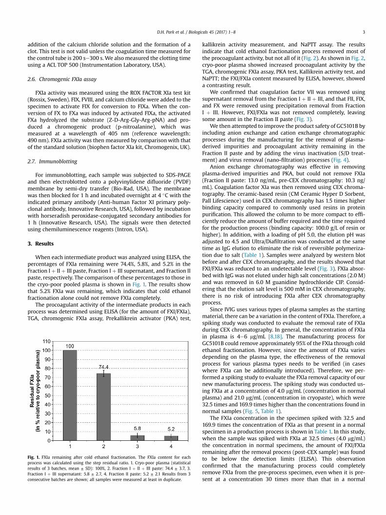
When each intermediate product was analyzed using ELISA, thepercentages of FXIa remaining were 74.4%, 5.8%, and 5.2% in theFraction I þ IIþ III paste, Fraction I þ III supernatant, and Fraction IIpaste, respectively. The comparison of these percentages to those inthe cryo-poor pooled plasma is shown in Fig. 1. The results showthat 5.2% FXIa was remaining, which indicates that cold ethanolfractionation alone could not remove FXIa completely.
The procoagulant activity of the intermediate products in eachprocess was determined using ELISA (for the amount of FXI/FXIa),TGA, chromogenic FXIa assay, Prekallikrein activator (PKA) test,
Fig. 1. FXIa remaining after cold ethanol fractionation. The FXIa content for eachprocess was calculated using the step residual ratio. 1. Cryo-poor plasma (statisticalresults of 3 batches, mean ± SD): 100%, 2. Fraction I þ II þ III paste: 74.4 ± 3.7, 3.Fraction I þ III supernatant: 5.8 ± 2.7, 4. Fraction II paste: 5.2 ± 2.1 Results from 3consecutive batches are shown; all samples were measured at least in duplicate.
kallikrein activity measurement, and NaPTT assay. The resultsindicate that cold ethanol fractionation process removed most ofthe procoagulant activity, but not all of it (Fig. 2). As shown in Fig. 2,cryo-poor plasma showed increased procoagulant activity by theTGA, chromogenic FXIa assay, PKA test, Kallikrein activity test, andNaPTT; the FXI/FXIa content measured by ELISA, however, showeda contrasting result.
We confirmed that coagulation factor VII was removed usingsupernatant removal from the Fraction I þ II þ III, and that FII, FIX,and FX were removed using precipitation removal from FractionI þ III. However, FXI/FXIa was not removed completely, leavingsome amount in the Fraction II paste (Fig. 3).
We then attempted to improve the product safety of GC5101B byincluding anion exchange and cation exchange chromatographicprocesses during the manufacturing for the removal of plasma-derived impurities and procoagulant activity remaining in theFraction II paste and by adding the virus inactivation (S/D treat-ment) and virus removal (nano-filtration) processes (Fig. 4).
Anion exchange chromatography was effective in removingplasma-derived impurities and PKA, but could not remove FXIa(Fraction II paste: 13.0 ng/mL, pre-CEX chromatography: 10.3 ng/mL). Coagulation factor XIa was then removed using CEX chroma-tography. The ceramic-based resin (CM Ceramic Hyper D Sorbent,Pall Lifescience) used in CEX chromatography has 1.5 times higherbinding capacity compared to commonly used resins in proteinpurification. This allowed the column to be more compact to effi-ciently reduce the amount of buffer required and the time requiredfor the production process (binding capacity: 100.0 g/L of resin orhigher). In addition, with a loading of pH 5.0, the elution pH wasadjusted to 4.5 and Ultra/Diafiltration was conducted at the sametime as IgG elution to eliminate the risk of reversible polymeriza-tion due to salt (Table 1). Samples were analyzed by western blotbefore and after CEX chromatography, and the results showed thatFXI/FXIa was reduced to an undetectable level (Fig. 3). FXIa absor-bed with IgG was not eluted under high salt concentrations (2.0 M)and was removed in 6.0 M guanidine hydrochloride CIP. Consid-ering that the elution salt level is 500 mM in CEX chromatography,there is no risk of introducing FXIa after CEX chromatographyprocess.
Since IVIG uses various types of plasma samples as the startingmaterial, there can be a variation in the content of FXIa. Therefore, aspiking study was conducted to evaluate the removal rate of FXIaduring CEX chromatography. In general, the concentration of FXIain plasma is 4e6 mg/mL [8,18]. The manufacturing process forGC5101B could remove approximately 95% of the FXIa through coldethanol fractionation. However, since the amount of FXIa variesdepending on the plasma type, the effectiveness of the removalprocess for various plasma types needs to be verified (in caseswhere FXIa can be additionally introduced). Therefore, we per-formed a spiking study to evaluate the FXIa removal capacity of ournew manufacturing process. The spiking study was conducted us-ing FXIa at a concentration of 4.0 mg/mL (concentration in normalplasma) and 21.0 mg/mL (concentration in cryopaste), which were32.5 times and 169.9 times higher than the concentrations found innormal samples (Fig. 5, Table 1).
The FXIa concentration in the specimen spiked with 32.5 and169.9 times the concentration of FXIa as that present in a normalspecimen in a production process is shown in Table 1. In this study,when the sample was spiked with FXIa at 32.5 times (4.0 mg/mL)the concentration in normal specimens, the amount of FXI/FXIaremaining after the removal process (post-CEX sample) was foundto be below the detection limits (ELISA). This observationconfirmed that the manufacturing process could completelyremove FXIa from the pre-process specimen, even when it is pre-sent at a concentration 30 times more than that in a normal

Fig. 2. Procoagulant activity, as determined by in-vitro assays performed during cold ethanol fractionation. Since each intermediate product has different IgG concentrations, theresults are shown relative to IgG level. Concentration of IgG (mean ± SD): 1. 7.2 ± 0.4 g/L, 2. 5.2 ± 0.2 g/L, 3. 3.8 ± 0.2 g/L, 4. 39.2 ± 2.3, 1. Cryo-poor plasma, 2. Fraction I þ II þ IIIpaste, 3. Fraction I þ III supernatant, and 4. Fraction II paste. 5. Normal citrated plasma for NaPTT. A. ELISA (statistical results of 3 batches, mean ± SD): 1. 478 ± 37.9, 2. 388.2 ± 25.5,3. 43.1 ± 20.7, 4. 42.5 ± 19.8. B. TGA: 1. 0.4 ± 0.1, 2. 1243.9 ± 219.0, 3. 24.0 ± 15.4, 4. 249.6 ± 124.1. C. Chromogenic FXIa assay: 1. 0.6 ± 0.2, 2. 60.8 ± 17.2, 3. 1.9 ± 1.1, 4. 4.5 ± 2.2. D. PKAtest: 1. 69.3 ± 42.6, 2. 1110.4 ± 235.3, 3. 3.7 ± 2.1, 4. 11.7 ± 5.5. E. Kallikrein activity measurement: 1. 2.8 ± 0.2, 2. 3.9 ± 0.9, 3. 0.7 ± 0.2, 4. 0.3 ± 0.1. F. NaPTT: 1. 165.1 ± 5.0, 2.135.8 ± 6.4, 3. 166.4 ± 4.2, 4. 174.9 ± 2.9, 5. 240.1 ± 7.5. Results from 3 consecutive batches are shown; all samples were measured at least in duplicate.
Fig. 3. Removal of coagulation factors (FII, FVII, FIX, FX, and FXIa) by cold ethanol fractionation and CEX chromatography. Loading amount is 5.0 mg of IgG and concentration of FXI/FXIa calculated by ELISA: 1. Cryo-poor plasma (219.0 ng/mL), 2. Fraction I þ II þ III paste (183.0 ng/mL), 3. Fraction I þ III paste (14.0 ng/mL), 4. Fraction II paste (13.0 ng/mL), 5. Pre-CEX chromatography (10.3 ng/mL), 6. Post-CEX chromatography (not detected). A. Image of the western blot with control (Non-reduced condition, 4e20% Tris-glycine gel, upperarrow: IgG monomer, 150 kDa, lower arrow: IgG polymer), B. Band density of FXI/FXIa calculated using Prizm ver. 6.0.
D.H. Park et al. / Biologicals 45 (2017) 1e84

Fig. 4. Overall flow chart of the manufacturing process for the IVIG preparation. 1) Thepurpose was to eliminate thrombogenic impurities such as FII, FVII, FIX, FX, and FXIa.In particular, FXIa was completely removed after CEX chromatography.
D.H. Park et al. / Biologicals 45 (2017) 1e8 5
specimen. However, when the specimen was spiked with 21.0 mg/mL of FXIa, a small amount of FXI/FXIa (2.93 ng/mL) remained inthe solution. Nevertheless, the residual ratio, based on which theprocess efficiency is measured, was 0.01%, indicating that a verysmall percentage of FXIa remained in the sample. Furthermore,FXIa activity, as determined using the TGA, was also found to bebelow the detection limits, thus suggesting that the potential risk ofthromboembolic events that may be caused by GC5101B preparedusing this new process would be very minor (Table 1).
Nine consecutive batches of GC5101B were produced on acommercial scale using the new process, and the potential risk ofthromboembolic events was estimated by measuring the procoa-gulant activity in the pharmaceutical GC5101B preparations. Theresults of the TGA and chromogenic FXIa assays were below thedetection limits (<1.56 mIU/mL and <0.16 mIU/mL, respectively),
Table 1Evaluation of the reduction of FXIa contents and activity by the spiking test in cation ex
Process step Prot. conc. (g/L) Purity (%) Polymer contents (%) Pre-CEX w
ELISA a (mg
Pre-CEX 21.1 99.6 0.16 123.6Post-CEX 10.4 99.8 0.12 <0.31IgG recovery (%) 97.1Residual ratio (%) N.C.c
All samples were measured at least in duplicate.a ELISA: Enzyme-linked immunosorbent assay.b TGA: Thrombin generation assay.c N.C.: Not calculated.d These concentrations are 32.5 times and 169.9 times higher than those in normal sp
and the coagulation time measured using the NaPTT assay was>250 s in all the batches, thus confirming that no procoagulantactivity existed in the final product. Moreover, the standard devi-ation between the batches was less than 5%, indicating that theproduction process was robust (Table 2).
4. Discussion
Among all thrombogenic factors, FXIa is known to play thegreatest role in causing thrombolytic events. Recently, a thrombo-embolic event that occurred in a patient who had received a highdose of IVIG was found to be induced by the FXIa remaining in thepharmaceutical preparation used [7,8]. In this study, we establisheda method to remove FXIa to undetectable levels (<0.31 ng/mL, asdetermined by ELISA). This was accomplished by using CEX chro-matography with a new resin with high binding capacities for IgG.By adjusting the pH of the elution buffer to 4.5 and conductingultra/diafiltration, we maximally inhibited IgG polymerization toestablish a stable manufacturing process for GC5101B (IgG recov-ery: 97.7 ± 2.2%, molecular distribution: pre-CEX polymer ratio0.16 ± 0.04%, post-CEX polymer ratio 0.12 ± 0.05%, purity:99.5 ± 0.1%) (Table 1). Thus, our method maximized the FXIaremoval efficiency during the manufacturing process of GC5101B.
Interestingly, both FXIa and PKA activities substantially increasein Fraction I þ II þ III as compared to the cryo-poor plasma sample.This is caused due to the activation of FXI to FXIa during the pre-cipitation and filtration step [19e21]. Further, PKA, known asHageman factor or FXIIa, is a type of serine protease that convertsPrekallikrein to Kallikrein, which also activates FXI, a Prekallikreinhomologue, in the upper level of the intrinsic pathway, or FVII inthe extrinsic pathway [22e25]. PKA activation in the plasma isinhibited by anti-thrombin III, C1 inhibitor, a1-antitrypsin, a2-antitrypsin, protease Nexin2, Kunitz-type protease inhibitor[26,27], but during Fraction I þ II þ III precipitation and filtration,inhibitors such as anti-thrombin III are removed as supernatants,thus allowing PKA activation. The increased levels of PKA activationcould be not only due to activated Prekallikrein, but could also bedue to activated FXI.
Furthermore, because the amount of FXIa can vary according tothe plasma source as a natural result of blood preparation, we alsoevaluated the robustness of the process through a spiking study.With this spiking assay, we showed that the high binding capacityof resin (>100.0 g/L) further increased the production efficiency.The spiking study including cation exchange chromatography wasconducted using 32.5 and 169.9 times the concentration of FXIa asthat present in normal specimens, and the results were analyzedusing ELISA and TGA. When the sample was spiked with FXIa at32.5 times the concentration present in normal specimens, theamount of FXIa remaining in the sample after the removal processwas below the detection limits. Even when the sample was spiked
change chromatography (CM Hyper D).
/o spiked Pre-CEX w/Spiked d
/L) TGA b (IU/L) ELISA (mg/L) TGA (IU/L) ELISA (mg/L) TGA (IU/L)
42.2 4020.1 72837.4 21002.2 447861.4<1.56 <0.31 <1.56 2.93 <1.56
95.3 99.4N.C. N.C. N.C. 0.01 N.C.
ecimens.
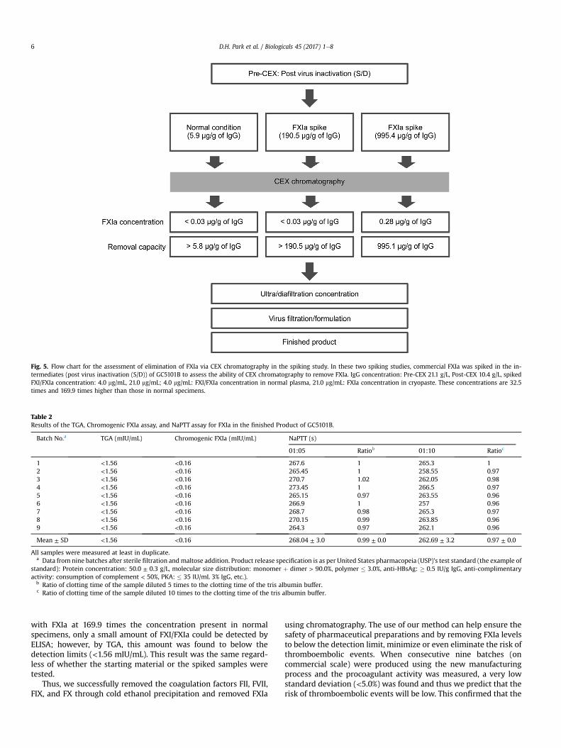
Fig. 5. Flow chart for the assessment of elimination of FXIa via CEX chromatography in the spiking study. In these two spiking studies, commercial FXIa was spiked in the in-termediates (post virus inactivation (S/D)) of GC5101B to assess the ability of CEX chromatography to remove FXIa. IgG concentration: Pre-CEX 21.1 g/L, Post-CEX 10.4 g/L, spikedFXI/FXIa concentration: 4.0 mg/mL, 21.0 mg/mL; 4.0 mg/mL: FXI/FXIa concentration in normal plasma, 21.0 mg/mL: FXIa concentration in cryopaste. These concentrations are 32.5times and 169.9 times higher than those in normal specimens.
Table 2Results of the TGA, Chromogenic FXIa assay, and NaPTT assay for FXIa in the finished Product of GC5101B.
Batch No.a TGA (mIU/mL) Chromogenic FXIa (mIU/mL) NaPTT (s)
01:05 Ratiob 01:10 Ratioc
1 <1.56 <0.16 267.6 1 265.3 12 <1.56 <0.16 265.45 1 258.55 0.973 <1.56 <0.16 270.7 1.02 262.05 0.984 <1.56 <0.16 273.45 1 266.5 0.975 <1.56 <0.16 265.15 0.97 263.55 0.966 <1.56 <0.16 266.9 1 257 0.967 <1.56 <0.16 268.7 0.98 265.3 0.978 <1.56 <0.16 270.15 0.99 263.85 0.969 <1.56 <0.16 264.3 0.97 262.1 0.96
Mean ± SD <1.56 <0.16 268.04 ± 3.0 0.99 ± 0.0 262.69 ± 3.2 0.97 ± 0.0
All samples were measured at least in duplicate.a Data from nine batches after sterile filtration andmaltose addition. Product release specification is as per United States pharmacopeia (USP)'s test standard (the example of
standard): Protein concentration: 50.0 ± 0.3 g/L, molecular size distribution: monomer þ dimer > 90.0%, polymer � 3.0%, anti-HBsAg: � 0.5 IU/g IgG, anti-complimentaryactivity: consumption of complement < 50%, PKA: � 35 IU/mL 3% IgG, etc.).
b Ratio of clotting time of the sample diluted 5 times to the clotting time of the tris albumin buffer.c Ratio of clotting time of the sample diluted 10 times to the clotting time of the tris albumin buffer.
D.H. Park et al. / Biologicals 45 (2017) 1e86
with FXIa at 169.9 times the concentration present in normalspecimens, only a small amount of FXI/FXIa could be detected byELISA; however, by TGA, this amount was found to below thedetection limits (<1.56 mIU/mL). This result was the same regard-less of whether the starting material or the spiked samples weretested.
Thus, we successfully removed the coagulation factors FII, FVII,FIX, and FX through cold ethanol precipitation and removed FXIa
using chromatography. The use of our method can help ensure thesafety of pharmaceutical preparations and by removing FXIa levelsto below the detection limit, minimize or even eliminate the risk ofthromboembolic events. When consecutive nine batches (oncommercial scale) were produced using the new manufacturingprocess and the procoagulant activity was measured, a very lowstandard deviation (<5.0%) was found and thus we predict that therisk of thromboembolic events will be low. This confirmed that the

D.H. Park et al. / Biologicals 45 (2017) 1e8 7
new purification process was robust and that it could ensure thesafety of the pharmaceutical preparation.
Several previous studies have attempted to correlate the con-centration of coagulant factors in IVIG with the risk of thrombo-embolic events. For example, Roemisch et al. demonstrated thatIVIG samples containing�2 mIU/mL did not provoke any thrombusformation using the Wessler test [28]. Funk et al. demonstrated thecorrelation between NaPTT and thromboembolic events in com-mercial IVIG batches for a period of 6 years [29]. NaPTT time >200 sand a NaPTT ratio >0.8 in IVIG resulted in low thromboembolicevent rates. In our study, TGA and NaPTT data from GC5101B werefound to be below 2 mIU/mL (<1.56 mIU/mL) and over 200 s(262.69 ± 3.2). However, our results are difficult to directly comparewith the previous studies as we have used different test andstandard materials to determine FXIa activity. Additional in vivoassays such as the Wessler test would be necessary to demonstratethe safety of the preparations more precisely.
Our findings indicate that the risk of thromboembolic eventsthat can occur with the use of pharmaceutical preparations may bepredicted by measuring the procoagulant activities using assays toevaluate thrombin generation, FXIa chromogenic activity, Pre-kallikrein activator activity, kallikrein activity, and NaPTT. For thisreason, we recommend that the same methods be used to deter-mine procoagulant activity to obtain comparable results. Then, bymeasuring the correlation between the results of the Wessler testand the procoagulant activity, relatively simple in vitro assay re-sults would be expected to be able to efficiently determine thesafety of the product.
Conflict of interest
D. H. Park, G. B. Kang, D. E. Kang, J. W. Hong, and K. Y. Kim aresalaried employees of Green Cross Corp. No other potential conflictsof interest are to be declared.
Funding
This work was supported by funding from the Green CrossCorporation andMedical Research Center programs granted to J. W.Han through the National Research Foundation of Korea (NRF)funded by the Ministry of Education, Science and Technology (NRF-2012R1A5A2A28671860). The funding source had no role in studydesign; in the collection, analysis, and interpretation of data; in thewriting of the report; and in the decision to submit the article forpublication.
Author contribution
Dong Hwarn Park: Study design and concept, data analysis andinterpretation, manuscript writing. Gil Bu Kang: data collection.Dae Eun Kang: data analysis and interpretation. Jeung Woon Hong:data collection. Min Gyu Lee: critical review of the manuscript. KiYong Kim: study supervision, critical review of the manuscript.Jeung Whan Han: critical review of the manuscript. All authorshave approved the final article.
Acknowledgments
We thank Dr. Ki Yong Kim at Green Cross Corp. for his scientificadvice, Jeung Woon Hong and Dae Eun Kang for help with the in-vitro bioassays, and Dr. Jeung Whan Han at SungkyunKwan Uni-versity School of Medicine for his academic advice. We alsoappreciate the leadership of Dr. Doo Hong Park as the Head of R&D,Green Cross Corp. We would also like to thank Cactus Communi-cations for their editorial support in the form of medical writing
based on authors' detailed directions, collating author comments,copyediting, fact checking, and referencing.
References
[1] Burnouf T. Modern plasma fractionation. Transfus Med Rev 2007;21:101e17.[2] Diez JM, Caballero S, Belda F, Otequi M, Gajardo R, Jorquera JI. Capacity of the
manufacturing process of Flebogamma DIF, a new human high purity intra-venous immunoglobulin, to remove a TSE model-agent. Biologicals 2010;38:670e4.
[3] Radosevich M, Burnouf T. Intravenous immunoglobulin G: trends in produc-tion methods, quality control and quality assurance. Vox Sang 2010;98:12e28.
[4] Parkkinen J, Rahola A, von Bonsdorff L, Toho H, Torma E. A modified caprylicacid method for manufacturing immunoglobulin G from human plasma withhigh yield and efficient virus clearance. Vox Sang 2006;90:97e104.
[5] Lebing W, Remington KM, Schreiner C, Paul HI. Properties of a new intrave-nous immunoglobulin (IGIV-C, 10%) produced by virus inactivation withcaprylate and column chromatography. Vox Sang 2003;84:193e201.
[6] Orbach H, Katz U, Sherer Y, Shoenfeld Y. Intravenous immunoglobulin:adverse effects and safe administration. Clin Rev Allergy Immunol 2005;29:173e84.
[7] Roemisch JR, Kaar W, Zoechling A, Kannicht C, Putz M, Kohla G, et al. Iden-tification of activated FXI as the major biochemical root cause in IVIG batchesassociated with thromboembolic events. Analytical and experimental ap-proaches resulting in corrective and preventive measures implemented intothe Octagam® Manufacturing process. WebmedCentral 2011;2. WMC002002.
[8] Wolberg AS, Kon RH, Monroe DM, Hoffman M. Coagulation factor FXI is acontaminant in intravenous immunoglobulin preparations. Am J Hematol2000;65:30e4.
[9] European Pharmacopeia 7.0. Human normal immunoglobulin for intravenousadministration. PA/PH/Exp. 6B/T (11) 14 PUB. 2012 Jan.. Report No.: 01/2012:0918.
[10] Cohn EJ, Strong LE, Hughes Jr WL, Mulford DJ, Ashworth JN, Melin M, et al.Preparation and properties of serum and plasma proteins III. A system for theseparation into fractions of the protein and lipoprotein components of bio-logical tissues and fluids. J Am Chem Soc 1946;68:459e75.
[11] Oncley JL, Melin M, Richert DA, Cameron JW, Gross Jr PM. The separation ofthe antibodies, isoagglutinins, prothrombin, plasminogen and beta-lipoprotein into subfractions of human plasma. J Am Chem Soc 1949;71:541e50.
[12] Burnouf-Radosevich M, Burnouf T. A therapeutic, highly purified factor XIconcentrate from human plasma. Transfusion 1992;32:861e7.
[13] Komenda M, Stadler D, Malinas T, Moses M, Pragst I, Herzog E. Assessment ofthe ability of the Privigen purification process to deplete thrombogenic factorXIa from plasma. Vox Sang 2014;107:26e36.
[14] Wu YW, Champagne J, Toueille M, Gantier R, Burnouf T. Dedicated removal ofimmunoglobulin (Ig) A, IgM, and Factor (F) XI/activated FXI from humanplasma IgG. Transfusion 2014;54:169e78.
[15] International Conference on Harmonisation of technical requirements forregistration of pharmaceuticals for human use. Validation of analytical pro-cedures: Text and methodology Q2 (R1).
[16] Jha NK, Shestopal SA, Gourley MJ, Woodle SA, Liang Y, Sarafanov AG, et al.Optimization of the thrombin generation test components to measure po-tency of factor VIII concentrates. Heamophilia 2016;22:780e9.
[17] European Pharmacopeia 7.0. Activated coagulation factors. 2.6.22. 2008 Jan..Report No.: 01/2008:20622.
[18] Hoffbrand V, Moss PAH. Chapter 24. Platelets, blood coagulation and hae-mostasis. In: Essential haematology. sixth ed. West Sussex: Wiley; 2011.
[19] Marzo N, Jose M, Lopez L, Bono M, Lopez M, Jorquera JI. Quantitative deter-mination of relevant amounts of blood coagulation factor XI activity in aspecific brand of intravenous immunoglobulin. WedmedCentral Immunother2011;2. WM001922.
[20] Jose M, Marzo N, Bono M, Carretero M, Maite L, Ristol P, et al. Pasteurizationinactivates clotting enzymes during Flebogamma® and Flebogamma® DIFproduction. WebmedCentral 2011;1. WMC001425.
[21] Jos�e M, Marzo N, Pons B, Herrerias A, L�opez L. Flebogamma® DIF(intravemousimmunoglobulin) purification process effectively eliminates procoagulantactivities. Biologicals 2013;41:393e9.
[22] Kuwahara SS. Prekallikrein activator (Hageman factor fragment) in humanplasma fractions. Transfusion 1980;20:433e9.
[23] Radcliffe R, Bagdasarian A, Colman R, Nemerson Y. Activation of bovine factorVII by hageman factor fragments. Blood 1977;50:611e7.
[24] Anton M, Suryakala S, Mao-Fu S, Sheehan JP, Vladimir S. Activation of factor XIby products of prothrombin activation. Blood 2011;118:437e45.
[25] Etscheid M, Breitner-Ruddock S, Gross S, Hunfeld A, Seitz R. Identification ofkallikrein and FXIa as impurities in therapeutic immunoglobulins: implica-tions for the safety and control of intravenous blood products. Vox Sang2012;102:40e6.
[26] Emsley J, McEwan PA, Gailani D. Structure and function of factor XI. Blood2010;115:2569e77.
[27] WuilleminWA, Minnema M, Meijers JCM, Roem D, Eerenberg AJM, Nuijens JH,et al. Inactivation of factor XIa in human plasma assessed by measuring factor

D.H. Park et al. / Biologicals 45 (2017) 1e88
XIa-protease inhibitor complexes: major role for C1-inhibitor. Blood 1995;85:1517e26.
[28] Roemisch J, Zapfl C, Zoechling A, Pock K. Comparison of thrombin generationassay (TGA) and non-activated partial thromboplastin time (NAPTT) for theassessment of enhanced procoagulant activity in immunoglobulin solutions.
WebmedCentral Immunother 2011;3. WMC003247.[29] Funk MB, Gross N, Gross S, Hunfeld A, Lohmann A, Guenay S, et al. Throm-
boembolic events associated with immunoglobulin treatment. Vox Sang2013;105:54e64.