a new in vitro cell line established from human large cell...
TRANSCRIPT

[CANCER RESEARCH 38, 3830-3835, November 1978]0008-5472/78/0038-0000$02.00
A New in Vitro Cell Line Established from Human Large Cell Variant of OatCell Lung Cancer1
Edwin R. Fisher2 and John D. Paulson
Departments of Pathology, Shadyside Hospital [E. R. F., J. D. P.], and University of Pittsburgh [E. R. F.], Pittsburgh, Pennsylvania 15232
ABSTRACT
A new tissue culture cell line, SHP-77, has been established from expiant cultures of a primary human lungcancer. Although the latter exhibited histológica! featuresof the so-called large or polygonal cell undifferentiatedvariant, neurosecretory granules were detected ultra-
structurally in cells of the original tumor, culture line, andneoplasms developing after transplantation of the latterin nude mice. These features attest to the identity of theoriginal tumor as a large-cell variant of oat cell cancer. In
addition, electron microscopy revealed the presence ofgland formation and intracytoplasmic lamellar bodies inthe cells of SHP-77. This information indicates the potential for varied differentiation and morphological expression of so-called undifferentiated lung cancer cells and ispertinent to nosological considerations concerning human lung cancer. The cell line has maintained its morphological, karyotypic, chromosomal, and growth characteristics after transplantation to nude mice. Because of thisstability, SHP-77 appears to represent a propitious cellline for in vitro and in vivo biological and therapeuticstudies of this type of lung cancer.
INTRODUCTION
A number of tissue culture cell lines of human lungcancer have been established. Reports of these have beenconcerned principally with methodology and descriptivefeatures of the cultured cells. Many of the primary cultureswere obtained from metastatic sites rather than the primarytumor. It is of interest that only two successfully culturedcell lines from a primary oat cell variant of human lungcancer have been described, both by Japanese investigators (6, 11). The most completely described line (OAT-1975)is that recently furnished by Ohara and Okamoto (11).Pertinent chromosomal, ultrastructural, and cell inactiva-tion analyses were performed. Suspensions of culturedcells were also successfully grown when transferred tonude mice and cheek pouches of conditioned Syrian hamsters. However, no comments concerning the ultrastructural, chromosomal, or growth characteristics of the transferred cells growing in vivo were made.
We have recently established a tissue culture cell line,designated SHP-77 (Shadyside Hospital, Pittsburgh, Pa.)which was derived from an undifferentiated, large-cell variant of lung cancer that was removed from a 54-year-oldCaucasian male patient with O+ blood type. No clinicalmanifestations or laboratory evidence of hormone production were present. He succumbed 2 months after pulmonary
' Supported by a grant from R. J. Reynolds Industries, Inc.2 To whom requests for reprints should be addressed, at Shadyside
Hospital, Institute of Pathology, 5230 Centre Avenue, Pittsburgh, Pa. 15232.Received May 30, 1978; accepted August 8. 1978.
resection. Suspensions of the cultured cells exhibitedgrowth in nude mice. Tumors from the latter were successfully grown in tissue culture. The commonality of chromosome constitutions and light and electron microscopicfeatures, as well as kinetics of growth in the culturesobtained before and after animal passage, indicates thesuitability of this cell line for contemplated therapeuticstudies in vitro as well as in vivo.
It was also observed that, despite the undifferentiatedhistological appearance of the primary tumor, neurosecretory granules could be recognized in cell cytoplasms. Thesewere more apparent in the cultured cells and those obtainedfrom the nude mice than in the original tumor. This, as wellas other information, prompts comments relevant to thenosological identity of this histological type of human lungcancer.
MATERIALS AND METHODS
Original Tumor. A firm, tan, circumscribed but not encapsulated neoplasm measuring 4 x 3 x 2.5 cm was encountered in the apical portion of the upper lobe of the resectedleft lung of a 54-year-old man. Its cut surface revealed fociof hemorrhage and necrosis.
Portions of the neoplasm and 7 hilar lymph nodes werefixed in Zenker's acetic fluid, dehydrated, and imbedded in
paraffin. Sections of these were stained with hematoxylinand eosin, as well as the periodic acid-Schiff technique,with and without antecedent diastase digestion for examination by light microscopy.
Aliquots of the tumor were also minced into 1-mm cubesand fixed in 3% glutaraldehyde in phosphate buffer for 3 hr,rinsed in buffer, and postfixed in 1% osmium tetroxide.After dehydration, blocks were imbedded in Maraglas. Ul-trathin sections were stained with lead citrate and uranylacetate and examined by electron microscopy.
Cells from solid portions of the tumor were also immediately teased into cold Hanks' balanced saline solutioncontaining antibiotic and anti-pleuropneumonia-like organism solution [Grand Island Biological Co. (Gibco) GrandIsland, N. Y.)] according to the method of Lasfargues andOzzello (10). After centrifugation, the cells were resus-pended in Roswell Park Memorial Institute Medium 1640that contained 10% virus-screened fetal calf serum andantibiotic and anti-pleuropneumonia-like organism solutions (Gibco) and seeded into Falcon tissue culture flasks.After incubation at 37°in a 5% CO2 humidified atmosphere
for 3 hr, the epithelial cells were then decanted with themedia and reseeded in new flasks at a concentration of 2 x10⁵ cells/flask. The medium was changed 3 times weekly.Subculture (1:4) was accomplished at 2-week intervals bytrypsinization and agitation of the flasks. Phase microscopywas continuously performed on all cultures, and aliquotsfrom the third, sixth, and 10th subcultures were prepared
3830 CANCER RESEARCH VOL. 38
on May 28, 2018. © 1978 American Association for Cancer Research. cancerres.aacrjournals.org Downloaded from
Lung Cancer Cell Line (SHP-77)
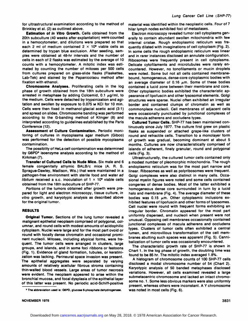
for ultrastructural examination according to the method ofBrinkley et al. (2) as outlined above.
Estimation of in Vitro Growth. Cells obtained from the20th subculture (40 weeks after explantation) were countedin a hemocytometer, and dilutions were prepared so thateach 2 ml of medium contained 2 x 10⁵ viable cells as
determined by trypan blue exclusion. After seeding, samples were obtained at 48-hr intervals and the number of
cells in each of 2 flasks was estimated by the average of 10counts with a hemocytometer. A mitotic index was estimated by counting the number of mitoses per 500 cellsfrom cultures prepared on glass-slide flasks (Flaskettes,Lab-Tek) and stained by the Papanicolaou method after
fixation with ethanol.Chromosome Analyses. Proliferating cells in the log
phase of growth obtained from the 18th subculture werearrested in metaphase by adding Colcemid, 0.1 pig/ml, tothe medium. Cells were detached by trypsinization and agitation and swollen by exposure to 0.075 M KCI for 10 min.Cells were then fixed in methanoliglacial acetic acid (3:1)and spread on slides. Chromosome banding was performedaccording to the G-banding method of Klinger (8) and
interpreted according to guidelines established by the ParisConference (12).
Assessment of Culture Contamination. Periodic monitoring of cultures in mycoplasma agar medium (Gibco)was performed for the detection of possible mycoplasmacontamination.
The possiblity of HeLa cell contamination was determinedby G6PD3 isoenzyme analysis according to the method of
Kirkman (7).Transfer of Cultured Cells to Nude Mice. Six male and 6
female congenially athymic BALB/c mice (A. R. S.Sprague-Dawley, Madison, Wis.) that were maintained in apathogen-free environment with sterile food and water adlibitum received a s.c. inoculation of 1 x 10s viable cellsobtained from the 18th subculture of SHP-77.
Portions of the tumors obtained after growth were prepared for light and electron microscopy, tissue culture, invitro growth, and karyotypic analysis as described abovefor the original tumor.
RESULTS
Original Tumor. Sections of the lung tumor revealed amalignant epithelial neoplasm comprised of polygonal, columnar, and round cells with modest amounts of acidophiliccytoplasm. Nuclei were large and for the most part ovoid orround with focally dense chromatin and occasional prominent nucleoli. Mitoses, including atypical forms, were frequent. The tumor cells were arranged in clusters, largegroups, and islands, and in some foci ribbons or festoons(Fig. 1). Evidence of gland formation, tubules, or keratini-
zation was lacking. Perineural space invasion was present.The epithelial aggregates were separated by varyingamounts of relatively acellular fibrous stroma containingthin-walled blood vessels. Large areas of tumor necrosis
were evident. The neoplasm appeared to arise within thebronchial mucosa, and focal dysplasia of the epithelial layerof this latter was present. No periodic acid-Schiff-positive
' The abbreviation used ¡s:G6PD, glucose 6-phosphate dehydrogenase.
material was identified within the neoplastic cells. Four of 7hilar lymph nodes exhibited foci of métastases.
Electron microscopy revealed tumor cell cytoplasms generally to contain abundant swollen mitochondria with fewinternal cristae. Rough endoplasmic reticulum was frequently dilated with invaginations of cell cytoplasm (Fig. 2).In some cells the rough endoplasmic reticulum was linearand in rarer instances disclosed an annulate configuration.Ribosomes were frequently present in cell cytoplasms.Delicate cytofilaments and microtubules were rarely encountered. However, no tonofilaments or mucin dropletswere noted. Some but not all cells contained membrane-bound, homogeneous, dense-core cytoplasmic bodies with
an average diameter of 0.16 /am. Some of these bodiescontained a lucid zone between their membrane and core.Other cytoplasmic bodies exhibited the characteristic appearance of lipofuscin and other lysosomal elements. Golgistructures were sparse. Nuclei often exhibited an irregularborder and contained clumps of chromatin as well ascommonplace nucleoli. Cell membranes of adjacent cellswere occasionally punctuated by junctional complexes ofthe macula adherens and occludens types.
Cultured Tumor Cells. SHP-77 has been maintained continuously since July 1977. The culture first grew on plasticflasks as suspended or attached grape-like clusters ofround and refractile cells. Transition to a monolayer formof growth was gradual, becoming dominant at about 5months. Cultures are now characteristically comprised ofislands of adherent, finely granular, round and polygonalcells (Fig. 3).
Ultrastructurally, the cultured tumor cells contained onlya modest number of pleomorphic mitochondria. The roughendoplasmic reticulum was for the most part narrow andlinear. Ribosomes as well as polyribosomes were frequent.Golgi complexes were also distinct in many cells. Occasional myelin figures were noted and some cells containedcongeries of dense bodies. Most of the latter exhibited ahomogeneous dense core surrounded in turn by a lucidhalo and membrane (Fig. 4). The average diameter of thesebodies was 0.15 ¿urn.Other cytoplasmic inclusions exhibited features of lipofuscin and other forms of lysosomes.Cell nuclei were round with frequent forms exhibiting anirregular border. Chromatin appeared for the most partuniformly dispersed, and nucleoli when present were notunusual. Opposing cell membranes occasionally containedjunctional complexes of macula adherens and occludenstypes. Clusters of tumor cells often exhibited a centrallumen, and microvillous transformation of the cell membranes abutting such spaces was apparent (Fig. 5). Canni-
balization of tumor cells was occasionally encountered.The characteristic growth rate of SHP-77 is shown in
Chart 1. In the log phase of growth, the doubling time wasfound to be 85 hr. The mitotic index averaged 1.8%.
A histogram of chromosome counts of 100 SHP-77 cells
disclosed a modal chromosome number of 54 (Chart 2).Karyotypic analysis of 50 banded metaphases disclosedvariations. However, all cells examined revealed a largesubmetacentric chromosome and lacked an intact chromosome no. 1. Some less obvious markers were also uniformlypresent, whereas others were inconstant. A Y chromosomewas noted in most cells (Fig. 6).
NOVEMBER 1978 3831
on May 28, 2018. © 1978 American Association for Cancer Research. cancerres.aacrjournals.org Downloaded from

E. fì.Fisher and J. D. Paulson
1413121110g
7-
6
5
2 3.
1
30-
4 6 8 10DAY S
Chart 1. Growth culture of SHP-77.
12 14
No evidence of mycoplasma contamination was encountered as revealed by negative cultures and the failure todetect these organisms in the many electron micrographsstudied.
Isoenzyme analysis revealed type B (slow-moving) G6PD.
Transplanted Tumors in Nude Mice. One male and 2female nude mice developed tumor nodules at the site ofinoculation. Two appeared first at 28 days as 2-mm nodules.
They measured 8 mm at Day 50. They were removed forpathological, karyotypic, and kinetic studies. The other wasobserved first at 56 days and exhibited a growth patternsimilar to that of those occurring earlier. This tumor isbeing utilized for transplantation into other nude mice.
Light microscopy revealed that s.c. transplants exhibitedhistological features that were, for the most part, indistinguishable from those of the primary tumor (Fig. 7). Thetumor appeared circumscribed but not encapsulated, withonly a slight degree of infiltration of the surrounding adipose tissue. Tumor necrosis, unlike that in the primarytumor, was not observed. A lymph node adjacent to one ofthe tumors disclosed marked sinusoidal dilation and focalcortical fibrosis and histiocyte infiltration but no evidenceof metastasis. No visceral métastases were observed in thenude mice at the time they were killed.
Ultrastructurally, the tumor was comprised of light andlesser numbers of dark cells, the latter representing degenerative forms. Cell cytoplasms contained a modest numberof round swollen mitochondria with relatively few delicatecristae. Rough endoplasmic reticulum was linear as well asdilated. Papillary cytoplasmic invaginations were noted inthe latter (Fig. 8). Free ribosomal particles and polyriboso-
mal aggregates and an occasional Golgi apparatus werepresent. Dense core secretory granules similar to thosenoted in the primary tumor and rare lysosomes and lipofus-
cin bodies were also present. Cell attachment plates ofmacula adherens and occludens types were evident, and insome instances adjacent cells exhibited a somewhat scalloped border due to pseudopodal extensions of one cell
<«2CH
uiO
111US10Q-
= BEFORE= TRAN SPL ANTATION
•¿�AFTERTRAN SPL ANTATION
42 45 50 54 58 97-115
NUMBER OF CHROMOSOMESChart 2. Histogram of chromosome numbers in cells of SHP-77 before
and after transplantation into nude mice, demonstrating commonality ofmodal number (54) and range.
into the adjacent member. Nuclei exhibited an irregular orround border with small clumps of chromatin. Nucleoliwere of the usual type.
Tissue culture characteristics of the transplanted tumorwere similar to those of the primary tumor. Comparison ofdistribution of chromosome counts in 100 cells revealedthat the modal chromosome number of 54 was maintained.Marker forms and Y chromosomes were similarly distributed.
DISCUSSIONExamination of the cell line established as SHP-77 has
provided pertinent information relative to the cytogenesisof lung cancer. Histopathological classification of the original tumor as well as those transplanted from SHP-77 into
nude mice, from which it was found to be morphologicallyindistinguishable, might be regarded as debatable if notdifficult. These neoplasms exhibited such light microscopicfeatures of oat cell carcinoma as marked anaplasia, frequent mitoses, and a tendency for the formation of ribbon-
like aggregates. Yet, their relatively large size, frequentpolygonal shape, and modest amount of acidophilic cytoplasm might also, according to prevailing criteria, warranta diagnosis of undifferentiated, large, or polygonal cellcarcinoma. It is this type of lung cancer that is frequentlythe source of interobserver disagreement. There is accumulating evidence, recently substantiated by the electronmicroscopic studies of Churg (3), which strongly impliesthat such undifferentiated carcinomas represent eitherpoorly differentiated squamous cell or adenocarcinomatypes. However, none of the cells from the original ortransplanted tumors or those of the cell line containedtonofilaments and other characteristics indicative of squamous cell carcinoma. On the other hand, all material examined by electron microscopy revealed that varying numbers
3832 CANCER RESEARCH VOL. 38
on May 28, 2018. © 1978 American Association for Cancer Research. cancerres.aacrjournals.org Downloaded from

of cells possessed dense core granules of the neurosecre-tory type, being in this regard consistent with their oat cellidentification (1, 5). In addition, lumen formation was notuncommon in electron microscopic preparations of the cellline. This latter criterion, despite the failure to demonstratemucin granules in electron microscopic preparations ortinctorially, is generally held as sufficient for the diagnosisof adenocarcinoma (3). Furthermore, the cultured cells alsooccasionally contained concentric whorls of osmophilicmembranes reminiscent of the lamellar bodies of type IIpneumocytes demonstrated by Coalson et al. (4) in thosepulmonary cancers considered to be of the bronchoalveolartype. Yet, the true identity and significance of these intra-cytoplasmic inclusions are tempered with the realizationthat comparable-appearing structures represent a relativelycommon "artifact" of cultured cells (13). Nevertheless, it isapparent that "undifferentiated" cells comprising pulmo
nary cancers may possess and indeed subtly exhibit thecapability of varied differentiation and morphological expression. In this light one might justifiably conclude thatthe extant classification (9), as well as recent studies concerning the nosological position of so-called "undifferentiated" carcinoma of the lung, may be too intransigent. The
above considerations also prompt us to regard this lungcancer as a variant of the oat cell type. It is of interest in thisregard to note that Spencer (14) briefly refers to such alarge-cell histological variant of oat cell cancer.
SHP-77 differs from the established cell line of oat celicarcinoma (OAT-1975) recently described by Ohara andOkamoto. The original tumor, which is utilized as well astransplants to nude mice, appears at least from the pertinent photographs, to represent the small-cell or moreclassical form of oat cell cancer of the lung. Interestingly,these investigators failed to find neurosecretory granules incultured cells, although such inclusions were noted inelectron microscopic preparations of the original tumor.This dichotomy was attributed to the vigorous growth of thecells in vitro with a resultant loss of functional stability.Furthermore, they provide no information concerning thechromosomal characteristics of transplanted tumors growing in nude mice. In regard to the latter, it is of interest thatwe obtained only 3 of 12 successful transplant takes,whereas Ohara and Okamoto (11) observed all cell transplants into nude mice to be successful and to exhibit earliergrowth, although similar numbers of cells were inoculated.Identical karyotypic features of the neoplastic cells in theestablished cell line and cells cultured from the transplantwere encountered in our experiments. The chromosomalcharacteristics of OAT-1975 and SHP-77 were also diver-
Lung Cancer Cell Line (SHP-77)
gent. OAT-1975 contained cells with a hypertriploid modalnumber, whereas that of cells in SPH-77 was 54.
Data obtained in this study clearly eliminate the possibilityof HeLa contamination of SHP-77. HeLa cells have neverbeen brought into or propagated in our laboratory. Thepresence of Y chromosomes, type B G6PD isoenzyme, anddivergent markers from those noted in HeLa substantiateour conclusion in this regard.
The persistence of the integrity of the structural, karyotypic, and growth characteristics of SPH-77, not only inculture but after in vivo transplantation into nude mice,indicates that it represents a stable and propitious cell linefor biological, immunological, and therapeutic studies ofthis type of tumor.
Although oat cell cancer of the human lung has a markedpropensity for métastases,such an event was not observedin the nude mice with transplanted tumors. Whether thisdivergence in behavior is related to the site of transplantation or duration of tumor growth is now being investigated.
REFERENCES
1. Bensch, K. G., Corrin, B. Pariente, R., and Spencer, H. Oat CellCarcinoma of the Lung: Its Origin and Relationship to Bronchial Carci-noid. Cancer, 22. 1163-1172, 1968.
2. Brinkley, B. R., Murphy, P., and Richardson, L. C. Procedure forEmbedding in Situ Selected Cells Cultured in Vitro. J. Cell Biol., 35. 279-283, 1967.
3. Churg, A. The Fine Structure of Large Cell Undifferentiated Carcinomaof the Lung. Human Pathol.,9. 143-156, 1978.
4. Coalson, J. J., Mohr, J. A, Pirtle, J. K., Dee, A. L., and Rhoades, E. R.Electron Microscopy of Neoplasms in the Lung with Special Emphasison the Alveolar Cell Carcinoma. Am. Rev. Resp. Dis., J07. 181-197,1970.
5. Hatori, S., Matsuda, M., Tateishi, R., Nishihara, H., and Horai, T. OatCell Carcinomas of the Lung: Clinical and Morphological Studies inRelation to Its Histogenesis. Cancer, 30: 1014-1024, 1972.
6. Hayata, Y., Tsuji, K., and Shimosato, Y. Technics and Cell Identificationin Cultures of Human Lung Cancer Cells (in Japanese). Igaku No Ayumi,90:36-41,1974.
7. Kirkman, H. N. Starch Gel Electrophoresis of Human Glucose-6-Phos-phate. Southern Med.J.,54: 1431-1432, 1961.
8. Klinger, H. Rapid Processing of Primary Embryonic Tissue for Chromosome Banding Pattern Analysis. Cytogenetics, 11: 424-435,1972.
9. Kreyberg, L. Histological Typing of Lung Tumors, pp. 9-80. Geneva:World Health Organization, 1967.
10. Lasfargues, E. Y.. and Ozzello, L. Cultivation of Human Breast Carcinomas. J. Nati. Cancer Inst.. 21: 1131-1148, 1958.
11. Ohara, H., and Okamoto, T. A New in Vitro Cell Line Established fromHuman Oat Cell Carcinoma of Lung. Cancer Res., 37: 3088-3095. 1977.
12. Paris Conference (1971): Standardization in Human Cytogenetics. BirthDefects: Original Articles Series, Vlll:7. New York: The National Foundation, 1972.
13. Seman, G . and Dmochowski, L. Ultrastructural Characteristics of Human Tumor Cells in Vitro. In: J. Fogh (ed.), Human Tumor Cells in Vitro,pp. 395-486. New York: Plenum Press, 1975.
14. Spencer, H. Pathology of the Lung, p. 811. Philadelphia: W. B. SaundersCo.,1977.
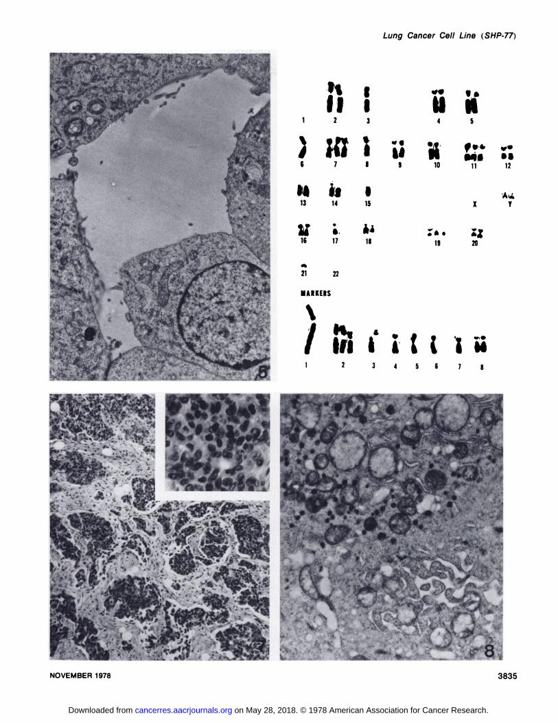
Fig. 1. Light microscopic appearance of original lung cancer. Tumor cells are arranged in clusters, islands, and ribbons. H & E, x 100. inset, tumor cellsat higher magnification, disclosing their pleomorphic nuclei and relatively abundant cytoplasm. H & E, x 400.
Fig. 2. Portions of tumor cells of original tumor revealing abundant, large mitochondria, dilated endoplasmic reticulum with invaginations, and occasionaldense core granules (arrow).
Fig. 3. Characteristic light microscopic appearance of SHP-77 cells in culture. Cell and nuclear pleomorphism and cannibalization are conspicuous.Papanicolaou stain, x 250.
Fig. 4. Portions of cells of SHP-77 revealing characteristic dense core neurosecretory granules and other cytoplasmic bodies resembling lamellar bodies,x 17,250.
Fig. 5. Cells of SHP-77 exhibiting acinus formation. The lumenal surfaces of the cells possess microvilli. x 8,300.Fig. 6. Karyotype of characteristic cell from SHP-77 revealing 50 chromosomes. 8 distinct marker forms, and nullisomy of chromosome 1.Fig. 7. Light microscopic appearance of tumor in nude mice after transplantation of SHP-77 cells. Topographical and cytological features are
indistinguishable from those noted in the original tumor as shown in Fig. 1. H & E, x 100. Inset: H & E, x 400.Fig. 8. Portions of tumor cells from tumor growing in nude mouse. Mitochondria are similar to those observed in original tumor, as are the foci of dilated,
invaginated, rough endoplasmic reticulum. Many neurosecretory granules are present, x 10,500.
NOVEMBER 1978 3833
on May 28, 2018. © 1978 American Association for Cancer Research. cancerres.aacrjournals.org Downloaded from

E. fì.Fisher and J. D. Paulson
3834 CANCER RESEARCH VOL. 38
on May 28, 2018. © 1978 American Association for Cancer Research. cancerres.aacrjournals.org Downloaded from
Lung Cancer Cell Line (SHP-77)
'
"•:.L«CJR-u«w-
1\i1II13y1621¡¡2;.'¡7it14•
è.172213i
•¿�•iu8
91ISA4uU
iÃ4SMf»0
*•M«»»10
1112.X
Y•
*•221920MARKERS\
t'tf «III i i i i i M
2 345678
g^
NOVEMBER 1978
-V M|
3835
on May 28, 2018. © 1978 American Association for Cancer Research. cancerres.aacrjournals.org Downloaded from
1978;38:3830-3835. Cancer Res Edwin R. Fisher and John D. Paulson Variant of Oat Cell Lung Cancer
Cell Line Established from Human Large Cellin VitroA New
Updated version
http://cancerres.aacrjournals.org/content/38/11_Part_1/3830
Access the most recent version of this article at:
E-mail alerts related to this article or journal.Sign up to receive free email-alerts
Subscriptions
Reprints and
To order reprints of this article or to subscribe to the journal, contact the AACR Publications
Permissions
Rightslink site. Click on "Request Permissions" which will take you to the Copyright Clearance Center's (CCC)
.http://cancerres.aacrjournals.org/content/38/11_Part_1/3830To request permission to re-use all or part of this article, use this link
on May 28, 2018. © 1978 American Association for Cancer Research. cancerres.aacrjournals.org Downloaded from