a neuropsychological model of free-drawing from memory in
TRANSCRIPT

American Journal of Psychiatry and Neuroscience 2014; 2(5): 60-75
Published online October 30, 2014 (http://www.sciencepublishinggroup.com/j/ajpn)
doi: 10.11648/j.ajpn.20140205.11
ISSN: 2330-4243 (Print); ISSN: 2330-426X (Online)
A neuropsychological model of free-drawing from memory in constructional apraxia: A theoretical review
Simon McCrea
Department of Psychology, University of Regina – Great Plains College, Swift Current, Saskatchewan, Canada S9H 4G3
Email address: [email protected]
To cite this article: Simon McCrea. A Neuropsychological Model of Free-Drawing from Memory in Constructional Apraxia: A Theoretical Review. American
Journal of Psychiatry and Neuroscience. Vol. 2, No. 5, 2014, pp. 60-75. doi: 10.11648/j.ajpn.20140205.11
Abstract: Constructional apraxia is an impairment in synthetic activities or abilities such as building, assembling and
drawing and it is a major reason for functional disability and for neuropsychological or neurological patients inability to return
to work. In this theoretical review a survey of the development of cognitive neuropsychological models of constructional
apraxia will be discussed as these relate specifically to drawing abilities. Persons with schizophrenia, somatoform disorders,
eating disorders, anosagnosia and right hemisphere disease conditions often present with impairments in the representations or
drawings of the body. A plausible cognitive neuropsychological model of constructional apraxia was developed by extracting
useful features of previous models and integrating common elements. It is hypothesized that given the relation of the integrity
of body-specific representations in the drawings of these neuropsychiatric groups a better understanding of the association
between constructional apraxia and free-drawing will provide insights into future clinical and applied neuroscience research.
Keywords: Constructional Apraxia, Free-Drawing, Unilateral Cortical Stroke Lesions, Functional MRI
1. Neuropharmacology and Drawing
Drawing is an ancient human ability as the discovery of the
Chauvet Caves in 1994 in southern France illustrates. A human
Venus figure found within the walls of the Chauvet Caves has
been dated at approximately 32, 000 years old. These ancient
drawings of humans and animals signify the value that human
societies have placed on visual artistic abilities for thousands
of generations [1]. Free format drawings of houses or
dwellings, trees and plant life, and persons are ubiquitous
concepts for humanity. These items have a multitude of
semantic associates unlike conventional neuropsychological
tasks such as clock drawings. Mechanical clocks only have a
history of 700 years. Drawings of persons have been used in
clinical psychology research since the 1890s to measure
intelligence [2] and subsequently to study the unconscious
mind and purported psychodynamic processes [3,4,5]. There is
an extensive history dating from the 1950s associated with the
use of human figure drawings in psychiatric experimentation
with psychopharmacological agents. Some of this drug
research, which involved powerful hallucinogens, was
conducted in Saskatchewan, Canada with the goal of
developing an experimental model of psychosis. Such basic
research studies have been credited, in part, with the eventual
elucidation and development of the monoaminergic, and,
eventually, the dopaminergic theory, of schizophrenia [6].
Much of this early psychopharmacological research has
been forgotten on library shelves, however recent efforts to
understand the cognitive neuroscience of human body
representations could change that situation. There is renewed
interest in studying body representations because of their
association with many psychiatric disorders, considerable
comorbidity with other more common mental disorders and
costs to society. As an example of research in this general
direction Schwoebel and Coslett (2005, p. 543) examined 70
stroke patients with an array of neuropsychological tasks and
found support for the existence of three types of body
representations [7]. In their model the body schema is a
dynamic representation of the relative positions of one’s body
parts derived from multiple sensory inputs, (including
proprioceptive), which interacts with the motor system. The
body structural description, is a topological map of locations
derived primarily from visual input that defines body part
boundaries and proximity relationships. Finally the body
image is a lexical–semantic representation of the body
including body part names, functions, and relations with
artifacts. It is presently unknown how these body-specific
representations are constructed in the brain, or how these
61 Simon McCrea: A Neuropsychological Model of Free-Drawing from Memory in Constructional Apraxia: A Theoretical Review
representations are related to the apraxias such as in the
inability to draw. The apraxias are a general class of
neuropsychological motor-based syndromes where the core
feature is an ability to perform purposeful planned
movements in the absence of a more fundamental primary
motor impairment [8]. Drawing then is classed as a particular
form of praxis behavior. Psychometric tests of human figure drawings in particular
have been developed to evaluate visuoconstructional abilities
across the entire human age span [9,10]. Although it is not
well known what the neuropsychological correlates of
performance on human figure drawing tasks might be, these
instruments may be of use to experimenters conducting
psychopharmacological research with severe mental illnesses
such as anorexia nervosa. Other older research in the
psychopharmacological vein has been illuminating on what
the neuropsychological correlates of performance on human
figure drawings may be.
By way of background, psychoactive drugs such as
scopolamine, sodium amytal, mescaline, and lysergic acid
diethylamide (LSD-25) began to be explored as potential
therapeutic agents in experimental psychiatric research in the
1940s and 1950s [6]. LSD, in particular, was used to study its
effects on body image with human figure drawings in healthy
participants [11]. Researchers sought to determine whether
experimental participants’ self-reported body image changes
might be manifest in their person drawings when
administered LSD. They found that trained clinical
psychologists and psychiatrists blind to the effects of the drug
(and with no previous experience in the psychodiagnostic use
of human figure drawings) were able to reliably discriminate
between drawings produced under conditions of mild LSD
intoxication and placebo control.
Since the psychopharmacology of many of the
hallucinogens such as LSD has now been characterized much
better compared to when first used in clinical
experimentation in the 1950’s, understanding of the
functional neuroanatomy associated with intoxication could
be invaluable to future researchers. Characteristically,
drawings performed under the influence of LSD intoxication
had substantially less attention to detail, and greater
frequency and severity of distortions - including disunity of
the body or dislocation of body parts. Impairments in shape,
symmetry, and proportion of drawn figures were also
common with LSD intoxication [12]. Savage (1955)
described three distinct stages in the process of body image
distortions as a consequence of LSD ingestion including (i)
an initial subjective pathological accentuation of body image,
(ii) progression towards manifold perceptual distortions, and
(iii) culmination in dissolution of ego boundaries
accompanied by intense feelings of depersonalization.
Liebert et al (1958) similarly found that administration of
LSD increased the perceived size of the subject’s body and
its parts without increasing the perceived size of external
objects [13].
It would appear, then, that LSD has specific and reliable
effects on body image construction in humans, and this
research suggests indirect support for the hypothesis that
dissolution of the integrity of the body schema may play
some role in evolution of pathological disorders such as
schizophrenia [14]. Support for these hypotheses is found in
a study conducted with administration of 100 mcg of LSD to
21 volunteers. Participants then completed the Wechsler-
Bellevue Intelligence Scale during and after intoxication
(mean interval = 2 months) which resulted in a temporary 11
point standard score drop in measured FSIQ [15,16]. These
researchers attributed this decline to a “loss of the abstract
attitude”. Judgment was also found to be impaired, however,
Object Assembly and Digit Span remained unaffected. Jarvik
et al. (1955) similarly showed that at a dosage of 100 mcg
healthy controls demonstrated impairments on recall of line
drawings of objects (visual memory) as well as recall of
verbal paired word associates (auditory memory) [17]. The
results of these early studies should not meant be interpreted
as suggesting that LSD studies in humans should begin in
earnest once again. LSD is a Schedule I drug with no current
accepted medical use and it is associated with significant
danger to the user.
These caveats aside, only recently have potential models
been developed to explain how LSD might affect cortical
functioning involved in somesthesis -- largely on the basis of
functional neuroimaging studies. Other psychoactive
substances such as psilocybin, dimethyltryptamine, and
mescaline, have largely been used in the place of LSD in
these imaging studies since LSD has remained a strictly
controlled substance for over 40 years [18]. However, there
are signs of liberalization of pharmaceutical regulatory
agencies restriction on the use of hallucinogens for
psychiatric research. For example, clinical trials of LSD have
recently been conducted by Swiss doctors for the treatment of
terminally ill cancer participants [19]. The goal of these
studies appears to have been to help terminally ill patients
cope with the metaphysical aspects of death. All of these
major hallucinogens - which have similar pharmacokinetic
properties to LSD - appear to activate the right hemisphere,
influence thalamic functioning and increase metabolism in
paralimbic structures and in the frontal cortex [18]. Snyder
and Reivich (1966) showed that LSD had a propensity
towards concentration in monkey brain thalami of an order
five times greater than in cortical tissue [20]. Other recent
neuroimaging research has examined the neural correlates of
dpersonalization previously associated with such experiences
as ingestion of LSD. For instance, DeRidder and colleagues
(2007) demonstrated that the neural correlates of autoscopy
or out-of-body experiences included prominent activation
within the right angular-supramarginal junction and the right
precuneus (within the right parietal lobes) and subcortically
within the posterior thalami [21].
2. History of the Study Constructional
Apraxia
Conceivably, these body image based perceptions and

American Journal of Psychiatry and Neuroscience 2014; 2(5): 60-75 62
distortions could potentially involve some of the same neural
elements associated with the neuropsychological syndrome
known as constructional apraxia. Constructional apraxia (CA)
has a very general meaning in neuropsychology. Tasks that
non-experts might associated with CA include the Block
Design subtest of the Wechsler Adult Intelligence Scale
(WAIS) [22], three dimensional cube drawing tasks [23], or
stick constructions [24] and various drawing tasks as
described by Lezak and colleagues (2004) – [25].
Constructional apraxia has been described as a disturbance
that is manifested in “formative activities such as assembling,
building, and drawing in which the spatial form of the
product proves to be unsuccessful, without there being an
apraxia for individual movements” [26]. Roncato et al. (1987)
was among the first published studies postulating a plausible
cognitive neuropsychological model of CA [27]. Roncato and
colleagues’ model was based on only three participants
studied with the WAIS and the copying tasks of houses,
persons, bicycles and cubes. Other clinical
neuropsychological tests included the Benton Visual
Retention Test [28], Token Test [29], and Warrington and
Taylor’s Unusual Views Test [30]. Experimental tasks
included copying with stimulus objects, copying to verbal
command, visual matching, sentence-design matching and
synonym judgments. Roncato et al.’s study was conducted on
participants in the post-acute phase of stroke on the
neurology ward [27].
Roncato et al. (1987, p. 127) purport that copying by
drawing has three main processing components [27]. The
first component or activity is exploration, which leads to the
encoding of a description of the stimulus constituents and
their relative relations. The second executive component
operates on this formulated description to activate the motor
programs after the choice of a scale factor. Scale factors
include preparatory activities such as positioning components
of a drawing so that these elements will fit on the page.
Finally, the matching component is used for comparison of
the drawer’s production with the stimulus model. Roncato et
al.’s first approximation model was exclusively qualitative
with little attempt to focus on the precise localization of
lesions that would cause the various types of CA. Critically,
there was no stepwise articulation or elaboration of the
hypothesized cognitive processes involved in the model. The
small sample of three participants with limited
neuropsychological assessments and no control group further
limits generalizability of the findings. Roncato et al. (1987)
stated that “…the model presented…has been used mainly for
identifying the different sorts of [constructional apraxia]
following a description of one or more of the principal
processing components. A further step which follows
logically requires using the model for analyzing the quality of
the errors of constructional apraxia patient’s drawings ”(p.
127-128) [27].
Constructional apraxia involves building, drawing, and
constructing elements into larger gestalts. However, in this
theoretical review and accompanying empirical study we will
focus entirely on the processes involved in free-drawing
rather than on copying or drawing of simple shapes. Free
drawing has an equivalent meaning as drawing-from-memory
and these terms are often used interchangeably. Although, in
recent years, rigorous attempts to characterize the neural
correlates of performance on structured constructional tasks
such as clock drawing have been undertaken [31]. In this
paper free-drawing or the drawing of richly semantically
textured complex objects will be examined under
standardized conditions with minimal prompts. Arguably,
free-drawings have much more ecological validity compared
to contrived mechanical clocks. CA has been studied since
the early part of the 20th
century, however, models of the
various processes did not emerge until the 1960’s [32]. In the
1980s, cognitive neuropsychology began to gain a better
understanding of drawing disorders beyond the simple
unilateral focal lesion approach. In the 1990s, compilations
of single cases and structural neuroimaging began to
highlight specific brain regions hypothesized to be involved
in composite drawings. More recently, functional
neuroimaging has begun to elucidate the various brain
networks involved in drawing-related CA. In this review, the
common features of the various theoretical models developed
over the last 25 years is elaborated upon with the goal of
synthesizing a contemporary model of constructional apraxia.
Table 1. Symptoms of constructional apraxia in left and right hemisphere cortical lesion patients. Note that poor visual acuity, apraxia for single movements,
proprioceptive and extrapyramidal disorders must be ruled out for a differential diagnosis of constructional apraxia [66].
Constructional Apraxia in Left Hemisphere Patients Constructional Apraxia in Right Hemisphere Patients
Reduced size of the figures [71] Increased number of lines [71]
Poor angle representation and incorrect number of angles [72] Exploded diagrams [72]
Motor tremor and right hemiparesis [72] Individual details accurately represented [72]
Poor verbal comprehension [72] Low Matrices and Block Design scores [72]
Impaired object recognition and naming [54] Failure in association of appropriate colour, shape and size of objects [54]
Loss of visual imagery [54] Loss of spatial relationships [51]
Vague description of the appearance of objects [54] Signs of unilateral left visual neglect and left-sided omissions [51]
Oversimplified diagrams with intact global outline [51] Difficulty in reproducing dimensionality [51]
Perseverative errors [51] A piecemeal copy of the design [51]
Comorbidity with dysexecutive syndromes [51] Tendency to orient drawings diagonally on the page [51]
Slow in drawing [66] Comorbidity with visuospatial agnosia [51]
Facilitation by copying [66] Wrong orientation between component parts [73]

63 Simon McCrea: A Neuropsychological Model of Free-Drawing from Memory in Constructional Apraxia: A Theoretical Review
Roncato et al. (1987) noted that CA can occur after lesions
in either hemisphere [27]. Costa and Vaughan (1962) found a
close correlation of drawing-related CA with deficient Block
Design and Raven’s Matrices [33] scores in right hemisphere
lesioned participants [34]. These studies are interesting, yet
limited, except from a componential perspective in
developing our model since well-structured drawing tasks
were invariably used. The associated deficiency between two
subtests that now comprise the Perceptual Reasoning Index
of the WAIS-IV [35] and CA would suggest a similarly high
correlation with nonverbal reasoning. Hence, these initial
studies implied that CA would be expected through
disruption of the integrated functioning of distributed neural
networks in close association with the right hemisphere [36].
Roncato and colleagues noted that drawings in left
hemisphere lesioned participants were characteristically
coherent with preservation of overall spatial relationships. In
contrast, right hemisphere lesioned participants’ drawings
were often over-simplified with a loss of overall contour [27].
These dual findings of left hemisphere sparsity of features
and right hemisphere loss of gestalt were originally
hypothesized to result from an under-specified executive
deficit in “planning the drawing”. Presumably this deficit
occurred as a consequence of a disconnection between
visuoperceptual and motor processes in the dominant
language hemisphere. These visuomotor disconnections were
first specified as originating within the left occipitoparietal
region [32]. As cognitive neuroscience has evolved, with powerful
imaging techniques, notions of hemispheric asymmetries
associated with the posterior cortices which build higher
order visuospatial percepts have been reconceptualized in
much more sophisticated terms of network interactions [37].
These newer models have little resemblance to linear
sequential processing models of higher cortical function
espoused by theorists such as Luria (1966) [38]. Insight into
these dynamic higher-order cognitive functions, associated
with the multimodal parietal cortex in particular [39] have
been aided by models of cortical interaction in which
‘metamodal’ brain centers perform particular computational
“perceptual-cognitive” operations without the necessity of
specific types of sensory input [40]. This means that
cognitive operations associated with CA could operate on
metamodal schemata that are independent of code type (e.g.,
visual, auditory, and tactile) although, it might be expected
that visuomanual motor-based codes and other metamodal
codes (semantic and lexically based codes) would
predominate at different stages of network interactions.
Other theory-based models of posterior cortical function,
such as the influential two cortical streams view of Milner
and Goodale (2006), describe the fundamental throughput
pathways that would be expected to instantiate and
fundamentally direct such metamodal processing [41].
The dorsal stream consists of two pathways, one in each
hemisphere, that transfers spatial location information from
the dorsal posterior cortices (occipitoparietal regions) to the
superior frontal cortices through the various arcuate and
superior fasciculi. The ventral stream also consists of two
pathways, one in each hemisphere, that transfers object based
information from ventral posterior cortices (occipitotemporal
regions) to the inferior frontal cortices through the
occipitotemporal, longitudinal, and uncinate fasciculi. These
four pathways can synchronize information processing across
the hemispheres via commissural fibers and also in the
superior/inferior plane via looping U-shaped white matter
fibers. Peterson and Rhodes (2003) stated that the posterior
aspect of the left hemisphere would instantiate analytic, local,
or part-based processing, whereas the posterior aspects of the
right hemisphere would be involved in holistic, global, or
configural processing [42]. A large body of the functional
neuroimaging and lesion research reviewed by Robertson
(2004) supports the view that the temporoparietal regions
within each hemisphere are chiefly responsible for building
up these dual representations [43].
At a more empirical level, drawings may be based on
direct visual inputs such as edges, line segments, line
drawings, portraits, photographs, or three-dimensional
objects that are presented immediately, or that are
subsequently removed after short durations.. These types of
drawing from copy, after a delay, characterize most of the
neuropsychological tasks associated with CA and used
presently. With such brief temporal durations, participants
would be expected to rely to a greater extent on visuospatial
encoding and memory processes. Alternatively, for the
production of facsimiles of real objects, participants may rely
on memory retrieval using language and speech based
mechanisms that evoke longer term visual representations
[44]. Casagrande (2010) hypothesized that such visual
representations may be conjured up from the subconscious
during hypnogogic imagery or during rapid eye movement
related dreaming and sleep as is described in the account of
how James Watson visualized the DNA helix [45]. However,
is there a seamless and contiguous appearance to these self-
generated mental images? van Sommers surmised that the
notion of imagery as being unified in a single topologically
coherent display has not been empirically supported. van
Sommers’ main support for the view of non-unitary
topological mental imagery displays in the mind’s eye
includes difficulties drawing complex designs (e.g.,
impossible objects such as the knot design) even when using
simple and didactic line-by-line copying strategies [44].
In an attempt to answer such fundamental questions as to
the part-based versus seamless nature of imagery, Kosslyn et
al. (1985) found that the neural specificity of dimensionally
simple unilaterally-presented visual field perceptual tasks
were separable from complex imagery tasks in patients
whose corpus callosum had been severed [46]. The complex
imagery tasks preferentially recruited the left hemisphere,
with the right hemisphere having specific difficulty
generating multi-part images. Corballis and Sergent (1988)
similarly undertook studies of simple and complex imagery
tasks in callosally-sectioned patients. These investigators
American Journal of Psychiatry and Neuroscience 2014; 2(5): 60-75 64
found that static imagery tasks employing size or angle part-
based descriptions were represented within the left
hemisphere, whereas mental rotation of the gestalt aspect of
objects was strongly localized to the right hemisphere [47].
These two viewpoints are not necessarily contradictory.
Mental rotation might be essential in developing the
formative position-independent view of an object and aiding
in compositional processes in both structural and descriptive
terms. These findings could be congruent with the hypothesis
of a strong role for both hemispheres in the overall formative
properties presumed to be deficient in fundamental cognitive
processes associated with CA.
van Sommers (1989) practically demonstrated aspects of
complex mental imagery with drawings of common real
objects [44]. van Sommers studied neurologically normal
participants’ representations of bicycles as only one instance
of objects with a rich structure and function knowledge. This
knowledge is logically encoded and inter-related verbally
within semantics. In these early studies it was shown that
participants undertook, (what appeared to be), experimentally
directed step-wise refinements of their own bicycle drawings,
presumably via access to their hierarchically structured
semantic knowledge bases. van Sommers discovered
examples of the necessity of access to such essential semantic
knowledge bases when drawing. Through task decomposition,
van Sommers demonstrated that the drawers must have
appreciated the nuances of strictly propositional forms such
as “a front wheel of a bicycle must be free to pivot” through
sequential approximations in their sketches. The functional
and structural knowledge that was encoded propositionally
then could augment perceptually-based visuoconstructional
processes occurring during various iterations of the drawing
plan.
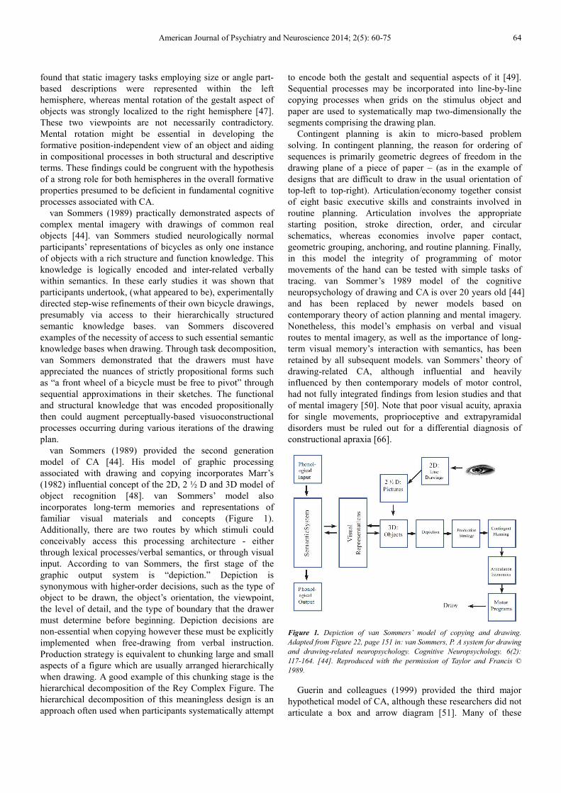
van Sommers (1989) provided the second generation
model of CA [44]. His model of graphic processing
associated with drawing and copying incorporates Marr’s
(1982) influential concept of the 2D, 2 ½ D and 3D model of
object recognition [48]. van Sommers’ model also
incorporates long-term memories and representations of
familiar visual materials and concepts (Figure 1).
Additionally, there are two routes by which stimuli could
conceivably access this processing architecture - either
through lexical processes/verbal semantics, or through visual
input. According to van Sommers, the first stage of the
graphic output system is “depiction.” Depiction is
synonymous with higher-order decisions, such as the type of
object to be drawn, the object’s orientation, the viewpoint,
the level of detail, and the type of boundary that the drawer
must determine before beginning. Depiction decisions are
non-essential when copying however these must be explicitly
implemented when free-drawing from verbal instruction.
Production strategy is equivalent to chunking large and small
aspects of a figure which are usually arranged hierarchically
when drawing. A good example of this chunking stage is the
hierarchical decomposition of the Rey Complex Figure. The
hierarchical decomposition of this meaningless design is an
approach often used when participants systematically attempt
to encode both the gestalt and sequential aspects of it [49].
Sequential processes may be incorporated into line-by-line
copying processes when grids on the stimulus object and
paper are used to systematically map two-dimensionally the
segments comprising the drawing plan.
Contingent planning is akin to micro-based problem
solving. In contingent planning, the reason for ordering of
sequences is primarily geometric degrees of freedom in the
drawing plane of a piece of paper – (as in the example of
designs that are difficult to draw in the usual orientation of
top-left to top-right). Articulation/economy together consist
of eight basic executive skills and constraints involved in
routine planning. Articulation involves the appropriate
starting position, stroke direction, order, and circular
schematics, whereas economies involve paper contact,
geometric grouping, anchoring, and routine planning. Finally,
in this model the integrity of programming of motor
movements of the hand can be tested with simple tasks of
tracing. van Sommer’s 1989 model of the cognitive
neuropsychology of drawing and CA is over 20 years old [44]
and has been replaced by newer models based on
contemporary theory of action planning and mental imagery.
Nonetheless, this model’s emphasis on verbal and visual
routes to mental imagery, as well as the importance of long-
term visual memory’s interaction with semantics, has been
retained by all subsequent models. van Sommers’ theory of
drawing-related CA, although influential and heavily
influenced by then contemporary models of motor control,
had not fully integrated findings from lesion studies and that
of mental imagery [50]. Note that poor visual acuity, apraxia
for single movements, proprioceptive and extrapyramidal
disorders must be ruled out for a differential diagnosis of
constructional apraxia [66].
Figure 1. Depiction of van Sommers’ model of copying and drawing.
Adapted from Figure 22, page 151 in: van Sommers, P. A system for drawing
and drawing-related neuropsychology. Cognitive Neuropsychology. 6(2):
117-164. [44]. Reproduced with the permission of Taylor and Francis ©
1989.
Guerin and colleagues (1999) provided the third major
hypothetical model of CA, although these researchers did not
articulate a box and arrow diagram [51]. Many of these
65 Simon McCrea: A Neuropsychological Model of Free-Drawing from Memory in Constructional Apraxia: A Theoretical Review
author’s contributions were thus conceptual in nature. These
authors noted that there had been little progress toward
understanding the neural basis and functional interactions of
distally located brain systems involved in CA. This was
despite the fact that CA is the most common form of apraxia
observed in neuropsychological evaluations [52]. Guerin et al.
attributed the limited progress in understanding CA to its
inherent complexity, as well as its less striking manifestations
compared to other disorders of higher cortical function (e.g.,
aphasia or agnosia). Guerin et al. note that familiar routine
drawings (e.g., geometric shapes and perhaps clock drawings)
tasks are often highly overlearned in a motor action
sequencing sense, and thus do not place heavy demands on
interactions between the semantic and visual representational
systems. Hence, this lack of theoretical progress in
understanding CA could also be a function of the fact that
simple copying or clock drawing tasks had been almost
exclusively used in the past. Continued use of such simple
visuospatial tasks might actually be hindering progress in this
regard.
To date, there have been widespread limitations in
experimental designs examining CA. The use of only
qualitative instead of both qualitative and quantitative
analyses of errors has continued to be a problem. A lack of
group and individualized cognitive neuropsychological
designs, as well as a lack of lesion analysis and functional
neuroimaging methods, until recently, also hindered progress.
In their review article, Guerin and colleagues’ (1999) argued
that the depiction decision components of van Sommers’
(1989) model are not specific to drawing, but rather are part
of a much more general visual imagery system [44,51]. Their
work has been influenced by Farah’s (1984) mental imagery
model, which posits that visual representations are recalled
from long-term visual memory and brought forth into the
visual buffer [50]. Guerin and colleagues noted that Farah’s
model requires an inspection process to examine the image
topologically once the images had been cued into the visual
buffer, especially when drawing from memory was required.
Inspection has been described as encompassing a top-down
hypothesis-testing process with a presumed executive
component. Participants with generative deficits cannot draw
or describe objects from memory yet they are capable of
copying and recognizing visual objects. Whereas participants
with visual memory deficits are only able to copy models.
Farah’s model of visual imagery is not specific to drawing-
related CA, yet it has significantly influenced thinking about
drawing abilities [50].
If depiction can be better understood through Farah’s
visual imagery model [50], then what is to be made of the
rest of van Sommers’ graphic production pathway to motor
output involving CA? [44] Guerin et al. (1999) observed that
van Sommers’ emphasis on production strategies could
alternatively reflect a deficiency in general motor planning
[53]. In this scenario, contingent planning would be
implemented in unfamiliar drawings (e.g., free-drawings)
whereas in familiar drawings co-activation of production
schemes in associative memory and motor representations in
procedural memory would be sufficient for performance. In
Guerin and colleagues’ view, van Sommers’ model
overemphasizes systems that constitute action programming
motor subroutines, rather than representational types of
encoding necessary for unfamiliar drawing (e.g., free-
drawing). Guerin and colleagues’ (1999) description of CA
involves three sets of systems to instantiate drawing abilities:
(i) visual perception, (ii) visual imagery and (iii) graphic
production [51].
In Guerin et al.’s (1999) model, visual imagery is not
always important in CA since familiar, routine, or
overlearned drawings can be implemented with activation of
associative or procedural motor memory systems alone [51].
In this sense, routine drawings and simple copying are
unlikely to tap the perceptual, cognitive, and motor processes
involved in pure forms of CA. Copying tasks are often used
in neuropsychology and these types of tasks may not use the
same neural mechanisms as free-drawing paradigms such as
in original artistic visual works or productions. Difficulty
with tasks such as clock drawing, which are often used as
single measures of CA, may have little relationship or
ecological validity to the types of neuropsychological deficits
that limit the functional capacity of brain-injured persons.
People view, hear, wind, read, wake up to, and interpret many
different types of clocks and time-keeping devices tens of
thousands of times in their lifetime. In fact, this common
neuropsychological test of constructional apraxia is likely
overlearned to the point of being automatic.
3. Constructional Apraxia and Mental
Imagery
Drawing from memory is generally regarded to be a
complex task with visual imagery as a core feature [54].
However, as van Sommers (1989) showed, copying can be
undertaken by highly automatized motor subroutines without
using mental images [44]. Farah posited that the left posterior
cortex was critical for mental imagery and that the inferior
occipital regions of this area were specifically involved in
imagery deficits [55,56]. Trojano and Grossi (1994) noted
that among eleven published comprehensive single cases of
patients with mental imagery deficits, the two most regular
co-occurring syndromes were visual recognition deficits
(agnosia) and naming disorders (anomia). The frequency of
optic aphasia, in which subjects are unable to name visually
presented objects (yet have no difficulty in naming those
objects on tactile presentation or verbal description) has also
been shown to be unusually high in patients with mental
imagery deficits. These patients had the greatest difficulties
either in describing imagined objects or drawing them from
memory. Among these participants with mental imagery
deficits, 50% had left unilateral posterior-inferior damage,
43% had bilateral hemispheric posterior-inferior damage, and
one left-handed participant had a right posterior-inferior
lesion [54].

American Journal of Psychiatry and Neuroscience 2014; 2(5): 60-75 66
Figure 2. Depiction of Farah’s model of mental imagery. Adapted from
Figure 12B, page 135 in: van Sommers, P. A system for drawing and
drawing-related neuropsychology. Cognitive Neuropsychology. 6(2): 117-
164. [44]. Reproduced with the permission of Taylor and Francis © 1989.
Adapted and redrawn [57] from Figure 1, page 250 in: Farah, M.J. (1985).
The neurological basis of mental imagery. In S. Pinker (Ed.). Visual
Cognition. Cambridge, MA: MIT Press, pp. 245-271.
Trojano and Grossi (1994) noted that it remains unknown
why similar types of lesions do not always elicit similarly
corresponding mental imagery deficits, although individual
differences in functional localization of cognitive modules
might play a role. The finding of regular associations of
visual recognition deficits with mental imagery deficits
originally led Levine (1978) to propose that there may be
redundancy in the primary pathways for visual perception
and those of the higher-order perceptual-cognitive process of
mental imagery [58]. Thus, empirical review of a number of
published cases of agnosia show a tendency for left
occipitotemporal lesions to result in visual object agnosia,
and right occipitotemporal lesions to result in prosopagnosia
or topographical (scene-based) agnosia [42, 59]. Farah (1984)
suggested that mental imagery deficits may also be due to
loss of access to long-term visual memories, a truism that
may be congruent with Levine’s hypothesis [50]. Problems in
access to long-term memories in this scenario would likely be
due to focal lesions in early visual cortex or occipitotemporal
cortex. Thus lesions in the left posterior-inferior cortex would
result in white matter deafferentiation, and hence blocking of
access to the anterior temporal lobe’s long-term visual
representations. In both Levine and Farah’s views there is an
assumption of the existence of shared visual recognition and
mental imagery perceptual processes.
Aside from studies that have hypothesized left posterior
localized neural instantiation of imagery, other studies have
examined the relationship between left visual field neglect and
mental imagery defects. Left-sided purely “representational
neglect” has been demonstrated in a number of participants,
beginning with Bisiach and Luzzatti’s (1978) famous
description of two cases of visualization impairment for
landmarks within a well-known piazza in Italy [60]. Their two
participants presented with either a right-sided frontal and right
temporo-occipital lesion, or a right temporoparietal lesion in
which left visual neglect was present in both cases. In such
instances, one would naturally presume that mental imagery
would still be intact. These two right-hemisphere lesioned
participants neglected the left side of the piazza scene and
naturally described the right side correctly. However, when a
180 degree change in viewpoint was made the participants
were unable to describe the original visual half field which
they had accurately described just seconds before! Importantly,
at the second viewing, the participants were now able to
describe the original left-sided half field neglected in the first
trial. If anything, these studies point to the considerable degree
that visual representations are coded across both visual fields
as well as the potent between-hemisphere integration processes
that must occur in real-time during this representational
encoding.
Trojano and Grossi (1994) subsequently described eight
such cases in the published literature of representational
neglect after right hemisphere lesions, and noted that the
originators of the representational neglect hypothesis
conceived of this as strong proof for a topologically
structured representation divided across the two hemispheres
[54]. With left neglect, drawings may demonstrate missing
left side elements, and therefore collectively such studies
imply that the right-sided temporoparietal lesions are apt to
adversely affect the building up or structuring of mental
representations across both the left and right visual fields.
Drawing from memory, however, has been shown to be less
sensitive than other tasks in identifying these various types of
representational neglect [61]. The reduced sensitivity of
drawing from memory to various forms of representational
neglect may be due to its complexity and potential for
reliance on secondary redundant encoding and lexical and
semantic pathways. Alternatively, output processes associated
with procedural motor memory, as well as reliance on
propositional and logic based processes, may provide
additional redundancies. Similarly, whereas participants with
visual agnosia have content-specific imagery defects in that
they cannot conjure up certain classes of mental images,
participants with representational neglect cannot conjure up
any mental images at all.
There is, presumably, then a representationally-flexible and
isomorphic nature to this system by which mental images are
generated, transposed within the visual buffer, and inspected
in this process-based framework. A process-based framework
of necessity implies some degree of co-operation between
distally located brain areas in the complex task of
instantiating mental imagery (e.g., see Figure 2). In this
scenario, left posterior-inferior sites implicated in mental
imagery and right temporoparietal areas involved in dual
visual field representational topography constitute a
synchronized system. In this process approach, however,
content-specific imagery is associated with visual long-term
memory. However there are further technical impediments to
fully describing and understanding the interactions of the
visual imagery and CA systems. Trojano and Grossi stated
that mere verbal reports or protocol analysis are heavily
reliant on participants’ capacity to describe their impairments
which are necessarily subjective and prone to error [62].

67 Simon McCrea: A Neuropsychological Model of Free-Drawing from Memory in Constructional Apraxia: A Theoretical Review
Drawings, by contrast, may have better objectivity in that
these verbal report factors can be controlled for and drawings
can be qualitatively and quantitatively scored. Similarly, the
Block Design subtest of the WAIS, Rey Complex Figure, and
Clock-Drawing [63] do not rely on visual and lexical
semantics as do free-drawing tasks. Thus, free-drawing tasks
could be optimal means of studying constructional apraxia in
its full richness and complexity.
Figure 3. Contemporary model of constructional apraxia-related free
drawing. Synthesis of common features of Farah’s (1985) [57], Roncato et
al.’s (1987) [27], van Sommers’ (1989) [44] and Guerin et. al’s (1999) [51]
model of free-drawing and visual imagery. Note that in this model the
graphic production system of van Sommers has been substantially simplified
in view of Guerin et al.’s more representational and less motor-based view of
drawing abilities. The visual buffer is analogous to a representational
medium for mental imagery.
Towards this end, Gainotti et al (1983) found that on
mental imagery tasks left-hemisphere lesioned aphasics
performed worst, left hemisphere lesioned participants
without aphasia performed second worst, right hemisphere
participants scored third and non-lesioned participants scored
best [64] These researchers’ findings strongly suggest a tight
link between imagery deficits and reliance on lexical and
semantic processes. Further, these results imply that
theoretically, mental imagery defects should have a degree of
independence from CA. A large study of ideal left and right
posterior cerebral artery (PCA) stroke participants (where
there is often damage to lateralized occipitotemporal regions
exclusively) found that participants with left PCA lesions
performed poorly on a pictorial version of the shape and
color test [65].
Left temporo-occipital patients (as opposed to left occipital
lesions) were selectively impaired on verbal and visual
questions. Right PCA patients were only impaired on
perceptual tests of shape and colour discrimination. Bilateral
PCA lesioned patients were not examined in this study [65].
Collectively, these findings suggest that a wide range of
lesions can result in mental imagery deficits pointing towards
a distributed model as depicted in Figure 3. It seems based on
this evidence that mental imagery is unequivocally related to
verbal abilities and semantic knowledge on both (i)
functional and (ii) neuroanatomical grounds [54].
4. Syndromes Associated with
Constructional Apraxia
Dupuy and Godefroy (2007) noted that diagnosis of CA is
often made on the basis of copying tasks such as the Clock-
Drawing test, the copy condition of the Rey Complex Figure
(Osterreith, 1944), or three-dimensional block constructions
[66]. In assessing CA, these investigators caution clinicians
to rule out competing hypotheses as to the origins of CA
involving other perceptual-cognitive mechanisms or damage
to elements of a distributed neural network illustrated as in
Figure 3. For example, dysexecutive syndromes in particular
must be ruled out, since such disorders can adversely affect
performance on virtually any complex motor function task
[66]. Thus, in theory, a definitive diagnosis of CA can only be
made with a comprehensive and individualized
neuropsychological assessment. Taken together, CA is largely
a diagnosis made on the basis of exclusion. In large samples
of patients referred for comprehensive general
neuropsychological evaluations, Gainotti (1985) reported a
frequency of CA in unilaterally lesioned hemispheric
participants of between 15% to 40% [67]. Larger lesions
would typically be associated with elevated rates of CA, as
would lesions to critical neuroanatomical loci or network
connections.
Hemianopia and fluent aphasia are often comorbid
symptoms of CA [68]. Dupuy and Godefroy (2007)
interpreted this comorbidity as support for their contention
that CA is a posterior cortical syndrome [66]. However,
historically the neuropsychological literature is replete with
CA cases with damage to prefrontal cortex and with
associated executive function impairments. Although not all
neuropsychologists agree that CA is frontal in origin. Among
the most common behavioral symptoms of frontal CA is a
“closing-in” behavior when participants are actively drawing
[69]. A patient with frontal-variant of CA and closing-in
behavior would often draw over the actual model when
attempting to copy it. Gainotti also demonstrated that diffuse
cortical damage was associated with closing-in related CA.
Whether this type of behavior is truly symptomatic of CA or
is indicative of a frontal-release, poor motor planning, mirror
movements or motor artifact is unknown. Recall that frontal-
release symptoms sometimes occur in the instance of frontal
damage where brain-lesioned participants exhibit behaviors
that would normally be inhibited. Conversely, mirror
movements are those that occur when the participant often
with a medial frontal lesion mimics the motor movements of
the examiner [70].
From a neurodevelopmental perspective closing-in
drawing behaviors can also be observed in young children,
presumably as a function of the immaturity of myelination
within the prefrontal cortex [69]. Proximal closing-in
behaviors were observed by Gainotti at an elevated frequency
with the presentation of the most complicated of models to be

American Journal of Psychiatry and Neuroscience 2014; 2(5): 60-75 68
copied. Luria and Tsvetkova (1964) demonstrated a frontal
variant of CA most often appearing when the most complex
of figures were presented [74]. Other studies showed that
examiners who provided verbal cues as to the correct
production that the subject should make [75] or cues to
sequencing [76] nonetheless facilitated performance for this
frontal-variant of CA. Collectively, these findings suggest
that executive processes such as planning and verification
maybe critical components in the motor programming
associated with complex drawing. It is unclear if these older
conceptions of CA are congruent with contemporary views
depicting CA as a perceptual process heavily-dependent upon
posterior cortical functions.
Clock drawings, the drawings associated with the Mini-
Mental Status Examination [77], and even the monolithic
Rey Complex Figure task lack definitive links with real-
world knowledge of language and lexicosematics – (e.g.,
however see also Lezak et al.’s, 2004 reference to the house-
tree-person drawing test and the bicycle test) [25]. These
mainstay diagnostic tests of clinical neuropsychology possess
little resemblance to the perceptual, cognitive, lexicosemantic,
and motor programming requirements of complex drawing
tasks used in naturalistic situations. Trojano and Conson
(2008) noted that drawing tasks are the most frequently used
tests to assess CA [73]. Drawing-related CA tasks include
free-drawing or drawing from memory of a named object
(e.g., house, tree or person) or simple copying tasks. Trojano
and Conson noted that in free-drawing tasks, specific scoring
techniques, relevant content analysis procedures, and
systematic control for premorbid drawing ability are required
for the evaluation of theoretical models of drawing. These
controls are rarely introduced in practice.
When visual neglect and severity of lesion are controlled
for researchers have found that left and right hemisphere
lesioned participants have a similar prevalence of CA [78,79].
These findings provide support for the hypothesis of a mutual
dependence between the two hemispheres in CA, as well as
qualitative differences in processes across the two
hemispheres depending on the lesion site. De Renzi (1982)
noted that CA is most often associated with parieto-occipital
lesions [80] although severity of CA did not differ between
frontal and posterior lesion variants [81]. That both frontal
and posterior cortical regions appear to be involved in CA
alludes to the complexity in the structuring and sequencing of
perceptual-cognitive processes in this disorder. Other
researchers, notably Kirk and Kertesz (1989), did not find
that there was specific intra-hemispheric localization in
determining CA [72]. Furthermore, Marshal and colleagues
(1994) found that subcortical right anterior lesions were
especially apt to cause CA [82] . As such, a modern view
might be if frontal regions are involved in CA, patient errors
are likely attributable to defective motor programming
components (see Figure 3) rather than perceptual encoding
and imagery-based processes.
These somewhat contradictory findings are consonant with
three conclusions. First, as Dupuy and Godefroy (2007) have
emphasized, CA has been consistently underestimated in its
degree of complexity [66]. Second, many assessment tasks
have been too easy for participants, or researchers have used
insufficient numbers and/or types of tests to rule out
competing alternative neuropsychological explanations.
Third, there have been too few models of the complete
picture of CA since van Sommers (1989) detailed attempt at
developing a cognitive neuropsychological box model [44].
Even in subsequent models [27,51,57] there has been
minimal specification as to what neural correlates correspond
to different modules and interactive systems.
The most comprehensive and modern account of CA to
date is Grossi’s (1991) model [83].
This model specifies that the lexical route works by
activation of familiar constructional verbal schemata (e.g.,
circle) and that a line-by-line procedure predominates in the
use of spatial analysis for which there are no extensive pre-
existing constructional representations (e.g, doodling).
Interestingly, line-by-line copying procedures are often
observed in certain visual agnosic participants, such as
integrative agnosics, who presumably have difficulties
accessing the lexical route [42,59]. Complex objects such as
houses, trees, and persons, are likely to place heavy demands
on lexicosemantic encoding (as opposed to copying simple
figures like circles or squares) and presumably would
accentuate visual imagery and the distributed free-drawing
network. Even familiar objects, which most people have
ample experience drawing (e.g., squares and circles) most
likely do not involve the lexical route and largely rely on the
integrity of procedural memory or a simple constructional
schema (e.g., see Figure 4). Trojano and Conson (2008)
argued that frontal lesions are not sufficient to produce “true
constructional apraxia”, and that comprehensive assessment
of frontal (e.g., executive function) and posterior functions
(e.g., agnosia, receptive language, visuospatial functions etc.)
is required for a differential diagnosis [73]. Complex free-
drawings with minimal prompts then would likely rely on
both a combination of the lexical and heuristic routes.
Comprehensive studies and compilations of single
participants using a broad cross-section of neuropsychological
instruments will be essential for developing contemporary
models. As an example, Trojano and Grossi (1998) described a
case of pure CA and emphasized the “need for detailed
assessments of visuoperceptual, representational and executive
abilities, as well as for analyses of drawing productions, to
gain insight into the nature of single participants’
disabilities...(p.49) – [84]. The complexity of the multiple
distributed perceptual, cognitive, and motor processes involved
in CA points to the necessity of using convergent research
methodologies. Conventional studies of CA in diffuse
dementing conditions with non-lexically based tasks will likely
impede theoretical developments in understanding this
neuropsychological disorder. Instead, studies of thoroughly
evaluated neuropsychological participants with single focal
contiguous stroke lesions demonstrating variants of CA with
different lesion topographies could be implemented using
computerized psychological experiments. Such comprehensive
analysis of single case studies using spatial attention, spatial

69 Simon McCrea: A Neuropsychological Model of Free-Drawing from Memory in Constructional Apraxia: A Theoretical Review
perception, executive function, spatial cognition, and object
recognition paradigms might then be able to provide more
fine-grained reaction time and accuracy data with which to
specify models of CA in detail. Finally, selected single
participants with CA could be studied using structural and
functional neuroimaging with which to test various hypotheses
associated with the model described herein (see Figure 4).
Figure 4. Contemporary model of constructional apraxia-related free drawing incorporating the lexical and line-by-line heuristic routes. Cognitive
neuropsychological model illustrating Grossi’s (1991) lexical route involved in drawing from memory (purple) and line-by-line heuristic used in copying from
a model (blue) [83].
5. Modeling Cortical Interactions in
Free-Drawing
Functional neuroimaging in combination with plausible
models offers a means of examining the neural correlates of
free-drawing related CA. Such studies have been aided by the
development of functional magnetic resonance imaging
(fMRI)-compatible writing tablets [85]. Computational
advances, such as structural equation modeling [86], are
likely to be essential in understanding CA in its natural
complexity. Neilson (1975) was among the first to question
whether CA might be construed of entirely as an inter-
hemispheric disconnection syndrome [71]. In his review,
Neilson examined similarities in drawing performances of
splenial-lesioned callosal participants using the left and right
hands. Studies by Gazzaniga et al. (1965) and LeDoux et al.
(1977) relatedly established that split-brain participants
demonstrated a left hand advantage on the Block Design
subtest of the WAIS [87,88]. These types of dual hemispheric
models of CA, in addition to subsequent models of imagery
[46,47], offer counterpoints to the usual single hemisphere
processing accounts of CA.
Alternatively, intra-hemispheric disconnections of left
mesial occipital white matter associated with acquired alexia,
visual object agnosia, and mental imagery deficits have long
been identified [54]. All of these related disconnection
studies point towards the critical involvement of large-scale
brain systems in free-drawing CA. Kirk and Kertesz (1993)
found that both left and right subcortical lesions were
significantly associated with impairment on the writing,
Block Design, and Raven’s Coloured Progressive Matrices
[89] tasks of the Western Aphasia Battery [90]. Left
subcortical lesions in striatal pathways proved to be
particularly detrimental and were associated with numerous
signs of CA (see Table 1). However, this study found that
neither left nor right subcortical lesions were associated with
differential subjective drawing errors. The authors concluded
that the lack of differential scoring errors after left or right
subcortical lesions suggest that cortical substrates, rather than
subcortical substrates, are involved in drawing processes [89].
Laeng (2006) examined CA in left and right hemisphere
stroke participants [91]. Laeng found that in a computer-
adapted task arranging identical items required a coordinated
metric representation constructed by the right hemisphere.
Laeng also showed that the degree of impairment between
specific types of spatial relations used, such as in categorical
or coordinate encoding, was highly correlated with the degree
of impairment found in constructional tasks such as stick
constructions. Laeng noted that up to 58% of variance in such
macro-level constructional tasks (e.g., stick constructions)
could be explained by performance patterns on simple
scaled-down computerized tests of categorical or coordinate
encoding - e.g., see Kosslyn et al, (1992) for a discussion of
categorical versus coordinate encoding [92]. To digress, Jager
and Postma (2003) summarized categorical representations as
representing the general attributes of the spatial structure of a
visual stimulus. In contrast, coordinate representations
specify precise spatial locations of objects in metrical units.
The left hemisphere appears to be specialized for the

American Journal of Psychiatry and Neuroscience 2014; 2(5): 60-75 70
computation of categorical spatial representations while the
right hemisphere is specialized for the computation of
coordinate ones [93]. Collectively, these recent studies point
to the modern view of CA as due to: (i) perceptual-cognitive
factors, (ii) predominance of posterior cortical function
involvement, and (iii) integrative cognitive processes
occurring across hemispheres.
6. Neuroimaging of Drawing-Related
Constructional Apraxia
Makuuchi et al (2003) examined drawing by copying using
fMRI and found activation within the superior parietal
lobules and intraparietal sulci, bilaterally [94]. The
supramarginal gyrus was activated in the left hemisphere in
29% of participants, whereas the right supramarginal gyrus
was activated in 41% of participants. Brodmann’s area (BA)
39 (angular gyrus) was not activated in participants where the
task was to trace an object using the dominant right hand.
Incidentally, there was an extended period of right
occipitotemporal activation found in the drawing by copying
condition by Makuuchi et al. This extended activation was
attributed to the temporal operational capacity of the visual
imagery buffer’s components involved in copying as opposed
to simple naming procedures [94]. In another study, Farias and colleagues (2006) compared
the effects of cuing by writing and by drawing in individuals
with aphasia using a combined lesion and fMRI study [95].
Drawing was found to facilitate verbal responses to a greater
degree than writing, which the researchers attributed to richer
priming of perceptual, structural, and functional motor
aspects of objects during drawing. Moreover, these authors
found that it was the motor act of drawing, rather than the
quality of the drawing, that was correlated with loci of
semantic activation and with subsequent accuracy in the
naming of the target object. Activation in the homologues of
Broca’s area in the right hemisphere, as well as within the left
fusiform gyrus and left dorsolateral prefrontal cortex
differentiated the drawing versus writing comparisons in
fMRI. The left fusiform gyrus has previously been shown to
be involved in object knowledge [94] and the left dorsolateral
prefrontal cortex in semantic selection [96] and phonological
processing [97]. Facilitation by naming in aphasia while drawing thus
appears to occur through the widely distributed semantic
network with recruitment of intact right hemisphere brain
regions probably through mechanisms of commissural fiber
lexical access. There are some limitations with these early
studies of the functional neuroimaging of drawing. Makuuchi
et al.’s (2003) study did not require hand movements and thus
the premotor and motor activations could be due entirely to
motor imagery or representational processes rather than
constituting true manifestations of CA related behavior [94].
The Makuuchi task also involved copying the object (e.g.,
object in a presented picture) with the index finger without
visual feedback, which is contrary to actual drawing tasks
that involves initiating and maintaining a flexible grip of a
writing instrument in conjunction with visual and
proprioceptive feedback. Other studies using advanced fiber
optics technologies in the functional neuroimaging
environment have come closer to the mark.
Ferber et al (2007) utilized an fMRI-compatible drawing
tablet in a task with both proprioceptive and visual feedback
[98]. In this experimental design there were two pivotal
conditions: (i) to draw a picture from a visually presented
word without an accompanying image, or (ii) to draw a copy
of a continuously displayed real object. Unlike the Makuuchi
(2003) study, the Ferber study was more akin to spontaneous
free-drawing in CA as opposed to mere copying. Drawing
from a word presentation minus the condition of copying of
realistic objects demonstrated activation of the right anterior
cingulate, right medial frontal gyrus, right middle frontal
gyrus, and right superior parietal lobule. In contrast to line-
by-line copying, drawing minus copying of realistic objects
would be expected to emphasize the importance of access to
the lexical route. In a third condition, copying versus mere
tracing was found to activate the anterior cingulate and the
medial frontal gyrus probably involved in hierarchical
planning and motor control mechanisms [98]. Copying minus
drawing also activated the left middle occipital gyrus, left
cuneus, and left lingual gyrus (left occipitotemporal regions)
which have been shown in lesion studies to be involved in
visual imagery [59] and uploading of schemata into the visual
buffer. Conceivably then, this online visual imagery could
help to ensure that the elements of the drawn copy match the
displaced target line drawing.
Ferber and colleagues (2007) posit that drawing-from-
memory requires “maintenance of attention, access to
memory systems to retrieve information about the stimulus,
and internal monitoring of whether the performed action
conforms to the original intention…Whereas copying
requires constant visual feedback processes and cross modal
shifts of attention to compare one’s own copy on the page
with the model object” (p. 1092) [98]. The visual feedback
hypothesis was supported by time-course analyses of regions
of interest within the left lingual gyrus and left cuneus, which
showed more sustained blood-oxygen level dependent
activation in the copy of realistic objects. Differential
patterns of activation between the Ferber and Makuuchi
studies were attributed to the lack of an appropriate motor
control condition in the Makuuchi study [94], as well as
reliance on a less complex copying paradigm. Collectively,
these studies underline the importance of the left occipital
and lingual gyrus in the integral functioning of the visual
buffer involved in visual imagery as well as in drawing when
copying line-by-line.

71 Simon McCrea: A Neuropsychological Model of Free-Drawing from Memory in Constructional Apraxia: A Theoretical Review
Figure 5. Hypothesized model of the functional neuroanatomy associated with free-drawing-related constructional apraxia. Numbers in parenthesis
correspond to specific Brodmann’s neuroanatomical regions. 17 = primary visual cortex, 18 = secondary visual cortex, 19 = associative visual cortex, 20 =
inferior temporal gyrus and 37 = fusiform gyrus involved in visual perception and imagery; 7 = precuneus or superior parietal lobule and 40 =
supramarginal gyrus associated with the visual buffer; 20 = inferior temporal gyrus, 21 = middle temporal gyrus and 38 = temporopolar region involved in
high level object representations and semantic memories; 41, 42 = primary and auditory association cortex involved in auditory input; 6 = premotor cortex
involved in contingent planning based on sensory feedback; 4 = primary motor cortex involved in action programming with output via the corticospinal tracts;
9 = dorsolateral prefrontal cortex and 24, 32 = anterior cingulate cortex involved in error monitoring associated with inspection; 7 = superior parietal lobule
and 46 = dorsolateral prefrontal cortex involved in spatial working memory maintenance; TPJ = temporoparietal junction including: 22 = superior temporal
gyrus, 39 = angular gyrus, 40 = supramarginal gyrus involved in generating mental images; L = left hemisphere, R = right hemisphere. Note that in this
diagram there is an assumption of right-handedness with left lateralized language and praxis as occurs in the largest proportion of the general population.
Harrington et al. (2007) compared the neural basis for
imagined writing and drawing in an fMRI study [99]. Brain
areas that were activated for imagined drawing (yet were not
activated for imagined writing) included the following:
bilateral insular cortex (BA 13), right posterior inferior
temporal cortex (BA 37), right middle and right inferior
frontal cortex (BA 46/9), and the left inferior frontal cortex
(BA 47). Activations associated only with actual drawing
minus writing were found in the bilateral associative visual
cortex (BA 19) as well as the right superior frontal gyrus (BA
9). Actual drawing minus writing would be expected to
emphasize visual imagery perhaps explaining the visual
associative cortex activation. The next stage of the
Harrington study was to compare the two imagined
conditions with actual motor-based drawing and writing.
Both tasks with motor output revealed, (not surprisingly),
greater activation within the left precentral gyrus (BA 4) in a
sub-sample of 6 right-handed participants. Motor-based
drawing as opposed to imagined drawing also revealed
greater activation of left superior frontal gyrus (BA 6).
The Harrington et al. (2007) study findings are congruent
with previous studies showing that mental imagery of motor
processes versus actual self-generated motor processes
require essentially the same motor cortices [100, 101]. The
Makuuchi study did not report activation in the left posterior
inferior temporal gyrus or within the anterior aspects of the
inferior frontal gyrus. These brain areas are involved in
naming and the Makuuchi study used naming as the baseline
subtraction condition and so activation in these two regions
would be expected to be cancelled out. In the Harrington et al.
(2007) study only drawing resulted in activation of the
anterior-most aspect of the posterior-inferior temporal gyrus
and fusiform gyrus [99]. The occipitotemporal cortex strip
appears to be involved in object recognition processes from
simple to complex. Moore and Price (1999) divided the
occipitotemporal cortex strip or BA 37 into anterior, middle,
and posterior segments involved in semantic processing,
naming and object feature-class processing, respectively
[102]. These studies show that essentially the same area is
involved in orthographic reading and visual feature extraction
of objects.
Harrington et al. (2007) found that the anterior, middle,
and posterior regions of BA 37 were all more active during
drawing compared with writing, implying increased
activation between object features and semantic processing
for the drawing condition. These researchers found that right
hemisphere activation within area BA 37 only extended from
the posterior to middle region, but not the anterior region,
which in the left rostral occipitotemporal region is involved
in semantic processing [102]. Increased activation of BA 37
bilaterally for drawing may facilitate increased
lexicosemantic access and activation of long-term visual
memories [103] for both visual and verbal object-based codes.
As Harrington et al. (2007, p. 456) note, this relationship
between lexical-semantic integration and drawing was first
suggested by Gainotti et al., (1983) – [64]. Collectively these
studies suggest that such enhanced lexicosemantic access
should be able to be distinguish neural activity between

American Journal of Psychiatry and Neuroscience 2014; 2(5): 60-75 72
drawings of equivalent structural difficulty in depiction but
different levels of richness of semantic associates using an
fMRI adaptation paradigm [104].
However, what is the precise mechanisms associated with
enhanced semantic and lexical word access facilitated by
drawing in functional neuroimaging studies? In so far as the
findings of the Harrington et al. (2007) study are concerned
there was robust activation of the right hemisphere
homologue of Broca’s area [99]. There was strong bilateral
activation of BA 7 or superior parietal lobule in drawing in
the Harrington study and previous studies have shown that
visual imagery tasks with high spatial transformation
demands normally elicit bilateral activation in BA 7 [105].
The motor areas that were activated in the Harrington et al.
study included the supplementary motor area, premotor gyrus,
and middle frontal gyrus. These findings are expected and not
new and thus could function as a verifiable “tight task”
functional MRI brain-task localizer. Deiber and colleagues
(1998) showed that the supplementary motor area is involved
in preparation of movements [106] and a premotor focus of
activation was found to be close to Exner’s area which is
involved in writing [107]. Contemporary views of the
behavioral functions associated with Exner’s area are that it
is involved in planning motor images rather than being
involved exclusively with writing [108].
In another imaging study, Ogawa and Inui (2009)
examined the relationship between drawing by copying with
activation in the posterior parietal cortex [109]. The Ogawa
and Inui study illustrates some of the regions of interest
involved in instantiating the line-by-line heuristic or drawing
via copying pathway in the model (see Figure 4). Specifically,
Ogawa and Inui found that copying versus tracing under
visual or memory guidance only activated the bilateral
intraparietal sulcus (BA 7, 40). These findings imply
dependence on visual buffer-augmented remembrance of line
drawings when transferring parts of graphics to a newer part
of the screen or paper. Behavioral data similarly showed a
significantly increased reaction time for copying compared
with tracing with both visual feedback and memory-guided
processes [109]. Since these tasks were well-equated for
visuomotor and visuoperceptual functions, the findings
suggest a specific time-limited spatial transformation that is
required for copying. Similarly, Graziano and Gross (1998)
demonstrated the involvement of the posterior parietal cortex
in transforming retinal coordinates into viewer-dependent
representations used for visuomotor control in monkeys [110].
The Ogawa and Inui (2009) study demonstrated bilateral
intraparietal sulcus (IPS) activation (BA 7, 40) in 26
participants. Seventy-three percent demonstrated left
intraparietal sulcus activation and seventy-six percent
demonstrated right intraparietal sulcus activation. With
availability of the visual model during copying, bilateral
middle occipital gyrus activation extending into the fusiform
gyri was also noted. The results imply that the bilateral
intraparietal sulci are activated independent of visual or
memory-guidance in copying and that these regions must
therefore play a primary role in coordinate transformations
[109]. Ogawa and Inui’s study used an effective motor
tracing control task as the comparison for the copying
condition unlike in the Makkuchi et al. (2003) study [94].
These differences in control comparison tasks may explain
the greater involvement of a distributed motor network in the
earlier study whereas bilateral IPS activation was the
predominant finding of the Ogawa and Inui (2009) study
[109]. It appeared that motor functioning requirements had
been successfully subtracted in the Ogawa study whereas the
ventral posterior premotor cortex (BA 44/6) was nonetheless
still shown to be activated in mental image generation (see
Figure 5). The results suggested that the ventral premotor
cortex plays a role in rotation of mental images based on
coordinate transformations occurring in the IPS.
7. Conclusions and Future Directions
Trojano and colleagues (2009) noted that many cognitive
models of drawing are not sufficiently supported by empirical
data [111]. The cognitive neuropsychological complexity of
drawing processes, not to mention free-drawing’s probably
greater inherent complexity, increases the probability that any
particular error could be a function of visuospatial, attention,
spatial representations, semantic representations, motor
planning and execution as well as spatial manipulation
variables. Systematic attempts to characterize errors in single
participants are thus scant. These authors noted that although
a special issue of Cortex (2009) was devoted to the Cognitive
Neurosciences of Drawing, there have been few reviews that
have dealt with the representational, semantic, lexical,
communicative, and symbolic facets of drawing. That issue
of Cortex was exclusively focused on the motor planning
basis of drawing. Hence, it appears that a broad range of
neurological pathologies can result in drawing disorders
through various mechanisms or disconnections of neural
pathways. Progress on the lexical and semantic research front
may pay higher scientific dividends in the short-term
compared to purely motor-based accounts of CA, drawing
and free-drawing in particular for reasons that have been
explained herein. The model depicted in Figure 5 is a
tentative model explicated here to stimulate further research.
It is unknown whether some of the established patterns of
neuropsychological findings integrated in this theoretical
overview will be borne out. However hopefully it provides a
better vantage point from which to order the multitude of
findings of the complex disorder of constructional apraxia.
References
[1] P. Turchin, “Long-term population cycles in human societies,” Ann. N.Y. Acad. Sci., vol. 1162, pp. 1-17, 2009.
[2] F. Goodenough, Measurement of Intelligence by Drawings. New York: World Books 1926.
[3] C.H. Swenson, “Empirical evaluations of human figure drawings,” Psychol. Bull.,vol. 54, pp. 431-466, 1957.
73 Simon McCrea: A Neuropsychological Model of Free-Drawing from Memory in Constructional Apraxia: A Theoretical Review
[4] C.H. Swenson, “Empirical evaluations of human figure drawings 1957-1966,” Psychol. Bull., vol. 70, pp. 20-44, 1968.
[5] S. Kahill, “Human figure drawings in adults: An update of the empirical evidence 1967-1982,” Can. Psychol., vol. 25, pp. 269-292, 1984.
[6] E. Dyck, “Flashback: Psychiatric experimentation with LSD in historical perspective,” Can. J. Psychiatry, vol. 50, pp. 381-388, 2005.
[7] J. Schwoebel and H.B. Coslett, “Evidence for multiple distinct representations of the human body,” J. Cogn. Neurosci., vol. 17, pp. 543-553, 2005.
[8] B. Kolb and I.Q. Whishaw, Fundamentals of Human Neuropsychology – Fourth Edition, New York: W.H. Freeman and Company, 1996.
[9] J.A. Naglieri, Draw-A-Person: A Quantitative Scoring System, San Antonio, Texas: Psychological Corporation, 1998.
[10] C.R. Reynolds and J.A. Hickman, DAP: IQ: Draw-A-Person Intellectual Ability Test for Children, Adolescents and Adults, Austin, Texas: ProEd, 2004.
[11] A.B. Silverstein and G.D. Klee, “A psychopharmacological test of the body image hypothesis,” J. Nerv. Ment. Dis., vol. 127, pp. 323-329, 1958.
[12] C. Savage, “Variations in ego feeling induced by d-lysergic acid diethylamide (LSD-25),” Psychoanalyt. Rev., vol. 42, pp. 1-16, 1955.
[13] R.S. Liebert, H. Werner and S. Wapner, “Studies in the effect of lysergic acid diethylamide (LSD-25): Self- and object-size perception in schizophrenics and normal adults,” AMA Arch. Neurol. Psychiatry, vol. 79, pp. 580-584, 1958.
[14] S. Priebe and F. Rohricht, “Specific body image pathology in acute schizophrenia,” Psychiatry Res., vol. 101, pp. 289-301, 2001.
[15] A. Levine, H.A. Abramson, M.R. Kaufman and S. Markham, “Lysergic acid diethylamide (LSD-25): XVI. The effect on intellectual functioning as measured by the Wechsler-Bellevue Intelligence Scale,” J. Psychol., vol. 40, pp. 385-395, 1955.
[16] D. Wechsler, The Wechsler-Bellevue Scale - Form II, New York: Psychological Corporation, 1946.
[17] M.E. Jarvik, H.A. Abramson and M.W. Hirsch, “Lysergic acid diethylamide (LSD-25): VI. Effect upon recall and recognition of various stimuli,” J. Psychol., vol. 39, pp. 443-454, 1955.
[18] T. Passie, J.H. Halpern, D.O. Stichtenoth et al. “The pharmacology of lysergic acid diethylamide: A review,” CNS Neurosci. Ther., vol. 14, pp. 295-314, 2008.
[19] The Economist. “LSD acid test: Research into hallucinogens begins to shake off decades of taboo,” June 23, 2011. http://www.economist.com/node/18864332
[20] S.H. Snyder and M. Reivich, “Regional localization of LSD in monkey brain,” Nature, vol. 209, pp. 1093-1095, 1966.
[21] D. De Ridder, K. Van Laere, P. Dupont, T. Menovsky and P. Van de Heyning, “Visualizing out-of-body experience in the brain,” New Eng. J. Med., vol. 357, pp. 1829-1833, 2007.
[22] D. Wechsler, The Wechsler Adult Intelligence Scale, New York: Psychological Corporation, 1955.
[23] K.A. Welsh, N. Butters, R.C. Mohs et al., “The consortium to establish a registry for Alzheimer’s disease (CERAD). Part V: A normative study of the neuropsychological battery,” Neurology, vol. 44, pp. 609-614, 1994.
[24] K.H. Goldstein KH and M. Scheerer M. “Tests of abstract and concrete behavior,” in Contributions to Medical Psychology, vol. II, A. Weidner, Ed. New York: Ronald Press, 1953.
[25] M.D. Lezak, D.B. Howieson and D.W. Loring, Neuropsychological Assessment – Fourth edition, New York: Oxford University Press, 2004.
[26] D.F. Benson and M.I. Barton, “Disturbances in constructional ability,” Cortex, vol. 6, pp. 19-46, 1970.
[27] S. Roncato, G. Sartori, J. Masterson and R. Rumiati, “Constructional apraxia: An information processing approach,” Cogn. Neuropsychol., vol. 4, pp. 113-129, 1987.
[28] A.L. Benton, “The visual retention test: A constructional praxis task,” Confin. Neurol., vol. 22, pp. 141-155, 1962.
[29] E. De Renzi and L.A. Vignolo, “ The token test: A sensitive test to detect receptive disturbances in aphasics,” Brain, vol. 85, pp. 665-678, 1962.
[30] E.K. Warrington and A.M. Taylor, “Contribution of the right parietal lobe to object recognition,” Cortex, vol. 9, pp. 152-164, 1973.
[31] D. Tranel, D. Rudrauf, E.P.M. Vianna and H. Damasio, “Does the clock drawing test have focal neuroanatomical correlates?,” Neuropsychology, vol. 22, pp. 553-562, 2008.
[32] E.K. Warrington, M. James and M. Kinsbourne, “Drawing disability in relation to laterality of lesion,” Brain, vol. 89, pp. 53-82, 1966.
[33] J.C. Raven, “Progressive Matrices: A Perceptual Test of Intelligence - Individual Form,” Oxford: Oxford University Press, 1938.
[34] L.D. Costa and H.G. Vaughn, “Performance of participants with lateralized cerebral lesions. 1: Verbal and perceptual tests,” J. Nerv. Ment. Dis., vol. 134, pp. 162-168, 1962.
[35] D. Wechsler, Wechsler Adult Intelligence Scale – Fourth Edition, San Antonio, Texas: Psychological Corporation, 2008.
[36] J. Glascher, D. Rudrauf, R. Colom et al., “Distributed neural system for general intelligence revealed by lesion mapping,” P. Natl. Acad. Sci. USA, vol. 107, pp. 4705-4709, 2010.
[37] K. Hugdahl and R. Westerhausen, The Two Halves of the Brain: Information Processing in the Cerebral Hemispheres, Cambridge, Massachusetts: MIT Press, 2010.
[38] A.R. Luria, “Higher Cortical Functions in Man, (Originally published by Moscow University Press, 1962), Translated by Basil Haigh, New York: Basic Books, 1966.
[39] J. Gottlieb and L.H. Snyder “Spatial and non-spatial functions of the parietal cortex,” Curr. Opin. Neurobiol., vol. 20, pp. 731-740, 2010.
[40] A. Pascual-Leone and R. Hamilton, “The metamodal organization of the brain,” Prog. Brain Res., vol. 134, pp. 427-445, 2001.
[41] A.D. Milner and M.A. Goodale, The Visual Brain in Action: Second Edition. New York: Oxford University Press, 2006.

American Journal of Psychiatry and Neuroscience 2014; 2(5): 60-75 74
[42] M.A. Peterson and G. Rhodes, Perception of Faces, Objects, and Scenes: Analytic and Holistic Processes, New York: Oxford University Press, 2003.
[43] L.C. Robertson, Space, Objects, Minds and Brains, New York: Psychology Press, 2004.
[44] P. van Sommers, “A system for drawing and drawing-related neuropsychology,” Cogn. Neuropsychol. Vol. 6, pp. 117-164, 1989.
[45] M. Casagrande, “Laterality and sleep,” in The Two Halves of the Brain: Information Processing in the Cerebral Hemispheres, K. Hugdahl and R. Westerhausen, Eds., Cambridge, Massachusetts: MIT Press, 2010, pp. 313-345.
[46] S.M. Kossyln, J.D. Holtzman, M.J. Farah and M.S. Gazzaniga, “A computational analysis of mental image generation: Evidence from functional dissociations in split-brain participants,” J. Exp. Psychol. Gen., vol. 114, pp. 311-341, 1985.
[47] M.C. Corballis and J. Sergent, “Imagery in a commissurotomised patient,” Neuropsychologia, vol. 26, pp. 13-26, 1988.
[48] D. Marr, Vision, New York: Freeman, 1982.
[49] P.A. Osterreith, “Le test de copie d’une figure complexe,” Arch. Psychologie, vol. 30, pp. 206-356, 1944.
[50] M.J. Farah, “The neurological basis of mental imagery: A componential analysis,” Cognition, vol. 18, pp. 245-272, 1984.
[51] F. Guerin, B. Ska and S. Bellevue, “Cognitive processing of drawing abilities,” Brain Cogn. vol. 40, pp. 464-478, 1999.
[52] J.L. Signoret and P. North, Les Apraxies Gestuelles, Paris: Masson, 1979.
[53] C. Semenza, G. Denes, V. D’Urso, O. Romano and T. Montorsi, “Analytic and global strategies in copying designs by unilaterally brain-damaged participants,” Cortex, vol. 14, pp. 404-410, 1978.
[54] L. Trojano and D. Grossi, “A critical review of mental imagery defects,” Brain Cogn. ,vol. 24, pp. 213-243, 1994.
[55] M.J. Farah, “Is visual imagery really visual? Overlooked evidence from Neuropsychology,” Psychol. Rev., vol. 9., pp. 307-317, 1988.
[56] M.J. Farah, “The neuropsychology of mental imagery,” in Handbook of Neuropsychology – Volume 2, F. Boller and J. Grafman, Eds. Amsterdam: Elsevier, 1989, pp. 395-414.
[57] M.J. Farah, “The neurological basis of mental imagery,” in Visual Cognition, S. Pinker Ed. Cambridge, Massachusetts: MIT Press, 1985, pp. 245-271.
[58] D.N. Levine, “Prosopagnosia and visual object agnosia: A behavioral study,” Brain Lang. vol. 5, pp. 341-365, 1978.
[59] M.J. Farah, Visual Agnosia – Second Edition. Cambridge, Massachusetts: MIT Press, 2004.
[60] E. Bisiach and C. Luzzatti, “Unilateral neglect of representational space,” Cortex, vol. 14, pp. 129-133, 1978.
[61] P.W. Halligan, J.C. Marshall and D.T. Wade, “Visuospatial neglect: Underlying factors and test sensitivity,” Lancet, vol. 2, pp. 908-911, 1989.
[62] K.A. Ericsson and H.A. Simon, Protocol Analysis: Verbal Reports as Data, Cambridge, Massachusetts, MIT Press, 1984.
[63] M. Critchley, The Parietal Lobes, New York: Hafner, 1953.
[64] G. Gainotti, M.C. Silveri, G. Villa and C. Caltagirone, “Drawing objects from memory in aphasia,” Brain, vol. 106, pp. 613-622, 1983.
[65] G. Goldenberg and C. Artner, “Visual imagery and knowledge about the visual appearance of objects in participants with posterior cerebral artery lesions,” Brain Cogn., vol. 15, pp. 160-186, 1991.
[66] D. Dupuy and O. Godefroy, “Disorders of visuoconstructive ability,” in The Behavioral and Cognitive Neurology of Stroke, O. Godefroy and J. Bogousslavsky, Eds. Cambridge, Massachusetts: Cambridge University Press, 2007, pp. 254-268.
[67] G. Gainotti, “Constructional apraxia,” in Handbook of Clinical Neurology – Volume 1, J.A.M. Frederiks, Ed. Amsterdam: Elsevier, 1985; pp. 491-506.
[68] R. Arena and G. Gainotti, “Constructional apraxia and visuoperceptive disabilities in relation to laterality of cerebral lesions,” Cortex, vol. 14, pp. 463-473, 1978.
[69] G. Gainotti, “A quantitative study of the ‘closing-in’ behavior symptom in children and in brain damaged participants,” Neuropsychologia, vol. 10, pp. 429-436, 1972.
[70] K. Heilman and E. Valenstein, Eds. Clinical Neuropsychology – Fourth Edition. New York: Oxford University Press, 2003.
[71] H. Nielsen, “Is constructional apraxia primarily an interhemispheric disconnection syndrome?,” Scand. J. Psychol., vol. 16, pp. 113-124, 1975.
[72] A. Kirk and A. Kertesz, “Hemispheric contributions to drawing,” Neuropsychologia, vol. 27, pp. 881-886, 1989. .
[73] L . Trojano and M. Conson, “ Visuospatial and visuoconstructive deficits,” in Handbook of Clinical Neurology – Volume 88: Neuropsychology and Behavioral Neurology, G. Goldenberg and B.L. Miller, Eds. Amsterdam, Elsevier, 2008; pp. 373-391.
[74] A.R. Luria and L.S. Tsvetkova, “The programming of constructive ability in local brain injury,” Neuropsychologia, vol. 2, pp. 95-108, 1964.
[75] J. Dérouesné, X. Seron and J.L. Signoret, “La rééducation de participants with lésion frontale,” Revue Neurol. (France), vol. 131, pp. 677-689, 1975.
[76] B. Pillon, “Troubles visuoconstructifs et méthods de compensation: Résultats de 85 participants atteints de lésions cérebrales,” Neuropsychologia, vol. 19, pp. 375-383, 1981.
[77] M.F. Folstein, S.E. Folstein and P.R. McHugh, “Mini-mental state: A practical method for grading the cognitive state of patients for the clinician,” J. Psychiat. Res. Vol. 12, pp. 189-198, 1975. .
[78] G. Villa, G. Gainotti and C. De Bonis, “Constructive disabilities in focal brain-damaged participants: Influence of hemispheric side, locus of lesion and co-existent mental deterioration,” Neuropsychologia, vol. 25, pp. 497-510, 1986.
[79] L. Trojano, N.A. Fragassi, L. Chiacchio et al. “Relationships between constructional and visuospatial abilities in normal participants and in focal brain-damaged participants,” J. Clin. Exp. Neuropsychol., vol. 26, pp. 1103-1112, 2004.

75 Simon McCrea: A Neuropsychological Model of Free-Drawing from Memory in Constructional Apraxia: A Theoretical Review
[80] E. De Renzi, Disorders of Space Exploration and Cognition, New York: Wiley and Sons, 1982.
[81] F.W. Black and B.A. Bernard, “Constructional apraxia as a function of lesion locus and size in participants with focal brain damage,” Cortex, vol. 20, pp. 111-120, 1984.
[82] R.S. Marshall, R.M. Lazar, J.R. Binder et al. “Intrahemispheric localization of drawing dysfunction,” Neuropsychologia, vol. 32, pp. 493-501, 1994.
[83] D. Grossi, La Riabilitazione dei Disturbi della Cognizione Spaziale, Milano: Masson, 1991.
[84] L. Trojano and D. Grossi, “Pure constructional apraxia – A cognitive analysis of a single case,” Behav. Neurol., vol. 11, pp. 43-49, 1998.
[85] F. Tam, N.W. Churchill, S.C. Strother and S.J. Graham, “A new tablet for writing and drawing during functional MRI,” Hum. Brain Mapp., vol. 32, pp. 240-248, 2011.
[86] L.A. Hayduk, Structural Equation Modeling with LISREL: Essentials and Advances. Baltimore, Maryland: John Hopkins University Press, 1987.
[87] M.S. Gazzaniga, J.E. Bogen and R.W. Sperry, “Observations on visual perception after disconnexion of the cerebral hemispheres in man,” Brain, vol. 88, pp. 221-236, 1965.
[88] J.E. LeDoux, D.H. Wilson and M.S. Gazzaniga, “Manipulospatial aspects of cerebral lateralization: Clues to the origin of lateralization,” Neuropsychologia, vol. 15, pp. 743-750, 1977.
[89] A. Kirk and A. Kertesz, “Subcortical contributions to drawing,". Brain Cogn. vol. 21, pp. 51-70, 1993.
[90] A. Kertesz, The Western Aphasia Battery, New York: Grune and Stratton, 1982.
[91] B. Laeng, “Constructional apraxia after left or right unilateral stroke,” Neuropsychologia, vol. 44, pp. 1595-1606, 2006.
[92] S.M. Kosslyn, C.F. Chabris, C.J. Marsolek and O. Koenig, “Categorical versus coordinate spatial relations: Computational analyses and computer simulations,” J. Exp. Psychol. Hum. Percept. Perform., vol. 19, pp. 562-577, 1992.
[93] G. Jager and A. Postma, “On the hemispheric specialization for categorical and coordinate spatial relations: a review of the current evidence,” Neuropsychologia, vol. 41, pp. 504-515, 2003.
[94] M. Makuuchi, T. Kaminaga and M. Sugishita, “Both parietal lobes are involved in drawing: A functional MRI study and implications for constructional apraxia,” Cogn. Brain. Res., vol. 16, pp. 338-347, 2003.
[95] D. Farias, C. Davis and G. Harrington, “Drawing: Its contribution to naming in aphasia,” Brain Lang., vol. 97, pp. 53-63, 2006.
[96] A. Martin, J.V. Haxby, F.M. Lalonde et al. “Discrete cortical regions associated with knowledge of color and knowledge of action,” Science, vol. 270, pp. 102-105, 1995.
[97] R.A Poldrack, A.D. Wagner, M.W. Prull et al., “Functional specialization for semantic and phonological processing in the left inferior prefrontal cortex,” NeuroImage, vol. 10, pp. 15-35, 1999.
[98] S. Ferber, R. Mraz, N. Baker and S.J. Graham, “Shared and differential neural substrates of copying and drawings: A functional magnetic resonance imaging study,” NeuroReport, vol. 18, pp. 1089-1093, 2007.
[99] G.S. Harrington, D. Farias, C.H. Davis and M.H. Buonocore, “Comparison of the neural basis for imagined writing and drawing,” Hum. Brain Mapp., vol. 28, pp. 450-459, 2007.
[100] J. Decety, “Do imagined and executed actions share the same neural substrate?,” Brain Res. Cogn. Brain Res., vol. 3, pp. 87-93,1996.
[101] C. Stippich, H. Ochmann and K. Sartor, “Somatotopic mapping of the human primary sensorimotor cortex during motor imagery and motor execution by functional magnetic resonance imaging,” Neurosci. Lett., vol. 331, pp. 50-54, 2002.
[102] C.J. Moore and C.J. Price, “Three distinct ventral occipitotemporal regions for reading and object naming,” NeuroImage, vol. 10, pp. 181-192, 1999.
[103] K. Usui, A. Ikeda, M. Takayama et al. “Conversion of semantic information into phonological representation: A function of left posterior basal temporal area,” Brain, vol. 126, pp. 632-641, 2003.
[104] K. Grill-Spector and R. Malach, “fMR-adaptation: a tool for studying the functional properties of human cortical neurons,” Acta Psychol. (Amsterdam), vol. 107, pp. 293-321, 2001.
[105] S.M. Kossyln, G. Ganis and W.L. Thompson, “Neural foundations of imagery,” Nat. Rev. Neurosci. vol. 2, pp. 635-642, 2001.
[106] M.P. Deiber, V. Ibanez, M. Honda et al. “Cerebral processes related to visuomotor imagery and generation of simple finger movements studied with positron emission tomography,” NeuroImage, vol. 7, pp. 73-85, 1998.
[107] S. Exner, Untersuchungen uber die Localisation der Functionen in der Grosshirnrinde des Menschen, Wien: W. Braumuller, 1881.
[108] M. Longcamp, J.L. Anton, M. Roth and J.L. Velay, “Visual presentation of single letters activates a premotor area involved in writing,” NeuroImage, vol. 19, pp. 1492-1500, 2003.
[109] K. Ogawa and T. Inui, “The role of the posterior parietal cortex in drawing by copying,” Neuropsychologia, vol. 47, pp. 1013-1022, 2009.
[110] M.S. Graziano and C.G. Gross, “Spatial maps for the control of movement,” Curr. Opin. Neurobiol., vol. 8, pp. 195-201, 1998.
[111] L. Trojano, D. Grossi and T. Flash, “Cognitive neuroscience of drawing: Contributions of neuropsychological, experimental and neurofunctional studies,” Cortex, vol. 45, pp. 269-277, 2009.