a modified surface on titanium deposited by a blasting process
TRANSCRIPT
Munster Technological University Munster Technological University
SWORD - South West Open Research SWORD - South West Open Research
Deposit Deposit
Publications Chemical & Process Engineering
2011-09-13
A Modified Surface on Titanium Deposited by a Blasting Process A Modified Surface on Titanium Deposited by a Blasting Process
Caroline O' Sullivan School of Pharmacy, Cavanagh Building, University College Cork, Cork, Ireland and Department of Chemical and Process Engineering, Cork Institute of Technology, Bishopstown, Cork, Ireland, [email protected]
Peter O'Hare The Nanotechnology and Integrated BioEngineering Centre, University of Ulster at Jordanstown, Newtownabbey, Co Antrim, BT37 OQB, Northern Ireland
Greg Byrne School of Electrical, Electronic & Mechanical Engineering, University College Dublin, Belfield, Dublin 4, Ireland
Liam O'Neill Research & Development, EnBIO, Carrigtohill, Cork, Ireland
Katie B. Ryan School of Pharmacy, Cavanagh Building, University College Cork, Cork, Ireland
See next page for additional authors
Follow this and additional works at: https://sword.cit.ie/dptchepengart
Part of the Biology and Biomimetic Materials Commons, Chemical Engineering Commons, Materials
Chemistry Commons, and the Other Chemistry Commons
Recommended Citation Recommended Citation O’Sullivan C, O’Hare P, Byrne G, O’Neill L, Ryan K B and Crean A M 2011 A Modified Surface on Titanium Deposited by a Blasting Process Coatings 1 53–71 Online: http://dx.doi.org/10.3390/coatings1010053
This Article is brought to you for free and open access by the Chemical & Process Engineering at SWORD - South West Open Research Deposit. It has been accepted for inclusion in Publications by an authorized administrator of SWORD - South West Open Research Deposit. For more information, please contact [email protected].

Authors Authors Caroline O' Sullivan, Peter O'Hare, Greg Byrne, Liam O'Neill, Katie B. Ryan, and Abina M. Crean
This article is available at SWORD - South West Open Research Deposit: https://sword.cit.ie/dptchepengart/1

Coatings 2011, 1, 53-71; doi:10.3390/coatings1010053
coatings ISSN 2079-6412
www.mdpi.com/journal/coatings/
Article
A Modified Surface on Titanium Deposited by a
Blasting Process
Caroline O’Sullivan 1,2,
*, Peter O’Hare 3, Greg Byrne
4, Liam O’Neill
5, Katie B. Ryan
1 and
Abina M. Crean 1
1 School of Pharmacy, Cavanagh Building, University College Cork, Cork, Ireland;
E-Mails: [email protected] (K.B.R.); [email protected] (A.M.C.) 2 Department of Chemical and Process Engineering, Cork Institute of Technology, Bishopstown,
Cork, Ireland 3 The Nanotechnology and Integrated BioEngineering Centre, University of Ulster at Jordanstown,
Newtownabbey, Co Antrim, BT37 OQB, Northern Ireland; E-Mail: [email protected] 4 School of Electrical, Electronic & Mechanical Engineering, University College Dublin, Belfield,
Dublin 4, Ireland; E-Mail: [email protected] 5 Research & Development, EnBIO, Carrigtohill, Cork, Ireland;
E-Mail: [email protected]
* Author to whom correspondence should be addressed; E-Mail: [email protected];
Tel.: +353-(0)21-490-1667; Fax: +353-(0)21-490-1656.
Received: 16 July 2011; in revised form: 23 August 2011 / Accepted: 1 September 2011 /
Published: 13 September 2011
Abstract: Hydroxyapatite (HA) coating of hard tissue implants is widely employed for its
biocompatible and osteoconductive properties as well as its improved mechanical
properties. Plasma technology is the principal deposition process for coating HA on
bioactive metals for this application. However, thermal decomposition of HA can occur
during the plasma deposition process, resulting in coating variability in terms of purity,
uniformity and crystallinity, which can lead to implant failure caused by aseptic loosening.
In this study, CoBlastTM
, a novel blasting process has been used to successfully modify a
titanium (V) substrate with a HA treatment using a dopant/abrasive regime. The impact of
a series of apatitic abrasives under the trade name MCD, was investigated to determine the
effect of abrasive particle size on the surface properties of both microblast (abrasive only)
and CoBlast (HA/abrasive) treatments. The resultant HA treated substrates were compared
OPEN ACCESS

Coatings 2011, 1
54
to substrates treated with abrasive only (microblasted) and an untreated Ti. The HA
powder, apatitic abrasives and the treated substrates were characterized for chemical
composition, coating coverage, crystallinity and topography including surface roughness.
The results show that the surface roughness of the HA blasted modification was affected
by the particle size of the apatitic abrasives used. The CoBlast process did not alter the
chemistry of the crystalline HA during deposition. Cell proliferation on the HA surface
was also assessed, which demonstrated enhanced osteo-viability compared to the
microblast and blank Ti. This study demonstrates the ability of the CoBlast process to
deposit HA coatings with a range of surface properties onto Ti substrates. The ability of the
CoBlast technology to offer diversity in modifying surface topography offers exciting new
prospects in tailoring the properties of medical devices for applications ranging from dental
to orthopedic settings.
Keywords: hydroxyapatite; grit blasting; CoBlast; hard tissue implants
1. Introduction
Hydroxyapatite (HA), Ca10(PO4)6(OH)2, a proven bioceramic for coating medical device implants is
widely known, not only for its biocompatible and osteoconductive properties, but also for its increased
mechanical properties when applied to bio-inert metals for orthopedic use [1-4]. Implant surface
modifications are often required in order to prescribe a particular surface roughness and increase
surface area for osteoblast attachment, as well as to enhance the bioactive and osteoconductive
properties of the underlying substrate. Such surface treatment methods include sand- or grit-blasting
using abrasives, chemical treatments and deposition of calcium phosphate (CaP) coatings [2-8].
Abrasive blasting involves impacting the implant metal surface with abrasive particles under
pressure to roughen the surface. Roughening orthopedic and dental implants utilizing alumina (Al2O3)
abrasives is a common practice to enhance implant osteointegration in vivo [5,6]. However, the use of
apatite abrasives are often preferred as it enhances bone formation [7,18]. It has been shown that this
technique can be effective in depositing a thin layer of CaP on the surface being roughened [18-20]. A
number of other HA coating deposition techniques have been employed to confer a bioactive layer
onto metallic and other inert substrates including plasma spraying, which is one of the most common
types of coating process for the generation of CaP thin films [3,4,9-14] and alternative deposition
processes including pulsed laser deposition (PLD) [15], radio frequency (RF) magnetron
sputtering [16], sol-gel immersion techniques, and electrophoretic deposition [17].
More recently, a novel approach CoBlast has been shown as an alternative process to deposit HA
and substituted apatites onto titanium (Ti) substrates [21-23]. The CoBlast technique is based on the
convergent flow of an abrasive and a dopant stream onto the implant surface which can effectively
impregnate the metal with the dopant material. The CoBlast approach manipulates the ability of
abrasive blasting to achieve surface roughening and bioactive layer deposition. The impregnation of
the dopant material onto the surface results from a combination of the mechanical interlocking and
tribo-chemical bond formation between the bioceramic material and the underlying metal
Coatings 2011, 1
55
substrate [21]. HA coatings prepared using the CoBlast technique demonstrated enhanced osteoblast
attachment in vitro and early stage lamellar bone growth in vivo compared to microblasted and
untreated Ti surfaces [21]. Additionally, a series of substituted apatites (AgA, SrA, ZnA) were
effectively deposited using the CoBlast technique and these modifications offered the dual benefits of
osteoconductive properties essential for bone integration with the added potential of microbial
colonization inhibition without cytotoxic effects [23]. The established research showed that <10 µm
thick coatings were applied with this technique employing alumina as the abrasive and that there was
no evidence of alumina being incorporated into the modified surface [21].
The objective of this study is to demonstrate the use of apatitic abrasives in the treatment of Ti
substrates using both the CoBlast technique (dopant/abrasive regime) and a control microblast surface
(abrasive only). The chemical, topological and osteo-viability advantages of treated Ti substrates was
characterised. The effect of abrasive particle size on the properties and performance of the CoBlast and
microblast modified surfaces was also investigated. A series of apatitic abrasives (sintered CaP under
the trade name MCD) with differing mean particle size values were employed for both techniques.
2. Results and Discussion
2.1. Chemical Characterization of HA and MCD Abrasive Powders
The particle size of the HA and MCD abrasives were measured using a laser light technique
(Mastersizer S), Table 1. The average particle size (d (0.5)) increased in the following order: HA <
MCD-106 < MCD-180 < MCD-425. The various powders were analyzed for their chemical
composition using energy dispersive X-ray (EDX) analysis, Table 1.
Table 1. Mean particle size analysis and energy dispersive X-ray (EDX) analysis of the
calcium phosphate powders (n = 3).
Powder
Mean
particle
Size (µµµµm)
O
% atm
Ca
% atm
P
% atm
Ca/P
HA 40 (±4) 71 18 11 1.66
MCD-106 44 (±2) 72 18 10 1.76
MCD-180 124 (±6) 73 17 10 1.73
MCD-425 355 (±6) 77 13 10 1.29
The calcium phosphate powders (HA and MCD abrasives) were found to be composed of O, P and
Ca. The Ca/P ratio for stoichiometric HA was found to be similar to the previously reported value of
1.67 [25]. The increase in Ca/P ratio for MCD-106 and MCD-180, as seen in Table 1, may be
explained by the presence of impurities such as tricalcium phosphate (TCP) phase as determined by
powder X-ray diffraction (PXRD) analysis (Figure 1). However, the Ca deficient nature for the more
amorphous MCD-425 results in a reduced Ca/P ratio (1.29).
Coatings 2011, 1
56
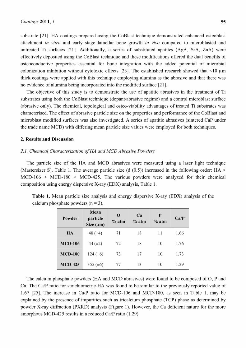
Relative crystallinity of each powder was investigated using PXRD, (Figure 1). HA was found to be
highly crystalline with well defined narrow peaks. The main characteristic peaks associated with HA
can be assigned to the 002, 102, 210, 211, 112, 300 and 202 reflections corresponding to 25.9°, 28.1°,
28.9°, 31.9°, 32.2°, 33.1° and 34.1°, as previously reported [25]. The resulting PXRD patterns for the
MCD apatite series indicate a lower crystallinity relative to the HA powder. The small peak present at
31° and 34.4° was attributed to the TCP phase [26]. Also in the MCD-425 pattern, peaks are poorly
resolved with low intensity relative to the other apatites, demonstrating the more amorphous nature of
this material [25,26].
Figure 1. Powder X-ray diffraction (PXRD) spectra of the powders (■ denotes HA peaks
and * represents tricalcium phosphate (TCP)).
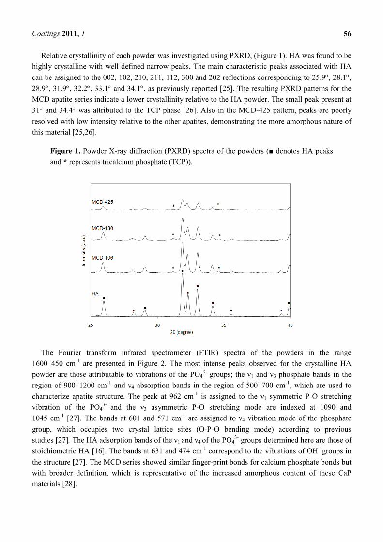
The Fourier transform infrared spectrometer (FTIR) spectra of the powders in the range
1600–450 cm-1 are presented in Figure 2. The most intense peaks observed for the crystalline HA
powder are those attributable to vibrations of the PO43- groups; the ν1 and ν3 phosphate bands in the
region of 900–1200 cm-1 and ν4 absorption bands in the region of 500–700 cm
-1, which are used to
characterize apatite structure. The peak at 962 cm-1 is assigned to the ν1 symmetric P-O stretching
vibration of the PO43-
and the ν3 asymmetric P-O stretching mode are indexed at 1090 and
1045 cm-1 [27]. The bands at 601 and 571 cm
-1 are assigned to ν4 vibration mode of the phosphate
group, which occupies two crystal lattice sites (O-P-O bending mode) according to previous
studies [27]. The HA adsorption bands of the ν1 and ν4 of the PO43- groups determined here are those of
stoichiometric HA [16]. The bands at 631 and 474 cm-1 correspond to the vibrations of OH
- groups in
the structure [27]. The MCD series showed similar finger-print bonds for calcium phosphate bonds but
with broader definition, which is representative of the increased amorphous content of these CaP
materials [28].
Coatings 2011, 1
57
Figure 2. Fourier transform infrared spectrometer (FTIR) spectra of the various apatite powders.
2.2. Characterization of the Modified Titanium Substrates
Titanium (V) was used as the base substrate and the untreated Ti surface was determined to contain
23% O and 77% Ti using EDX analysis. The chemical composition of the microblast surfaces
(abrasive blast only, no dopant) are presented in Table 2 and were analyzed for O, Ca, P and Ti only.
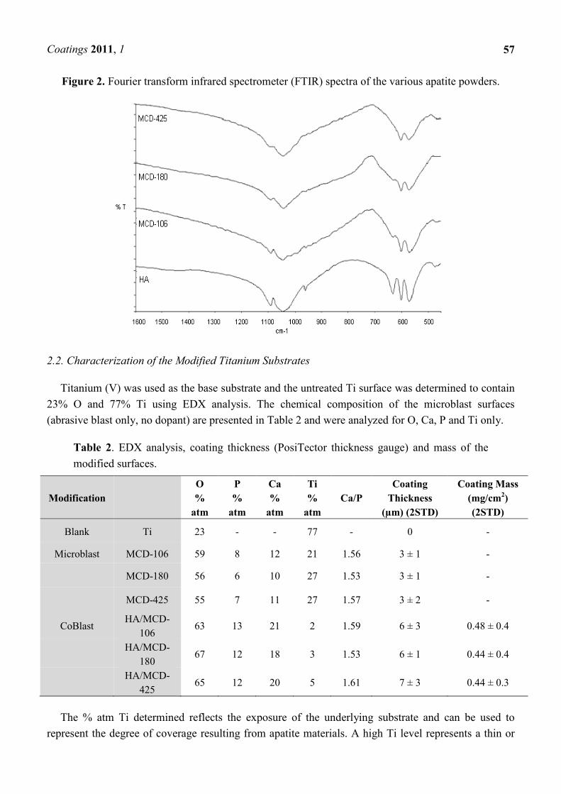
Table 2. EDX analysis, coating thickness (PosiTector thickness gauge) and mass of the
modified surfaces.
Modification
O
%
atm
P
%
atm
Ca
%
atm
Ti
%
atm
Ca/P
Coating
Thickness
(µm) (2STD)
Coating Mass
(mg/cm2)
(2STD)
Blank Ti 23 - - 77 - 0 -
Microblast MCD-106 59 8 12 21 1.56 3 ± 1 -
MCD-180 56 6 10 27 1.53 3 ± 1 -
MCD-425 55 7 11 27 1.57 3 ± 2 -
CoBlast HA/MCD-
106 63 13 21 2 1.59 6 ± 3 0.48 ± 0.4
HA/MCD-
180 67 12 18 3 1.53 6 ± 1 0.44 ± 0.4
HA/MCD-
425 65 12 20 5 1.61 7 ± 3 0.44 ± 0.3
The % atm Ti determined reflects the exposure of the underlying substrate and can be used to
represent the degree of coverage resulting from apatite materials. A high Ti level represents a thin or

Coatings 2011, 1
58
patchy coating and conversely, a low Ti concentration signifies a thick coating. The EDX results
reveal that after a wash treatment, the MCD microblasted surfaces show a reduction in Ti
concentrations to 21–27% atm, compared to 77% atm for the Ti substrate. Also the presence of Ca and
P which are the main constituents of the MCD abrasive is noted on the surface. This illustrates that a
thin coating of Ca/P material has been blasted onto the surface and successfully deposited as a stable
layer onto the Ti surface. The Ca/P values obtained for the microblasted samples treated with the MCD
series of the abrasives ranged between 1.53 and 1.57 which is consistent with similar grit blasted
studies [18].
The chemical composition of the CoBlast surfaces (blasting with both abrasive and dopant) on Ti
are also presented in Table 2. For CoBlast coatings, the levels of O, P, Ca and Ti obtained were
determined to be in the range of 63–67%, 12–13%, 18–21% and 2–5% atm, respectively. The reduced
level of Ti detected in these samples, compared to the Ti substrate and microblasted surfaces, is
indicative of a high degree of coating coverage across all CoBlast samples. The Ca/P values were
found to display a ratio of between 1.53 and 1.61, which are relatively close to the value for
stoichiometric HA [25]. The % atm Ti, determined using EDX analysis, was observed to increase as
the MCD series particle size order increased, indicating a decrease in the thickness of the deposited
layer, as outlined in Table 2. This suggests that the smaller the particle size of the MCD abrasive the
more HA was deposited, although the coating thickness determined using the PosiTector thickness
gauge, and the coating mass values were found to be similar. The coating thickness of all the CoBlast
samples was <10 microns which is in agreement with a previous study which used Al2O3 as the
abrasive [22].
The scanning electron microscopy (SEM) image of the untreated Ti substrate can be seen in
Figure 3a which has similar topography to that observed in a previous study [6]. This image reveals a
very smooth surface and the morphology of a machined metal. The SEM images of the microblast
MCD-106 surfaces, as well as the corresponding CoBlast HA/MCD-106 surfaces, are presented in
Figure 3b and c respectively. (More images can be seen as supporting information)
As expected, the microblast process was observed to roughen the untreated Ti surface. Examination
of the CoBlast surfaces suggests that the co-introduction of the HA with the abrasive appears to have
in-filled some of the surface features that are evident on the microblast sample (Figure 3b). The
CoBlast process results in a roughened, highly regular and uniform surface which is consistent with
other calcium phosphate coatings produced using simple grit blasting technologies [6,19,20]. It was
noted that as the particle size (d90) of the MCD abrasive increased from 106 to 425 microns, the
texture (presence of surface features) and the apparent roughness of the resultant surfaces was also
observed to increase for both the microblast and CoBlast treatments and this was confirmed by surface
roughness measurements.

Coatings 2011, 1
Figure 3. Scanning electron microscopy (
(a) titanium; (b) microblast MCD 106;
(a)
(b)
Scanning electron microscopy (SEM) images (×1000 magnification) of
) microblast MCD 106; (c) CoBlast HA/MCD-106.
59
1000 magnification) of

Coatings 2011, 1
(c)
The surface roughness was measured using a stylus method and the results obtained are given in
Figure 4. The arithmetical mean roughness
tended to increase as the particle size of the
between the surface treated and the untreated Ti.
(0.4 µm) increased to 0.5, 0.8, 1.4 µm when MCD
employed for microblast treatments. It has been previously reported that an increase in surface
roughness was observed with the introduction of HA with the Al
technique and the same trend was observed here
abrasives [21]. Statistically significant differences were noted between the roughness of the microblast
and CoBlast samples prepared using MCD
was used. A large standard error was observed for the MCD
possibly be a feature of the crude microblast process. As per the microblast samples, the level of
roughness and irregularity of the Co
particle size, with a significant increase in surface roughness produced by larger abrasive particle
sizes (p < 0.05).
Implants with rougher surfaces result in a higher removal torque force and
osteointegration when compared to those with smoother surfaces [5]. As seen in this study,
microblasting offers increased roughening of machined Ti substrates as expected and the use of MCD
abrasives results in the deposition of a thi
roughness of the microblast process can be tuned between 0.5
of the apatite abrasive employed. This is consistent with previously studies [5,6]. Unfortunately, the
coatings deposited using this microblast process have demonstrated poor adhesion to the metal and
have not been widely employed as final surface treatments for this reason [21].
Figure 3. Cont.
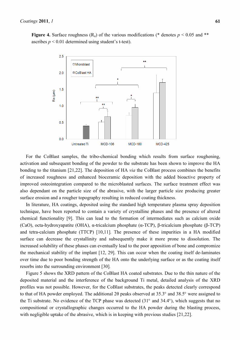
The surface roughness was measured using a stylus method and the results obtained are given in
The arithmetical mean roughness (Ra) was used as a measure of the surface roughness
the particle size of the abrasive increased. Significant differences were observed
between the surface treated and the untreated Ti. The average surface roughness of the blank titanium
to 0.5, 0.8, 1.4 µm when MCD-106, MCD-180 and MCD
employed for microblast treatments. It has been previously reported that an increase in surface
roughness was observed with the introduction of HA with the Al2O3 abrasive using the CoBlast
technique and the same trend was observed here on the introduction of
Statistically significant differences were noted between the roughness of the microblast
and CoBlast samples prepared using MCD-106 and MCD-180 abrasives, though not when MCD
was used. A large standard error was observed for the MCD-425 microblasted surface, which may
possibly be a feature of the crude microblast process. As per the microblast samples, the level of
roughness and irregularity of the CoBlast surface was visibly altered by changes in the abrasive
particle size, with a significant increase in surface roughness produced by larger abrasive particle
Implants with rougher surfaces result in a higher removal torque force and
osteointegration when compared to those with smoother surfaces [5]. As seen in this study,
microblasting offers increased roughening of machined Ti substrates as expected and the use of MCD
abrasives results in the deposition of a thin coating layer of calcium phosphate. Furthermore, the
roughness of the microblast process can be tuned between 0.5–1.4 µm depending on the particle size
of the apatite abrasive employed. This is consistent with previously studies [5,6]. Unfortunately, the
coatings deposited using this microblast process have demonstrated poor adhesion to the metal and
have not been widely employed as final surface treatments for this reason [21].
60
The surface roughness was measured using a stylus method and the results obtained are given in
) was used as a measure of the surface roughness, which
Significant differences were observed
surface roughness of the blank titanium
180 and MCD-425, respectively were
employed for microblast treatments. It has been previously reported that an increase in surface
abrasive using the CoBlast
on the introduction of HA with the MCD
Statistically significant differences were noted between the roughness of the microblast
s, though not when MCD-425
425 microblasted surface, which may
possibly be a feature of the crude microblast process. As per the microblast samples, the level of
Blast surface was visibly altered by changes in the abrasive
particle size, with a significant increase in surface roughness produced by larger abrasive particle
Implants with rougher surfaces result in a higher removal torque force and demonstrate excellent
osteointegration when compared to those with smoother surfaces [5]. As seen in this study,
microblasting offers increased roughening of machined Ti substrates as expected and the use of MCD
n coating layer of calcium phosphate. Furthermore, the
1.4 µm depending on the particle size
of the apatite abrasive employed. This is consistent with previously studies [5,6]. Unfortunately, the
coatings deposited using this microblast process have demonstrated poor adhesion to the metal and
Coatings 2011, 1
61
Figure 4. Surface roughness (Ra) of the various modifications (* denotes p < 0.05 and **
ascribes p < 0.01 determined using student’s t-test).
For the CoBlast samples, the tribo-chemical bonding which results from surface roughening,
activation and subsequent bonding of the powder to the substrate has been shown to improve the HA
bonding to the titanium [21,22]. The deposition of HA via the CoBlast process combines the benefits
of increased roughness and enhanced bioceramic deposition with the added bioactive property of
improved osteointegration compared to the microblasted surfaces. The surface treatment effect was
also dependant on the particle size of the abrasive, with the larger particle size producing greater
surface erosion and a rougher topography resulting in reduced coating thickness.
In literature, HA coatings, deposited using the standard high temperature plasma spray deposition
technique, have been reported to contain a variety of crystalline phases and the presence of altered
chemical functionality [9]. This can lead to the formation of intermediates such as calcium oxide
(CaO), octa-hydroxyapatite (OHA), α-tricalcium phosphate (α-TCP), β-tricalcium phosphate (β-TCP)
and tetra-calcium phosphate (TTCP) [10,11]. The presence of these impurities in a HA modified
surface can decrease the crystallinity and subsequently make it more prone to dissolution. The
increased solubility of these phases can eventually lead to the poor apposition of bone and compromize
the mechanical stability of the implant [12, 29]. This can occur when the coating itself de-laminates
over time due to poor bonding strength of the HA onto the underlying surface or as the coating itself
resorbs into the surrounding environment [30].
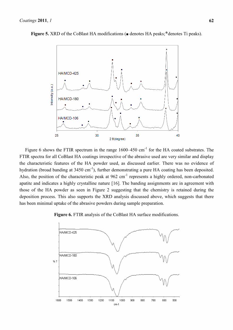
Figure 5 shows the XRD pattern of the CoBlast HA coated substrates. Due to the thin nature of the
deposited material and the interference of the background Ti metal, detailed analysis of the XRD
profiles was not possible. However, for the CoBlast substrates, the peaks detected clearly correspond
to that of HA powder employed. The additional 2θ peaks observed at 35.3° and 38.5° were assigned to
the Ti substrate. No evidence of the TCP phase was detected (31° and 34.4°), which suggests that no
compositional or crystallographic changes occurred to the HA powder during the blasting process,
with negligible uptake of the abrasive, which is in keeping with previous studies [21,22].
Coatings 2011, 1
Figure 5. XRD of the CoBlast
Figure 6 shows the FTIR spectrum in the range 1600
FTIR spectra for all CoBlast HA coatings irrespective of the abrasive used are very similar and display
the characteristic features of the HA powder used, as discussed earlier. There was no evidence of
hydration (broad banding at 3450 cm
Also, the position of the characteristic peak at 962 cm
apatite and indicates a highly crystalline nature [16]. The banding assignments are in agreement with
those of the HA powder as seen in
deposition process. This also supports the
has been minimal uptake of the abrasive powders during sample preparation.
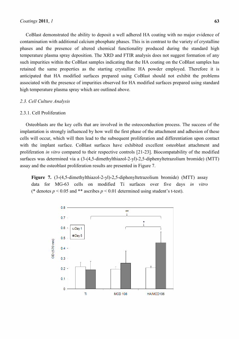
Figure 6. FTIR analysis of the
CoBlast HA modifications (■ denotes HA peaks;*denotes Ti peaks)
6 shows the FTIR spectrum in the range 1600–450 cm-1 for the HA coated substrates. The
HA coatings irrespective of the abrasive used are very similar and display
the characteristic features of the HA powder used, as discussed earlier. There was no evidence of
hydration (broad banding at 3450 cm-1), further demonstrating a pure HA coating ha
Also, the position of the characteristic peak at 962 cm-1 represents a highly ordered, non
apatite and indicates a highly crystalline nature [16]. The banding assignments are in agreement with
those of the HA powder as seen in Figure 2 suggesting that the chemistry is retained during the
This also supports the XRD analysis discussed above, which
has been minimal uptake of the abrasive powders during sample preparation.
FTIR analysis of the CoBlast HA surface modifications.
62
denotes Ti peaks).
for the HA coated substrates. The
HA coatings irrespective of the abrasive used are very similar and display
the characteristic features of the HA powder used, as discussed earlier. There was no evidence of
), further demonstrating a pure HA coating has been deposited.
represents a highly ordered, non-carbonated
apatite and indicates a highly crystalline nature [16]. The banding assignments are in agreement with
suggesting that the chemistry is retained during the
which suggests that there
modifications.
Coatings 2011, 1
63
CoBlast demonstrated the ability to deposit a well adhered HA coating with no major evidence of
contamination with additional calcium phosphate phases. This is in contrast to the variety of crystalline
phases and the presence of altered chemical functionality produced during the standard high
temperature plasma spray deposition. The XRD and FTIR analysis does not suggest formation of any
such impurities within the CoBlast samples indicating that the HA coating on the CoBlast samples has
retained the same properties as the starting crystalline HA powder employed. Therefore it is
anticipated that HA modified surfaces prepared using CoBlast should not exhibit the problems
associated with the presence of impurities observed for HA modified surfaces prepared using standard
high temperature plasma spray which are outlined above.
2.3. Cell Culture Analysis
2.3.1. Cell Proliferation
Osteoblasts are the key cells that are involved in the osteoconduction process. The success of the
implantation is strongly influenced by how well the first phase of the attachment and adhesion of these
cells will occur, which will then lead to the subsequent proliferation and differentiation upon contact
with the implant surface. CoBlast surfaces have exhibited excellent osteoblast attachment and
proliferation in vitro compared to their respective controls [21-23]. Biocompatability of the modified
surfaces was determined via a (3-(4,5-dimethylthiazol-2-yl)-2,5-diphenyltetrazolium bromide) (MTT)
assay and the osteoblast proliferation results are presented in Figure 7.
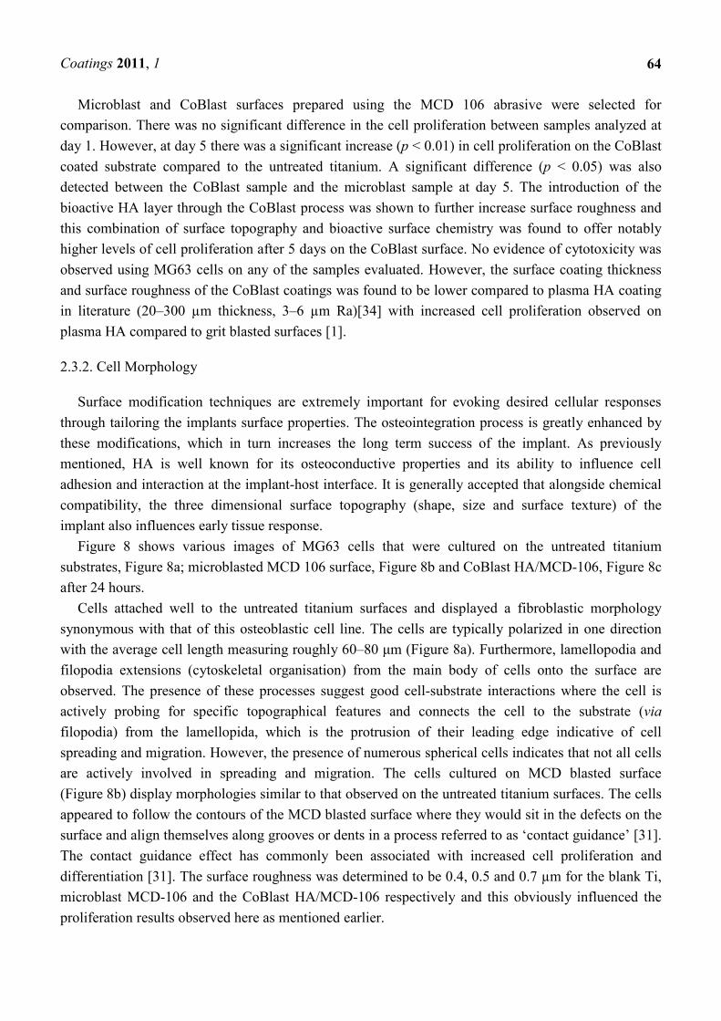
Figure 7. (3-(4,5-dimethylthiazol-2-yl)-2,5-diphenyltetrazolium bromide) (MTT) assay
data for MG-63 cells on modified Ti surfaces over five days in vitro
(* denotes p < 0.05 and ** ascribes p < 0.01 determined using student’s t-test).
Coatings 2011, 1
64
Microblast and CoBlast surfaces prepared using the MCD 106 abrasive were selected for
comparison. There was no significant difference in the cell proliferation between samples analyzed at
day 1. However, at day 5 there was a significant increase (p < 0.01) in cell proliferation on the CoBlast
coated substrate compared to the untreated titanium. A significant difference (p < 0.05) was also
detected between the CoBlast sample and the microblast sample at day 5. The introduction of the
bioactive HA layer through the CoBlast process was shown to further increase surface roughness and
this combination of surface topography and bioactive surface chemistry was found to offer notably
higher levels of cell proliferation after 5 days on the CoBlast surface. No evidence of cytotoxicity was
observed using MG63 cells on any of the samples evaluated. However, the surface coating thickness
and surface roughness of the CoBlast coatings was found to be lower compared to plasma HA coating
in literature (20–300 µm thickness, 3–6 µm Ra)[34] with increased cell proliferation observed on
plasma HA compared to grit blasted surfaces [1].
2.3.2. Cell Morphology
Surface modification techniques are extremely important for evoking desired cellular responses
through tailoring the implants surface properties. The osteointegration process is greatly enhanced by
these modifications, which in turn increases the long term success of the implant. As previously
mentioned, HA is well known for its osteoconductive properties and its ability to influence cell
adhesion and interaction at the implant-host interface. It is generally accepted that alongside chemical
compatibility, the three dimensional surface topography (shape, size and surface texture) of the
implant also influences early tissue response.
Figure 8 shows various images of MG63 cells that were cultured on the untreated titanium
substrates, Figure 8a; microblasted MCD 106 surface, Figure 8b and CoBlast HA/MCD-106, Figure 8c
after 24 hours.
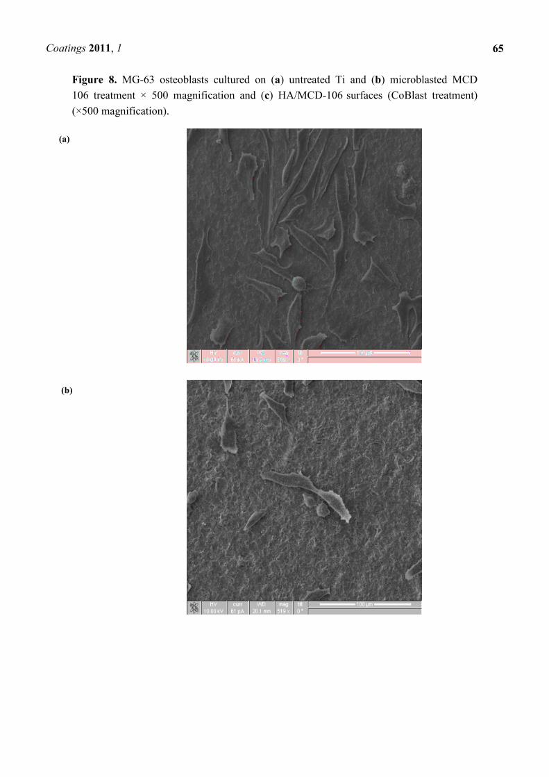
Cells attached well to the untreated titanium surfaces and displayed a fibroblastic morphology
synonymous with that of this osteoblastic cell line. The cells are typically polarized in one direction
with the average cell length measuring roughly 60–80 µm (Figure 8a). Furthermore, lamellopodia and
filopodia extensions (cytoskeletal organisation) from the main body of cells onto the surface are
observed. The presence of these processes suggest good cell-substrate interactions where the cell is
actively probing for specific topographical features and connects the cell to the substrate (via
filopodia) from the lamellopida, which is the protrusion of their leading edge indicative of cell
spreading and migration. However, the presence of numerous spherical cells indicates that not all cells
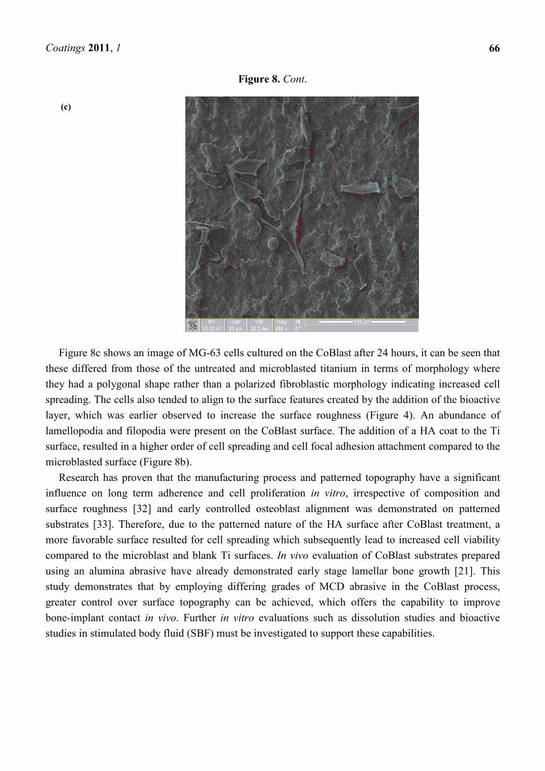
are actively involved in spreading and migration. The cells cultured on MCD blasted surface
(Figure 8b) display morphologies similar to that observed on the untreated titanium surfaces. The cells
appeared to follow the contours of the MCD blasted surface where they would sit in the defects on the
surface and align themselves along grooves or dents in a process referred to as ‘contact guidance’ [31].
The contact guidance effect has commonly been associated with increased cell proliferation and
differentiation [31]. The surface roughness was determined to be 0.4, 0.5 and 0.7 µm for the blank Ti,
microblast MCD-106 and the CoBlast HA/MCD-106 respectively and this obviously influenced the
proliferation results observed here as mentioned earlier.
Coatings 2011, 1
Figure 8. MG-63 osteoblasts cultured on
106 treatment × 500 magnification and
(×500 magnification).
(a)
(b)
63 osteoblasts cultured on (a) untreated Ti and (b) microblasted MCD
500 magnification and (c) HA/MCD-106 surfaces (CoBlast treatment)
65
) microblasted MCD
(CoBlast treatment)
Coatings 2011, 1
(c)
Figure 8c shows an image of MG
these differed from those of the untreated and microblasted
they had a polygonal shape rather than a polari
spreading. The cells also tended to align to the surface features created by the addition of the bioactive
layer, which was earlier observed to increase the surface roughness (
lamellopodia and filopodia were present on the CoBlast surface. The addition of a HA coat to the Ti
surface, resulted in a higher order of
microblasted surface (Figure 8b).
Research has proven that the manufacturing process and patterned topography have a significant
influence on long term adherence and cell proliferation
surface roughness [32] and early controlled osteoblast alignment was demonstrated on patterned
substrates [33]. Therefore, due to the patterned nature of the HA surface after CoBlast treatment, a
more favorable surface resulted for cell spreading w
compared to the microblast and blank Ti surfaces.
using an alumina abrasive have already demonstrated early stage lamellar bone growth [21]. This
study demonstrates that by employing differing grades of MCD abrasive in the CoBlast process,
greater control over surface topography can be achieved, which offers the capability to improve
bone-implant contact in vivo. Further
studies in stimulated body fluid (SBF) must be investigated to support these capabilities.
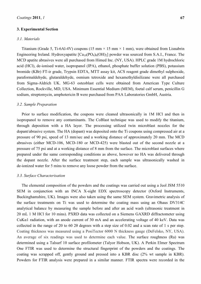
Figure 8. Cont.
8c shows an image of MG-63 cells cultured on the CoBlast after 24 hours, it can be seen that
these differed from those of the untreated and microblasted titanium in terms of morphology where
they had a polygonal shape rather than a polarized fibroblastic morphology indicating increased cell
spreading. The cells also tended to align to the surface features created by the addition of the bioactive
ich was earlier observed to increase the surface roughness (Figure
lamellopodia and filopodia were present on the CoBlast surface. The addition of a HA coat to the Ti
higher order of cell spreading and cell focal adhesion attachment
Research has proven that the manufacturing process and patterned topography have a significant
influence on long term adherence and cell proliferation in vitro, irrespective of composit
surface roughness [32] and early controlled osteoblast alignment was demonstrated on patterned
[33]. Therefore, due to the patterned nature of the HA surface after CoBlast treatment, a
more favorable surface resulted for cell spreading which subsequently lead to increased cell viability
compared to the microblast and blank Ti surfaces. In vivo evaluation of CoBlast substrates
alumina abrasive have already demonstrated early stage lamellar bone growth [21]. This
onstrates that by employing differing grades of MCD abrasive in the CoBlast process,
surface topography can be achieved, which offers the capability to improve
. Further in vitro evaluations such as dissolution studies and bioactive
studies in stimulated body fluid (SBF) must be investigated to support these capabilities.
66
63 cells cultured on the CoBlast after 24 hours, it can be seen that
titanium in terms of morphology where
ed fibroblastic morphology indicating increased cell
spreading. The cells also tended to align to the surface features created by the addition of the bioactive
Figure 4). An abundance of
lamellopodia and filopodia were present on the CoBlast surface. The addition of a HA coat to the Ti
adhesion attachment compared to the
Research has proven that the manufacturing process and patterned topography have a significant
, irrespective of composition and
surface roughness [32] and early controlled osteoblast alignment was demonstrated on patterned
[33]. Therefore, due to the patterned nature of the HA surface after CoBlast treatment, a
hich subsequently lead to increased cell viability
evaluation of CoBlast substrates prepared
alumina abrasive have already demonstrated early stage lamellar bone growth [21]. This
onstrates that by employing differing grades of MCD abrasive in the CoBlast process,
surface topography can be achieved, which offers the capability to improve
tion studies and bioactive
studies in stimulated body fluid (SBF) must be investigated to support these capabilities.
Coatings 2011, 1
67
3. Experimental Section
3.1. Materials
Titanium (Grade 5, Ti-6Al-4V) coupons (15 mm × 15 mm × 1 mm), were obtained from Lisnabrin
Engineering Ireland. Hydroxyapatite [Ca10(PO4)6(OH)2] powder was sourced from S.A.I., France. The
MCD apatite abrasives were all purchased from Himed Inc. (NY, USA). HPLC grade 1M hydrochloric
acid (HCl), de-ionised water, isopropanol (IPA), ethanol, phosphate buffer solution (PBS), potassium
bromide (KBr) FT-ir grade, Trypsin EDTA, MTT assay kit, ACS reagent grade dimethyl sulphoxide,
paraformaldehyde, glutaraldehyde, osmium tetroxide and hexamethyldisilizane were all purchased
from Sigma-Aldrich UK. MG-63 osteoblast cells were obtained from American Type Culture
Collection, Rockville, MD, USA. Minimum Essential Medium (MEM), foetal calf serum, penicillin G
sodium, streptomycin, amphotericin B were purchased from PAA Laboratories GmbH, Austria.
3.2. Sample Preparation
Prior to surface modification, the coupons were cleaned ultrasonically in 1M HCl and then in
isopropanol to remove any contaminants. The CoBlast technique was used to modify the titanium,
through deposition with a HA layer. The processing utilized twin microblast nozzles for the
dopant/abrasive system. The HA (dopant) was deposited onto the Ti coupons using compressed air at a
pressure of 90 psi, speed of 13 mm/sec and a working distance of approximately 20 mm. The MCD
abrasives (either MCD-106, MCD-180 or MCD-425) were blasted out of the second nozzle at a
pressure of 75 psi and at a working distance of 8 mm from the surface. The microblast surfaces where
prepared under the same corresponding conditions as above, however no HA was delivered through
the dopant nozzle. After the surface treatment step, each sample was ultrasonically washed in
de-ionized water for 5 mins to remove any loose powder from the surface.
3.3. Surface Characterisation
The elemental composition of the powders and the coatings was carried out using a Jeol JSM 5510
SEM in conjunction with an INCA X-sight EDX spectroscopy detector (Oxford Instruments,
Buckinghamshire, UK). Images were also taken using the same SEM system. Gravimetric analysis of
the surface treatments on Ti was used to determine the coating mass using an Ohaus DV314C
analytical balance by measuring the sample before and after an acid wash (ultrasonic treatment in
20 mL 1 M HCl for 10 mins). PXRD data was collected on a Siemens GAXRD diffractometer using
CuKα1 radiation, with an anode current of 30 mA and an accelerating voltage of 40 keV. Data was
collected in the range of 20 to 60 2θ degrees with a step size of 0.02 and a scan rate of 1 s per step.
Coating thickness was measured using a PosiTector 6000 N thickness gauge (DeFelsko, NY, USA).
An average of six readings was used to determine each value. The surface roughness (Ra) was
determined using a Talsurf 10 surface profilometer (Talyor Hobson, UK). A Perkin Elmer Spectrum
One FTIR was used to determine the structural fingerprint of the powders and the coatings. The
coating was scrapped off, gently ground and pressed into a KBR disc (2% wt sample in KBR).
Powders for FTIR analysis were prepared in a similar manner. FTIR spectra were recorded in the
Coatings 2011, 1
68
1600–400-cm-1 range, with 4 cm
-1 resolution using 20 scans and background subtraction. The spectra
gave approximately 70–90% transmittance however the results are presented in an overlay fashion.
3.4. In Vitro Cell Culture
Sample modifications including CoBlast HA/MCD-106 and microblast MCD-106 and the blank Ti
were evaluated for osteoconductivity and cytotoxicity using cell culture tests. Prior to cell culture
analysis, each sample set was steam autoclaved at 121 °C for 20 minutes. MG-63 cells were used to
assess cell proliferation. Cells were cultured in the MEM media supplemented with 10% foetal calf
serum and antibiotic/antimycotic (penicillin G sodium 100 U/mL, streptomycin 100 µg/mL,
amphotericin B 0.25 µg/mL) in 75 cm3 tissue culture flasks. Cells were maintained in a humidified
atmosphere with 5% CO2 at 37 °C and were sub-cultured when they reached confluence using 0.25%
Trypsin EDTA solution to provide adequate numbers of cells for the various in vitro culture
studies undertaken.
3.4.1. Cell Proliferation
MG-63 cell attachment to the various treated and untreated Ti substrates was determined after 4
hours in culture using a commercial MTT assay and employing a modified Mosmann method [24].
Cells were seeded onto the samples at a concentration of 1x10⁵ cells/cm
2and allowed to adhere during
incubation at 37 °C in 5% CO2 for 4 hrs. The MTT assay reagent was prepared as a 5 mg/mL stock
solution in PBS, sterilized by filtration, and stored in the dark. An aliquot of the MTT stock solution
(10% of total volume) was added to each well of a six well plate containing the samples (n = 4 for each
sample type). After 3 hrs incubation at 37 °C in 5% CO2, 200 µl of dimethyl sulfoxide was added to
dissolve the formazan crystals. The solution was agitated for 15 min on a shaker to ensure adequate
dissolution. The optical density of the formazan solutions was read by spectrophotometry using an
ELISA plate reader (Tecan Sunrise, Tecan Austria) at 570 nm with the background absorbance value
measured at 650 nm. The absorbance values recorded were determined to be proportional to the
number of cells attached to the surface in each case. All data reported are expressed as mean ±
standard deviation.
3.4.2. Cell Morphology
MG-63 cells were seeded onto each of the above substrates at a cell density of 5 × 10⁵ cells/cm
2 in
6-well plates and were incubated for 24 hours. After cell culture, the samples were gently rinsed with
PBS to remove any unattached cells and fixed in a modified Karnovsky’s Fixative (2%
paraformaldehyde/ 2% glutaraldehyde in PBS) for 1 hour. The samples were then rinsed in PBS and
post-fixed in 1% osmium tetroxide and rinsed three times with PBS. The specimens were dehydrated
by rinsing in an alcohol series (20, 30, 50, 70, 80, 90 and 95% ethanol), and finally rinsing 3 times in
100% ethanol. The samples were then were chemically dried in hexamethyldisilizane (HMDS)
overnight. A 50 nm layer of gold-palladium was deposited onto the substrates using a Polaron E5000
SEM Sputter Coating Unit. The sputtering conditions used a set voltage of 1.4 kV, with a plasma
current of 18 mA (argon gas), a deposition time of 2 minutes at a vacuum pressure of 0.05 Torr. The

Coatings 2011, 1
69
samples were then analyzed using the Jeol JSM 5510 SEM and subsequently using a FEI Quanta 200
Focused Ion Beam and SEM in backscatter electron mode.
4. Conclusions
Detailed surface studies have shown that the combination of an apatite abrasive and a HA dopant in
the CoBlast process produces surfaces with a combination of optimized apatite chemistry and
controlled surface structure. The CoBlast process has the ability to retain the chemistry of the starting
HA material. This offers advantages over conventional high temperature plasma processing which
alters the HA material from its desired chemical, structural and dissolution requirements for its use as
an in vivo implant material. The study also shows that employing MCD abrasives offer an alternative
to alumina for deposition using CoBlast process. In vitro studies clearly show that increased roughness
of treated surfaces favors enhanced cell proliferation and the CoBlast process offers the ability to tailor
the surface texture to produce an optimized surface for osteointegration of a HA modified implant.
Enhanced cell proliferation was observed for CoBlast modified surfaces compared to the microblasted
surface. The ability of the CoBlast technology to offer diversity in modifying surface topography is
clearly shown in this and previous studies and represents foundation work, which supported by
bioactivity studies and in vivo trials, offers exciting new prospects in tailoring the properties of medical
devices for applications ranging from dental to orthopedic settings.
Acknowledgments
The authors would like to acknowledge EnBio for supplying the CoBlast samples for this study.
References and Notes
1. Borsari, V.; Giavaresi, G.; Fini, M.; Torricelli, P.; Salito, A.; Chiesa, R.; Chiusoli, L.; Volpert, A.;
Rimondini, L.; Giardino, R. Physical characterization of different-roughness titanium surfaces,
with and without hydroxyapatite coating and their effect on human osteoblast-like cells.
J. Biomed. Mater. Res. Part B 2005, 75B, 359-368.
2. Stoch, A.; Jastrze, B.W.; Dlugon, E.; Lejda, W.; Trybalska, B.; Stoch, G.J.; Adamczyk, A. Sol-gel
derived hydroxyapatite coatings on titanium and its alloy Ti6Al4V. J. Mol. Struct. 2005, 744,
633-640.
3. Oh, I.H.; Nomura, N.; Chiba, A. Microstructures and bond strengths of plasma-sprayed
hydroxyapatite coatings on porous titanium substrates. J. Mater. Sci. Mater. Med. 2005, 16,
635-640.
4. Lu, Y.P.; Li, M.S.; Li, S.T.; Wang, Z.G.; Zhu, R.F. Plasma-sprayed hydroxyapatite + titania
composite bond coat for hydroxyapatite coating on titanium substrate. Biomaterials 2004, 25,
4393-4403.
5. Wennerberg, A.; Ektessabi, A.; Albrektsson, T.; Johansson, C.; Andersson, B.A. 1-year follow-up
of implants of differing surface roughness placed in rabbit bone. Inter. J. Oral Maxillofac.
Implants 1997, 12, 486-494.
Coatings 2011, 1
70
6. Abron, A.; Hopfensperger, M.; Thompson, J.; Cooper, L.F. Evaluation of a predictive model for
implant surface topography effects on early osseointegration in the rat tibia model. J. Prosthet.
Dent. 2001, 85, 40-46.
7. Nakada, H.; Sakae, T.; Legeros, R.Z.; Legeros, J.P.; Suwa, T.; Numata, Y.; Kobayashi, K. Early
tissue response to modified implant surfaces using back scattered imaging. Implant Dent. 2007,
16, 281-289
8. Gil, F.J.; Planell, J.A.; Padros, A.; Aparicio, C. The effect of shot blasting and heat treatment on
the fatigue behavior of titanium for dental implant applications. Dent. Mater. 2007, 23, 486-491.
9. Chen, J.; Wolke, J.G.C.; De Groot, K. Microstructure and crystallinity in hydroxyapatite coatings.
Biomaterials 1994, 15, 396-399.
10. Gross, K.A.; Berndt, C.C.; Herman, H. Amorphous phase formation in plasma-sprayed
hydroxyapatite coatings. J. Biomed. Mater. Res. 1998, 39, 407-414.
11. Gross, K.A.; Berndt, C.C. Thermal processing of hydroxyapatite for coating production.
J. Biomed. Mater. Res. 1998, 39,580-587.
12. Heimann, R.B.; Wirth, R. Formation and transformation of amorphous calcium phosphates on
titanium alloy surfaces during atmospheric plasma spraying and their subsequent in vitro
performance. Biomaterials 2006, 27, 823-831.
13. Weng, J.; Liu, Q.; Wolke, J.G.; Zhang, X.; De Groot, K. Formation and characteristics of the
apatite layer on plasma-sprayed hydroxyapatite coatings in simulated body fluid. Biomaterials
1997, 18, 1027-1035.
14. Li, H.; Li, Z.X.; Li, H.; Wu, Y.Z.; Wei, Q. Characterization of plasma sprayed
hydroxyapatite/ZrO2 graded coating. Mater. Design 2009, 30, 3920-3924.
15. Katto, M.; Kurosawa, K.; Yokotani, A.; Kubodera, S.; Kameyama, A.; Higashiguchi, T.;
Nakayama, T.; Tsukamoto, M. Poly-crystallized hydroxyapatite coating deposited by pulsed laser
deposition method at room temperature. Appl. Surf. Sci. 2005, 248,365-368.
16. Hong, Z.; Luan, L. Paik, S.E.; Deng, B.; Ellis, D.E.; Ketterson, J.B. Mello, A.; Eon, J.G., Terra,
J., Rossi, A. Crystalline hydroxyapatite thin films produced at room temperature—An opposing
radio frequency magnetron sputtering approach. Thin Solid Films 2007, 515, 6773-6780.
17. Stoch, A.; Brozek, A.; Kmita, G.; Stoch, J.; Jastrzebski, W.; Rakowska, A. Electrophoretic
coating of hydroxyapatite on titanium implants. J. Mole. Struct. 2001, 596, 191-200.
18. Mano, T.; Ueyama, Y.; Ishikawa, K.; Matsumura, T.; Suzuki, K. Initial tissue response to a
titanium implant coated with apatite at room temperature using a blast coating method.
Biomaterials 2002, 23, 1931-1926.
19. Gbureck, U.; Masten, A.; Probst, J.; Thull, R. Tribochemical structuring and coating of implant
metal surfaces with titanium oxide and hydroxyapatite layers. Mater. Sci. Eng. C 2003, 23,
461-465.
20. Ishikawa, K.; Miyamoto, Y.; Nagayama, M.; Asaoka, K. Blast coating method: New method of
coating titanium surface with hydroxyapatite at room temperature. J. Biomed. Mater. Res. 1997,
38,129-134.
21. O’Hare, P.; Meenan, B.J.B.; George, A.; Byrne, G.; Dowling, D.; Hunt, J.A. In vitro and in vivo
response of hydroxyapatite surfaces deposited via a novel co-incident microblasting technique for
improved orthopaedic implant performance. Biomaterials 2010, 31, 515-522.

Coatings 2011, 1
71
22. O’Neill, L.; O’Sullivan, C.; O’Hare, P.; Sexton, L.; Keady, F.; O’Donoghue, J. Deposition of
substituted apatites onto titanium surfaces using a novel blasting process. Surf. Coat. Technol.
2009, 204, 484-488.
23. O’Sullivan, C.; O’Hare, P.; O’Leary, N.D.; Crean, A.M.; Ryan, K.; Dobson, A.D.; O’Neill, L.D.
Deposition of substituted apatites with anticolonizing properties onto titanium surfaces using a
novel blasting process. J. Biomed. Mater. Res. Part B 2010, 95,141-149.
24. Mosmann, T. Rapid colorimetric assay for cellular growth and survival: Application to
proliferation and cytotoxicity assays. J. Immunol. Methods 1983, 65, 55-63.
25. Kim, T.N.; Feng, Q.L.; Kim, J.O.; Wu, J.; Wang, H.; Chen, G.C.; Cui, F.Z. Antimicrobial effects
of metal ions (Ag+, Cu
2+, Zn
2+) in hydroxyapatite. J. Mater. Sci.: Mater. Med. 1998, 9, 129-134.
26. Fathi, M.H.; Hanifi, A.; Mortazavi, V. Preparation and bioactivity evaluation of bone-like
hydroxyapatite nanopowder. J. Mater. Proc. Technol. 2008, 202,536-542.
27. Varma, H.K.; Babu, S.S. Synthesis of calcium phosphate bioceramics by citrate gel pyrolysis
method. Ceram. Int.2005, 31, 109-114.
28. Pleshko, N.; Boskey, A.; Mendelsohn, R. Novel infrared spectroscopic method for the
determination of crystallinity of hydroxyapatite minerals. Biophy. J.1991, 60,786-793.
29. Legeros, R.Z.; Daculsi, G.; Orly, I.; Gregoire, M. Substrate surface dissolution and interfacial
biological minerals. In The Bone-Biomaterial Interface; Davies, J.E., Ed.; University of Toronto:
Toronto, Canada, 1991; pp. 76-88.
30. Masmoudi, M.; Assoul, M.; Wery, M.; Abdelhedi, R.; El Halouani, F.; Monteil, G. Friction and
wear behaviour of cp Ti and Ti6Al4V following nitric acid passivation. Appl. Surf. Sci. 2006, 253,
237-2243.
31. Anselme, K. Osteoblast adhesion on biomaterials. Biomaterials 2000, 21, 667-681.
32. Bigerelle, M.; Anselme K. Statistical correlation between call adhesion and proliferation on
biocompatible metallic materials. J. Biomed. Mater. Res. A 2005, 75, 530-540.
33. Puckett, A.; Pareta, R.; Webster, T.J. Nano rough micron patterned titanium for directing
osteoblast morphology and adhesion. Inter. J. Nanomed. 2008, 2, 229-241.
34. Sun, L.; Berndt, C.C.; Gross, K.A.; Kucuk, A. Material fundamentals and clinical performance of
plasma-sprayed hydroxyapatite coatings: A review. J Biomed. Mate. Res. 2001, 58, 570-592.
© 2011 by the authors; licensee MDPI, Basel, Switzerland. This article is an open access article
distributed under the terms and conditions of the Creative Commons Attribution license
(http://creativecommons.org/licenses/by/3.0/).