a method for detection of mutations, using … · a method for detection of mutations, using...
TRANSCRIPT
A METHOD FOR DETECTION O F MUTATIONS, USING STREPTOMYCIN DEPENDENCE I N
ESCWERICHIA COLI * G. BERTAN11
Department of Genetics, Carnegie Institution of Washington, Cold Spring Harbor, N e w York
Received December 3, 1950
N studies of induced mutation it is particularly desirable to use well-con- I trolled methods for the detection of mutations. MULLER’S introduction of the ClB method for detecting lethal mutations in Drosophila first opened this field of study and much work has been done with this organism in spite of the laborious technical procedures required. Microorganisms offered a simpler material for this kind of work. DEMEREC’S studies ( DEMEREC, WALLACE, WITKIN and BERTANI 1949) of spontaneous and radiation-induced mutation in the streptomycin resistance system in Escherichia coli indicated that a suitable test material for induced mutation might be the back-mutation from streptomycin dependence to nondependence. The present paper is concerned with the development of such a test, based on plating streptomycin-dependent bacteria on agar containing no streptomycin. On this medium, nondependent cells (back-mutants) are able to form visible colonies; whereas the dependent cells divide a few times (residual growth), thus increasing the chances for back-mutation. If all or most back-mutations occur after plating, exposure to a mutagenic agent before plating can be expected to increase the number of back-mutants. We shall examine in this paper the factors that affect the re- sults obtained with this technique and that must be controlled in quantitative studies of induced mutation.
MATERIALS AND METHODS
Streptomycin-dependent bacterial strains were first described by MILLER and BOHNHOFF (1947). Dependent strains have since been isolated from a number of bacterial species (see WAKSMAN 1948, 1949).
In Escherichia coli, dependent mutants are found among the resistant mu- tants that can easily be obtained from sensitive strains by suitable selection techniques (NEWCOMBE and HAWIRKO 1949; DEMEREC et al. 1949). The strains used in our work were isolated from the sensitive strain B of E. coli; they will be referred to by the symbols B / S d or simply Sd (= streptomycin- dependent). Sd strains of different mutational origin will be distinguished by means of a number; most data concern Sd-4 (obtained from DEMEREC), al- though a few observations have also been made on Sd-26, Sd-3, and other
* This study was conducted in part under a grant from the AMERICAN CANCER SOCIETY,
1 Present address : Department of Bacteriology, University of Illinois, Urbana, Illinois. through the COMMITTEE ON GROWTH of the NATIONAL RESEARCH COUNCIL.
GENETICS 36: 598 November 1951

STREPTOMYCIN DEPENDENCE 599
dependent strains. Sensitive mutants derived by back-mutation from depen- dent strains will be called R (= reversion), to distinguish them from B, the original sensitive strain, to which they are phenotypically similar ; resistant, nondependent mutants derived from dependent strains will be designated K S . In some of the experiments two strains marked with mutations for phage re- sistance were used: B/Sd-4/1,3,4,5 and B/Sd-4/3,4. The first is resistant to phages T l , T3, T4, T 5 ; the second to T3 and T4.
The dependent strains were cultivated in nutrient broth containing strepto- mycin (usually 25 micrograms per milliliter). Colony counts (assays) were made on streptomycin agar (nutrient agar to which 50 pg of streptomycin per ml was added just before pouring). Streptomycin sulfate and dihydro- streptomycin sulfate were used ; no essential difference was observed.
The plate-" washing " technique described by BEALE (1948) was used to determine the total number of bacteria present on a plate after incubation; each plate was " washed " once with 10 ml of broth. On the average, 84 per- cent of the bacteria present on a plate can be recovered.
A nephelometer designed by UNDERWOOD and DOERMANN (1947) was used to measure the generation time of different strains in the exponential phase.
RESIDUAL GROWTH OF Sd-4 IN THE ABSENCE OF STREPTOMYCIN
General Properties of Sd-4. The generation time of Sd-4 in broth (aerated culture) containing 20 pg of streptomycin per ml, measured nephelometrically at 37"C, is approximately 34 minutes. The final titer reached by Sd-4 in broth does not change with concentrations of streptomycin between 1 and 100 p g per ml, and is usually between 1.5 and 3.9 x 1Og bacteria per ml. As the concentration increases beyond 100 pg per ml the final titer slowly decreases.
Residual growth in liquid cultures. Sd bacteria grown with streptomycin, washed, and transferred to fresh streptomycin-free medium, do not stop grow- ing at once, but undergo a certain number of divisions. If the inoculum is small (of the order of lo3 cells per ml) this residual growth is very slight. If the inoculum is of the order of 5 x lo7 bacteria per ml, full growth is obtained in 12 to 24 hours. Figure 1 shows an experiment of this kind. Parallel plat- ings on plain and on streptomycin agar at the end of the experiment showed that growth was due to dependent cells and not to resistant mutants that might have overgrown the Sd cells.
Residual growth is affected by the concentration of streptomycin in the culture from which the inoculum is taken: the higher this concentration, the greater the amount of residual growth (fig. 2 ) .
Residual growth on agar. Residual growth on agar has to be studied by different methods : microscopic count of bacteria per microcolony (which gives the total number of bacteria), and plate-washing technique (which gives the number of viable bacteria present on the plate). The two methods sup-
2 The word " viable " is used throughout this paper, with reference to streptomycin- dependent bacteria, to mean : " able to start dividing again when transferred to nutrient medium containing streptomycin."

600 G. BERTANI
TIME OF INCUBATION IN HOURS
FIGURE 1.--Multiplication of streptomycin-dependent E. coli in broth containing no streptomycin, followed both nephelometrically and by colony counts. Inoculum : 5.9 x 10⁷ per ml. Ordinate 1 represents the initial turbidity and the initial number of viable bacteria.
TIME OF INCUBATION IN HOURS
FIGURE 2.-Multiplication of streptomycin-dependent E. coli in broth containing no streptomycin. Heavy inocula, from cultures grown in 50 pg and 25 pg of streptomycin per ml, respectively.

STREPTOMYCIN DEPENDENCE 60 1
ply the same kinds of information, respectively, as the nephelometric tech- nique and the colony count in the case of liquid cultures. Figure 3 shows the extent of residual growth of dependent bacteria plated on plain nutrient agar, as observed by these two methods. A significant discrepancy is noted between the total counts and the numbers of viable cells.
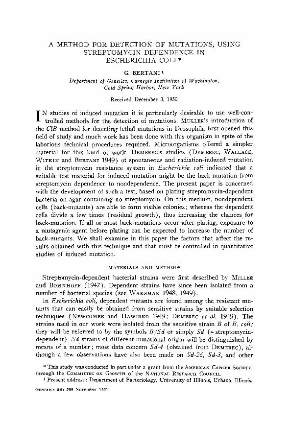
Figure 4 shows the effect of crowding. For initial concentrations higher than lo7 cells per plate, the larger the number of cells plated the greater is the extent of residual growth.
FIGURE 3.-Multiplication of streptomycin-dependent E. coli on agar containing no streptomycin. The curve through the solid dots represents the increase in average num- bers of bacteria per microcolony, as observed microscopically; each point is based on observation of 200 microcolonies. Inoculum from a culture with 25 pg of streptomycin per ml.
Inoculum as above. Data from five experiments. 1.6 to 3.8 X 10⁷ bacteria per plate i t time 0. The limits k 2 standard error are given. The circles give values obtained using an inoculum from a culture with 10 pg of streptomycin per ml. Data from two experiments. 1.8 X 10⁷ bacteria per plate at time 0.
The triangles give the corresponding values for viable bacteria.
Less extensive data are available concerning the effect of previous con- centration of streptomycin on residual growth on agar. Nevertheless, micro- scopical observations show that if cells are grown in 1 pg of streptomycin per ml and plated on plain agar, only a few are able to undergo even one cell division. Cells grown in 10 ccg of streptomycin undergo fewer divisions man cells grown in 25 pg (see fig. 3 ) .
Formation of “ snakes ” during residual growth. After 12 hours of incuba- tion the number of bacteria has reached a maximum (fig. 3). Thereafter very few cell divisions occur, but cell elongation and nuclear division continue, so that the cells gradually develop into long polynucleated filaments (snakes). The cytology of these forms has been studied by DELAPORTE (1949). Snakes
602 z 9 2 50 m
z fn 40
a a
eJ- - 0
5 - 3 0 - t i 4 -
1"0- 9'
w lo
d o
a z
$
G. BERTANI
-
-
. .<
- . ,'Qyo/o*. . : - I
FIGURE 4.-EA ect of crowding on the residual growth of streptomycin-dependent E. coli plated on agar. Inocula from cultures with 25 pg of streptomycin per ml. Every point represents the factor of increase in number of the cells originally plated, at the end of 12 hours' incubation, as determined by the plate-washing technique.
exhibit peculiar movements on the surface of agar, which makes it difficult to count the bacteria of a microcolony after the beginning of snake formation. It was necessary, therefore, to use a different technique for studying residual growth after more than 12 hours of incubation.
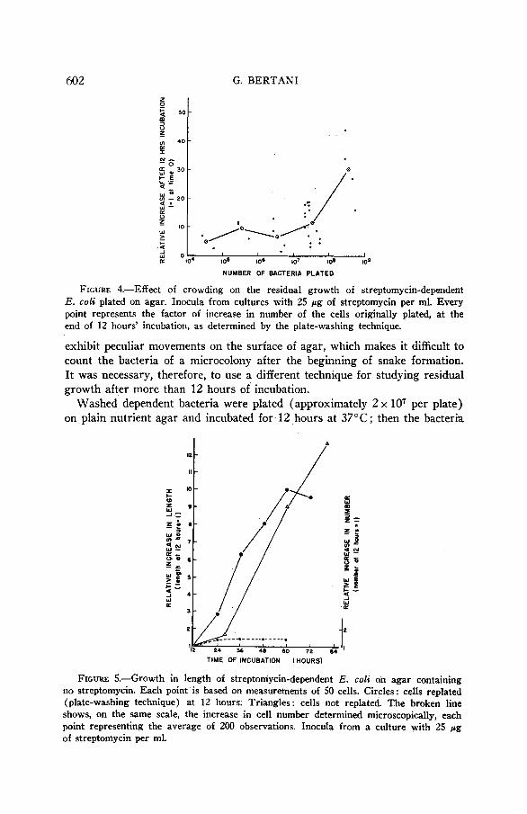
Washed dependent bacteria were plated (approximately 2 x lo7 per plate) on plain nutrient agar and incubated for 12 hours at 37°C; then the bacteria
TIME OF INCUBATION (HOURS)
FIGURE 5.-Growth in length of streptomycin-dependent E. coli on agar containing no streptomycin. Each point is based on measurements of 50 cells. Circles: cells replated (plate-washing technique) at 12 hours. Triangles : cells not replated. The broken line shows, on the same scale, the increase in cell number determined microscopically, each point representing the average of 200 observations. Inocula from a culture with 25 pg of streptomycin per ml.

STREPTOMYCIN DEPENDENCE 603 were transferred by the washing technique to new plain-agar plates. In this way the microcolonies present at 12 hours were broken up; the single bacteria could be examined microscopically for new cell divisions and easily measured with a camera lucida. Figure 5 shows that cell divisions are rare after the first 12 hours of residual growth, whereas cell length, which has remained normal during the first 12 hours, increases enormously. At the same time viability decreases ; more and more cells lose the ability to start dividing again when returned to streptomycin medium (fig. 6).
TIME OF INCUBATION IN HOURS
FIGURE 6.-Decrease of viability of " snakes " on agar containing no streptomycin, following the first 12 hours of residual growth. Two experiments. After 12 hours on plain agar, the bacteria were washed, replated on plain agar, incubated further for different periods of time, and then covered with a layer of streptomycin agar, which allowed development of colonies from the cells that were still viable.
BACK-MUTATION IN Sd-4 When streptomycin-dependent bacteria are plated on nutrient agar without
streptomycin, any nondependent mutants that may be present will divide and give origin to visible colonies. These nondependent mutants may be expected to include two types: sensitive to streptomycin (R) and resistant to strepto- mycin (RS). Both types were found. They were classified by picking the mutant colonies and streaking on plain nutrient agar and on streptomycin agar (50 or more pg of streptomycin per ml) ; only the RS types are able to grow on streptomycin agar. In many experiments, the colonies were first transferred to tubes containing 2 ml of plain nutrient broth, incubated for 48 hours, and then tested by streaking.
Contaminants might be scored as mutants. This would result in a negligible error when the number of mutants per plate was high, but in a serious error when the number was low. To reduce this source of error, the strain Sd- 4/1,3,4,5 was used in many experiments, and the mutant colonies were tested by cross-streaking against phages TZ and T3 (or T 4 ) . Those that were either not sensitive to T2 or not resistant to T3 (or T 4 ) were discarded as con-

604 G. BERTANI
TABLE 1 Frequency o/ nondependent mutants among &penden# c e l l s grown in 25 pg of streptomycin per ml and plated on plain nutrient agar. Pautteetz experiments.
- Bacteria plated
per plate ( X 10')
No. of Mutants per plate plates AV. fS.E.
Murants 10' cells plated
10 12.60 fl.ll 3.8 33.2 15 6.07 f0.66 2.6 23.4 8 6.63 k0.96 2.3 28.5
6.22 f0.64 2.7 23.0 47.6
7 7 10.00 f1.72 2.1 8 7.50 k1.07 4.0 18.8 7 11.00 f1.95 3.8 28.9 8 11.50 f0.80 3.0 38.3 8 15.88 k0.92 2.6 61.1 7 11.89 f1.64 2.7 41.0 10 11.70 f1.26 3.0 39.0
6.56 f0.60 2.4 27.3 57.8
7 7 13.86 k3.20 2.4 7 10.00 k1.65 2.7 37.0
taminants. They never represented more than a small proportion of the total colonies.
Time of appearance of mutant colonies. No mutant colonies were visible on the plain-agar plates before 8 to 12 hours of incubation; most colonies be- came visible between 24 and 72 hours of incubation ; no new mutant colonies appeared after 7 days of incubation (fig. 7). The total counts of mutants after 7 days' incubation are given in table 1. Figure 7 also shows that the resistant mutants appeared later than the sensitive ones, with an average delay of about 24 hours. The final ratio of RS type to R type among the total number of scored mutants did not vary much from experiment to experiment (table 2) , the average ratio being 0.29 RS to 0.71 R.
The pattern of appearance of mutant colonies is essentially the result of two factors: the time of Occurrence of mutations and the rate of growth of the mutant colonies.
Time of occurrence o f back-mutations. If a large part of the mutations detected in the manner described above occurred before plating, that is, if the
TABLE 2 Ratio of resistant to sens i t ive among nondependent mutants.
Exper*ment
a b C d e Total %
~~ ~~
Sensitive mutants (R) 114 2 21 50 42 229 71.1 Resistant mutants (RS) 41 5 5 20 22 93 28.9 Total 155 7 26 70 64 322
Chi-square for homogeneity = 8.75, P = 0.07 = 2.48, P = 0.50, eliminating Experiment b.

STREPTOMYCIN DEPENDENCE 605
mutant colonies stemmed from nondependent cells present as such in the inoculum, this would complicate the use of the method for detecting muta- tions induced by B mutagenic agent applied immediately before plating on plain agar, The following considerations, however, show that only a small proportion of the total nondependent colonies may be due to mutations that occurred before plating,
Sensitive mutants that may arise during the growth of the dependent bac- teria in the presence of streptomycin have no chance of surviving if the con- centration of gtreptomydn is higher than 3 pg per ml. Resistant mutants, on
100
W
TIME OF INCUBATION IN HOURS
FIGURE 7.-Time of appearance of mutant colonies among streptomycin-dependent bacteria (less than 3.8 x 10⁷ cells per plate) grown to 25 p g of streptomycin per ml and plated on plain nutrient agar. The percentages, are calculated from the total number of mutants (29, 70 and 126 for experiments U, b and c, respectively). Triangles : same for RS mutants alone (results of the three experiments pooled). Crosses: same for I2 mutants alone.
the other hand, may survive, increase in number, and appear as nondependent colonies on plain agar. Although this possibility cannot be ruled out com- pletely, there is indirect evidence against it. (a) The ratio of RS to R mu- tants does not vary greatly from experiment to experiment, as might be expected if this were so. (b) Strain Sd-4, serially subcultured (large inocula) for as many as twelve times, gave, when plated on plain agar, the same num- ber of mutants as at the beginning of the experiment. This rules out the possibility of selection in favor Qf RS mutants against the.Sd cells in the presence of streptomycin. (c) Several RS mutants proved to be at a disad- vantage when mixed with Sd cells in the presence of ktreptomycin.

606 G. BERTANI
Since snakes are polynucleate, they may contribute greatly to the total number of mutations, if mutations occur and are expressed in these cells. Ultraviolet irradiation of Sd cells plated on plain agar and incubated for 12 hours increases the number of mutations as compared with non-irradiated con,trols. No statistically significant increase is detectable when the irradia- tion is done after 24 or more hours of incubation.
We conclude, therefore, that most mutations occur after plating, during the residual cell divisions, although these data do not exclude the possibility that a few also occur and are expressed after the completion of the residual di- visions, in the very first stages of snake formation.
Development of inutant colonies. The generation time of different mutant strains, measured nephelometrically in nutrient broth, was 29, 29, 35, 42 and 54 minutes for five R strains, and 29, 33 and 37 minutes for three RS strains. The generation time of mutant colonies growing on the plain-agar plates on which they are detected must be considerably slower, in order to account for the shape of the curves of figure 7. Assuming that most of the mutations occur in the first 12 hours of incubation, an approximate estimate of the generation time of the mutant cells during this period can be obtained from the curve of residual growth on agar (fig. 3, triangles), the average number of mutant colonies per plate (table l), and the average number of mutant cells per plate after 12 hours of incubation (table 3 ; the 1 : 100 dilution factor must be
TABLE 3 Number of nondependent cells of mutational origin present among Sd bacteria
(grown in 25 pg of streptomycin per ml) plated on plain agar plates and incubated 12 and 14 hours respectively. The plates were "washe&' either at 12 or at 14 hours of incubation, and samples of the suspension were replated on a number of plain agar plates; a 1 : 100 dilution is involved in this procedure.
Washed at 12 hours
No. of mutant No. of
Exp. colonies plates No. of No. of RS RS t%) R per plate (replat- mutants mucanCS
(replating) ing) Av.fS.E.
A 1.10f0.31 10 .... . .. . 1.70 f0.42 10 2.20fO.53 10 14 2.20f0.53 10 10 12
B l . l l f 0 . 5 1 9 7 2 0.70f0.21 10 5 2 33.4 0.56f0.24 9 2 3
C 0.57f0.30 7 0 3 1.44 f0.47 9 0
2.67f0.71 9 24 l9 0 2.88f0.91 8
l3 ; 39.3
't 27.1
AV. 1.56 33.3
Washed at 14 hours
No. of No. of RS mutant No. of No. of
colonies plates
(replating) ing) AV.* S.E.
per plate (replat- RS (%)
2.70f0.63 10 27 0 5.70f0.67 10 55 2 6.7 3.GOf0.78 10 30 6
4.67f0.82 9 35 7 3.30f0.47 10 25 8 16.3
23.20f2.00 10 1% 35 4.50f0.99 6 20 5
1 0.67f0.37 9 4 1.89fO.31 9 13 24.6 3.44f0.53 9 21 9
5.37 15.3 (3.39).
Calculated disregarding item 23.20 of Exp. B.

STREPTOMYClN DEPENDENCE 607
taken into account in comparing with data of table 1). An independent esti- mate of the average generation time of the mutants can be made from the increase in number of mutant cells per plate between 12 and 14 hours of incubation (table 3) . The two separate estimates are consistent ; the genera- tion time of mutated cells, in the period of time considered, is somewhat less than two hours. This means either that the crowding on the plates is already critical (which seems unlikely in the first hours of incubation, because all ex- periments reported above were done with not more than 3 x 10' cells per plate; compare fig. 4), or that the transition of the mutated cells from the slow division rate typical of the dependent cells in residual growth to the normal division rate of the nondependent cells (i.e., the adjustment of the mutated cells to the new gene-cytoplasm-environment balance) is not a rapid process.
To explain the difference between the curves for R and RS mutants in figure 7, one must assume either that the transition period is longer for RS cells or that such cells are hindered to a greater extent than R cells by the presence of dependent bacteria. As a matter of fact, a comparison of the ratios of sensitive to resistant cells after 12 and 14 hours of incubation (table 3) shows that resistant cells under these conditions divide on the average more slowly than sensitive ones. Unfortunately, the type of experiment supplying the data for table 3 does not allow any simple statistical treatment. Addi- tional evidence was obtained from measurements o€ the diameters of mutant colonies after 168 hours of incubation; on the average, RS colonies were al- ways smaller than the R ones that appeared at the same time (table 4).
Factors influencing the total mutant count. As the majority of the back- mutations occur during residual growth, one expects that any factor that in- creases the extent of the latter will also increase the number of mutants. One such factor is the concentration of streptomycin in which the Sd cells used as inoculum have been grown (see fig, 3). Table 5 shows the results for con- centrations of streptomycin between 10 and 50 pg per ml. For lower concen- trations (2 to 6 pg per ml), the mutant frequency ranges between 1.5 and 3.6 per 108 bacteria plated.
From the observations on residual growth reported in figure 4, one might expect an increase in frequency of mutants when more than 5 x lo7 bacteria
TABLE 4 Size of mutant colonies, classified according to the time of tbeir appearance.
AI1 measurements made a/ter 168 hours of incubation.
Mutant me
Time of appearance (hours of incubation)
36 48 60 72 84 96 108 120
No. of colonies 24 34 11 7 4 1 4 3 I Average diameter 14.3 11.1 8.8 7.5 5.5 4.5 3.4 3.0
No. of colonies 10 3 1 0 1 3 4 2 RS I A verage diameter 8.5 5.7 6.8 4.0 3.3 2.9 3.3

60s G. BERTANI
TABLE 5
Effect of streptomycin concentration in the culture from which the inoculum was The number of cells plated taken on the frequency of mutants per-bacteria plated
was always between 1.6 and 2.8 x lo' per plate.
10 p d m l 25 w / m l 50 w / m l
Mutants Total Mutants Mutants per 10' per 10' No. of no. of per lo* No. of
plates bacteria plates bacteria plates bacteria plated plated plated
no. of Exp. No. of Total
no. of mutants
A 9 50 22.6 7 97 57.0 8 174 87.5 B 10 37 22.5 9 70 36.4 8 109 74.3 C 9 68 28.6 9 90 37.4 9 181 72.5
are plated per plate. O n the contrary, the extent of the residual growth under these conditions is such as to hinder the development of mutant colonies ; the total mutant counts are lower than when fewer bacteria are plated. This holds for bacteria grown in 25 pg per ml of broth. With bacteria grown in 10 pg of streptomycin per 1111, differences in frequency of mutants due to different de- grees of crowding are much smaller ; and with lower streptomycin concentra- tions they may be completely negligible. These effects of crowding must be carefully considered when one plates large inocula-for example, in experi- ments using mutagenic agents that kill a high percentage of the bacteria treated.
Estimate of rate of mutation to nondependence. Counts of mutants, ex- pressed up to this point as the number of mutant colonies per IOs plated bacteria (mutant frequency), include mostly mutants originating during the residual cell divisions, plus a few that may have been carried over with the inoculum, and possibly some originating in young snake forms. If ,the number of residual divisions is taken into account, it is possible to estimate a mutation rate per cell per generation that will be exaggerated because mutations in snakes and carried over mutants will be included. The numbers of residual cell divisions for bacteria grown at 10 and 25 pg of streptomycin per ml are given in figure 3. The average mutant frequencies (calculated from data of table 5 and table 1) are 24.5 and 36.1 per IOs, respectively. Calculations give the following values for mutation rate per cell per division: 4.0 and 4.4 per lo8, respectively. These are maximum estimates, being based on viable, not total, cell counts (fig. 3 ) .
The method proposed by LURIA and DELBRUCK (1943), based on the dis- tribution of mutants over a large number of cultures started from a small inoculum, was used to reach an independent estimate of the rate of mutation to nondependence. S d bacteria were grown with 1 pg/ml of streptomycin, a t which concentration sensitive mutants are not killed and dependent bacteria can still grow normally. One experiment of this type (sixty 0.2-ml cultures inoculated with 300 S d cells, grown to turbidity a s d plated on plain agar) gave an estimate of mutation rate of 1.4 per IOs per cell p e r generation, as calculated from the frequency of cultures with no mutants. Of the 15 inde-

STREPTOMYCIN D E P E N D E N C E 609
pendent mutants found in this experiment, 4 n-ere resistant to streptomycin and 11 were sensitive.
OBSERVATIONS ON OTHER DEPEi’iDEPiT STRAINS
A few observations were made on streptomycin-dependent strains other than S d - 4 .
The residual growth of strain S d - 3 was studied both in liquid culture and on agar. No striking differences from the behavior of S d - 4 were observed. Growth in length also occurs to the same extent as in S d - 4 .
Strain S d - 2 6 , although it has the same streptomycin requirements for optimal growth as S d - 4 , shows a more extensive residual growth when plated on plain agar. Residual cell divisions occur as long as 24 hours after plating, and after 48 hours 14 percent of the cells present at 12 hours are still able to recover if streptomycin is added. Only a few cells develop into snakes. An increase in number of mutants can be produced by irradiating with ultra- violet after as long as 36 hours of incubation on plain agar. Determinations of the frequency of mutants for this strain (grown on 25 pg of streptomycin per ml; 2.1 to 3.4 x lo7 bacteria per plate) give an average value of 27.3 per los plated bacteria. The ratio of RS to R types among mutants in this strain is completely different from that typical for S d - 4 : among a total of 124 mu- tants tested, 56 percent were found to be resistant to streptomycin.
Observations were made on two other dependent strains, isolated from a resistant strain in which dependent mutants were easy to select on strepto- mycin agar, because of their ability to form faster-growing colonies than the resistant cells. Both these strains, when grown in 25 pg of streptomycin per ml, showed fewer residual cell divisions on agar than S d - 4 . Filamentous forms were very short, or absent, and mutant frequencies were low (0.34 per lo* in one strain, 1.S per los in the other). One of the strains, analyzed for ratio of RS to R among the mutants, gave 74 percent RS.
DISCUSSION
The method described above permits selection and scoring of cells that undergo mutation from streptomycin dependence to nondependence, even when the frequency of mutations is as low as 1 in los cells plated. Increases of mutation frequency induced by some chemical or physical treatment of the bacteria can be easily detected.
Other types of transformations, which also have the character of true niuta- tions but occur with much higher frequency, have been used as tests for in- duced mutation (see, for instance, KAPLAN 1947, 1949). In these cases, the methods involve no selection, but simple scoring of the mutated and non- mutated types. Until more is known about these high-rate transformations, however, it does not seem advisable to use them as routine tests for induced mutation, in the same way that it would not be advisable to use highly muta- ble loci in higher organisms for the -same purpose. Furthermore, these high- frequency transformations may present problems in scoring of colonies of

610 G. BERTANI
alternative types, as every colony is expected to be a mixture of cells of the different types.
More strictly comparable with the method described in this paper is the test for induced mutation based on bacterial resistance to phage (DEMEREC and LATARJET 1946; WITKIN 1947; BEALE 1948). Here the selection is effected either by plating bacteria on agar previously coated with phage, or by spraying phage on bacteria previously plated and incubated for a certain period of time to allow a known number of cell divisions to take place after the mutagenic treatment. The first technique does not reveal all the induced mutations, whereas the spraying technique is too laborious a procedure to be used routinely. The method based on streptomycin dependence offers the ad- vantages of the spraying technique, allowing control of the residual growth by simply varying the concentration of streptomycin in which the dependent bacteria are grown, and at the same time is technically much simpler than the method based on phage resistance. The two methods, involving mutation to phage resistance and back-mutation from streptomycin dependence, may also be used together in a double test employing a streptomycin-dependent phage- sensitive strain, because phage can multiply normally on streptomycin-de- pendent bacteria.
Residual growth is common to many dependent strains of bacteria when transferred to media lacking the required substance ; strains dependent on other substances, therefore, might be used in tests for induced mutation. The streptomycin-dependence method has the advantage that streptomycin acts as a selective agent against back-mutants to sensitivity and, as pointed out be- fore, against most of the resistant nondependent mutants. This would not happen with substances that are not antibiotic.
The few observations made on dependent strains other than Sd-4 demon- strate the possibility of adjusting the test to particular needs by using strains that do or do not form snakes, strains that have low or high spontaneous mutation rates, and so forth.
The problem of the nature of the back-mutations-that is, whether they are gene mutations at the same locus involved in mutation to dependence, or mutations of modifier genes-cannot be elucidated with the strain of E. coli used in these studies. This problem has recently been investigated by NEW- COMBE and NYHOLM (1949) and DEMEREC (1950) with strain K-12, which exhibits genetic recombination ( LEDERBERG and TATUM 1946).
SUMMARY
Illhen mutant strains of Escherichia coli that require streptomycin for growth are transferred to a medium containing no streptomycin, they are able to continue growing for a limited period before they die. This residual growth consists in a number of residual cell divisions, followed by an increase in length of the cells (nuclear division without cell division). The pattern of residual growth is different for different dependent strains. The extent of residual growth is mainly a function of the concentration of streptomycin in which the dependent strains have previously been cultured. During residual

STREPTOMYCIN DEPENDENCE 61 1
growth on plain nutrient agar, nondeyendent back-mutants either sensitive or resistant to streptomycin have a chance to form colonies, which can be scored and used to calculate mutation rate. The method described can be used as a very siinple and rapid test for induced mutation.
ACKNOWLEDGMENT
The writer is greatly indebted to DR. M. DEMEREC, who suggested the problem, and to D R . S. E. LURJA and MISS A. FISHER for discussion and re- vision of the manuscript.
LITERATURE CITED
BEALE, G. H., 1948 A method for the measurement of mutation rate from phage sensi-
DELAPORTE, B., 1949 Cytology of bacteria. Carnegie Inst. Wash. Year Book 48: 166-170. DEMEREC, M., 1950 Reaction of populations of unicellular organisms to extreme changes
in environment. Amer. Nat. 84: 5-16. DEMEREC, M., and R. LATARJET, 1946 Mutations in bacteria induced by radiations.
Cold Spring Harbor Symp. Quant. Biol. 11: 38-50. DEAIEREC, M., B. WALLACE, E. M. WITKIN and G. BERTANI, 1949 The gene. Carnegie
Inst. Wash. Yr. Bk. 48: 154-165. KAPLAN, R., 1947 Spontane Mutabilitat bei Bacterium prodigiosum. Z. Naturforschung.
2b: 308-312. 1949 Mutations by photodynamic action in Bacterium prodigiosum. Nature 163: 573-574.
LEDERBERG, J., and E. L. TATUM, 1946 Gene recombination in Escherichia coli. Nature 158: 558.
LURIA, S. E., and M. DELBRUCK, 1943 Mutations of bacteria from virus sensitivity to virus resistance. Genetics 28: 491-511.
MILLER, C. P., and M. BOHNHOFF, 1947 Development of streptomycin-resistant variants of Meningococcus. Science 105: 620621.
NEWconrBE, H. B., and R. HAWIRKO, 1949 Spontaneous mutation to streptomycin re- sistance and dependence in Escherichia coli. J. Bact. 57: 565-572.
NEWconmE, H. B., and M. H. NYHOLM, 1949 Crosses with streptomycin resistant and dependent mutants of Escherichia coli. Genetics 35: 126-127.
UNDERWOOD, N., and A. H. DOERMANN, 1917 A photoelectric nephelometer. Review Scient. Instruments. 18: 665-669.
WAKSMAN, S. A., 1948 The literature on streptomycin 1944-1948. x v + 112 pp. New Brunswick : Rutgers University Press. 1949 Streptomycin ; nature and practical applications. ix + 618 pp. Baltimore : Williams and Wilkins.
Mutations in Escherichia coli induced by chemical agents. Cold Spring Harbor Symp. Quant. Biol. 12: 256-269.
tivity to phage resistance in Escherichia coli. J. Gen. Microbiol. 2: 136142.
WITKIN, E. M., 1947