a laser-cooled electron source for single-shot … gfa and swissfel accelerator seminar a...
TRANSCRIPT
A laser-cooled electron source for single-shotfemtosecond X-ray and electron diffractionLuiten, O.J.
Published in:Proceedings of the Paul Scherrer Instituut (PSI), 29 november 2010, Villingen, Switzerland
Published: 01/01/2010
Document VersionPublisher’s PDF, also known as Version of Record (includes final page, issue and volume numbers)
Please check the document version of this publication:
• A submitted manuscript is the author's version of the article upon submission and before peer-review. There can be important differencesbetween the submitted version and the official published version of record. People interested in the research are advised to contact theauthor for the final version of the publication, or visit the DOI to the publisher's website.• The final author version and the galley proof are versions of the publication after peer review.• The final published version features the final layout of the paper including the volume, issue and page numbers.
Link to publication
General rightsCopyright and moral rights for the publications made accessible in the public portal are retained by the authors and/or other copyright ownersand it is a condition of accessing publications that users recognise and abide by the legal requirements associated with these rights.
• Users may download and print one copy of any publication from the public portal for the purpose of private study or research. • You may not further distribute the material or use it for any profit-making activity or commercial gain • You may freely distribute the URL identifying the publication in the public portal ?
Take down policyIf you believe that this document breaches copyright please contact us providing details, and we will remove access to the work immediatelyand investigate your claim.
Download date: 14. Jul. 2018
I
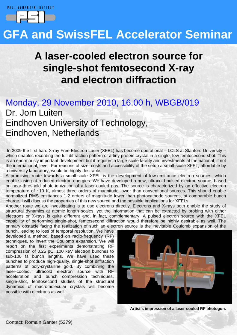
GFA and SwissFEL Accelerator Seminar
A laser-cooled electron source for single-shot femtosecond X-ray
and electron diffraction
Monday, 29 November 2010, 16.00 h, WBGB/019 Dr. Jom Luiten Eindhoven University of Technology,
Eindhoven, Netherlands
In 2009 the first hard X-ray Free Electron Laser (XFEL) has become operational – LCLS at Stanford University – which enables recording the full diffraction pattern of a tiny protein crystal in a single, few-femtosecond shot. This is an enormously important development but it requires a large-scale facility and investments at the national, if not the international, level. For reasons of size, costs and accessibility of the setup a small-scale XFEL, affordable by a university laboratory, would be highly desirable. A promising route towards a small-scale XFEL is the development of low-emittance electron sources, which enable lasing at reduced electron energies. We have developed a new, ultracold pulsed electron source, based on near-threshold photo-ionization of a laser-cooled gas. The source is characterized by an effective electron temperature of ~10 K, almost three orders of magnitude lower than conventional sources. This should enable normalized RMS emittances 1-2 orders of magnitude lower than photocathode sources, at comparable bunch charge. I will discuss the properties of this new source and the possible implications for XFELs. Another route we are investigating is to use electrons directly. Electrons and X-rays both enable the study of structural dynamics at atomic length scales, yet the information that can be extracted by probing with either electrons or X-rays is quite different and, in fact, complementary. A pulsed electron source with the XFEL capability of performing single-shot, femtosecond diffraction would therefore be highly desirable as well. The primary obstacle facing the realization of such an electron source is the inevitable Coulomb expansion of the bunch, leading to loss of temporal resolution. We have developed a method, based on radio-frequency (RF) techniques, to invert the Coulomb expansion. We will report on the first experiments demonstrating RF compression of 0.25 pC, 100 keV electron bunches to sub-100 fs bunch lengths. We have used these bunches to produce high-quality, single-shot diffraction patterns of poly-crystalline gold. By combining the laser-cooled, ultracold electron source with RF acceleration and bunch compression techniques, single-shot, femtosecond studies of the structural dynamics of macromolecular crystals will become possible with electrons as well.
Artist’s impression of a laser-cooled RF photogun.
Contact: Romain Ganter (5279)
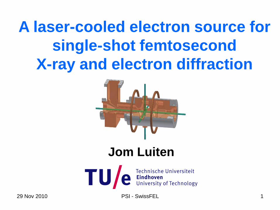
29 Nov 2010 PSI - SwissFEL 1
A laser-cooled electron source for
single-shot femtosecond
X-ray and electron diffraction
Jom Luiten
29 Nov 2010 PSI - SwissFEL 2
Thijs van Oudheusden – PhD student
Peter Pasmans – PhD student
Wouter Engelen – PhD student
Adam Lassise – PhD student
Marloes van der Heijden – Master student
Joris Kanters – Master student
Bas van der Geer, Marieke de Loos – Pulsar Physics (GPT)
Peter Mutsaers
Edgar Vredenbregt
Netherlands Technology
Foundation
NL Foundation for Fundamental
Research on Matter
FEI Company
Technical support
Louis van Moll
Jolanda van de Ven
Eddie Rietman
Ad Kemper
Harry van Doorn
29 Nov 2010 PSI - SwissFEL 3
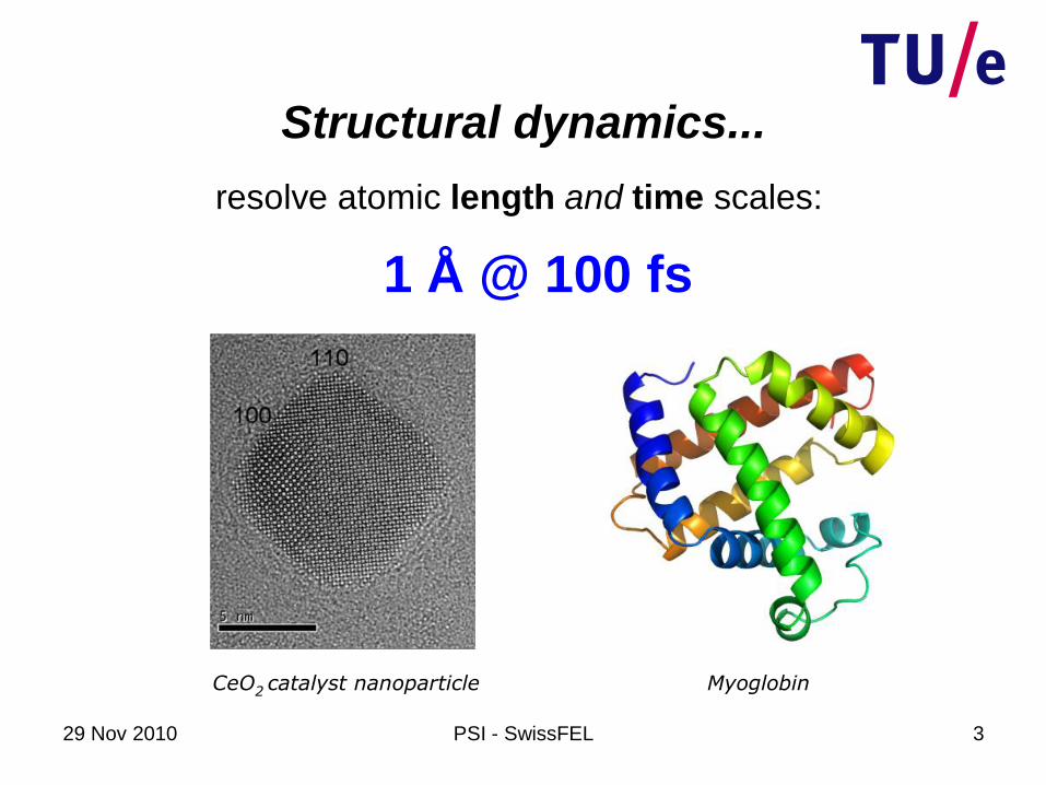
resolve atomic length and time scales:
Structural dynamics...
CeO2 catalyst nanoparticle Myoglobin
1 Å @ 100 fs

29 Nov 2010 PSI - SwissFEL 4
X-ray or electron pulse
ultrafast diffraction
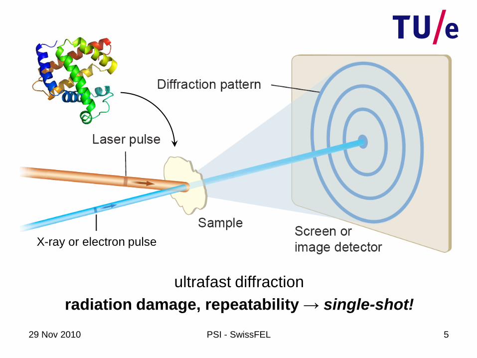
X-ray or electron pulse
29 Nov 2010 PSI - SwissFEL 5
radiation damage, repeatability → single-shot!
ultrafast diffraction
29 Nov 2010 PSI - SwissFEL 6
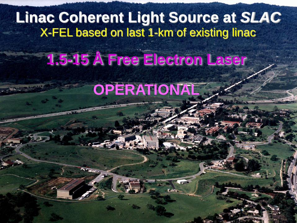
Linac Coherent Light Source at SLACX-FEL based on last 1-km of existing linac
1.5-15 Å Free Electron Laser
OPERATIONAL
29 Nov 2010 PSI - SwissFEL 7
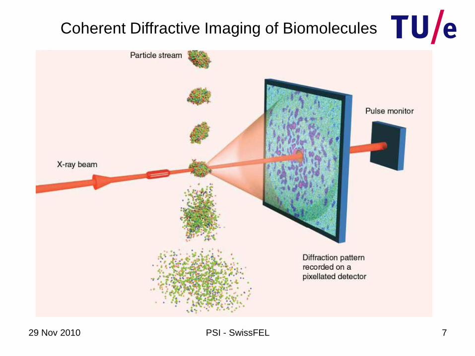
Coherent Diffractive Imaging of Biomolecules

29 Nov 2010 PSI - SwissFEL 8
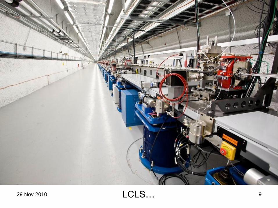
29 Nov 2010 PSI - SwissFEL 9LCLS…

29 Nov 2010 PSI - SwissFEL 10
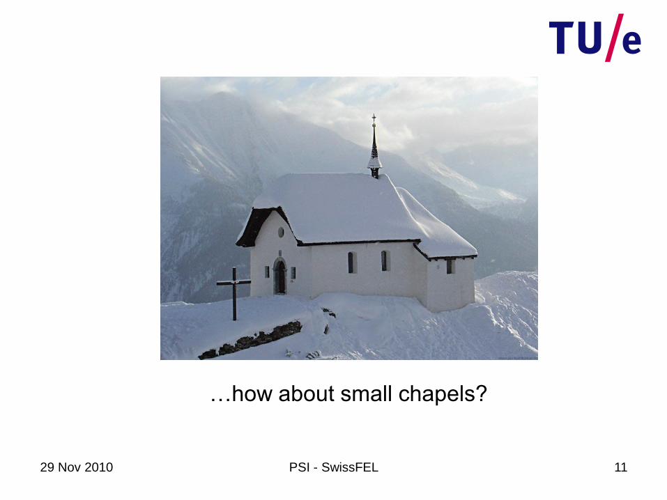
Modern day cathedrals…
X-ray light sources: modern day cathedrals…
29 Nov 2010 PSI - SwissFEL 11
…how about small chapels?

29 Nov 2010 PSI - SwissFEL 12
5 cmu
undulator
1 0 G e V1 0
21 0 m
2
u
ra d
Single-pass X-ray FEL
~ k Ap ea k
I
→ single-pass gain

29 Nov 2010 PSI - SwissFEL 13
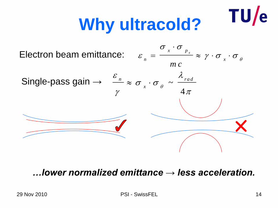
Electron beam emittance:
Why ultracold?
xx p
n x
m c
~4
n ra d
x
Single-pass gain →
Overlap between electron and X-ray beam…
29 Nov 2010 PSI - SwissFEL 14
Electron beam emittance:
Why ultracold?
xx p
n x
m c
~4
n ra d
x
Single-pass gain →
…lower normalized emittance → less acceleration.

29 Nov 2010 PSI - SwissFEL 15
Electron beam emittance:
Why ultracold?
xx p
n x
m c
~4
n ra d
x
Single-pass gain →
s o u rc e 2
e
n
kT
m c
…lower normalized emittance → less acceleration.

29 Nov 2010 PSI - SwissFEL 16
Electron beam emittance:
Why ultracold?
xx p
n x
m c
~4
n ra d
x
s o u rc e 2
e
n
kT
m c
Single-pass gain →
cannot be reduced
very much
(bunch charge)

29 Nov 2010 PSI - SwissFEL 17
so u rce 2
e
n
kT
m c
Electron beam emittance:
Why ultracold?
xx p
n x
m c
~4
n ra d
x
500× lower!!
Single-pass gain →
cannot be reduced
very much
(bunch charge)

29 Nov 2010 PSI - SwissFEL 18
so u rce 2
e
n
kT
m c
Electron beam emittance:
Why ultracold?
xx p
n x
m c
~4
n ra d
x
500× lower!!
Single-pass gain →
Cold source → compact X-FEL!
cannot be reduced
very much
(bunch charge)
29 Nov 2010 PSI - SwissFEL 19
Laser-cooled charged particle source
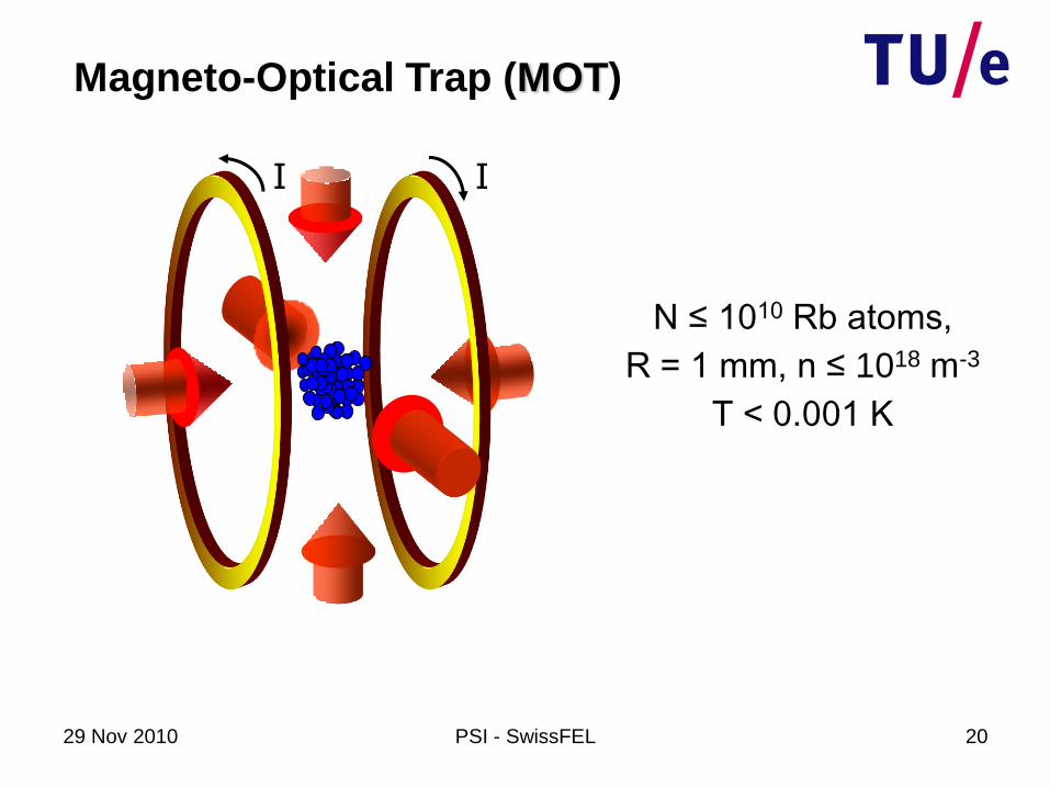
29 Nov 2010 PSI - SwissFEL 20
I I
Magneto-Optical Trap (MOT)
N ≤ 1010 Rb atoms,
R = 1 mm, n ≤ 1018 m-3
T < 0.001 K
29 Nov 2010 PSI - SwissFEL 21
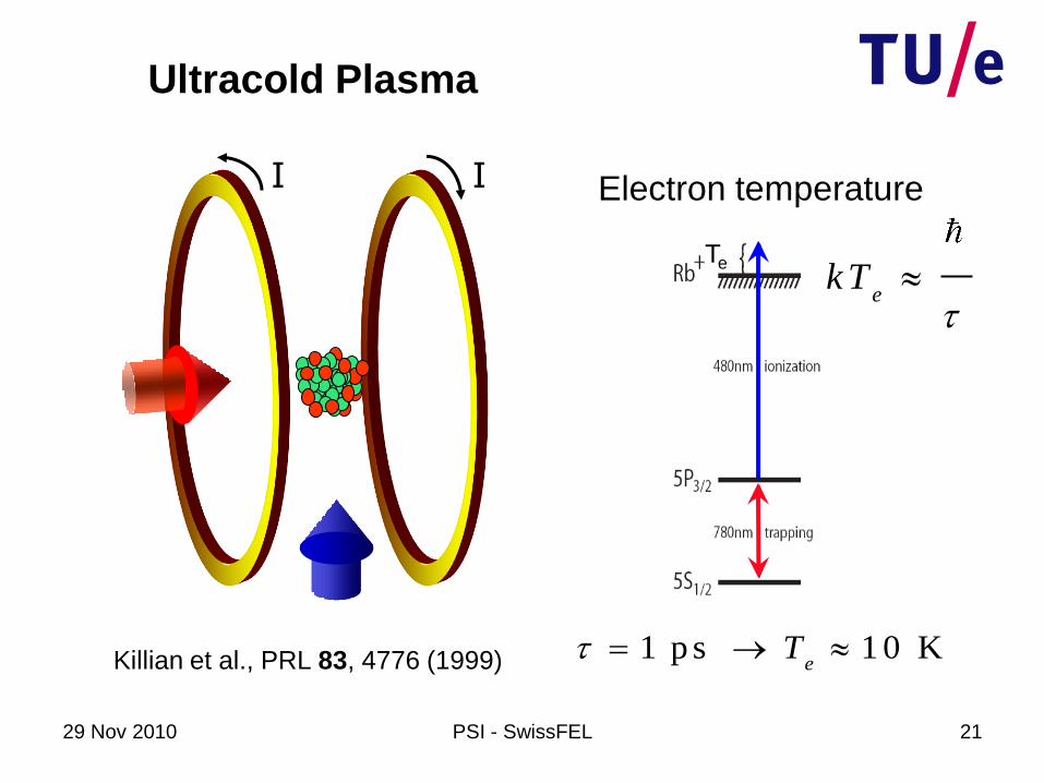
Te
Ultracold Plasma
I I Electron temperature
Killian et al., PRL 83, 4776 (1999)
ekT
1 p s 1 0 Ke
T
29 Nov 2010 PSI - SwissFEL 22
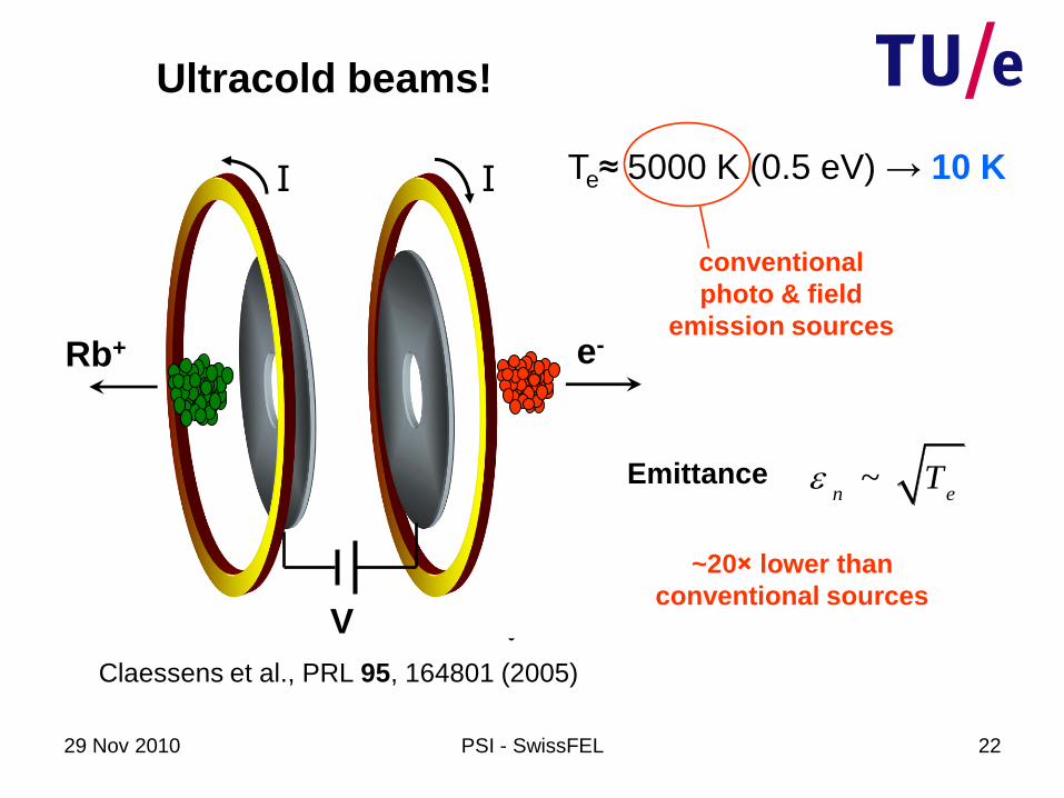
V
Rb+ e-
V
I I
Ultracold beams!
Te≈ 5000 K (0.5 eV) → 10 K
conventional
photo & field
emission sources
Claessens et al., PRL 95, 164801 (2005)
Emittance ~n e
T
~20× lower than
conventional sources
29 Nov 2010 PSI - SwissFEL 23
Moreover...
• Each shot a new source – no cathode problems;
• Up to 10 nA average current: 10 pC @ 1kHz;
• Ionization volume fully controlled by laser beam
overlap;
• ultracold ion bunches → model system for space
charge dynamics.
29 Nov 2010 PSI - SwissFEL 24
Ultracold electron beams
• photo-ionization experiments
• implications for compact X-FEL
Outline
Single-shot, femtosecond electron diffraction
• RF bunch compression
• ultracold electron source
29 Nov 2010 PSI - SwissFEL 25
Ultracold beam experiments

29 Nov 2010 PSI - SwissFEL 26
Ultracold beam experiments
Claessens et al., PRL 95, 164801 (2005);
Claessens et al., Phys. Plasmas 14, 093101 2007;
Taban et al., PRSTAB 11, 050102 (2008);
Reijnders et al., PRL 102, 034802 (2009);
Taban et al., EPL91, 46004 (2010);
Reijnders et al., PRL 105, 034802, (2010).

29 Nov 2010 PSI - SwissFEL 27
Ultracold beam experiments
2 cm
cathode
UHV
UHV
laser beams
(trapping, ionization)
-30 kV
300 pse-

29 Nov 2010 PSI - SwissFEL 28
z = 50 mm
Accelerator
VA ≤ 30 kV
y
z
1 m
MCP
Ultracold beam experiments
Solenoidal lens
y = 50 mm
Phosphorscreen
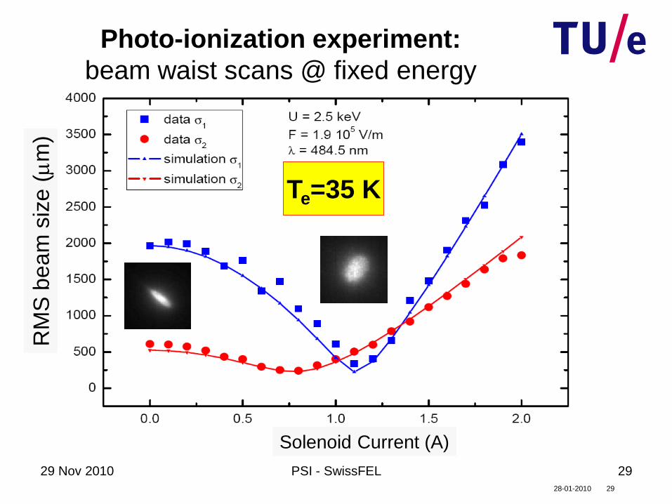
29 Nov 2010 PSI - SwissFEL 29
2928-01-2010
σxi = 30 μm
U = 2.1 keV
F = 1.13 kV/cm
λ = 474 nm
T = 405 ± 43 K
Solenoid Current (A)
RM
S b
ea
m s
ize
(m
m)
Photo-ionization experiment:
beam waist scans @ fixed energy
Te=35 K

29 Nov 2010 PSI - SwissFEL 30
0 0
1 14
e x c
FE h c R y
F
Excess energy
0
1 1h c
0
4F
R yF
0
0
0
0F 0F
3 / 25 P
1 / 25 S
R b
→ Stark shift0F
Electric field strength F
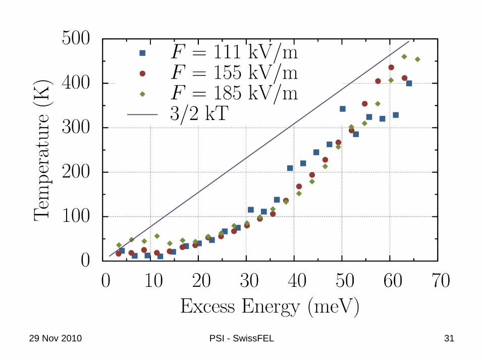
29 Nov 2010 PSI - SwissFEL 31

29 Nov 2010 PSI - SwissFEL 32
Te≈10 K

29 Nov 2010 PSI - SwissFEL 33
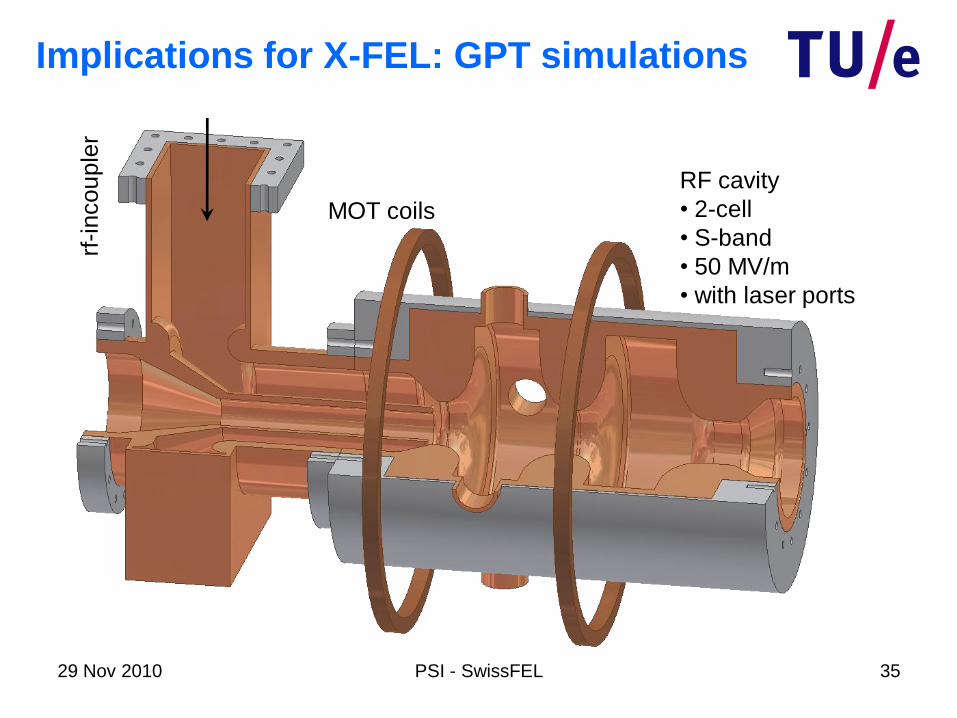
Implications for X-FEL: GPT simulations
RF cavity
• 2-cell
• S-band
• 50 MV/m
• with laser ports

29 Nov 2010 PSI - SwissFEL 34
rf-i
nco
up
ler
RF cavity
• 2-cell
• S-band
• 50 MV/m
• with laser ports
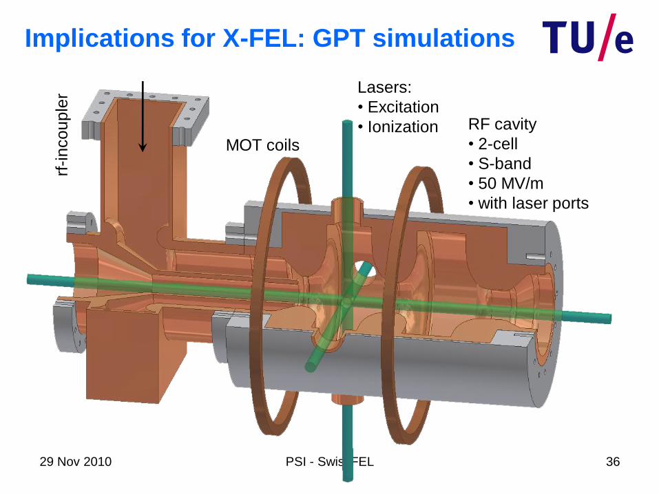
Implications for X-FEL: GPT simulations
29 Nov 2010 PSI - SwissFEL 35
rf-i
nco
up
ler
MOT coils
RF cavity
• 2-cell
• S-band
• 50 MV/m
• with laser ports
Implications for X-FEL: GPT simulations
29 Nov 2010 PSI - SwissFEL 36
rf-i
nco
up
ler
MOT coils
Lasers:
• Excitation
• Ionization RF cavity
• 2-cell
• S-band
• 50 MV/m
• with laser ports
Implications for X-FEL: GPT simulations
29 Nov 2010 37PSI - SwissFEL
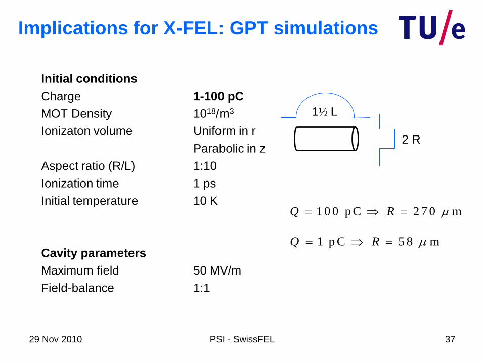
Initial conditions
Charge 1-100 pC
MOT Density 1018/m3
Ionizaton volume Uniform in r
Parabolic in z
Aspect ratio (R/L) 1:10
Ionization time 1 ps
Initial temperature 10 K
Cavity parameters
Maximum field 50 MV/m
Field-balance 1:1
2 R
1½ L
Implications for X-FEL: GPT simulations
1 p C 5 8 mQ R m
1 0 0 p C 2 7 0 mQ R m
29 Nov 2010 PSI - SwissFEL 38
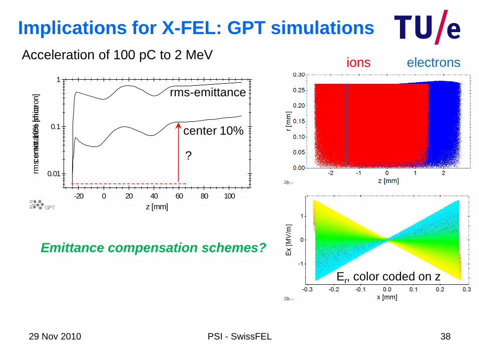
-20 0 20 40 60 80 100
GPT z [mm]
0.01
0.1
1
cente
r 10%
slice
-20 0 20 40 60 80 100
GPT z [mm]
0.01
0.1
1
rms e
mitta
nce [m
icro
n] rms-emittance
center 10%
Acceleration of 100 pC to 2 MeV
?
Implications for X-FEL: GPT simulations
Er, color coded on z
electronsions
Emittance compensation schemes?

29 Nov 2010 PSI - SwissFEL 39
4
radn
21
2
2
2
Ku
rad
g
u
FEL
L
34
1
rad
u
g
IeK
mcL
m
2
32
2342
3
1
FELI
e
mcP
2
e
mcFEL
W
2
2
Wz
QI
/max
Basic FEL equations:
29 Nov 2010 PSI - SwissFEL 40
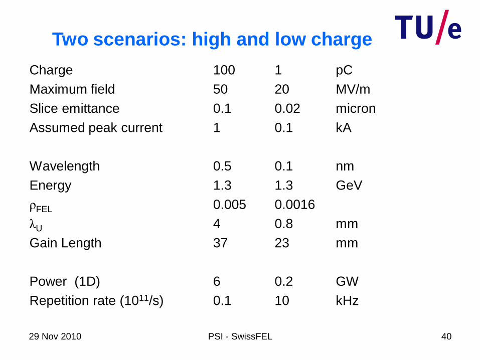
Charge 100 1 pC
Maximum field 50 20 MV/m
Slice emittance 0.1 0.02 micron
Assumed peak current 1 0.1 kA
Wavelength 0.5 0.1 nm
Energy 1.3 1.3 GeV
ρFEL 0.005 0.0016
λU 4 0.8 mm
Gain Length 37 23 mm
Power (1D) 6 0.2 GW
Repetition rate (1011/s) 0.1 10 kHz
Two scenarios: high and low charge
29 Nov 2010 PSI - SwissFEL 41
Single-shot femtosecond
electron diffraction
29 Nov 2010 PSI - SwissFEL 42
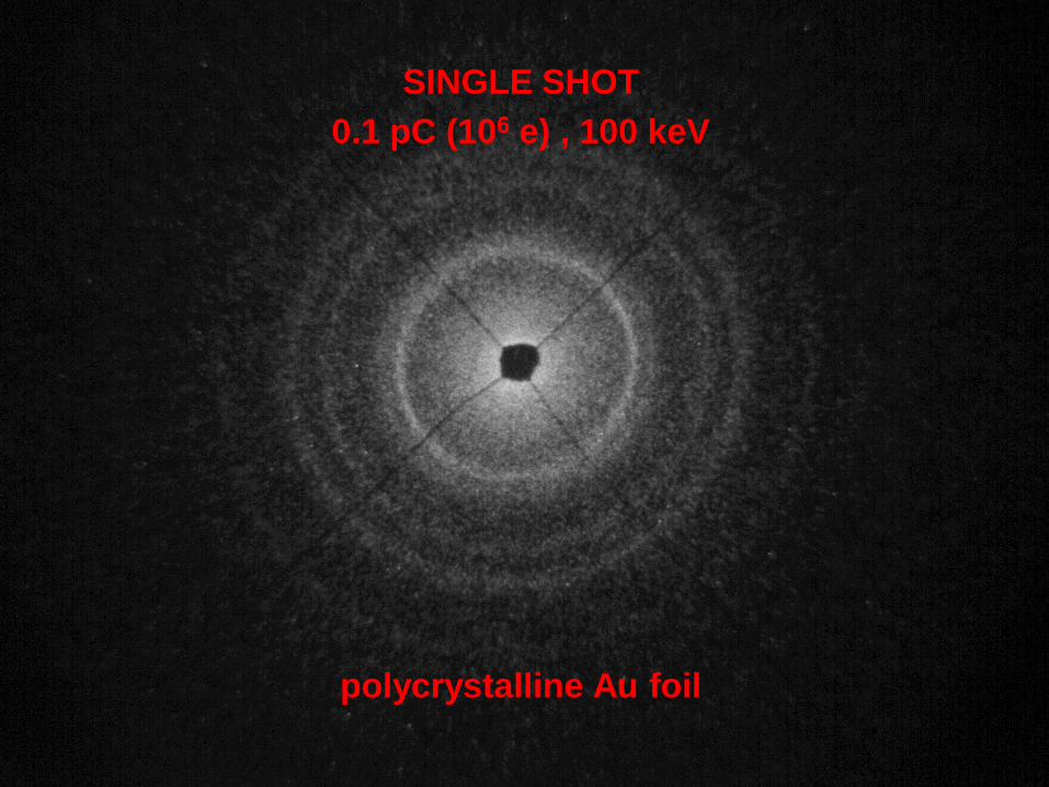
SINGLE SHOT
polycrystalline Au foil
0.1 pC (106 e) , 100 keV

29 Nov 2010 PSI - SwissFEL 43
average of 20 single shot pictures
polycrystalline Au foil
29 Nov 2010 PSI - SwissFEL 44
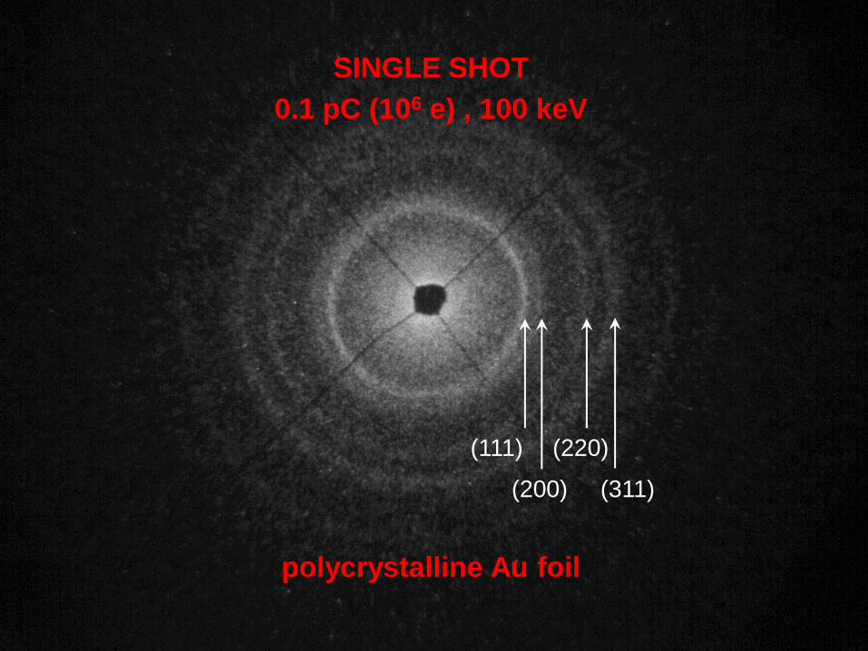
SINGLE SHOT
polycrystalline Au foil
0.1 pC (106 e) , 100 keV
(200) (311)
(111) (220)

29 Nov 2010 PSI - SwissFEL 45
9 nm Au foil
U = 95 keV
Q = 0.2 pC
Van Oudheusden et al.,
PRL (2010)
29 Nov 2010 PSI - SwissFEL 46
Why use electrons?

29 Nov 2010 PSI - SwissFEL 47
X-rays:Thomson scattering
Electrons:Rutherford scattering
Complementary information!
high density, bulk gas phase, surfaces
2 9 26 .6 1 0 m
T
2 4 21 0 m
R
29 Nov 2010 PSI - SwissFEL 48
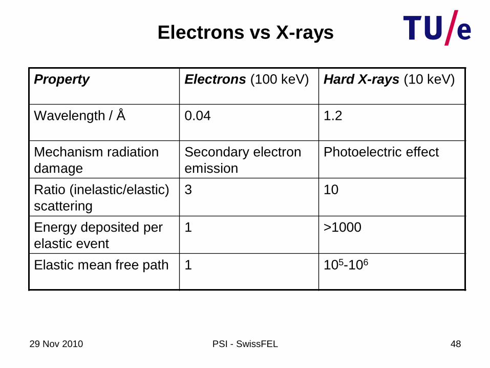
Electrons vs X-rays
Property Electrons (100 keV) Hard X-rays (10 keV)
Wavelength / Å 0.04 1.2
Mechanism radiation
damage
Secondary electron
emission
Photoelectric effect
Ratio (inelastic/elastic)
scattering
3 10
Energy deposited per
elastic event
1 >1000
Elastic mean free path 1 105-106

29 Nov 2010 PSI - SwissFEL 49
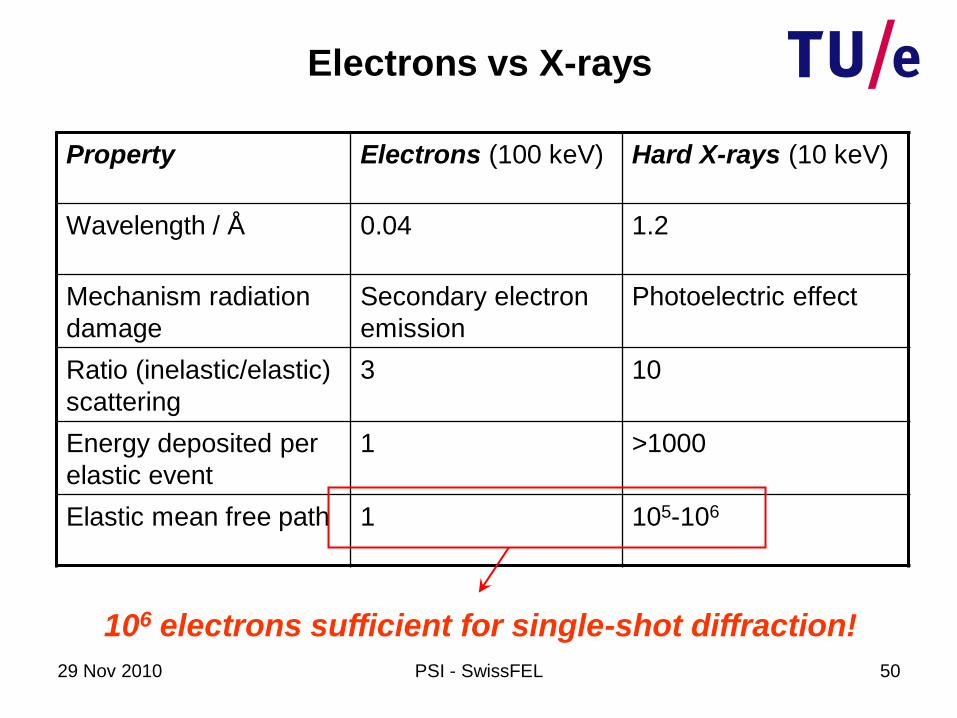
Electrons vs X-rays
Property Electrons (100 keV) Hard X-rays (10 keV)
Wavelength / Å 0.04 1.2
Mechanism radiation
damage
Secondary electron
emission
Photoelectric effect
Ratio (inelastic/elastic)
scattering
3 10
Energy deposited per
elastic event
1 >1000
Elastic mean free path 1 105-106
1000× less radiation damage per count!
29 Nov 2010 PSI - SwissFEL 50
Electrons vs X-rays
Property Electrons (100 keV) Hard X-rays (10 keV)
Wavelength / Å 0.04 1.2
Mechanism radiation
damage
Secondary electron
emission
Photoelectric effect
Ratio (inelastic/elastic)
scattering
3 10
Energy deposited per
elastic event
1 >1000
Elastic mean free path 1 105-106
106 electrons sufficient for single-shot diffraction!
29 Nov 2010 PSI - SwissFEL 51
Electrons – why not?
29 Nov 2010 PSI - SwissFEL 52
Electrons – why not?
Source brightness & Coulomb forces

29 Nov 2010 PSI - SwissFEL 53
femtosecond laser photoemission...
100 μm
106 electrons from 100 μm spot
29 Nov 2010 PSI - SwissFEL 54
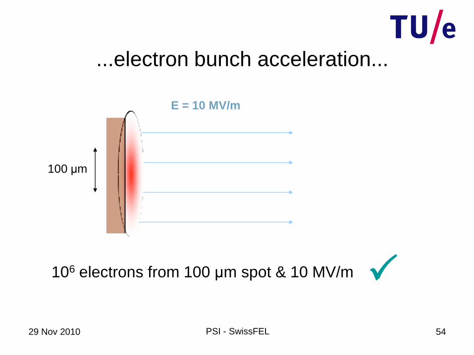
...electron bunch acceleration...
E = 10 MV/m
100 μm
106 electrons from 100 μm spot & 10 MV/m

29 Nov 2010 PSI - SwissFEL 55
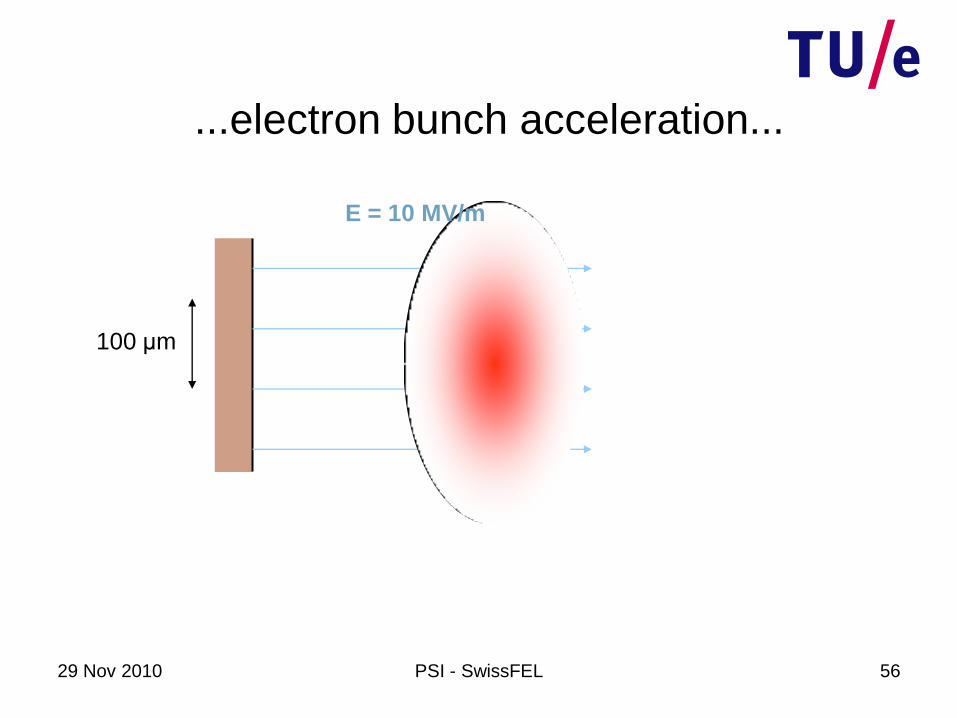
...electron bunch acceleration...
100 μm
E = 10 MV/m
29 Nov 2010 PSI - SwissFEL 56
...electron bunch acceleration...
100 μm
E = 10 MV/m
29 Nov 2010 PSI - SwissFEL 57
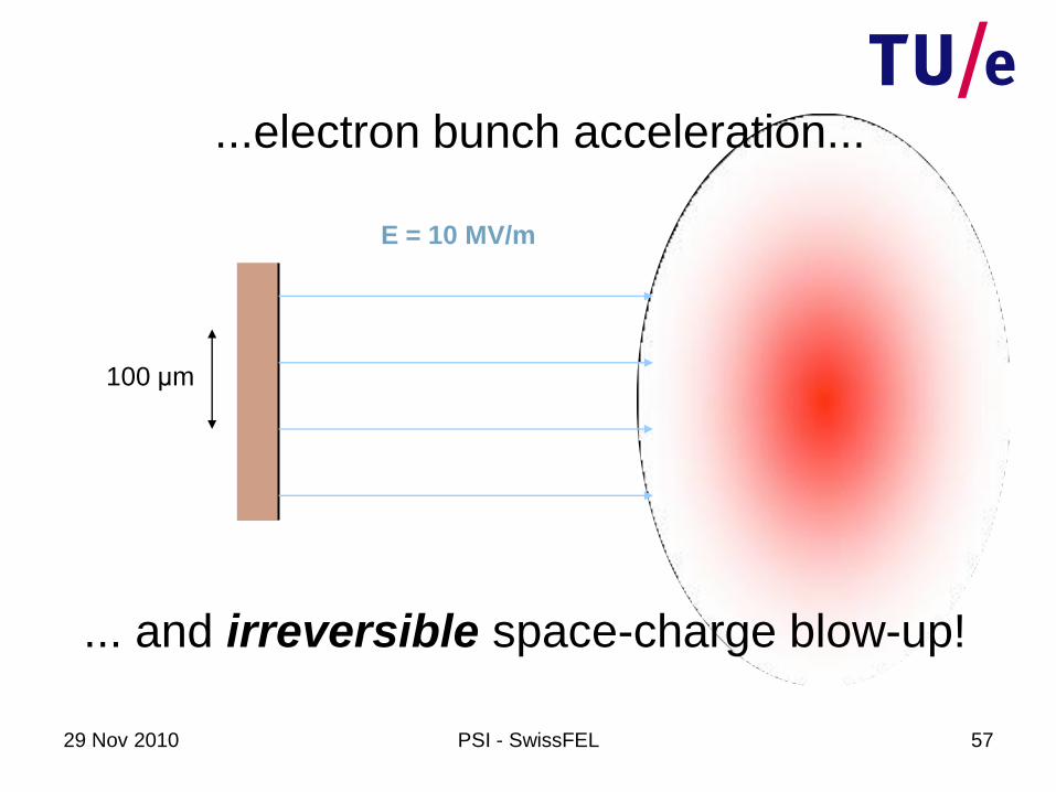
... and irreversible space-charge blow-up!
...electron bunch acceleration...
100 μm
E = 10 MV/m
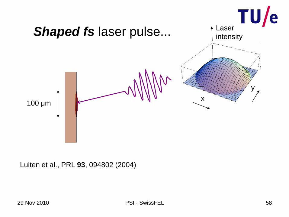
29 Nov 2010 PSI - SwissFEL 58
100 μmx
y
Laser
intensity
Luiten et al., PRL 93, 094802 (2004)
Shaped fs laser pulse...
29 Nov 2010 PSI - SwissFEL 59
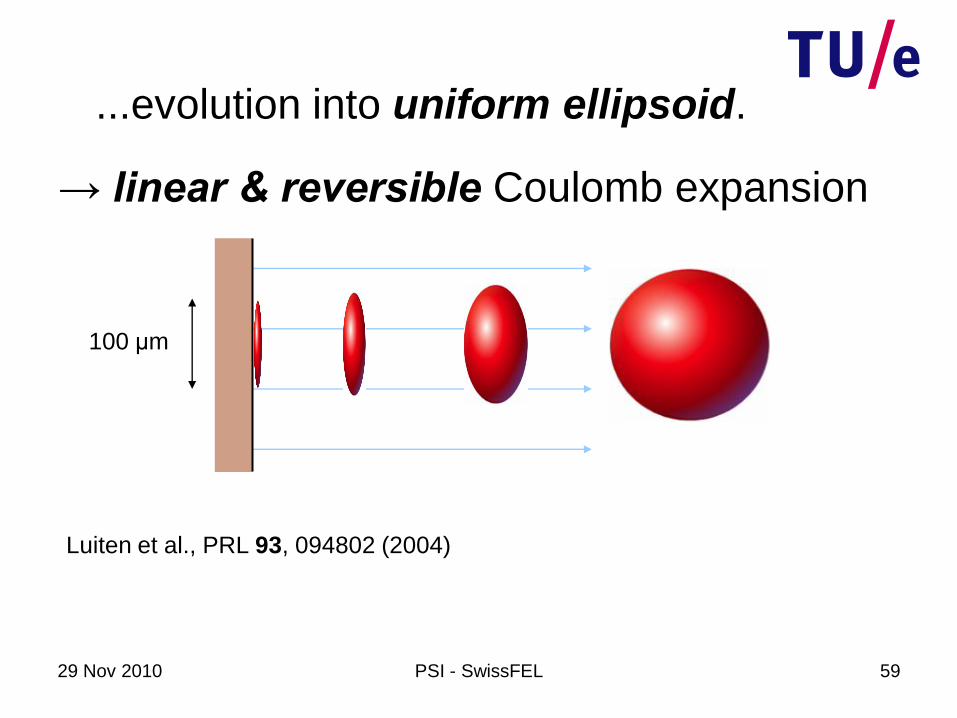
...evolution into uniform ellipsoid.
100 μm
Luiten et al., PRL 93, 094802 (2004)
→ linear & reversible Coulomb expansion
29 Nov 2010 PSI - SwissFEL 60
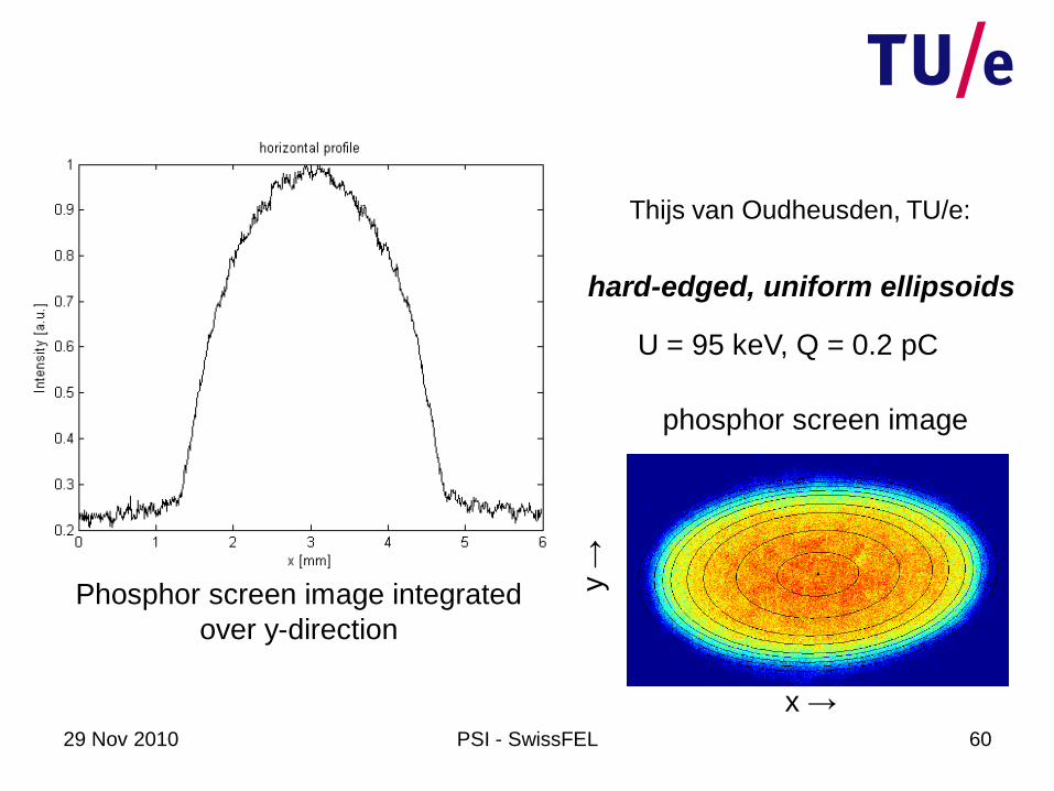
U = 95 keV, Q = 0.2 pC
hard-edged, uniform ellipsoids
Thijs van Oudheusden, TU/e:
Phosphor screen image integrated
over y-direction
phosphor screen image
x →
y →
29 Nov 2010 PSI - SwissFEL 61
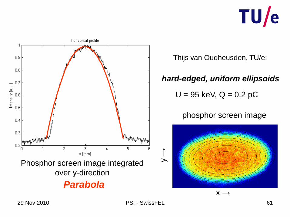
hard-edged, uniform ellipsoids
Thijs van Oudheusden, TU/e:
Phosphor screen image integrated
over y-direction
Parabola
phosphor screen image
x →
y →
U = 95 keV, Q = 0.2 pC
29 Nov 2010 PSI - SwissFEL 62
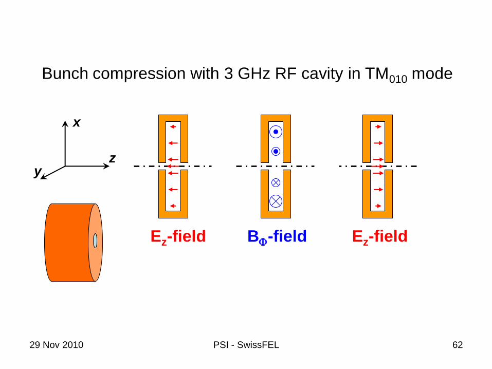
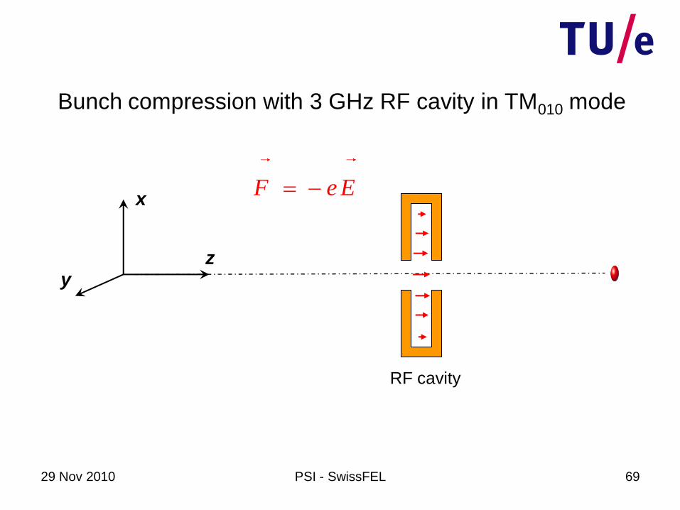
Bunch compression with 3 GHz RF cavity in TM010 mode
Ez-field BF-field Ez-field
x
yz

29 Nov 2010 PSI - SwissFEL 63
TM010 : Ez-field TM010 : BF-field
x
y
Ez
Bunch compression with 3 GHz RF cavity in TM010 mode

29 Nov 2010 PSI - SwissFEL 64
RF cavity
Bunch compression with 3 GHz RF cavity in TM010 mode
F e E x
yz
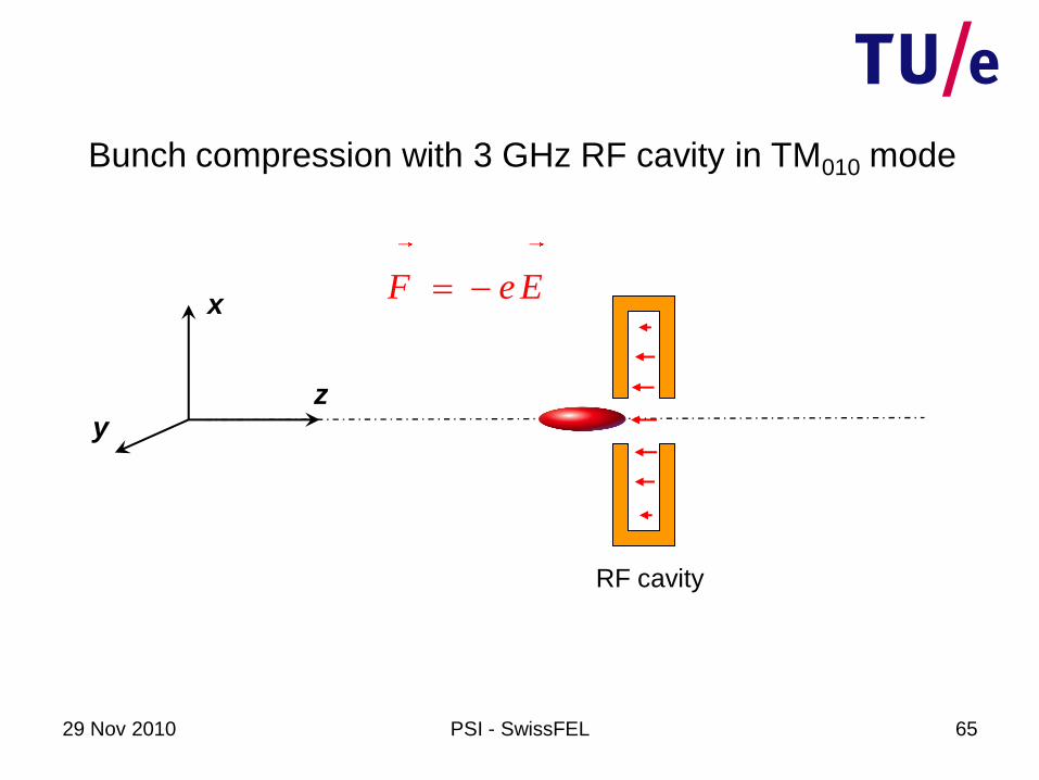
29 Nov 2010 PSI - SwissFEL 65
RF cavity
Bunch compression with 3 GHz RF cavity in TM010 mode
F e E x
yz
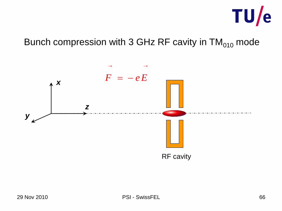
29 Nov 2010 PSI - SwissFEL 66
RF cavity
Bunch compression with 3 GHz RF cavity in TM010 mode
F e E x
yz

29 Nov 2010 PSI - SwissFEL 67
RF cavity
Bunch compression with 3 GHz RF cavity in TM010 mode
F e E x
yz
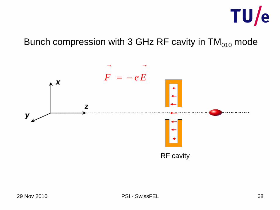
29 Nov 2010 PSI - SwissFEL 68
RF cavity
Bunch compression with 3 GHz RF cavity in TM010 mode
F e E x
yz
29 Nov 2010 PSI - SwissFEL 69
RF cavity
Bunch compression with 3 GHz RF cavity in TM010 mode
F e E x
yz

29 Nov 2010 PSI - SwissFEL 70
3 GHz RF cavity
longitudinal E-field
50 fs
100 kV
Van Oudheusden et al., JAP 102, 093501 (2007)
The setup
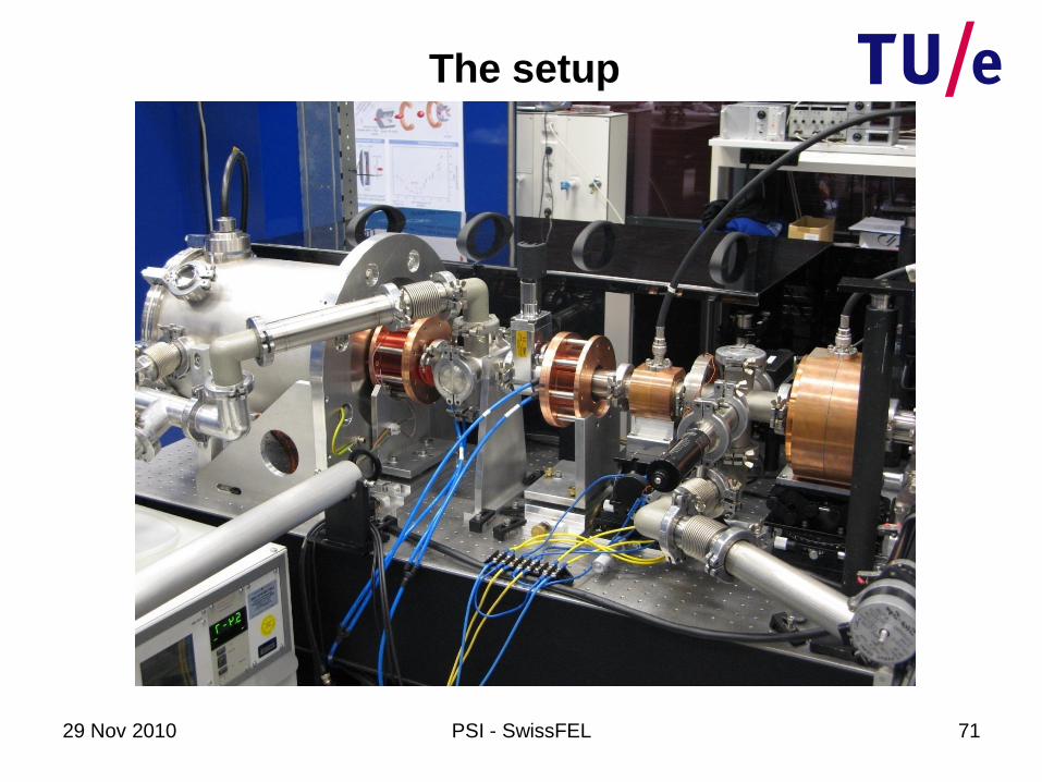
29 Nov 2010 PSI - SwissFEL 71
The setup
29 Nov 2010 PSI - SwissFEL 72
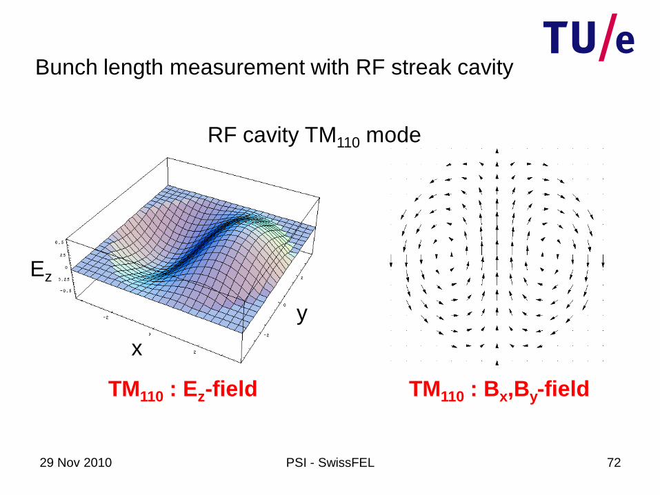
TM110 : Ez-field TM110 : Bx,By-field
x
y
Ez
RF cavity TM110 mode
Bunch length measurement with RF streak cavity
29 Nov 2010 PSI - SwissFEL
v
B
Bunch length measurement with RF streak cavity
x,ty
z
RF cavity in TM110 mode
transverse B field
73
F ev B

29 Nov 2010 PSI - SwissFEL 74
Cavity off
x,ty
z
RF cavity in TM110 mode
transverse B field
Limitation temporal resolution:
v
29 Nov 2010 PSI - SwissFEL 75
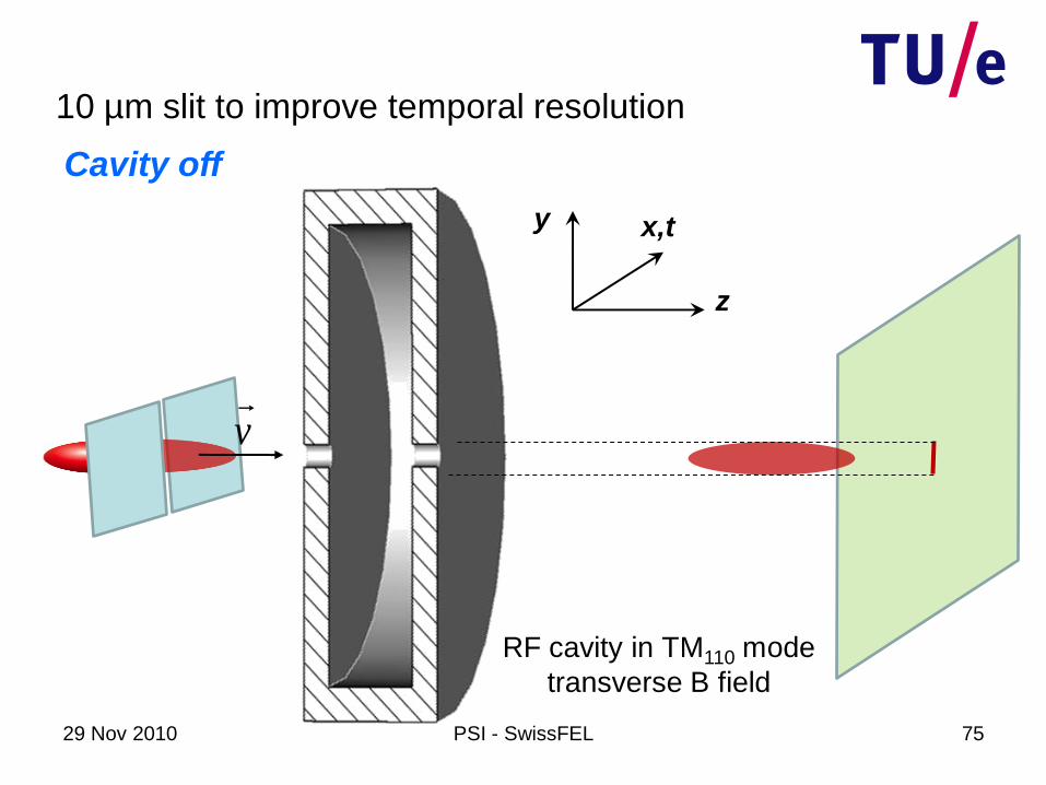
Cavity off
x,ty
z
RF cavity in TM110 mode
transverse B field
10 µm slit to improve temporal resolution
v
29 Nov 2010 PSI - SwissFEL 76
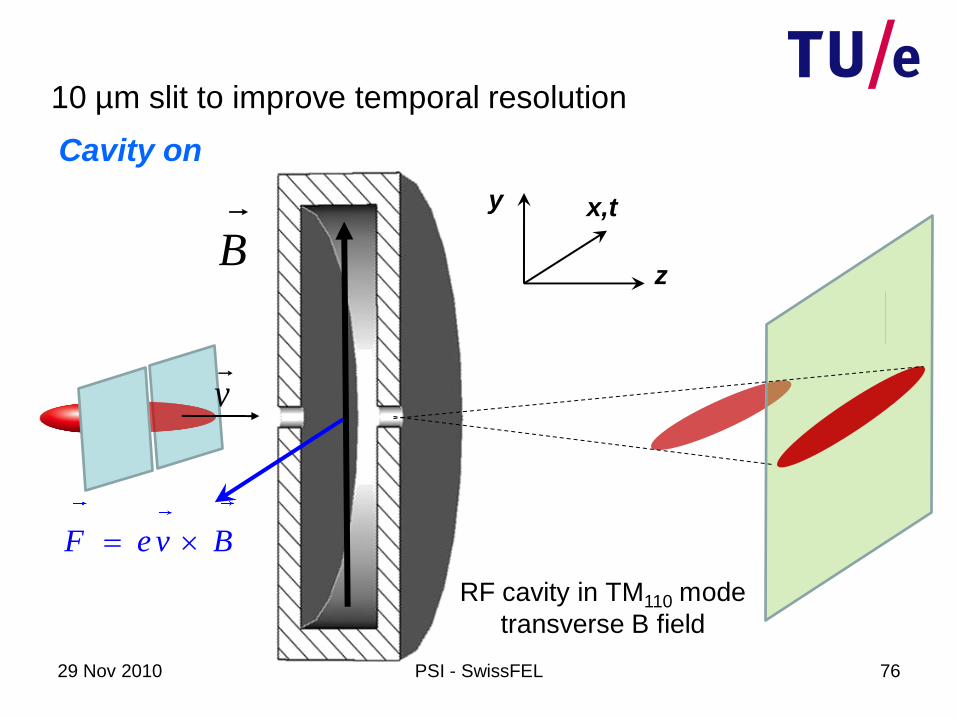
10 µm slit to improve temporal resolution
Cavity on
x,ty
zB
F e v B
RF cavity in TM110 mode
transverse B field
v

29 Nov 2010 PSI - SwissFEL 77
Streak image on screen
Streak cavity on cavity off
29 Nov 2010 PSI - SwissFEL 78
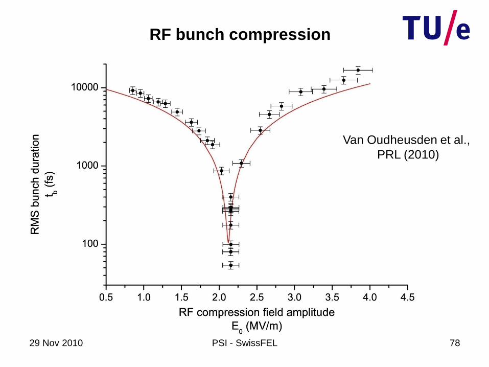
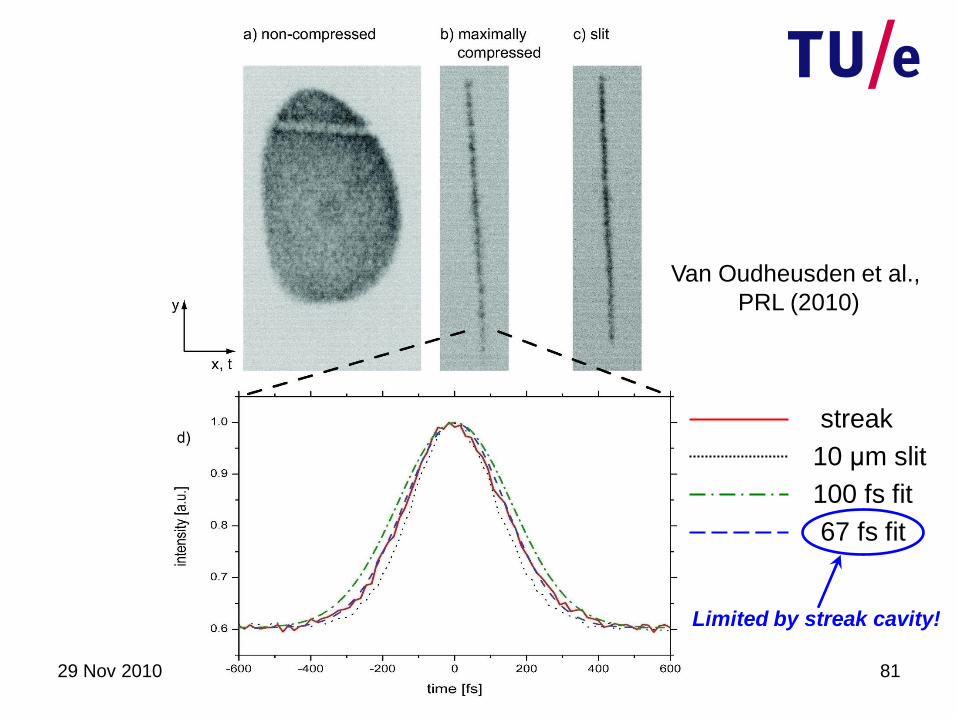
RF bunch compression
Van Oudheusden et al.,
PRL (2010)
29 Nov 2010 PSI - SwissFEL 79
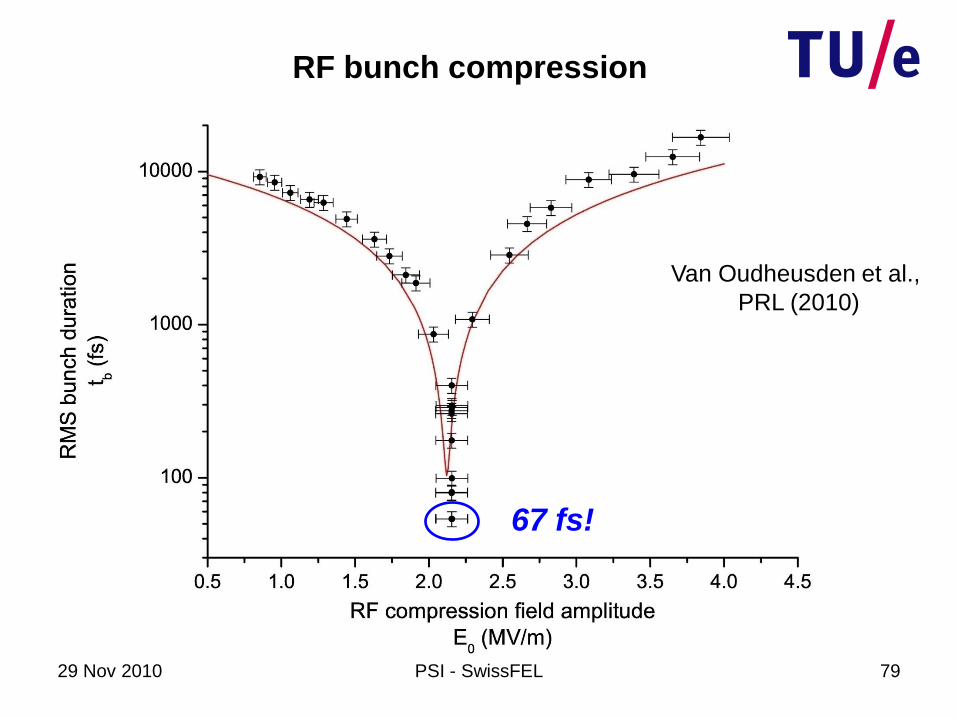
RF bunch compression
67 fs!
Van Oudheusden et al.,
PRL (2010)
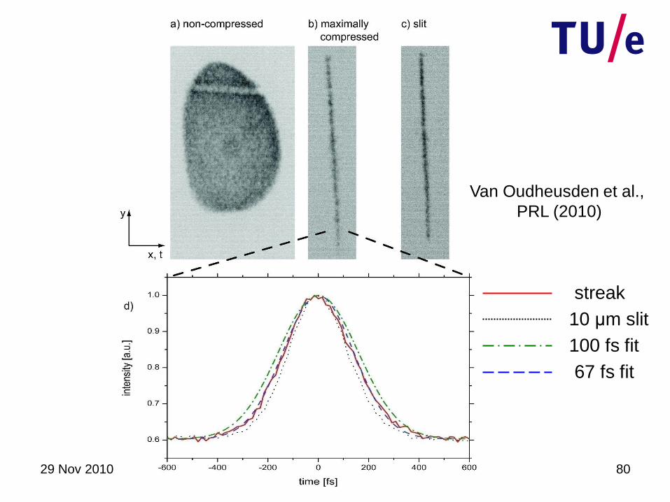
29 Nov 2010 PSI - SwissFEL 80
10 μm slit
100 fs fit
67 fs fit
streak
Van Oudheusden et al.,
PRL (2010)
29 Nov 2010 PSI - SwissFEL 81
10 μm slit
100 fs fit
67 fs fit
streak
Van Oudheusden et al.,
PRL (2010)
Limited by streak cavity!
29 Nov 2010 PSI - SwissFEL 82
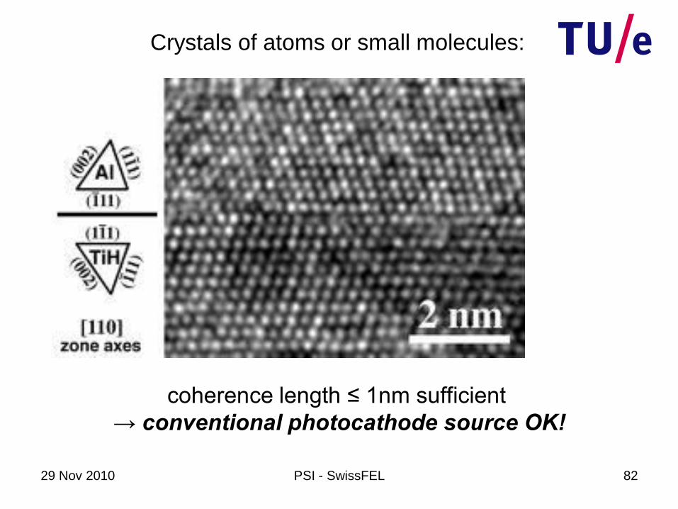
coherence length ≤ 1nm sufficient
→ conventional photocathode source OK!
Crystals of atoms or small molecules:
29 Nov 2010 PSI - SwissFEL 83
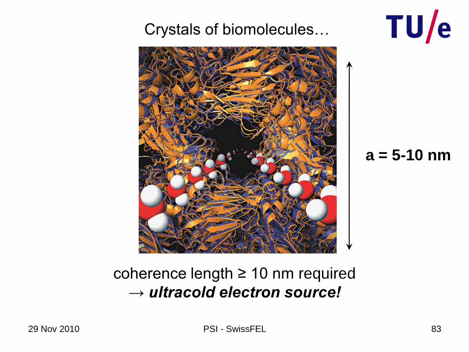
Crystals of biomolecules…
a = 5-10 nm
coherence length ≥ 10 nm required
→ ultracold electron source!
29 Nov 2010 PSI - SwissFEL 84
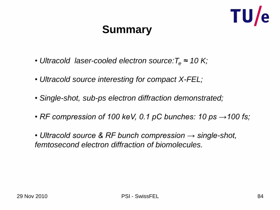
Summary
• Ultracold laser-cooled electron source:Te ≈ 10 K;
• Ultracold source interesting for compact X-FEL;
• Single-shot, sub-ps electron diffraction demonstrated;
• RF compression of 100 keV, 0.1 pC bunches: 10 ps →100 fs;
• Ultracold source & RF bunch compression → single-shot,
femtosecond electron diffraction of biomolecules.