a human na /h antiporter sharing evolutionary origins with ... · a human na /h antiporter sharing...
TRANSCRIPT

A human Na�/H� antiporter sharing evolutionaryorigins with bacterial NhaA may be a candidategene for essential hypertensionMinghui Xiang, Mingye Feng, Sabina Muend, and Rajini Rao*
Department of Physiology, Johns Hopkins University School of Medicine, 725 North Wolfe Street, Baltimore, MD 21205
Edited by H. Ronald Kaback, University of California, Los Angeles, CA, and approved September 27, 2007 (received for review July 30, 2007)
Phylogenetic analysis of the cation/proton antiporter superfamilyhas uncovered a previously unknown clade of genes in metazoangenomes, including two previously uncharacterized human iso-forms, NHA1 and NHA2, found in tandem on human chromosome4. The NHA (sodium hydrogen antiporter) family members sharesignificant sequence similarity with Escherichia coli NhaA, includ-ing a conserved double aspartate motif in predicted transmem-brane 5. We show that HsNHA2 (Homo sapiens NHA2) resides onthe plasma membrane and, in polarized MDCK cells, localizes to theapical domain. Analysis of mouse tissues indicates that NHA2 isubiquitous. When expressed in the yeast Saccharomyces cerevisiaelacking endogenous cation/proton antiporters and pumps,HsNHA2 can confer tolerance to Li� and Na� ions but not to K�.HsNHA2 transformants accumulated less Li� than the salt-sensitivehost; however, mutagenic replacement of the conserved aspar-tates abolished all observed phenotypes. Functional complemen-tation by HsNHA2 was insensitive to amiloride, a characteristicinhibitor of plasma membrane sodium hydrogen exchanger iso-forms, but was inhibited by phloretin. These are hallmarks ofsodium–lithium countertransport activity, a highly heritable traitcorrelating with hypertension. Our findings raise the possibilitythat NHA genes may contribute to sodium–lithium countertrans-port activity and salt homeostasis in humans.
sodium–lithium countertransport � yeast expression � red blood cell �pancreas
The regulation of salt, pH, and volume is prerequisite to allforms of life, and central to these homeostatic mechanisms
is the transmembrane exchange of H� for cations (Na� or K�).In bacteria, an array of Na�/H� antiporters convert the protonmotive force (established by the respiratory chain or the F1Fo-ATPase) into sodium gradients that drive other energy-requiringprocesses (solute transport or flagellar motors), transduce en-vironmental signals into cell responses, and even function in drugefflux (TetL and MdfA) (1–3). In plants, newly discoveredmembers of the NHE (sodium hydrogen exchanger) family ofNa�/H� exchangers sequester Na� and K� within vacuoles as aprincipal means of salt tolerance, regulate vacuolar pH to controlf lower color, and are essential for development (4, 5). Inmetazoans, the role of NHE in cytosolic and compartmental pHregulation has been implicated in the control of cell cycle and cellproliferation, vesicle trafficking, and compartmental biogenesis(6–8). Specific isoforms of NHE localize distinctly to basolateralor apical membranes to control transepithelial sodium fluxes (4,9). In mammals, NHE dysfunction leads to a host of pathophys-iological conditions that include hypertension, epilepsy, post-ischemic myocardial arrhythmias, gastric and kidney disease,diarrhea, and glaucoma.
The last decade witnessed an explosion in the number ofgenomic sequences deposited in databases worldwide, and theseare now awaiting functional analysis. Automated annotationprograms have identified �550 sequence entries as putativeNa�/H� exchangers. In an effort to understand the evolutionaryorigins and distribution of Na�/H� exchangers, we undertook a
comprehensive phylogenetic analysis of the superfamily ofmonovalent cation/proton antiporters (CPA) that have in com-mon a transmembrane organization of 12 predicted hydropathichelices with detectable sequence similarity (10). This superfam-ily has two main subdivisions, named CPA1 and CPA2, accord-ing to the nomenclature of Transport Classification Databaseestablished by Milton Saier (http://www.tcdb.org/). Members ofthe CPA1 group include the well characterized NHE family ofelectroneutral Na�(K�)/H� exchangers represented by nineparalogs in human (NHE1–9). In contrast, we found that virtu-ally all eukaryotic members of the CPA2 group were previouslyunknown or poorly characterized. Among these was a new familyof related genes in animals that we named NHA on the basis oftheir similarity to fungal NHA genes and bacterial NhaA genes(10). There are two paralogs, NHA1 and NHA2, in all com-pletely sequenced metazoan genomes, including nematodes, f ly,puffer fish, mouse, and human.
The identification of an entire family of phylogeneticallydistinct antiporters that are conserved from bacteria to humansopens up a new line of investigation. In this study, we describeHomo sapiens NHA2 (HsNHA2) as a prototypic metazoanexample of the NHA family. Sequence similarity with Esche-richia coli NhaA (EcNhaA) served to guide functional charac-terization by heterologous expression in yeast. Patterns of tissuedistribution, chromosomal location, and inhibitor sensitivitypoint to this gene as a likely candidate for the sodium–lithiumcountertransport (SLC) activity reported in red blood cells (11),lymphoblasts, and fibroblasts (12–14), and suggest an importantpotential physiological role in hypertension.
ResultsThe presence of two paralogous NHA genes in the metazoangenomes of nematodes, insects, fish, and mammals is indicativeof an early gene duplication event. Indeed, NHA1 and NHA2appear in tandem on human chromosome 4, and the syntenicmouse chromosome 3 (Fig. 1A), and share amino acid identityof 55% over 515 aa [69% similarity; supporting information (SI)Fig. 5]. An alignment of human NHA2 sequence with that ofEcNhaA reveals a significant conservation of residues (18%identity and 33% similarity over 388 aa) extending over 12predicted transmembrane domains (Fig. 1B). Of note, twoaspartic acid residues in TM5 of EcNhaA that have previouslybeen shown to be critical for ion transport (15) are conserved inthe metazoan NHA. Other residues of interest include H356
Author contributions: M.X., M.F., and R.R. designed research; M.X., M.F., and S.M. per-formed research; M.X. and M.F. contributed new reagents/analytic tools; M.X., M.F., S.M.,and R.R. analyzed data; and M.X., S.M., and R.R. wrote the paper.
The authors declare no conflict of interest.
This article is a PNAS Direct Submission.
*To whom correspondence should be addressed. E-mail: [email protected].
This article contains supporting information online at www.pnas.org/cgi/content/full/0707120104/DC1.
© 2007 by The National Academy of Sciences of the USA
www.pnas.org�cgi�doi�10.1073�pnas.0707120104 PNAS � November 20, 2007 � vol. 104 � no. 47 � 18677–18681
MED
ICA
LSC
IEN
CES

(TM8) and R432 (TM10) in NHA2, which may be equivalent tofunctionally important residues H225 and K300, respectively, inEcNhaA (16). Compared with the related NHE family of CPA1antiporters that have long, hydrophilic C-terminal domains(150–300 aa) (10), the NHA proteins are distinguished by muchshorter C-terminal tails following predicted TM12.
Antibodies raised against a unique N-terminal peptide ofHsNHA2 (Fig. 1B, underlined) identified a band of the expectedsize in membranes from yeast heterologously expressing His9-tagged HsNHA2 (55 kDa), or GFP-HsNHA2 (90 kDa), whereas
no cross-reactivity was seen in membranes from yeast trans-formed with His9-HsNHA1 (Fig. 2A). The partial conservationof residues (7 of 15) within the antigenic peptide between mouseand human NHA2 allowed us to test expression in mouse: Astrongly reacting band of �50 kDa was observed in pancreaticlysates, and preincubation of antibody with the antigenic peptidegreatly attenuated the band intensity (Fig. 2 A, lanes 13–14).Expression of NHA2 appeared to be ubiquitous in all mousetissues examined (Fig. 2B), with high levels in pancreas. Indirectimmunofluorescence of pancreatic � cells derived from ratindicated plasma membrane distribution of the endogenousprotein (Fig. 2C). In the polarized, kidney-derived MDCK cellline transfected with GFP-HsNHA2, confocal microscopyshowed that fluorescence was restricted to the apical domain, asseen by colocalization with fluorescent lectin (Rhodamine-wheat germ agglutinin) (Fig. 2D). Similarly, yeast cells express-ing GFP-HsNHA2 showed predominant plasma membranelocalization, albeit with some retention within intracellularcompartments (Fig. 2E).
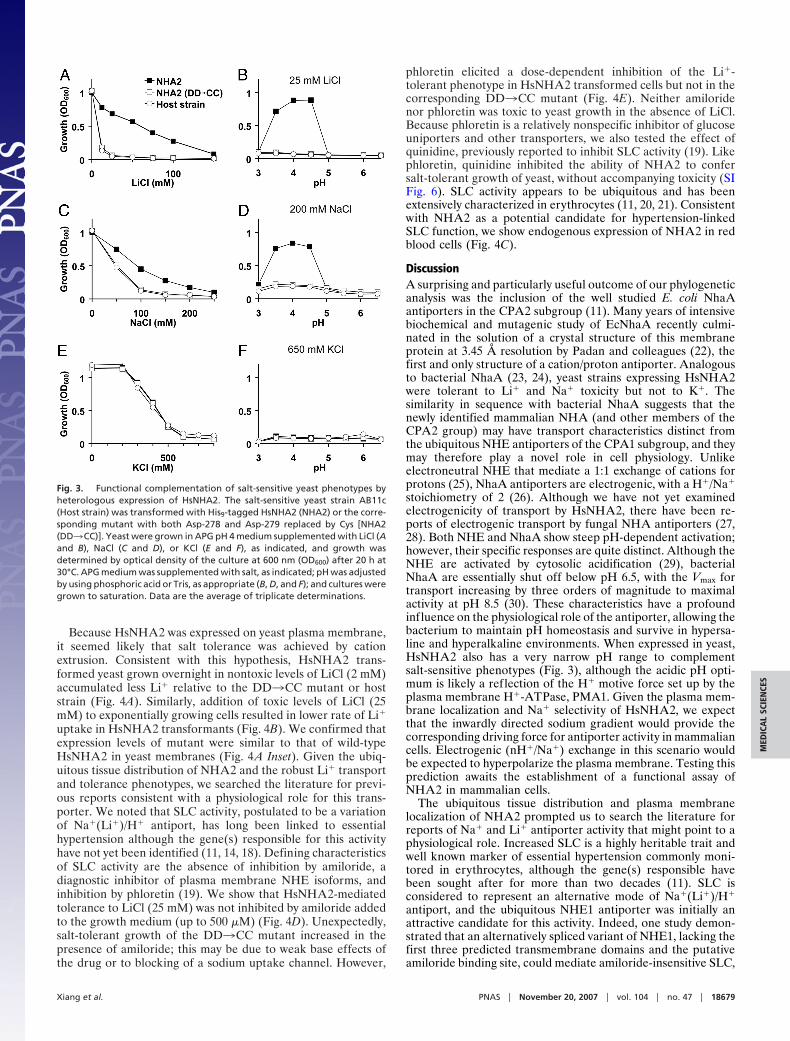
A useful strategy to obtain functional insight on a new gene isby phenotype complementation of the orthologous gene knock-outs in yeast. We evaluated the ability of heterologously ex-pressed HsNHA2 to rescue the salt-sensitive growth phenotypeof the yeast strain AB11c (ena1-4�nhx1�nha1�) lacking threemajor salt-handling mechanisms. As expected, the host strainshowed dose-dependent growth sensitivity to cationic salts ofLi�, Na�, and K�. We show that HsNHA2 conferred toleranceto Li� and Na� but not to K� (Fig. 3 A, C, and E). Mutagenicreplacement of the two aspartate residues, D278 and D279, ofHsNHA2 that align with residues D163 and D164 of EcNhaA asshown in Fig. 1B, abolished the ability of HsNHA2 to confersalt-tolerant growth (double mutant DD3CC) (Fig. 3), consis-tent with a conserved critical role of these acidic residues inmediating cation transport. The complementation efficacy ofHsNHA2 in yeast, for both LiCl and NaCl sensitivity, wasoptimal between pH 3.5 and 4.5 (Fig. 3 B and D), whereas therewas no complementation of KCl sensitivity at all tested pH (Fig.3F). In contrast, heterologous expression of mammalian NHE6and NHE9 could confer robust growth at 1 M KCl in yeast strainslacking endogenous antiporters (17).
Fig. 1. Gene location and sequence similarity of NHA. (A) Tandem arrange-ment of NHA1 and NHA2 on human chromosome 4 and the syntenic mousechromosome 3 showing relative locations of genes. (B) Sequence alignment ofhuman NHA2 (HsNHA2) with EcNhaA, showing locations of transmembranedomains (TM1–TM12), based on the structure of EcNhaA (22). Asterisks markthe conserved aspartate motif in TM5. Black and gray boxes highlight se-quences of identity and similarity, respectively. The N-terminal peptide ofHsNHA2 used for antibody generation is underlined.
Fig. 2. Distribution and subcellular localizationof NHA2. (A) Western blot of membranes isolatedfrom yeast AB11c expressing HsNHA1 (lanes 1, 4, 7,and 10), HsNHA2 (lanes 2, 5, 8, and 11), or neither(lanes 3, 6, 9, and 12). Antibodies raised against anN-terminal peptide of HsNHA2 recognizes a �55kDa polypeptide in yeast membranes expressingHsNHA2 only (lanes 2 and 8) and not HsNHA1(lanes 1 and 7). Control antibodies against epitopetags recognize both NHA proteins (anti-His, lanes4 and 5; anti-GFP, lanes 10 and 11). Mouse pancre-atic lysates were treated with anti-NHA2 antibodyafter preincubation in the absence (�, lane 13) orpresence (�, lane 14) of antigenic peptide for 1 h.(B) Western blot, generated by using anti-NHA2antibody, of lysates (100 �g) from the indicatedmouse tissues. A prominent band of �50 kDa wasobserved in all tissues. (C) Immunofluorescencemicrograph of rat pancreatic � cells INS-1 (832/13)generated by using anti-NHA2 antibody (�, Left)and peptide-blocked anti-NHA2 antibody (�,Right). (D Upper) Confocal fluorescence image ofpolarized MDCK cells treated with Rhodamine-labeled wheat germ agglutinin (red) and trans-fected with GFP-HsNHA2 (green). (Lower) Twocells expressing GFP fluorescence show apical lo-calization in the z plane. (E) Confocal fluorescenceimage of B31 yeast expressing GFP-HsNHA2 at thecell surface.
18678 � www.pnas.org�cgi�doi�10.1073�pnas.0707120104 Xiang et al.
Because HsNHA2 was expressed on yeast plasma membrane,it seemed likely that salt tolerance was achieved by cationextrusion. Consistent with this hypothesis, HsNHA2 trans-formed yeast grown overnight in nontoxic levels of LiCl (2 mM)accumulated less Li� relative to the DD3CC mutant or hoststrain (Fig. 4A). Similarly, addition of toxic levels of LiCl (25mM) to exponentially growing cells resulted in lower rate of Li�uptake in HsNHA2 transformants (Fig. 4B). We confirmed thatexpression levels of mutant were similar to that of wild-typeHsNHA2 in yeast membranes (Fig. 4A Inset). Given the ubiq-uitous tissue distribution of NHA2 and the robust Li� transportand tolerance phenotypes, we searched the literature for previ-ous reports consistent with a physiological role for this trans-porter. We noted that SLC activity, postulated to be a variationof Na�(Li�)/H� antiport, has long been linked to essentialhypertension although the gene(s) responsible for this activityhave not yet been identified (11, 14, 18). Defining characteristicsof SLC activity are the absence of inhibition by amiloride, adiagnostic inhibitor of plasma membrane NHE isoforms, andinhibition by phloretin (19). We show that HsNHA2-mediatedtolerance to LiCl (25 mM) was not inhibited by amiloride addedto the growth medium (up to 500 �M) (Fig. 4D). Unexpectedly,salt-tolerant growth of the DD3CC mutant increased in thepresence of amiloride; this may be due to weak base effects ofthe drug or to blocking of a sodium uptake channel. However,
phloretin elicited a dose-dependent inhibition of the Li�-tolerant phenotype in HsNHA2 transformed cells but not in thecorresponding DD3CC mutant (Fig. 4E). Neither amiloridenor phloretin was toxic to yeast growth in the absence of LiCl.Because phloretin is a relatively nonspecific inhibitor of glucoseuniporters and other transporters, we also tested the effect ofquinidine, previously reported to inhibit SLC activity (19). Likephloretin, quinidine inhibited the ability of NHA2 to confersalt-tolerant growth of yeast, without accompanying toxicity (SIFig. 6). SLC activity appears to be ubiquitous and has beenextensively characterized in erythrocytes (11, 20, 21). Consistentwith NHA2 as a potential candidate for hypertension-linkedSLC function, we show endogenous expression of NHA2 in redblood cells (Fig. 4C).
DiscussionA surprising and particularly useful outcome of our phylogeneticanalysis was the inclusion of the well studied E. coli NhaAantiporters in the CPA2 subgroup (11). Many years of intensivebiochemical and mutagenic study of EcNhaA recently culmi-nated in the solution of a crystal structure of this membraneprotein at 3.45 Å resolution by Padan and colleagues (22), thefirst and only structure of a cation/proton antiporter. Analogousto bacterial NhaA (23, 24), yeast strains expressing HsNHA2were tolerant to Li� and Na� toxicity but not to K�. Thesimilarity in sequence with bacterial NhaA suggests that thenewly identified mammalian NHA (and other members of theCPA2 group) may have transport characteristics distinct fromthe ubiquitous NHE antiporters of the CPA1 subgroup, and theymay therefore play a novel role in cell physiology. Unlikeelectroneutral NHE that mediate a 1:1 exchange of cations forprotons (25), NhaA antiporters are electrogenic, with a H�/Na�
stoichiometry of 2 (26). Although we have not yet examinedelectrogenicity of transport by HsNHA2, there have been re-ports of electrogenic transport by fungal NHA antiporters (27,28). Both NHE and NhaA show steep pH-dependent activation;however, their specific responses are quite distinct. Although theNHE are activated by cytosolic acidification (29), bacterialNhaA are essentially shut off below pH 6.5, with the Vmax fortransport increasing by three orders of magnitude to maximalactivity at pH 8.5 (30). These characteristics have a profoundinfluence on the physiological role of the antiporter, allowing thebacterium to maintain pH homeostasis and survive in hypersa-line and hyperalkaline environments. When expressed in yeast,HsNHA2 also has a very narrow pH range to complementsalt-sensitive phenotypes (Fig. 3), although the acidic pH opti-mum is likely a reflection of the H� motive force set up by theplasma membrane H�-ATPase, PMA1. Given the plasma mem-brane localization and Na� selectivity of HsNHA2, we expectthat the inwardly directed sodium gradient would provide thecorresponding driving force for antiporter activity in mammaliancells. Electrogenic (nH�/Na�) exchange in this scenario wouldbe expected to hyperpolarize the plasma membrane. Testing thisprediction awaits the establishment of a functional assay ofNHA2 in mammalian cells.
The ubiquitous tissue distribution and plasma membranelocalization of NHA2 prompted us to search the literature forreports of Na� and Li� antiporter activity that might point to aphysiological role. Increased SLC is a highly heritable trait andwell known marker of essential hypertension commonly moni-tored in erythrocytes, although the gene(s) responsible havebeen sought after for more than two decades (11). SLC isconsidered to represent an alternative mode of Na�(Li�)/H�
antiport, and the ubiquitous NHE1 antiporter was initially anattractive candidate for this activity. Indeed, one study demon-strated that an alternatively spliced variant of NHE1, lacking thefirst three predicted transmembrane domains and the putativeamiloride binding site, could mediate amiloride-insensitive SLC,
Fig. 3. Functional complementation of salt-sensitive yeast phenotypes byheterologous expression of HsNHA2. The salt-sensitive yeast strain AB11c(Host strain) was transformed with His9-tagged HsNHA2 (NHA2) or the corre-sponding mutant with both Asp-278 and Asp-279 replaced by Cys [NHA2(DD3CC)]. Yeast were grown in APG pH 4 medium supplemented with LiCl (Aand B), NaCl (C and D), or KCl (E and F), as indicated, and growth wasdetermined by optical density of the culture at 600 nm (OD600) after 20 h at30°C. APG medium was supplemented with salt, as indicated; pH was adjustedby using phosphoric acid or Tris, as appropriate (B, D, and F); and cultures weregrown to saturation. Data are the average of triplicate determinations.
Xiang et al. PNAS � November 20, 2007 � vol. 104 � no. 47 � 18679
MED
ICA
LSC
IEN
CES

although this truncated form of the transporter is devoid ofNa�/H� exchange function and may not have a physiological role(31). However, a comprehensive genetic analysis appears to haveruled out linkage between the NHE1 gene locus and SLCactivity, pointing instead to the possibility of an unknown genemediating SLC (32). Given the expression of HsNHA2 in redblood cells (Fig. 4C), we considered the possibility that thispreviously uncharacterized antiporter contributed to SLC activ-ity. We showed that inhibitor characteristics of NHA2 expressedin yeast matched those of SLC activity, including insensitivity toamiloride (a hallmark of plasma membrane NHE), and sensi-tivity to phloretin and quinidine. In addition to genetic predis-position to hypertension and cardiovascular disease, elevatedSLC has also been reported in subjects with diabetic and IgAnephropathy (33, 34). There is evidence that renal Li� clearance,which is inversely related with proximal tubule Na� reabsorb-tion, is reduced in patients with high SLC (35). In this regard, theapical localization of NHA2 in the kidney-derived MDCK cellline is consistent with a role in Na� reabsorption. Finally, wenote that the gene loci of HsNHA1 and HsNHA2 (4q24) liewithin a chromosomal region associated with hypertension innumerous linkage studies, including a sib-pair study linking SLCactivity to MN blood group (4q28-q31) on chromosome 4 (36).In particular, one study in baboon mapped quantitative trait lociassociated with SLC activity to chromosome 5, which is syntenicto human chromosome 4, with a maximum multipoint LODscore of 9.3 near microsatellite marker D4S1645 (37). Takentogether, these findings raise the possibility that the previouslyuncharacterized NHA antiporters contribute to SLC activity andare candidate genes for essential hypertension.
Materials and MethodsChemicals. Amiloride hydrochloride hydrate (catalog no. A7410–5G), phloretin (catalog no. P7912–250MG), and quinidine (cat-alog no. Q3625–5G) were all purchased from Sigma.
Plasmids, Site-Directed Mutagenesis, Yeast Strains, and Growth Me-dia. Human NHA1 (HsNHA1) cDNA clone (accession numberBM908201.1) and human NHA2 (HsNHA2) cDNA clone (ac-
cession number NM�178833) were purchased from Open Bio-systems. The gene was amplified by PCR to include an N-terminal MluI site immediately before codon 1 (ATG) and aNotI site following the termination codon, and subsequentlycloned into the equivalent sites of plasmid pSM1052 (a gift fromSusan Michaelis, The Johns Hopkins University, Baltimore).This placed the ORF behind an N-terminal His9 or GFP epitopetag and under control of the constitutive PGK1 promoter in theyeast expression vector, as described earlier for yeast PMR1 (38,39). Two conserved aspartic acid residues (D278 and D279) inHsNHA2 were mutated into cysteine (DD3CC) by usingmutagenic primers in conjunction with the PCR. HsNHA2 wasalso subcloned into mammalian expression vector pEGFPC2(Clontech) by using existing EcoRI and engineered NotI re-striction sites.
Saccharomyces cerevisiae strains AB11c or B31W lackingendogenous Na� pumps and antiporters (40) were used as hostfor heterologous expression of HsNHA2. Yeast cultures weregrown at 30°C in APG, a synthetic minimal medium withminimal salt (10 mM arginine, 8 mM phosphoric acid, 2%glucose, 2 mM MgSO4, 1 mM KCl, 0.2 mM CaCl2, and traceminerals and vitamins) (41). Salt-sensitive growth was moni-tored by inoculating 0.2 ml of APG medium in a 96-wellmicroplate with 4 �l of a saturated seed culture. After incubationat 30°C for 20–72 h, cultures were gently resuspended, and theOD600 was recorded on a FLUOStar Optima plate reader (BMGLabtechnologies).
Antibody Generation and Purification. Polyclonal antibodies wereraised in rabbit against a 15-aa peptide of HsNHA2(24SMHQEAQEETVMKLK38C). HsNHA2 peptide (1 mg/ml)was immobilized on SulfoLink Coupling Gel (Pierce) throughthe C-terminal cysteine according to manufacturer’s instruc-tions. Peptide-specific antibodies were purified from antisera asdescribed, concentrated by Centricon filtration (YM30; Milli-pore), and stored in PBS supplemented with 0.02% NaN3 at 4°C.
SDS/PAGE and Biochemical Techniques. Total yeast lysates wereprepared by using glass bead methods (42) from equal numbers
Fig. 4. NHA2 has hallmarks of SLC activity. (A) Li� accumulation was measured in yeast strain AB11c (Host) expressing His9-HsNHA2 (NHA2) or the correspondingmutant [NHA2 (DD3CC)] after overnight incubation in low levels of LiCl (2 mM). (Inset) Both WT (lane 1) and mutant NHA2 (lane 2) are expressed at similar levelsin host strain AB11c (lane 3). (B) Li� uptake was monitored in yeast strains expressing His9-HsNHA2 (NHA2) or mutant [NHA2 (DD3CC)]. Cultures were incubatedin APG pH 4 medium supplemented with 25 mM LiCl for up to 1 h, as indicated. (C) Western blot of human erythrocyte lysate with anti-NHA2 antibody, showinga prominent band at �52 kDa (arrow). Amiloride (D) or phloretin (E) was added to yeast cultures, at the concentrations indicated. Yeast strains expressing NHA2or DD3CC mutant were grown in APG pH 4 medium in the absence or presence of 25 mM LiCl as indicated by the line above the graphs. Dose-dependentinhibition of salt-tolerant growth (NHA2 transformed cells) was seen with phloretin (E) but not amiloride (D). Neither drug inhibited growth in the absence ofLiCl (500 �M; last bar).
18680 � www.pnas.org�cgi�doi�10.1073�pnas.0707120104 Xiang et al.

of cells (4 OD600 units). Total lysates from mouse tissues wereextracted by using N� buffer (60 mM Hepes, pH 7.4, 150 mMNaCl, 3 mM KCl, 5 mM Na3EDTA, 3 mM EGTA, and 1% TritonX-100). Samples (50 �g) were subjected to SDS/PAGE andWestern blotting. HsNHA2 was detected on a Western blot byanti-His6 antibody (1:5,000 dilution; BD Biosciences), anti-GFPantibody (1:2,000; Abcam), and anti-HsNHA2 antibody (1:2,000dilution), as described in the figure legends. Where specified,purified anti-HsNHA2 antibody (1:2,000) was preincubated withantigenic peptide (5 �g/�l) at 25°C for 1 h. Horseradish perox-idase-coupled anti-rabbit (1:5,000; GE Healthcare) or anti-mouse secondary antibody (1:10,000; Amersham Biosciences)was used in conjunction with ECL reagents (Amersham Bio-sciences) to visualize protein bands.
Confocal Microscopy. Confocal images were taken on a ZeissLSM410 laser confocal microscope equipped with a Zeiss �100oil immersion lens. Live yeast cells harboring GFP-HsNHA2were grown overnight and then visualized and imaged directlyunder microscope. MDCK cells were cultured in RPMI medium(Mediatech) containing 10% FBS (Invitrogen). Cells weregrown on glass slides and transiently transfected with pEGFP-HsNHA2 by using LipofectAmine 2000 (Invitrogen) accordingto the manufacturer’s instructions. One day after transfection,cells were placed at 4°C for 20 min and then treated withRhodamine-conjugated wheat germ agglutinin (WGA) for 20min to label apical membrane. After brief wash with PBS buffer,cells were visualized and imaged under microscope.
Immunofluorescence. Rat pancreatic � cell line INS-1 (832/13)(43) was kindly provided by Christopher Newgard of DukeUniversity (Durham, NC). The cells were cultured in RPMI 1640medium with 10% FCS, 10 mM Hepes, 2 mM L-glutamine, 1 mMsodium pyruvate, 0.05 mM 2-mercaptoethanol. � cells were fixed
in 4% paraformaldehyde for 20 min, washed three times in PBS,and blocked with BSA (10% for 60 min). The coverslips wereincubated with affinity-purified anti-HsNHA2 antibody (1:1,000dilution) for 1 h. Where specified, purified anti-HsNHA2 anti-body (1:1,000) was preincubated with antigenic peptide (5 �g/�l)at 25°C for 1 h. Secondary antibodies used were anti-rabbit AlexaFluor 488 (1:1,000; Molecular Probes). Cells were imaged on afluorescence microscope.
Li� Uptake in Yeast. For measurement of overnight Li� accumu-lation, yeast strains were seeded in 24-well plates in APG pH 4.0medium with or without 2 mM LiCl. After growth for 20 h at30°C, yeast cells were harvested by centrifugation. For measure-ment of time-dependent Li� uptake, LiCl was at a final con-centration of 25 mM, and 1-ml yeast samples were harvested atthe indicated time points. Harvested samples were washed threetimes with water and then resuspended in 1,050 �l of water. Fiftymicroliters of the sample was used for measurement of celldensity (OD600). The remainder (1,000 �l) was centrifuged, andthe pellet was dissolved in 25 �l of 1 M nitric acid. Afterovernight incubation at 37°C, 25 �l of 1 M NaOH was added toneutralize nitric acid. Then 950 �l of water was added to makefinal volume of 1 ml of solution. The diluted samples wereanalyzed for Li� concentration by using a PerkinElmer LifeSciences AAnalyst 600 graphite furnace atomic absorption spec-trophotometer.
We thank Michael Poli for technical assistance, Dr. Hana Sychrova forproviding yeast strains, and Dr. Olga Kovbasnjuk and the HopkinsDigestive Diseases Basic Research Development Core Center (R24DK64388) for help with confocal microscope. This work was supportedby a grant from the National Institutes of Health to R.R. (DK54214) anda grant from the Binational Science Foundation jointly awarded to R.R.and Prof. Etana Padan (BSF2005013).
1. Krulwich TA, Jin J, Guffanti AA, Bechhofer H (2001) J Mol MicrobiolBiotechnol 3:237–246.
2. Krulwich TA, Lewinson O, Padan E, Bibi E (2005) Nat Rev Microbiol 3:566–572.3. Lewinson O, Padan E, Bibi E (2004) Proc Natl Acad Sci USA 101:14073–14078.4. Akhter S, Nath SK, Tse CM, Williams J, Zasloff M, Donowitz M (1999) Am J
Physiol Cell Physiol 276:C136–C144.5. Fukada-Tanaka S, Inagaki Y, Yamaguchi T, Saito N, Iida S (2000) Nature 407:581.6. Putney LK, Barber DL (2003) J Biol Chem 278:44645–44649.7. Brett CL, Tukaye DN, Mukherjee S, Rao R (2005) Mol Biol Cell 16:1396–1405.8. Denker SP, Barber DL (2002) J Cell Biol 159:1087–1096.9. Gekle M, Mildenberger S, Sauvant C, Bednarczyk D, Wright SH, Dantzler WH
(1999) Am J Physiol 277:F251–F256.10. Brett CL, Donowitz M, Rao R (2005) Am J Physiol Cell Physiol 288:C223–C239.11. Canessa M, Adragna N, Solomon HS, Connolly TM, Tosteson DC (1980)
N Engl J Med 302:772–776.12. Morrison AC, Boerwinkle E, Turner ST, Ferrell RE (2005) Am J Hypertens
18:653–656.13. Schork NJ, Gardner JP, Zhang L, Fallin D, Thiel B, Jakubowski H, Aviv A
(2002) Hypertension 40:619–628.14. Zerbini G, Podesta F, Meregalli G, Deferrari G, Pontremoli R (2001)
J Hypertens 19:1263–1269.15. Inoue H, Noumi T, Tsuchiya T, Kanazawa H (1995) FEBS Lett 363:264–268.16. Padan E, Venturi M, Gerchman Y, Dover N (2001) Biochim Biophys Acta
1505:144–157.17. Hill JK, Brett CL, Chyou A, Kallay LM, Sakaguchi M, Rao R, Gillespie PG
(2006) J Neurosci 26:9944–9955.18. Zerbini G, Gabellini D, Ruggieri D, Maestroni A (2004) J Am Soc Nephrol
15(Suppl 1):S81–S84.19. Pandey GN, Sarkadi B, Haas M, Gunn RB, Davis JM, Tosteson DC (1978)
J Gen Physiol 72:233–247.20. Strazzullo P, Siani A, Cappuccio FP, Trevisan M, Ragone E, Russo L, Iacone
R, Farinaro E (1998) Hypertension 31:1284–1289.21. Romero JR, Rivera A, Monari A, Ceolotto G, Semplicini A, Conlin PR (2002)
J Hum Hypertens 16:353–358.
22. Hunte C, Screpanti E, Venturi M, Rimon A, Padan E, Michel H (2005) Nature435:1197–1202.
23. Padan E, Schuldiner S (1994) J Exp Biol 196:443–456.24. Ros R, Montesinos C, Rimon A, Padan E, Serrano R (1998) J Bacteriol
180:3131–3136.25. Demaurex N, Orlowski J, Brisseau G, Woodside M, Grinstein S (1995) J Gen
Physiol 106:85–111.26. Taglicht D, Padan E, Schuldiner S (1993) J Biol Chem 268:5382–5387.27. Ohgaki R, Nakamura N, Mitsui K, Kanazawa H (2005) Biochim Biophys Acta
1712:185–196.28. Kinclova-Zimmermannova O, Gaskova D, Sychrova H (2006) FEMS Yeast Res
6:792–800.29. Aronson PS, Nee J, Suhm MA (1982) Nature 299:161–163.30. Taglicht D, Padan E, Schuldiner S (1991) J Biol Chem 266:11289–11294.31. Zerbini G, Maestroni A, Breviario D, Mangili R, Casari G (2003) Diabetes
52:1511–1518.32. Lifton RP, Hunt SC, Williams RR, Pouyssegur J, Lalouel JM (1991) Hyper-
tension 17:8–14.33. Mangili R, Bending JJ, Scott G, Li LK, Gupta A, Viberti G (1988) N Engl J Med
318:146–150.34. Boero R, Fabbri A, Degli Esposti E, Guarena C, Forneris G, Lucatello A,
Sturani A, Quarello F, Fusaroli M, Piccoli G (1993) Am J Kidney Dis 21:61–65.35. Weder AB (1986) N Engl J Med 314:198–201.36. Weder AB, Schork NJ, Julius S (1991) Hypertension 17:977–981.37. Kammerer CM, Cox LA, Mahaney MC, Rogers J, Shade RE (2001) Hyper-
tension 37:398–402.38. Wei Y, Marchi V, Wang R, Rao R (1999) Biochemistry 38:14534–14541.39. Mandal D, Rulli SJ, Rao R (2003) J Biol Chem 278:35292–35298.40. Maresova L, Sychrova H (2005) Mol Microbiol 55:588–600.41. Rodriguez-Navarro A, Ramos J (1984) J Bacteriol 159:940–945.42. Sorin A, Rosas G, Rao R (1997) J Biol Chem 272:9895–9901.43. Hohmeier HE, Mulder H, Chen G, Henkel-Rieger R, Prentki M, Newgard CB
(2000) Diabetes 49:424–430.
Xiang et al. PNAS � November 20, 2007 � vol. 104 � no. 47 � 18681
MED
ICA
LSC
IEN
CES