a home study course offered by nurses … home study course offered by nurses research publications...
TRANSCRIPT

1
A HOME STUDY COURSE OFFERED BY
NURSES RESEARCH PUBLICATIONS
PHYSICAL ASSESSMENT Nurses Research Publication PO Box 480 Hayward CA 94543-0480 Office: 510-888-9070 Fax: 510-537-3434 No unauthorized duplication photocopying of this course is permitted Editor: Nurses Research

2
HOW TO USE THIS COURSE
Thank you for choosing Nurses Research Publication home study for your continuing education. This course may be completed as rapidly as you desire. However there is a one-year maximum time limit. If you have downloaded this course from our website you will need to log back on to pay and complete your test. After you submit your test for grading you will be asked to complete a course evaluation and then your certificate of completion will appear on your screen for you to print and keep for your records. Satisfactory completion of the examination requires a passing score of at least 70%. No part of this course may be copied or circulated under copyright law.

3
TABLE OF CONTENTS
Course Contents 4
Lungs and Thorax Assessment 5
Mental Status Assessment 27
Neurological Assessment 41
Cardiovascular Assessment 55
Post Examination
103
Course Contents
4
PART I Lungs and thorax Assessment
A. Anatomy and Physiology updates
B. Assessment Techniques
C. Recording the Results of the Exam PART II Mental Status Examination
A. Mental status as part of the overall physical assessment
B. Step-by-step assessment technique
C. Recording the examination PART III Acute-care Neurological Assessment
A. General neurological examination
B. Assessment of specific acute disease conditions
C. Recording the examination PART IV Cardiovascular Assessment
A. General Exam of the Thorax and Heart
B. Heart Sounds and Auscultation
C. Assessment in Select Disease Conditions The four parts of this course are individual programs of assessment by themselves. For this course, the four have been combined because they have been the most requested parts of the overall patient physical examination. These four parts are perhaps the most important skills for most acute-care nurses to comprehend. The acute-care setting is defined for this text as the acute-care nursing unit, the ICU, ER, OR, or any other setting where assessment skills are of vital importance, As you study this course, keep in mind the objectives for each section. Let this course add to your knowledge base in the skills of acute-care physical assessment.
Lungs and Thorax Assessment

5
COURSE OUTLINE FOR LUNGS AND THORAX PART I Anatomy and Physiology
A. Chest wall anatomy
B. Lungs in relationship to the chest wall
C. Mechanisms of breathing
D. Physiology of breathing
PART II Manual inspection of the Lungs and Thorax
A. Techniques for palpation of the thorax
B. Techniques for percussion of the lungs and chest wall
PART III Auscultation of the Lungs
A. Description of normal breath sounds
B. Description of abnormal breath sounds
C. Techniques for auscultation
PART IV Charting the results of your assessment
A. Nursing implications of chest and lung assessment
B. Different charting methods and forms
C. Sample charting of the physical assessment findings
Course Objectives: At the end of this course, each participant will be able to: Name and discuss at least 5 important anatomical landmarks of the respiratory
system used for assessment purposes Name and discuss each of the individual steps involved in the process of
palpation of the lungs and thorax

6
Name and discuss each of the individual steps involved in the process of percussion of the lungs and thorax
Name and discuss each of the individual steps involved in the process of auscultation of the lungs and thorax
Obtain a score of 70% or better on an objective examination at the conclusion of the test
In the clinical area, be better able to assess the lungs and thorax; and to chart assessment findings
(to be evaluated by each nurse in their own clinical facility) Because of this increase in the skill level of each participant, the nurse will be better able to care for all their patients; not just those with diseases of the respiratory tract. ASSESSMENT OF THE LUNGS AND THORAX These same steps are very popular today in assessment of all systems of the body. These steps are designed to take the nurse through the assessment in a logical and organized sequence. You first start with a very general inspection and history of the patient; then your exam becomes more detailed as you begin to examine the interaction of all body systems. Visual Inspection - is the first step of the examination. This is a very important part of the exam, since many abnormalities can be detected by merely inspecting the thorax as the patient is breathing. Palpation - is the first step of the assessment, where we will touch the patient. Many breathing difficulties can be seen during this step. Some systemic problems can be detected during this part of the exam as well as just mechanical breathing problems. Percussing - is usually helpful only in a limited capacity to the examiner, as we will discuss later. Ausculation - is the process of listening to the breath sounds with the use of a stethoscope. In this text, we will describe the characteristics of normal and common abnormal breath sounds.
PATIENT HISTORY
Following is a guide to the history-taking process. (Lehrer, 1990). The history is very important to obtain before you begin your examination. The nursing history may repeat some of the same items that the medical history has obtained but the nurse will have

7
different objectives in mind when asking questions and gathering data, The following guide can be used to obtain information from the patient and nursing-related information.
A. Reason for Hospitalization (medical diagnosis from chart)
B. Family Medical History
1. Family history (TB, allergy, asthma, smoking) 2. Social history of family
C. Occupational history
1. Type of work patient is engaged in; are they exposed to air or chemical pollutants
2. Exposure to exotic animals, birds; pigeons, parrots, parakeets 3. Consider the part of the country they are from;
Some diseases are endogenous
D. General Patient Medical History
1. Major medical problems; heart, GI, GU, respiratory surgery, etc. 2. Allergy 3. Childhood diseases
E. Specific Medical History (specific to this hospitalization)
1. Pain – pulmonary pain, pleural pain, muscular pain, cardiac pain, describe in detail.
2. Cough – type of cough, type and character of sputum, blood in sputum 3. Hemoptysis 4. Dyspnea – ask circumstances surrounding trouble breathing 5. Hoarseness 6. Wheezing
THE LUNGS AND THORAX The lungs are the cone-shaped organs located in the pleural spaces in the right and left sides of the bony thorax. The right lung is divided into three separate and distinct lobes by deep fissures. The left lung has only two lobes. The purpose of the lungs is most importantly the exchange of gases in the body. Air is moved into the lungs through the air passages by the use of the respiratory muscles. In this text, we will not go into detail about these structures, because most nurses are already quite familiar with the respiratory muscles, primary and secondary. If you need a refresher, you may use any basic anatomy test. In this text, we wish to update you on assessment of the lungs and thorax.
8
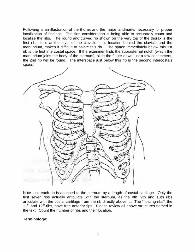
Following is an illustration of the thorax and the major landmarks necessary for proper localization of findings. The first consideration is being able to accurately count and localize the ribs. The round and curved rib shown on the very top of the thorax is the first rib. It is at the level of the clavicle. It’s location behind the clavicle and the manubrium, makes it difficult to palate this rib.. The space immediately below this 1st rib is the first intercostal space. If the examiner finds the suprasternal notch (which the manubrium joins the body of the sternum), slide the finger down just a few centimeters, the 2nd rib will be found. The interspace just below this rib is the second intercostals space.
Note also each rib is attached to the sternum by a length of costal cartilage. Only the first seven ribs actually articulate with the sternum, as the 8th, 9th and 10th ribs articulate with the costal cartilage from the rib directly above it,. The “floating-ribs”, the 11th and 12th ribs, have free anterior tips. Please review all above structures named in the text. Count the number of ribs and their location. Terminology:

9
1. Tidal volume The volume of air in and out of the lungs with a normal breath, approx. 500 ml)
2. Residual Volume
Volume of air which remains in the lungs after a forced expiration (approx. 1200 ml)
3. Inspsiratory reserve volume This term is the extra volume of air which can be inhaled after the person has taken in a normal breath (“forced inspiration”) approx. 3000 ml.
4. Expiratory reserve volume
Is the extra amount of air that can be expired by forceful expiration after the person has expired a normal breath of air, (“forced expiration”) approx, 1100 ml of air.
5. Inspsiratory capacity
The sum of: tidal volume + inspiratory reserve volume; amount of air you can breathe when you forcefully inspire, after taking a normal breath (tidal volume) (3500 ml).
6. Functional residual capacity
Is the sum of: expiratory reserve volume + residual volume. This volume is the amount f air left in lungs after a normal expiration. approx 2300 ml.
7. Vital capacity
The sum of: inspiratory reserve volume + tidal volume + expiratory reserve volume. This volume is the maximum amount of air that a person can expel from the lungs after first filling lungs to maximum and then expel air to maximum extent approx. 4600 ml.
8. Total lung capacity
Is the sum of the maximum volume to which the lungs can be expanded with the greatest possible inspiratory force. approx. 5800 ml.
EXAMINATION OF THE THORAX In the clinical setting, examination of the thorax first includes a gross examination of the patient. The patient will be comfortably seated on the edge of the bed, if possible, to best visualize the thorax and breathing patterns. Keeping in mind the structures of the bony thorax, visually inspect the thorax.

10
Assess the following: Respiratory rate and rhythm
Gross deformities; curvatures, scars, discolorations, etc.
Abnormal breathing patterns (retractions included)
Keep in mind, if there is any gross breathing difficulty, or any other condition which may compromise the patient; emergency first aid should be administered. We will not try to continue the entire respiratory examination if another emergency condition exists. However, for our “routine” examination, the first step is visual inspection. Perhaps during this part of the exam, the nurse should make a mental note of any abnormality which will need to be further assessed by some other method, such as palpation. During the time of the visual inspection, the nurse will be aware of any mental status changes in the patient such as restlessness or lethargy or confusion. These changes may be indicative of respiratory difficulty. Vital signs should be taken prior to the examination for a baseline. Taking all of the above information, the nurse may now continue to the next phase of our examination. Remember to make notes on paper of any abnormal findings as well as the normal findings of the exam. These notes will help you later for charting the findings on the patient’s chart. Accurate information is always important when documenting the patient’s condition. Observing Respirations: The normal respiratory rate is 12 – 18 breaths per minute in adults. DEFORMITIES OF THE THORAX

11
There are many possible deformities of the thorax. The illustration shows two common deformities Funnel Chest and Barrel Chest. While performing your examination you may be unsure as to the proper term or name of the particular deformity. If you are unsure about the correct term, just describe the problem in detail and describe the subjective and/or objective symptoms that may be present.
Funnel Chest may be described as a deformity where the sternum is depressed and results in pressure placed upon the underlying organs. This may cause a change in the blood pressure or pulse; chart these changes if present. Barrel Chest may be described as a deformity which causes a rounded chest where ribs are elevated and separated more than normal. The slope of the ribs is also changed, they are almost completely horizontal. PALPATION: Expose the patient’s thorax providing for as much comfort and privacy as possible. Use the fingertips and flat of the hand in order to palpate the thorax. Use firm but gentle pressure to assess the breathing and movements of the thorax. Next, palpate any abnormalities which you noticed from the first step of this assessment the inspection phase. Palpate the following:
1. Size and shape of the thorax during respirations 2. Intercostal spaces (for bulging or retractions) 3. Any scars or other skin abnormalities (skin temperature as well) 4. Tenderness or pain (palpate gently)
Palpation should be done in an orderly method. First start at either the top or bottom and palpate through the entire thorax surface. Next observe and palpate the posterior thorax. Use the same methodical approach. As with the previous findings, take notes and carefully chart any abnormal findings. Locations: Describe the location of your abnormal findings according to the way your hospital requires. A general method of describing location is to use landmarks such as nipples, nipple line, the midsternal line, etc. Indicate anterior or posterior thorax, and use the midaxillary line location when applicable. When charting your findings, especially abnormal ones, be very descriptive as to specific types of abnormality. If the breathing is abnormal, describe the rate and rhythm. If there is an abnormal node or mass, describe its location and size and

12
whether tender or non-tender. These locations will apply to the posterior thorax as well as to the anterior. Additional landmarks:
1. Midsternal line A line extending downward from the sterna notch.
2. Midclavicular line
A vertical line parallel to the midsternal line and extending downward from the midpoint of each clavicle.
3. Anterior Axillary Line
A line extending downward from the anterior axillary fold.
4. Posterior Axillary Line A line parallel to the anterior axillary line beginning at the posterior axillary fold.
5. Midaxillary Line
A vertical line starting at a point midway between the anterior and posterior axillary lines.
6. Midspinal Line
A vertical line in the center of the back running along the spinal process.
7. Midscapular Lines Vertical lines on the back, parallel to the midspinal line, extending through the apices of the scapulae.
8. Infrascapular Area
Area of the posterior thorax lying below the scapulae
9. Interscapular Area Area of the posterior thorax lying between the scapulae.
PERCUSSION Percussion is of limited use to most nurses. The technique can reveal abnormalities which might be better assessed by palpation of auscultation. Use percussion in conjunction with these other methods of assessment in order to confirm suspicions of underlying pathology. The technique of percussion, striking the chest wall, sets up sound waves. These waves set the underlying tissues in motion, producing audible and palpable vibrations. Using this method is effective for only a depth of about 5 to 7 cm. into the chest wall

13
tissues. Therefore, deeper problems are virtually undetectable by this method. Percussion will be help to indicate the presence of air or fluids in underlying tissues.
As shown in the illustration:
a. Hyperextend the fingers as shown, pressing the middle distal phalanx firmly on the surface of the chest wall. (Do not let the hand rest upon the area as it will decrease the sounds heard).
b. Hold the opposite hand close to the hand on the patient. Flex the middle finger
as shown, retract and strike the area shown by the arrow. Use a quick motion of the wrist.
c. After striking at a right angle, quickly withdraw the finger (the striking finger), so as not to dampen the sounds.
d. Strike an area twice, in order to get a clear sound. Move on to next spot quickly, using a uniform blow each time for comparison.
Always use a quick wrist motion and listen carefully to the pitch of the resulting vibrations set up by the blow. Use this chart for comparison:
1. FLAT High pitch solid tissue beneath 2. DULL Medium pitch firm tissue beneath 3. RESONANT Low pitch normal resounding lung tissue
(semi-hollow or “fluffy” sound) 4. HYPERRESONANT very low pitch very hollow sound, (emphysema) 5. TYMPANY musical very hollow sound (air bubble)

14
Percuss across the tops of both shoulders (apex of each lung; see fig. 4, page 22). Next, move down and move across the thorax, striking each spot as you proceed and noting the sounds produced. Normal healthy lung tissue should resound with a resonant and full sound; as there is normal “fluffy” lung tissue beneath. If there is a hemothorax or pneumonia with fluid present, the sound will be flatter or duller as described in chart above. Remember to percuss anterior and posterior thorax if you decide to use this technique. AUSCULTATION This technique has many indications and is widely used today. It will indicate that there is normal air flow through the trachea and the bronchi, and into the lungs. It can indicate the presence of fluid and/or other obstruction in the air passages. Also the condition of the surrounding tissues can be assessed by this method. Just as with the other methods of assessment, have the patient sit comfortably and have them breath just slightly deeper than normal through an open mouth,. Listen at each spot (described in the following paragraphs) for at least one full breath. Compare the symmetry of the sounds (each side of the thorax). If the patient becomes faint (from hyperventilation during the exam), stop temporarily, and then proceed. You will be listening for:
1. Quality and intensity are they full and easily audible, rate, rhythm, are they diminished?
2. Adventitious sounds abnormal breath sounds; these sounds are
distinguished from the variations of normal breath sounds which can occur due to hypoventilation or hyperventilation.
SOUND: Before we discuss auscultation of breath sounds, we will discuss some of the characteristics of sound. Sound has three basic characteristics; intensity frequency, and duration. Frequency refers to the number of vibrations per unit of time; and it is measured in cycles per second, or Hz (Hertz). What is called a high frequency sound will have a large number of vibrations per second. Wheezes can be of high frequency or low frequency, or can be described as high-pitched or low-pitched. The “quality” of sound is also known as its “timbre.” It is a result of the component frequencies that will make up a particular sound. The quality of a sound will allow us to recognize if a musical note was played on a guitar or a violin or a piano. You may play the exact same note on all instruments, but that note will have a different quality on each instrument. This quality is important when listening to breath sounds and in noting changes in the spoken voice through the stethoscope.

15
The intensity is described simply as the loudness of the sound. Intensity is affected by these factors:
a. Amplitude b. Energy source c. Distance the sound travels d. Medium through which the sound travels
The above factors will determine if the sound is loud or faint or distant. For example, if you are listening to lung sounds through lung tissue that is full of fluid, the sound will be louder because fluid conducts sound better. Sound travels better through material that is denser. Our ears usually hear sounds using normal air conduction of sounds. In a vacuum, no sound is transmitted. Duration of vibrations of sound will determine if our ears interpret sounds as short or long sounds. For example, a patient can have short or long wheezes. The average adult can hear vibrations with frequencies from 16 to 16,000 Hz. Our maximum sensitivity is between 1,000 and 2,000 Hz. Below 1,000 Hz our sensitivity falls off rapidly. Most breath sounds are below 500 Hz, therefore we must listen carefully to all breath sounds, as our ears are not very sensitive to these frequencies. NORMAL AND ABNORMAL BREATH SOUNDS Normally, it is difficult to hear breath sounds. Normal breath sounds will seem faint or distant when auscultating. This is a normal feature of breath sounds. Many authorities have described abnormal breath sounds using varied terminology. The Joint Committee on Pulmonary Nomenclature of the American College of Chest Physicians-American Thoracic Society renamed abnormal breath sounds into two main categories in 1975. These two categories are:
1. Rales: for a discontinuous sound (crackle) 2. Rhonchus: for a continuous sound (wheezes)
In 1980, they further categorized the sounds as:
1. Crackles (course and fine) 2. Wheeze 3. Rhonchus Cugell, in 1978, and Hudson, et al., in 1976 coined terms such as dry rales, sticky rales, bubbling rales, and others. Still other authorities use further derivations of these terms. Each nurse must follow their own hospital policy when they attempt to describe breath sounds. In this text, we use the definitions below for breath sounds:
1. Normal VESICUCLAR low pitch heard over most of normal lung
2. Normal BRONCRO-VESICULAR medium pitch heard over mainstream bronchi
16
3. Normal BRONCHIAL-(TRACHEAL) high pitch normally heard over trachea,
SOUNDS tubular (like wind tunnel)
4. ABNORMAL RALES discrete non-continuous sound, produced by moisture in the lung tissues; can be fine in quality or coarse.
5. ABNORMAL RHONCRI continuous sounds produced by air being forced through narrowed passages, narrowed by secretions and/or constriction of the air passage.
6. ABNORMAL WHEEZES continuous musical sounds produced as air is forced through narrowed passages, like rhonchi, can occur in inspiration or expiration; with
rales, may change character after coughing
7. ABNORMAL STRIDOR loud musical sound of constant pitch, most prominent during inspiration can be heard very well at a distance due to its loud intensity; sound is produced by obstruction of the airway, laryngeal tumors, tracheal stenosis or aspirated foreign body.
8. ABNORMAL PLEURAL FRICTION RUB non-musical sound, usually longer and lower pitch
than lung crackles, sounds like the creaking of old leather; etiology; coarsened surface of the normal pleura, due to fibrin deposits, thickened or inflamed or with neoplastic cells.
9. ABNORMAL MEDIASTINAL CRUNCH (Harman’s Sign) This is a coarse, crackling sound or vibration that is synchronous with systole and is frequently heard over the precordium in the presence of mediastinal emphysema. This distinctive popping or crunching sound is thought to originate from air separating the parietal and visceral pericardium during the contraction of the heart.
10. ABNORMAL BRONCHIAL LEAK SQUEAK This is heard in patients with bronchopleurocutaneous
fistula; a high-pitched squeak over the affected chest area during sustained Valsalva maneuver, the pitch being higher in smaller fistules than in larger ones.
11. ABNORMAL INSPIRATORY SQUAWK A musical sound, squawk found in some patients with
diffuse pulmonary fibrosis; this squawk is usually accompanied by rales (crackles) and also predisposed by hypersensitivity pneumonitis caused by inhaling antigens.
Other abnormal sounds will also be presented in this booklet. They are sounds which can be helpful in diagnosing certain conditions of the lungs (diagnosis here refers to assessing possible abnormal conditions of the lungs….nursing diagnosis). Always place the diaphragm of the stethoscope firmly over the area of the thorax and move it from right to left in order to assess symmetry of the sounds your will hear.(refer to the diagram).
17
Terms relating to the formation of breath sounds:
1. Turbulence Sound that is caused by the uneven flow of air in the human airway; turbulence is thought to be the source for all normal breath sounds.
2. Laminar Flow Air flow in a straight, smooth pipe; if it flows unobstructed, the pressure will gradually decrease and no sound will be created, because there is no turbulence.
3. Turbulent Flow Sound is created by this type of air flow. In the airway, the laminar air flow is broken, and currents form. Currents cause uneven air flow and produce sounds.
4. Vortices This is a whirlpool of air that is started when air enters a wider channel from a narrow one. Vortices are created in the airway and will help to form some of the lung sounds heard when air is also turbulent.
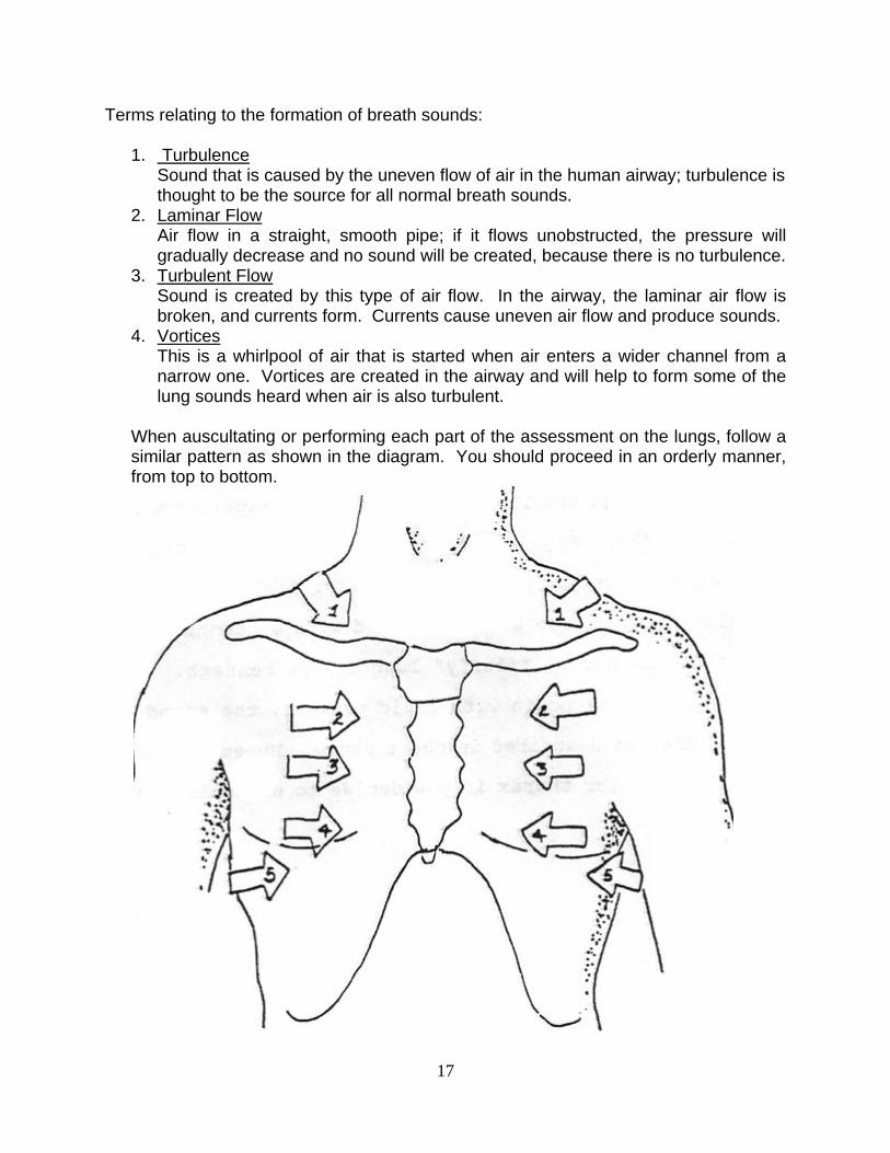
When auscultating or performing each part of the assessment on the lungs, follow a similar pattern as shown in the diagram. You should proceed in an orderly manner, from top to bottom.

18
Begin at the apex of the lung; go right to left side. Next, place the stethoscope on the chest wall, going from side to side, in the same spot on each side. Proceed down the length of the chest wall and using several different spots in the lung field. Remember to be symmetrical, *compare on side, same spot, to other side of chest, same spot. Perform same for posterior thorax. Right lung: 3 lobes a. RUL right upper lobe b. RML right middle lobe c. RLL right lower lobe Left lung: 2 lobes a. LUL left upper lobe b. LLL left lower lobe When performing any of the preceding assessments, be sure to assess all lobes of the lungs. Note that anterior only, a small part of the left lower lobe is able to be assessed. The same holds true for the right lower lobe.

19

20
In the figures, the posterior view is seen, and the largest mass of the LLL can only be assessed by carefully examining the posterior of the chest wall. The nurse must then be sure that the lung fields are auscultated to the lowest point on the posterior of the thorax in order to assess the lower lobes completely. Many disorders such as pneumonia will virtually go undetected if the examiner does not carefully look at and assess the lower posterior borders of the lungs. Also, patients who will lie for long times on their backs may develop fluid collecting in these lobes; another reason for the careful assessment of these lower lobes.
This figure shows a lateral view of the lower lobes; pointing out how the bulk of the mass of the lower lobes is toward the posterior of the thorax.
21
ADDITIONAL BREATH SOUNDS If you are unsure of what you are hearing through the stethoscope, or if breath sounds are diminished, ask him/her to breathe deeper and/or open the mouth wider. Perhaps ask him to breath faster; that may enhance the quality of the sounds you are hearing. Bronchophony This term represents a test to perform on the patient which may indicate that there is consolidation of the lung. Consolidation refers to increased density of the lung tissue, due to it being filled with fluid and/or blood or mucus. Ask the patient to say the words: “ninety-nine” while you listen through the stethoscope. Normally the sound of “ninety-nine” will sound very faint and muffled. When you listen through normal lung tissue, sounds are normally muffled. If it sounds clear through the stethoscope, there is probably consolidation of the lung and Bronchophony is present. This occurs because sound transmission through consolidated tissues will be greater and clearer because dense tissue transmits sound better than normal “fluffy” lung tissue. Egophony This is a term that indicates that there is consolidation of the lung or possible collapse of the lung. Ask the patient to repeatedly say the sound “ee” while you listen with the stethoscope. Normally, it will sound muffled, but it will remain with the long sound of “ee” when you listen over most of the lung field. If the sound changes to “ay” sound, while the patient is saying “ee” then egophony is present. This indicates consolidation, or that there is fluid in the lungs.
Whispered Pectoriloquy This is another term to determine the presence of consolidation of the lungs. You will ask the patient to whisper a number or short phrase and repeat it; such as counting, “1, 2, 3” “1, 2, 3”, etc. and listen through the stethoscope. Normally the whispered voice will be distant and very muffled through the stethoscope. If consolidation is present in a section of the lung field, the whispered voice will sound unusually clear and loud, instead of muffled and distant. Consolidation of the lung tissue causes filling of the air spaces of the alveoli and voice transmission through that part of the lung will be unusually clear and louder than normal. Thus if pectoriloquy is present, it indicates consolidation of some portion of the lung field.

22
COMMON PULMONARY DISORDERS AND PHYSICAL ASSESSMENT FINDINGS USUALLY PRESENT.
a. Bronchial Asthma:
hyperinflation of lungs, impaired expansion, use of accessory muscles of respiration, prolonged expiration and wheezes present.
b. Pneumothorax:
decreased expansion on affected side, hyper resonant or tympanic sounds or even absent sounds in affected areas.
c. Pleural Effusion:
decreased expansion of affected side, trachea & heart shifted away from affected side, dullness or flatness or absent breath sounds.
d. Atelectasis:
decreased expansion on affected side, dull or flat sound or absent breath sounds, trachea and heart shifted toward affected side.
e. Consolidation:
bronchial breath sounds, bronchophony, pectoriloquy, possible splinting on the (pneumonia) affected side.
Summary of Assessment factors:
1. When Inspecting
Look for the slope of the ribs, bilateral and symmetrical chest wall expansion, abnormal breathing patterns, thoracic or abdominal breathing. Look for the shape of the thorax; evaluate anteroposterior diameter relative to lateral diameter of chest wall, pectus carinatum (pigeon breast), pectus excavatum (funnel chest), kyphosis (spine curvature), scoliosis (lateral spine curvature), kyphoscoliosis, and note tracheal position. Look for breathlessness wheezing, sputum, cough, cyanosis, pallor, eruptions, nodules, scars, neck vein distention, fingers for tobacco stains, finger and toes for clubbing, which can be a sign of chronic respiratory disease.

23
2. When Palpating
Feel for masses, nodules, pain, tenderness, examine the: neck, axillae, supraclavicular fossae for lymph nodes, palpate trachea for midline placement. Feel for skin temperature and moisture Feel for other mentioned in the text.
3. When Percussing
Listen for symmetry of sounds from each side. Listen to patient to tell you of pain or tenderness when percussing.
4. When Auscultating
Listen for intensity of sounds one each side of the thorax (symmetry) Listen for normal and abnormal breath sounds.
Following, we will present detailed outlines of the method for assessment. Today, nurses are taking increased responsibility for assessment of lungs, including auscultation. However, there are still many differences in levels of responsibilities among nurses in different hospitals. Some hospitals do not allow any nurses to chart any breath sounds at all. Other facilities want all nurses to listen and record all patients’ breath sounds. There is also every situation in between these two extremes. We will present guidelines for those nurses who will have this responsibility of listening and charting breath sounds. If you are in a facility that does not allow you to record breath sounds, you may still listen to the lungs and at least chart that you notified someone that the patient sounds “congested.” In most facilities around the country, you may at least chart “congested” lungs if you are not allowed to chart terms like: “rales,” “rhonchi,” etc. CHARTING THE EXAMINATION FINDINGS When charting the normal exam, most nurses, for brevity, will chart only that respirations are “normal” and there is no “SOB.” In most cases, that is acceptable for a routine or normal examination. However, it is very possible to be brief and thorough.
1. Inspection observe: shape of chest; include deformities width or costal angle, movements of intercostal spaces during respirations use of accessory muscles of

24
respirations local impairment of respiratory movements rate and rhythm of respirations.
Charting of these normal findings might be: resp rate-20/min, regular, no SOB1
2. Palpation a. identify areas of tenderness
b. assess any observed areas of abnormality c. assess respiratory excursion (expansive movements of the chest during breathing) d. assess skin condition (temperature, etc.)
3. Percussion a. assess any areas of dullness, flatness, tympany
b. assess areas found to be abnormal from previous examinations.
4. Auscultation a. assess quality and intensity of breath sounds
b. assess patient for abnormal breath sounds c. assess patient for areas of consolidation
When charting your findings, you may not be sure as to exactly what you are hearing. Most hospitals do not require that palpation and percussion results be charted. If the nurse carefully assesses the breath sounds, those others may not need to be charted, but are still used to confirm the nurse’s assessment of the patient’s problem. If the nurse is unfamiliar with naming the individual breath sounds, you should be very descriptive when charting. 1
For example: chart the location and sound that you hear….. moist respirations in LLL and RLL……or fine rales in LLL and RLL (either is correct) Do not feel that you must always tag a name to the type of abnormal respirations that
you hear. It is sufficient to accurately describe the abnormal breathing. Another important function is to follow up the results of your exam if there is an abnormality. Your nursing diagnosis will include nursing orders to turn the patient more frequently or to suggest that respiratory therapy be performed on the patient. Therefore, communications is important, but so is the nursing follow-up on your findings.
1 SOB may mean other things to other people; only use abbreviations that are accepted and approved by your hospital. Also remember that your facility may require other items to be included in your charting; always follow procedure. -25-
25
GUIDE TO ASSESSMENT OF LUNGS AND THORAX
1. Assemble Equipment 2. History-taking 3. Explains Procedure to the patient 4. Washes hands 5. Gowns or drapes patient to prevent unnecessary exposure 6. Provides a quiet place for patient comfort and for auscultation 7. Provide adequate lighting 8. Use of proper Techniques:
a. inspection, palpation, percussion, auscultation b. compares symmetry of thorax (each hemothorax) c. starts at neck, then posterior, right and left lateral, then anterior thorax
9. Respiratory rate determination 10. Rhythm determination 11. Depth determination 12. Abnormalities:
a. defines boundaries of abnormality is found; describes accurately b. do not allow patient to hyperventilate during the exam. c. avoids bony prominences during the exam (poor sound conduction) d. records findings accurately
POSTERIOR THORAX EXAMINATION
1. Patient seated with arms folded across chest 2. Inspects symmetry, contour, color, skin condition 3. Palpates posterior interspaces for masses, lesions, etc. 4. Palpates ribs and scapulae for masses, breaks, etc. 5. Evaluates tactile fremitus 6. Evaluates respiratory excursion 7. Percussion – 5 cm intervals from apex to base contra laterally 8. Diaphragmatic excursion 9. Ausculate breath sound 10. Ausculate voice and whispered sounds
RIGHT AND LEFT LATERAL THORAX
1. Patient seated with arms on head 2. Begin in the axillae and proceed downward contra laterally using at least 4 or 5
sites for comparison 3. Inspects for symmetry, color, condition of skin 4. Palpate ribs for masses or bulges 5. Palpates tactile fremitus

26
6. Percusses lateral thoraces 7. Auscultates breath sounds 8. Auscultates voice and whispered sounds
ANTERIOR THORAX
1. Patient is supine with arms abducted; child is placed totally flat and head is not allowed to turn.
2. Inspect anterior chest for symmetry, contour, color, skin condition 3. Palpate ribs and interspaces for bulges and masses 4. Palpate for tactile fremitus 5. Palpate trachea 6. Percuss anterior chest at 5 cm intervals 7. Auscultate for breath sounds 8. Auscultate for voice and whispered sounds.
CHARTING EXERCISE: This is not part of Posttest for this course: for practice only.
1. Chart a brief narrative of a “normal” lung assessment 2. Chart on a patient who has COPD with an acute attack.
a. General - - - - - - b. Rate, rhythm, depth (difficulty) - - - - - c. Auscultation results - - - - - -
S- (subjective) O- (objective) A- (assessment) P- (plan - -nursing orders)
ADVENTITIOUS SOUNDS: RALES: (or crackles) Definition:
a. Clusters or showers of sounds b. Produced by bubbling air through the alveoli, bronchioles bronchi c. Non-continuous d. Variable quality:
Types:
Fine rales: terminal bronchioles and alveoli, sounds like hair being rubbed between fingers. Medium rales: larger air passages, bubbling sound of opening a carbonated beverage. Coarse rales: louder and lower-pitched from larger passages

27
RHONCHI: Definition:
a. Produced by air travelling through narrowed passages or through mucus in the passages.
b. Varying sound quality c. Continuous sound
Types: Fine rhonchi Coarse rhonchi (sonorous) Sibilant rhonchi (wheezes)
FRICTION RUB: Definition:
a. Coarse grating sound b. Inflamed surfaces of the pleura rub together during respirations c. Usually over anterolateral thorax
PART II MENTAL STATUS ASSESSMENT Course Outline: MENTAL STATUS EXAMINATION Part I Introduction to the Mental Status Assessment
A. Reason for performing the exam B. Examination techniques
PART II Overall Assessment
A. General Appearance, Grooming B. General Behavior
PART III Intellectual Functioning
A. Orientation B. Abstract Reasoning C. Judgment
PART IV Mood and Affect

28
A. Factors used to assess mood B. Clinical implications
PART V Thought Cohesiveness
A. Normal and abnormal thought content B. Nursing implications
PART VI Performing the Exam
A. Step-by-step procedure B. Nursing implications C. Deviation from the above procedure
PART VII Documentation of the Examination
Course Objectives Behavioral Objectives: At the end of this course, each participant will be able to:
1. Name and discuss at least two reasons for performing the mental status examination
2. Name and discuss the five general observations used while performing a complete mental status examination.
3. Name and discuss five areas of the overall physical assessment which relate to the mental status assessment.
4. Name and discuss at least five parts of the mental status assessment. 5. Name and discuss five areas of intellectual function; describe each one in detail. 6. Name and describe at least one method of assessing each of the six areas of
intellectual assessment. 7. Describe nursing implications of patients with disturbances in each of the six
areas of intellectual functioning. Introduction The mental status examination should always be included in the overall physical assessment of all patients. The assessment you perform may be either an initial admission assessment or it may be the daily, on-going assessment. In either case, the mental status assessment is an essential part of the examination. As you perform your medical assessment of the patient you will perform parts of the mental status assessment, almost without being aware of it.
29
In general terms, mental status could be described as an individual’s state of awareness and responsiveness to the environment. It also includes the more complex areas of a person’s mental functioning, such as intelligence, orientation, thought process and judgment. As you see, mental status is very dependent upon other body systems. Physical illness may certainly impair mental status. In describing the techniques of assessing mental status, you should remember to incorporate parts of this examination into the patient’s general physical exam. If the mental status exam is presented in one separate group to the patient, the patient will usually become very anxious over the types of questions being asked. You can assess mental status as you perform the medial exam. For example, memory is assessed while taking the history. Mood can be assessed when you meet the patient. Mental status findings are important. Patient care plans may have to be altered in order to properly care for patients with impaired mental status.
The following guidelines should be used for the assessment:
1. First impressions Record your first impressions of the patient. Some very subtle thought disorders may be detected. As an example, the patient acts just a little peculiarly, but you don’t see anything grossly wrong.
2. Medical survey
Incorporate the mental status exam into your medical survey (general physical assessment). There are parts of the mental status assessment that may indicate an organic process as well as a mental illness.
3. Explain procedures
Always explain to the patient what you are about to do. Most patients are very anxious just to be in the hospital, not to mention the sensitive questions you are going to ask them.
4. Take notes
Take notes during the assessment. Always explain to the patient what you are doing. You are taking notes so that you will not forget anything important.
5. Use common sense
If they say they are depressed, and might want to die, finish the remainder of your interview and have someone stay with the patient; report your findings, but make sure someone constantly stays with the patient; safety first. The following outline is the basic mental status exam. As each section is presented, techniques will be discussed.

30
PART I Overall Assessment PART II Intellectual Functioning PART III Mood and Affect PART IV Thought Cohesiveness Again, use common sense; the examination does not have to be performed in this exact order. The exam is presented in this order only to give the nurse a guide to follow. It is a way to organize your own thinking before you begin to assess patient’s thoughts. Now proceed through steps of the exam in the following pages.
PHYSICAL ASSESSMENT: The following topics are part of the routine daily assessment of most patients. As you read and review each system, be aware of the possible abnormalities of the mental status examination. A. Neurological Assessment
Changes in level of consciousness; restlessness, listlessness, confusion, disorientation, others. Any of these changes may or may not accompany functional disorders, or can be only temporary symptom of a medical problem.
B. Respiratory System
Dyspnea, hyperventilation, others. Persons with certain disorders of the nervous system will manifest signs of respiratory distress; therefore, the mental status may also be affected.
C. Cardiovascular System
Rapid or irregular pulse or even the opposite may occur, a slowed pulse rate; many other changes may be obvious, such as the depressed patient who may have a slowing of all body systems.
D. Gastrointestinal System
These changes may include the minor complaints such as chronic indigestion, nausea, cramps, vague stomach pains, can also include symptoms such as vomiting or diarrhea or constipation.
E. Genital-Urinary System
Urinary symptoms may include the following: frequency or retention, scant urine which is concentrated may be present in the person who is depressed and not taking adequate fluids.

31
There may be other symptoms in addition to these. This is not to say that if a person has any of these symptoms, they also have a change in their mental status. All we can conclude is that if there has been no diagnosed reason for the particular symptoms, the nurse should always consider that a possibility can be an altered mental status with physical symptoms. Now we will discuss the next step which is mental status assessment with each step listed for clarity. Overall Assessment 1. History
This is usually obtained during the medical history. Items to be included are drugs taken, trauma surgery, etc.
2. General Appearance
This part of the exam is often overlooked. Be sure to note the manner of dressing, grooming, and hygiene: abnormal: sloppy clothes, body odor, dirty clothes, all could mean OBS or depression. Very tidy or meticulous grooming may mean obsessive-compulsive personality.
3. Facial Appearance
Note facial expressions and appearance. Abnormal: depression and some other diseases can cause an inappropriate facial expression.
4. Posture A normal reaction to hospitalization can make a person tense and unable to relax. Do note if they are too tense or too relaxed.
5. Motor Assessment
Includes patient’s gait, speech, and general motor activity. Speech and motor activity are the most pertinent to our mental status examination. Abnormal: altered speech and motor activity can indicate depression, organic disease or other functional disease.
6. General Behavior
This is the: “First Impressions” category. Is patient open to your questions? Is the patient cooperative? Is the patient relaxed? Abnormal: belligerence, hostility, combativeness, would be considered a sign of some type of disease process.
A. Intellectual Functioning
Intellectual here means the higher brain functions of cognition which were mentioned earlier. By the higher brain functions, we mean that there must be some thought used, the brain must be used to its fullest capacity; i.e. thought, integration of memory and the conscious mind, Presented now, are those higher brain functions which will be assessed in our exam.

32
1. Orientation
Most nurses are familiar with this phase of brain function. Orientation is measured in time, person, and place. During your interview, it should become apparent the person is confused. Be precise with questions; time of day, day of week, date, month and the year. Start questions from the general to more precise questions. The patient may not know it is August 24, but they may know it is the month of August. Use this method for person and place as well.
2. Communications Skills
This category includes vocabulary used, information facts, spelling and reading.
Assess these qualities only if needed. To perform a detailed examination in these areas, it might be necessary to consult another reference which lists vocabulary words, spelling word exams, reading tests, etc. Most nurses would not ordinarily need this much detail, unless they work in a specialty area such as neurology. Abnormal: Vocabulary is an excellent method of assessing intelligence, as well as information facts. Some examples are:
a. How many days are there is a week? b. What is a major city in Italy? c. How many ounces in a pound?
3. Abstract Reasoning
This area includes the ability of the person to be able to interpret abstract concepts. To test the person, ask proverbs and their meaning:
What do these mean?:
a. A stitch in time, saves nine. b. A rolling stone gathers no moss. c. The proof of the pudding is in the eating.
Abnormal: The disorder of not being able to think abstractly is called concrete thinking. The person takes the words by their literal and actual meaning. If you were to direct the patient to: “take a seat, please” the person would pick up the chair and ask you where to “take” it. Test them by using proverbs. Always remember that there are other factors which can influence this test. Persons who are from a different country and are not proficient in the English language, will also test poorly in this area unless they are tested in their own language.
33
4. Attention Span
Clinically speaking, this category includes the ability to pay attention to the interviewer and to concentrate on the subject of the interview. There are some methods of testing for this ability. Use of the digit span and the serial numbers sequence is the method of choice. For our purposes, however, these methods are also too time-consuming, and the information gained would have to be evaluated by an expert. In most instances, the physician performs this test and the others like it.
5. Memory
Most memory deficits will be apparent during the history-taking process. First, test the patient for long-term (remote) memory by asking birthdays, anniversary children’s birthdays, etc. Test short-term memory by asking recent events. Also to test the recent memory, you can tell the patient a fact that he did not know previously, then ask them to recall the fact at a later time. Start at five minute intervals and then make the time longer or shorter, depending upon how the patient performs. Abnormal: Of course, lack of memory is abnormal. If the patient exhibits partial loss or transient loss of memory, that is significant, and needs to be assessed carefully. Also of great significance, is if the patient makes up answers to your questions (confabulation). When testing memory it is best to ask questions which can be easily verified. Recent memory can be assessed by asking the name of an object or address. If the patient answers wrongly, then ask the question again, very clearly, as he may not have heard you and answer the question wrong.
Another way to test memory is to ask information questions such as:
a. How many days are there in a week? b. What is the capital of Italy? c. What must you do to water to make it boil? d. When is Memorial Day? e. What are the four seasons of the year? f. What is a prime number? g. Where does the sun set? h. Who wrote Moby Dick?
Persons of average ability should be able to answer up to 75% of the questions correctly. There are cultural differences to consider. There are intellectual considerations. However, the average person will answer eight out of ten such questions posed. If you are unsure of the patient’s intelligence when answering the above questions, use the test below in order to determine if they are of at least average intelligence.

34
Vocabulary Test: Ask the meaning of the following: apple donkey diamond join fur shilling bacon seclude spangle flout recede amanuensis dilatory microscope
Using this test, and some of your own words, you can determine if the person is of average intelligence. Again, cultural and educational backgrounds may prevent a person from performing well on this test, so use your judgment when interpreting the results. Above 50% would be considered normal (using increasingly difficult words in a list of at least 14 words).
6. Judgment This area can also be assessed during the medical survey. Note if the patient has acted with good judgment prior to admission to the hospital. Do they continue to use good judgment while in the hospital? For example, do they keep trying to get out of bed even though they have been instructed not to?
Abnormal: Judgment is one of the higher brain functions. It is usually the first quality of the patient to deteriorate in the event of disease. Even the administration of hypnotics or narcotics can “cloud” the patient’s thoughts enough that they can show poor judgment, so take drugs into consideration. Trust your judgment in assessing the patient’s judgment, and their behavior.
B. MOOD AND AFFECTS Assessment of mood is usually a simple task for most nurses. There are physical signs that the patient exhibits, that will give a clue to their mood. Also do not forget that mood changes can be subtle and can happen quite unexpectedly. It is usually easy to detect the depressed patient; but what about the patient who has just the opposite symptoms? There is also great difficulty in assessing the patient who is just slightly “high” or “manic” as we usually do not know what they were like before they entered the hospital. In these cases, you should consult the family of the patient. Ask questions such as, “Is he more manic than usual?” or “Is he more restless than usual?” If a patient seems depressed or “down” to you, do not be afraid to ask them, “Are you depressed?” If they answer “yes,” “Do you feel that you might hurt yourself?”
If a person is depressed, they should be assessed for any suicidal tendencies. Always take into consideration the physical evidence as well as the mood of the patient when planning your nursing care. If your patient is depressed, or very upset, they should be evaluated for a psychiatric condition.
Now, to deal with the term, “affect.” This term goes one step beyond the definition of mood. Affect means assessing the patient’s mood and their behavior. Affect means the

35
“appropriateness” of their mood and behavior. As an example, a patient’s spouse has just died in a car accident.
The person acts and speaks “normally” without emotion. This is not a normal affect, they should be showing grief. The person’s outward mood may be “normal” for anyone else, but considering that there was JUST a death in the family, their affect is not normal.
Therefore, “affect” is a combination of mood and behavior of the person. A slightly depressed affect might be normal in the above situation. I would worry more about a person who seemed normal, than the person who was crying over the death of a spouse. At least their affect is normal for the situation. When you assess mood and affect, take into consideration all of the above facts; this is why a good history is important.
C. THOUGHT COHESIVENESS
The previous section describing Mood and Affect, will now lead us into discussing this section on the thought process. If a person’s affect is inappropriate or grossly pathological, there is probably a thought process disturbance. Listed in this section, are most of the major thought disorders and their definitions. Always remember that these disorders are functional; but in any case, psychiatric attention and treatment may be necessary.
NEUROTIC DISORDERS These are associated with neurosis … the patient is probably functioning at an adequate level, outside the hospital, but still does need some type of medical attention. In neurosis, the person is still in touch with reality; meaning that there is no major thought disorder, however, the conditions may become worse and can interfere greatly with the person’s life.
a. Obsessive-Compulsive Behavior: The obsession is the recurrent thought that the patient has that they should perform some type of behavior that most other people would consider abnormal. The compulsion is the actual performing of the act, or in other words, acting upon the obsession. A very common example of this is that of compulsive cleaning. The person who is so absorbed with cleaning, that they take clean clothes out of the closet and wash then again. This behavior will not actually hurt anyone but it will tend to limit the life of the person who is afflicted with this obsession and compulsion.
-37- b. Ruminations: This term refers to repetitive thoughts or ideas that the patient
expresses. These thoughts are usually centered around abstract ideas or concepts.
36
c. Phobias: These are irrational fears expressed by the patient. They can be extremely anxiety-producing for those persons. Most of us are familiar with the common phobias. Normally, these phobias do not interfere with the person’s life, The person will usually just avoid contacting the situation which makes them anxious.
d. Anxiety: (Free-floating anxiety) the person expresses a sense of dread. They
are usually unable to define exactly what they are not afraid of, but it can become very strong and the patient has feelings of impending doom.
PSYCHOTIC DISORDERS
These are usually associated with the more severe thought disorders and usually with schizophrenia. The person with a psychotic disorder is usually gravely ill. This disorder can interrupt and limit the quality of the person’s life.
a. Feelings of depersonalization: The patient has feelings that he/she is not real. He/she exhibits feelings of changes in him/herself and in his/her personality.
b. Feelings of unreality: The patient has feelings that everything in his/her
environment is unreal. This is different from the above, where the patient feels that he/she, him/herself, is unreal. In this case, he/she feels everything around him/her is unreal, or changed in some way.
c. Feelings of persecution: The patient has vague feelings that all people are plotting against him/her or that they do not like him/her. This disorder obviously borders on paranoia.
d. Feelings of Influence: The patient feels that others are controlling him/her and his/her life.
e. Feelings of reference: The patient has feelings that everything is about him/her. The radio is talking to him/her alone so does the television, so do all other events take place due to him/her.
f. Delusions: Delusions are false beliefs which the patient holds. These beliefs are usually well fixed in his/her mind. The delusions may be very simple, such as he/she believes him/herself to be the president. They may also be very elaborate, such as the patient acts out his/her entire life as if he/she were Jesus; on a day-to-day basis he/she acts like he/she were Jesus and believes that he/she is.
g. Illusions: The patient misinterprets outside stimuli. In other words, the patient
may look at a pencil, but he/she sees a snake. This disorder is not usually as elaborate as a hallucination, and there is a stimulus. The patient just misinterprets the object(s).

37
f. Hallucinations: A completely unreal sensory experience by the person. A hallucination has no basis in reality as did an illusion. Hallucinations can be visual, auditory, olfactory, or by any other of the senses, such as touch. The last several disorders may, in part, be caused by a perceptual disorder. They can all be caused by a physical or emotional disorder as well. Abnormal Findings: Drugs, OBS, fever, dehydration, and other organic stimulants may be the cause of the preceding psychotic disorders, as well as being functional in origin. The nurse can be very important to the proper diagnosis of these conditions. Many times these disorders may be classified as a psychiatric disorder, when actually the patient was just extremely dehydrated and started to hallucinate. Be careful to document these following items when you are assessing your patients:
1. Symptoms get better or worse at different times of the day. 2. Drug history of the patient 3. Changes in vital signs coincide with changes in the symptoms 4. Psychiatric history (if any) 5. Venereal disease history of the patient 6. Any historical information
These and any other factors you might notice can be very important. Many nurses have developed that “sixth sense.” You know that something is wrong, but you just cannot put your finger on it. Sometimes a patient may say something just a slightly odd. Don’t dismiss it! Investigate everything! Those remarks that the patient may make might be significant. So look for any other signs of a thought disturbance. D. Performing the Examination
Those nurses who work in a psychiatric setting will have some very specific guidelines for performing the mental status examination. Therefore, we will not spend much time going over those settings. However, a word of caution to even the most “seasoned” psychiatric nurses; do not become so “routine” with your exams that you overlook some obvious signs and symptoms. If you do use a form for taking your interview, stop first and take a critical look at the patient. No patient can fit your form exactly. No matter how comprehensive your outline is, each patient will have some sign or peculiarity that needs further documentation. In summary, look at your patient first, and not the form that you may be using. For medical-surgical nurses reading this; do not be afraid to ask your patient any of these questions. Those nurses who are not accustomed to asking these questions, will feel uncomfortable asking the patient certain questions. For example:

38
1. Do you feel like hurting yourself? 2. Do you feel people are against you? 3. Do you see disturbing sights that other people do not see? Ask these questions tactfully, and if the patient acts or feels uncomfortable with these questions, it could be that there is a problem; it could be a significant finding. Chart: “The patient denies having hallucinations, but becomes very anxious when questioned about it.” This will alert the doctor that maybe the patient does have a problem that needs looking into. Charting will be discussed later. The following outline should be followed by complete mental status examination:
1. These will be obtained in your medical history: a. General appearance b. General behavior c. Orientation (general) d. Motor activity e. Level of consciousness f. Mood and affect
2. Assess abnormalities in any of the above:
a. Dirty clothing b. Depression c. Hallucinations d. Anxiety e. Disorientation f. Others mentioned
3. Intellectual functioning:
a. Assess orientation b. Judgment c. Abstract reasoning d. Others mentioned in text
4. Mood and Affect:
a. Is their mood normal b. Others mentioned in the text c. Is affect appropriate for the situation
5. Thought Cohesiveness:
a. Thought content – are responses appropriate b. Nursing measures – are any emergency measures necessary.

39
Summary: The main point to remember when performing this assessment is that many of the areas mentioned will be assessed during the general medical assessment. Always, however treat the mental status exam as a separate examination. It is an important step, and it should be handled separately in order to fully be able to assess the patient’s level of mental health and adjustment to their hospitalization. In a real-life situation most nurses do not have time to perform a detailed and separate mental status assessment. Therefore, one purpose of this course is to force you to think of each part of the exam, and to be more observant while performing the general physical assessment. The observant nurse can perform the complete mental status exam in a very brief time. So even to those nurses who say, “I do not have time to ask all of those questions.” Even for those nurses, we say that you do have time, if you are able to sharpen your skills as an observer. E. Charting As we have seen in the previous section, all of your observations are worthless unless you chart them accurately and completely. In this section, we will not try to change your method of charting but rather we will try to help you organize your thoughts better and to chart more completely. Most nurses have their own method of organizing their charting. On a more practical note, we will instead try to present guidelines to increase your awareness of what is important to chart, and to draw valid conclusions. 1. Chart general findings first: (even if normal)
Does the patient appears stated age, dressed casually, is cooperative, follows instructions well is alert, responds appropriately to questions. Also report history pertinent to the medical exam.
2. Intellectual:
Patient is oriented in three spheres, shows no impaired judgment, nor impaired memory.
3. Mood:
If mood is very depressed or very inappropriate, this category will be mentioned first in our charting, (that goes without saying). However, if the mood is normal, you might just chart that the patient is in good humor or in good spirits; Affect is appropriate or not.
4. Thought Cohesiveness If this is normal, there is no need to mention it except to say, “Responds appropriately.” If abnormalities present, describe them.
5. General Impressions:
All nurses should allow themselves a space for narrative description of the patient. Perhaps your patient will not nearly fit into any of the above descriptions perfectly.
40
Perhaps you are not sure what is wrong with him/her, and need to describe it. Also remember that many facilities now use checklists for charting must of the “routine” assessments. Be careful to check each item carefully, and to write any abnormal findings that do not fit into the categories of the checklist type of charting.
Acute and Chronic OBS The following is a comparison of two major types of organic brain disorders. The two will first be discussed, followed by a comparison of signs/symptoms. The two types of disorders are acute OBS (delirium), and chronic OBS (dementia). With the acute disorder many of the same symptoms may be present as with the chronic disorder. Delirium, however, seems to have a more fluctuating level of consciousness than does dementia. Cognitive functions for both disorders are the same. Both patients will be disoriented (usually), attention and memory will become worse. Judgment and perceptions are usually poor. This is a comparison of the two (Chronic vs. Acute OBS): Acute OBS (delirium) Chronic OBS (dementia)
a. cognitive functions deteriorate a. cognitive functions deteriorate b. mood—anxious, fearful, labile b. irritable, labile c. behavior – restless with fluctuating c. deterioration of personal habits
level of consciousness d. thought content – delusions, d. if any of these three are present
illusions, hallucinations, might (delusions, illusions, be confused. hallucinations) they will be transient and mild’; patients are confused
As you see, one of the most distinguishable factors of chronic OBS, is confused. However, if the patient has delusions or hallucinations, they are transient (come and go quickly). Causes for the acute type are numerous, Chemical toxicity can cause the acute type. Drugs, whether prescribed or non-prescribed, can cause acute OBS (organic brain syndrome). If the body cannot tolerate high doses of any drug, toxic symptoms will be seen. Other causes for the acute type are: fluid and electrolyte imbalance (severe ones), heavy metal poisoning, chemical poisoning (other than drugs already mentioned), and others. When the nurse performs the mental status examination keep in mind the above factors about OBS. Also remember the safety of the patient. Whether the patient is young or old, they can hurt themselves when they are in this state of agitation or confusion. Make sure you pad the bed side rails if necessary, restrain if necessary. However, many is the time the unsuspecting nurse was injured by that “frail little old lady” in room 210. So think safety! Restrain the patient if needed; for the safety of all!

41
PART III NEUROLOGICAL ASSESSMENTS COURSE OBJECTIVES: At the end of this course, each participant will be able to:
1. Discuss the parts of the general medical examination performed before beginning the neurological assessment of the patient.
2. Explain in detail at least four diagnostic tests relating directly to the nervous
system.
3. Discuss the importance of the diagnostic tests and their results, in relation to the neurological assessment which each nurse will perform.
4. Discuss important neurological findings, both normal and abnormal, for each of the following conditions: brain tumors, C.V.A.’s, meningitis, subdural hematoma.
5. Discuss important medical findings and specific neurological findings relating to the neurological findings, both normal and abnormal, for the following: cardiac surgery patient, polyradiculitis, diabetic neuropathy.
6. Discuss at least five possible abnormal findings relating to the neurological examination for the infant with cerebral palsy.
7. Name and discuss the five areas of assessment as they pertain to recording the neurological assessment.
8. Name and discuss at least three items that would be significant of pathology in the patient history.
9. Name and discuss the two significant parts of the motor examination
10. Name and discuss the assessment of the cranial nerves, and the three main parts of the examination.
11. Name and discuss the three main considerations of the nursing care plan for the neurologically impaired patient.
INTRODUCTION: The neurological examination that is performed by many nurses today includes only a gross examination of the patient, Most chronic and acute medical and surgical nurses

42
have no need for a detailed exam. However, this course deals with an exam for nurses who are involved with very special nursing situations in which a more detailed neurological examination is necessary. It will be assumed in this course that you have a basic understanding of the anatomy and physiology of the nervous system. If you need to refresh yourself, you may use any basic anatomy and physiology textbook as a reference. This course will present the nurse with a quick and thorough technique for performing a neurological exam. It will also describe in detail some special nursing situations which are common to the acute care med/surg nurse and the critical care nurse. As you begin your general physical assessment, you may notice some symptoms that will reveal the need for a more detailed neuro examination. This text will provide a step-by-step procedure for performing the exam. GENERAL NEUROLOGICAL EXAMINATION 1. Patient History A detailed history is always important before starting the examination. If this exam is the first one given to a patient, such as the admission assessment, the nurse will usually complete a general form or questionnaire stating the history of the patient. If any of the questions suggest a neurological problem, the nurse will then ask questions more pertinent to the neuro status of the person. As always, the nurse must intervene if there is an emergency discovered during the examination. However, for this course, we will assume that the patient is “THE AVERAGE PATIENT.” Some specific items to include in the history are: syncope, pain, bladder or bowel incontinence, seizures, diplopia and others. Often the relatives can give a better history, especially if the patient’s level of coherence is diminished. In summary, the neurological history is often part of the general medical history. The nurse must be careful to intervene should there be a potentially life-threatening problem encountered. Otherwise, the nurse will continue to assess the neurological status of the patient, while obtaining the general medical history of the patient. After the general questions are asked and answered, the nurse can more carefully assess the neuro status of the patient. Following, there is a list of items to be included in the medical and neuro exam of the patient. Remember that this is only a partial list. Some items can be excluded if the answers were already obtained at an earlier time; there may be other items which the nurse may wish to add to the list, due to specific patient problems or responses to questions.

43
History-taking: Relatives are often a source of information, especially when the patient is unclear or unconscious. Remember to be complete to start with a general medical history if you have not already obtained one,
A. General medical considerations:
1. surgeries 7. diabetes 2. cancers 8. hypertension 3. major illnesses 9. Vascular diseases 4. anemia 10. taking any medications 5. childhood illnesses 11. infectious diseases 6. metabolic disorders
B. Specific Neurological history
1. seizures 6. diplopia 2. pain 7. muscular Weakness 3. bowel or bladder incontinence 8. headaches 4. nervous disorders 9. blackouts 5. syncope
If any of the above or related problems are present, the nurse will follow up the problem by asking further, very specific questions regarding the problem. For example, if seizures were answered “yes”, the nurse would ask questions such as:
1. When did you begin having seizures? 2. When was the last one that you ever had? 3. How long did they usually last?
4. Have you now or ever taken medication for the seizures, and if so, what is the
name of the medication(s)?
You will try to localize the problem as much as possible. Always read the medical history that the physician obtained first. You can save asking the patient many repetitive questions, if you first find out what information has already been obtained. Keep all this information in mind, as you proceed through the steps of the neuro exam.
44
2. PERMORMING THE NEUROLOGICAL ASSESSMENT In basic nursing school, you undoubtedly learned a specific order in which to conduct the assessment. In fact, the order of the exam is usually unimportant. Use any specific order for the exam that is logical and makes sense for your patient. For purposes of this text, we will discuss the neuro exam in terms of the three major divisions of the neurological system, and then proceed with the examination: Parts of the nervous system:
A. Central Nervous System B. Autonomic Nervous System C. Peripheral Nervous System
A. Central Nervous System (CNS)
1. Brain – ventricles, skull, brain stem 2. Spinal Cord
reflexes, (deep tendon reflexes): biceps, triceps, ankle, brachioradialis, knee superficial reflexes: abdominal, and others
B. Autonomic Nervous System Sympathetic and parasympathetic divisions: heart rate, respiratory rate, constriction and dilation of pupils, constriction and dilation of blood vessels, salivation, many others.
C. Peripheral Nervous System (spinal nerves) Pain, temperature, balance and the cranial nerves.
The above is a guide to the general sequence of the neurological assessment. Some other authorities divide the exam into different sections, such as cerebral function, motor function, sensory function, etc. However, these are just different terms for the same examination. The above divisions and the guide to follow will be the best method to use if you are a beginner. As you proceed through the exam, keep the following times in mind:
Cerebral function: General behavior, level of consciousness, intellectual functioning, emotional status, thought content (mental status), cortical sensory interpretation, language, etc. Cranial Nerves: Special senses, facial nerves, other combined sensory/motor nerves Motor: Muscle tone, muscle size, involuntary movements, muscle strength.

45
All these systems are a part of basic nursing assessment. However, if you need a review, you may use any textbook in assessment in order to refresh your memory. Once you have reviewed the above, you may proceed to the next section of this text which discusses the aspects of nursing assessment and the acutely ill patient.
3. DIAGNOSTIC TESTS Skull X-Ray is usually one of the first tests performed in cases of known or suspected neural injury. This will reveal configuration, intracranial tumors, calcifications, vascular markings and densities. Assessment of the patient, during the procedure, is usually limited to the stabilizing the vital signs and possibly assisting the technicians. CAT studies (Computerized Axial Tomography) is an imaging method used to provide a cross-sectional view of the skull (or other body part), and shows varying densities of select tissues. This test can be diagnostic of tumors, infarcts and other lesions of the brain and/or spinal cord. Assessment of the patient during the procedure includes stabilization of vital signs as the patient is usually moved to a remote area of the hospital for the test. A permit must usually be signed and the patient instructed to lie very still in order to obtain the best results. EEF, (electroencephalography) is a non-invasive test. It provides for physiological assessment of the electrical activity of the brain. The test may be done while awake, asleep or during activity. Nursing assessment will include reassurance of the patient since electrodes are placed upon the scalp. Many of the patients think that they are going to get a shock. Also withhold medications as per doctor’s order before the test. Usually any stimulant drug(s) are withheld; coffee, tea, stimulant drugs, etc. EMG, (electromyography) is used to diagnose the presence of neuromuscular disorders. Needle electrodes are paced into the skeletal muscles in order to study the changes in electrical potentials. Assessment of the patient is usually limited to observation after the test, since there will be some discomfort and muscle soreness, similar to the discomfort experienced after an intramuscular injection. Air contrast studies, include pneumoencephalogram, fractional pneumoencephalogram and ventriculogram. Air replaces the fluid in the closed spaces of the cranial cavity, in different and selected locations. The air acts as a contrast medium and it is less dangerous than injecting chemical contrast medium. Air is less dense that fluid medium and will outline shadows on x-ray. Assessment includes observations for signs of increased intracranial pressure, level of consciousness, neurological signs, infection, fever, and hydration status. Radioisotope Brain Scanning, involves intravenous injection of a radioactive substance, and the subsequent measuring of the particles emitted after scanning of the patient. There is usually an increased uptake or decreased uptake of this “dye”” at areas of pathology. The nurse’s role in assessment of patients undergoing this test is limited.

46
There is a minimal danger from radiation, and other than the injection, the test is non-invasive. Cerebral Angiography, uses an injected contrast medium in X-Ray studies designed to view specific arterial blood flow. This test can help to detect the location of tumors, aneurysms, hematomas, and others. The nursing assessment of these patients includes a neurological assessment, motor assessment, sensory and circulatory assessment. Especially observe for weakness, speech disturbances, blood pressure fluctuations and arrhythmias. Also observe the injection site and evaluate peripheral pulses.
Myelogram, is a test which outlines the subarachnoid space, showing the presence of tumors, cysts, herniated intervertebral discs and other lesions. A lumbar puncture is first performed, and with this, goes all the usual nursing precautions. During this procedure, the contrast medium is injected and x-rays are obtained in order to visualize the subarachnoid space. Most hospitals today, use an absorbable contrast medium which does not have to be aspirated. This helps to reduce the danger t the patient. The nursing considerations include measuring the level of patient anxiety, as sedation may be needed for some persons. Also included is a neurological assessment, with the measurement of sensory and motor functioning, especially the legs, any headaches present, their ability to void, stiff neck, photophobia and fever. Summary: There certainly are many other diagnostic tests that can be performed to determine neurological functioning. When assessing the patient with a suspected neuro disorder, follow the guidelines at the beginning of this text for the general assessment. If more detailed facts are needed, always approach the assessment with a goal in mind. For example, if the patient has leg weakness, the nurse will examine the history, spinal cord, spinal column and the extremities thoroughly in order to find an abnormality.
A. History-taking B. General medical exam findings C. Neurological exam findings D. Intervention (if needed) E. Recording and/or reporting findings F. Nursing care plan:
Formulate a nursing care plan for that patient, considering abnormal findings (if any) from your assessment of the patient. Also consider any diagnostic test results. Nursing care plans may include assessment of the patient after an invasive diagnostic test; or any test which may adversely affect the patient.
47
The next section will present specific patient conditions often seen in the acute care nursing units and/or the intensive care units. As you study these conditions, keep in mind the objectives of this section dealing with specific conditions. SPECIFIC DISEASE CONDITIONS A. Neurological Assessment of the Cardiac Surgery Patient Assessment of this type of patient can be difficult, especially immediately after surgery. These patients will usually be admitted directly to the ICU after surgery. Usually after the first 24-48 hours, they will be stabilized and may be transferred to a less acute unit, depending upon their condition. The neuro assessment of these patients will be performed along with the usual assessment of cardiovascular and respiratory assessment. The problem being that many nurses may forget to give the neuro assessment the proper consideration. They are primarily concerned with cardio status and may forget to perform a thorough assessment. One of the most severe complications of this type of surgery is that of “postcardiotomy delirium.” It is characterized by impairment of orientation, memory, judgment, perception, visual and/or auditory hallucinations, and paranoid delusions. Neuro Assessment, first 24-48 hours, (usually in the ICU, along with the usual general physical assessment)
1. Restlessness 3. Confusion 5. Hypotension 2. Headache 4. Dyspnea 6. Cyanosis
You will remember that immediately after surgery, patients are subject to hypo perfusion and microembolli (air emboli). The nurse should also perform an assessment every hour, then less frequently as the patient stabilizes. Also pay attention to the emotional needs by offering reassurance, orientation to time and place, and by just talking to the patient in order to help prevent delirium. Neuro Assessment, after the first 48 hours:
1. Orientation 3. Depression (mental status) 2. Sleep patterns 4. Postoperative cardiac status
Patients who will be confined in the acute care setting for long periods of time, can possibly develop a psychosis due to disturbances of sleep and rest periods. They can often become depressed due to the fact that they have had a life-threatening illness for a long period of time. The nurse must constantly assess the cardiac status and the mental status even after the first 48 hours.
Try to include the family in the patient’s recovery plan. Provide as many distractions as possible for the patient, also taking into consideration their age, culture and other
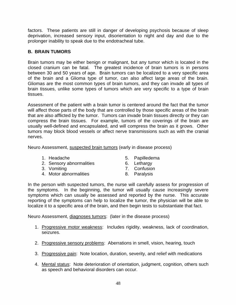
48
factors. These patients are still in danger of developing psychosis because of sleep deprivation, increased sensory input, disorientation to night and day and due to the prolonger inability to speak due to the endotracheal tube. B. BRAIN TUMORS Brain tumors may be either benign or malignant, but any tumor which is located in the closed cranium can be fatal. The greatest incidence of brain tumors is in persons between 30 and 50 years of age. Brain tumors can be localized to a very specific area of the brain and a Glioma type of tumor, can also affect large areas of the brain. Gliomas are the most common types of brain tumors, and they can invade all types of brain tissues, unlike some types of tumors which are very specific to a type of brain tissues. Assessment of the patient with a brain tumor is centered around the fact that the tumor will affect those parts of the body that are controlled by those specific areas of the brain that are also afflicted by the tumor. Tumors can invade brain tissues directly or they can compress the brain tissues. For example, tumors of the coverings of the brain are usually well-defined and encapsulated, and will compress the brain as it grows. Other tumors may block blood vessels or affect nerve transmissions such as with the cranial nerves. Neuro Assessment, suspected brain tumors (early in disease process)
1. Headache 5. Papilledema 2. Sensory abnormalities 6. Lethargy 3. Vomiting 7. Confusion 4. Motor abnormalities 8. Paralysis
In the person with suspected tumors, the nurse will carefully assess for progression of the symptoms. In the beginning, the tumor will usually cause increasingly severe symptoms which can usually be assessed and reported by the nurse. This accurate reporting of the symptoms can help to localize the tumor, the physician will be able to localize it to a specific area of the brain, and then begin tests to substantiate that fact. Neuro Assessment, diagnoses tumors: (later in the disease process)
1. Progressive motor weakness: Includes rigidity, weakness, lack of coordination, seizures.
2. Progressive sensory problems: Aberrations in smell, vision, hearing, touch
3. Progressive pain: Note location, duration, severity, and relief with medications
4. Mental status: Note deterioration of orientation, judgment, cognition, others such as speech and behavioral disorders can occur.

49
The prognosis of the tumor, of course, depends upon early diagnosis and treatment. Some tumors are not treatable if they are widespread and/or invasive. The nurse will continue to give emotional support to the patient and the family, and referrals when needed. Some tumors can be treated effectively, depending upon the age and condition of the patient. Radiation, surgery and/or chemotherapy can be used. C. CVA’s (stroke, or cerebrovascular accident) Persons acutely ill with a CVA need special attention paid to the neurological assessment. The nurse will maintain life support systems as necessary, since the cardiovascular and respiratory systems will usually be compromised. The nurse should also keep in mind the underlying cause of the stroke. There will be a different treatment for strokes caused by hemorrhagic disorders as opposed to the thrombo-embolitic disorder. Neurological Assessment, CVA victim, Acute Phase
1. Responsiveness: Changes in level of consciousness, changes in response to stimuli
2. Spontaneous movements: Changes in muscle tone, movements in the
extremities, body posture, position of head and/or neck.
3. PERL: Perform a complete pupil check with recording the size of pupils
4. Skin: Temperature of skin, moisture, and color
5. Speech: If able/ if conscious, note changes in ability and/or quality
6. Reflexes: Assess deep tendon reflexes and the superficial reflexes During the acute phase, the nurse should perform all assessment and interventions necessary. If the patient is unconscious, the nurse will need to maintain the airway and all other aspects of the unconscious patient. The neuro assessment is to ensure that the patient’s brain is being adequately oxygenated, and to prevent further neurological damage. During the rehabilitation phase, the nurse will still need to perform detailed neuro exams. They need not be as frequent as in the acute phase. However, the patient’s progress still must be recorded accurately at regular intervals.

50
D. MENINGITIS Meningitis can be caused by bacteria, mycobacterium or viral agents. We know that the patient will be isolated, will usually have a high fever and could have numerous neurological symptoms. The following guide to the assessment of the patient will be helpful for the acute phase of the disease.
1. Altered consciousness – maintain airway, assess level of consciousness and record hourly, confusion, delirium, irritability
2. Seizure activity—assess for muscle tremors, twitching or any other seizure
activity.
3. Post lumbar puncture assessment—patient will have one or several diagnostic studies involving the lumbar puncture, always assess the patient carefully after procedure.
4. Complications—accesses for complications of meningitis, including neck and back stiffness, petechiae and/or ecchymosis, infection, heart failure, shock, disseminated intravascular coagulation.
In the acute phase, the patient may be in the ICU, and will have life support as needed. The nurse should still perform a complete neuro assessment at least every hour and record these findings, in order to obtain a baseline of information during the acute phase of the illness. When the acute phase is over, the neuro assessment will not have to be performed as frequently, but still be alter to changes in neuro status. E. Polyradiculitis, (Guillain-Barre’ Syndrome) This disorder is a clinical syndrome, possibly caused by allergic response or immunological reaction, involving the peripheral nervous system and cranial nerves. It is characterized by muscle weakness and paralysis and parasthesias of extremities. Neurological Assessment includes:
1. Paresthesia - tingling and numbness of lower extremities 2. Muscle weakness starts in legs and can progress up the trunk 3. Face difficulty chewing, swallowing, talking, facial muscle
paralysis can occur 4. Incontinence loss of sphincter control; bladder and rectum 5. Deep Tendon Reflexes may be absent
In severe cases, the disease may cause cardiac and respiratory failure. Assess for progressive respiratory weakness, irregular breathing, rapid pulse rate and arrhythmias. The disease usually causes deteriorating functions. You should always record assessment findings so as to chart the progress of the disease. The prognosis can be better if treated early and complications are prevented.
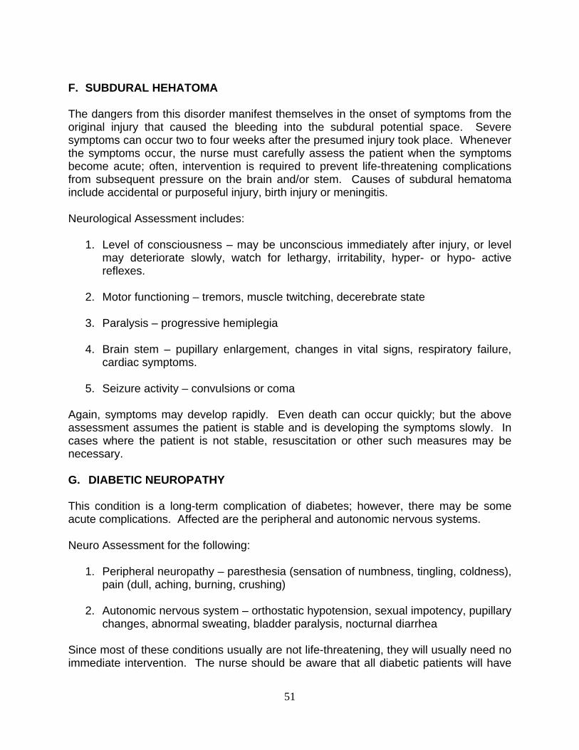
51
F. SUBDURAL HEHATOMA The dangers from this disorder manifest themselves in the onset of symptoms from the original injury that caused the bleeding into the subdural potential space. Severe symptoms can occur two to four weeks after the presumed injury took place. Whenever the symptoms occur, the nurse must carefully assess the patient when the symptoms become acute; often, intervention is required to prevent life-threatening complications from subsequent pressure on the brain and/or stem. Causes of subdural hematoma include accidental or purposeful injury, birth injury or meningitis. Neurological Assessment includes:
1. Level of consciousness – may be unconscious immediately after injury, or level may deteriorate slowly, watch for lethargy, irritability, hyper- or hypo- active reflexes.
2. Motor functioning – tremors, muscle twitching, decerebrate state
3. Paralysis – progressive hemiplegia
4. Brain stem – pupillary enlargement, changes in vital signs, respiratory failure,
cardiac symptoms.
5. Seizure activity – convulsions or coma
Again, symptoms may develop rapidly. Even death can occur quickly; but the above assessment assumes the patient is stable and is developing the symptoms slowly. In cases where the patient is not stable, resuscitation or other such measures may be necessary. G. DIABETIC NEUROPATHY This condition is a long-term complication of diabetes; however, there may be some acute complications. Affected are the peripheral and autonomic nervous systems. Neuro Assessment for the following:
1. Peripheral neuropathy – paresthesia (sensation of numbness, tingling, coldness), pain (dull, aching, burning, crushing)
2. Autonomic nervous system – orthostatic hypotension, sexual impotency, pupillary
changes, abnormal sweating, bladder paralysis, nocturnal diarrhea
Since most of these conditions usually are not life-threatening, they will usually need no immediate intervention. The nurse should be aware that all diabetic patients will have

52
these and other long-term complications of diabetes, and that the assessment should include all of the above criteria plus careful assessment of the general medical status of the patient. The feet, skin and extremities will be of special importance. H. PEDIATRICS CONSIDERATIONS This section is a guide to assessment of the infant and small child with these possible neurological disorders. It should be used in conjunction with any existing guidelines at your facility.
1. Cerebral Palsy: Malfunction of the motor centers of the brain and of the pathways. Assess for the following:
a. Asymmetry in motion or contour b. Generalized muscle weakness c. Listless or irritability d. Abnormal postures e. Difficulty in feeding or swallowing f. Defective speech g. Excessive or feeble cry h. Long, thin infants who are slow in gaining weight i. Delayed motor development
These areas above are some of the clinical manifestations of the disorder. Also associated with this are seizures, hearing deficits, visual defects, perceptual disorders, language disorders and others. These latter problems will be apparent in the older infant/child, and a generalized lack of motor development will also be noted. There are several types of palsy, exhibiting spastic symptoms or “jerky” movements. Be aware of abnormal motor symptoms.
2. Hydrocephalus: Increased amount of cerebrospinal fluid within the closed cranial cavity, causing enlargement of the child’s head and the related symptoms.
Recording the Examination Each hospital has its own form for recording the results of your examination. Usually it will call for the general observation first, then the more specific findings. If your facility has no model for charting the results, you can use this as a guide.
A. General Survey (mental status)
1. Mood and affect 3. State of awareness 2. Dress and grooming 4. Posture
B. Cranial Nerve Examination
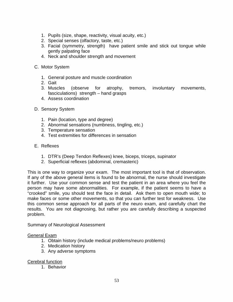
53
1. Pupils (size, shape, reactivity, visual acuity, etc.) 2. Special senses (olfactory, taste, etc.) 3. Facial (symmetry, strength) have patient smile and stick out tongue while
gently palpating face 4. Neck and shoulder strength and movement
C. Motor System
1. General posture and muscle coordination 2. Gait 3. Muscles (observe for atrophy, tremors, involuntary movements,
fasciculations) strength – hand grasps 4. Assess coordination
D. Sensory System
1. Pain (location, type and degree) 2. Abnormal sensations (numbness, tingling, etc.) 3. Temperature sensation 4. Test extremities for differences in sensation
E. Reflexes
1. DTR’s (Deep Tendon Reflexes) knee, biceps, triceps, supinator 2. Superficial reflexes (abdominal, cremasteric)
This is one way to organize your exam. The most important tool is that of observation. If any of the above general items is found to be abnormal, the nurse should investigate it further. Use your common sense and test the patient in an area where you feel the person may have some abnormalities. For example, if the patient seems to have a “crooked” smile, you should test the face in detail. Ask them to open mouth wide; to make faces or some other movements, so that you can further test for weakness. Use this common sense approach for all parts of the neuro exam, and carefully chart the results. You are not diagnosing, but rather you are carefully describing a suspected problem. Summary of Neurological Assessment General Exam
1. Obtain history (include medical problems/neuro problems) 2. Medication history 3. Any adverse symptoms
Cerebral function
1. Behavior

54
2. Level of consciousness 3. Intellectual function 4. Emotional status 5. Thought content 6. Language
Cranial Nerve Testing
1. Olfactory 2. Optic 3. Oculomotor 4. Others (as mentioned earlier)
Cerebral Function
1. Finger to nose with eyes open and shut 2. Fine finger movement 3. Walking in a straight line (heel-to-toe) 4. General coordination
Motor Testing
1. Muscle size and tone 2. Muscle strength
Sensory Testing
1. Pain (superficial and deep pain testing) 2. Motion and position 3. Point localization 4. Texture discrimination 5. Temperature
Reflex Testing
1. Stretch reflex 2. Cutaneous reflex 3. Corneal reflex 4. Gag reflex 5. Babinski reflex
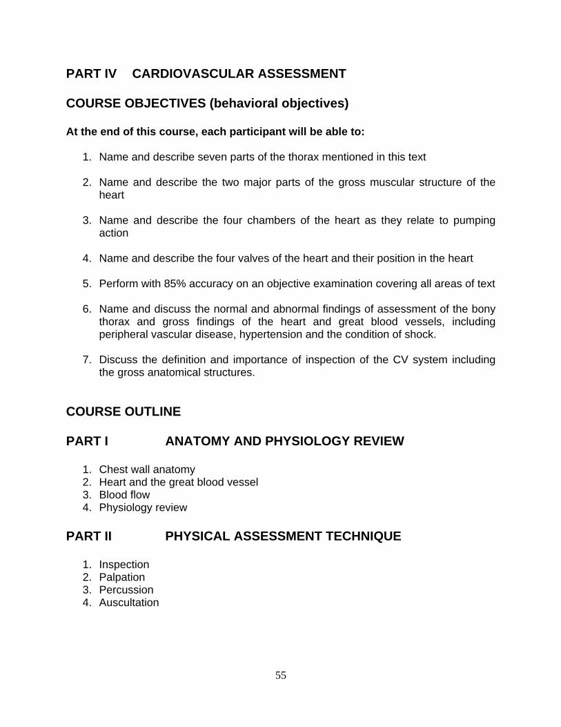
55
PART IV CARDIOVASCULAR ASSESSMENT COURSE OBJECTIVES (behavioral objectives) At the end of this course, each participant will be able to:
1. Name and describe seven parts of the thorax mentioned in this text
2. Name and describe the two major parts of the gross muscular structure of the heart
3. Name and describe the four chambers of the heart as they relate to pumping
action
4. Name and describe the four valves of the heart and their position in the heart
5. Perform with 85% accuracy on an objective examination covering all areas of text
6. Name and discuss the normal and abnormal findings of assessment of the bony thorax and gross findings of the heart and great blood vessels, including peripheral vascular disease, hypertension and the condition of shock.
7. Discuss the definition and importance of inspection of the CV system including the gross anatomical structures.
COURSE OUTLINE PART I ANATOMY AND PHYSIOLOGY REVIEW
1. Chest wall anatomy 2. Heart and the great blood vessel 3. Blood flow 4. Physiology review
PART II PHYSICAL ASSESSMENT TECHNIQUE
1. Inspection 2. Palpation 3. Percussion 4. Auscultation

56
PART III RECORDING THE EXAMINATION
1. Recording normal findings 2. Recording abnormal findings 3. Legal implications
PART IV SPECIAL NURSING SITUATIONS
1. Cardiovascular pharmacology 2. Chest tubes 3. Myocardial infraction 4. Peripheral vascular disease 5. Conduction of the heart and the EKG 6. Use and abuse of cocaine 7. Others
PART I ANATOMY AND PHYSIOLOGY UPDATE
A. The Thorax The first part of this course is an update of the anatomical structures and the physiology of the cardiovascular system in relationship to the nursing care of the patient with a related disorder. If you need to review the related structures and/or function which we will be discussing, please refer to any basic anatomy textbook; one of which you probably have hidden away somewhere in your closet or garage since nursing school. However, it is not required that you use any other reference. All information needed to pass the test at the end of this course, will be included in the text. We will list the most important structures of the thorax and entire CV system only so that you can relate these to the clinical approach that we will use. Following is an illustration of the thorax and the heart in relationship to other structures which will be noticed upon visual examination. The thorax has a characteristic shape, size and movement.

57
Thorax – Be sure you can identify the following:
a. Sternum – mid chest, flat, non-protruding b. Ribs – slope of ribs, intercostal spaces, costal margins c. Heart – heartbeat in some cases, can be visible as a pulsation in the thorax at
lower costal margin d. Shoulder – patient’s shoulder should be relaxed, and at a 90 degree angle to
the patient – look for abnormal angles and musculature which might indicate overuse of accessory muscles
e. Neck veins – should not normally be visible f. Clavicle – clavicular line horizontal with no protrusions during breathing g. Respirations – normal respiration should be unlabored and comfortable
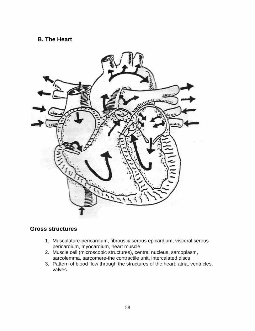
58
B. The Heart
Gross structures
1. Musculature-pericardium, fibrous & serous epicardium, visceral serous pericardium, myocardium, heart muscle
2. Muscle cell (microscopic structures), central nucleus, sarcoplasm, sarcolemma, sarcomere-the contractile unit, intercalated discs
3. Pattern of blood flow through the structures of the heart; atria, ventricles, valves

59
Chambers – Right side of Heart
1. Right atrium – the thin-walled atrium, low relative pressure receives blood from superior and inferior vena cavae, the coronary sinus and thebesian veins. The outflow of blood through tricuspid valve.
2. Right ventricle – relatively thin muscle wall, crescent-shaped, papillary muscles, chordae tendineae, low pressure. Outflow through the pulmonic valve to the pulmonary artery.
Left side of the heart
1. Left atrium, - thicker muscle, medium pressure of blood, inflow of blood through the four pulmonary veins. Outflow is through the mitral valve.
2. Left ventricle – largest muscle mass, high pressure blood flow, papillary muscles, spring-like pump action. Outflow of blood through the aortic valve and the aorta.
Heart Valves
Atrioventricular Valves:
1. Tricuspid – has three leaflets, controlled by papillary muscles; chordate tendineae
2. Mitral valve – two cusps, controlled by papillary muscles and the chordae tendineae
Semilunar Valves:
1. Pulmonic valve – three-leaflet valve, formed by fibrous ring, tendinous tubercle midpoint free edges
2. Aortic valve – three leaflets, also formed by fibrous ring, tendinous tubercle midpoint free edges.
Vasculature of the Heart
1. Right coronary artery – most branches of this artery anastomose distally with left anterior descending.
2. Left coronary artery – divides into two main branches, left ant. descending and left circumflex artery.
3. Great cardiac vein – largest system, forms coronary sinus, drains left ventricle primarily.
4. Anterior cardiac veins – empty directly into right atrium. 5. Thebesian veins – smallest system, empty into right atrium

60
Conduction System of the Heart
1. SA (Sino-atrial node) 2. Atrial preferential pathways – anter, internodal, middle, posterior internodal 3. AV (Atrio-ventricular node) 4. Bundle of HIS 5. Left Bundle Branch 6. Right Bundle Branch 7. Purkinje fibres
61
Contractility of Heart Muscle At this time it is appropriate to mention the physiology of muscle contractility. Electrical conduction in the heart is unique and remarkable. Heart muscle possesses the following properties:
1. Automaticity- pacemaker ability 2. Conductivity-each cell has the ability to conduct impulses 3. Contractility-ability to contract (make each cell shorter or longer) 4. Irritability-each cell has ability to contract on its own, to send impulses to cells
without it first being stimulated from another source These properties make the myocardium different from other muscle cells in the body. The normal activity of the heart conducts impulses from one point (SA node) to another point (individual cells), thus stimulating a uniform and effective contraction. Various factors affect the activity of cardiac muscle. The availability of oxygen, afterload, nervous control, muscle condition and other factors can affect the force of the contraction and affects blood flow through the heart. Drugs can also affect the contraction of the heart. The nurse should be aware of all the factors which influence heart activity and heart sounds. The below figure shows points on the thorax where heart sounds are best heard. Later in this text, these points on the thorax and heart sounds will be discussed in greater detail.

62
BLOOD FLOW THROUGH THE HEART Refer to the figure below to view blood flow through the heart. Blood is shown as it enters the heart, circulates and then leaves the heart. In relation to the physical assessment performed by most nurses, keep in mind the changes in circulation which will be assessed. Impeded flow may cause extra heart sounds and/or physical changes. Also, reduced flow will usually cause changes that can be assessed by the nurse.

63
Physical characteristics important to blood flow:
1. Diameter of the blood vessels 2. Cross-section areas of the chambers and vessels 3. Length of the vessels
Quantities of blood:
1. Heart 18% 2. Pulmonary vessels 12% 3. Large arteries 8% 4. Small arteries 5% 5. Arterioles 2% 6. Capillaries 5% 7. Small veins 25% 8. Large veins 25%

64
Velocities of blood flow: The velocity of blood flow is directly related to the amount of circulating blood volume and the area of the vessels. Blood returns to the heart from the general circulation. Almost 50% of all blood in the body is in the systemic veins of the body. This includes small veins and venules and blood in the pulmonary circulation. The small veins usually offer little resistance to blood flow. The large veins do offer much resistance to the flow of blood to the heart. This is an important nursing implication, as the patient who is more active will have better flow of blood back to the heart. With reduced activity, the blood tends to pool in the large vessels and can lead to severe venous stasis. Blood returns to the heart via the superior and inferior vena cavae and into the right atrium. From the right atrium blood flows to the right ventricle and is then propelled into pulmonary circulation. After blood is aerated with fresh oxygen, it is returned to the left side of the heart into the left atrium. From the left atrium the blood is ejected into the left ventricle. The left ventricle then pumps the blood out of the heart into the general circulation. The aorta is the first vessel to carry blood, and, at the same time, the coronary arteries are being fed oxygenated blood to circulate through the heart. The above is only a brief outline of the circulation of blood. Be sure you can trace the blood through the heart. Be sure that you can name all the valves and chambers of the heart as blood flows through. You should also be able to list the major arteries of the body. Later, when performing the assessment, it will be necessary for you to know these vessels and their location. PHYSIOLOGY OF THE CARDIOVASCULAR SYSTEM a. Blood Pressure is determined by:
Blood Pressure is determined by many factors in the body. Normal blood pressure is determined by the cardiac output, the velocity, the resistance of the blood vessels and by other factors. Systolic pressure refers to the initial force of contraction of the heart. Diastolic pressure refers to the pressure of the blood vessels after the initial force of contraction of the heart. In other words, the diastolic pressure is due to the elasticity of the arteries as they “snap” back after the initial “stretch” due to the systolic pressure. Blood pressure is probably one of the most important measures of the overall cardiovascular system that exists. Take the patient’s blood pressure lying down and then standing up. If there is a difference of more than 15-20 MM Hg, this may be an indication of one or more problems. Persons with hypertension that is poorly controlled, may have orthostatic hypertension. This may also indicate aortic disease or cerebrovascular disease.

65
As you remember, the pulse pressure is the difference between the two pressure readings (systolic & diastolic). The pulse pressure is very important because it can indicate certain major problems in the cardiovascular system. Shock can be diagnosed, in part, by blood pressure readings and the pulse pressure. The pulse pressure is normally determined by the general condition of the heart, arteries and the amount of circulating blood. b. Autonomic Nervous system control: Autonomic Nervous system control of the cardiovascular system includes control of the heart rate, cardiac output, blood vessels and amount of blood volume. Cardiac muscle is under the influence of the sympathetic and parasympathetic nervous systems. The sympathetic (cervical) system secretes norepinephrine and innervates the cardiac plexus. It increases the SA node rate, it increases the AV node conduction and increases the contractile force of the myocardium. The parasympathetic division (cholinergic fibres) secretes acetylcholine and also innervates the cardiac plexus. This branch decreases the SA node, AV node and the contractility of the heart. Together, they work to regulate the heart rate, blood pressure and other vital cardiovascular functions. Blood vessel contraction is controlled in the like manner. The sympathetic division causes vasoconstriction of most blood vessels, thus raising the blood pressure and circulating volume. The parasympathetic division causes vasodilation for most vessels. c. The Pressoreceptor System: The Pressoreceptor System originates in the arch of the aorta. Here are located sensitive nerve endings which help to control blood pressure and heart rate. Receptors are also located in the carotid sinus, vena cavae and the pulmonary arteries. When these receptors sense low pressure, the signal is fed to the medulla, where then the sympathetic division impulse is increased, causing the pressure to rise (either due to increased pulse rate or increased contractility). The opposite actions occur when there is high blood pressure. The nervous stimulation acts to bring the pressure back to normal by either vasoconstriction or vasodilation; by increased heart rate or decreased rate; by increased contractility or decreased contractility. Whichever combination occurs, the result will be the same; to bring the pressure back to normal. Summary (history and chief complaint) Now that you have refreshed your memory concerning anatomy and physiology of the cardiovascular system, you are ready to begin the examination. Before you go to the patient’s room, read pertinent data that has already been recorded. In many hospitals, the patient’s old charts from previous admissions are available. Read the old charts and obtain a summary of important data such as:
66
a. Previous admissions to hospital b. Reasons for admission c. Allergies d. Chronic medical problems e. Current and past drugs taken f. Social supports g. Discharge information
Next, interview the patient to discover the history of the present illness. The History of present illness means: What contributed to their coming to the hospital? You will ask for present symptoms and other recent symptoms that would be pertinent to this present illness. Perhaps they had some symptom at home that is now gone. You should question them in detail to confirm any historical data that you found in the old chart. Do not record any information from the old chart as fact, until confirmed by the patient in your interview. When you enter the room to interview your patient, the first thing to do is introduce yourself and explain that you will be interviewing them. Try to make them relax as much as possible. If they are just being admitted to the hospital, they will probably be very apprehensive. Use a blend of subtle humor and calm soothing conversation to help the patient relax. One important consideration is not to rush them. Often times you are interviewing the patient under rushed conditions. Rushing them will make them even more anxious. Give them time to answer questions. Do not interrupt them when they are trying to answer; let them finish before you ask another question. We will discuss charting and recording later. However, be sure that you record patient’s statements accurately. Put what they say in quotes when applicable. This is extremely important to remember…..be accurate! Many times the patient is unfamiliar with medical terms, and they might express a symptom in lay terms. Quote them as much as possible when you record their statements or comments. You might not understand what the patient means when they describe something in lay terms. For example, the patient might describe that they feel “crawly” when they took penicillin last year. Perhaps “crawly” means they were having an allergic reaction or maybe not. In any case, bring it to the attention of the physician and record what they say in quotes. Once you have completed the history of the present illness, ask the patient to describe any symptoms that seem important. Ask them to describe in detail, their symptoms, pain or bowel habits; what ever is important and relates to their diagnosis. For example, eating habits for a patient who has abdominal pain: Ask how many meals do they eat each day; Do you eat meat? Do you eat dairy products? Do you drink coffee? Then ask: How much of these foods; what times of day do you eat? Etc. You might get some interesting responses to the detailed questions that might explain some of their symptoms. Record the responses and ask more detailed questions if necessary. Remember to have the patient fully explain all symptoms in detail.

67
Since we are concentrating on the cardiovascular system for this text, we will ask the patient to give us those pertinent details. First ask the patient if he/she has been told that they have any of the following conditions:
a. “heart attack” or “heart trouble” b. Myocardial infraction c. Angina or “heart pain” or angina pectoris d. Congestive heart failure e. Congenital heart problems or heart problems you were born with f. Rheumatic fever or rheumatic heart disease g. Coronary heart disease h. Heart arrhythmias or heart murmurs
You should use the precise medical terms mentioned above. Many patients might remember the term if they heard it. Some patients have been hospitalized so many times that they will be able to use the exact term for their illness. Now remember that you looked at their chart (if it was available). That will give you an edge, but remember to confirm the problems with them directly. Some patients will not know the medical term for any past problems they had. In that case, you should ask them to describe the condition. Use some of the terms mentioned above. Next, ask what medications they have taken for their heart problem. Of course, they might have answered this question and others, in the above general medical history. However, have them give more details, since we are now concerned primarily with the cardiovascular part of the history. Find out the following for each drug:
1. Drub name and dosage 2. What you take it for 3. Frequency 4. Does it work for you or help at all? 5. When was the last dose you took?
Next, assess their use of alcohol and nicotine. Get details of the type of alcohol and/or “recreational” drugs used and the frequency and their smoking habits. Alcohol, cocaine, nicotine and other drugs can directly affect the heart, causing arrhythmias and other cardiac conditions. Assess social factors that might affect the patient should major surgery be required. If the patient is facing a severe debilitating condition, the physician would need to know the patient’s educational background and the resources they have at home and at the job. Will this illness affect their job or career? These are questions the nurse should anticipate will be important later on in their hospitalization.

68
Recording the findings: There are many different ways to record your observations. Some facilities utilize narrative charting forms. Other facilities use problem-oriented charting. Still others have computerized medical records. However, it does not matter how you record the findings; just be sure that you do record the findings. This includes vital signs, history and other data. PART II ASSESSMENT TECHNIQUES 1. INSPECTION As you prepare to begin the actual exam, you already have obtained and recorded the patient history and you arm yourself with pertinent data such as their chief complaint and allergic history. Also keep in mind to allow a certain amount of time in order to complete a thorough exam. Many nurses do not have large blocks of time for completion of the assessment, but you must be as thorough as possible. If this is an admission assessment, you must allow enough time to be complete. If this is an on-going assessment, not as much time will be required. Begin exam:
a. Patient undresses, but allow for privacy b. Have the patient sit upright and inspect the thorax from the front c. Now inspect from the back of the patient
You will inspect for posture and symmetry of the thorax, color of the skin, gross deformities of the skin or bone structure, the neck, face, eyes and any abnormal contours. Breathing patterns will also be noted. Be especially aware of cyanosis. Central cyanosis is a condition which will cause the lips, mouth and conjunctiva to become blue. Peripheral cyanosis will cause blue discoloration mainly in the lips, ear lobes and nail beds. Peripheral cyanosis might indicate a peripheral problem of vasoconstriction, and would generally be less severe than central cyanosis, which could indicate heart disease and poor oxygenation.
a. Thorax:
Inspect for symmetry of thorax, PMI (point of maximum intensity), PMI is easier to find if patient will lie on left side. PMI may also be palpated (see next section): skin color of thorax. b. Eyes:
Arcus senilis; is a light gray ring surrounding the iris, common in older patients; in younger patients it might indicate a type of lipid metabolism disorder, which is a precursor to coronary artery disease.

69
Xanthelasma; yellowish raised plaques on skin surrounding eyes; can also appear on the elbows; this is a possible indication of, or sign of, hypercholesterolemia, often a precursor to coronary artery disease (atherosclerosis).
2. PALPATION Palpation, or touching, is the next part of the exam. In the step above, if we noted any abnormalities, we will now palpate and evaluate them further.
a. Skin: temperature, texture, moisture, lumps, bumps, tenderness
Examination of extremities for edema might also indicate a cardiovascular problem. Examine the feet, ankles, sacrum, abdomen, trunk and face for edema. If you notice puffiness or frank edema, then palpate the area for pitting edema. Most facilities recognize the following scale:
+1 pitting edema - 0 to ¼ inch indentation +2 pitting edema - ¼ to ½ inch indentation +3 pitting edema - ½ to 1 inch indentation +4 pitting edema - more than 1 inch indentation
b. Breathing: lay hands on chest at different locations and feel the respiratory patterns, feel the ribs elevate and separate during normal breathing.
c. Pre-cordial areas: feel the pounding of the heartbeat, normal and abnormal pulsations on the chest wall; PMI, as mentioned above
d. Arteries: Assess all pulses: You undoubtedly assessed the apical pulse earlier when you took the patient’s vital signs, if not, now is the time. Assess the following pulses: 1. Apical heart rate: monitor for a full minute, note rhythm, rate, regularity 2. Radial pulse: monitor for a full minute, note rhythm, rate, regularity. Note any
differences from right to left radial, a large difference, might indicate arterial blockage or even enlarged ventricles. If pulse is regular but volume diminishes from beat to beat, this might indicate left-sided heart failure and is called pulsus alternans. If the volume of the pulse diminishes on inspiration, might indicate constrictive pericardial disease, the condition is called pulsus paradoxus.
3. Carotid, brachial, femoral, popliteal, posterior tibialis and dorsalis pedis pulses: when checking these pulses, do it the same way as the others mentioned in this section; right then left side. When you check the carotid, press gently and do not rub. Do not palpate carotid on persons with known carotid disease or bruits; listen with stethoscope instead; and do not palpate both carotid pulses at the same time.

70
Carotid artery: a. Plateau pulse—slow rise and slow collapse pulse; may be caused by
aortic stenosis; slow ejection of blood through a narrowed aortic valve. b. Decreased amplitude—(grade I pulse); due to hemorrhagic shock; pulse is
weak due to decreased blood volume. c. Bounding pulse (grade IV)—can be due to hypertension, thyrotoxicosis,
others; associated with high pulse pressure, the upstroke and downstroke of the pulse waves are very sharp.
d. It is common to use +1, +2, etc., when recording pulses: 0 = absent +1 = diminished +2 = normal pulses +3 = full pulse or slight increase in pulse volume +4 = bounding pulse or increased volume
e. Veins: neck, arms, legs, etc.
VEINS: CVP, Central Venous Pressure: In order to assess the patients CVP, start by having the patient sit in bed and then lean backwards at about a 45 degree angle. Let the patient relax for a few seconds while you look for the internal jugular vein. In most persons in which the vein’s pulsating is visible, the vein will be seen to pulsate at the level of the sternal notch (Angle of Louis). If the level of pulsation is more than three (3) cm above the level of the sternal notch, it is a sign that the CVP is elevated. An elevated CVP may be indicative of right sided heart failure, obstruction of the superior vena cava, or constrictive pericarditis. Normal pressure in the venous circulation run from five (5) to twelve (12) centimeters of water pressure. The CVP would usually be measured by placing a catheter into a large vein and attach it to manometer or strain gauge. If you find evidence of elevated CVP, may further confirmation the findings that you just saw. The hepato-jugular reflux test may be used. This test is performed by placing your hand in the area of the right upper quadrant of the abdomen. Once you have placed your hand on the abdomen, exert firm pressure directly into the abdomen for one full minute, and at the same time, observe the jugular vein. If the pulsation you observed begins to definitely rise over the highest level of pulsation seen, then this confirms that the CVP is elevated. Jugular veins:
The pulsations from veins are different from the arterial pulsations that can be palpated in the neck area: 1. the venous pulse is easily compressed by gentle pressure, in contract
to the carotid pulse, which requires firm pressure to obliterate.
71
2. the normal venous pulse descends upon inspiration and rises on expiration, but carotid pulse remains unaffected by respirations.
3. a venous pulse usually collapses in the sitting position, while the carotid arterial pulse is not affected by changes in position.
4. a venous pulse normally has more components than the arterial pulse. It consists of three positive deflections
----the a, c, and v waves--- and two negative deflections, the x and y descents. Normally this is when the venous pulse waves are the most prominent. If venous pressure is high, the pulsations may be better observed if client is sitting.
The “a” Wave: The predominate wave in the neck; reflects the pressure transmission caused by arterial contraction; begins just before the first heart sound; it can be palpated by feeling the jugular pulse, while auscultating the apex of the heart; the a wave also occurs just prior to the carotid pulsation.
The “c” Wave: This is a reflection of the onset of right ventricular contraction; begins at the end of the first heart sound; is usually not visible in the neck veins.
The “v” Wave: Represents arterial filling with atrioventricular valve closed; very small and is considered a passive filling wave.
The “x” Decent: Is a negative wave following the c wave; represents atrial diastole. The “v” wave is followed by a y descent, a negative wave produced when the tricuspid valve opens, allowing blood to pour into the right ventricle.
f. Hair: Observe and feel the consistency and texture of the person’s hair. Very fine hair shafts may indicate hyperthyroidism. Very course hair, might indicate hypothyroidism. Both conditions of the thyroid, may have adverse effects on the cardiovascular system.
3. PERCUSSION This technique has a very limited place for most nurses in assessment of our patients. As discussed earlier, percussion refers to “tapping” the chest wall with the fingers, in order to elicit sounds which indicate abnormalities. We will discuss and demonstrate the technique, but remember that it is a very limited tool. Much information obtained by percussion, can be more easily be determined by auscultation.

72
The technique for percussion involves hyperextending the fingers of one hand and placing the middle distal phalanx firmly on the chest wall. Hold your opposite hand close to the hand on the patient. Retract the middle finger of that second hand; strike the finger firmly at the top of the distal phalanx. After striking the finger, quickly remove it and move to another area and repeat the same motions. Percuss the precordial area of the chest, listening for a resonant sound which indicates normal tissue beneath the fingers. When percussing over the lung tissue, the sound will be resonant, a semi-hollow, medium pitched sound. When percussing over denser tissues, the heart, the sound will be flat or “dull” in pitch. These are normal sounds. If the patient is sensitive or indicates pain or difficulty breathing, stop the percussion and go on with the other parts of your assessment. 4. AUSCULTATION
Auscultation is defined as listening to the sounds produced by the body, with or without the use of a stethoscope. Some sounds may be loud enough to hear without the use of the stethoscope. Although, when we think of auscultation, and the cardiovascular system, most nurses think of listening to the chest and heart through the stethoscope. As you know, the heart sounds heard are due to the closure of the heart valves under pressure of the blood flow. The stethoscope can also be used to listen to other sounds that could be of importance to the cardiovascular system. Heart Sounds: Auscultation of heart sounds should usually follow the general medical assessment and the general assessment of the cardiovascular system. The nurse should first think about the results of the general assessment and then proceed to listen to heart sounds. In many cases, the first part of the assessment will give you a clue of what to listen for upon auscultation. For example, if the patient states in his/her history that he/she has had cardiac surgery, or a valve replaced, etc., then it will alert the nurse to listen for particular sounds or murmurs. In most persons, there are two major sounds that can be heard. The “lub” and “dub” are called S1 and S2 respectively, and are the two most prominent and easily heard sounds. S1 and S2 follow each other closely. The time between 1 and 2 is shorter than the time from end of S2 to the beginning of the next cycle and S1 of the next beat. The time interval between S1 and S2 also corresponds to systolic phase of the cardiac cycle. Two additional heart sounds may be audible in the cardiac cycle, these are S3 and S4. S3 can sometimes be heard immediately after S2. S3 is the sound of early, rapid diastolic filling of the ventricles. It is not often heard in adults, but is heard very commonly in children. S4 is the last heart sound, and like S3, it is rarely heard in the adult, except in disease conditions such as congestive heart failure or multiple sclerosis.

73
Origins of heart sounds:
S1 - Closing of the atrioventricular valves (Mitral and Tricuspid): corresponds with the carotid pulse; ventricular systole heard loudest at the mitral and tricuspid areas S2 - closing of the aortic and 73ulmonic valves heard loudest at the aortic area
Each of the two major heart sounds is made up of rushing blood and of two valves closing at the same time. Normally the pair of valves open and close at the same time, causing a clear and distinct sound. At certain times, the valves may close at slightly different times, or one may close very slightly slower than the corresponding valve. This causes one of the heart sounds to be distinctly “split” or having and “echo” sound. This may be a naturally occurring phenomenon, called “physiological splitting”, or it may be due to a disease, called pathological splitting. When S1 valve closures can be heard separately, there may be a conduction defect present or even a mechanical defect. Of course, even young and healthy children and some adults can still have normal splitting of S1 and S2. In review, the two common heart sounds are S1 and S2. They each are made up of two distinctly separate sounds of two valves closing in unison. When assessing the patient for heart sounds, the nurse must first obtain a patient history; inspection, palpation and possibly percussion will also usually be performed before you begin to auscultate. Ideally, the nurse must know the patient’s heart rate and the regularity of rhythm before auscultation is performed. S3 and S4 are two “normal” heart sounds that may sometimes be heard in the cardiac cycle. Splitting is usually a normal situation arising from asynchronistic closure of two valves responsible for each of S1 and S2.
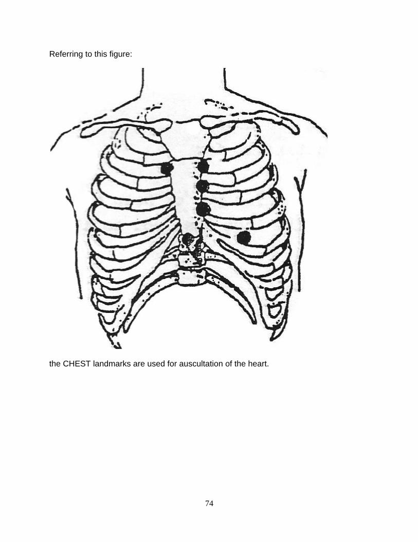
74
Referring to this figure:
the CHEST landmarks are used for auscultation of the heart.

75
In the below figure these same positions are marked ON THE HEART ITSELF (note blood flow).
The next figure illustrates the names of these precise positions.
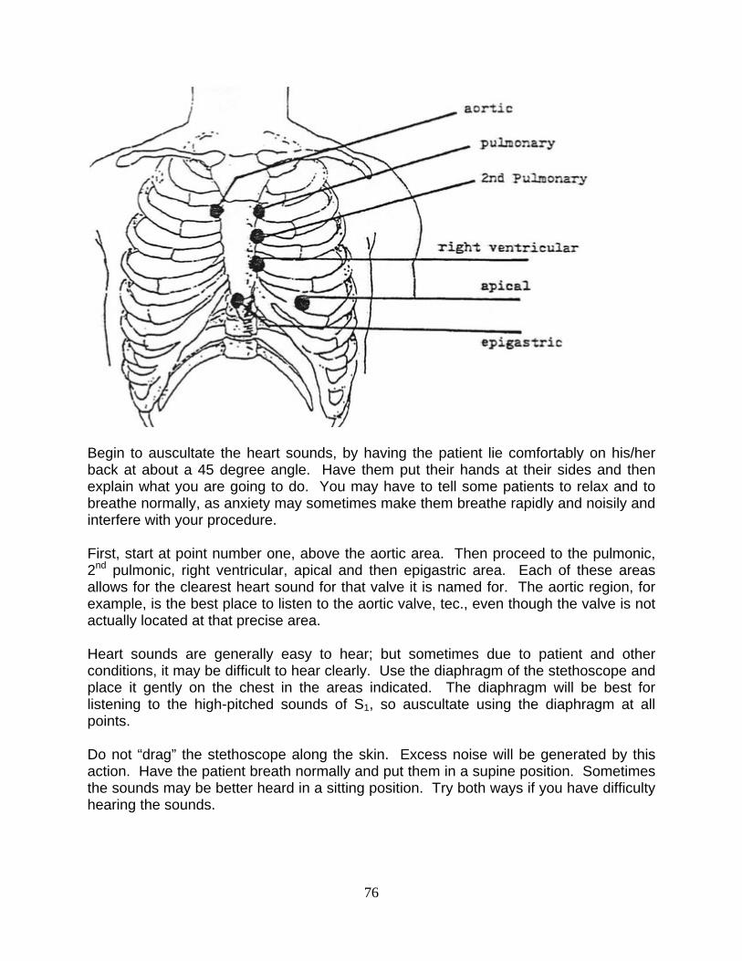
76
Begin to auscultate the heart sounds, by having the patient lie comfortably on his/her back at about a 45 degree angle. Have them put their hands at their sides and then explain what you are going to do. You may have to tell some patients to relax and to breathe normally, as anxiety may sometimes make them breathe rapidly and noisily and interfere with your procedure.
First, start at point number one, above the aortic area. Then proceed to the pulmonic, 2nd pulmonic, right ventricular, apical and then epigastric area. Each of these areas allows for the clearest heart sound for that valve it is named for. The aortic region, for example, is the best place to listen to the aortic valve, tec., even though the valve is not actually located at that precise area. Heart sounds are generally easy to hear; but sometimes due to patient and other conditions, it may be difficult to hear clearly. Use the diaphragm of the stethoscope and place it gently on the chest in the areas indicated. The diaphragm will be best for listening to the high-pitched sounds of S1, so auscultate using the diaphragm at all points. Do not “drag” the stethoscope along the skin. Excess noise will be generated by this action. Have the patient breath normally and put them in a supine position. Sometimes the sounds may be better heard in a sitting position. Try both ways if you have difficulty hearing the sounds.
77
Murmurs: A heart murmur is a very general term used to describe any one of the variety of abnormal sounds heard in the heart due to turbulent or rapid blood flow through the heart, great blood vessels and/or heart valves (whether the heart valves are normal or are diseased). Most nurses associate murmurs with an abnormal heart valve. However, there are a variety of other conditions that can cause murmurs. Murmurs can also be caused by the forward flow of blood across a constricted or otherwise irregular valve, or into a dilated heart chamber or dilated vessel. They can also be caused by the backward flow of blood through an incompetent valve or a septal defect. Murmurs are usually described as a “rushing” or “swooshing” sound. Murmurs are usually related to defects in valves or ventricular septal defect, or atrial septal defect. When auscultating murmurs, the nurse should record the timing, characteristics,* location and radiation of murmur. *characteristic include: loudness, intensity, pitch and quality of murmur. These assessment factors are discussed in more detail in the next few paragraphs.
Gallops: The bell of the stethoscope may be used for low frequency sounds (they are better amplified by the bell). S3 and S4 gallops are generally low-pitched sounds and are heard best with the bell of the stethoscope while the patient is stretched out on his left side. Many nurses prefer to auscultate the heart sounds a second time with the bell of the stethoscope in order to detect any sounds that might be missed with the diaphragm. S3 gallop, the ventricular gallop, occurs at the end of ventricular systole. It is often caused by the sound of blood prematurely rushing into the ventricle that is stiff or dilated due to failure, coronary artery disease or pulmonary hypertension. Clicks: Sounds described as “clicks” are extra sounds often heard in those patients with mitral valve prolapsed, aortic stenosis or those with prosthetic heart valves. Opening “snaps” are usually caused by mitral stenosis or stenosis of the tricuspid valves. Rubs: Sounds referred to as “rubs’ occur when the visceral and parietal layers of the pericardium rub together. The sound is produced when inflammation is present due to uremic pericarditis, myocardial infarction or other inflammatory condition. Discussion of Heart Sounds The loudness and intensity of heart sounds are important when you are listening. S1
and S2 are heard at different levels of loudness, depending upon where you listen on the

78
chest. The loudness of S1 is mainly determined by the position of the heart valves when ventricles contract. If valve leaflets are wide open at the time of contraction, the sound is very loud. The loudness of the sound is also affected by the pressure of the blood. It is this pressure that “slams” the valves shut and generates the sound. If you recall that the interval between S1 and S2 corresponds to the systolic phase, then a murmur that is heard between S1 and S2 would be called a systolic murmur. Then a diastolic murmur would be called a murmur heard between S2 and S1, which corresponds to the diastolic phase of the cardiac cycle. Next, these two murmurs, systolic and diastolic, can further be pinpointed by describing exactly when in the phase it occurs. The murmur can be described as: early systolic midsystolic late systolic early diastolic middiastolic late diastolic These above terms describe murmurs in the exact position that they fall in the phase. For example, an early systolic murmur would be “timed” as occurring early in the phase of systole; and so on for all the phases. Another term called holosystolic (also pansystolic), is used to for a murmur heard throughout the entire systolic phase (S1 to S2). Holodiastolic will be used to refer to the murmur heard throughout the entire diastolic phase (S2 to S1). The timing of the murmur above is very difficult to assess in some patients. In other patients, the timing will be very easy to assess. An important factor is that the nurse have experience in listening to a variety of “normal” variations of normal heart sounds. It is this author’s opinion that most nurses with little experience listening to what a normal heart sounds like, will not have much success in determining the timing of heart murmurs. You must first listen to many different normal heart sounds. Once you have some experience at differentiating normal S1 and S2 sounds, then you will be able to identify abnormal sounds, and to determine the timing of those abnormal sounds. The valves are at their widest when blood is actually filling into the ventricle. As the ventricle fills and the atria empty, the leaflets of the valve begin to close or to narrow. At that point, when the atria are empty, the ventricle is contracting and slams the valve shut. This is the dynamic force behind the loudness and intensity of the heart sounds. Other factors affect closure. Exercise, fever, anemia and other factors can affect heart rate and the force of the closure of the valves. Loudness, of course, is also affected. Note the following changes due to disease:
1. Mitral Stenosis - loud S1 and delayed closing of valve 2. Heart Block - varying intensity of S1 and S2 due to incomplete emptying of the
atria, leaflets and may be partially open at some times, completely open at others 3. Atrial flutter; fibrillation - sounds vary in intensity for the same reason as above
79
4. Bundle Branch Block – S1 may be widely split (two separate sounds make up S1) S2 can also be affected
5. Normal Respirations – physiological splitting of S2 during late part of inspiration 6. Pulmonic Stenosis – delayed emptying of right side—can cause splitting of S2 7. Atrial-Septal Defect – causes delayed emptying of right side of heart, as in above
condition, splitting of S2 will occur 8. Hypertension – increases back pressure on aortic valve, causes increase in
loudness of S2 These are some disease conditions and the resulting change in the heart sounds. The term murmur refers generally to any “extra” or unusual heart sound. Most nurses will not be expected to fully “diagnose” all murmurs and/or abnormal heart sounds. However, the nurse should be able to recognize whether or not the two normal sounds are present, and if they are not, what sounds that are present, should be described carefully. When charting heart sounds for your nurse’s notes, chart only the sounds that are abnormal. Chart basic information such as heart rate, rhythm, intensity and abnormal sounds. Describe carefully their location in the cycle. Describe any coincidental factors that may be influencing the rhythm such as respirations or movement of the patient. For murmurs, chart where it occurs in the cardiac cycle, loudness, pitch, the location of where it is heard the best and other locations where it can be heard. Also record the general type of sound heard and if anything makes the sound change in any way. For example, if you reposition the patient, does the sound change. If the patient happened to breathe deeply and the sound changed; all these would be notable events. Following is a guide to auscultation of sounds of the heart. It is only a guide, and should be used with existing guidelines at your facility. The methods of charting are different at each hospital, so are responsibility levels for each type of nurse. Always use terms which are acceptable at your facility. If a heart sound or murmur is accompanied by adverse clinical symptoms, results should be reported. GUIDE TO AUSCULTATION OF HEART SOUNDS Step I Prepare patient
Have patient relax, remove cloths from waste up, cover with gown and provide for privacy
Step II Vital Signs Record vital signs, TPR and Blood Pressure, note any abnormalities

80
Step III Heart Rate Listen to apical pulse and record the rate, even though you already took the radial pulse rate from the above step; note any abnormal sounds while listening to apex
Step IV Rhythm Determine the regularity of rhythm; regular or irregular? a. Is the irregularity due to respirations b. Auscultate at the points mentioned on previous page (6 points) c. Listen especially for any murmurs you previously heard d. Timing – describe where the sound is heard in the cardiac cycle
Step V Sounds
Describe carefully the sound heard that is abnormal. Is it a “rubbing sound” or a “clicking sound” or “swooshing” or other? Chart the sound just as you heard it. This course is not designed to make you a cardiologist, but the nurse should be able to recognize and chart anything abnormal in the cardiac cycle
Loudness of heart murmurs: Once you have determined the timing and other gross characteristics of the murmur, you should determine and record the loudness. Loudness is graded on a numerical scale as shown below. Grade I is the softest and Grade VI is the loudest. *Remember, however, that you will see other scales used to measure loudness. Some authorities use a scale of I, II, III, IV. Some others use a scale of only I, II, III; so be aware of the scale that is generally used at your facility and apply the same principles to it. Grade I Soft Grade II Medium Soft Grade III Loud **All of these ratings are very subjective Grade IV Medium Loud Grade V Louder Grade VI Loudest Intensity of heart murmurs: Crescendo begins softly and becomes louder Decrescendo begins loudly and becomes softer Crescendo/Decrescendo begins softly, peaks at a certain intensity and then becomes soft again Decrescendo/Crescendo begins loudly, becomes softer, then becomes loud again Bolosystolic or (pansystolic) stays same intensity through systole and diastole Bolodiastolic the same intensity throughout diastole; pandiastolic murmur

81
Further characteristics of murmurs include the quality and pitch. The quality of a murmur may be harsh, blowing, musical or rumbling. The pitch may be described as high or low pitched. Other terms may also be: dull-sounding, sharp, others. In many cases, you may simply describe how the murmur sounds to you. Be descriptive and try to use established terms. However, be precise in location and changes in the sounds that you hear. Do not be afraid to say, “it is a swooshing murmur when the patient is lying on left side, and has a ‘tapping” sound when sitting up”. Describe exactly what you hear. Types of Murmurs: The heart murmur associated with mitral stenosis is caused by the flow of blood across the constricted mitral valve during the period of rapid ventricular filling. There are two periods of this rapid ventricular filling in the cardiac cycle. These are: early diastole, shortly after the opening of the atrioventricular valves; and immediately prior to the onset of systole just as the atria contract. This explains why there are two murmurs heard in mitral stenosis. The first is an early to middiastolic murmur and a presystolic murmur. There is also another distinct sound heard with the condition of mitral stenosis. This sound is referred to as the opening snap of mitral stenosis. Normally, the opening of the mitral valve is silent. In the presence of mitral stenosis, there is a sharp, high-pitched click can be auscultated. The click is best heard between the apex and the lower left sterna border. The murmur of aortic insufficiency is caused by the backward flow of blood from the aorta into the left ventricle across an aortic valve that is incompetent. This backward pressure exerted by the blood in the aorta is the greatest, just after the closure of the aortic valve and progressively falls thereafter throughout diastole. The sound produced is described as regurgitation. The murmur usually begins immediately after S2 and can progress throughout the entire diastole. This murmur is best heard at the base of the heart and will often radiate to the apex. The murmur will usually be very high pitched and will usually have a “blowing” quality; although it many times has a very harsh quality to the sound and may be very loud.
-81- The third heart sound, S3 was discussed earlier as being normal in some adults and in children. In the case of a pathological S3, it may be noted with the event of damage to the myocardium. This heart sound, when auscultated, sounds like the gallop of a horse. Indeed, it is described as a gallop. The S3 creates an extra heart sound that can be rapid and very distinctive. The S3 G (S3 Gallop) is caused by early diastolic vibrations that are probably the result of ventricular distensibility associated with the myocardial damage. The S3 G can be best heard at the apex as a short, low-pitched sound. It may possibly be palpated, since it is associated with the ventricle and can create a “thrust” from the heart. A gallop rhythm is very suggestive of myocardial damage and the possible presence of early congestive heart failure.

82
Cardiac Arrhythmias: An arrhythmia is described as an abnormal heartbeat. It might be caused by one or more of several factors:
a. SA node – variation of the rate of discharge of the node b. Conduction defect – an abnormality of conduction of electrical impulses from
sinoatrial node through the heart c. Myocardial irritability – initiation of contraction of heart muscle by impulse on a
random area of the heart, other than SA node The “average” person’s heart rate is usually 70 to 100 BPM, Beats Per Minute. However, we all know that some persons may have a normal heart rate that is slightly higher or lower than these normal ranges. Of course metabolism, exercise and other factors will affect a person’s normal heart rate. A rapid heart rate is called tachycardia, and a slower than normal rate is called bradycardia. When assessing a patient’s heart rate, you must consider that the heart rate is also controlled by the sympathetic and parasympathetic nervous systems. The sympathetic nervous innervation increases the rate at which the SA node fires. The parasympathetic nervous innervation decrease the rate of firing and subsequently the heart rate slows. Normally, these two influences keep the heart rate in balance and produce a normal heart rate. However, in disease conditions, either or both of these nerves may dominate and produce a fast or a slowed heart rate. The normal heart beat is also referred to as sinus rhythm, or being initiated in the sinoatrial node. Very specifically, if the rate is normal and the heart beat is a sinus rhythm, it is called “normal sinus rhythm”. Using this terminology, it would be easy to see why a rapid, but otherwise normal (sinus) rhythm, is called sinus tachycardia. Conversely, a slow, but otherwise normal (sinus) rhythm, is called sinus bradycardia. Both of these arrhythmias are not normal, but they usually are not severe or life-threatening. The nurse might easily understand that sinus tachycardia could be caused as a normal reaction to anxiety. In fact, just being sick and in the hospital might produce anxiety and also sinus tachycardia. The nurse would need to assess the patient very carefully if a rapid heart rate is present; especially if there are no adverse clinical signs or symptoms present. Of course, an EKG tracing and interpretation would be necessary to absolutely confirm that only sinus tachycardia is present, and that there is no immediate danger to the patient. Factors that affect the sympathetic nervous system are: anxiety, fear, fever, extreme physical exercise, others. These conditions will generally cause an increase in the sympathetic stimulation of the SA node and subsequently, tachycardia.

83
Factors that affect the parasympathetic nervous system will induce a slowed heart rate. The parasympathetic nervous system exerts its influence through the vagus nerve. This stimulation of the vagus nerve will cause bradycardia. In summary, prepare the patient; take vital signs and the history of the patient before you auscultate the heart. Compare your findings to what a normal heart sounds like. Then, report findings, especially if the patient has adverse clinical systems, such as cyanosis or some other important symptom.
Recording the Physical Assessment Findings As an introduction to charting, it should be known that there are many different ways to record an assessment. Some hospitals have their own form for recording findings, and other facilities, a narrative or “story” form. This guide for charting will present one method. If your facility uses a different method of charting, you may still derive some benefit from this exercise below. You can study terminology and the presentation, then apply it to your facility. Even if your facility uses a “checklist’ style charting, you still may have to record certain observations that do not exactly fit those checklists. Therefore, remember to observe and carefully describe and record your findings for each patient. Narrative style: Begin with: Vital signs, radial pulse, BP, temperature, respirations and history. “Patient is a 78 year old male, in no acute distress, reports a “heart attack” five years ago and has been in good health since then; came into the ER today feeling weak, dizzy and pounding in the chest.” Next…The General Medical Exam: Patient is alert, oriented, no respiratory difficulty, no complaints of pain now, skin turgor good, skin color good, skin is warm and dry, no problems voiding, no bowel movement for two days. Takes Digoxin and Lasix QD, dosage unknown, lungs sound slightly congested, but no dyspnea, as stated above Cardiovascular: Peripheral pulses all present and strong, neck veins slightly distended when laying down, heart rate regular and strong, thorax normal shape, no masses, no tenderness, heart sounds clear and strong, with faint murmur between S1 and S2, sounds like faint clicking noise, MD was notified, no treatment because patient has had condition for many years. This is a sample of a fairly healthy patient. Some facilities might want the cardiovascular system charted first in the nurse’s note section. Others will want all cardiovascular findings together in one place on the chart.

84
In the above example, we placed skin color together with the other skin findings. Skin color could be considered a cardiovascular sign. It does not matter where you put it; just remember to include all pertinent findings. How do you know what is pertinent? That is a difficult question, but always remember to include all findings that you would expect to be abnormal if the patient did have a definite cardiovascular problem; things such as skin color, respiratory difficulty, poor pulses, poor heart sounds, low BP, etc. This is why it is important to have the history and the general medical exam reviewed by the nurse before you concentrate on your cardiovascular exam. Once you know the general findings, it will be easier for you to review the cardiovascular system. As you finish recording your findings, remember to include all actions that you took for your patient. If you started your exam and the patient was having a severe asthma attack, you would not say, “wait”, I have to do my cardiovascular assessment first. You would take the appropriate emergency measures first. Remember to chart all such treatments or emergency measures. Legally, you might be held responsible, even if you did take the appropriate measures, if you did not chart that you notified the MD, then you could be held responsible for some adverse occurrence. Charting is a method of recording that you did take the appropriate action for the situation; “notified MD and no treatment at this time”. This charting protects the patient, and protects the nurse. It lets everyone know that you performed the correct action in response to your abnormal findings. If you are ever in doubt as to how you should chart something; remember to be as objective as possible. Chart the findings (be descriptive), and then chart what you did about it. That is how good charting protects you and the patient.
SPECIAL NURSING SITUATIONS Topic One – Cardiovascular Pharmacology We are including this section, because it is vitally important that all nurses be up-to-date on the cardiovascular drugs. When assessing the heart, the nurse must take into consideration the effects of drugs upon the cardiovascular system. This section will be concerned with the most common drugs used in a cardiac arrest situation. Each drug will be presented along with its uses and other helpful information. During a code, or cardiac arrest, it is not unusual for even the most experienced nurse to have fears concerning the administration of these potent drugs. Since this is a life and death situation, the nurse must learn not to be overwhelmed by the numbers of new drugs being used today. Be sure to study the drugs before the code and not during. Some of these drugs will be new to you and others have been around for a long time. It wouldn’t hurt to study these drugs, see if they have any new uses of which you were not aware. lidocaine HCI dopamine HCI sodium bicarbonate isoproterenol HCI procainamide HCI epinephrine HCI

85
calcium chloride atropine sulfate verapamil (isoptin) bretylium tosylate dobutamine HCI
1. Sodium bicarbonate: corrects metabolic acidosis during a cardiac arrest. It is administered by IV push in a dose of 50ml, D5W solution, 44.6mEq of sodium bicarbonate. Metabolic acidosis occurs after the heart stops, due to a build-up of the acid waste materials in the body. This condition will be corrected by regularly administering (approx every 10 minutes) sodium bicarbonate. Defibrillation will be more affective if the body pH is adjusted, so will other drugs be more affective. Arterial blood gas results will tell you the patient’s pH and if it needs correcting. Do not administer too much sodium bicarbonate, as alkalosis may occur. If alkalosis does occur, the patient can develop arrhythmias and other problems.
2. Lidocaine: is used for reducing the irritability of the heart muscle. Specifically, it treats PVC’s and other ventricular arrhythmias. The drug is usually administered intravenously, but in an emergency it can be delivered via ET tube, intratracheally. The usual dose is 50 to 100 mgm IV push, followed by a continuous IV drip. The continuous drip helps to overcome reappearance of the arrhythmia. It is usually mixed in the following ratio: lidocaine, 2Gms mixed in 50ml of D5W…… delivers 4mg of lidocaine per ml….. 60gtts per minute, of this solution, gives 4mg per minute
3. Epinephrine: is a potent stimulant. It increases the contractility of the myocardium and stimulates spontaneous contraction. It is administered IV push or via the ET tube when necessary. The dosage is 0.5mg to 1.0mg (5 to 10 ml) of a 1:10,000 solution. This drug in the smaller dose above, can be given intracardiac as well. This drug will also in certain conditions, make the myocardium susceptible to defibrillation.
4. Dopamine: is used for the treatment of insufficient cardiac output and for
hypotension. Dopamine is often preferable during codes because it also acts as a vasodilator, bringing better circulation to the brain, myocardium and the kidneys. The main action of this drug is due to its alpha receptor stimulation effect. This increases blood pressure and cardiac output. The usual dosage is 2 to 5 mcg/kg/min given in a continuous IV drip. The dosage can be as high as 50 mcg/kg/min. Dopamine is mixed: 400mg (2 x 200mg vials) in 500ml of D5W. This results in 800 mcg/ml. 15gtts/min of this solution would result in 200 mcg/min. The blood pressure should be monitored closely, every 5 to 10 minutes at the beginning of the infusion and while dosage is being adjusted.

86
Once stabilized, the blood pressure need be taken only as often as clinical signs warrant, approximately every half hour or one hour. Urine output should also be monitored; dopamine will cause greater kidney perfusion, but output will still need to be monitored for persistent renal failure.
5. Atropine: is a cardiac stimulant. It is used for severe cases of bradycardia. The drug is administered by IV push, slowly. The action is that of blocking the vagus nerve. The smallest dose to give is 0.5 mg, if a smaller dose is given it may have the opposite effect and cause further slowing of the heart. The dose is then repeated every 5 minutes for up to 4 doses (2.0 mg). Atropine will usually not be given if the patient has had an acute MI. With atropine the heart rate is increased which causes increased oxygen demands upon the myocardium. MI patients usually can not tolerate added O2 demands.
6. Isoproterenol: Isuprel is used to combat sinus bradycardia and is infused in a
drip solution containing 2mg (2 mg ampules) in 500 ml of D5W which gives a dilution of 4 mcg/ml. Dosage range is from 0.5 mcg/min to 5 mcg/min. and then the patient is titrated as the patient improves. Very similar to atropine, the oxygen demands must be considered when using Isuprel.
7. Procainamide: is usually administered IV push at a dosage of 100 mgm pushed at a rate of 20 mg/min so as not to be given too rapidly. The drug is used to stop PVC’s and is the second choice if lidocaine fails. The above dosage can be repeated every 5 minutes, up to a dose of 1 gram. If any adverse effects occur, such as hypotension or widening of the QRS complex, then the drug should be stopped immediately
8. Calcium Chloride: This drug is administered IV push at a dose of 5 to 10 ml at lml/min. It is used to stimulate the heart to make a more forceful contraction. In cases of asystole, the drug can be used to start spontaneous contractions. The drug can also be used in cases of electromechanical dissociation, a condition in which electrical impulses are being produced by the heart, but the heart does not respond to them. On EKG, it seems like the heart should be beating just fine. However, the patient has no effective contractions. Calcium Chloride can be given in order to make the heart respond effectively.
9. Verapamil: is one of the newer drugs which is in common use today. It is a calcium channel blocker (also classified as slow channel blocking agents or calcium antagonists). Verapamil and others in this group are used for slowing conduction of the heart’s electrical impulses and treating such arrhythmias as atrial flutter, atrial fibrillation, atrial tachycardia and for supraventricular tachycardia. The drug is also used for angina and the group of drugs is being investigated for treating other disorders; such as hypertension and cardiomyopathy
87
The drugs in this class have many different and varied chemical structures but they all perform the same function. They inhibit calcium flux across the cell membrane. The drugs do not seem to affect sodium flux, or any other electrolyte in the cells. Calcium, as you know, is important for the contraction of all muscle cells, especially cardiac cells. Some cells are more dependent upon calcium than other cells. They do not all utilize it equally. Therefore, cardiac muscle cells react differently to each of the drugs in this class. There are many different effects which can occur because of this action, it depends upon: a. Which cardiac cells are affected by the slowing b. The chemical structure of the particular blocker drug being used c. The dosage d. The route of administration e. The extent to which that particular cardiac cell depends upon calcium Verapamil and some of the others in this class affect the SA node and the AV node. It depresses the SA node and also slows conduction through the AV node. The overall effects of Verapamil are: a. Negative chronotropic effect (slowed SA rate) b. Negative dromotropic effect (slowed AV conduction) c. Prolonged PR interval on EKG d. Decreased myocardial contractility
Coronary artery dilation is another effect of this group of drugs. Blood flow through these arteries is increased by relaxing the arterial smooth muscle. Therefore, the drug can be useful for treating angina. Myocardial oxygen consumption is also reduced because both the preload and afterload are decreased. Of course, just the fact that the heart rate is reduced, lowers the work load on the heart. The side effects of these drugs can be severe. They are related to the way in which the drugs work. The drugs will have a systemic effect on the body. Peripheral circulation is also affected, resistance is decreased and peripheral blood flow is increased. Possible side effects are: more serious less serious bypotension dysesthesias bradycardia constipation AV heart block pedal edema
88
dizziness vertigo headache flushing Dosage of Verapamil: 60 to 80 mg PO Q8 hours 75 to 150 mcg/kg IV…or continuous infusion at: 0.005 mg/kg/min
10. Nifedipine: (Procardia) is another calcium blocker drug. Actions and side effects are very similar to above drug. Dosage: 10 to 30 mg PO Q4 to Q8 hours; 10 mg SL Caution must be observed when administering this drug. If given to patients with refractory angina pectoris, the “coronary steal syndrome” may be set off. This syndrome has been observed in patients taking this drug and also taking combinations of nitrates and beta blockers at the same time. The syndrome is characterized by multiple episodes of chest pain, about 30 minutes after taking a dose of the nifedipine. It is caused when the combination of drugs reduces the coronary perfusion pressure too greatly. This can cause blood to be diverted into the extremely dilated systemic arterioles. By slightly reducing the dosage of the drug, less dilation will occur and the syndrome is usually relieved. Symptoms of the syndrome could include, (in addition to the chest pain), hypotension and reversible myocardial ischemia.
11. Diltiazem (Cardizem) is another calcium channel blocker. Dosage: 60 to 90 mg PO Q8 hours 75 to 150 mcg/kg IV *note…each calcium antagonist drug has its own particular uses. Each MD will use one of the drugs as they have had success with treatment. In the near future you will see many more uses for the drugs, as research progresses. Before you administer any of the drugs above, be sure to read the literature concerning that particular drug, including its side effects.
12. Bretylium: is also one of the newer drugs in common use today. It works directly on the heart to slow the refractory period allowing the heart to have a longer recovery period between beats. This drug is used for some of the life-threatening arrhythmias in which there is no response to Lidocaine. Some physicians prefer Bretylium over Lidocaine for its effects upon the heart. Dosage: 1 to 8 mg/kg per minute IV (push or infusion) (given IV push slowly, 1 or 2 mg/kg/minute) It is used to control severe ventricular arrhythmias such as ventricular tachycardia and/or ventricular fibrillation. Bretylium is supplied in ampules of 10ml which contain 500 mg and can be used for IM or IV injection.

89
13. Dobutamine: is similar to dopamine in that they both increase contractility of the myocardium. It can be used to treat hypotension and/or shock. Dobutamine (Dobutrex) works directly on the heart muscle to increase cardiac output, whereas dopamine works indirectly via the kidneys. Dobutrex, thereby, does a better job increasing the cardiac output; but dopamine treats hypotension better. Dosage Dobutrex: 2.5 to 10 mcg/kg/min is administered by infusion only and must be reconstituted from a powder just before use.
Summary: Arrhythmia Drug. In order of use 1. Sinus bradycardia 1. Atropine 2. Complete heart block 1. Atropine 2. Isoproterenol 3. PVC’s 1. Lidocaine 2. Procainamide 3. Bretylium2 4. Ventricular tachycardia 1. Lidocaine 2. Procainamide 3. Bretylium 5. Ventricular fibrillation 1. Lidocaine 2. Procainamide 3. Bretylium
(Sodium bicarbonate may be administered concurrently. Other drugs may be used To treat underlying arrhythmias)
6. Ventricular asystole 1. Epinephrine 2. Calcium Chloride 3. Atropine (Sodium bicarb & other drugs may also be used)
Presented here are the most basic drugs used today in a code or emergency cardiac situation. This list will vary with each hospital and each physician. However, once you as the nurse, becomes familiar with these drugs, it will be easier for you to respond to that tense situation. During a code, try to anticipate the next drug that will be given. The nurse giving drugs during a code should have several bottles of D5W ready to be mixed with infusion drips if necessary. Be aware that there are also pre-mixed infusions available today with common drugs such as dopamine, Lidocaine and others. If your hospital uses these pre-mixed solutions, have them handy for use. Also ask the MD how often the sodium bicarbonate is to be administered. Some doctors will want to be reminded every 5-10 minutes so he/she can evaluate the patient for “bicarb”. Others will want to give it automatically every 5 to 15 minutes during the code. Be sure you know the protocol at your facility for administering bicarb and the other drugs. (i.e. proper strength and route etc.) Remember that some prefer to use bretylium first, before Lidocaine; however, more MD’s will try Lidocaine first.
90
Other Cardiovascular Drug Updates
1. Vascor, Bepridil, a calcium channel blocker is up for approval by the FDA. It will be marketed by McNeil Pharmaceuticals as a treatment for chronic stable angina pectoris. It has a longer effect than all other calcium blockers so far. The patient takes the drug once a day in 300mg to 400mg doses. So far, studies show that the drug significantly reduces frequency of angina attacks and consumption of nitroglycerine tablets. Side effects are nausea, dyspepsia, diarrhea, dizziness and nervousness, similar to other calcium blockers. The company reports that Vascor may be safely used with other drugs commonly used by these angina patients (AJN, January 1985 page 16). Rare reaction to Verapamil: One patient using the calcium channel blocker, Verapamil, (Calan, Isoptin), developed myoclonic dystonia from the drug. The symptoms were uncontrolled, irregular, symmetrical jerking movements of the arms and legs with accompanying twisting movements of the trunk (Hicks, C, Abraham, K., Verapamil and myoclonic dystonia (letter), (AIM 103:154 July 1985). The patient was also taking nitroglycerin, a diuretic and a potassium supplement drug at the same time. It is unknown the exact cause for the problem, but another calcium blocker was substituted for the Verapamil and the problem stopped.
2. Norpace-Induced liver damage: Norpace is a commonly used antiarrhythmic agent. It is used for ventricular arrhythmias such as PVC’s. The normal side effects can include anticholinergic reactions such as dry mouth, blurred vision, urinary retention and also constipation. Some patients experience severe hypotension and congestive heart failure. Liver enzyme abnormalities have been reported and even Norpace-Induced cholestatic jaundice. Recently, a case of direct hepatocellular toxicity has been reported. It is the first case ever reported. The treatment was to withdraw the medication.
3. Long-term Amiodarone Therapy: Amiodarone is a very new antiarrhythmic and antianginal drug. The drug contains 75mg of iodine per 200 mg tablet and recent studies have shown it can cause thyroid dysfunctions. These include hyperthyroidism (most commonly), goiter or hypothyroidism. The extent of the problem depends upon the “normal” intake of iodine by the person from the environment (foods). Persons who already have sufficient iodine intake would be prone to develop hyperthyroidism faster and more severely. The problem is that the drug causes T4 levels to increase and cause T3 levels to decrease. Patients on short-term therapy seem to be affected the most. The hormone levels seem to go back to normal in about three months, even if the patient continues the drug. More studies are being done on this new drug to determine why there is such a fluctuation in the hormone levels.

91
4. Levarterenol (Levophed): This drug is also called Norepinephrine, a naturally occurring catecholamine. It is used rarely today because there are several other drugs which are preferred by most physicians. However, in some areas of the country, this drug is still used quite extensively. It is a potent peripheral vasoconstrictor. An alpha-receptor stimulating agent, it results in an increase in the blood pressure. The drug is also a powerful beta-stimulating agent which works mainly upon blood vessels. It also causes coronary vasodilation. Levophed is also used in peripheral vascular collapse, manifested by hypotension. However, this drug is used only in the absence of significant peripheral vasoconstriction. Levophed works well in hypotension, but it will also cause renal and mesenteric vasoconstriction. This is why a drug such as Dopamine usually is preferable over Levophed. For the use of this drug, see the directions packed with the drug. It is usually supplied in ampules of 4ml of a 0.2% solution. Each ampule contains 8.0 mg of Levophed. It is usually mixed by adding two ampules in a liter of D5W. This produces a concentration of 16 mg/L, or 16 ug/ml.
5. Verapamil: (Calan, Isoptin) a calcium antagonist, is used for slowing arrhythmias such as atrial flutter, fibrillation or supraventricular tachycardia; recently, recommended for angina. Nursing implications – a. Dose: 60-80 mg, PO, Q8 hours; or 75 to 150 mcg/kg IV b. Possible headache, hypotension, AV block, constipation c. Can also be given as a continuous IV drip at 0.005 mg/kg/minute
VERAPAMIL UPDATE (AJN, Nurses’ Drug Alert, May 1986). Verapamil Decreasing Efficacy A 68-year old man suddenly stopped responding to his ordinary doses of the calcium-channel blocker Verapamil. For two years he successfully used oral Verapamil, 240 mg/day, to prevent the episodes of the supraventricular tachycardia. Then he was hospitalized with fever and leukopenia. Intravenous gentamicin and carbenicillin were started; he continued taking the oral Verapamil. On the first hospital day, supraventricular tachycardia developed and reverted to sinus rhythm only after injection of 2.5 mg of Verapamil. His oral Verapamil dosage was increased to 360 mg/day, but tachyarrhythmic episodes recurred. On the second and third hospital days, 5 mg and 10 mg of IV Verapamil, respectively, were needed to reverse the arrhythmia. By the fourth hospital day, arrhythmia could not be controlled even by 30 mg of IV Verapamil, infused over one hour, followed by 0.5 mg of digoxin. Finally, reversion to sinus rhythm was achieved with the IV administration of the antiarrhythmic, amiodarone, Cardarone, 150 mg. The man was placed on a

92
regimen of oral amiodarone, 400 mg/day and his heart remained in sinus rhythm during a one-month follow-up. When a drug or hormone is administered repeatedly, resistance to its effects can build up gradually. The phenomenon is known variously as tachyphylaxis, refractoriness, desensitization and tolerance. The above is apparently the first reported case of tachyphylaxis to Verapamil.
Topic Two - Chest Tubes Assessment of patients with chest tubes and/or underwater drainage systems is extremely important. The principle of this type of drainage is simple. The end of the tube from the thoracic cavity is placed below the level of the water in a closed bottle. The water prevents air from entering the thorax, yet allows for drainage of the pleural space. Remember the dynamics of breathing; pressure is increased during expiration and pressure is reduced during inspiration. See the following illustrations if you have questions regarding the principles of a closed drainage system. PATIENT ASSESSMENT: A patient will usually need chest tubes after any type of surgery that enters the thorax, or for treatment of atelectasis, etc. We will try to confine our discussion to the cardio system, but the lungs must also be assessed carefully. Assess for:
1. Subjective symptoms a. breathing, any dyspnea or pain b. anxiety, patient feels uncomfortable c. neuro--level of consciousness, level of understanding
2. Objective symptoms:
a. breathing--rate, rhythm, depth, breath sounds b. site--dressing intact, drainage, subcutaneous emphysema (crepitus) c. tubing--taped properly, kinks, no dependent loops, check suction. d. heart sounds--regular, rate, clear e. drainage--measure volume, type, color, note any solid drainage (clots) f. suction--set at proper level, bubbling gently and continuously g. other--assess entire cardiovascular system, skin color, pulses.

93
Study the example of bottle drainage and the Pleurevac system, Chest Tube. There are also other companies that manufacture a similar product. Newer models of drainage systems are made highly portable and with fewer ways of disconnecting. These enclosed systems give good control over the amount of suction applied to patient, and allows for large amounts of drainage. It can be disconnected from the suction with no adverse effects. Topic Three – Myocardial Infarction As a review, we remember that an MI, myocardial infarct, is death of the heart muscle tissue. The area of infarct can be small or large, depending upon the amount of the blood supply which was cut off. The treatment for the MI patient is divided into two phases. First the acute stage, where the patient is in the ICU. The second phase of medical treatment is the rehabilitation state. The person is placed on the nursing care unit where rehabilitation starts. ASSESSMENT during the Acute Phase: (day 1-4)
1. Assess for possible complications of the MI a. arrhythmias b. ventricular aneurysm c. ventricular septal rupture d. cardiogenic shock e. mitral regurgitation f. congestive heart failure g. pericarditis

94
h. Dressler’s Syndrome (post MI syndrome)
2. Progressive activity x starting with self-care items, then progress as per the individual’s capability, MD will order activity levels.
ASSESSMENT during the Rehabilitation Phase: (semi-acute phase 4-10 days)
1. Up to bathroom and assess patient for any arrhythmia which is still a concern at this time, bedside activities only also assess vital signs regularly, especially after activity.
2. Daily care; assess short walks in room and hallway, can usually do all hygiene-related activities at this time.
ASSESSMENT during late Rehabilitation Stage: (10-14 days) discharge phase
1. Counseling on discharge; Does the person return to their same job and lifestyle?
2. Patient teaching; The patient should be taught what to look for, any adverse symptoms should be reported to the physician immediately, careful assessment before discharge is important. These are only guidelines to assessing the MI patient. Each person will progress differently, and must be assessed on their own merits. Individual programs will be worked out with their cardiologist. The nurse should be familiar with each program so that it can be followed carefully. Continually assess for above complications which can occur at any time during rehabilitation phase of the MI patient. Complications such as arrhythmias, CHF, shock and angina will slow the recovery of the patient. These persons with complications will have to be assessed even more carefully to prevent life-threatening further complications. Topic Four – Peripheral Vascular Disease When we discuss peripheral vascular diseases, we will limit the topics to the most common ones. Many of the diseases of the vessels in the extremities will not be observed by most nurses, as these patients are usually treated in the doctor’s office and rarely reach the hospital. Peripheral vascular problems can be divided into two main sections dealing with arterial problems and then venous problems. As you know, peripheral vascular disease is defined clinically as ischemia to a part due to decreased circulation. In this section, we will concentrate on assessment concerning peripheral vascular disorders dealing with both the arterial system and the venous system. Below
95
are some criteria to use for assessing these conditions. Keep in mind that these criteria below are primarily for the acute disease conditions. Chronic occlusive disorders will tend to cause chronic problems with pain and sensory and/or motor systems. Chronic disorders are usually asymptomatic at rest, and then symptoms appear or get worse on exertion. Arterial Vessel Assessment: Begin by performing the routine assessment that you would for any patient. Begin with vital signs, routine pulse determinations, including pedal pulses and then proceed to the more specific assessment below: a. Pain
The most prevalent sign of acute arterial problems is pain. Question patient as to the type, location, severity of pain. In chronic cases, pain will get worse upon exertion; symptoms might be intermittent in nature. Pain is usually cramping in nature, but gait is usually not affected.
b. Skin Color us usually pale and the skin would be cold in acute conditions of ischemia. Nail blanching response in distal beds will be poor. Assess for atrophic changes in the skin; thickened nails; hair loss. Often in arterial disease, the leg can have rubor, a blue-red discoloration when the leg is in a dependent position. Elevate leg 12 inches above heart for 30 seconds, assess for pallor of toes, sole, heel or leg.
c. Sensory Test sensory function by touch, pressure, and/or nail blanching. Assess the amount of loss of sensation if any. Assess for numbness or tingling which will probably be present in acute disease. Numbness is prevalent also in chronic disease and gets worse on exercise.
d. Motor Inability to move extremity can be a serious complication. Remember that ischemic conditions can progress rapidly.
Venous disease Assessment: Peripheral venous problems usually develop from increased venous pressure. These conditions may include: valve damage from inflammation or stretching, dilation from defective vein walls, thrombus formation secondary to endothelial lining damage, venous stasis or hypercoagulability. The symptoms of peripheral venous vascular disease will usually also correspond to the extent of the damage of the vessels. Also remember that today there are many sophisticated ways of diagnosing peripheral vascular disease. However, they still do not replace a through hands-on assessment from the nurse.
96
Acute disease assessment: Acute peripheral venous disease is usually associated with conditions such as immobility, dehydration, blood dyscrasias and malignancies. Acute phlebitis and acute thrombophlebitis are the most common problems. One of the major indicators of peripheral venous conditions is edema. Increased hydrostatic pressure within a vein can cause a fluid shift into the interstitial space, edema is the result. Assessment should include recording any edema and the amount present. First, you inspect both legs for symmetry in color, temperature and size. In some cases, you may need to measure the exact diameter of the leg at various points in order to detect if the problem might be getting worse. Measure the leg at several different points with the same tape measure and at the same points every day. You may have to mark the exact locations on the leg to be sure that you are measuring the same place every day. Continue your assessment of the patient by gently palpating the legs for nodules, lumps or inflamed veins. Assess for Homan’s sign as well as for general feelings of malaise, fever or fatigue which are often present with inflamed veins. Chronic disease assessment: The symptoms seen with chronic peripheral venous disease are similar to those of the acute type. Those with chronic disorders however, will tend to have other medical problems and tend to have both legs involved. Some symptoms might be: chronic pain and edema, cramping, fatigue in legs after standing or sitting for short periods; there is often more discomfort at the end of the day. Also assess for feelings of burning and itching of the legs that usually is due to a build-up of catabolic wastes. This might also lead to eczematoid dermatitis. Their legs feel heavy and tight. Assess for skin ulcers, pigmentation and trophic changes. In summary: It is sometimes very difficult to differentiate between peripheral arterial and venous conditions. When there are many systemic and chronic medical problems present, along with edema and/or fatigue in the extremity, chances are the problem is venous. When the symptoms include numbness, tingling and/or sensory and/or motor changes, the indications might indicate an arterial problem. Whichever is the case, the nurse must still assess the patient very carefully and keep in mind the immediate nursing measures that should be taken.

97
Topic Five – Conduction of the Heart and the EKG
Represented on the above is the electrical pathway of the impulses through the heart. Each wave on the EKG is related to a portion of those impulses. When the heart muscle is stimulated by electrical impulses, blood is ejected from the corresponding chamber of the heart. This figure shows the electrical pathway of the heart and the EKG wave formed by that electrical stimulation. Below are listed the waves of the EKG and origin of the impulses: P wave impulses going through the atria QRS complex impulses going through the ventricles T wave repolarization (recovery phase) no heart contraction The SA node is referred to as ‘the pacemaker” of the heart. The SA node is where the electrical impulses originate, which eventually stimulate the entire cardiac cycle. The SA node is located near the top of the atrium and is also called the “normal physiological pacemaker”. The impulse then spreads from the atria to the AV node. The QRS complex begins in the AV node which is located on the septum of the heart near the superior aspect of the ventricle.

98
Both SA and AV nodes are innervated by the autonomic nervous system. Branches of the vagus nerve cause heart rate to slow or to increase. The T wave, as mentioned earlier, represents repolarization. The heart muscle is readying for the next contraction. There is electrical activity at this time, but it is related to recovery, not to an impulse to contract the heart muscle. THE EKG PAPER In order to begin understanding the interpretation of the EKG’s, one must have a knowledge of the EKG paper.
Shown in the illustration is one of the large blocks on the EKG paper. The time intervals are shown as well as the measurements of each block.

99
Measuring each wave of the EKG

100
P wave three small blocks tall; three small blocks wide T wave up to ten small blocks high in the precordial leads; up to five small blocks high in the remaining leads PR interval up to five small blocks long; .2 sec.; lengthened if there is scarring in the area of the atrium and AV node area QRS complex consists of: Q wave, R wave, S wave the QRS complex refers to the ventricular impulse and the contraction St segment begins at end of S wave; ends at beginning of T wave; if elevated, can mean MI if it is depressed can mean hypoxia to myocardium
The locations of EKG leads and their significance: All leads of the EKG record the same electrical impulses of the heart muscle. However, a lead placed on a different area of the body, records the electrical activity from a slightly different position, or “angle”. This means that by using the EKG tracing from different positions, various EKG waves will be accentuated. The diagnosis of arrhythmias may be made easier by examination of 12 different leads, or positions of the EKG recording.
The 12-lead EKG tracing is a standard. Six of the 12 leads are recorded by placing wire/electrodes on limbs. The other six leads are recorded by placing wire/electrodes on the chest in varying positions. Limb Leads I, II, III, IV, V, VI Lead IV also called AVR, Lead V also called AVL, Lead VI also called AVF Chest Leads: V1, V2, V3, V4, V5, V6
For diagnosis of most arrhythmias, lead II is most commonly used. Lead II, and the chest leads, most consistently show the most clear P wave, which can be diagnostic of many common arrhythmias.

101
These leads are listed to show the relationship to myocardium: V1, AVR right side of the heart V2, V3, V4 transition between right and left sides of heart V5, V6, I, AVL left side of the heart II, III, AVF inferior aspect of the heart If changes in the EKG tracing are seen in a group of the above leads, the disease can be localized to a particular area of the heart. In the case of an MI which shows changes in leads V1 and AVR only, the damage to the heart is in the right side. If the MD can thus localize the damage to the heart they can usually diagnose other possible problems in the heart. Valvular problems may show up as a specific change in one or more leads of the EKG tracing. Blockages in one of the major arteries or veins may show up as an altered deflection in the EKG. Below is a diagram of the chest and the placement of leads on the chest, so as to trace I and II:
When the patient is being monitored for a specific arrhythmia, it will help to connect the wires to the spot on the chest which will show that arrhythmia most clearly. In assessing the electrical activity of the heart, the nurse should obtain vital signs, including the apical pulse. Rate and rhythm abnormalities will indicate that there is a problem which requires an EKG tracing. EKG interpretation is a lengthy course, which would be too long to discuss here. Topic Six – Use and Abuse of Cocaine The U.S. Food and Drug Administration has classified cocaine as a schedule II narcotic (controlled substance with a high potential for abuse). (Swinyard 1985) Cocaine has several approved medical uses, and is mostly used as a local anesthetic. Cocaine is also classified as a sympathomimetic agent (Gay 1982). However, cocaine today has become a highly abused drug, and there is not much known about its effects on humans. There have only been a few anecdotal reports published about its effects, and there is still much controversy about the drug. Cocaine is a tropane alkaloid (benzoylmethylecogognine), of the evergreen shrub, Erythroxylon coca. It is extensively
102
grown in Bolivia and Peru. The strong sympathomimetic effects of cocaine have been compared to amphetamines. It causes the “fight and flight” reaction; tachycardia, dilated pupils, increased muscle contractility, increased blood glucose and peripheral vasoconstriction. It has been found to primarily block the uptake of neurotransmitters at the nerve terminals, thereby potentiating sympathetic stimulation and central nervous system effects of euphoria. This is due to the effects of dopamine and serotonin. (Langer 1974) (Gropetti 1976) Of course, the response to cocaine is a phenomenon unique to each individual. Even the feelings of dysphoria experienced when the effects of the cocaine are wearing off, are different for many individuals. When assessing patients who have taken cocaine, the nurse should remember that the drug has quite variable effects upon individuals. Physical tolerance and withdrawal symptoms do not occur with cocaine. However, it is still considered to be very addictive due to its effect as the drug is wearing off. During this time, the person experiences extreme dysphoric sensations, which leads to a craving for the drug. Therefore, the need for cocaine could be considered an obsession rather than a physical addition (Cohen 1975). Many authorities consider this a psychological addiction rather that a physical addition. Cocaine will usually cause the following: Central Nervous System: Causes euphoria, a sensation of “soaring”, elevation, laughing, talkativeness, flighty, irritability, apprehensiveness, unable to sit still, nausea, vomiting, headaches, cold sweats, vertigo, twitching of small muscles especially in the face, fingers, feet, tremors, generalized tics, possible psychosis and hallucinations, core body temperature rises. Advanced effects: may be unresponsive to voice, decreased responsiveness to all stimuli, increased deep tendon reflexes, possible convulsions, status epilepticus, incontinence. In some persons, cocaine may have the opposite effect; depression; flaccid paralysis of muscles, coma, pupils fixed and dilated, loss of reflexes, respiratory arrest, cocaine has also been known to precipitate CVA’s. Cardiovascular System: Initially the pulse may be irregular, and then become very slow, later, pulse may rise dramatically, hypertension, skin pallor caused by vasoconstriction, PVC’s increased respiratory rate and depth. Advanced symptoms may include: more increases in pulse and blood pressure, then blood pressure can fall
-103- due to ventricular arrhythmias that can occur, pulse becomes rapid, weak, and irregular, peripheral and then central cyanosis, Cheyne-Stokes respirations, gross pulmonary edema, may lead to MI or ventricular fibrillation and death. Dosage and absorption of cocaine: As with all other drugs, the dosage and the route of administration will greatly influence the effects of the cocaine. The maximum therapeutic dose of cocaine for local anesthesia is 200mg to 300mg (Pearman 1979). Cocaine can be in the form of a

103
powder, paste, liquid or crystal and may be administered by oral, parenteral, intranasal routes. In fact, newer forms of abusing cocaine include the inhalation of concentrated forms of ‘street” cocaine, “freebasing”, that can be “smoked’ like a cigarette or just inhaled. Many street forms of cocaine will also contain other drugs such as amphetamines, caffeine and other stimulants, or Lidocaine (Allred 1981). However, cocaine is not taken orally very frequently, because the effect is diminished due to the acidity of the stomach and the alkalinity of the small intestine; absorption is poor from these environments. Nurses today are most likely to encounter patients with cocaine toxicity in the intensive care unit of the hospital. However, it is certain, that other patients, some of whom may be seriously ill, may be suffering from the effects of cocaine use. Every nurse will need to remember that there is a very wide prevalence of cocaine use today, and be able to recognize and assess those signs and symptoms of the use of cocaine. Post Examination
1. Which should be obtained before beginning assessment of the lungs and thorax? a. Lung sounds b. Tidal volume c. Heart sounds d. Patient’s insurance e. Patient history
2. Tidal volume is described as the volume of air in and out of the lungs with:
a. Forced inspiration b. Normal inspiration c. Forced expiration d. Forced inspiration and expiration
3. In the normal adult, the total lung capacity is the estimated to be approximately
a. 1,300 cc b. 2,200 cc c. 2,500 cc d. 3,900 cc e. 5,800 cc

104
4. In examining the thorax, the gross assessment of visual inspection would include
the notation of: a. Gross deformities b. Rate and lung sounds c. Lung sounds d. Speech pattern e. Heart sounds
5. The normal adult respiratory rate is about _____________ breaths per minute
a. 5-8 b. 7-9 c. 10-12 d. 12-18 e. 16-24
6. During palpation of the thorax, you should assess the intercostal spaces for
a. Tenderness and retractions b. Swelling and color changes c. Bulging or retractions d. Scars of swelling
7. The lung sound that is considered “normal” lung sounds when purcussing is
a. Rales b. Rhonchi c. Flat sound d. Dull sound e. Resonant sound
8. The intensity of auscultated lung sounds is also called:
a. Loudness b. Rales c. Resonant sound d. Quality e. Amplitude
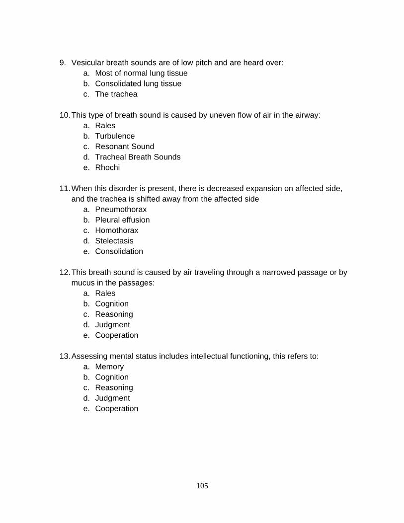
105
9. Vesicular breath sounds are of low pitch and are heard over:
a. Most of normal lung tissue b. Consolidated lung tissue c. The trachea
10. This type of breath sound is caused by uneven flow of air in the airway:
a. Rales b. Turbulence c. Resonant Sound d. Tracheal Breath Sounds e. Rhochi
11. When this disorder is present, there is decreased expansion on affected side,
and the trachea is shifted away from the affected side a. Pneumothorax b. Pleural effusion c. Homothorax d. Stelectasis e. Consolidation
12. This breath sound is caused by air traveling through a narrowed passage or by
mucus in the passages: a. Rales b. Cognition c. Reasoning d. Judgment e. Cooperation
13. Assessing mental status includes intellectual functioning, this refers to:
a. Memory b. Cognition c. Reasoning d. Judgment e. Cooperation

106
14. Abstract reasoning disorder means the person cannot think in the abstract. They
have the disorder of: a. Concrete thinking b. Disorientation c. Concrete reasoning d. Poor short term memory
15. In neurosis, the person is in touch with reality, however, the condition may:
a. Become worse b. Become overtly neurotic c. Lead to a memory disorder d. Become abstract
16. A delusion is defined as:
a. False hallucination b. Seeing something that is not there c. False belief d. Hearing something that is not there
17. The difference between a hallucination and an illusion is that with illusions there
is always a ______ present. a. Vision b. Stimulus c. Sensation d. Doctor e. Neurosis
18. The best way to chart that the patient is in a “good mood” is: the patient is
______ a. In good spirits b. Is cooperative c. In a good mood d. Is ambivalent
19. A symptom of chronic OBS would be:
a. Irritable b. Anxious c. Fearful d. Constant hallucinations

107
20. The nervous system that is composed of the sympathetic and parasympathetic systems is the:
a. Peripheral system b. Autonomic system c. Central nervous system d. Cranial nervous system
21. The nervous system that is responsible for the special senses is the _____
nervous system. a. Cranial b. Autonomic c. Peripheral d. Central e. Cerebral
22. This test is an imaging test and provides for a cross-sectional view of the skull,
showing various densities: a. `EEG b. EMG c. CAT study d. Myelogram e. Angiogram
23. This test outlines the subarachnoid space and shows the presence of tumors:
a. EEG b. EMG c. CAT study d. Myelogram e. Angiogram
24. This test requires needle electrodes to be placed into skeletal muscles in order to
study electrical impulses: a. EEG b. EMG c. CAT study d. Myelogram e. Angiogram
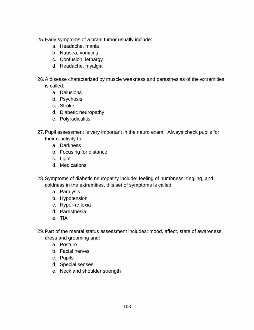
108
25. Early symptoms of a brain tumor usually include:
a. Headache, mania b. Nausea, vomiting c. Confusion, lethargy d. Headache, myalgia
26. A disease characterized by muscle weakness and parasthesias of the extremities
is called: a. Delusions b. Psychosis c. Stroke d. Diabetic neuropathy e. Polyradiculitis
27. Pupil assessment is very important in the neuro exam. Always check pupils for
their reactivity to: a. Darkness b. Focusing for distance c. Light d. Medications
28. Symptoms of diabetic neuropathy include: feeling of numbness, tingling, and
coldness in the extremities, this set of symptoms is called: a. Paralysis b. Hypotension c. Hyper-reflexia d. Paresthesia e. TIA
29. Part of the mental status assessment includes: mood, affect, state of awareness,
dress and grooming and: a. Posture b. Facial nerves c. Pupils d. Special senses e. Neck and shoulder strength

109
30. Assessment of motor system includes: general posture and muscle coordination,
muscles, coordination, and: a. Gait b. Paralysis c. Hypotension d. Cranial nerves
31. Examination of cerebral function include: behavior, level of consciousness,
intellectual functioning and: a. Thought content b. Blood pressure c. Gait d. Pupils e. Affect
32. Pulmonary blood vessels carry approx. what percentage of blood is in the body?
a. 10% b. 12% c. 18% d. 25% e. 50%
33. Arcus Senilis is a disorder of old age. When seen in younger patients it might be
a precursor to: a. Hypertension b. Myocardial infraction (MI) c. Stroke (CVA) d. Coronary artery disease
34. A patient’s edema is measured as being 1 inch in depth. The edema is
described as: a. +1 pitting edema b. +2 pitting edema c. +3 pitting edema d. +4 pitting edema
110
35. The volume of a patient’s radial pulse diminishes upon inspirations, this is
a. Pulses altering b. Bounding pulse c. Pulses obliterans d. Pulses paradoxus
36. Your patient has a regular radial pulse. However, the volume diminishes from
beat to beat, this is called a. Pulsus alterans b. Bounding pulse c. Pulsus obliterans d. Pulsus paradoxus
37. The normal jugular, venous pulse varies upon inspiration and expiration. Upon
inspiration, the normal jugular venous pulse: a. Rises b. Descends c. Weakness d. Pulsates
38. If the jugular vein pulsation is more than 3 cm above the sterna notch, it could
possibly indicate: a. Lowered CVF b. Hypotension c. Increased CVF d. Hypertension e. Congestive heart failure
39. The “c wave” in the jugular vein, is a reflection of onset of right ventricular
contraction. It begins at the end of: a. S1 b. S2 c. S3 d. S4

111
40. The heart sounds, S1, originates from the closing of the mitral valve and the:
a. Aortic valve b. Pulmonic lunar valve c. Pulmonic valve d. Tricuspid valve
41. Gallops are low-pitched sounds usually heard best with which part of the
stethoscope? a. Diaphragm b. Bell c. Cannot be heard with a stethoscope
42. A person with hypertension will have a louder sound with which heart sound?
a. S1 b. S2 c. S3 d. S4
43. Pulmonic stenosis causes a delayed emptying of the right side of the heart and a
splitting of which sound? a. S1 b. S2 c. S3 d. S4
44. In mitral stenosis there may be a louder than normal heart sound. Which heart
sound is often louder when mitral stenosis is present? a. S1 b. S2 c. S3 d. S4
45. Step I of the cardiovascular assessment is very important; it includes:
a. Putting the patient at ease b. Assessing the vital sounds c. Counting the heart rate

112
46. In the classification of heart murmurs, the loudest murmur is in the group below
is: a. Grade I b. Grade II c. Grade III d. Grade IV e. Grade V
47. A murmur that begins softly and becomes louder is referred to as:
a. Crescendo b. Decrescendo c. Holosystolic d. Pansystolic
48. When listening to heart sounds, you describe where the sound is in the cardiac
cycle. This is called: a. Grading b. Ausculating c. Palpating d. Timing e. Intensity
49. A heart sound can sometimes sound like two sounds at once. This dividing of a
heart sound is called: a. Crescendo b. Timing c. Splitting d. Grading e. Intensity
50. The time interval between S1 and S2 corresponds to the ___ phase of the
cardiac cycle. a. Pansystolic b. Systolic c. Diastolic d. Pandiastolic e. Resting