a histological study of mineralised tissue formation around … · in technovit 7200 vlc (heraeus...
TRANSCRIPT

57Osteogenesis of peri-implant tissue in vitro
Introduction
Many previous studies on osseointegration have eluci-dated that cell adhesion and proliferation were sensitive to substrate surface characteristics of dental implant (IP)s1–11). We have 3D-cultured JCRB1119:KUSA/A1 bone marrow-derived mesenchymal stem cell (MSC)s obtained from C3H/He mice (JCRB/HSRRB, Osaka, Japan) in a neutralised type I collagen gel (Cellmatrix Type I-A; Nitta Gelatin Inc., Osaka, Japan) with IPs, and observed hard tissue formation to mimic contact and the static osteogenesis in the GBR tissue surrounding titanium (Ti) IPs2, 9, 12–19). Recently, we have investigated initial contact osteogenesis of HMS0014 MSCs (Yub621b, human bone-derived MSC line; Riken BRC, Tsukuba, Japan) on Ti discs (plates) subject to different surface modifications. The study observed that HMS0014 cells
A histological study of mineralised tissue formation aroundimplants with 3D culture of HMS0014 cells in
Cellmatrix Type I-A collagen gel scaffold in vitro
By
Aiko MORISHITA, Shunji KUMABE, Michiko NAKATSUKA and Yasutomo IWAI
Department of Oral Anatomy, Osaka Dental University, 8-1 Kuzuhahanazono-cho, Hirakata-shi, Osaka 573-1121, Japan
–Received for Publication, November 25, 2014–
Key Words: Turn over, mesenchymal stem cells (MSCs), 3D collagen scaffold, GBR tissue
Summary: We cultured HMS0014 Yub621b cells within a 3D collagen gel scaffold (Cellmatrix Type I-A) and aimed to study the fate and contribution of human bone-derived mesenchymal stem cells (MSCs) in the guided bone regeneration(GBR)-en-gineered tissue which has developed around the titanium (Ti) test dental implant (IP) in vitro. The light microscopy (LM) and transmission electron microscopy (TEM) results of the peri-IP tissue indicated that collagen fibrils of the Cellmatrix Type I-A gel were accumulated and fabricated to provide a 3D meshwork for proliferation and differentiation of the HMS0014 cells in the top (cell) layer; mineralisation of the GBR tissue had commenced since day 1 and became markedly deposited between days 7 and 14 of the experiment. TEM observation revealed sedimentation of cement line at the periphery of the interwoven Cellmatrix fibres and fibrils in the ECM scaffold of the GBR tissue; matrix vesicle-mediated and appositional collagen-mediated mineralisation were identified in the peri-IP ECM scaffold. The fine structure study of the plurimorphic osteoblast(Ob)-like osteogeneic cells demonstrated numerous membranous organelles related with vesicular trafficking, secretion and endocytosis in the cytoplasm; well-developed cytoskeleton networks and intercellular junctional complexes were also observed. The specimens on fluorescence immunohistochemistry (IHC) by confocal laser-scanning microscopy (LSM) showed the expression of LC3 and Cx43 associated with autophagic-lysosomal degeneration pathway and signal conduction mediated with gap junctions (GJS) in maintaining tissue homeostasis of the Ob-like cells which grew and degen-erated in the 3D scaffold. Results from this in vitro study suggest that Ob-like HMS0014 cells actively regulate turnover of the peri-IP ECM to recapitulate the development and formation of osteoid tissue-engineered material which might contribute to augment osseointegration around the dental implant.
differentiated into osteoblast(Ob)-like cells secreting abundant extracellular matrix (ECM) under induction condition, and mineralisation of the ECM was initi-ated since day 1 and thereby became markedly depos-ited on day 7 of the experiment. We therefore surmised that mineralisation was prominently and significantly progressed between days 7 and 14 under osteogeneic induction; more calcium (Ca) and osteocalcin (OC) volumes (in µg/µgDNA) were acquired in 3D cultures10,
11). Many studies have observed that immature MSCs (e.g., HMS0014 cells) were osteoinduced to migrate and attach onto the substrate of the Ti material surfaces subjected to modifications, and then differentiated into mature Ob-like cells which extended cell processes within 60 min of culture2–4, 6, 10–12, 20–22). Subsequently, the spherical-to-polygonal (diameter (d) = 10–40 µm) HMS0014 cells were differentiated into flat, large polyg-
Okajimas Folia Anat. Jpn., 91(3): 57–71, November, 2014
Corresponding author: Shunji Kumabe, D.D.S., PhD., Dept. of Oral Anatomy, Osaka Dental University, 8-1 Kuzuhahanazono-cho, Hirakata-shi, Osaka 573-1121, Japan. E-mail: [email protected]

58 A. Morishita et al.
onal (dimension = 30 µm × 90 µm to 100 µm × 200 µm) cells sending out prominent lamellipodia and filopodia within 180 min culture; we observed that the prolifer-ating and differentiating Ob-like cells were more expan-sively adhered on the surface of anodic-oxidized (AO) Ti alloy discs. In addition, we demonstrated the expression and co-localisation of CD51 (the αv/β3 integrin: ITGAV, an interfacing specific receptor protein) and F-actin (fila-mentous actin cytoskeleton) in the matured Ob-like cells by immunohistochemistry (IHC) visualised using fluores-cent microscopy11).
There have some studies on GBR with osteogeneic cells seeded on 3D scaffolds that enhanced cell-biomate-rial interactions to support growth and differentiation of cultured mesenchymal cells9, 23–28). As we have observed that the establishment of bone-to-implant direct contact (BIC) interfaces was initiated with attachment of pre-Obs which secreted non-collagen ECM proteins to enhance the sedimentation with a layer of calcified afibrillar cement line, and then the phenomenon was followed with additional crystal growth and collagen assembly to facil-itate matricial mineralisation in the GBR peri-IP tissue to attain an appositional process of peri-IP contact osteogen-esis12–19, 29–31). In the present in vitro study, we cultured HMS0014 Yub621b cells with anodic-oxidized Ti dental IPs (AO-IPs) in the Cellmatrix Type I-A gel that devel-oped a substance essentially similar to the peri-IP tissue observed in our previous studies9, 32). We studied hard tissue formation in the tissue-engineering material mainly by transmission electron microscopy (TEM) and IHC by confocal laser-scanning microscopy (LSM) examina-tion, aimed to reveal the fine structure and turnover of the osteoid tissue which might be utilised to offer augmenta-tion of osseointegration tissue for dental IP therapy by the GBR concepts.
Materials and Methods
Immature HMS0014 : Yub621b (Human bone-derived mesenchymal, osteogeneic linage cells; Riken BRC) cells were filtrated, centrifuged and incubated/maintained in POWEREDBY10 (Glyco Technica Ltd., Sapporo, Japan) supplemented with 1% antibiotic-antimycotic agent (100 units/mL penicillin + 100 μg/mL streptomycin; Nacalai Tesque, Kyoto, Japan) in cell culture 75 cm2 flasks (TPP, Switzerland) at 37°C in humid air with 5% CO2 for 72h.
Tissue engineering of HMS0014 cells on Ti dental IPsPrimarily (finely-blasted) and secondarily (anod-
ic-oxidized; AO) processed dental root-shaped and straight α-β type titanium alloy (Ti-6Al-4V) FINAFIX dental implants (d = 3.7 mm, l = 14.0 mm, POI system; KYOCERA Medical Co., Osaka, Japan; AO-IP) were laid in dishes paved with Cellmatrix Type I-A gel (base layer), cultured together with the cell cluster containing
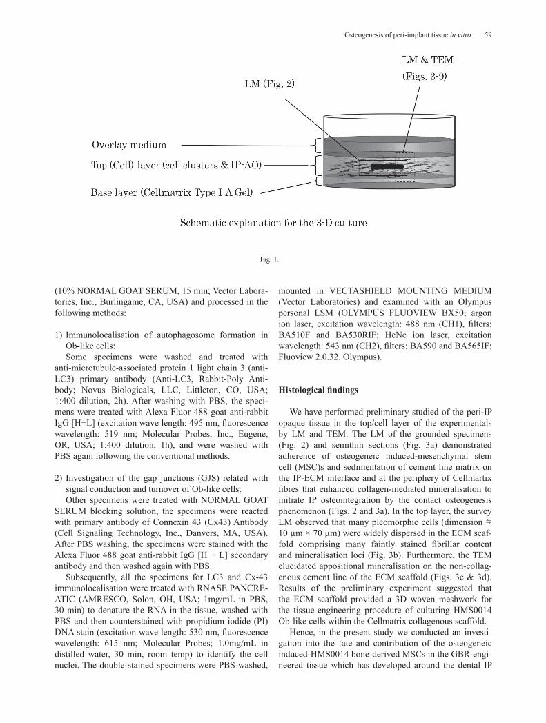
immature HMS0014 mesenchymal stem cells (MSCs) in Cellmatrix Type I-A collagen gel (top layer, cell layer), and overlaid with POWEREDBY10 (added with osteo-geneic induction medium: ascorbic acid (AA) + β-glyc-erophosphate (β-GP) + dexametazone (DEX); overlay medium) hence the HMS0014 cells have been induced to different into mature osteogeneic osteoblast (Ob)-like cells on AO-IPs in a 3D microenvironment (1×10⁶ cells/mL; humid 5% CO2/37°C, for 21 days) since the day 3 of experiment according to the “Collagen Gel Embedded Culture Method” (http://www.nitta-gelatin.co.jp; Nitta Gelatin Inc.) to mimic osseointegration of AO-IPs in 10 cm dishes (IWAKI, Tokyo, Japan) under the induction condition in vitro (Fig. 1).
In contrast, HMS0014 MSCs 3D-cultured under non-inducing condition (without addition of the osteo-geneic supplements) for 3 days following the same Collagen Gel Embedded Culture Method were prepared and designated to be the controls for the purpose of comparison with the experimentals (cultured under osteo-geneic condition) in the present TEM study.
Histological studies 1. Light microscopy (LM)
Test IPs with surrounding mineralizing tissue (a visu-ally opaque layer) were dissected from the cultures, fixed with 4% paraformaldehyde, dehydrated and embedded in Technovit 7200 VLC (Heraeus Kulzer GmbH & Co. KG, Wehrheim, Germany). The samples were sectioned with EXAKT BS-300CP-A Band Saw Machine and MG-400CS Microgrinding Machine (MEIWAFOSIS, Tokyo, Japan) by using the Cutting-Grinding Tech-nique. Subsequently, the specimens were prepared for light microscopy (LM) study by an Olympus BX41 LM connected with a FX380 system (Olympus, Tokyo, Japan).
On the other hand, some of the dissected peri-IP tissue were immersion-prefixed (1/2 Karnovsky’s fixatives), post-fixed (1.0% OsO4), embedded in EPON 812 (TAAB, Berkshire, UK), trimmed and semithin sectioned with diamond knives on an ultratome (ULTROTOME; LKB, Stockholm, Sweden), and then prepared for LM survey specimens to select areas for the ultrathin sections.
2. Transmission electron microscopy (TEM)The Epon 812 embedded specimens (the controls and
experimentals) were ultrathin-sectioned with diamond knives using the ULTROTOME, picked up on copper grids, double electron stained (uranyl acetate and lead citrate), and then examined and photographed with a Hitachi H-7100 TEM (Hitachi, Tokyo, Japan).
3. Fluorescence immunohistochemistry (IHC) by confocal laser-scanning microscopy (LSM)Some frozen-sectioned specimens subjected to acetone
fixation were washed with PBS (5 min, 3 times), blocked
59Osteogenesis of peri-implant tissue in vitro
(10% NORMAL GOAT SERUM, 15 min; Vector Labora-tories, Inc., Burlingame, CA, USA) and processed in the following methods:
1) Immunolocalisation of autophagosome formation in Ob-like cells:Some specimens were washed and treated with
anti-microtubule-associated protein 1 light chain 3 (anti-LC3) primary antibody (Anti-LC3, Rabbit-Poly Anti-body; Novus Biologicals, LLC, Littleton, CO, USA; 1:400 dilution, 2h). After washing with PBS, the speci-mens were treated with Alexa Fluor 488 goat anti-rabbit IgG [H+L] (excitation wave length: 495 nm, fluorescence wavelength: 519 nm; Molecular Probes, Inc., Eugene, OR, USA; 1:400 dilution, 1h), and were washed with PBS again following the conventional methods.
2) Investigation of the gap junctions (GJS) related with signal conduction and turnover of Ob-like cells:Other specimens were treated with NORMAL GOAT
SERUM blocking solution, the specimens were reacted with primary antibody of Connexin 43 (Cx43) Antibody (Cell Signaling Technology, Inc., Danvers, MA, USA). After PBS washing, the specimens were stained with the Alexa Fluor 488 goat anti-rabbit IgG [H + L] secondary antibody and then washed again with PBS.
Subsequently, all the specimens for LC3 and Cx-43 immunolocalisation were treated with RNASE PANCRE-ATIC (AMRESCO, Solon, OH, USA; 1mg/mL in PBS, 30 min) to denature the RNA in the tissue, washed with PBS and then counterstained with propidium iodide (PI) DNA stain (excitation wave length: 530 nm, fluorescence wavelength: 615 nm; Molecular Probes; 1.0mg/mL in distilled water, 30 min, room temp) to identify the cell nuclei. The double-stained specimens were PBS-washed,
mounted in VECTASHIELD MOUNTING MEDIUM (Vector Laboratories) and examined with an Olympus personal LSM (OLYMPUS FLUOVIEW BX50; argon ion laser, excitation wavelength: 488 nm (CH1), filters: BA510F and BA530RIF; HeNe ion laser, excitation wavelength: 543 nm (CH2), filters: BA590 and BA565IF; Fluoview 2.0.32. Olympus).
Histological findings
We have performed preliminary studied of the peri-IP opaque tissue in the top/cell layer of the experimentals by LM and TEM. The LM of the grounded specimens (Fig. 2) and semithin sections (Fig. 3a) demonstrated adherence of osteogeneic induced-mesenchymal stem cell (MSC)s and sedimentation of cement line matrix on the IP-ECM interface and at the periphery of Cellmartix fibres that enhanced collagen-mediated mineralisation to initiate IP osteointegration by the contact osteogenesis phenomenon (Figs. 2 and 3a). In the top layer, the survey LM observed that many pleomorphic cells (dimension ≒ 10 µm × 70 µm) were widely dispersed in the ECM scaf-fold comprising many faintly stained fibrillar content and mineralisation loci (Fig. 3b). Furthermore, the TEM elucidated appositional mineralisation on the non-collag-enous cement line of the ECM scaffold (Figs. 3c & 3d). Results of the preliminary experiment suggested that the ECM scaffold provided a 3D woven meshwork for the tissue-engineering procedure of culturing HMS0014 Ob-like cells within the Cellmatrix collagenous scaffold.
Hence, in the present study we conducted an investi-gation into the fate and contribution of the osteogeneic induced-HMS0014 bone-derived MSCs in the GBR-engi-neered tissue which has developed around the dental IP
Fig. 1.

60 A. Morishita et al.
Fig. 2. LM of day 14 osseointegration in vitro (Olympus BX41/FX380 system). Fig. 2a LM of ground-sectioned AO-IP. High magnification of the peri-AO-IP tissue (Figs. 2b–d) shows attachment of Obs ( ▲ ), sedimentation of an afibrillar cement line (
➡
), and collagen-related ECM min-eralisation ( △ ) to obtain a peri-IP osteogenesis.
Fig. 3. Preliminary experimental study (day 21). Fig. 3a LM of a semithin section showing sedimentation of cement line matrix on the periphery of Cellmatrix scaffold (☆). Fig. 3b LM indicates that pleomorphic cells (*) are widely dispersed in the ECM scaffold comprising many faintly stained fibrillar content and mineralisation loci (↑). Figs. 3c, d The TEM shows sedimentation of an afibrillar cement line (▲) at the periphery of Cellmatrix scaffold (☆) and collagen-mediated ECM mineralisation (◆), and therefore attains a direct osteogenesis of tis-sue-engineering tissue on the Cellmatrix Type I-A 3-D scaffold.
61Osteogenesis of peri-implant tissue in vitro
in vitro.
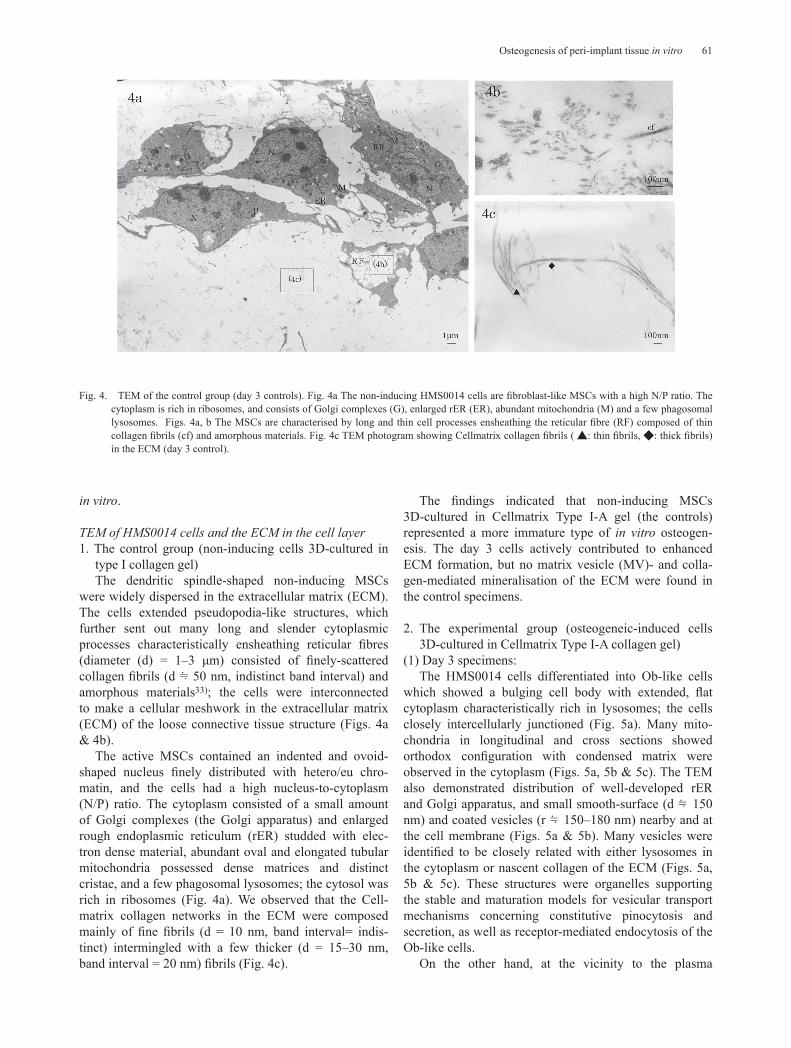
TEM of HMS0014 cells and the ECM in the cell layer 1. The control group (non-inducing cells 3D-cultured in
type I collagen gel)The dendritic spindle-shaped non-inducing MSCs
were widely dispersed in the extracellular matrix (ECM). The cells extended pseudopodia-like structures, which further sent out many long and slender cytoplasmic processes characteristically ensheathing reticular fibres (diameter (d) = 1–3 μm) consisted of finely-scattered collagen fibrils (d ≒ 50 nm, indistinct band interval) and amorphous materials33); the cells were interconnected to make a cellular meshwork in the extracellular matrix (ECM) of the loose connective tissue structure (Figs. 4a & 4b).
The active MSCs contained an indented and ovoid-shaped nucleus finely distributed with hetero/eu chro-matin, and the cells had a high nucleus-to-cytoplasm (N/P) ratio. The cytoplasm consisted of a small amount of Golgi complexes (the Golgi apparatus) and enlarged rough endoplasmic reticulum (rER) studded with elec-tron dense material, abundant oval and elongated tubular mitochondria possessed dense matrices and distinct cristae, and a few phagosomal lysosomes; the cytosol was rich in ribosomes (Fig. 4a). We observed that the Cell-matrix collagen networks in the ECM were composed mainly of fine fibrils (d = 10 nm, band interval= indis-tinct) intermingled with a few thicker (d = 15–30 nm, band interval = 20 nm) fibrils (Fig. 4c).
The findings indicated that non-inducing MSCs 3D-cultured in Cellmatrix Type I-A gel (the controls) represented a more immature type of in vitro osteogen-esis. The day 3 cells actively contributed to enhanced ECM formation, but no matrix vesicle (MV)- and colla-gen-mediated mineralisation of the ECM were found in the control specimens.
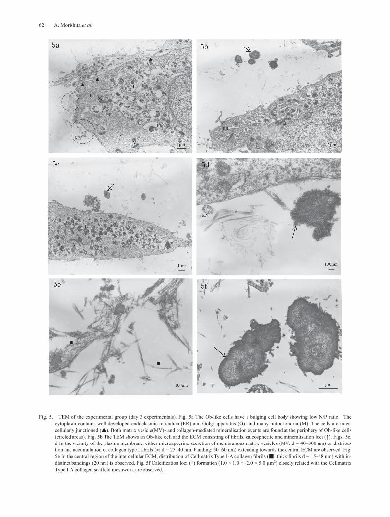
2. The experimental group (osteogeneic-induced cells 3D-cultured in Cellmatrix Type I-A collagen gel)
(1) Day 3 specimens:The HMS0014 cells differentiated into Ob-like cells
which showed a bulging cell body with extended, flat cytoplasm characteristically rich in lysosomes; the cells closely intercellularly junctioned (Fig. 5a). Many mito-chondria in longitudinal and cross sections showed orthodox configuration with condensed matrix were observed in the cytoplasm (Figs. 5a, 5b & 5c). The TEM also demonstrated distribution of well-developed rER and Golgi apparatus, and small smooth-surface (d ≒ 150 nm) and coated vesicles (r ≒ 150–180 nm) nearby and at the cell membrane (Figs. 5a & 5b). Many vesicles were identified to be closely related with either lysosomes in the cytoplasm or nascent collagen of the ECM (Figs. 5a, 5b & 5c). These structures were organelles supporting the stable and maturation models for vesicular transport mechanisms concerning constitutive pinocytosis and secretion, as well as receptor-mediated endocytosis of the Ob-like cells.
On the other hand, at the vicinity to the plasma
Fig. 4. TEM of the control group (day 3 controls). Fig. 4a The non-inducing HMS0014 cells are fibroblast-like MSCs with a high N/P ratio. The cytoplasm is rich in ribosomes, and consists of Golgi complexes (G), enlarged rER (ER), abundant mitochondria (M) and a few phagosomal lysosomes. Figs. 4a, b The MSCs are characterised by long and thin cell processes ensheathing the reticular fibre (RF) composed of thin collagen fibrils (cf) and amorphous materials. Fig. 4c TEM photogram showing Cellmatrix collagen fibrils ( ▲: thin fibrils, ◆: thick fibrils) in the ECM (day 3 control).
62 A. Morishita et al.
Fig. 5. TEM of the experimental group (day 3 experimentals). Fig. 5a The Ob-like cells have a bulging cell body showing low N/P ratio. The cytoplasm contains well-developed endoplasmic reticulum (ER) and Golgi apparatus (G), and many mitochondria (M). The cells are inter-cellularly junctioned (▲). Both matrix vesicle(MV)- and collagen-mediated mineralisation events are found at the periphery of Ob-like cells (circled areas). Fig. 5b The TEM shows an Ob-like cell and the ECM consisting of fibrils, calcospherite and mineralisation loci (↑). Figs. 5c, d In the vicinity of the plasma membrane, either microapocrine secretion of membranous matrix vesicles (MV: d = 40–300 nm) or distribu-tion and accumulation of collagen type I fibrils (*: d = 25–40 nm, banding: 50–60 nm) extending towards the central ECM are observed. Fig. 5e In the central region of the intercellular ECM, distribution of Cellmatrix Type I-A collagen fibrils (■: thick fibrils d = 15–48 nm) with in-distinct bandings (20 nm) is observed. Fig. 5f Calcification loci (↑) formation (1.0 × 1.0 ~ 2.0 × 5.0 µm2) closely related with the Cellmatrix Type I-A collagen scaffold meshwork are observed.
63Osteogenesis of peri-implant tissue in vitro
membrane, either microapocrine secretion of MVs (d = 40–300 nm) or distribution and accumulation of thin collagen type I fibrils (d = 25–40 nm, band interval = 50–60 nm) extending towards the central portion of the ECM were observed; MV- and subsequent collagen-me-diated mineralisation adjacent to the Ob-like cells were demonstrated (Figs. 5c & 5d). In the centre of the ECM, about 2–5 µm distant from the neighboring cells, distri-bution of Cellmatrix Type I-A collagen fibrils (d = 15–48 nm) with indistinct fine band intervals (20 nm) was found; collagen-mediated mineralisation (d = 100–280 nm) and calcification loci formation (1.0 µm × 1.0 µm to 2.0 µm × 5.0 µm) closely related with the Cellmatrix Type I-A collagen scaffold meshwork were also found (Figs. 5b, 5e & 5f).
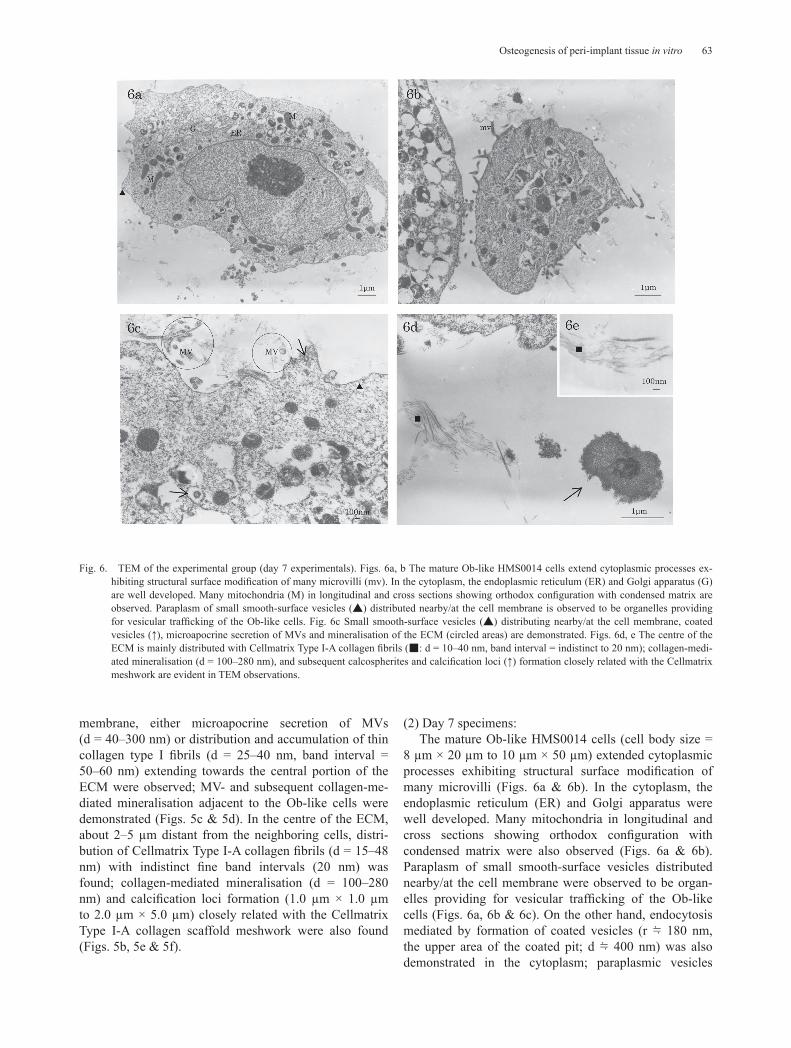
(2) Day 7 specimens:The mature Ob-like HMS0014 cells (cell body size =
8 µm × 20 µm to 10 µm × 50 µm) extended cytoplasmic processes exhibiting structural surface modification of many microvilli (Figs. 6a & 6b). In the cytoplasm, the endoplasmic reticulum (ER) and Golgi apparatus were well developed. Many mitochondria in longitudinal and cross sections showing orthodox configuration with condensed matrix were also observed (Figs. 6a & 6b). Paraplasm of small smooth-surface vesicles distributed nearby/at the cell membrane were observed to be organ-elles providing for vesicular trafficking of the Ob-like cells (Figs. 6a, 6b & 6c). On the other hand, endocytosis mediated by formation of coated vesicles (r ≒ 180 nm, the upper area of the coated pit; d ≒ 400 nm) was also demonstrated in the cytoplasm; paraplasmic vesicles
Fig. 6. TEM of the experimental group (day 7 experimentals). Figs. 6a, b The mature Ob-like HMS0014 cells extend cytoplasmic processes ex-hibiting structural surface modification of many microvilli (mv). In the cytoplasm, the endoplasmic reticulum (ER) and Golgi apparatus (G) are well developed. Many mitochondria (M) in longitudinal and cross sections showing orthodox configuration with condensed matrix are observed. Paraplasm of small smooth-surface vesicles (▲) distributed nearby/at the cell membrane is observed to be organelles providing for vesicular trafficking of the Ob-like cells. Fig. 6c Small smooth-surface vesicles (▲) distributing nearby/at the cell membrane, coated vesicles (↑), microapocrine secretion of MVs and mineralisation of the ECM (circled areas) are demonstrated. Figs. 6d, e The centre of the ECM is mainly distributed with Cellmatrix Type I-A collagen fibrils (■: d = 10–40 nm, band interval = indistinct to 20 nm); collagen-medi-ated mineralisation (d = 100–280 nm), and subsequent calcospherites and calcification loci (↑) formation closely related with the Cellmatrix meshwork are evident in TEM observations.
64 A. Morishita et al.
closely related with electron-dense lysosomes which were also characteristically abundant in the day 7 Ob-like cells (Fig. 6a, 6b & 6c).
It was near to the plasma membrane, MV- and colla-gen-mediated mineralisation arose spatially and struc-turally by the nucleation phenomena (Fig. 6a, 6c & 6d). On the other hand, the centre of the ECM were mainly distributed with Cellmatrix Type I-A collagen fibrils (d = 10–40 nm, band interval = indistinct to 20 nm); colla-gen-mediated mineralisation (d = 100–280 nm), and subsequent calcospherites and calcification loci formation were evident in TEM of the Cellmatrix meshwork (Fig. 6d & 6e).
(3) Day 14 specimens:The day 14 HMS0014 cells cultured with Cellma-
trix Type I-A showed similar TEM findings to the day 7 specimens; abundant cytoplasm distributed with well-de-
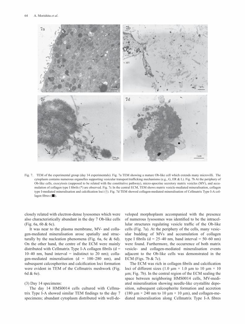
veloped morphoplasm accompanied with the presence of numerous lysosomes was identified to be the intracel-lular structures regulating vesicle traffic of the Ob-like cells (Fig. 7a). At the periphery of the cells, many vesic-ular budding of MVs and accumulation of collagen type I fibrils (d = 25–40 nm, band interval = 50–60 nm) were found. Furthermore, the occurrence of both matrix vesicle- and collagen-mediated mineralisation events adjacent to the Ob-like cells was demonstrated in the ECM (Figs. 7b & 7c).
The ECM was rich in collagen fibrils and calcification loci of different sizes (1.0 µm × 1.0 µm to 10 µm × 10 µm; Fig. 7b). In the central region of the ECM sealing the space between neighboring HMS0014 cells, MV-medi-ated mineralisation showing needle-like crystallite depo-sition, subsequent calcospherite formation and accretion (180 µm × 240 nm to 10 µm × 10 µm), and collagen-me-diated mineralisation along Cellmatrix Type I-A fibres
Fig. 7. TEM of the experimental group (day 14 experimentals). Fig. 7a TEM showing a mature Ob-like cell which extends many microvilli. The cytoplasm contains numerous organelles supporting vesicular transport/trafficking mechanisms (e.g., G, ER & L). Fig. 7b At the periphery of Ob-like cells, exocytosis (supposed to be related with the constitutive pathway), micro-apocrine secretory matrix vesicles (MV), and accu-mulation of collagen type I fibrils (*) are observed. Fig. 7c In the central ECM, TEM shows matrix vesicle-mediated mineralisation, collagen type I-mediated mineralisation and calcification loci (↑). Fig. 7d TEM showed collagen-mediated mineralisation of Cellmatrix Type I-A col-lagen fibres (■).

65Osteogenesis of peri-implant tissue in vitro
forming calcification loci were found (Fig. 7d).
(4) Day 21 specimens:The day 21 cells showed similar histology with the
days 7 and 14 specimens; TEM observed that they were cells abundant in morphoplasm. However, some Ob-like cells with nuclear envelopes had the perinuclear cisternal space dilated and filled with condensed substance. The nucleus was morphologically characterized with disin-tegrating nucleolemma showing dilation of nuclear pores to envelop condensed chromatin and many auto-phagic vacuoles (Fig. 8a). The histology indicated that Ob-like cells in the day 21 growing tissue-engineered material were entering the terminal phase of autophagic degeneration, a type of non-apoptotic programmed cell death. GJS connecting degenerative cells and adjacent cells were evident (Figs. 8a & 8b). Exocytotic vesicles, as well as membranous MVs were demonstrated at the periphery cytoplasm and cell membrane (Fig. 8c). On the other hand, the fine structure examination showed that the ECM was abundantly distributed with bundles of collagen fibrils and calcification loci (Figs. 8d & 8e). The TEM also revealed MV- and Type I collagen-mediated calcification in the ECM; Cellmatrix Type I-A collagen fibrils were hardly identified in the day 21 specimens (Figs. 8d & 8e).
The day 21 osteogeneic-inducted Ob-like cells spread and were intercellularly connected with many fine cyto-plasmic protrusions (Fig. 8a). In the abundant cytoplasm, it was rich in kinds of membranous organelles and ribo-somes, and the cytosol was filled with a well-developed cytoskeletal network of microfilaments (d = 3–6 nm), intermediate filaments (d ≒ 10 nm) and microtubules (Figs. 8a, 8c, 8e & 8f).
Fluorescence IHC by LSMThe results were summarised and demonstrated the
protein expression related with autophagic-lysosomal degeneration pathway and cell death signal conduction mediated with GJS of Ob-like bone-derived MSCs (Table 1; Figs. 9a & 9b, 10a & 10b).
1. LC3 autophagy-related protein expressionThe autophagic vacuole marker protein LC3 was
not immumolocalised in the day 3 controls (Fig. 9a). However, the expression of LC3 was evident in the days 3, 7, 14 and 21 experimental osteoproductive Ob-like cells (Fig. 9b).
2. Cx43 GJS protein expressionThe GJS are type of surface domains essentially
related with moderation of cell death, signal conduction and turnover of the Ob-like cells.
Cx43 expression was observed in HMS0014 cells of both the controls and experimentals (Fig. 10a). The LSM results indicated that the number of GJS between the
adjacent Ob-like cells have increased over time of experi-ment (Fig. 10b).
The present fluorescence IHC by LSM indicated that there was autophagic-lysosomal degeneration pathway modified with cell death signal conducted of neigh-bouring Ob-like cells that maintained tissue homeostasis by regulating the life and death of the bone-derived MSCs in the growing peri-IP tissue-engineering material.
Discussion
Several studies on dental implant (IP) osseointegra-tion have elucidated that peri-IP bone bonding began with osteoinduction allowing for attachment and subse-quent differentiation of osteogeneic linage cells to initiate osteoconduction to facilitate the direct bone-to-implant contact (BIC) of IPs3, 4, 6, 9, 12, 15, 18, 20–22, 29, 30, 34). Never-theless, the studies have emphasized that the develop-ment of focal contacts of the attached osteogeneic cells essentially mediated the IP surface substrate responses to affect cell spreading and signaling and thereby modi-fied contact osteogenesis at the cell-substratum inter-face1, 3, 4, 6, 12, 15, 19, 20, 35–39). Previously, we have studied the histochemical properties (e.g., by ALPase activity, Ca volume, osteocalcin volume studies) and osteo-conduction of mouse KUSA/A1 (JCRB/HSRRB) and human HMS0014 Yub621b (Riken BRC) osteogeneic linage MSCs in monolayer and 3D cultures, and then cultured the cells on titanium (Ti) discs with different surface substrates (i.e., anodic oxidation, hydroxyapatite coating, precision blasting). The results revealed that the immature bone-derived MSCs were condition-induced to actively proliferate, migrate and spread to acquire focal adhesions and intercellular connections with well-de-veloped lamellipodia (extended sheet-like cytoplasmic protrusions supported with a meshwork of branched cytoskeletal proteins) and filopodia (finger-like protru-sions supported with a parallel array of microfilaments assembled in long bundles) within 180 minutes, and were differentiated into mature osteoblast (Ob)-like cells during three days of culture; the SEM study observed that HMS0014 cells were more expansively adhered on the anodic-oxidized (AO) Ti discs. Furthermore, fluores-cent immunomicroscopy findings revealed co-expression of F-actin (actin filament, a cytoskeleton protein) and CD51 (the αv/β3 integrin: ITGAV, an interfacing specific receptor protein) in the differentiated Ob-like cells 8,11).
The integrin family of cell adhesion receptor mole-cules found in cell anchoring junctions has been eluci-dated to be the trans-membrane linker (anchoring) protein which actively transducts signals across the plasma membrane by tying cytoskeletal protein anchors (e.g., actin filaments) to ECM proteins with focal adhe-sions40, 41). On the other hand, there have been studies of

66 A. Morishita et al.
Fig. 8. TEM of the experimental group (day 21 experimentals). Fig. 8a The nucleolemma shows dilated nuclear pores and perinuclear cisternal space; the disintegrating nucleolamma envelopes many enlarged autophagic vacuoles (↑) (L: lysosome, M: mitochondrion). Fig. 8b The TEM shows the GJS (↑) connecting adjacent Ob-like cells. Fig. 8c Exocytotic vesicles (↑), as well as membranous MVs are demonstrated at the periphery cytoplasm and cell membrane. Figs. 8d, e The TEM shows that the ECM is abundantly distributed with bundles of collagen fibrils (*) and calcification loci (↑). The TEM reveals MV- and Type I collagen-mediated calcification (↑) in the ECM; Cellmatrix Type I-A collagen fibrils are hardly identified in the day 21 specimens. Fig. 8f The day 21 cells are rich in kinds of membranous organelles (e.g., M: mitochondrion, L: lysosome, ▲: vesicle), ribosomes and a well-developed cytoskeletal network (↑).

67Osteogenesis of peri-implant tissue in vitro
Ob adhesion properties on biomaterials that elucidated the expression of integrin adhesion receptors involving in Ob-material interactions was essentially modified according to the surface characteristics of the biomate-rials. The studies also elucidated that Obs would prolif-erate and differentiate into cells showing lamellipodia and filopodia with enhanced expression of F-actin filaments coupling with large focal contacts to adhere to the oste-oinductive substratum; the IP surface substrate responses vice versa played active roles in events that governed not
only osteoinduction (adherence and spreading, migration, proliferation, maturation and modulation of the Ob-like cells) but also the development of focal adhesion struc-tures which mediated contact osteogenesis at the direct BIC interface1, 3, 4, 6, 12, 15, 20, 35, 37–39, 42, 43). In contrast, there has been a study demonstrating that inappropriate cell-matrix interactions might evoke signals transducted from type I collagen fibrils (e.g., of the ECM or scaffold) into surrounding cells through integrin, thereby inducing disoriented cell death to ensure the polarity of cells.
Figs. 9 & 10. Fluoresence IHC by LSM. The LC3 autophagy-related protein and Cx43 GJS protein expression. The expression of autophagic vacuole marker protein LC3 expression and Cx43 GJS protein is not immumolocalised in the controls (Figs. 9a, 10a), but is evident in the experimental osteoproductive Ob-like cells (Figs. 9b, 10b) (represented by day 3 specimens).

68 A. Morishita et al.
The study surmised that apoptosis played the important cell death mechanism in physiological developmental processes or under pathological conditions42).
Some histological studies have indicated that certain chemical and physical signals occurring at the focal adhesions were transmitted to the cell body and subse-quently shared with neighboring cells via intercellular junctions (e.g., adherence, gap junctional intercellular communication)44–49). On the other hand, the architecture of actin microfilamentous networks build up a F-actin cortex which was essential for the maintenance and regulation of cell shape and the generation of mechan-ical forces in cell migration, as well as the development of stress fibres to couple with focal adhesion plaques for transmission of forces towards the substrate and scaf-folds6, 42, 49). Many other studies have elucidated that intracellular cytoskeletal elements were organised to be tension-bearing structures of beams (microtubules) and cables (micro- and intermediate filaments), giving struc-tural support to hold and regulate cell shape by resisting buckling due to compressive forces and filament fracture, as well as a dynamic 3D structure to carry out cellular movements and convey information/signals about the microenvironment of the cells6, 42, 49–51). Our previous SEM studies have demonstrated that the actively prolifer-ating and differentiating Ob-like HMS0014 cells adhered to and spread well on the surface of tested Ti discs and IPs under inducing conditions11, 32). In the present TEM study, we observed that the mature Ob-like HMS0014 cells contained many non-membranous cell organelles of microfilaments, intermediate filaments and microtubules in the cell body and peripheral cytoplasm. We speculated that the distribution of the F-actin cytoskeleton corre-sponds with the actin stress fibres, which have fibrillar ends co-localised with CD51 to represent focal adhesion formation of the HMS0014 cells under inducing condi-tions11, 32).
The present TEM fine structure study revealed that the well-developed cytoskeleton and abundant membra-nous organelles seemed to be general characteristics of the mature Ob-like HMS0014 cells; a well-developed Golgi apparatus, rER, sER, polymorphic mitochondria in orthodox configuration containing dense matrix and other morphoplasm were evident in the cells. We also observed
that distribution of coated vesicles and endosomes were spatially closely related with numerous electron-dense lysosomes, and identified vesicular transport and traf-ficking mechanisms of the osteoinducted cells2, 52–54). On the other hand, the changes in the number and size of mitochondria present in a cell are directly related to the cell’s metabolic activity, and mitochondria undergo reversible ultrastructural transformations between a condensed and an orthodox conformation in relationship to either the osmotic balance between cytosol and the matrix or respiratory state of the cell. A previous study has elucidated that mitochondria with matrix of slow metabolite diffusion rate in the condensed state were dominant in a tissue with high level of oxidative phos-phorylation55). However, it has been noted that the onset of phosphorylation was accompanied by a reduction of the matrix volume of orthodox mitochondria, but there is no evidence that the decrease in matrix volume affected the phosphorylation efficiency56). Nevertheless, other studies have reported that volume-dependent regulation of matrix protein packing may modulate metabolite diffu-sion and, in turn, mitochondrial metabolism of cells57–59).
On the other hand, by TEM examination of the cell (top) layer as presented in this study, budding of para-plasm of small smooth-surface vesicles and formation of coated vesicles nearby/at the cell membrane, as well as accumulation of nascent/thin collagen type I fibrils and microapocrine secretion of MVs at the periphery of cell membrane in the ECM were demonstrated; we identified that the ECM contained hybrid of collagen secreted by Ob-like cells and distributed by the Cellmatrix Type I-A gel. Furthermore, the study revealed a temporo-spatial replacement of the finely divergent Cellmatrix networks by collagen type I fibrils secreted from Ob-like HMS0014 cells in the ECM; the Cellmatrix collagen fibrils were not evident in the cell layer of day 21 specimens. Meanwhile, it was similar to what has described for contact osteogen-esis during osseointegration, we identified cement line formation at the periphery of Cellmatrix Type I-A fibres and fibrils in the cell layer. Collectively, we have eluci-dated osteoconduction of MSCs to commence MV- and collagen-mediated mineralisation in a collagenous 3D ECM scaffold to develop a tissue-engineering peri-IP osteoid tissue in vitro2, 9, 13–19, 30, 32).
Table 1. Results
Fluorescence IHC by LSM
Protein expressionControl Experimental
LC3Day 3
–Day 3
+Day 7
+Day 14
+Day 21
+
Cx43 – + + + +
Alexa Fluor 488 goat anti-rabbit IgG [H + L] (excitation wavelength: 495 nm, fluorescence wavelength: 519 nm and PI DNA stain (excitation wavelength: 530 nm, fluorescence wavelength: 615 nm)
69Osteogenesis of peri-implant tissue in vitro
In the present study, LC3 localisation was used as a marker of autophagosome formation in the Ob-like cells of the tissue-engineered material60). The fluorescence IHC by LSM detected the expression of LC3 expres-sion in experimental Ob-like cells. Meanwhile by using TEM for observation of Ob-like cells, we observed that day 21 experimentals had the nuclear enveloped with dilated perinuclear cisternal space filled with condense substance, and the nucleus was morphologically charac-terized with disintegrating nucleolemma showing dila-tion of nuclear pores to envelop condensed chromatin and many enlarged autophagic vacuoles. The histolog-ical studies observed that day 21 Ob-like cells in the growing GBR tissue were entering the terminal phase of non-apoptotic programmed cell death (PCD) -the type 2 physiological cell death showing autophagic degenera-tion-, thereby indicated immunohistochemical and ultra-structural evidence for progressive autophagic degener-ation resulting in PCD of the Ob-likes cells in the GBR tissue61, 62).
In addition, the present TEM and LSM observed that other than adherens junctions, the cells were connected by GJS for allowing the transmission of electrical impulses among their neighbouring cells. GJS have been elucidated to be channels playing an important func-tion in maintaining cellular energy homeostasis and cell survival by spreading cell-killing signals initially gener-ated by a single cell that spontaneously initiated apoptosis into healthy non-apoptotic neighbours. Thus, the GJS regulated important roles in the life and death of cells by moderating cell death signal conduction to migrate the bystander effect between contacting cells51, 63–66). Some studies have further stated that the recycling function of autophagy through a lysosomal degradation pathway was a decisive factor not causing the unaffected healthy bystander cells to also die and thereby maintained cellular energy homeostasis and survival. Moreover, PCD has been elucidated to be a self-limited survival strategy rather than a primary or irreversible death execution program62, 67–69).
Tissue engineering for GBR by the placement of osteogeneic cells within matrices to recapitulate the development and formation of biological implantation tissue has been proposed to be a predictable strategy for a decrease in the need for recruitment of cells at the defect site70–73). Previously, we have in vitro studied osseointe-gration of a peri-IP tissue-engineered material by mouse KUSA/A1 bone marrow-derived MSCs cultured with Cellmatrix Type I-A Gel9). The GBR method engineered mouse osteogeneic MSCs in the 3D collagen networks to initiate contact osteogenesis at the cell-substrate inter-faces followed with mineral apposition of the growing peri-IP tissue to acquire osteoid tissue which provided bone-to-implant direct contact (BIC) without intervening soft tissue at the interfaces9, 12, 16, 30).
Taken together, in the present study by embedment
of the AO-IP with cell clusters containing human osteo-geneic HMS0014 Yub621b BMSCs in the Cellmatrix Type I-A Gel fashioned from natural material, we inves-tigated histological events associated with growth of a biological tissue-engineered osseointegrative material to surround the dental IP70–74). Consequently, the TEM and LSM results elucidated that the active Ob-like HMS0014 cells contained the morphoplasm providing for the vesic-ular transport and trafficking mechanisms, as well as structures related with osteoconduction of the cells in osseointegration of Ti IPs. Moreover, we identified the LC3 autophagic gene expression and configuration of autophagy-mediated programmed cell death to main-tain cellular energy homeostasis and cellular survival of the Ob-like cells distributed in the peri-IP mineralising tissue62, 67, 68). The study concluded that the present GBR method osteoinducted human bone-derived MSCs within a 3D ECM scaffold to actively regulate a homeostatic microenvironment for commencing osteoconduction of the peri-IP tissue in vitro. Furthermore, we surmised that the tissue-engineered osteoid tissue might be utilised as a supplementary material for the IP in the host drilled socket, thereby employs a biocompatible and biodegrad-able BIC tissue which would be remolded and modeled to optimize functional osseointegration for the endosseous implant therapy9, 75–77).
Acknowledgements
This work was partly supported by a grant-in-aid for scientific research (C) (24592976) from the Ministry of Education, Culture, Sports, Science, and Technology of Japan. The human mesenchymal stem cells line, HMS0014 (Yub621b), was provided by the RIKEN BRC through the National Bio-Resource Project of the MEXT, Japan. The study was performed using the Laboratory Animal, Morphological Research and Tissue Culture Facilities in the Institute of Dental Research, Osaka Dental University. The study results were presented at the 56th Annual Meeting of Japanese Association for Oral Biology in Fukuoka on September 27, 2014.
References
1) Anselme K. Osteoblast adhesion on biomaterials. Biomaterials 2000; 21:667–681.
2) Albrektsson T, Johansson C. Osteoinduction, osteoconduction and osseointegration. Eur Spine J 2001; 10 (Suppl 2): S96–101.
3) Boyan BD, Lossdörfer S, Wang L, Zhoa G, Lohmann CH, Cochran DL, Schwartz Z. Osteoblasts generate an osteogenic microenvironment when growth on surfaces with rough microto-pographies. Eur Cell Mater 2003; 6:22–27.
4) Nasatzky E, Gultchin J, Schwartz Z. The role of surface roughness in promoting osteointegration. Refuat Hapeh Vehashinayim 2003; 20:8–19, 98.
5) Diener A, Nebe B, Lüthen F, Becker P, Beck U, Neumann HG,

70 A. Morishita et al.
Rychly J. Control of focal adhesion dynamics by material surface characteristics. Biomaterials 2005; 26:383–392.
6) Salido M, Vilches Jl, Gutiérrez JL, Vilches J. Actin cytoskeletal organization in human osteoblasts grown on different dental tita-nium implant surfaces. Histol Histopathol 2007; 22:1355–1364.
7) Ferguson SJ, Langhoff JD, Voelter K, von Rechenberg B, Scharn-weber D, Bierbaum S, Schnabelrauch M, Kautz AR, Frauchinger VM, Mueller TL, van Lenthe GH, Schlottig F. Biomechanical comparison of different surface modifications for dental implants. Int J Oral Maxillofac Implants 2008; 23:1037–1046.
8) Nakatsuka M, Mikami T, Hashimoto Y, Kon-I H, Chen HS, Hsiao SY, Kumabe S, Huang ST, Iwai Y, Huang HC. A preliminary study of osseointegration in the dental implant therapy in vivo —culture of mouse KUSA/A1 cells on titanium plates with different surface modifications—. TWJ Oral Med Sci 2009; 25:4–19.
9) Mikami T, Kumabe S, Y Iwai. A study of osseointegration -3D culture of KUSA/A1 cells with a collagen scaffold on titanium implants with different surface modifications. J Oral Tissue Engin 2010; 8:60–73.
10) Nakatsuka M, Hashimoto Y, Kumabe S, An C, Ueda K, Mikami H, Huang HC, Iwai Y. The study of cell attachment and hard tissue formation on the dental implant surface [Abstract]. Kaibougaku Zasshi 2010; 85 (Suppl): 177. (in Japanese)
11) Iwai Y, Matsuda Y, Nakatsuka M, Mikami Y, Kumabe S. A prelim-inary study of the dental implant therapy —initial osteogenesis of human mesenchymal stem (HMS0014) cells on titanium discs with different surface modifications—. Okajimas Folia Anat Jpn 2012; 88:133–140.
12) Davies JE. In vitro modeling of the bone/implant interface. Anat Rec 1996; 245:426–445.
13) Murai K, Takeshita F, Ayukawa Y, Kiyoshima T, Suetsugu T, Tanaka T. Light and electron microscopic studies of bone-tita-nium interface in the tibiae of young and mature rats. J Biomedical Mater Res 1996; 30:523–533.
14) Rosengren A, Johansson BR, Danielsen N, Thomsen P, Ericson LE. Immunohistochemical studies on the distribution of albumin, fibrinogen, fibronectin, IgG and collagen around PTFE and tita-nium implants. Biomaterials 1996; 17:1779–1786.
15) Davies JE. Mechanisms of endosseous integration. Intl J Prostho-dont 1998; 11:391–401.
16) Puelo DA, Nanci A. Understanding and controlling the bone-im-plant interface. Biomaterials 1999; 20:2311–2321.
17) Ferretti M, Palumbo C, Contri M, Marotti G. Static and dynamic osteogenesis: Two different types of bone formation. Anat Embryol 2002; 206:21–29.
18) Davies JE. Understanding peri-implant endosseous healing. J Dent Educ 2003; 67:932–949.
19) Marco F, Milena F, Gianluca G, Vittoria O. Peri-implant osteogen-esis in health and osteoporosis. Micron 2005; 36:630–644.
20) Grössner-Schreiber B, Tuan RS. Die Bedeutung der Oberfläche von Titaniimplantaten in Osteointegrationvorgang (The influence of the titanium implant surface on the process of osseointegration). Dtsch Zanhartzl Z 1991; 46:691–693.
21) Meyer U, Büchter A, Wiesmann HP, Joos U, Jones DB. Basic reactions of osteoblasts on structured material surfaces. Eur Cell Mater 2005; 9:39–49.
22) Issac J, Loty S, Hamdan A, Kokubo T, Kim HM, Berdal A, Sautier JM. Bone-like tissue formation on a biomimetic titanium surface in an explant model of osteoconduction. J Biomed Mater Res A 2009; 89:585–593.
23) Holmes TC. Novel peptide-based biomaterial scaffolds for tissue engineering. Trend Biotechnol 2002; 20:16–21.
24) Kisiday J, Jin M, Kurz B, Hung H, Semino C, Zhang S, Grodz-insky AJ. Self-assembling peptide hydrogel fosters chondrocyte extracellular matrix production and cell division: implications for cartilage tissue repair. Proc Natl Acad Sci USA 2002; 99:9996–10001.
25) Zhang S. Fabrication of novel biomaterials through molecular self assembly. Nat Biotechnol 2003; 21:1171–1178.
26) Kisiday JD, Jin M, DiMicco MA, Kurz B, Grodzinsky AJ. Effects of dynamic compressive loading on chondrocyte biosynthesis in self-assembling peptide scaffolds. J Biomech 2004; 37:595–604.
27) Bokhari MA, Akay G, Zhang S, Birch MA. The enhancement of osteoblast growth and differentiation in vitro on a peptide hydrogel- polyHIPE polymer hybrid material. Biomaterials 2005; 26:5198–5208.
28) Fillies T, Wiesmann HP, Sommer D, Joos U, Meyer U. Osteoblast reaction on SLA and microgroved implant surfaces. Mund Kiefer Gesichtschir 2005; 9:24 –28.
29) Inoue T. Mechanisms of metabolism and wound healing. In: Yamazaki M, Takahashi T, Katsuyama H, Inoue T, Hayashi Y, eds, The Ultimate Guide to Implant Dentistry. Tokyo, Japan: Ishiyaku Publishers Inc.; 2004: 38–53.(in Japanese)
30) Inoue T. Histology (Bone Soft tissue). In: Akagawa Y et al., eds. Fundamental concepts and techniques of oral implants. 2nd ed. Tokyo, Japan: Ishiyaku Publishers, Inc.; 2005: 40–60.(in Japanese)
31) Koyano K, Matsushita Y, Ayukawa Y. Basic science for the dental implantology. In: Koyano K, Matsuura M, eds. Essential oral implantology. Tokyo, Japan: Ishiyaku Publishers, Inc.; 2009: 10–17.(in Japanese)
32) Iwai Y, Mikami Y, Nakatsuka M, Huang HC, Mikami H, Kumabe S. A preliminary study of the dental implant therapy —initial osteogenesis of human mesenchymal stem (HMS0014) cells on titanium discs and implants with different surface modifications—. J Tissue Eng Regen Med 2012; 6 (Suppl. 1): 234–235.
33) Mohammad AAY, Asai J. Ultrastructural and morphometrical studies on the reticular framework and reticular fibers in the retic-uloendothelial system of rats. Nagoya J Med Sci 1993; 55:47–56.
34) Joos U, Meyer U. New paradigm in implant osseointegration. Head Face Med 2006; 2:19.
35) Lange R, Lüthen F, Beck U, Rychly J, Baumann A, Nebe B. Cell-extracellular matrix interaction and physico-chemical charac-teristics of titanium surfaces depend on the roughness of the mate-rial. Biomol Eng 2002; 19:255–261.
36) Jayaraman M, Meyer U, Bühner M, Joos U, Wiesmann HP. Influ-ence of titanium surfaces on attachment of osteoblast-like cells in vitro. Biomaterials 2004; 25:625–631.
37) Lenhert S, Meier MB, Meyer U, Chi L, Wiesmann HP. Osteo-blast aligment, elongation and migration on grooved polystyrene surfaces patterned by Langmuir-Blodgett lithography. Biomate-rials 2005; 26:563–570.
38) Graf HL, Stoeva S, Armbruster FP, Neuhaus J, Hilbig H. Effect of bone sialoprotein and collagen coating on cell attachment to TICER and pure titanium implant surfaces. Int J Oral Maxillofac Surg 2008; 37:634–640.
39) Coelho PG, Granjeiro JM, Romanos GE, Suzuki M, Silva NR, Cardaropoli G, Thompson VP, Lemons JE. Basic research methods and current trends of dental implant surface. J Biomed Mater Res B Appl Biomater 2009; 88:579–596.
40) Albert B, Johnson A, Lewis J, Raff M, Roberts K, Walter P. Molecular Biology of the Cell. 4th edition. New York, NY: Garland Science; 2002:1–29.
41) Stricker J, Falzone T, Gardel ML. Mechanics of the F-actin cyto-skeleton. J Biomechanics 2010; 43:9–14.
42) Lim JY, Taylor AF, Li Z, Vogler EA, Donahue HJ. Integrin expres-sion and osteopontin regulation in human fetal osteoblastic cells mediated by substratum surface characteristics. Tissue Eng 2005; 11:19–29.
43) Tang MJ, Hu JJ, Lin HH, Chiu WT, Jiang ST. Collagen gel overlay induces apoptosis of polarized cells in cultures: disoriented cell death. Am J Physiol 1998; 275:C921–931.
44) Rosendaal M, Green CR, Rahman A, Morgan D. Up-regulation of the connexin43+ gap junction network in haemopoietic tissue before the growth of stem cells. J Cell Sci 1994; 107:29–37.

71Osteogenesis of peri-implant tissue in vitro
45) Shapiro F. Variable conformation of GAP junctions linking bone cells: a transmission electron microscopic study of linear, stacked linear, curvilinear, oval, and annular junctions. Calcif Tissue Int 1997; 61:285–293.
46) Donahue HJ. Gap junctions and biophysical regulation of bone cell differentiation. Bone 2000; 26:417–422.
47) Chen CS, Alonso JL, Ostuni E, Whitesides GM, Ingber DE. Cell shape provides global control of focal adhesion assembly. Biochem Biophys Res Commun 2003; 307:355–361.
48) Yap AS, Kovacs EM. Direct cadeherin-activated cell signalling: a view from the plasma membrane. J Cell Biol 2003; 160:11–16.
49) Lim JY, Donahue HJ. Biomaterial characteristics important to skeletal tissue engineering. J Musculoskelet Neuronal Interact 2004; 4:396–398.
50) Guo Y, Martinez-Williams C, Rannels DE. Gap junction-microtu-bule associations in rat alveolar epithelial cells. Am J Physiol Lung Cell Mol Physiol 2003; 285: L1213-L1221.
51) Lu JG, Sun YN, Wang C, Jin DJ, Liu M. Role of the αv-integrinin subunit in cell proliferation, apoptosis and tumor metastasis of laryngeal and hypopharyngeal squamous cell carcinomas: a clin-ical and in vitro investigation. Eur Arch Otorhinolaryngol 2009; 266:89–96.
52) Thyberg J, Friberg U. Electron microscopic enzyme histochemical studies on the cellular genesis of matrix vesicles in the epiphyseal palate. J Ultrastruct Res 1972; 41:43–59.
53) Nimura A, Egawa K. High resolution scanning electron micros-copy of formative bone surface by endochondral ossification. J Showa Univ Dent Soc 1985; 5:118–126.(in Japanese)
54) Watanabe T, Sakai Y, Hira Y, Bochimoto H, Hosaka M. Molecular and cellular basis for the secretory granule biogenesis in endocrine cells. Microscopy 2008; 43:29–34.(in Japanese)
55) Kassik A, Safiulina D, Zharkosky A, Veksler V. Regulation of mitochondrial matrix volume. Am J Physiol Cell Physiol 2007; 292:C157–C163.
56) Lang RD, Bronk JR. A study of rapid mitochondrial struc-tural changes in vitro by spary-freeze-etching. J Cell Biol 1978; 77:134–147.
57) Scalettar BA, Abney JR, Hackenbrock CR. Dynamics, structure, and function are coupled in the mitochondrial matrix. Proc Natl Acad Sci USA 1991; 88:8057–8061.
58) Gottlieb E, Armour SM, Harris MH, Thompson CB. Mitochon-drial membrane potential regulates matrix configuration and cyto-chrome c release during apoptosis. Cell Death and Differ 2003; 10:700–717.
59) Dąbrowska E, Balunowska M, Letko R, Szynaka B. Ultrastruc-tural study of the mitochondria in the submandibular gland, the pancreas and the liver of young rats, exposed to NaF in drinking water. Rocz Akad Med Bialymst 2004; 49 Suppl 1:180–181.
60) Martinet W, De Meyer GR, Andries L, Herman AG, Kockx MM. Detecttion of autophagy in tissue by standard immunohistochem-istry: possibilities and limitations. Autophagy 2006; 2:55–57.
61) Mizushima N. Methods for monitoring autophagy. Int J Biochem
Cell Biol 2004; 36:2491–2502.62) Kitanaka C. Non-apoptotic programmed cell death: existence and
significance of programmed cell deaths having morphology and mechanism distinct from apoptosis. Yamagata Med J 2005; 23:83–96.
63) Mesnil M, Piccoli C, Triraby G, Willecke K, Yamasaki H. Bystander killing of cancer cells by herpes simplex virus thymi-dine kinase gene is mediated by connexions. Proc Natl Acad Sci USA 1996; 93:1831–1835.
64) Krutovskikh VA, Piccoli C, Yamasaki H. Gap junction intercel-lular communication propagates cell death in cancerous cells. Oncogene 2002; 21:1989–1999.
65) Frank DK, Szymkowiak B, Josifovska-Chopra O, Nakashima T, Kinnaly KW. Single-cell microinjection of cytochrome c can result in gap junction-mediated apoptotic cell death of bystander cells in head and neck cancer. Head Neck 2005; 27:794–800.
66) Wang CM, Lincoln J, Cook JE, Becker DL. Abnormal connexin expression underlies delayed wound healing in diabetic skin. Diabetes 2007; 56:2809–2817.
67) Bi WL, Parysek LM, Warnick R, Stambrook PJ. In vitro evidence that metabolic cooperation is responsible for the bystander effect observed with HSV tk retroviral gene therapy. Hum Gene Ther 1993; 4:725–731.
68) Levine B, Yuan JY. Autophagy in cell death: an innocent convict? J Clin Invest 2005; 115:2679–2688.
69) Apel A, Zentgraf H, Büchler MW, Herr I. Autophage- A double-edged sword in oncology. Intl J Cancer 2009; 125:991–995.
70) Langer R, Vacanti J. Tissue engineering. Science 1993; 260:920–926.
71) Bartold PM, McCulloch C, Narayanan AS, Pitaru S. Tissue engi-neering: A new paradigm for periodontal regeneration based on molecular and cell biology. Periodontol 2000 2000; 24:253–269.
72) Hämmerle C, Jung RE. Bone augmentation by means of barrier membranes. Periodontol 2000 2003; 33:36–53.
73) Sparks MS, Kerns DG, Wilson TG, Hallmon WW, Spears R, Haghighat N. Bone regeneration around implants in the canine mandible with cultured fibroblasts in polyglactin mesh. J Peri-odontol 2007; 78:1276–1287.
74) Paolantonio M, Dolci M, Scarano A, d’ Archivio D, di Placido G, Turnini V, Piattelli A. Immediate implantation in fresh extraction sockets. A controlled clinical and histological study in man. J Periodontol 2001; 72:1560–1571.
75) Hoshaw SJ, Brunski JB, Cochran GVB. Mechanical loading of Brånemark implants affects interfacial bone modeling and remod-elling. Intl J Oral Maxillofac Implants 1994; 9:345–360.
76) Skedros JG, Mason MW, Bloebaum RD. Modeling and remod-eling in a developing artiodactyl calcaneus: a model for evaluating Frost’s mechanostat hypothesis and its corollaries. Anat Rec 2001; 263:167–185.
77) Seeman E. Bone modeling and remodeling. Crit Rev Eukaryot Gene Expr 2009; 19:219–233.