a histological evaluation of the human pulp in teeth with varying … · 2015-07-08 · on 104...
TRANSCRIPT

JOURNAL OF ENDODONTICS ] VOL 5, NO 8, AUGUST 1979
A histological evaluation of the human pulp in teeth with varying degrees of periodontal disease
R o g e r T. Czarneck i , DDS, MScD, MA, and Herber t Schi lder , BA, DDS, B o s t o n
Careful pe r iodon ta l d o c u m e n t a t i o n and su b seq u en t histological examina t ion of 46 h u m a n tee th wi th varying degrees of per iodonta l i nvo lvemen t showed that their pulps r ema ined wi th in normal limits regardless of the severi ty of the pe r iodon ta l disease. Fur the rmore , it was obse rved that ve ry deep caries or extensive corona l res tora t ions were associated with pulpal changes regardless of the degree of pe r iodonta l involvement . O n the basis of the teeth e x a m i n e d in this study, no corre la t ions could be m a d e be tween the p resence or severi ty of pe r iodonta l disease and pulpal changes.
Both periodontal and endodontic therapy is concerned with maintain- ing the health of the attachment apparatus. Periodontal therapy deals with many aspects of the supporting structures, including the prevention and repair of lesions of the gingival sulcus. Endodontics deals primarily with diseases of the pulp and periapi- cal tissues, and often, because of the presence of accessory canals, disease processes at some distance from the apex as well. Success of both perio- dontal and endodontic therapy on a given tooth depends on the elimina- tion of both disease processes, wheth- er they exist separately or as a combined lesion.
The interrelationship of periodon- tal and endodontic disease has been a subject of speculation for many years. Investigators have expressed an entire spectrum of opinions on this interrelationship, ranging from statements that the pulps of all peri- odontally involved teeth be removed
to redirect the nutritional supply from the pulp to the periodontium, to statements that the histological condition of the pulp is completely independent of periodontal disease and that the amount of periodontal involvement in no way affects the health of the pulp. These hypotheses are obviously incompatible.
R E V I E W O F T H E L I T E R A T U R E
Much has been written concerning the histological condition of the pulp in the presence of periodontal disease. The extraordinary variety of results reported reinforces the need for careful investigations that can lead to reproducible experimental results and clinically useful conclu- sions.
Many studies in the literature indicate, for example, that combined periodontal and endodontic therapy is essential for successful healing of a
periodontal-endodontic lesion. It has been said that either endodontic or periodontic treatment alone would not lead to a satisfactory prognosis if both disease entities were present and that both must be considered together.l-~' Hiatt and Amen '~ claimed that persistent periodontal disease may clear up only after defi- nitive periodontal therapy is fol- lowed by successful endodontic treat- ment. Most authors agree that both forms of therapy are essential for successful healing of the combined lesion. However, the problem arises as to which lesion came first and which caused or perpetuated the clinical problem.
There is general agreement that pulpal disease could initiate or perpetuate (or both) periodontal disease; the opposite theory is con- troversial. Seltzer and others ~ dem- onstrated in dogs and monkeys that, after mechanically inducing pulpal disease, interradicular lesions were
242

JOURNAL OF ENDODONTICS [ VOL 5, NO 8, AUGUST 1979
initiated and perpetuated opposite lateral canals by inflamed or necrotic pulps. In humans, Johnston and Orban ~ showed that periodontal disease that remained after unsuc-
cessful endodontic therapy cleared up after successful endodontic thera- py. Several authors""" have shown remission of severe periodontal bone loss after endodontic therapy alone. Forrest' stated that disease originat-
i n g in the pulp may later affect the periodontium. Simring and Gold- berg'" postulated that endodontic therapy is indicated in the treatment of terminal periodontal disease that does not respond to periodontal ther-
a p y . Stahl'""-' questioned the rela- tionship of pulpal pathosis and peri- odontal disease, noting that the material in the literature was con- fused on the subject and further studies were needed before definitive information would be available.
It appears reasonably clear that disease of the pulp plays a significant role in the initiation, perpetuation, and healing of periodontal condi- tions, and that combined therapy is essential for healing of the combined periodontal-endodontic lesion. How the pulp of a tooth is affected by the periodontal condition, however, is still controversial. There is no agree- ment on this subject in the litera- ture.
Many early and recent studies suggest that a definite cause-and- effect relationship exists between periodontal disease and pulpal pa- thosis. Brammer, ':~ in a study with neither documentation nor controls, noticed atrophic changes in the pulps of teeth with periodontal disease. Lang and McConnell, 1' in another uncontrolled study, found calcified masses in the pulps of eight of 20 teeth with pyorrhea. Bauch- witz,':' again without the use of
control teeth, concluded that teeth loosened by pyorrhea showed pulps that were "different" from the pulps of normal teeth.
Cahn TM reported large channels (accessory canals) running from the periodontal ligament to the pulp, indicating a close relationship be- tween the periodontium and the pulp. I te concluded that he had not seen a "normal pulp" in a tooth with periodontal disease.
Craney '; made a serious at tempt to classify the amount of periodontal disease by apportioning teeth into three groups to indicate varying amounts of resorption of alveolar bone. His findings indicated a full array of pulpal changes, and normal pulps, in each of his three groups. Paradoxically, in spite of finding normal -pulps in periodontally in- volved teeth, he then concluded that pyorrhea did have some effect on the pulp. He subsequently qualified his conclusion by admitt ing that this influence was neither constant nor regular.
Rubach and Mitchell, TM in a well- documented study of periodontal disease, concluded that pulpitis or necrosis, or both, could occur as a result of periodontal inflammation. However, no normal control teeth from the same mouth were used, and no mention was made of previous history of periodontal therapy.
In 1963, Seltzer and others '~ intro- duced the concept of "retrograde pulpitis." They concluded that, through interference with the nutri- tional supply, periodontal lesions routinely produced atrophic and degenerative changes in the pulps of involved teeth. The histological ob- servations may have been correct, but conclusions cannot be drawn solely on the basis of such observa- tions. No statement was made
regarding previous periodontal ther- apy, no periodontally normal teeth were examined, and, most impor- tant, no control teeth from the same mouths were examined to see if perhaps all pulps from these patients were atrophic, regardless of the extent of the periodontal disease. Such sweeping generalizations on the etiology of observed pulpal changes cannot be made without careful documentation, clinical controls, and history of previous periodontal therapy.
These are the most significant studies currently indicating a direct effect of periodontal disease on pulpal pathosis. None of them was conducted with the strict experimen- tal control and documentation neces- sary to obtain the base of this prob- lem. On the basis of the previous studies, it has been assumed that periodontal disease directly affects the pulp, an assumption found throughout the literature.
Posteraro -~" said that, in advanced periodontal disease, the pulp be- comes infected by the mechanism of lymph drainage or direct extension and, without endodontic interven- tion, the pulp dies. This was based on his own general information and on one clinical case presentation in which he did not specify the vitality of the pulp, pulpal necrosis, or possi- ble occlusal trauma. Forrest 9 said that disease may initiate in the pulp with later effects in the periodon- tium, or vice versa, again an unsup- ported assumption. Serene, a on the basis of a superficial review of the literature and on four clinical cases, concluded that destruction of the periodontium had been responsible for the contamination of many teeth. Stallard, -01 at a meeting of the Amer- ican Institute of Oral Biology, at tempted to summarize the existing
2 4 3
JOURNAL OF ENDODONTICS [ VOL 5, NO 8, AUGUST 1979
evidence and report current investi- gative findings on the interrelation- ship of periodontal disease and pulpal pathology. In reviewing the literature, he said that all the hypo- theses concerning this relationship cannot be true because they conflict radically. Nevertheless, he concluded that an interrelationship did exist between pulpal and periodontal tissues, with diseases of either adversely affecting the other, despite his previous statements.
However, several studies demon- strate that periodontal disease as such does not have a direct or constant effect on pulpal pathosis and may be independent of it. Fish and MacLean 22 performed an experi- ment that invalidated previous stud- ies that found bacteria in the pulps of teeth with pyorrhea. '-':~2~ By cauteriz- ing the gingival sulcus before extrac- tion of the periodontally involved teeth, they found a complete absence of microorganisms. They concluded that the presence of microorganisms in nearly all teeth of these previous studies '-'3-'-'~ was the result of the pumping action of luxation and extraction, and was unrelated to pulpal infection. Unfortunately, in spite of their findings, the earlier bacterial studies are still quoted as evidence that periodontal disease results in infection of the pulp.
Sauerwein '-'~ undertook a study to determine whether, and to what degree, a relationship exists between periodontal conditions, both inflam- matory and dystrophic, and degener- ative changes in the pulp. After reviewing the literature, he said that most authors assumed that periodon- tal disease caused degenerative alter- ations of the pulp. He then reported on 104 teeth with periodontal disease, the pulps of which he exam- ined histologically. He found no
244
regressive alterations in 15% of the teeth, and said that changes in the remaining teeth were no different from alterations he observed in a normal dentition. He further stated that these observed changes are not necessarily a sequel of periodontal breakdown but could be caused by obstruction of the nutritional chan- nels by denticles as well as by exoge- nous irritants.
Mazur '-'s and Mazur and MassleV '~ reported on a two-part study, involv- ing 106 cariesfree teeth from 26 patients and then 22 teeth from four additional patients, correlating pul- pal changes with varying amounts of peridontal disease. The second part of the study was well controlled, and the severity of the periodontal disease was documented carefully. They found no relationship between the amount of periodontally exposed root and pulpal changes. A full array of pulpal conditions was found in each group of periodontal diseases, a finding seen also by Craney, '7 but which he interpreted incorrectly. In the same patient, Mazur and Mass- ler found no correlation between the severity of periodontal disease and pulpal changes. In fact, the pulps of the same patient showed similar appearances regardless of the amount of periodontal involvement. Teeth with normal periodontium showed the same pulpal changes found in the periodontally involved teeth.
Much of the pulpal pathology associated with many previous stud- ies involved so-called degenerative conditions termed "reticular atro- phy," "hydropic degeneration," or "fatty degeneration." Langeland, :"~3'-' Arey, ~3 and Zander :~*':~5 contend that these alterations do not reflect true pathosis but rather the difficulty in obtaining good fixation of the dental
pulp. In addition, the question also exists as to whether previous investi- gators paid sufficient attention to normal histologic variation in diag- nosing so-called pathosis in the pulp.
In addition, it was not clear whether these cases of periodontal disease had or had not received previous treatment. In our opinion, extensive periodontal manipulation may at times compromise the well- being of the pulp. We have been unable to decide whether such pulpal pathosis reported in the liter- ature was associated with the perio- dontal lesions per se or was caused by their treatment.
Further evidence is needed to help resolve this question. Therefore, a study was designed with the follow- ing considerations:
- A n y case involving previous periodontal therapy was to be excluded.
-Al l aspects of the periodontal condition would be documented clinically and adequate controls would be maintained.
- T h e procedures would ensure histologic preparation adequate to differentiate fixation artifact from true pathosis.
In this way, it was hoped to estab- lish the degree to which pulpal pathosis is associated with periodon- tal disease, as opposed to causation by other complications such as decay, operative procedures, or, most important, periodontal manipula- tion.
M A T E R I A L S A N D M E T H O D S
The study involved the clinical and histological examination of 46 human teeth in patients ranging in age from 14 to 89 years. These teeth were designated for extraction by the

JOURNAL OF ENDODONTICS ] VOL 5, NO 8, AUGUST 1979
oral diagnosis depar tment of the Boston University School of Grad- uate Dentistry. Before extraction, each tooth was carefully docu- mented, both periodontally and en- dodontically. The sample included 38 intact, caries-free teeth, and 12 teeth with evidence of caries or a history of operative manipulation.
In addition to a routine medical history, the data recorded (Fig 1) included the tooth number and reason for extraction. Periodontal evaluation included measurement of pocket depth at six positions around each tooth, mobility patterns, soft tissue recession, occlusal evaluation, radiographic analysis of bone level, and verification that no previous periodontal therapy had been per- formed. Furcation involvements, if any, were noted, and the teeth were given scores of 0 to 8, according to Russell's Periodontal Index. Each tooth was then placed into one of three general periodontal categories: normal, gingivitis, or periodontitis.
Endodontic evaluation included notation of the condition of the clin- ical crown; that is, whether the tooth was intact, carious, or had a history of operative manipulation. Both electrical and thermal tests of pulp vitality were performed, and tender- ness to percussion was noted. Radio- graphs were studied for depth of restorations, possible pulp capping, or periapical pathosis, to determine to which of these general endodontic groups the tooth belonged: normal, hyperemic, pulpitic, or necrotic. Ac- cording to these classifications, 39 teeth were normal, six were necrotic, one was hyperemic, and none was pulpitic. However, of the pulps clas- sified not within normal limits, all but one were necrotic. In the case of each necrotic pulp, the tooth had either extensive caries or massive
SCORE SHEET
Name: Age: Sex:
Tooth Number:
Reason f o r E x t r a c t i o n :
P e r i o d o n t a l E v a l u a t i o n :
Pocket ~ e a s u r e a e n t : b u c c a l m e s i a l b u c c a l d i s t a l
M o b i l i t y : ( 0 , 1 , 2 , 5 ) * or -
f i i n g i v a l Hargin f ron CEJ (Recession) :
l i n g u a l s e s i a l l i n g u a l - -
d i s t a l
0 c c l u s a l E v a l u a t i o n :
R a d i o g r a p h i c E v a l u a t i o n :
P r e v i o u s P e r i o d o n t a l H i s t o r y or Therapy:
R u s s e l l ' s P e r i o d o n t a l Index Score :
Furca I n v o l v e m e n t :
Genera l P e r i o d o n t a l Group:
E n d o d o n t i c E v a l u a t i o n :
Crown C o n d i t i o n :
V i t a l i t y T e s t :
Thermal T e s t s : Cold: Heat:
PUTCUSSiO~: R a d i o g r a p h i c Find ings:
Genera l E n d o d o n t l c Group:
Fig 1-In addition to routine medical history, data recorded for each tooth determined general periodontal group of normal, gingivitis, or per- iodontitis, and its general endodontic classification of normal, hyper- emic, pulpitic, or necrotic.
restorations. The absence of teeth with inflamed vital pulps may be explained by the symptomfree na- ture of the teeth randomly selected for the study. Inflamed vital pulps do exist often enough under carious lesions or extensive restorations. Fig- ure 2 illustrates a severe pulpal inflammation from an unrelated investigation :"~ for purposes of histo- logical comparison only. With use of local anesthesia, the teeth were extracted as atraumatically as possi-
Fig 2-Section from another histopathologic study of dental pulp (courtesy of Dr. M. Pie- koff) illustrates severe inflammation of pulp caused by deep caries.
245
JOURNAL OF ENDODONTICS ] VOL 5, NO 8, AUGUST 1979
ble to prevent the histological seque- lae of t r aumat ic extract ion. The histological t echnique used in this s tudy has been used at Boston Univers i ty School of G r a d u a t e Den- t istry since 1969. :"~ By far the most crucial step in the histological prepa- ra t ion is p rompt and a d e q u a t e fixa- t ion of the pulp. Lange l and ' s s tud- ies :"'''r-' have demons t r a t ed that merely d ropp ing a tooth in a j a r of Formal in , even if done immed ia t e ly after extract ion, is i nadequa t e to permi t subsequent cr i t ical examina- tion of the denta l pulp. This has been a serious p rob lem in m a n y previous studies.
O u r fixation procedure involved ref inements of the basic methods developed by Lange land , 3132 Zan- der, 3~3~ and others. I t represents the most pred ic tab le me thod known to us of achieving adequa t e pu lpa l fixa- tion. The teeth were ob t a ined imme- d ia te ly after extract ion. T h e apical ends of the roots were cut off p romp t -
ly and the sides of the teeth were ground away under an a b u n d a n t flow of cool water unti l the pulps were near ly exposed longi tudinal ly . The teeth were p laced immed ia t e ly
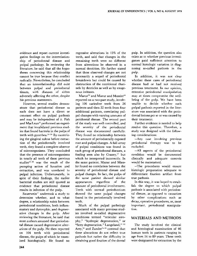
into 10% neut ra l buffered Fo rma l in solution and left there for at least 48 hours. T h e teeth were then decalci- fied in 5% nitr ic acid for ten days. After paraff in e m b e d d i n g under vacuum, serial sections were m a d e and s ta ined with hematoxy l in and esoin. Each pulp was examined serially. T h e his topathologic condi- tion was eva lua ted and recorded accord ing to the protocol i l lus t ra ted in the Table .
This char t was p repared in accor- dance with Lange land ' s cr i ter ia :~-~ for true pathosis as opposed to res idual technical ar t i fact or no rma l range of var ia t ion in an a symptoma t i c , hea l thy pulp. Cavi ty format ion in the pu lp sections both in the odonto- blast layer and centra l ly served to evaluate , on a 0 to 3 basis, the degree
of ar t i fact associated with incom- plete or de layed fixation of the pulp tissue. Ind ica t ion of true pathosis included degenera ted odontoblas ts or asp i ra ted odontob las t nuclei, in- f l ammatory cells ei ther in the odon- toblast layer or central ly , d i la t ion o f capillaries, ex t ravasa t ion or break- down of erythrocytes , and calcific changes. T h e final diagnosis evalu- ated not only the severity of any one cri terion, bu t also the n u m b e r of cri teria met and the combina t ion in which they occurred. The final histo- logical eva lua t ion of each tooth ranged from wi th in normal limits, through mode ra t e and severe inflam- mat ion, to comple te necrosis, or autolysis.
RESULTS
The results of our histological eval- uat ions are summar ized graphica l ly in Figures 3 and 4. Figure 3 illus- trates the d is t r ibu t ion of the teeth
Table �9 Periodontitis group.
Tooth
Cavity formation
Odonto- blast Cen- layer trally
Changes Aspi- Inflammatory cells in rated Dilation Extra-
odonto- odonto- Odonto- Centrally of vasation blast blast blast In Outside capil- of layer nuclei layer vessels vessels laries RBS's
Calcifications
Second- ary or General
Brown Pulp tertiary histological pigment stones dentin evaluation
F1 2 2 F2 1 2 F3 0 1 F4 2 2 F5 1 1 G1 2 1
G2 1 2
H1 0 0 H2 0 1 H3 1 2 H4 1 2
0 0 0 1 1 1 0 0 0 0 0 1 1 1 0 0 0 0 1 1 0 0 0 0 0 0 1 0 0 0 0 0 0 1 1 2 2 1 1 2 2 2
2 1 0 1 1 1 0
0 0 0 0 0 1 1 0 0 0 0 0 1 0 0 0 0 0 0 1 1 0 0 0 0 0 1 0
0 0 1 NL* 0 0 0 NL 0 1 0 NL 0 0 1 NL 0 1 0 NL 1 0 2 Severe
inflamma- tion
0 0 1 Moderate inflamma- tion
0 0 0 NL 0 1 0 NL 0 I 0 NL 0 3 0 NL
*NL indicates within normal limits.
246
JOURNAL OF ENDODONTICS I VOL 5, NO 8, AUGUST 1979
according to their periodontal condi- tion. Four teeth were clinically normal, eight showed evidence of gingivitis, and 34 showed periodonti- tis. None of the teeth in the normal periodontal group showed histologi- cal evidence of pulpal pathosis. In the gingivitis group, only one tooth z contained a pulp that was outside normal limits histologically. In the largest category, the periodontitis group, six teeth showed evidence of pathologic pulpal changes.
Figure 4 further analyzes the data by segregating and comparing the intact cariesfree teeth in each perio- dontal group from the teeth in that group that were carious or had histo- ries of operative manipulation. In the periodontally normal group, all the teeth were intact, and all the pulps appeared within normal limits. In the gingivitis group, six teeth were intact, and all the pulps appeared within normal limits. Of the two teeth in this group that were not intact, one had a pulp within normal limits, and the other had a pulp with
18 moderate inflammatory changes. This last tooth contained a deep mesial occlusal and distal amalgam and also a large buccal amalgam. All 20 intact teeth in the periodontitis
lz group contained pulps that were E histologically within normal limits. t.- Fourteen teeth in this group were not intact; of these 14 teeth, eight contained pulps that were histologi- cally within normal limits, and the s remaining six showed pathologic changes. Seven of the eight teeth in the group with normal pulps con- tained shallow restorations or mini- mal decay; only one tooth had a 0 large restoration. On the other hand, the remaining six teeth in this perio- dontitis group had extensive restora- tions or decay; only these six teeth
35
24
INCIDENCE OF PULP PATHOSIS
] WITHIN NORMAL LIMITS'
PATHOLOGIC
NORMAL GINGIVITIS
GROUP GROUP PERIODONTITIS
GROUP
Fig 3-Graph of results. Of four teeth in normal group, all appeared histologically within normal h'mits. Only one tooth in gingivitis group contained pulp that was not within normal hmits. Of 34 teeth in periodontitis group, six teeth contained pulps that were not within normal h'mits.
INCIDENCE OF
[ ~ WITHIN NORMAL LIMITS'
PATHOLOGIC
ROT] INTACT INTACT
NORMAL GROUP
PULP PATHOSIS
: ; : ; : . : . :o NOT
INTACT INTACT
GINGIVITI S GROUP
| , o % % % o
..:.;o:.:.:., - : - : ' : ' : .b
. : . : . : . : . : . : , ".'.~.T-'."
NOT INTACT INTACT
PERIOOONTITIS GROUP
Fz~ 4--Graph illustrates same data as in Figure 3 but segregates intact teeth in each group.
247
JOURNAL OF ENDODONTICS I VOL 5, NO 8, AUGUST 1979
had pulps that were not histological- ly within normal limits.
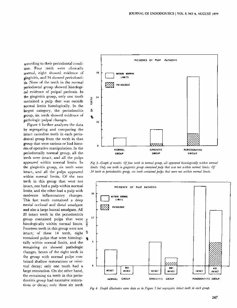
Figures 5 through 18 illustrate representative pulp sections and radiographs of teeth with varying degrees of periodontal involvement and, where significant, compare the radiographic appearance of the at- tachment apparatus with the histo- logical appearance of the pulp. The legends for each illustration and the following discussion give a detailed description of the periodontal condi- tion, as a combination of many factors contributed to the periodon- tal evaluation of each tooth.
DISCUSSION
A thorough analysis of the results of this study shows a pattern of noncorrelation between the pulpal appearance and the coincidental periodontal condition of each tooth. In the experimental sample, only teeth with extensive decay or opera- tive manipulation showed pulpal pathosis, regardless of the absence, presence, or severity of associated periodontal disease. Our a t tempt to identify periodontal disease as a causative agent in pulpal pathosis has been completely unsuccessful�9 The pulps of all intact teeth, as well as many that were not intact, were diagnosed histologically as within normal limits regardless of the sever- ity of any associated periodontal disease.
Figure 5 illustrates an example of a pulp from a young patient that was interpreted to be within normal limits from the periodontally normal group. Notice the relative absence of morphologic changes and total ab- sence of inflammatory cells, com- pared with pulp shown in Figure 2.
Figure 6 shows a pulp from the gingivitis group. Notice the intact
, . �9
Fig 5--Pulp from b~tact tooth in normal group, from young person, might be considered ideally normal. Note intact zones and vessels, complete absence of inflammatory changes.
Fig 7-Radiograph of tooth from 89-year-old woman with severe periodontal involvement. Note degree of bone loss. A llfurcations were exposed, and distobuccal root (histology in Figure 8 and 9) could be probed nearly to apex.
. " 4 " " " :
i �9 J . "
, , j !
Fig 6--Pulp from gingivitis group shows nor- mal appearance of cell zones, even with im- perfect fixation. Diagnosis as within normal limits was made owing to absence of patho- logic criteria.
Fig 8--Pulp in distobuccal root of tooth in Figure 8. It is from periodontitis group in this stud), and shows variation of normal pulp classification because of age. Degree of fibro- sis and diffuse calcification is not surprising as pulp came from 89-year-old woman. Same fibrosis and calcification were seen in nine other teeth with vital pulps extracted from patient at same time�9 Though tooth was asso- ciated with severe periodontal disease with bone loss involving almost complete length of root, pulp was diagnosed as within normal limits.
Fzir 9-H~ir magnification of pulp of distobuccal root of tooth shown in Ftgures 7 and 8. Note errthrocytes in intact functioning blood vessels (indicative of vital pulp)�9 Even with extensive calcifications and fibrous stro- ma, pulp was classified as within normal limits because no criteria of pathology, such as inflammatory changes, could be seen.
248
JOURNAL OF ENDODONTICS [ VOL 5, NO 8, AUGUST 1979
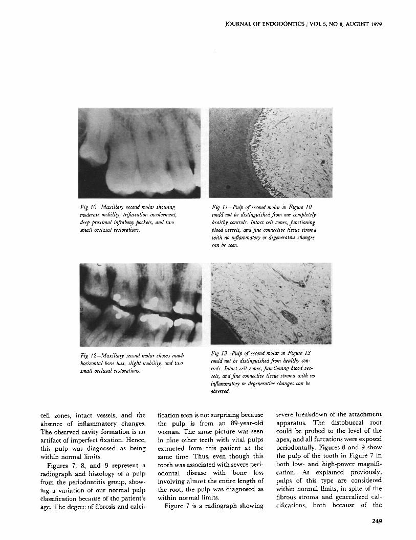
Fig lO--Maxilla~ second molar showing moderate mobility, trifurcation involvement, deep proximal infrabony pockets, and two small occlusal restorations.
Fig ll--Pulp of second molar in Figure 10 could not be distinguished from our completely healthy controls. Intact cell zones, functioning blood vessels, and fine connective tissue stroma with no inflammato?y or degenerative changes can be seen.
Fig 12-Maxilla?y second molar shows much horizontal bone loss, slight mobility, and two small occlusal restorations.
t . ~ , ' . . d �9 �9 , �9 " " J r " ,
Fig 13--Pulp of second molar hz Figure 13 could not be distinguished fiom healthy con- trols. Intact cell zones, functioning blood ves- sels, and fine connective tissue stroma with no inflammatory or degenerative changes can be observed.
cell zones, intact vessels, and the absence of inflammatory changes. The observed cavity formation is an artifact of imperfect fixation. Hence, this pulp was diagnosed as being within normal limits�9
Figures 7, 8, and 9 represent a radiograph and histology of a pulp from the periodontitis group, show- ing a variation of our normal pulp classification because of the patient's age. The degree of fibrosis and calci-
fication seen is not surprising because the pulp is from an 89-year-old woman. The same picture was seen in nine other teeth with vital pulps extracted from this patient at the same time. Thus, even though this tooth was associated with severe peri- odontal disease with bone loss involving almost the entire length of the root, the pulp was diagnosed as within normal limits.
Figure 7 is a radiograph showing
severe breakdown of the a t tachment apparatus. The distobuccal root could be probed to the level of the apex, and all furcations were exposed periodontally. Figures 8 and 9 show the pulp of the tooth in Figure 7 in both low- and high-power magnifi- cation. As explained previously, pulps of this type are considered within normal limits, in spite of the fibrous stroma and generalized cal- cifications, both because of the
249

JOURNAL OF E N D O D O N T I C S I VOL 5, N O 8, A U G U S T 1979
Fig 16--F,gures 16 to 18 show that pulp can remain viable and within normal limits, not only in presence of severe periodontal dis- ease, but also with restorations of such depth as to produce significant secondary dentin for- mation. Radiograph of maxillary first molar shows severe horizontal bone loss, severe tri-
furcation involvement, and deep coronal resto- ration. Distobuccal root could be probed al- most to apex.
advanced age of the patient and because of the total absence of vascu- lar or inflammatory changes.
The teeth discussed so far from all three periodontal groups were all intact. The extremes of normal limits in both the very young and the very old patient should be noticed. The next group of illustrations are from middle-aged patients in whom both
Fig 14-In same patient as shown in Figures 12 and 13, maxillary second molar had severe horizontal and vertical bone loss, especially distally, involving more than half length of root. All furcations were exposed and tooth showed moderate mobility.
big 17-Histological appearance of periodon- tal lesion in trifurcation of first molar shown in Figure 16. Note inflamed granulation tis- sue that has completely replaced alveolar bone in region of furcation.
periodontal disease and coronal res- torations were present.
Figures 10 and 11 show a radio- graph and pulp section of a maxil- lary second molar with much hori- zontal bone loss, deep infrabony pockets on the mesial and distal aspects, involvement of all three furcations, two small occlusal resto- rations, and moderate mobility. The
Fzg 15-Pulp stroma of second molar in Fig- ure 15 appeared well organized and free of inflammatory changes in spite of severe perio- dontal involvement of tooth. Normal appear- ance of pulp was not an isolated finding in this patient, as several other teeth with perio- dontal involvement were also studied and showed identical appearances.
b'z~g 18--Even in presence of large trifurcation lesion, pulp remained within normal limits. Note buildup of secondary dentin on root of pulp chamber because of extensive operative manipulation. Under this calcification, pulp shows normal structure with intact cell zones, functioning blood vessels, normal connective tissue stroma, and absence of inflammatory changes, and is indistinguishable from any pulp in normal control group.
2 5 0
JOURNAL OF ENDODONTICS ] VOL 5, NO 8, AUGUST 1979
pulp of this tooth (Fig 11) could not be distinguished from completely healthy controls. Perfectly intact cell zones, intact vessels, and a fine connective tissue stroma with no inflammatory or degenerative changes were observed.
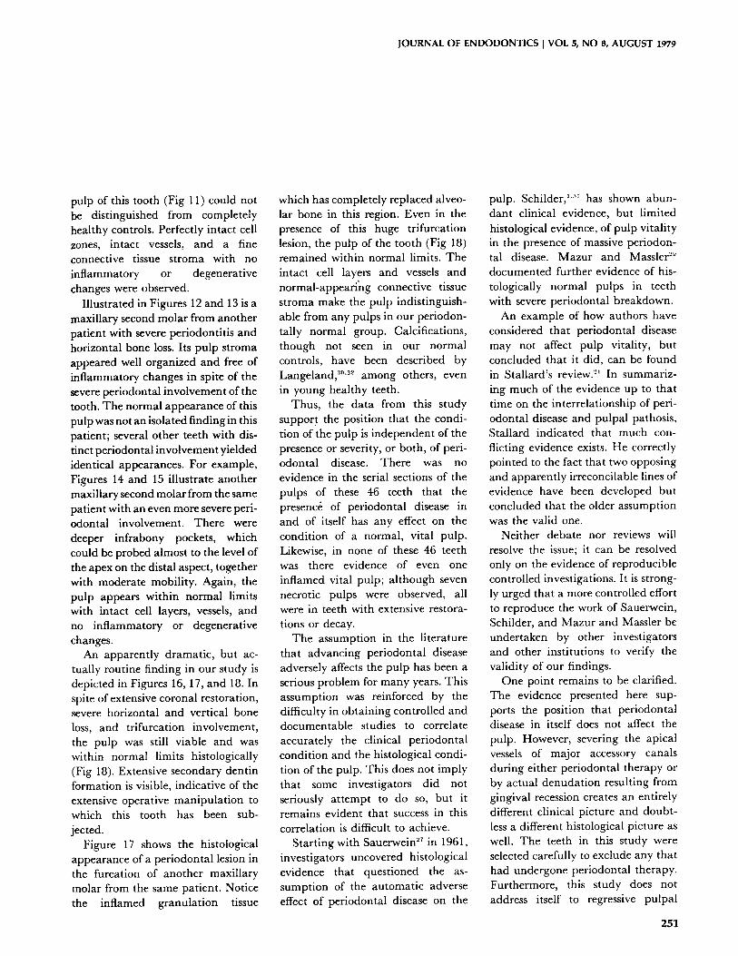
Illustrated in Figures 12 and 13 is a maxillary second molar from another patient with severe periodontitis and horizontal bone loss. Its pulp stroma appeared well organized and free of inflammatory changes in spite of the severe periodontal involvement of the tooth. The normal appearance of this pulp was not an isolated finding in this patient; several other teeth with dis- tinct periodontal involvement yielded identical appearances. For example, Figures 14 and 15 illustrate another maxillary second molar from the same patient with an even more severe peri- odontal involvement. There were deeper infrabony pockets, which could be probed almost to the level of the apex on the distal aspect, together with moderate mobility. Again, the pulp appears within normal limits with intact cell layers, vessels, and no inflammatory or degenerative changes.
An apparently dramatic, but ac- tually routine finding in our study is depicted in Figures 16, 17, and 18. In spite of extensive coronal restoration, severe horizontal and vertical bone loss, and trifurcation involvement, the pulp was still viable and was within normal limits histologically (Fig 18). Extensive secondary dentin formation is visible, indicative of the extensive operative manipulation to which this tooth has been sub- jected.
Figure 17 shows the histological appearance of a periodontal lesion in the furcation of another maxillary molar from the same patient. Notice the inflamed granulation tissue
which has completely replaced alveo- lar bone in this region. Even in the presence of this huge trifurcation lesion, the pulp of the tooth (Fig 18) remained within normal limits. The intact cell layers and vessels and normal-appearing connective tissue stroma make the pulp indistinguish- able from any pulps in our periodon- tally normal group. Calcifications, though not seen in our normal controls, have been described by Lange land , :~~ among others, even in young healthy teeth.
Thus, the data from this study support the position that the condi- tion of the pulp is independent of the presence or severity, or both, of peri- odontal disease. There was no evidence in the serial sections of the pulps of these 46 teeth that the presenc~ of periodontal disease in and of itself has any effect on the condition of a normal, vital pulp. Likewise, in none of these 46 teeth was there evidence of even one inflamed vital pulp; although seven necrotic pulps were observed, all were in teeth with extensive restora- tions or decay.
The assumption in the literature that advancing periodontal disease adversely affects the pulp has been a serious problem for many years. This assumption was reinforced by the difficulty in obtaining controlled and documentable studies to correlate accurately the clinical periodontal condition and the histological condi- tion of the pulp. This does not imply that some investigators did not seriously at tempt to do so, but it remains evident that success in this correlation is difficult to achieve.
Starting with Sauerwein 27 in 1961, investigators uncovered histological evidence that questioned the as- sumption of the automatic adverse effect of periodontal disease on the
pulp. Schilder, ''~7 has shown abun- dant clinical evidence, but limited histological evidence, of pulp vitality in the presence of massive periodon- tal disease. Mazur and Massler 29 documented further evidence of his- tologically normal pulps in teeth with severe periodontal breakdown.
An example of how authors have considered that periodontal disease may not affect pulp vitality, but concluded that it did, can be found in Stallard's review.'-" In summariz- ing much of the evidence up to that time on the interrelationship of peri- odontal disease and pulpal pathosis, Stallard indicated that much con- flicting evidence exists. He correctly pointed to the fact that two opposing and apparently irreconcilable lines of evidence have been developed but concluded that the older assumption was the valid one.
Neither debate nor reviews will resolve the issue; it can be resolved only on the evidence of reproducible controlled investigations. It is strong- ly urged that a more controlled effort to reproduce the work of Sauerwein, Schilder, and Mazur and Massler be undertaken by other investigators and other institutions to verify the validity of our findings.
One point remains to be clarified. The evidence presented here sup- ports the position that periodontal disease in itself does not affect the pulp. However, severing the apical vessels of major accessory canals during either periodontal therapy or by actual denudation resulting from gingival recession creates an entirely different clinical picture and doubt- less a different histological picture as well. The teeth in this study were selected carefully to exclude any that had undergone periodontal therapy. Furthermore, this study does not address itself to regressive pulpal
251

JOURNAL OF ENDODONTICS [ VOL 5, NO 8, AUGUST 1979
changes associated with d is rupted supply of blood to the pulp . One of the authors ' has repor ted on this type of endodon t i c -pe r iodon ta l interrela- t ionship in b i furca t ion areas and elsewhere along the root. A l though we recognize the great impor t ance of this type of in ter re la t ionship , it is not the issue under s tudy in this report .
SUMMARY
Forty-six h u m a n teeth tha t had not undergone previous pe r iodonta l t he rapy were examined and were appor t ioned on the basis of their clinical per iodonta l condi t ion into three groups accord ing to Russell 's Per iodonta l Index: normal , gingivi- tis, and periodonti t is .
After extract ion, the pulps of the teeth were p repared for histological s tudy and each was e x a m i n e d in serial section. Pu lp condi t ions were recorded as normal , hype remia , put- pitis, and necrosis.
Four teeth were ca tegor ized perio- don ta l ly normal ; all of thei r pulps were normal . Eight teeth were in the gingivitis group. Seven of these teeth had normal pulps; one was min imal - ly inf lamed. Th i r ty - four teeth were in the per iodont i t i s group. Twenty- eight teeth had pulps wi th in normal l imits, and six had necrot ic pulps. All of the necrotic pulps a n d the sl ightly inf lamed pulp were found in teeth with caries or large restorat ions, or both. All teeth with in tac t clinical crowns in all three pe r iodonta l groups had pulps tha t were within normal limits.
CONCLUSIONS
No correla t ion could be found between the severity of pe r iodonta l disease in itself and the presence or absence of pu lpa l pathosis. Normal
pulps were seen in teeth with advanced per iodonta l disease.
Pu lpa l pathosis is often associated with deep decay or extensive restora- tions. This re la t ionship, when it occurs, is independen t of the ab- sence, presence, or severity of perio- donta l disease.
No conclusions can be d r a w n from this s tudy as to the effect of pe r iodon- tal t he rapy on the condi t ion of the pulp , because the teeth examined had no history of pe r iodonta l t rea t - ment; this var iable was in ten t iona l ly e l imina ted from the study.
M a n y previous studies inade- quate ly considered the mul t ip le vari- ables associated with decay, size a n d depth of coronal restorat ions, age, and his tological art i facts in examin- ing the re la t ionship of pe r iodon ta l and pu lpa l disease.
A para l le l s tudy of teeth tha t have undergone per iodonta l t he r a py and root p l an ing should be unde r t aken to clarify fur ther the concept of the inherent resistance of the pu lp to regressive changes in the presence of pe r iodonta l disease.
Dr. Czarnecki is in private practice in Williamsville, NY, and is director of graduate endodontics, State University of New York at Buffalo. Dr. Schilder is in private practice in Boston and is chairman of the department of endodontics, Boston University Goldman School of Graduate Dentistry, 100 E Newton St, Boston, 02118. Requests for reprints should be directed to Dr. Czarnecki, 5353 Main St, Williamsville, NY 14221.
References 1. Schilder, H. Endodontic-periodontal
therapy. In Grossman, L.I. (ed.). Endodontic practice, 6th ed. Philadelphia, Lea & Febiger, 1965.
2. Simon, P., and Jacobs, D. The so-called combined periodontal-pulpal problem. Dent Clin North Am 13:45-52, 1969.
3. Serene, T.P. Interrelationship between endodontics and periodontics. J Georgia Der~t Assoc 40:14-17, 1967.
4. Biddington, W.R. Relation of endodon. tics to periodontics. W Virg Dent J 37:2-5, 1963.
5. Archambault, M.B. L'endontie et ses rapports avec la periodontie. J Can Dent Assoc 28:205-216, 1962.
6. Hiatt, W., and Amen, C. Periodontal pocket elimination by combined therapy. Dent Clin North Am 133-144, 1964.
7. Seltzer, S., and others. Pulpitis-induced interradicular periodontal changes in experi- mental animals. J Periodontol 38:124-129, 1967.
8. Johnson, H.B., and Orban, B.J. Interra- dicular pathology as related to accessory root canals. J Endodontia 3:21-25, 1948.
9. Forrest, J.O. Periodontal endodontics. J Br Endod Soc 1:8-10, 1967.
10. Simring, M., and Goldberg, M. The pulpal pocket approach: retrograde periodon- titis. J P eriodontol 35:22-48, 1964.
11. Stahl, S.S. Pulpal response to gingival injury in adult rats. Oral Surg 16:1116-1119, 1963.
12. Stahl, S.S. The pathogenesis of inflam- matory lesions in pulp and periodontal tissues. Periodontics 4:190-196, 1966.
13. Brammer, F. Uber atrophis pulpae reti- cularis bei chronisch endzundlichen verander- rungen des paradontium. Sitzungsb Gesellsch z Beford d ges Naturw zu Marb 62:547, 1927.
14. Lang, A., and McConnell, G. Calcifica- tion in the pulps of teeth affected by pyorrhea. J Dent Res 2:203-213, 1920.
15. Bauchwitz, M. Veranderungen der zahnpulpa und des paradontium bei paraden- tose. Zahnarztl Rundschau 41:430, 1228, 1271, 1932.
I6. Cahn, L. Pathology of pulps found in pyorrhetic teeth. Dent Items Interest 49:598- 617, 1927.
17. Craney, L. Die pathologisch-anatom- ischen veranderungen der pulpa bei pyorrhea alveolaris. Kor-Bl f Zahn 49:317, 343, 369, 1925.
18. Rubach, W.C., and Mitchell, D.F. Peri- odontal disease, accessory canals, and pulp pathosis. J Periodontol 36:34-38, 1965.
19. Seltzer, S.; Bender, I.B.; and Ziontz, M. The interrelationship of pulp and periodontal disease. Oral Surg 16:1474-1490, 1963.
20. Posteraro, A.F. The pulp and periodon- tal disease. Ann Dent 20:104-105, 1961.
252

JOURNAL OF ENDODONTICS [ VOL 5, NO 8, AUGUST 1979
21. Stallard, R.E. Periodontal disease and its relationship to pulpal pathology. Am Inst Oral Biol Ann Meet 197-203, 1967.
22. Fish, E.W., and MacLean, I. The distri- bution of oral streptococci in the tissues. Br Dent J 61:336-362, 1936.
23. Henrici, A., and Hartzel, T.B. A micro- scopic study of pulps from infected teeth. Br Dent J 42:497-498, 1921.
24. Henrici, A., and Hartzel, T.B. Bacterio- logy of vital pulps. Dent Cosmos 43:91, 1921.
25. Collins, K.R., and Lyne, H.C. Prelimi- nary report on bacteria found in apical tissues and pulps of extracted teeth. JADA 6(3):370- 373, 1919.
26. Golyer, S.F. Infection of the pulp of
pyorrhetic teeth. Br Dent J 45:558-559, 1924.
27. Sauerwein, E. Histopathology of the pulp in instances of periodontal disease. Dent Abst 1:467-468, 1956.
28. Mazur, B. Influence of periodontal disease on the pulp. Thesis, University of Illinois, 1961.
29. Mazur, B., and Massler, M. The influence of periodontal disease on the dental pulp. Oral Surg 17:598-603, 1964.
30. Langeland, K., and Langeland, L.K. Histologic study of 155 impacted teeth. Odont T 73:527-549, 1965.
31. Langeland, K. Criteria for the evalua- tion of dentin and pulp reactions. Am Inst Oral Biol Ann Meet 23:61-69, 1966.
32. Langeland, K. Tissue changes in the dental pulp. Oslo, Oslo University Press, 1957.
33. Arey, L.B. Developmental anatomy, ed 6. Philadelphia, W. B. Saunders Co., 1954.
34. Zander, H.A. The physiology of the dental pulp. Queensland Dent J 6:33, 1953- 1954.
35. Zander, H.A. Phagocytes in the dental pulp. J Endodontia 1:26-28, 1946.
36. Piekoff, M. The effect of dental caries on the pulp: a correlation of clinical and histopathologic findings. Thesis, Boston Uni- versity, 1969.
37. Schilder, H. Lecture series on periodon- tal-endodontic considerations given at Boston University School of Graduate Dentistry, 1971.
253