a guide to glaucoma for primary health care providers · glaucoma is a global health problem...
TRANSCRIPT

A GUIDE TO GLAUCOMA FOR PRIMARY HEALTH
CARE PROVIDERS
A companion document to NHMRC Guidelines for the screening, prognosis, diagnosis, management and prevention of glaucoma 2010
WORKING TO BUILD A HEALTHY AUSTRALIA

© Commonwealth of Australia 2011
Paper-based publicationsThis work is copyright. You may reproduce the whole or part of this work in unaltered form for your own personal use or, if you are part of an organisation, for internal use within your organisation, but only if you or your organisation do not use the reproduction for any commercial purpose and retain this copyright notice and all disclaimer notices as part of that reproduction. Apart from rights to use as permitted by the Copyright Act 1968 or allowed by this copyright notice, all other rights are reserved and you are not allowed to reproduce the whole or any part of this work in any way (electronic or otherwise) without first being given the specific written permission from the Commonwealth to do so. Requests and inquiries concerning reproduction and rights are to be sent to Strategic Communications, National Health and Medical Research Council, GPO Box 1421, Canberra ACT 2600 or via e-mail to [email protected].
Electronic documentThis work is copyright. You may download, display, print and reproduce this material in unaltered form only (retaining this notice) for your personal, noncommercial use or use within your organisation. Apart from any use as permitted under the Copyright Act 1968, all other rights are reserved. Requestsand inquiries concerning reproduction and rights should be addressed to Commonwealth Copyright Administration, Attorney-General’s Department, National Circuit, Barton ACT 2600 or posted at www.ag.gov.au/cca.
ISBN: 1864964995
Published: June 2011
NHMRC (National Health and Medical Research Council) (2010). A guide to glaucoma for primary health care providers – A companion document to NHMRC Guidelines for the screening, prognosis, diagnosis, management and prevention of glaucoma.
Contact:National Health and Medical Research Council Level 1, 16 Marcus Clarke Street Canberra ACT 2601 GPO Box 1421 Canberra ACT 2601
Ph: 61 2 6217 9000 Fax: 61 2 6217 9100 Email: [email protected]
NHMRC publication reference: CP113b
A GUIDE FOR PRIMARY HEALTH CARE PROVIDERS A COMPANION DOCUMENT TO NHMRC GUIDELINES FOR THE SCREENING, PROGNOSIS, DIAGNOSIS, MANAGEMENT
AND PREVENTION OF GLAUCOMA
NATIONAL HEALTH AND MEDICAL RESEARCH COUNCIL iii
Table of Contents
Introduction 1
The context of glaucoma 3
Identifying those at risk of glaucoma 5
Questions to ask patients with suspected glaucoma 6 Risk factors explained: 7
Age 7 Family history and genetics 8 Ethnic origin 8 Myopia 9 Long-term steroid users 9 Frequency of visits to eye care providers 9 Smoking 9 Diabetes 9 Migraine and peripheral vasospasm 10 Eye injury 10 Systemic blood pressure 10
Glaucoma prognosis 13
Normal tension glaucoma 14 Ocular hypertension 14 Early primary open angle glaucoma 15 Advanced primary open angle glaucoma 15 Angle closure glaucoma 16
Diagnosis of glaucoma 17
Professional roles in diagnosis 181. Degree of diagnostic suspicion 18 2. Degree of urgency and severity 18 3. Referral/cooperative management 19
What should be examined to identify angle closer? 20What should be examined to identify open angle glaucoma? 21

NATIONAL HEALTH AND MEDICAL RESEARCH COUNCIL iv
A GUIDE FOR PRIMARY HEALTH CARE PROVIDERSA COMPANION DOCUMENT TO NHMRC GUIDELINES FOR THE SCREENING, PROGNOSIS, DIAGNOSIS, MANAGEMENT AND PREVENTION OF GLAUCOMA
Managing patients with glaucoma 23
Open angle glaucoma 23 Angle closure glaucoma 23
Monitoring: long-term care 27
Indicators to change regimen 27After surgery for primary open angle glaucoma 29 After surgery for angle closure 29
Professional roles within the team 29 Questions to ask your patients with glaucoma at review 29
Medication 31Side effects 32 Topical medications 35
Initiating treatment 35 Application of topical medications 35
Changing medication regimes 36 Medication in acute angle closure crisis 36 Managing glaucoma successfully within specific comorbid conditions 36 Medication-induced glaucoma 36
Open angle glaucoma 36 Angle closure and angle closure glaucoma 37
Specific patient groups 37
Laser therapy and surgery 39
Laser therapy 39Laser iridotomy 39 Laser iridoplasty 39 Laser trabculoplasty 39 Combination laser surgery 39 Cyclodestructive procedures 39
Common surgical interventions 40Trabeculectomy 40

A GUIDE FOR PRIMARY HEALTH CARE PROVIDERS A COMPANION DOCUMENT TO NHMRC GUIDELINES FOR THE SCREENING, PROGNOSIS, DIAGNOSIS, MANAGEMENT
AND PREVENTION OF GLAUCOMA
NATIONAL HEALTH AND MEDICAL RESEARCH COUNCIL v
Resources 41
Consumer-orientated organisations 41 Profession-specific organsiations 42 Pregnancy-specific information 44
References 45
Appendix 1: Definitions and anatomy of the eye related to glaucoma 51
Appendix 2: Companion document details 57
Appendix 3: Abbreviations and Glossary 59


A GUIDE FOR PRIMARY HEALTH CARE PROVIDERS A COMPANION DOCUMENT TO NHMRC GUIDELINES FOR THE SCREENING, PROGNOSIS, DIAGNOSIS, MANAGEMENT
AND PREVENTION OF GLAUCOMA
NATIONAL HEALTH AND MEDICAL RESEARCH COUNCIL 1
Introduction
A companion document to NHMRC Guidelines for the screening, prognosis, diagnosis, management and prevention of glaucoma 2010 (the Guide) has been developed to provide guidance for primary health care providers who encounter patients with the symptoms of glaucoma or with patients who have already been diagnosed with glaucoma. The Guide is a companion document to the NHMRC Guidelines for the Screening, Prognosis, Diagnosis, Management and Prevention of Glaucoma 2010 (the Glaucoma Guidelines).
This Guide aims to inform primary healthcare providers on key strategies and their responsibilities in:
• Identifying those at risk of developing glaucoma
• Risk factors that can be elicited from the patient’s history
• Questions to ask patients with suspected glaucoma
• Examining eye function
• Questions to ask patients with glaucoma
• Managing glaucoma successfully within specific comorbid conditions
• Having an understanding of angel closure glaucoma
• Seeking additional resources for professionals and consumers are available part of the glaucoma network
A summary of recommendations are provided at the beginning of each chapter in the Guide to assist the primary health care provider in the screening, prognosis, diagnosis, management and prevention of glaucoma. Definitions and anatomy related to glaucoma has been provided in the Appendices.
For more detailed information on the recommendations please consult the Glaucoma Guidelines that are available on NHMRC website at www.nhmrc.gov.au/publications/index.htm.


A GUIDE FOR PRIMARY HEALTH CARE PROVIDERS A COMPANION DOCUMENT TO NHMRC GUIDELINES FOR THE SCREENING, PROGNOSIS, DIAGNOSIS, MANAGEMENT
AND PREVENTION OF GLAUCOMA
NATIONAL HEALTH AND MEDICAL RESEARCH COUNCIL 3
The context of glaucoma
In Australia Glaucoma is a leading cause of blindness and vision loss6. There is robust data to suggest that in most first world countries, 50% of glaucoma cases are undetected1. Glaucoma Australia is a national charity that promotes eye health. This group estimates that over 300,000 Australians have glaucoma2, although many people may not know it.
There is limited information regarding the prevalence and incidence of glaucoma within the Indigenous population of Australia. The Australian Bureau of Statistics5 ‘National Aboriginal and Torres Strait Islander Health Survey 2004-05’ provided data on long-term eyesight problems, however, glaucoma was not specifically reported in the data. It is currently assumed that Indigenous Australians are no more at-risk of glaucoma than Caucasians.
Glaucoma in the global populationGlaucoma is a global health problem expected to affect 60.5 million people by 20106. Glaucoma affects individuals through the full age span. It is estimated that up to two-thirds of people with glaucoma are undetected7. Once established, glaucoma is progressive and usually relentless, and the damage to the eye is irreversible. Significant reductions in annual healthcare costs have been proposed if individuals with glaucoma are diagnosed and treated early8,9. In general, in white populations around the world, the prevalence and incidence of glaucoma increases significantly with age in adults.1,3,4 People aged 70+ years have approximately three-times greater risk of developing glaucoma than people aged 40 years1,10.
Major risk factors for developing glaucoma include elevated intraocular pressure (IOP), increased cup:disc ratio, disc rim haemorrhage, reduced central corneal thickness, age older than 40 years, strong family history and specific ethnicity.
Whilst people of African-American and Inuit descent are more at-risk of developing different forms of glaucoma, on current data, Australian Indigenous populations appear to be no more at-risk of developing any form of glaucoma than Caucasian-descent Australians.

NATIONAL HEALTH AND MEDICAL RESEARCH COUNCIL 4
A GUIDE FOR PRIMARY HEALTH CARE PROVIDERSA COMPANION DOCUMENT TO NHMRC GUIDELINES FOR THE SCREENING, PROGNOSIS, DIAGNOSIS, MANAGEMENT AND PREVENTION OF GLAUCOMA
Population screening for glaucoma is currently not considered to be cost-effective. There is no consensus in the literature on the recommended frequency of screening even for at-risk subgroups. An optimal test, or group of tests, for glaucoma screening has not been identified. However there is emerging evidence that several tests are potentially feasible for detecting glaucoma in a screening program, including assessment of optic disc, visual field and IOP.1,11,12

A GUIDE FOR PRIMARY HEALTH CARE PROVIDERS A COMPANION DOCUMENT TO NHMRC GUIDELINES FOR THE SCREENING, PROGNOSIS, DIAGNOSIS, MANAGEMENT
AND PREVENTION OF GLAUCOMA
NATIONAL HEALTH AND MEDICAL RESEARCH COUNCIL 5
Identifying those at-risk of glaucoma
Recommendation 5 Identify and assess glaucoma patients and suspects (those at high risk of the disease).Good Practice Points • Identification is essential in order to make therapeutic decisions, whom to treat,
and how aggressively to treat each person. • All involved in their health care need to adopt a standard approach to risk factor
assessment for each individual.
Recommendation 6 Detect glaucoma earlierGood Practice Points• Perform regular eye health checks for Caucasians over the age of 50, and for
African-descended people over the age of 40.• Perform regular eye health checks for all first-degree relatives of glaucoma patients,
commencing 5-10 years earlier than the age of onset of glaucoma in their affected relative. Remind all glaucoma patients to alert first-degree relatives of the benefits of early and regular eye checks.
• Survey for glaucoma particularly in patients greater than 50 years of age, with any myopia, with abnormal blood pressure, with a history of migraine, with diabetes, with peripheral vasospasm, with eye injury and/or with ongoing steroid use.
• Monitor for glaucoma particularly in patients greater than 70 years of age, with IOP >21 mmHg, large and/or asymmetric cup-to-disc ratio (compared with disc size), disc haemorrhage, and thin central corneal thickness.

NATIONAL HEALTH AND MEDICAL RESEARCH COUNCIL 6
A GUIDE FOR PRIMARY HEALTH CARE PROVIDERSA COMPANION DOCUMENT TO NHMRC GUIDELINES FOR THE SCREENING, PROGNOSIS, DIAGNOSIS, MANAGEMENT AND PREVENTION OF GLAUCOMA
There is a strong body of research, developed over many years, that has established the risk factors for glaucoma development and progression. Many of these can be elicited from a patient history taken during the course of a consultation by primary health care providers, and by an eye examination with an ophthalmoscope (no pupil dilation). Full eye examinations are not usually performed by General Practitioners.
Where appropriate, it is a primary health care provider’s responsibility to educate patients about glaucoma and to alert them to the risk factors associated with it.
Questions to ask patients with suspected glaucoma
What are your symptoms?
Do you have any first-degree relatives with eye disorders (e.g. parents or siblings)?
What is your age?
Do you have existing eye conditions? (e.g. myopia and hypermetropia, eye trauma)
Do you have other medical problems? (e.g. diabetes)
Are you of African or Asian descent?
Are you taking any prescription or over-the-counter medicines, if so what?
Are you pregnant or breastfeeding?
When did you last have an eye examination?
Have you heard of glaucoma?
What do you know about glaucoma?
Would you like details on how to contact organisations offering glaucoma education and
support services?

A GUIDE FOR PRIMARY HEALTH CARE PROVIDERS A COMPANION DOCUMENT TO NHMRC GUIDELINES FOR THE SCREENING, PROGNOSIS, DIAGNOSIS, MANAGEMENT
AND PREVENTION OF GLAUCOMA
NATIONAL HEALTH AND MEDICAL RESEARCH COUNCIL 7
Risk factors explainedAge
Advancing age is a major risk factor for the development of glaucoma.
The prevalence of glaucoma is four to 10 times higher in the older age groups than in individuals in their forties.13 Damage to the optic nerve from glaucoma is uncommon before the age of 50 years in Caucasians, however it occurs at least a decade earlier in people of African descent.14
Age-specific glaucoma prevalence data is available from the two Australian studies, based mainly on white Australians3,4 (Figure 2). Updates are required to this data given the increasingly multicultural nature of the Australian population (see Recommendation 6).
Figure 1: Age-specific prevalence of open angle glaucoma (extracted from Weih et al4 p1969)
12
10
8
6
4
2
050-59 60-69 70-79 80+40-49
Inci
denc
e(%
)
PossibleProbableDe�nite
Age at baseline
POINT OF NOTE
Caucasians over 50 years of age are at moderate risk, and those over 80 years of age are at high risk of developing glaucoma. A rational approach to screening for glaucoma is therefore required for Caucasians over the age of 50 years.
For Caucasians without other significant risk factors for glaucoma, a glaucoma assessment could be included in the health assessment for people aged 45-49 years (inclusive) who are at risk of developing chronic disease and the health assessment for people aged 75 and older using one of four time-based Medicare Item Numbers 701 to 707 undertaken by general medical practioners.

NATIONAL HEALTH AND MEDICAL RESEARCH COUNCIL 8
A GUIDE FOR PRIMARY HEALTH CARE PROVIDERSA COMPANION DOCUMENT TO NHMRC GUIDELINES FOR THE SCREENING, PROGNOSIS, DIAGNOSIS, MANAGEMENT AND PREVENTION OF GLAUCOMA
Family history and geneticsA family history of glaucoma puts an individual at greater risk of developing the disease.13 In close relatives of individuals with primary open angle glaucoma (POAG), the prevalence is three to six times that of the general
population.
POINT OF NOTE
A primary health care provider should advise all patients with glaucoma to inform all close relatives to undergo ocular examination as early as possible. This should occur at the age that is recommended for their ethnic group, or five to ten years earlier than the age of onset of glaucoma in their relative.
Ethnic originPeople of African descent have been reported to have an age-adjusted prevalence for POAG, 4.3 times greater than Caucasians. Prevalence of primary angle closure glaucoma (PACG) is highest among those of Asian or Inuit descent, with rates in these populations reported to be three to 10 times higher than in other ethnic groups.15
POINT OF NOTE
The Working Committee note a new report ‘National Indigenous Eye Health Survey: Minum Barreng Full Report’ prepared by Anna-Lena Arnold, Ross A. Dunn and members of the National Indigenous Eye Health Survey Team (NIEHS) which was published 02 October 2009.
POINT OF NOTE
For individuals from high-risk ethnic backgrounds, appropriate surveillance activities include, but are not limited to, patient education regarding glaucoma, individual risk, consideration of concurrent medications, and advice to attend regular standard ocular examinations.

A GUIDE FOR PRIMARY HEALTH CARE PROVIDERS A COMPANION DOCUMENT TO NHMRC GUIDELINES FOR THE SCREENING, PROGNOSIS, DIAGNOSIS, MANAGEMENT
AND PREVENTION OF GLAUCOMA
NATIONAL HEALTH AND MEDICAL RESEARCH COUNCIL 9
MyopiaGlaucoma and myopia have a strong familial basis and thus may share a common genetic link. The prevalence of open angle glaucoma (OAG) among people with myopia ranges from 1.4% to 4.3%.1 A dose–response relationship between OAG and myopia has been postulated (the higher the myopia, the more likely an individual would be to develop OAG).
Long-term steroid usersCorticosteroids are the main cause of drug-induced glaucoma (secondary glaucoma related to an external causation).6,16 Steroids administered by any route are associated with increases in IOP.17 Steroidal-like substances can also be found in traditional and natural medicines.
POINT OF NOTE
Surveillance activities include, but are not limited to, patient education about risk, consideration of concurrent medications, and encouraging attendance at basic ocular checks.
Frequency of visits to eye care providersThe longer the time since the last visit to an eye care provider, the higher the risk of undiagnosed glaucoma.4
SmokingEvidence for the association of smoking with POAG is controversial. There is some evidence to suggest that current smoking is associated with POAG, but not past smoking.18
DiabetesThe association between diabetes mellitus and POAG is controversial. The most recent data from Burr et al1 reported almost twice the risk of OAG onset among individuals with diabetes, compared with those without diabetes. The current understanding is that people with diabetes are at increased risk of developing POAG, and should be targeted for blindness-prevention programs.19

NATIONAL HEALTH AND MEDICAL RESEARCH COUNCIL 10
A GUIDE FOR PRIMARY HEALTH CARE PROVIDERSA COMPANION DOCUMENT TO NHMRC GUIDELINES FOR THE SCREENING, PROGNOSIS, DIAGNOSIS, MANAGEMENT AND PREVENTION OF GLAUCOMA
Migraine and peripheral vasospasmMigraine headache and peripheral vasospasm have been identified as risk factors for progressive glaucomatous optic nerve damage.3,20 Peripheral vasospasm has also been proposed as one possible mechanism for, or a factor contributing to, optic nerve damage in glaucoma.
Eye injuryEye trauma is widely accepted as a risk factor for glaucoma. Traumatic glaucoma can occur immediately after a blunt trauma or penetrating eye injury, or years later.21 It is usually considered as secondary OAG. Eye trauma with angle recession is a risk factor for OAG.
Systemic blood pressureDiastolic blood pressure largely determines perfusion pressure to the eye and is related to IOP. Low systemic blood pressure, including the nocturnal dip, may pose a risk for normal tension glaucoma (NTG)13, and POAG22, There is limited evidence concerning the relationship between high systemic blood pressure and glaucoma. It is likely to be a complex relationship as patient age and the duration of systemic hypertension both impact upon the hypertensive state.
POINT OF NOTE
Recent publications, which were outside the scope of the literature review undertaken for these guidelines, indicate that reduced ocular perfusion pressure is strongly associated with glaucoma progression.

A GUIDE FOR PRIMARY HEALTH CARE PROVIDERS A COMPANION DOCUMENT TO NHMRC GUIDELINES FOR THE SCREENING, PROGNOSIS, DIAGNOSIS, MANAGEMENT
AND PREVENTION OF GLAUCOMA
NATIONAL HEALTH AND MEDICAL RESEARCH COUNCIL 11
Reminder to health care providersHealth care providers should use a patient’s history to elicit information about risk factors that are significantly associated with developing most types of glaucoma:• elevated or fluctuating intraocular pressure• strong family history of glaucoma• advanced age• African or Asian ethnicity• current diabetes• myopia• rural location.
Health care providers should use a patient examination to elicit information about other risk factors that are significantly associated with developing most types of glaucoma:• elevated or fluctuating intraocular pressure• significant alterations in cup:disc ratio and cup:disc ratio asymmetry• nerve fibre layer defects• optic disc haemorrhage.
An assessment of these risk factors should aid in therapeutic decision-making regarding who to treat, when to treat, how to treat, and how aggressively to treat.
If appropriate, health care providers may also consider other risk factors which have more limited evidence of their association with developing most types of glaucoma:• central corneal thickness• current smoking• current migraine and peripheral vasospasm• long-term steroid use• previous eye injury.


A GUIDE FOR PRIMARY HEALTH CARE PROVIDERS A COMPANION DOCUMENT TO NHMRC GUIDELINES FOR THE SCREENING, PROGNOSIS, DIAGNOSIS, MANAGEMENT
AND PREVENTION OF GLAUCOMA
NATIONAL HEALTH AND MEDICAL RESEARCH COUNCIL 13
Glaucoma prognosis
Recommendation 4 Assess risk of conversion from ocular hypertension to glaucoma.Good Practice Points • Patients at low risk of conversion should be considered for monitoring.• Patients at high risk of conversion should be considered for treatment.• Educate patients on the risks and consequences of conversion to glaucoma.
Recommendation 2 Reduce intraocular pressure.
Recommendation 3 Monitor visual field and determine rate of any field loss.
In the literature, the natural history of glaucoma is poorly defined and heterogeneous. An individual’s risk of progressive and sight-threatening glaucoma cannot be predicted with precision, however there is improving evidence to specifically identify candidates for treatment. If treatment decisions wait until there are overt signs of disease, this generally results in irreversible optic damage and likely disease progression.
However, all glaucoma treatments carry some side effects (e.g. development of cataracts), and therefore the trade-off between risk and benefit should be carefully considered in each patient’s case.12

NATIONAL HEALTH AND MEDICAL RESEARCH COUNCIL 14
A GUIDE FOR PRIMARY HEALTH CARE PROVIDERSA COMPANION DOCUMENT TO NHMRC GUIDELINES FOR THE SCREENING, PROGNOSIS, DIAGNOSIS, MANAGEMENT AND PREVENTION OF GLAUCOMA
Communication with patientsWhile lowering intraocular pressure slows or halts glaucoma progression, all interventions carry risk. Potential benefit and possible harm (the therapeutic index) need to be balanced carefully, with patient involvement where possible, in decision making.
Normal tension glaucoma
Individuals with glaucoma who have IOP in the ‘normal’ range are labelled as having NTG. There is sound evidence that IOP-lowering treatment is effective in preserving visual field in people with NTG.36
Ocular hypertension10% of patients with ocular hypertension will progress to POAG within five years1. However, the mean probability of progressing to POAG becomes greater with increasing IOP levels.1 Conversion time to POAG from ocular hypertension is significantly shorter for individuals not undergoing treatment.36
Risk factors for progression from ocular hypertension to glaucoma include elevated IOP, increased cup:disc ratio, older age, and thinner corneas.37 The relative proportion of the population converting from ocular hypertension to
POAG can be reduced by 50% with appropriate IOP-lowering treatment.38,39
Communication with patientsIt is essential that patients understand the risks for, and consequences of, progression to glaucoma and the value of treatment.
Rates of conversion to glaucoma are initially low, however any progression and visual loss is irreversible. Timely treatment can reduce the chance of progression and/or conversion by 50%.

A GUIDE FOR PRIMARY HEALTH CARE PROVIDERS A COMPANION DOCUMENT TO NHMRC GUIDELINES FOR THE SCREENING, PROGNOSIS, DIAGNOSIS, MANAGEMENT
AND PREVENTION OF GLAUCOMA
NATIONAL HEALTH AND MEDICAL RESEARCH COUNCIL 15
Early primary open angle glaucomaThe literature provides sound evidence that without treatment, individuals with ocular hypertension and early POAG will convert more rapidly to advanced stages of the disease, with the inherent risks of visual field loss.
Allowing for considerable variability reported in the literature, an estimate of the average time for patients with POAG to progress to blindness without treatment is 23 years, and with treatment, 35 years.1 Topical IOP-lowering treatment is effective for most individuals, as it reduces the rate of progression of glaucomatous damage.38,39
Communication with patientsWith treatment 20 years ago, the average time to unilateral blindness for patients with primary open angle glaucoma was approximately 17 years. Untreated patients progress at approximately twice the speed of treated patients. In the last 20 years the rates of glaucoma blindness have dropped due to earlier diagnosis and more effective intraocular pressure-lowering treatment which significantly improves prognosis in the majority of cases. It is therefore important to comply with treatment and discuss any concerns with treatment with your health care provider.
Advanced primary open angle glaucomaPatients with more severe glaucoma at diagnosis (i.e. those diagnosed later) are more likely to go blind.40
There is scant evidence on the impact of risk factors on the progression and outcomes of patients with severe and advanced glaucoma. The Advanced Glaucoma Intervention Study suggests that older age, lower formal education, male gender and diabetes are significant risk factors for the progression of advanced glaucoma to blindness.37 Reduction in maximal IOP and IOP fluctuation has some benefits for some patients, even in the advanced stage of glaucoma. However not every individual will gain the same benefits from treatment. A larger reduction in IOP is required to prevent progression in patients with more advanced glaucoma, when loss of vision is threatened. Patients with IOP below 14mmHg are reported to have the least progression.41

NATIONAL HEALTH AND MEDICAL RESEARCH COUNCIL 16
A GUIDE FOR PRIMARY HEALTH CARE PROVIDERSA COMPANION DOCUMENT TO NHMRC GUIDELINES FOR THE SCREENING, PROGNOSIS, DIAGNOSIS, MANAGEMENT AND PREVENTION OF GLAUCOMA
Communication with patientsHigher rates of progression and visual loss may occur in patients who have been diagnosed late, or who already suffer from more advanced forms of glaucoma. However, evidence continues to support the benefits of active intraocular pressure reduction, even when patients have advanced stage glaucoma.
Angle closure glaucomaPrimary angle closure suspect (PACS) is an anatomical predisposition to closure with signs of narrowing of the angle (appositional contact between iris and trabecular meshwork) but without permanent occlusion or signs of adhesion (synechiae) between the iris and trabecular meshwork.
The treatment of primary angle closure-related glaucoma is two-fold; one is to manage the compromised angle and the other is to manage the glaucomatous nerve damage which is no different to the management of primary open angle glaucoma. Moreover, the prognosis regarding visual field and optic disc damage is thought to be identical, depending upon the pressure and patient susceptibility, so the previous section on primary open angle glaucoma prognosis is thought to pertain to angle closure.

A GUIDE FOR PRIMARY HEALTH CARE PROVIDERS A COMPANION DOCUMENT TO NHMRC GUIDELINES FOR THE SCREENING, PROGNOSIS, DIAGNOSIS, MANAGEMENT
AND PREVENTION OF GLAUCOMA
NATIONAL HEALTH AND MEDICAL RESEARCH COUNCIL 17
Diagnosis of glaucoma
Recommendation 8 Assess with a comprehensive medical history, a full eye examination and investigate appropriately.Good Practice Points • Acomprehensivemedicalhistory: identify all relevant risk factors, relevant co-
morbidities and concurrent topical and systemic medications, and assess the impact of visual dysfunction, social environment and support networks that may affect adherence to a treatment program. Co-morbidities include hypertension, diabetes, thyroid disease, depression, asthma, liver and renal disease.
• Afulleyeexamination: anterior segment evaluation including gonioscopy, optic nerve and retinal nerve fibre layer exam stereoscopic optic disc and retinal nerve fibre assessment with a permanent record, IOP and corneal thickness measurements.
• Appropriateinvestigations: standard automated perimetry (white-on-white) including comparison with age-corrected normals on a point-wise, regional (e.g. hemifield) and global basis, optic disc photography and imaging of the optic nerve and optic nerve fibre layer.
• Careful and informedinterpretationofresults from all imaging and functional tests in order to detect disease or to detect progression. With the multi-faceted nature of glaucoma and the large variability in normal values of all tests, consider results from all tests and assessments.
A diagnosis of glaucoma should be made on the basis of multiple sources of information, including the presenting history, an assessment of relevant risk factors, and an ocular examination reflecting structure and function of the eye. Initial consultation should elicit a complete medical, surgical, personal and occupational history, and ascertain relevant risk factors.42 This consultation should be followed by a comprehensive clinical examination including slit lamp examination, tonometry, fundus and optic nerve head examination, gonioscopy, and corneal thickness measurement. This examination may be in conjunction with special investigations to document the extent of structural damage to the optic nerve head and the retinal nerve fibre layer, using optic nerve and retinal

NATIONAL HEALTH AND MEDICAL RESEARCH COUNCIL 18
A GUIDE FOR PRIMARY HEALTH CARE PROVIDERSA COMPANION DOCUMENT TO NHMRC GUIDELINES FOR THE SCREENING, PROGNOSIS, DIAGNOSIS, MANAGEMENT AND PREVENTION OF GLAUCOMA
nerve fibre layer analysis or disc photography, computer-assisted visual field analysis. Children with suspected glaucoma should be referred to a specialist health care provider in the field.
A systematic approach should be used to elicit diagnostic information. In certain cases, glaucoma can present as a medical emergency. Confirmatory diagnosis of glaucoma may require more than one consultation with a health care provider, and the involvement of an ophthalmologist. A diagnosis is generally made on the basis of characteristic degenerative changes in the optic disc, and matching defects in visual fields.
Professional roles in diagnosisThe NHMRC guidelines encourage the establishment and nurturing of networks between primary health care providers, and between primary health care providers and ophthalmologists, to ensure that the best quality comprehensive care is provided to patients suspected of having, or diagnosed with, glaucoma.
Given the limited evidence base and ongoing changes in professional skills and responsibilities in Australia, the Working Committee notes that there are three essential issues that direct the most appropriate management pathway for a patient.
1. Degree of diagnostic suspicion: In the primary health care setting, if the degree of diagnostic suspicion of glaucoma is low, unnecessary referral of a patient to an ophthalmologist may lead to system overload. Low-risk patients may well be monitored by the most appropriate primary health care provider within the patient’s location, using established ocular health care networks for advice. If the degree of diagnostic suspicion of glaucoma is high however, the network should still be used for advice, and the appropriate decision may be a direct referral to a health care provider able to initiate treatment.
2. Degree of urgency and severity: If suspicion is very high with marked signs of nerve damage, and/or the IOP is very high then patients need urgent referral, with or without IOP-lowering treatment in the meantime, depending upon the waiting period for referral. Acute angle closure presents as a medical emergency and requires immediate referral to a specialist.

A GUIDE FOR PRIMARY HEALTH CARE PROVIDERS A COMPANION DOCUMENT TO NHMRC GUIDELINES FOR THE SCREENING, PROGNOSIS, DIAGNOSIS, MANAGEMENT
AND PREVENTION OF GLAUCOMA
NATIONAL HEALTH AND MEDICAL RESEARCH COUNCIL 19
3. Referral/cooperative management: The Working Committee recommends that the professional roles, responsibilities and referral pathways are best determined in individual cases based on location, resources, skill-base of local health care providers and patient choice. Classically, referral occurs to an ophthalmologist when significant suspicion of glaucoma is raised. In some parts of Australia optometrists and or general practitioners can initiate treatment.
Irrespective of their location and professional networks, health care providers involved in diagnosing glaucoma should have the skills and equipment to measure IOP (by Goldmann Applanation Tonometry or a well calibrated non-contact tonometer, anterior chamber assessment and gonioscopy), visual field and optic disc.
Health care providers involved only with screening for, and/or diagnosis of, glaucoma, should receive appropriate training and continuing support from health care providers who manage glaucoma.1,43 Students in each health care discipline should be alerted to the importance of cooperation between disciplines in the screening, diagnosis and management of glaucoma.
POINT OF NOTE
In rural/remote settings, fundus photography is valuable if the results are to be relayed to a diagnosing health care provider.

NATIONAL HEALTH AND MEDICAL RESEARCH COUNCIL 20
A GUIDE FOR PRIMARY HEALTH CARE PROVIDERSA COMPANION DOCUMENT TO NHMRC GUIDELINES FOR THE SCREENING, PROGNOSIS, DIAGNOSIS, MANAGEMENT AND PREVENTION OF GLAUCOMA
What should be examined to identify angle closure?• Assess anterior chamber and angle with gonioscopy and biomicroscopy
Key signs of closure
− peripheral anterior synechaie − trabecular meshwork pigment patches − iris insertion above a scleral spur − angle structures (trabecular meshwork) not being visible
• Assess and record eye structure with the best available instrument
− refractive status − pupil size and reactivity − external appearance eye
• Assess and record eye function with best available instrument
• Assess IOP using best available instrument and taking patient preference into consideration

A GUIDE FOR PRIMARY HEALTH CARE PROVIDERS A COMPANION DOCUMENT TO NHMRC GUIDELINES FOR THE SCREENING, PROGNOSIS, DIAGNOSIS, MANAGEMENT
AND PREVENTION OF GLAUCOMA
NATIONAL HEALTH AND MEDICAL RESEARCH COUNCIL 21
What should be examined to identify open angle glaucoma?
• Assess anterior chamber and angle with gonioscopy and biomicroscopy
Key signs
− abnormal trabecular meshwork − abnormal ciliary base (angle or cyclodialysis cleft) − blood reflux in Schlemm’s canal
• Assess and record eye structure with the best available instrument − ocular examination including
− refractive status − pupil size and reactivity − external appearance eye − optic nerve head − visual field
Key signs
− typically superotemporal or inferotemporal optic disc neuroretinal rim loss with excavation
− disc haemorrhage − increased cup:disc ratio and cup:disc ratio asymmetry − nerve fibre layer atrophy − peripapillary atrophy
• Assess and record eye function with best available instrument
Key signs
− defects that are − asymmetrical and cross midline − located in mid periphery − clustered in neighbouring points − correlate to defects on optic disc
• Assess IOP using best available instrument and taking patient preference into consideration
Key levels
− less than 21mmHg – consider NTG − over 26mmHg consider ocular hypertension − consider diurnal variation


A GUIDE FOR PRIMARY HEALTH CARE PROVIDERS A COMPANION DOCUMENT TO NHMRC GUIDELINES FOR THE SCREENING, PROGNOSIS, DIAGNOSIS, MANAGEMENT
AND PREVENTION OF GLAUCOMA
NATIONAL HEALTH AND MEDICAL RESEARCH COUNCIL 23
Managing patients with glaucoma
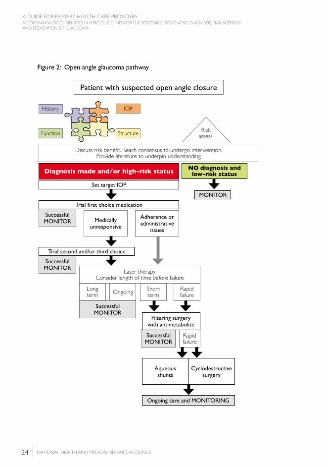
Open angle glaucomaFigure 2 presents the current best evidence for the most appropriate medical care of the ‘average’ patient with OAG. There are many factors which impact on the decision-making of the different health care providers who are responsible for the management of individual patients. This guideline does not consider the wider aspects of a patient journey with glaucoma, for instance social, emotional and economic elements. This guideline notes however that it is important that appropriate support is provided by health care providers to the patient with glaucoma and their carer(s). This may be provided by ensuring that they establish links to important consumer groups such as Glaucoma Australia, and by providing clear written and verbal communication. In advanced stages, low vision rehabilitation is a valuable adjunct to the glaucoma management plan.
Angle closure glaucomaFigure 3 represents the current best evidence for the most appropriate care of the ‘average’ patient with intermittent or chronic angle closure. Angle closure may present as an acute crisis, requiring emergency management. These guidelines provide clear information regarding the signs and symptoms of an angle closure crisis.
NATIONAL HEALTH AND MEDICAL RESEARCH COUNCIL 24
A GUIDE FOR PRIMARY HEALTH CARE PROVIDERSA COMPANION DOCUMENT TO NHMRC GUIDELINES FOR THE SCREENING, PROGNOSIS, DIAGNOSIS, MANAGEMENT AND PREVENTION OF GLAUCOMA
Figure 2: Open angle glaucoma pathway
Risk assess
Discuss risk benefi t. Reach consensus to undergo interviention. Provide literature to underpin understanding
Set target IOP
NO diagnosis and low-risk statusDiagnosis made and/or high-risk status
Rapid failure
Ongoing care and MONITORING
MONITOR
Trial fi rst choice medication
SuccessfulMONITOR Medically
unresponsive
Adherence or administrative
issues
Aqueousshunts
Cyclodestructivesurgery
SuccessfulMONITOR
Filtering surgery with antimetabolite
Rapid failure
Short termOngoingLong
term
Successful MONITOR
Laser therapyConsider length of time before failure
Trial second and/or third choice
SuccessfulMONITOR
History
Function Structure
IOP
Patient with suspected open angle closure

A GUIDE FOR PRIMARY HEALTH CARE PROVIDERS A COMPANION DOCUMENT TO NHMRC GUIDELINES FOR THE SCREENING, PROGNOSIS, DIAGNOSIS, MANAGEMENT
AND PREVENTION OF GLAUCOMA
NATIONAL HEALTH AND MEDICAL RESEARCH COUNCIL 25
Figure 3: Intermittent or chronic angle closure patient pathway
Patient with suspected angle closure
Risk assess
Discuss risk benefi t. Reach consensus to undergo interviention. Provide literature to underpin understanding
Angle closure diagnosed
Peripheral Iridotomy (P.I.)
YAG Laser P.I.
Unilateral for chronic
closure
Unilateral Surgical P.I.
For acute closure where YAG P.I. not possible
Bilateral for acute or whenever access to medical
services restricted
Ongoing monitoring for ALL patients
Consider patency of angle
Open
MONITORClosing rapidly
and/or signfi cantlyClosing slowly
and/or minimally
Consider medication
Consider cause
Synechial closurewithout
posterior force
Plateau iris or mild ciliary block
NO cataract
Plateau iris or mild ciliary blockWITH cataract
Ciliary block(severe)
Drainage surgerytrabeculectomy Iridoplasaty Lens extraction and IOL
MONITOR Consider patency of angle
Narrow post iridoplasty
Closing
Vitrectomy and anterior hyloidectomy
Ongoing monitoring for ALL patients
Open
MONITOR
History
Function Structure
IOP


A GUIDE FOR PRIMARY HEALTH CARE PROVIDERS A COMPANION DOCUMENT TO NHMRC GUIDELINES FOR THE SCREENING, PROGNOSIS, DIAGNOSIS, MANAGEMENT
AND PREVENTION OF GLAUCOMA
NATIONAL HEALTH AND MEDICAL RESEARCH COUNCIL 27
Monitoring: long-term care
The aims of monitoring patients diagnosed with glaucoma are to detect progression, evaluate the effects of treatment, re-assess risk factors for progression and note changes in health that may influence glaucoma management plans. Monitoring requires the performance of visual field, optic disc, gonioscopy and intraocular pressure measurement by suitably trained and equipped practitioners. Primary health care providers play an important role by prompting attendance in order to monitor glaucoma progression. Appropriate monitoring plans will ensure that patients who are at risk of glaucoma, and patients with established glaucoma, do not worsen through inadequate, or inappropriate medical care.
While it is not always possible to stop disease progression, it can usually be slowed significantly with appropriate treatment. Similar to diagnosis, monitoring is not based on a single test; rather it is based on a combination of test methodologies and technological tools. Lowering IOP is the strategy with the greatest evidence of effectiveness to achieve these goals. Therefore IOP measurement is vital in follow-up, with changes in visual field and optic nerve being the criteria for the alteration of target IOP. Once glaucoma has been diagnosed and patients placed on a treatment regimen, monitoring the patient’s capacity for adherence to the regimen and engaging the patient with treatment maintenance (including attendance at future appointments) is essential to best practice. The patient’s risk profile, disease state and capacity to self-manage dictate the frequency of review.
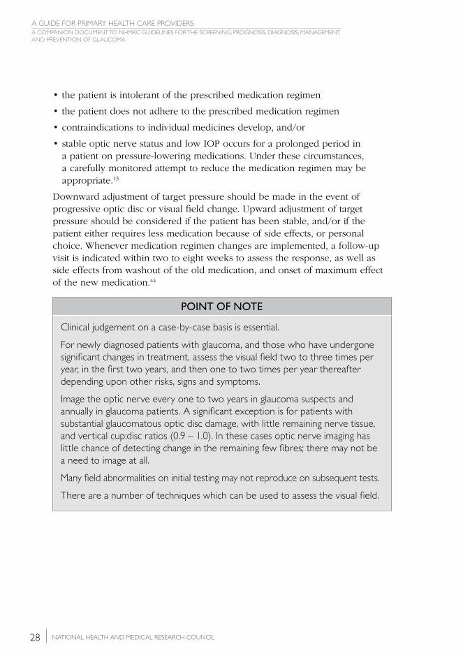
Indications to change regimenThe indications for adjusting a glaucoma management plan are:
• target IOP is not achieved
• the patient has progressive optic nerve or visual field damage despite achieving the target IOP. The validity of the diagnosis and target pressure should be reassessed. Additional evaluation may identify conditions that are contributing to the progression of damage, and serve as a justification to escalate treatment. These evaluations include obtaining diurnal IOP measurements, repeating the central corneal thickness measurement to verify a thin cornea or a change in corneal thickness after refractive surgery, or seeking evidence of unrecognised low blood pressure. A neurologic evaluation also may be considered.
NATIONAL HEALTH AND MEDICAL RESEARCH COUNCIL 28
A GUIDE FOR PRIMARY HEALTH CARE PROVIDERSA COMPANION DOCUMENT TO NHMRC GUIDELINES FOR THE SCREENING, PROGNOSIS, DIAGNOSIS, MANAGEMENT AND PREVENTION OF GLAUCOMA
• the patient is intolerant of the prescribed medication regimen
• the patient does not adhere to the prescribed medication regimen
• contraindications to individual medicines develop, and/or
• stable optic nerve status and low IOP occurs for a prolonged period in a patient on pressure-lowering medications. Under these circumstances, a carefully monitored attempt to reduce the medication regimen may be appropriate.13
Downward adjustment of target pressure should be made in the event of progressive optic disc or visual field change. Upward adjustment of target pressure should be considered if the patient has been stable, and/or if the patient either requires less medication because of side effects, or personal choice. Whenever medication regimen changes are implemented, a follow-up visit is indicated within two to eight weeks to assess the response, as well as side effects from washout of the old medication, and onset of maximum effect of the new medication.44
POINT OF NOTE
Clinical judgement on a case-by-case basis is essential.
For newly diagnosed patients with glaucoma, and those who have undergone significant changes in treatment, assess the visual field two to three times per year, in the first two years, and then one to two times per year thereafter depending upon other risks, signs and symptoms.
Image the optic nerve every one to two years in glaucoma suspects and annually in glaucoma patients. A significant exception is for patients with substantial glaucomatous optic disc damage, with little remaining nerve tissue, and vertical cup:disc ratios (0.9 – 1.0). In these cases optic nerve imaging has little chance of detecting change in the remaining few fibres; there may not be a need to image at all.
Many field abnormalities on initial testing may not reproduce on subsequent tests.
There are a number of techniques which can be used to assess the visual field.

A GUIDE FOR PRIMARY HEALTH CARE PROVIDERS A COMPANION DOCUMENT TO NHMRC GUIDELINES FOR THE SCREENING, PROGNOSIS, DIAGNOSIS, MANAGEMENT
AND PREVENTION OF GLAUCOMA
NATIONAL HEALTH AND MEDICAL RESEARCH COUNCIL 29
After surgery for primary open angle glaucomaAfter laser therapy or surgical treatment, a proportion of patients will be able to reduce or cease their medication. This may raise issues for monitoring. Health care providers should be sure that patients understand the chronic nature of their disease and the continued need for monitoring. A member of the health care team should take responsibility for monitoring these patients despite their independence from medication management.
After surgery for angle closureAfter iridotomy, patients may be classified as residual open angle, or a mix of open angle and peripheral anterior synechaie. Patients in whom glaucomatous damage has occurred should be monitored as recommended for POAG. Patients who do not have glaucomatous optic neuropathy should be monitored in a manner similar to a POAG suspect.44
Professional roles within the teamUsing established networks, primary health care providers are encouraged to ensure the patients with glaucoma whom they are monitering see an ophthalmologist at proscribed intervals.
Questions to ask your patient with glaucoma at review
How are you? How are your eyes and vision?
Are you managing to take your medication as discussed? If no, what are the problems and difficulties you face?
Is there anything about your problems or your treatment plan that you would like explained?
Are you experiencing any side effects from the medication?
Do you have other medical problems? If yes, have they been exacerbated recently?
Are you taking any prescription or over-the-counter medicines, if so what?
Do you have plans to conceive/are you already pregnant? If yes, do you plan to breastfeed?
When did you last attend an eye examination or have your condition monitored?
Do you require further glaucoma information, at a patient level or would you benefit from contacting a glaucoma patient association?


A GUIDE FOR PRIMARY HEALTH CARE PROVIDERS A COMPANION DOCUMENT TO NHMRC GUIDELINES FOR THE SCREENING, PROGNOSIS, DIAGNOSIS, MANAGEMENT
AND PREVENTION OF GLAUCOMA
NATIONAL HEALTH AND MEDICAL RESEARCH COUNCIL 31
Medication
Medication is the first management choice1 for most patients with glaucoma. Medication reduces IOP by enhancing aqueous outflow and/or reducing aqueous production. There are five main families of glaucoma medications, each with recognised actions, side effects and contraindications: beta-blockers, prostaglandin analogues, alpha2-agonists, carbonic anhydrase inhibitors and cholinergic agonists (see Table 1). Hyperosmotic medications such as mannitol are given to lower IOP in emergency situations.
The time taken to achieve maximal reduction in IOP is dependent on both the individual and the type of medication used. Initial reduction in IOP typically occurs within minutes to hours after administration, while maximal reduction in IOP can take weeks to months. Response to newly-initiated medications should be assessed after two to four weeks.
When prescribing glaucoma medication, many factors should be considered including IOP-lowering potency, additive effects, interaction with concomitant medications and disease states, side effects and ease of administration. Persistence with and adherence to medication regimens is vital in the management of chronic disease. Glaucoma medication must be suited to an individual’s capacity to effectively self-administer.
Many pharmacies have the capacity to provide a medicines profile, listing the prescription, OTC and complementary medicines being taken by a particular patient. The profiles are used to support patients in managing their medicines and can also be used as an effective communication tool when seeing other health professionals.
Primary health care providers should note that medications and how to use them are constantly being refined by research, and the development of new products. It is recommended that a pharmacist is involved in multidisciplinary networks of health care providers to provide specialist knowledge of medicines and their actions.
Ophthalmologists often authorise changes in a patient’s medication and should do in communication with a General Practitioner.
1 NB First choice refers to medications that a treating health care provider prefers to use as the initial intervention. First line refers to a medication that has been approved by an official controlling body for initial intervention (European Guideline Society 2003). This guideline refers to first choice as it provides guidance to health care provider

NATIONAL HEALTH AND MEDICAL RESEARCH COUNCIL 32
A GUIDE FOR PRIMARY HEALTH CARE PROVIDERSA COMPANION DOCUMENT TO NHMRC GUIDELINES FOR THE SCREENING, PROGNOSIS, DIAGNOSIS, MANAGEMENT AND PREVENTION OF GLAUCOMA
Side effectsHealth care providers should not underestimate the potentially significant side effects associated with medications for glaucoma. Side effects can be life-threatening. Particular caution should be exercised when prescribing medications for infants and the elderly, who may be more susceptible to side effects26. Some side effects occur immediately, but most occur over time. This is why optimum management of patients with glaucoma should include regular monitoring and review of medication regimens. Details of side effects related to glaucoma medications are reported in Table 1.

A GUIDE FOR PRIMARY HEALTH CARE PROVIDERS A COMPANION DOCUMENT TO NHMRC GUIDELINES FOR THE SCREENING, PROGNOSIS, DIAGNOSIS, MANAGEMENT
AND PREVENTION OF GLAUCOMA
NATIONAL HEALTH AND MEDICAL RESEARCH COUNCIL 33
Tabl
e 1:
M
edic
atio
ns a
vaila
ble
in A
ustr
alia
tha
t ar
e us
ed in
the
man
agem
ent
of g
lauc
oma
Prep
arat
ions
by
clas
sM
echa
nism
of
act
ion
Effic
acy
Dai
ly
dosa
geTo
pica
l oph
thal
mic
sid
e ef
fect
s Sy
stem
ic s
ide
effe
cts
Ord
er o
f tr
eatm
ent
choi
ces
Pros
tagl
andi
n A
nalo
gues
Lata
nopr
ost
0.00
5%Tr
avop
rost
0.0
04%
Bim
atop
rost
0.0
3%
Incr
ease
aq
ueou
s ou
tflow
25-3
0%1x
• Bl
urre
d vi
sion
• Bu
rnin
g•
Stin
ging
• C
onju
nctiv
al h
yper
aem
ia•
Fore
ign-
body
sen
satio
n•
Itchi
ng•
Rev
ersib
le m
acul
ar o
edem
a•
Incr
ease
d pi
gmen
tatio
n of
the
iri
s/pe
riorb
ital s
kin
• Lo
nger
-dar
ker,
and
thic
ker
lash
es•
Rea
ctiv
atio
n of
her
petic
infe
ctio
n•
Iritis
/uve
itis
• U
nlik
ely,
but
poss
ible
• C
onsu
lt pr
oduc
t in
form
atio
n
FIR
ST
Beta
-blo
cker
sN
on-s
elec
tive
agen
tsT
imol
ol 0
.25%
, 0.5
%, 0
.1%
Levo
buno
lol 0
.25%
Sele
ctiv
e ag
ents
Beta
xolo
l 0.2
5%, 0
.5%
Dec
reas
e aq
ueou
s pr
oduc
tion
20-2
5%1x
to
2x•
Burn
ing
• St
ingi
ng•
Phot
opho
bia
• Itc
hing
• Te
arin
g•
Dec
reas
ed c
orne
al s
ensit
ivity
• H
yper
aem
ia•
Punc
tate
ker
atiti
s•
Dip
lopi
a
• Br
onch
ospa
sm•
Hyp
oten
sion
• Br
adyc
ardi
a•
Hea
rt b
lock
• M
ask
hypo
glyc
aem
ia•
Adv
erse
ly a
ffect
s lip
id p
rofil
e•
Impo
tenc
e•
Fatig
ue•
Dep
ress
ion
• R
educ
ed e
xerc
ise t
oler
ance
• Sy
ncop
e•
Con
fusio
n•
Alo
peci
a
FIR
ST
Alp
ha2-
agon
ists
Brim
onid
ine
0.2%
Apr
aclo
nidi
ne 0
.5%
Incr
ease
aq
ueou
s ou
tflow
and
de
crea
se
aque
ous
prod
uctio
n
20-2
5%2x
to
3x•
Ocu
lar
alle
rgic
rea
ctio
n•
Burn
ing
• St
ingi
ng•
Blur
ring
• Fo
reig
n-bo
dy s
ensa
tion
• Itc
hing
• H
yper
aem
ia•
Lid
retr
actio
n•
Con
junc
tival
bla
nchi
ng•
Phot
opho
bia
• M
ydria
sis (
Apr
aclo
nidi
ne)
• C
entr
al n
ervo
us s
yste
m
depr
essio
n•
Ora
l dry
ness
• H
eada
che
• Fa
tigue
• D
row
sines
sSE
CO
ND

NATIONAL HEALTH AND MEDICAL RESEARCH COUNCIL 34
A GUIDE FOR PRIMARY HEALTH CARE PROVIDERSA COMPANION DOCUMENT TO NHMRC GUIDELINES FOR THE SCREENING, PROGNOSIS, DIAGNOSIS, MANAGEMENT AND PREVENTION OF GLAUCOMA
Prep
arat
ions
by
clas
sM
echa
nism
of
act
ion
Effic
acy
Dai
ly
dosa
geTo
pica
l oph
thal
mic
sid
e ef
fect
s Sy
stem
ic s
ide
effe
cts
Ord
er o
f tr
eatm
ent
choi
ces
Prop
riet
ary
fixed
co
mbi
natio
nsC
ombi
gan
(brim
onid
ine
0.2%
/tim
olol
0.5
%)
Cos
opt
(dor
zola
mid
e 2%
/tim
olol
0.5
%)
Duo
Trav
(tr
avop
rost
0.
004%
/tim
olol
0.5
%)
Xal
acom
(la
tano
pros
t 0.
005%
/tim
olol
0.5
%)
As
for
indi
vidu
al
com
pone
nts
25-3
0%
2x 2x 1x 1x
• A
s fo
r in
divi
dual
com
pone
nts
• A
s fo
r in
divi
dual
com
pone
nts
SEC
ON
D
Car
boni
c an
hydr
ase
inhi
bito
rs
Topi
cal
Dor
zola
mid
e 2%
Brin
zola
mid
e 1%
Dec
reas
e aq
ueou
s pr
oduc
tion
15-2
0%2x
to
3x•
Burn
ing
• St
ingi
ng•
Itchi
ng•
Punc
tate
epi
thel
ial k
erat
opat
hy
• Bi
tter
tas
te•
Hea
dach
e•
Nau
sea
• Fa
tigue
SEC
ON
D
Syst
emic
Ace
tazo
lam
ide
250m
g25
-30%
2x t
o 4x
• Tr
ansie
nt m
yopi
a•
(Up
to 5
0% o
f pat
ient
s do
no
t to
lera
te a
ceta
zola
mid
e)•
Fatig
ue/le
thar
gy•
Ano
rexi
a/w
eigh
t lo
ss•
Gas
tro
inte
stin
al u
pset
• Pa
raes
thes
ia•
Dep
ress
ion
• Lo
ss o
f lib
ido
TH
IRD
Cho
liner
gics
(M
iotic
s)Pi
lcar
pine
1%
, 2%
Car
bach
ol 1
.5%
, 3%
Incr
ease
aq
ueou
s ou
tflow
20-2
5%3x
to
4x•
Eye
pain
• D
ecre
ase
in n
ight
visi
on•
Blur
red
visio
n•
Mio
sis
• H
eada
che
• Sa
livat
ion
• U
rinar
y fr
eque
ncy
• D
iarr
hoea
• A
bdom
inal
cra
mps
TH
IRD

A GUIDE FOR PRIMARY HEALTH CARE PROVIDERS A COMPANION DOCUMENT TO NHMRC GUIDELINES FOR THE SCREENING, PROGNOSIS, DIAGNOSIS, MANAGEMENT
AND PREVENTION OF GLAUCOMA
NATIONAL HEALTH AND MEDICAL RESEARCH COUNCIL 35
Topical medications
Initiating treatmentThere is general consensus that for most patients with glaucoma, initial topical medication management should commence in one eye only, using the other eye as a control to check for therapeutic response. The response to lowering the IOP should be checked within two to six weeks, as should adherence to the medication regimen and instillation method. Two to four weeks is generally considered to be a suitable time frame for the medication to reach full effect before extending treatment to the fellow eye.
Application of topical medicationsPatient adherence to their medication regimen, and their capacity to self-instill eye drops safely and effectively is of paramount importance when determining the most appropriate medication regimen for the individual. Patients should therefore be instructed carefully on how best to administer the medication to ensure accurate, effective and appropriate instillation. The preferred method for eye drop self-instillation includes holding the head horizontal with punctual occlusion and eyelid closure for three minutes (DOUBLE DOT: Digital Occlusion of Tear Duct and Don‘t Open Technique) as systemic absorption can be reduced (by up to 70%) with this technique. If two or more drops are being instilled to the same eye, there should be an interval of at least five minutes between drops.
Glaucoma Australia has produced a DVD on ‘How to instill eye drops’ with funding from the Department of Health and Ageing. Health care providers are encouraged to become familiar with this resource, and to recommend it to their patients.
POINT OF NOTE
Effective education on the instillation of eye drops includes:
• demonstrating the technique to the patient and carers
• observing patient and carers instilling the drops correctly
• repeating education, demonstration and observation until the heath care provider is satisfied that patient and carers are fully capable of instilling the drops correctly.

NATIONAL HEALTH AND MEDICAL RESEARCH COUNCIL 36
A GUIDE FOR PRIMARY HEALTH CARE PROVIDERSA COMPANION DOCUMENT TO NHMRC GUIDELINES FOR THE SCREENING, PROGNOSIS, DIAGNOSIS, MANAGEMENT AND PREVENTION OF GLAUCOMA
Changing medication regimensChange in well-tolerated medication regimens, and the use of additional medications are only supported in situations where target IOP has not been reached, despite the patient’s adherence to the regimen.
Medication in acute angle closure crisisWhen acute angle closure presents as a crisis, medication management is usually initiated to lower IOP quickly, to reduce pain and to clear corneal oedema in preparation for laser therapy. Medications that suppress aqueous humor formation may be ineffective because they will have decreased capacity to reduce aqueous formation if the ciliary body is ischemic.45 Refer to the Therapeutic Guidelines: Emergency Medicine 2008 for clear guidance on the latest medications to be used in an emergency situation.
Managing glaucoma successfully within specific comorbid conditionsGlaucoma often occurs comorbidly to pre-existing health conditions. Primary health care providers should consider the potential interaction of glaucoma medications with medications for other conditions, as well as the effect of glaucoma medications on the pathophysiology of the pre-existing health conditions. Problematic pre-existing conditions include diabetes, depression, hyperthyroidism, asthma, renal and hepatic disease, chronic obstructive pulmonary disease, and cardiovascular disease. Further information is available in the NHMRC Glaucoma Guidelines (p122-128).
Medication-induced glaucoma
Open angle glaucomaCorticosteroids are the main culprits in medication-induced glaucoma.46 Medication-induced glaucoma should be considered as secondary glaucoma related to its external causation.6 Corticosteroids raise the IOP when administered in any form.

A GUIDE FOR PRIMARY HEALTH CARE PROVIDERS A COMPANION DOCUMENT TO NHMRC GUIDELINES FOR THE SCREENING, PROGNOSIS, DIAGNOSIS, MANAGEMENT
AND PREVENTION OF GLAUCOMA
NATIONAL HEALTH AND MEDICAL RESEARCH COUNCIL 37
POINT OF NOTE
Any patient taking steroids on a long-term basis is advised to undergo regular ocular checks to monitor intraocular pressure.
Angle closure and angle closure glaucomaAdrenergic, anticholinergics (many antidepressants) and sulpha-based medications can precipitate an acute angle closure crisis. Further information is available in the NHMRC Glaucoma Guidelines (p127-128).
POINT OF NOTE
A large number of over-the-counter and prescription medications have been linked with acute angle closure crisis and/or raised intraocular pressure. Counseling could be offered for individuals who are identified as being at risk of angle closure glaucoma, regarding medication use.
Specific patient groupsWhen prescribing or monitoring glaucoma medications, health care providers should consider the special needs of pregnant women, breastfeeding mothers, children and other vulnerable groups of patients at risk of, or with, glaucoma. Further information regarding these specific considerations are available in the NHMRC Glaucoma Guidelines. (p129-135).


A GUIDE FOR PRIMARY HEALTH CARE PROVIDERS A COMPANION DOCUMENT TO NHMRC GUIDELINES FOR THE SCREENING, PROGNOSIS, DIAGNOSIS, MANAGEMENT
AND PREVENTION OF GLAUCOMA
NATIONAL HEALTH AND MEDICAL RESEARCH COUNCIL 39
Laser therapy and surgery
Laser therapyLaser therapy is currently considered for patients who fail to maintain IOP within the specified target range with medications.13 Emerging evidence suggests that laser therapy may become a first choice strategy for IOP reduction for some patients.
Laser iridotomyLaser iridotomy is used to treat angle closure. This technique creates a hole in the iris in order to break the pupil block, which is the most common cause of angle closure. It is most frequently undertaken by Nd:YAG laser iridotomy, however when this form is not available, an argon laser may be utilised.25
Laser iridoplastyLaser iridoplasty is used in angle closure following iridotomy when the angle remains appositionally closed or occludable. Contraction burns are applied to the peripheral iris to pull it away from the trabecular meshwork.
Laser trabculoplastyLaser trabeculoplasty is used in OAG. Applications to the trabecular meshwork alter the drainage tissue, generally increasing aqueous outflow.
Combination laser surgeryIridotomy is often combined with iridoplasty, where laser is applied to shrink the peripheral iris away from the trabecular meshwork to improve the aqueous flow.
Cyclodestructive proceduresTranscleral cyclophotocoagulation is a form of laser therapy which treats glaucoma by damaging the ciliary body. The laser is aimed through the sclera at the ciliary body, which secretes aqueous humor. This form of laser

NATIONAL HEALTH AND MEDICAL RESEARCH COUNCIL 40
A GUIDE FOR PRIMARY HEALTH CARE PROVIDERSA COMPANION DOCUMENT TO NHMRC GUIDELINES FOR THE SCREENING, PROGNOSIS, DIAGNOSIS, MANAGEMENT AND PREVENTION OF GLAUCOMA
treatment lowers IOP by decreasing aqueous humor production. Currently, cyclodestructive procedures are commonly performed using a transscleral laser delivery system, however they can also be performed endoscopically.11
Common surgical interventionsTrabeculectomyIncisional filtering microsurgery involves surgically creating a drainage channel between the anterior chamber and subconjunctival space (see Figure 4). This is not a true fistula because the subconjunctival space is only loose connective tissue with a large capacity for fibrosis. The surgical dissection and subsequent aqueous flow are believed to stimulate this fibrosis which reduces the outflow of aqueous over time. Standard trabeculectomy five-year survival is reported to be 80%.35
Trabeculectomy may be undertaken as a primary procedure, or when laser therapy does not successfully lower IOP, or if the IOP begins to rise.
Since excessive scarring in the operative area will lead to failure of filtering surgery, anti-fibrotic medications such as 5-fluorouracil and Mitomycin C are frequently applied locally to retard healing.
Figure 4: Purpose of incisional filtering microsurgery (Source: Members of the Working Committee)
Bleb
Fluid flowsthrough cutchannel
Optic nerve now underless pressure

A GUIDE FOR PRIMARY HEALTH CARE PROVIDERS A COMPANION DOCUMENT TO NHMRC GUIDELINES FOR THE SCREENING, PROGNOSIS, DIAGNOSIS, MANAGEMENT
AND PREVENTION OF GLAUCOMA
NATIONAL HEALTH AND MEDICAL RESEARCH COUNCIL 41
Resources
Consumer-orientated organisationsGlaucoma Australiahttp://www.glaucoma.org.au/
Glaucoma Australia’s (formerly The Glaucoma Foundation of Australia lnc.) mission is to minimise visual disability from glaucoma. Their sole purpose is:
• increasing community awareness and understanding of glaucoma and the need for regular eye checks
• supporting glaucoma patients and their families, especially with information
• funding glaucoma research.
Vision Australiahttp://www.visionaustralia.org/
Vision Australia is a living partnership between people who are blind, sighted or have low vision. They are united by their passion that in the future people who are blind or have low vision will have access to and fully participate in every part of life they choose.
Royal Society for the Blindhttp://www.rsb.org.au/
The Royal Society for the Blind (RSB) is the primary provider of services for South Australians who have severe vision impairment. These services are delivered by a professional, committed and highly qualified team supported by volunteers, drawn from all age groups and walks of life. Blindness or vision impairment can have a severe impact on a person’s lifestyle. The RSB is here to assist people to overcome their vision impairment and participate independently in the community.

NATIONAL HEALTH AND MEDICAL RESEARCH COUNCIL 42
A GUIDE FOR PRIMARY HEALTH CARE PROVIDERSA COMPANION DOCUMENT TO NHMRC GUIDELINES FOR THE SCREENING, PROGNOSIS, DIAGNOSIS, MANAGEMENT AND PREVENTION OF GLAUCOMA
Guide Dogs Australiahttp://www.guidedogsaustralia.com/
Guide Dogs Australia is a brand that represents all of Australia’s state based Guide Dog organisations. Together, as the nation’s leading providers of orientation and mobility services, including Guide Dogs, they assist people who are blind or have a vision impairment gain the freedom and independence to move safely and confidently around the community and to fulfil their potential.
Association for the Blind of WAhttp://www.abwa.asn.au/
Their mission is to maximise the quality of life of people who are blind or vision impaired by building confidence, promoting wellness, and creating connection.
Health Insitehttp://www.healthinsite.gov.au/topics/glaucoma
Through this site you will find a wide range of up-to-date and quality assessed information on important health topics such as glaucoma, diabetes, cancer, mental health and asthma.
Profession-specific organisationsAustralian Ophthalmic Nurses Associationwww.aonavic.com.au www.aonansw.org.au www.aona.org.au
The Australian Ophthalmic Nurses Association is the professional association for Ophthalmic Nursing in Australia, with branches located NSW, QLD and VIC. The association aims to provide and communicate current information from a variety of clinical aspects, including nursing, medical and allied health professionals.
Royal Australian College of General Practitionershttp://www.racgp.org.au/
The Royal Australian College of General Practitioners is the professional organisation that focuses on supporting general practitioners in improving

A GUIDE FOR PRIMARY HEALTH CARE PROVIDERS A COMPANION DOCUMENT TO NHMRC GUIDELINES FOR THE SCREENING, PROGNOSIS, DIAGNOSIS, MANAGEMENT
AND PREVENTION OF GLAUCOMA
NATIONAL HEALTH AND MEDICAL RESEARCH COUNCIL 43
health and wellbeing for all Australians and improving the safety and quality of general practice.
The Royal Australian College of General Practitioners has branches across Australia.
Orthoptic Association of Australiahttp://orthoptics.org.au
The Orthoptic Association of Australia Inc (OAA) is the national peak body for Orthoptists in Australia. The role of the OAA is to:
• promote and develop the profession of orthoptics
• represent and support its members
• contribute to excellence in eye health care in the community.
Optometrists Association Australiahttp://www.optometrists.asn.au/
Optometrists Association Australia is the professional association for Australian optometrists. The website includes a ‘find an optometrist’ function, enabling consumers to find an optometrist by location.
The Pharmacy Guild of Australiahttp://www.guild.org.au
The Pharmacy Guild of Australia is the professional body representing community pharmacies in Australia. The website includes a ‘Find a Pharmacy’ function, enabling consumers and health professionals to find a pharmacy by location.
The Royal Australian and New Zealand College of Ophthalmologists (RANZCO)http://www.ranzco.edu/
The College’s mission is the improvement of the already high standard of eye care in Australia and New Zealand. In pursuit of this mission, the College provides a variety of services centered on its core roles as a higher educational institution and learned society.

NATIONAL HEALTH AND MEDICAL RESEARCH COUNCIL 44
A GUIDE FOR PRIMARY HEALTH CARE PROVIDERSA COMPANION DOCUMENT TO NHMRC GUIDELINES FOR THE SCREENING, PROGNOSIS, DIAGNOSIS, MANAGEMENT AND PREVENTION OF GLAUCOMA
Pregnancy-specific informationNew South WalesMother SafePhone (02) 9382 6539, Toll free (NSW) 1800 647 848
QueenslandQueensland Drug Information CentrePhone (07) 3636 7098
Information for health professionals only.
South AustraliaWomen’s and Children’s HospitalPhone (08) 8161 7222
VictoriaRoyal Women’s HospitalPhone (03) 9344 2277
Western AustraliaWomen’s and Children’s Health ServicesPhone (08) 9340 2723
A GUIDE FOR PRIMARY HEALTH CARE PROVIDERS A COMPANION DOCUMENT TO NHMRC GUIDELINES FOR THE SCREENING, PROGNOSIS, DIAGNOSIS, MANAGEMENT
AND PREVENTION OF GLAUCOMA
NATIONAL HEALTH AND MEDICAL RESEARCH COUNCIL 45
References
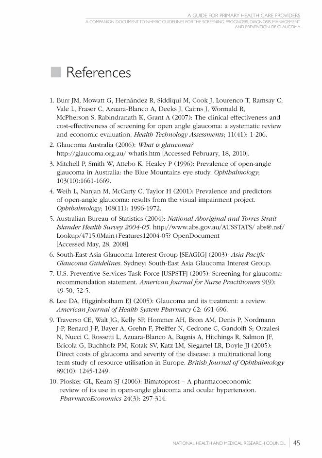
1. Burr JM, Mowatt G, Hernández R, Siddiqui M, Cook J, Lourenco T, Ramsay C, Vale L, Fraser C, Azuara-Blanco A, Deeks J, Cairns J, Wormald R, McPherson S, Rabindranath K, Grant A (2007): The clinical effectiveness and cost-effectiveness of screening for open angle glaucoma: a systematic review and economic evaluation. Health Technology Assessments; 11(41): 1-206.
2. Glaucoma Australia (2006): What is glaucoma? http://glaucoma.org.au/ whatis.htm [Accessed February, 18, 2010].
3. Mitchell P, Smith W, Attebo K, Healey P (1996): Prevalence of open-angle glaucoma in Australia: the Blue Mountains eye study. Ophthalmology; 103(10):1661-1669.
4. Weih L, Nanjan M, McCarty C, Taylor H (2001): Prevalence and predictors of open-angle glaucoma: results from the visual impairment project. Ophthalmology; 108(11): 1996-1972.
5. Australian Bureau of Statistics (2004): National Aboriginal and Torres Strait Islander Health Survey 2004-05. http://www.abs.gov.au/AUSSTATS/ [email protected]/Lookup/4715.0Main+Features12004-05? OpenDocument [Accessed May, 28, 2008].
6. South-East Asia Glaucoma Interest Group [SEAGIG] (2003): Asia Pacific Glaucoma Guidelines. Sydney: South-East Asia Glaucoma Interest Group.
7. U.S. Preventive Services Task Force [USPSTF] (2005): Screening for glaucoma: recommendation statement. American Journal for Nurse Practitioners 9(9): 49-50, 52-5.
8. Lee DA, Higginbotham EJ (2005): Glaucoma and its treatment: a review. American Journal of Health System Pharmacy 62: 691-696.
9. Traverso CE, Walt JG, Kelly SP, Hommer AH, Bron AM, Denis P, Nordmann J-P, Renard J-P, Bayer A, Grehn F, Pfeiffer N, Cedrone C, Gandolfi S; Orzalesi N, Nucci C, Rossetti L, Azuara-Blanco A, Bagnis A, Hitchings R, Salmon JF, Bricola G, Buchholz PM, Kotak SV, Katz LM, Siegartel LR, Doyle JJ (2005): Direct costs of glaucoma and severity of the disease: a multinational long term study of resource utilisation in Europe. British Journal of Ophthalmology 89(10): 1245-1249.
10. Plosker GL, Keam SJ (2006): Bimatoprost – A pharmacoeconomic review of its use in open-angle glaucoma and ocular hypertension. PharmacoEconomics 24(3): 297-314.

NATIONAL HEALTH AND MEDICAL RESEARCH COUNCIL 46
A GUIDE FOR PRIMARY HEALTH CARE PROVIDERSA COMPANION DOCUMENT TO NHMRC GUIDELINES FOR THE SCREENING, PROGNOSIS, DIAGNOSIS, MANAGEMENT AND PREVENTION OF GLAUCOMA
11. American Academy of Ophthalmology [AAO] (2005b): Primary open-angle glaucoma preferred practice pattern. San Francisco: American Academy of Ophthalmology.
12. Sycha T, Vass C, Findl O, Bauer P, Groke I, Schmetterer L, Eichler H (2003):Interventions for normal tension glaucoma. Cochrane Database of Systematic Reviews; 1.
13. American Optometric Association [AOA] (2002): Optometric clinical practice guideline: Care of the patient with open angle glaucoma (2nd edn.). St Louis: American Optometric Association.
14. Japan Glaucoma Society [ JGS] (2004): Guidelines for glaucoma. Tokyo: Japan Glaucoma Society.
15. Schmier JK, Halpern MT, Jones ML (2007): The economic implications of glaucoma – A literature review. PharmacoEconomics; 25(4): 287-308.
16. Adis International (2004): Corticosteroids are the main culprits in drug-induced glaucoma. Drugs Therapy Perspective; 20(8): 19-22.
17. Tripathi RC, Tripathi BJ, Haggerty C (2003): Drug-Induced Glaucomas Mechanism and Management. Drug Safety; 26(11): 749-767.
18. Bonovas S, Filioussi K, Tsantes A, Peponis V (2004a): Epidemiological association between cigarette smoking and primary open-angle glaucoma: a meta-analysis. Public Health 118(4): 256-261.
19. Bonovas S, Peponis V, Filioussi K (2004b): Diabetes mellitus as a risk factor for primary open-angle glaucoma: a meta-analysis. Diabetic Medicine 21(6): 609-614.
20. Budenz DL, Anderson DR, Feuer WJ, Beiser JA, Schiffman J, Parrish RK, Piltz-Seymour JR, Gordon MO, Kass MA (2006): Detection and Prognostic Significance of Optic Disc Hemorrhages during the Ocular Hypertension Treatment Study. Ophthalmology; 113(12): 2137-2143.
21. Williams R (1999): Traumatic Eye Injury. Gleams Spring Issue. Glaucoma research Foundation. http://www.glaucoma.org/learn/traumatic_glauc.php
22. Tielsch JM, Katz J, Singh K, Quigley HA, Gottsch JD, Javitt J, Sommer A (1991): A Population-based Evaluation of Glaucoma Screening: The Baltimore Eye Survey. American Journal of Epidemiology; 134(10): 1102-1110.
23. Hatt S, Wormald R, Burr J (2006): Screening for prevention of optic nerve damage due to chronic open angle glaucoma. Cochrane Database of Systematic Reviews; 4.

A GUIDE FOR PRIMARY HEALTH CARE PROVIDERS A COMPANION DOCUMENT TO NHMRC GUIDELINES FOR THE SCREENING, PROGNOSIS, DIAGNOSIS, MANAGEMENT
AND PREVENTION OF GLAUCOMA
NATIONAL HEALTH AND MEDICAL RESEARCH COUNCIL 47
24. Sommer A, Katz J, Quigley HA, et al. Clinically detectable nerve fiber layer atrophy precedes the onset of glaucomatous field loss. Archives of Ophthalmology 1991; 109:77-83.
25. European Glaucoma Society [EGS] (2003): Terminology and guidelines for glaucoma (2nd edn.). Savona, Italy: European Glaucoma Society.
26. Royal College of Ophthalmologists [RCO] (2004): Guidelines for the management of open angle glaucoma and ocular hypertension. London: Royal College of Ophthalmologists.
27. Dueker DK, Singh K, Lin SC, Fechtner RD, Minckler DS, Samples JR, Schuman JS (2007): Corneal Thickness Measurement in the Management of Primary Open-angle Glaucoma. A Report by the American Academy of Ophthalmology. Ophthalmology; 114(9):1779-1787.
28. Gordon MO, Beiser JA, Brandt JD, Heuer DK, Higginbotham EJ, Johnson CA, Keltner JL, Miller JP, Parrish II RK, Wilson MR, Kass MA (2002): The Ocular Hypertension Treatment Study: Baseline factors that predict the onset of primary open-angle glaucoma. Archives of Ophthalmology 120: 714-720.
29. Miglior S, Guareschi M, Albe E, Gomarasca S, Vavassori M, Orzalesi N (2003): Detection of glaucomatous visual field changes using the moorfields regression analysis of the heidelberg retina tomograph. American Journal of Ophthalmology; 136:26–33.
30. Burr J, Azuara-Blanco A, Avenell A (2004): Medical versus surgical interventions for open angle glaucoma. Cochrane Database of Systematic Reviews; 1.
31. Leske MC, Heijl A, Hyman L, Bengtsson B, Komaroff E (2004): Factors for progression and glaucoma treatment: The Early Manifest Glaucoma Trial. Current Opinion in Ophthalmology; 15:102–106.Heigl, Leske, Bengtsson et al 2002.
32. Heigl A, Leske C, Bengtsson B, Hyman L, Bengtsson B, Hussien M (2002): Reduction of intraocular pressure and glaucoma progression: results from the early manifest glaucoma trial. Archives of Ophthalmology; 120:1268-1279.
33. Canadian Glaucoma Study Group (2006): Canadian glaucoma study: 1. Study design, baseline characteristics and preliminary analyses. Canadian Journal of Ophthalmology; 41:566-75.
34. The AGIS Investigators (2000): The Advanced Glaucoma Intervention Study (AGIS): 7. The relationship between control of intraocular pressure and visual field deterioration. American Journal of Ophthalmology 130: 429-440.
35. The AGIS Investigators [AGIS] (2002): The Advanced Glaucoma Intervention Study (AGIS) 11. Risk Factors for Failure of Trabeculectomy and Argon Laser Trabeculoplasty. American Journal of Ophthalmology 134: 481-498.

NATIONAL HEALTH AND MEDICAL RESEARCH COUNCIL 48
A GUIDE FOR PRIMARY HEALTH CARE PROVIDERSA COMPANION DOCUMENT TO NHMRC GUIDELINES FOR THE SCREENING, PROGNOSIS, DIAGNOSIS, MANAGEMENT AND PREVENTION OF GLAUCOMA
36. Fleming C, Whitlock EP, Beil T, Smit B, Harris RP (2005): Screening for primary open-angle glaucoma in the primary care setting: an update for the US preventive services task force. Annals of Family Medicine; 3(2):167-170.
37. Friedman DS, Wilson MR, Liebmann JM, Fechtner RD, Weinreb RN (2004): An evidence-based assessment of risk factors for the progression of ocular hypertension and glaucoma. American Journal of Ophthalmology; 138(3):S19-31.
38. Maier PC, Funk J, Schwarzer G, Antes G, Falck-Ytter YT (2005): Treatment of ocular hypertension and open angle glaucoma: meta-analysis of randomised controlled trials. British Medical Journal; 331(7509):134.
39. Rolim de Moura RC, Paranhos Jr A, Wormald R (2007): Laser trabeculoplasty for open angle glaucoma (Review). Cochrane Database of Systematic Reviews; 4.
40. Oliver JE, Hattenhauer MJ, Herman D, Hodge DO, Kennedy R, Fang-Yen M, Johnson DH, Hattenhauer MG (2002): Blindness and Glaucoma: A comparison of patients progressing to blindness from glaucoma with patients maintaining vision. American Journal of Ophthalmology; 133: 764–772.
41. Tuulonen A, Airaksinen PJ, Erola E, Forsman E, Friberg K, Kaila M, Klemetti A, Mäkelä M, Oskala P, Puska P, Suoranta L, Teir H, Uusitalo H, Vainio-Jylhä E, Vuori M (2003): The Finnish evidence-based guideline for open-angle glaucoma. Acta Ophthalmologica Scandinavica; 81(1): 3-18.
42. South African Glaucoma Society [SAGS] (2006): Glaucoma algorithm and guidelines for glaucoma. Hatfield: South African Glaucoma Society.
43. Azuara-Blanco A, Burr J, Thomas R, Maclennan G, McPherson S (2007): The accuracy of accredited glaucoma optometrists in the diagnosis and treatment recommendations for glaucoma. British Journal of Ophthalmology; 91: 1639-1643.
44. American Academy of Ophthalmology [AAO] (2005c): Primary open-angle glaucoma suspect preferred practice pattern. San Francisco: American Academy of Ophthalmology.
45. American Academy of Ophthalmology [AAO] (2005a): Primary angle closure preferred practice pattern. San Francisco: American Academy of Ophthalmology.
46. Adis International (2004): Corticosteroids are the main culprits in drug-induced glaucoma. Drugs and Therapy Perspectives; 20(8): 19-22.
47. Moore W, Nischal KK (2007): Pharmacologic management of glaucoma in childhood. Pediatric Drugs; 9(2):71-79.
48. Papadopoulos M (2001): What’s New in Primary Open Angle Glaucoma? Journal Community Eye Health; 14(39): 35-36.

A GUIDE FOR PRIMARY HEALTH CARE PROVIDERS A COMPANION DOCUMENT TO NHMRC GUIDELINES FOR THE SCREENING, PROGNOSIS, DIAGNOSIS, MANAGEMENT
AND PREVENTION OF GLAUCOMA
NATIONAL HEALTH AND MEDICAL RESEARCH COUNCIL 49
49. Minckler DS, Vedula SS, Li TJ, Mathew MC, Ayyala RS, Francis BA (2006): Aqueous shunts for glaucoma. Cochrane Database of Systematic Reviews; 2.
50. Sycha T, Vass C, Findl O, Bauer P, Groke I, Schmetterer L, Eichler H (2003): Interventions for normal tension glaucoma. Cochrane Database of Systematic Reviews; 1.
51. Vass C, Hirn C, Sycha T, Findl O, Sacu S, Bauer P, Schmetterer L (2007): Medical interventions for primary open angle glaucoma and ocular hypertension. Cochrane Database of Systematic Reviews; 4.
52. Foster PJ, Buhrmann R, Quigley HA, Johnson GJ (2002): The definition and classification of glaucoma in prevalence surveys. British Journal of Ophthalmology; 86(2):238-242.


A GUIDE FOR PRIMARY HEALTH CARE PROVIDERS A COMPANION DOCUMENT TO NHMRC GUIDELINES FOR THE SCREENING, PROGNOSIS, DIAGNOSIS, MANAGEMENT
AND PREVENTION OF GLAUCOMA
NATIONAL HEALTH AND MEDICAL RESEARCH COUNCIL 51
Appendix 1Definitions and anatomy of the eye related to glaucoma
There is no definitive definition of glaucoma, largely due to the number of separate classifications of the disease. There is broad agreement that any form of glaucoma can be described as a progressive optic neuropathy with characteristic visual field loss, optic disc and nerve fibre degeneration. The NHMRC Glaucoma Working Committee recommended the use of the definition by Burr, Azuara-Blanco & Avenell30 ‘Glaucoma describes a group of eye diseases in which there is progressive damage to the optic nerve characterised by specific structural abnormalities of optic nerve head and associated patterns of visual field loss’ (p2). There are four main ways to classify glaucoma:
1. Mechanism of aqueous flow impairment leading to open angle glaucoma or angle closure glaucoma.
2. Aetiology and presence of associated factors with the impairment (primary or secondary glaucoma).
3. Age of the population (congenital, infantile, juvenile or adult). Glaucoma of any age group may be classified within the above broad headings.
4. Temporal nature of symptoms i.e. acute/chronic or intermittent angle closure. This is most often used in angle closure glaucoma.
The pathophysiology of IOP elevation in glaucoma is primarily related to alteration in aqueous flow, although research is beginning to understand the role of optic nerve vulnerability and particular mechanisms of injury, including connective tissue and neural alteration, vascular dysfunction, genetic factors and responses to local excitotoxic conditions.
Open angle glaucomaOAG is a progressive optic neuropathy.23 In the absence of other identifiable causes, it is classified as POAG. There is a characteristic acquired atrophy of the optic nerve, and loss of retinal ganglion cells and their axons. POAG is a disease in which elevated IOP is combined with a progressive optic neuropathy, resulting in characteristic excavation of the optic nerve head and corresponding visual field defects.50 This is associated with an open anterior chamber angle, by gonioscopic appearance.11 POAG is arbitrarily distinguished from NTG using an IOP cut-off of 21mmHg.51

NATIONAL HEALTH AND MEDICAL RESEARCH COUNCIL 52
A GUIDE FOR PRIMARY HEALTH CARE PROVIDERSA COMPANION DOCUMENT TO NHMRC GUIDELINES FOR THE SCREENING, PROGNOSIS, DIAGNOSIS, MANAGEMENT AND PREVENTION OF GLAUCOMA
Secondary OAG can be caused by a variety of substances that mechanically block the outflow of aqueous through the anterior chamber angle, resulting in an elevation of IOP. These substances include pigment, exfoliation material, and red blood cells. Secondary OAG can also result from alterations in the structure and function of the trabecular meshwork, due to insults such as trauma, inflammation, and ischemia.11 In secondary OAG, elevated IOP causes progressive typical glaucomatous optic neuropathy and visual field loss. In several forms of secondary glaucoma, pathomechanisms are combined, leading to both secondary OAG and angle closure glaucoma.42
Normal tension glaucomaNTG is a subtype of POAG where IOP remains within the statistically determined usual range, but progressive optic neuropathy and visual defects typical of glaucoma occur. The criteria that are widely used to define NTG include:
• a mean untreated IOP consistently equal to or less than 21mmHg or median IOP equal to or less than 20mmHg on diurnal testing, with no single measurement greater than 24mmHg
• open drainage angles on gonioscopy
• typical optic disc damage with glaucomatous cupping and loss of neuroretinal rim
• absence of any secondary cause for a glaucomatous optic neuropathy (trauma, steroids, uveitis)
• visual field defect compatible with glaucomatous cupping (disc to field correlation).51

A GUIDE FOR PRIMARY HEALTH CARE PROVIDERS A COMPANION DOCUMENT TO NHMRC GUIDELINES FOR THE SCREENING, PROGNOSIS, DIAGNOSIS, MANAGEMENT
AND PREVENTION OF GLAUCOMA
NATIONAL HEALTH AND MEDICAL RESEARCH COUNCIL 53
Angle closure glaucomaAngle closure glaucoma can present in either primary or secondary forms, in acute or chronic situations. Patients may have both, and present with acute attacks superimposed on a chronic condition. In primary angle closure, the eye is at risk of developing glaucomatous optic disc damage, particularly when associated with elevated IOP. When optic disc damage occurs, the eye is deemed to have progressed from primary angle closure to PACG.45 If acute angle closure is suspected, some components of the examination (optic disc imaging, visual field testing) may need to be postponed as patients may present as medical emergencies. Providing appropriate and timely treatment becomes the priority.
Primary angle closure is defined as appositional or synechial closure of the anterior-chamber angle, the commonest mechanism of which is usually pupillary block.45 In pupillary block, the front lens surface is anterior to the plane of the iris insertion into the ciliary body base, which causes resistance to aqueous humor flow. The resistance increases as the lens position is further forward, whether in primary angle closure or secondary angle closure. The resultant pressure gradient between the posterior and anterior chambers causes a forward bowing of the peripheral iris so that the iris covers all or part of the filtering portion of the trabecular meshwork (appositional angle closure), which can lead to elevation of IOP. Prolonged or repeated contact of the peripheral iris with the trabecular meshwork may lead to adhesions known as peripheral anterior synechiae and residual functional damage to the trabecular meshwork. The angle closure may or may not be associated with elevated IOP or glaucomatous optic neuropathy, and may occur in either an acute or chronic form.
Acute angle closure has at least two of the following symptoms (ocular or periocular pain, nausea and/or vomiting, history of intermittent blurring of vision with halos) and at least three of the following signs (raised IOP 21mmHg, conjunctival injection, corneal epithelial oedema, mid-dilated unreactive pupil, or shallow anterior chamber in the presence of an occludable angle).52
Secondary angle closure is associated with a closed anterior chamber as identified on gonioscopy. The European Glaucoma Society25 suggests that the pathophysiology of secondary angle closure includes phakomorphic eyes and cases of inflammation.

NATIONAL HEALTH AND MEDICAL RESEARCH COUNCIL 54
A GUIDE FOR PRIMARY HEALTH CARE PROVIDERSA COMPANION DOCUMENT TO NHMRC GUIDELINES FOR THE SCREENING, PROGNOSIS, DIAGNOSIS, MANAGEMENT AND PREVENTION OF GLAUCOMA
Figure 5: The anatomy of the eye (Source: Members of the NHMRC Working Committee)
Ciliary Body
Lens
Sclera
Cornea
Trabecular Meshin Angle
Anterior Chamber
Posterior Chamber
Zonules
Figure 6: An illustration of open angle and angle closure glaucoma with trabecular meshwork (Source: www.angleclosureglaucoma.cn)
Lens
Lens
trabecular
meshwork
closed angle
open angle
Cornea
Cornea
Ciliary body Ciliary body
trabe
cular
meshwork

A GUIDE FOR PRIMARY HEALTH CARE PROVIDERS A COMPANION DOCUMENT TO NHMRC GUIDELINES FOR THE SCREENING, PROGNOSIS, DIAGNOSIS, MANAGEMENT
AND PREVENTION OF GLAUCOMA
NATIONAL HEALTH AND MEDICAL RESEARCH COUNCIL 55
Childhood glaucomaChildhood glaucoma is a potentially blinding optic neuropathy invariably associated with raised IOP, characteristic optic disc changes and associated with specific visual field defects.47 Typically in infants, the signs of glaucoma are corneal haze, tearing of the eye with striking photophobia, and enlargement of the eyeball (buphthalmos). Childhood glaucoma is defined by age and mechanism of onset.
Pigmentary glaucomaPigmentary glaucoma presents similarly to POAG, with additional key signs including:
• pigment on the anterior surface of the iris often as concentric rings within the iris furrows
• spoke-like transillumination defects in the midperiphery of the iris
• pigment in the anterior and posterior chambers, and possibly Krukenberg’s spindles on the corneal endothelium
• a dense, homogeneously pigmented trabecular meshwork, especially posteriorly
• an open, deep anterior chamber angle with possible posterior bowing (concavity) of the iris
• rise of the IOP to rather high levels, with dramatic fluctuation
• pigment release resulting from pupillary dilation or strenuous exercise which requires assessment of the IOP after dilation.
Pseudoexfoliation glaucomaPseudoexfoliation glaucoma presents similarly to POAG, with additional key signs of:
• distribution of pseudoexfoliative material on the pupillary margin of the iris and, on the surface of the lens, as a central translucent disc with curled edges surrounded by an annular clear zone
• a peripheral granular zone on the anterior surface of the lens, best viewed through a dilated pupil

NATIONAL HEALTH AND MEDICAL RESEARCH COUNCIL 56
A GUIDE FOR PRIMARY HEALTH CARE PROVIDERSA COMPANION DOCUMENT TO NHMRC GUIDELINES FOR THE SCREENING, PROGNOSIS, DIAGNOSIS, MANAGEMENT AND PREVENTION OF GLAUCOMA
• transillumination defects in the iris near the pupil, and patchy pigmentation of the trabecular meshwork located in the superior angle and anterior to Schwalbe’s line. Pigment granules may form a whorled pattern over the sphincter muscle on the surface of the iris
• depigmentation of pupillary ruff
• poor pupillary response to topical mydriatic medications
• accelerated cataract formation
• trabecular pigmentation which may precede the appearance of pseudoexfoliative material on the surface of the lens, even though this material is present in the conjunctiva.
Neovascular glaucomaNeovascular glaucoma is a secondary glaucoma variously referred to as hemorrhagic glaucoma, thrombotic glaucoma, congestive glaucoma, rubeotic glaucoma, and diabetic hemorrhagic glaucoma. Causes of neovascular glaucoma include numerous secondary ocular and systemic diseases that share one common element, this being retinal ischemia/hypoxia and subsequent release of an angiogenesis factor. This angiogenesis factor causes new blood vessel growth from pre-existing vascular structures. Depending on the progression of neovascular glaucoma, it can cause glaucoma either through secondary open angle or secondary closed angle mechanisms. This is accomplished through the growth of a fibrovascular membrane over the trabecular meshwork in the anterior chamber angle, resulting in obstruction of the meshwork and/or associated peripheral anterior synechiae.

A GUIDE FOR PRIMARY HEALTH CARE PROVIDERS A COMPANION DOCUMENT TO NHMRC GUIDELINES FOR THE SCREENING, PROGNOSIS, DIAGNOSIS, MANAGEMENT
AND PREVENTION OF GLAUCOMA
NATIONAL HEALTH AND MEDICAL RESEARCH COUNCIL 57
Appendix 2Companion document details
Purpose of the companion documentThis companion document presents a summary for primary health care providers of the current best evidence presented in the NHMRC guideline for Screening, Prognosis, Diagnosis, Management and Prevention of Glaucoma. The purpose of the guideline is to inform practice for Australian health care providers, particularly utilising a multidisciplinary team approach.
Guideline development periodThe systematic review of the literature underpinning these guidelines was completed in September 2008 (available on www.nhmrc.gov.au). The guideline was completed in June 2009, and public consultation occurred during October-November 2009. This guideline was commissioned and funded by NHMRC.
Centre for Allied Health Evidence University of South Australia (Technical Team)Professor Karen Grimmer-Somers
Ms Janine Dizon
Ms Judith Lowe
Ms Anthea Worley
Ms Lucylynn Lizarondo
NHMRC Expert Working Committee Professor William Morgan (Ophthalmologist) Lions Eye Institute (CHAIR)
Associate Professor Ivan Goldberg (Ophthalmologist)

NATIONAL HEALTH AND MEDICAL RESEARCH COUNCIL 58
A GUIDE FOR PRIMARY HEALTH CARE PROVIDERSA COMPANION DOCUMENT TO NHMRC GUIDELINES FOR THE SCREENING, PROGNOSIS, DIAGNOSIS, MANAGEMENT AND PREVENTION OF GLAUCOMA
Eye Associates Glaucoma Services Sydney Eye Hospital
Professor Jonathon Crowston (Ophthalmologist) Centre for Eye Research Australia
Associate Professor David Mackey (Epidemiologist/Ophthalmologist) Royal Victorian Eye and Ear Hospital
Professor Algis Vingrys (Optometrist) Department of Optometry and Visual Sciences University of Melbourne
Dr Philip Anderton (Optometrist rural)
Associate Professor Amanda McBride (General Practitioner with interest in Glaucoma)Head of General Practice School of Medicine The University of Notre Dame Australia, Sydney and GP in Woollahra
Dr Genevieve Napper (Optometrist, low-vision service provider) Victorian College of Optometry
Mr Grant Martin (Director, Professional Services) Pharmaceutical Society of Australia
Ms Jill Grasso (Ophthalmic Nurse) Representing the Ophthalmic Nurses Association
Ms Beverly Lindsell (Glaucoma Australia Representative) Glaucoma Australia
Ms Tania Straga (Orthoptist) Representing the Orthoptic Association of Australia
Ms Helen Robbins Representing the Optometrists Association Australia (Observer)
Internal reference groupMr Luke Grzeskowiak, Pharmacist University of South Australia
NHMRC project staffMs Vesna Cvjeticanin
Ms Carla Rodeghiero
Mr Fethon Ileris
Ms Tess Winslade
Ms Kay Currie
Ms Marion Hewitt

A GUIDE FOR PRIMARY HEALTH CARE PROVIDERS A COMPANION DOCUMENT TO NHMRC GUIDELINES FOR THE SCREENING, PROGNOSIS, DIAGNOSIS, MANAGEMENT
AND PREVENTION OF GLAUCOMA
NATIONAL HEALTH AND MEDICAL RESEARCH COUNCIL 59
Appendix 3Abbreviations
IOP Intraocular pressure
mmHg Millimetres of mercury
NHMRC National Health and Medical Research Council (Australia)
nm Nanometre
NTG Normal tension glaucoma
OAG Open angle glaucoma
PACG Primary angle closure glaucoma
POAG Primary open angle glaucoma

NATIONAL HEALTH AND MEDICAL RESEARCH COUNCIL 60
A GUIDE FOR PRIMARY HEALTH CARE PROVIDERSA COMPANION DOCUMENT TO NHMRC GUIDELINES FOR THE SCREENING, PROGNOSIS, DIAGNOSIS, MANAGEMENT AND PREVENTION OF GLAUCOMA
Glossary of terms
Afferent pupillary defect A defect of the pupillary reflex characterised by less constriction of both pupils when the affected eye is stimulated by light relative to that occurring when the unaffected eye is stimulated, as with the swinging flashlight test. The defect is also known as the Marcus Gunn pupil.
African The literature variably refers to the increased risk of glaucoma occurring in people of African descent. This refers to people who trace their ancestry to Africa, whether this be African-Americans, African-Carribbeans, East Africans, Sub-Sarharan Africans or West Africans.
Anterior chamber The space in the eye, filled with aqueous humor that is bordered anteriorly by the cornea and a small portion of the sclera and posteriorly by a small portion of the ciliary body, the iris, and that portion of the lens which presents through the pupil.
Argon laser trabeculoplasty
Light stimulation of the trabecular meshwork of the angle of the anterior chamber by an argon laser beam to facilitate aqueous humor outflow.
Aqueous humor The clear, watery fluid that fills the anterior and posterior chambers of the eye.
Biomicroscopy Examination of ocular tissue using a bright focal source of light with a slit of variable width and height and a binocular microscope with variable magnification.
Confocal scanning laser ophthalmoscopy
The recording of two-dimensional sectional images for the evaluation of ocular tissue, using a confocal laser imaging system displayed digitally in real time.
Confocal scanning laser tomography
The recording of a series of images along the axial axis of the eye enabling the three-dimensional reconstruction of the topography of the surface of the specific tissue under examination using a confocal laser imaging system.

A GUIDE FOR PRIMARY HEALTH CARE PROVIDERS A COMPANION DOCUMENT TO NHMRC GUIDELINES FOR THE SCREENING, PROGNOSIS, DIAGNOSIS, MANAGEMENT
AND PREVENTION OF GLAUCOMA
NATIONAL HEALTH AND MEDICAL RESEARCH COUNCIL 61
Cup:disc ratio The ratio of the diameter of the area of excavation of the surface of the optic disc to that of the diameter of the optic disc in any given meridian, often either the horizontal or vertical meridian. Cup:disc ratio is a value obtained by dividing the cup diameter by the disc diameter. The closer this value is to 1, the greater the level of tissue loss and therefore damage to the disc.
Excitotoxicity The stimulation of neurons to death by excessive levels of excitatory neurotransmitters.
Filtration surgery Surgical procedures (e.g. thermal sclerostomy, posterior or anterior lip sclerectomy, trephination, trabeculectomy) used to create an alternative pathway for the outflow of aqueous humor to lower intraocular pressure.
Fundus photography The use of a camera with optics and an illumination system that permits photographing the fundus of the eye.
Genetic mutation The alteration of DNA sequencing by changes in the genome.
Glaucoma Glaucoma describes a group of eye diseases in which there is progressive damage to the optic nerve characterised by specific structural abnormalities of optic nerve head and associated patterns of visual field loss.
Glaucoma suspect A person suspected of having glaucoma has some but not all of the criteria required for a glaucoma diagnosis. They may have one or more of the following: suspicious optic disc, optic disc margin haemorrhage, occludable drainage angle, peripheral anterior synechiae or elevated intraocular pressure.
Gonioscopy A diagnostic procedure to examine the angle of the anterior chamber in which a specialised corneal contact lens and a biomicroscope are used.
Health care provider Any member of the glaucoma team who provides input into the patient’s glaucoma journey. Health care providers involved with glaucoma in Australia may include, but are not limited to, ophthalmologists, general medical practitioners, optometrists, ophthalmic nurses and orthoptists.
Hypermetropia A defect of vision in which a person is able to focus on objects in the distance, but not on close objects.

NATIONAL HEALTH AND MEDICAL RESEARCH COUNCIL 62
A GUIDE FOR PRIMARY HEALTH CARE PROVIDERSA COMPANION DOCUMENT TO NHMRC GUIDELINES FOR THE SCREENING, PROGNOSIS, DIAGNOSIS, MANAGEMENT AND PREVENTION OF GLAUCOMA
Intraocular pressure The pressure within the eye due to the balance between the formation and drainage of the aqueous humor.
Multifactorial inheritance The determination of phenotype by multiple genetic and environmental factors, each making a small contribution.
Myopia A vision condition in which close objects are seen clearly, but objects farther away appear blurred.
Myocilin A protein believed to be associated with primary open angle glaucoma found both extraocular and in the trabecular meshwork, optic nerve, retina, cornea, iris, ciliary body, and sclera.
Nerve fibre layer The layer of the retina that comprises unmyelinated axons of retinal ganglion cells.
Neuroprotection The use of pharmacological, genetic alteration, and other means to attenuate a destructive cellular environment thereby protecting neurons from secondary degeneration caused by a variety of primary insults (ischemia/hypoxia, stroke, trauma, degeneration).
Neuroretinal rim The tissue between the optic cup and disc margins.
Nocturnal dip The decrease in systemic blood pressure during sleep.
Optic nerve The cranial nerve (N II) that carries visual impulses from the retina to the brain.
Perimetry Determination of the extent of the visual field for various types and intensities of stimuli for the purpose of diagnosing and localising disturbances in the visual pathway.
Peripapillary area Tissue surrounding the optic nerve head.
Polygenic The traits or diseases caused by the impact of many genes, each with a small additive effect on phenotype.
Posterior chamber The space in the eye delimited by the posterior surface of the iris, the ciliary processes, and the valleys between them, the zonule of Zinn, and the anterior surface of the crystalline lens. It includes the canal of Hanover, the canal of Petit, and the retrolental space of Berger.

A GUIDE FOR PRIMARY HEALTH CARE PROVIDERS A COMPANION DOCUMENT TO NHMRC GUIDELINES FOR THE SCREENING, PROGNOSIS, DIAGNOSIS, MANAGEMENT
AND PREVENTION OF GLAUCOMA
NATIONAL HEALTH AND MEDICAL RESEARCH COUNCIL 63
Pulsatile ocular blood flow
The indirect assessment of choroidal blood flow by estimating the influx of blood into the eye during cardiac systole from an evaluation of the continuous IOP pulse wave.
Puncta Puncta are tiny openings along the eyelid margin through which tears drain.
Refraction Clinically, the determination of the refractive errors of an eye, or eyes (e.g. myopia, hyperopia, astigmatism, anisometropia).
Reverse pupillary block Blockage of the movement of aqueous from the anterior to the posterior chamber leading to a concave anatomical configuration of the peripheral iris.
Selective laser trabeculoplasty
Use of a q-switched Nd:YAG laser to target trabecular meshwork endothelial cells without provoking coagulative necrosis, to improve aqueous outflow.
Short-wavelength automated perimetry
A form of automated perimetry that isolates the blue cone mechanism of the visual system by utilising a two-colour incremental thresholding technique consisting of a large blue target on a bright yellow background.
Tonometry A procedure for measurement of the pressure within the eye. Clinically, tonometry measures the intraocular tension.
Trabecular meshwork The meshwork of connective tissue that is located between the canal of Schlemm and the anterior chamber, and which is involved in drainage of aqueous humor from the eye.
Trabeculectomy
Surgical creation of a fistula to allow aqueous outflow from the anterior chamber to the subconjunctival tissue space, bypassing the trabecular meshwork/Canal of Schlemm outflow pathway.
Visual acuity The clearness of vision that depends on the sharpness of the retinal image and the integrity of the retinal and visual pathway. It is expressed as the angle subtended at the anterior focal point of the eye by the detail of the letter or symbol recognised.
Visual field The area or extent of space visible to an eye in a given position.

Notes

Notes

Notes