a glucose biosensor based on tio2–graphene composite
TRANSCRIPT
Biosensors and Bioelectronics 38 (2012) 184–188
Contents lists available at SciVerse ScienceDirect
Biosensors and Bioelectronics
0956-56
http://d
n Corr
E-m1 Th
journal homepage: www.elsevier.com/locate/bios
A glucose biosensor based on TiO2–Graphene composite
Hee Dong Jang a,n,1, Sun Kyung Kim a,b,1, Hankwon Chang a, Ki-Min Roh a, Jeong-Woo Choi b,Jiaxing Huang c
a Rare Metals Research Center, Korea Institute of Geoscience and Mineral Resources, Daejeon 305-350, Koreab Interdisciplinary Program of Integrated Biotechnology, Sogang University, Seoul 121-742, Koreac Department of Materials Science and Engineering, Northwestern University, Evanston, Illinois 60208, USA
a r t i c l e i n f o
Article history:
Received 23 March 2012
Received in revised form
23 May 2012
Accepted 24 May 2012Available online 6 June 2012
Keywords:
Glucose biosensor
TiO2–GR composite
Aerosol assisted self-assembly
Cyclic voltammetry
63/$ - see front matter & 2012 Elsevier B.V. A
x.doi.org/10.1016/j.bios.2012.05.033
esponding author. Tel.: þ82 42 868 3612; fax
ail address: [email protected] (H.D. Jang).
ese authors contributed equally to this work
a b s t r a c t
A novel glucose biosensor was developed based on the adsorption of glucose oxidase at a TiO2–
Graphene (GR) nanocomposite electrode. A TiO2–GR composite was synthesized from a colloidal
mixture of TiO2 nanparticles and graphene oxide (GO) nanosheets by an aerosol assisted self-assembly
(AASA). The particle morphology of all TiO2–GR composites was spherical in shape. It was observed that
micron-sized TiO2 particles were encapsulated by GR nanosheets and that the degree of encapsulation
was proportional to the ratio of GO/TiO2. The amperometric response of the glucose biosensor
fabricated by the TiO2–GR composite was linear against a concentration of glucose ranging from 0 to
8 mM at �0.6 V. The highest sensitivity was noted at about 6.2 mA/mM cm2. The as prepared glucose
biosensor based on the TiO2–GR composite showed higher catalytic performance for glucose redox than
a pure TiO2 and GR biosensor.
& 2012 Elsevier B.V. All rights reserved.
1. Introduction
Glucose biosensors are a common item of concern among allthe enzyme-based biosensors due to their importance in themonitoring of blood glucose for the treatment and control ofdiabetes (Wang, 2008). Glucose oxidase (GOD) is employed as theenzyme in most glucose biosensors. The general principle of theglucose biosensor is based on the amperometric detection of Hþ
via the following chemical reactions (Kuila et al., 2011):
GlucoseþGOD-FAD-gluconolactoneþGOD-FADH2
FADH22FADþ2Hþþ2e�
Glucose is the substrate of GOD, which will result in anenzyme-catalyzed reaction and decrease the concentration ofGOD-FAD on the electrode surface. GOD assembled materialsexhibit a fast electron transfer reaction on the electrode. Itsoxidized form, FAD, is electrochemically oxidized at the electrodesurface producing its reduced form, FADH2 (Wu et al., 2010).
Graphene (GR) exhibits a unique structure of a two-dimen-sional (2D) nanosheet composed of hexagonal arrayed sp2 hybri-dized carbon atoms with one-atomic thickness (Liu et al., 2011).The unique structure of GR instills in its fast electron
ll rights reserved.
: þ82 42 868 3415.
as the first author.
transportation, a high specific surface area, high thermal con-ductivity, excellent mechanical stiffness and good biocompatibil-ity, which result in promising applications in nanocomposites,electromechanical resonators, solar cells and electrochemicalsensors (Li et al., 2008).
Chen et al. (2010) developed Nafion-GR/GOD film-modifiedelectrodes for the detection of glucose, which showed goodstability. Shan et al. (2010) reported a good amperometricresponse to glucose with wide linear ranges using chitosan-GR/Au nanocomposite films for glucose sensing applications. Liu et al.(2011) synthesized self-assembled graphene platelet-glucose oxi-dase nanostructures for glucose biosensing applications. Thus, GRis expected to play an important role in improving the catalyticperformance of biosensing applications due to its uniqueproperties.
Many types of materials, such as SiO2, gold, silver, CeO2, MnO2
and TiO2 nanoparticles, have been used to construct nano-biosensors (Rahman et al., 2010). Among them, TiO2 nanoparticleshave attracted considerable interest due to the superior proper-ties, such as their large specific surface area, high uniformity, andexcellent biocompatibility (Li et al., 2009). Bao et al. (2008)reported that modified TiO2 on an electrode could enhance theapplication of a glucose biosensor by the immobilization of theenzyme. They hydrothermally synthesized a new slack TiO2 layerwith a uniform porous nanostructure through the use of a MWNTtemplate. They showed that TiO2 provided the enzyme with abetter immobilization environment because it enlarged the spe-cific surface area. However, few studies have investigated TiO2

H.D. Jang et al. / Biosensors and Bioelectronics 38 (2012) 184–188 185
nanoparticles with GR nanosheets as an electrode material forglucose biosensors.
In the present study, we present an enhanced glucose biosen-sor based on the TiO2–GR composite. The TiO2–GR composite wassynthesized by evaporating droplets composed of TiO2 nanopar-ticles, GO and water by an aerosol-assisted self-assembly (AASA)method. The AASA method is of interest as it has many advan-tages; it is a simple system, it is cost-effective, and it can be scaledup to very high quantities (Jang et al., 2008).
GO can be reduced to form chemically modified GR in whichthe oxygen functional groups (–OH, –COOH, –O–) are removed byreduction reactions with chemical reducing agents such ashydrazine or by heat treatment in a reducing atmosphere (Coteet al., 2009a). However, the use of chemical reducing agentsrequires much time to prepare the composites due to many typesof chemical reaction steps, long reaction time, necessity ofrepeated washing, and long drying time. Moreover, the processrequires the use of poisonous and potentially explosive chemicals(Liu et al., 2011). In this study, we synthesized the TiO2–GRcomposite using the AASA method without any reducing agentsfor the GR. The effects of the weight ratio of the GR/TiO2 in thecolloidal mixture on the particle properties of the morphologyand specific surface area were examined. In addition, the electro-chemical properties of the as-prepared TiO2–GR composites foruse in a glucose biosensor were evaluated by means of cyclicvoltammetry measurements.
2. Experimental
2.1. Synthesis of the graphene oxide colloid
Graphene oxide (GO) was synthesized by the oxidation ofgraphite powder (Alfa Aesar, 99.9%) using H2SO4 (Junsei, 95%) andKMnO4 (Junsei, 99.3%) according to the modified Hummers’method (Hummers and Offeman, 1958) with a pre-oxidation step,where the micrographic powder was mixed with a strong oxidiz-ing agent and was then filtered, washed and dried. The dried GOwas redispersed in water with mechanical agitation or mildsonication using a table-top ultrasonic cleaner, giving a colloidalsolution of exfoliated GO, as noted in earlier reports (Cote et al.,2009a, 2009b; Hummers and Offeman, 1958; Kim et al., 2010a).
Fig. 1. X-Ray photoelectron spectroscopy of the as-prepared GO.
2.2. Synthesis of TiO2–GR composite
For the synthesis of the TiO2–GR composite, a colloidalmixture solution as an aerosol precursor was prepared by disper-sing TiO2 nanoparticles (P25, Degussa) and the as-prepared GOcolloid into distilled water. The precursor was prepared withdifferent weight ratios of GO/TiO2 from 0.05 to 2.0 while theconcentration of the TiO2 was fixed at 0.1 wt% in the colloidalmixture. The experimental apparatus for the AASA process con-sisted of an ultrasonic atomizer, an electrical tubular furnace, anda filter sampler. The ultrasonic atomizer was used to generatemicron-sized droplets of the colloidal precursor. The dropletswere then carried into the furnace by 1.0 l/min of argon. Theevaporation of water, the self-assembly of GO and TiO2, and thereduction of GO were carried out in sequence in the tubularfurnace, where the length and diameter of the heating zone were54 cm and 25 mm, respectively, and the operating temperaturewas 800 1C. The fabricated TiO2–GR composites were then col-lected by a Teflon membrane filter. The filter was heated toprevent water from condensing on it. It took only several secondsto prepare the composite without the need for any post-heat-treatment or purification steps.
2.3. Synthesis of TiO2–GR/GOD electrode
1 mg of the as-prepared TiO2–GR composite was dispersed in1 ml of 10,000 ppm (10 mg/ml) GOD solution (Sigma Aldrich,Aspergillus niger, 200 units/mg). The TiO2–GR/GOD colloid wasstored at 4 1C for 24 h and separated by a centrifugal separatoroperating at 10,000 rpm for 1 h in order to ensure complete GODimmobilization. The precipitate was re-dispersed by 1 ml of0.01 M PBS solution (Fluka). For the cyclic voltammetry measure-ment, 5 ml of TiO2–GR/GOD colloid was dropped onto the surfaceof a glassy carbon electrode (GCE) and left to dry at roomtemperature. Then, 10 ml of 0.05 wt% Nafion solution was addi-tionally cast on the surface of the modified GCE and was driedbefore the electrochemical experiments. 0–8 mM of D-(þ)-glu-cose (Sigma Aldrich) was used as the reaction solution.
2.4. Analysis
The particle morphology and size of the TiO2–GR compositewere characterized by a field emission scanning electron micro-scope (FE-SEM; Sirion, FEI). The BET specific surface areas weremeasured by N2 adsorption–desorption isotherms on a Quadra-sorb Quantachrome analyzer. The crystallinity of the as-preparedcomposite was analyzed by X-ray diffractometer (XRD, RigakuRTP 300 RC). X-ray photoelectron spectroscopy (XPS) measure-ment was carried out for an elemental analysis of the compositeusing a Sigma Probe photoelectron spectrometer (Thermo VG).The electrochemical properties of the glucose biosensor weremeasured by cyclic voltammetry (CV) method using an electro-chemical interface instrument (VSP, Bio-Logics). A conventionalthree-electrode cell was used, including a glassy carbon electrodeas the working electrode, an Ag/AgCl electrode as the referenceelectrode, and a platinum foil as the counter electrode.
3. Result and discussion
3.1. Synthesis of TiO2–GR composite
Fig. 1 shows the X-ray photoemission spectroscopy (XPS)spectrum of the as-prepared GO showed that C and O peaksappeared at 285 and 533 eV, respectively. Also, C 1s spectrumshows two peaks at 284.7 and 286.6 eV, which are attributed toC–C and C–OH/C¼O, respectively (Yang et al., 2009). According to

H.D. Jang et al. / Biosensors and Bioelectronics 38 (2012) 184–188186
the result of the XRD analysis, the peak of GO appeared at 10.21,which matched earlier results (Ishikawa et al., 2010). Theseresults confirmed that GO was successfully synthesized fromgraphite by the modified Hummers’ method.
In Fig. 2, the FE-SEM analysis exhibits the morphology of aTiO2–GR composite prepared at different ratios of GR/TiO2. It wasobserved that the TiO2 particles become encapsulated by the GR,as shown in Fig. 2(a). The formation of the TiO2–GR composite viathe aerosol-assisted co-assembly of GO nanosheets and TiO2
nanoparticles is explained as follows. As the aerosol dropletswere introduced into the hot zone of the furnace, rapid
Fig. 2. FE-SEM images of TiO2–GR composite at different weight ratios of GO/TiO2 (a) 2
fixed experimental condition (TiO2: 0.1 wt%, temp.: 800 1C, carrier gas flowrate: 1 l/mi
evaporation caused the shrinkage of the droplets, concentratingthe TiO2 nanoparticles and the GO sheets, and then subsequentlycompressing them into micron-sized composite (Jang et al., 2008;Jang et al., 2010; Luo et al., 2011). The GO sheets were alsothermally reduced to chemically modified graphene. Because GOis effectively a 2D amphiphile with a hydrophilic periphery and alargely hydrophobic center, it was noted that GO sheets aresurface-active with the ability to lower interfacial energies (Kimet al., 2010b; Cote et al., 2011). GO can accumulate at the watersurface of a sprayed droplet during evaporation and finally form aGR ball. In contrast, hydrophilic TiO2 nanoparticles agglomerate
, (b) 0.5, (c) 0.1, (d) 0.05, and (e,f,g,h) shows the magnified image of (a,b,c,d) at the
n).
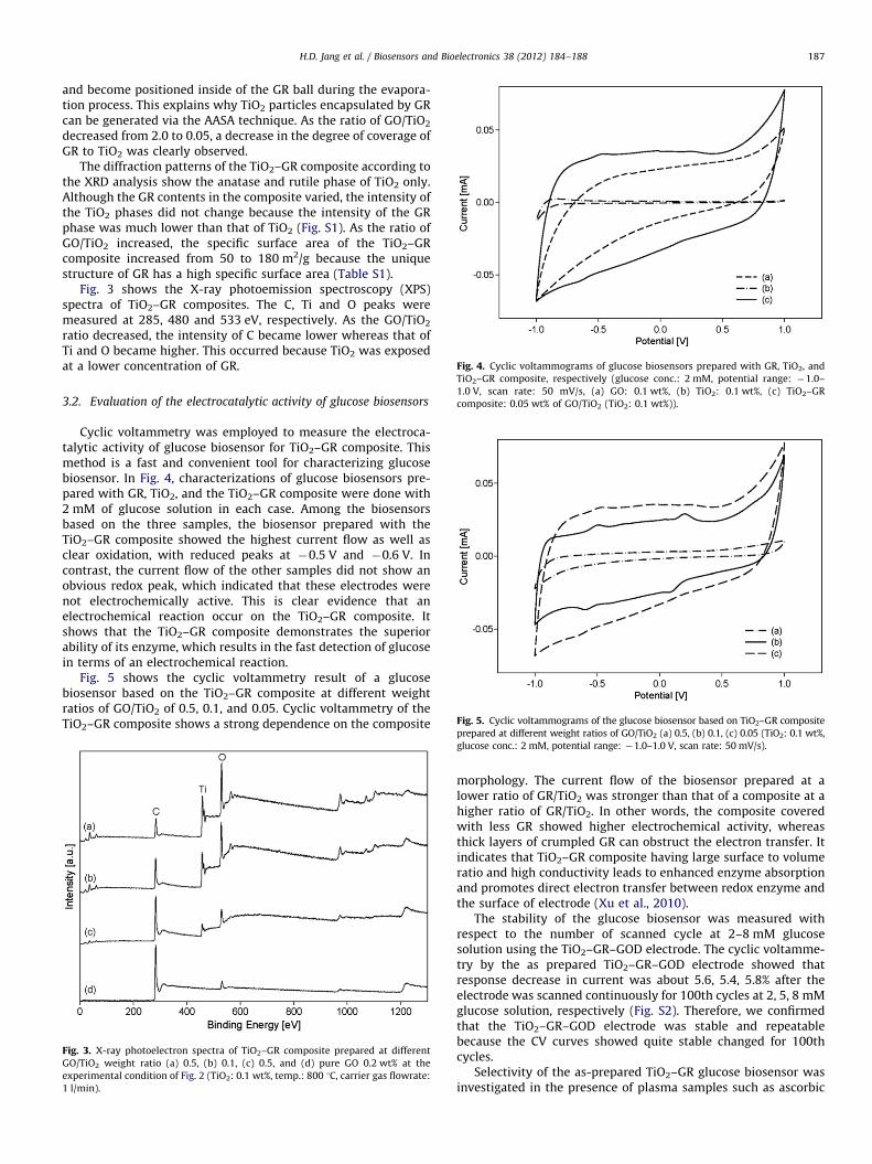
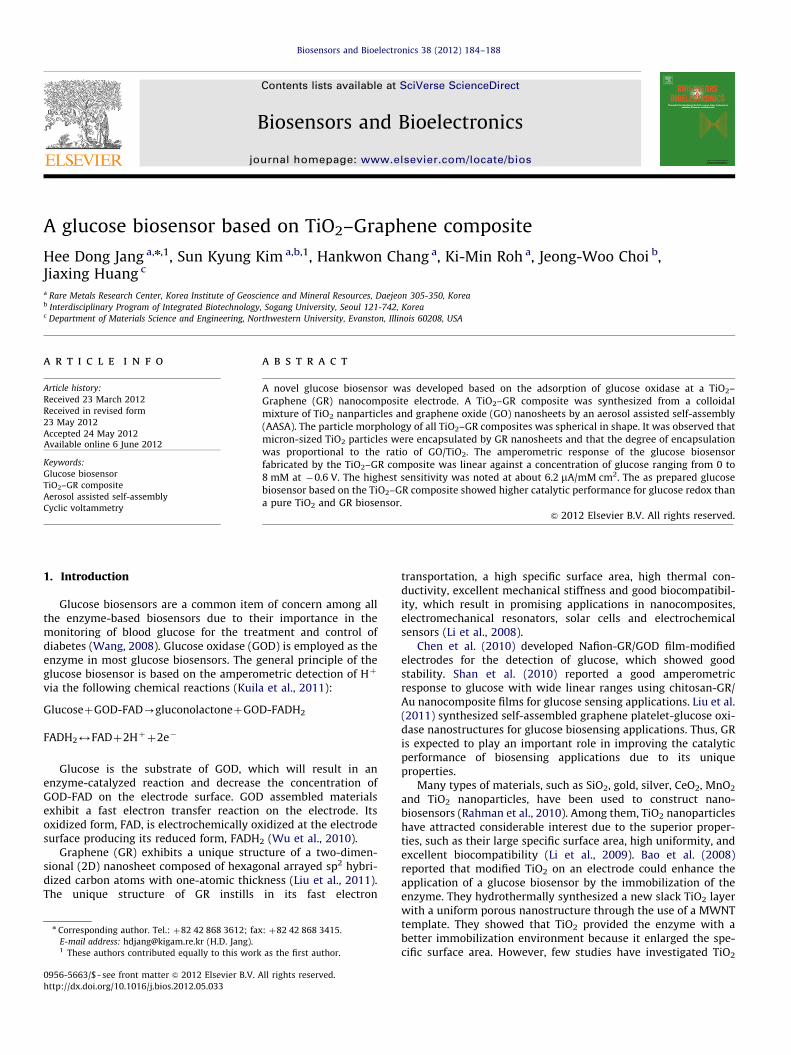
Fig. 4. Cyclic voltammograms of glucose biosensors prepared with GR, TiO2, and
TiO2–GR composite, respectively (glucose conc.: 2 mM, potential range: �1.0–
1.0 V, scan rate: 50 mV/s, (a) GO: 0.1 wt%, (b) TiO2: 0.1 wt%, (c) TiO2–GR
composite: 0.05 wt% of GO/TiO2 (TiO2: 0.1 wt%)).
Fig. 5. Cyclic voltammograms of the glucose biosensor based on TiO2–GR composite
H.D. Jang et al. / Biosensors and Bioelectronics 38 (2012) 184–188 187
and become positioned inside of the GR ball during the evapora-tion process. This explains why TiO2 particles encapsulated by GRcan be generated via the AASA technique. As the ratio of GO/TiO2
decreased from 2.0 to 0.05, a decrease in the degree of coverage ofGR to TiO2 was clearly observed.
The diffraction patterns of the TiO2–GR composite according tothe XRD analysis show the anatase and rutile phase of TiO2 only.Although the GR contents in the composite varied, the intensity ofthe TiO2 phases did not change because the intensity of the GRphase was much lower than that of TiO2 (Fig. S1). As the ratio ofGO/TiO2 increased, the specific surface area of the TiO2–GRcomposite increased from 50 to 180 m2/g because the uniquestructure of GR has a high specific surface area (Table S1).
Fig. 3 shows the X-ray photoemission spectroscopy (XPS)spectra of TiO2–GR composites. The C, Ti and O peaks weremeasured at 285, 480 and 533 eV, respectively. As the GO/TiO2
ratio decreased, the intensity of C became lower whereas that ofTi and O became higher. This occurred because TiO2 was exposedat a lower concentration of GR.
3.2. Evaluation of the electrocatalytic activity of glucose biosensors
Cyclic voltammetry was employed to measure the electroca-talytic activity of glucose biosensor for TiO2–GR composite. Thismethod is a fast and convenient tool for characterizing glucosebiosensor. In Fig. 4, characterizations of glucose biosensors pre-pared with GR, TiO2, and the TiO2–GR composite were done with2 mM of glucose solution in each case. Among the biosensorsbased on the three samples, the biosensor prepared with theTiO2–GR composite showed the highest current flow as well asclear oxidation, with reduced peaks at �0.5 V and �0.6 V. Incontrast, the current flow of the other samples did not show anobvious redox peak, which indicated that these electrodes werenot electrochemically active. This is clear evidence that anelectrochemical reaction occur on the TiO2–GR composite. Itshows that the TiO2–GR composite demonstrates the superiorability of its enzyme, which results in the fast detection of glucosein terms of an electrochemical reaction.
Fig. 5 shows the cyclic voltammetry result of a glucosebiosensor based on the TiO2–GR composite at different weightratios of GO/TiO2 of 0.5, 0.1, and 0.05. Cyclic voltammetry of theTiO2–GR composite shows a strong dependence on the composite
Fig. 3. X-ray photoelectron spectra of TiO2–GR composite prepared at different
GO/TiO2 weight ratio (a) 0.5, (b) 0.1, (c) 0.5, and (d) pure GO 0.2 wt% at the
experimental condition of Fig. 2 (TiO2: 0.1 wt%, temp.: 800 1C, carrier gas flowrate:
1 l/min).
prepared at different weight ratios of GO/TiO2 (a) 0.5, (b) 0.1, (c) 0.05 (TiO2: 0.1 wt%,
glucose conc.: 2 mM, potential range: �1.0–1.0 V, scan rate: 50 mV/s).
morphology. The current flow of the biosensor prepared at alower ratio of GR/TiO2 was stronger than that of a composite at ahigher ratio of GR/TiO2. In other words, the composite coveredwith less GR showed higher electrochemical activity, whereasthick layers of crumpled GR can obstruct the electron transfer. Itindicates that TiO2–GR composite having large surface to volumeratio and high conductivity leads to enhanced enzyme absorptionand promotes direct electron transfer between redox enzyme andthe surface of electrode (Xu et al., 2010).
The stability of the glucose biosensor was measured withrespect to the number of scanned cycle at 2–8 mM glucosesolution using the TiO2–GR–GOD electrode. The cyclic voltamme-try by the as prepared TiO2–GR–GOD electrode showed thatresponse decrease in current was about 5.6, 5.4, 5.8% after theelectrode was scanned continuously for 100th cycles at 2, 5, 8 mMglucose solution, respectively (Fig. S2). Therefore, we confirmedthat the TiO2–GR–GOD electrode was stable and repeatablebecause the CV curves showed quite stable changed for 100thcycles.
Selectivity of the as-prepared TiO2–GR glucose biosensor wasinvestigated in the presence of plasma samples such as ascorbic

Fig. 6. Amperometric responses of the glucose biosensor based on TiO2–GR
composite prepared at different weight ratios of GO/TiO2 (b) 0.5, (d) 0.1,
(a) pure TiO2 0.1 wt%, and (c) pure GO 0.2 wt% (potential: �0.6 V, scan rate:
50 mV/s, error bar¼7standard deviation).
H.D. Jang et al. / Biosensors and Bioelectronics 38 (2012) 184–188188
acid (AA) and uric acid (UA). We prepared 1 mM AA and UAsolution, respectively. As those are minutely added into 20 ml PBSsolution, there was no change in current after adding 1 ml of AAor UA, respectively. These results show that the as-prepared TiO2–GR glucose biosensor has the good selectivity for glucose.
The amperometric response by the glucose biosensor based onthe TiO2–GR composite was linear against the concentration ofglucose ranging from 0 to 8 mM at �0.6 V (Fig. 6). The relativestandard deviation (RSD) of the current response to 8 mM glucosewas 5.6% for 4 measurements, which assures stability of the TiO2–GR biosensor. The sensitivity of the TiO2–GR composite wasdetermined by measuring the slope between glucose concentra-tion and current. The highest sensitivity of the glucose biosensorbased on the TiO2–GR composite was about 6.2 mA/mM cm2 whenthe composite was prepared at 0.1 of GR/TiO2. It is well knownthat the glucose concentration for a normal person ranges from4.4 to 6.6 mM (Kuila et al., 2011). This indicates that the as-prepared TiO2–GR glucose biosensor is suitable for detecting thehuman blood sugar concentration for the diagnosis of diabetesmellitus. The response current of the glucose biosensor based onTiO2–GR was larger than those of the biosensors based on TiO2
and GR. This suggests a synergistic effect of the electrocatalyticreaction for a glucose biosensor when TiO2 nanoparticles areemployed with GR as electrode materials due to the combinationof the unique properties of GR and TiO2.
When we compared the electrochemical properties of theglucose biosensor based on TiO2–GR composite to those ofpreviously reported sensors, our sensor shows the highest sensi-tivity and stability in the absence of novel metals (Table S2).
4. Conclusions
A spherical TiO2–Graphene composite was successfully pre-pared by rapidly evaporating droplets of a colloidal dispersion ofGO nanosheets and TiO2 nanoparticles via the aerosol assisted
self-assembly method. The TiO2–GR composite showed bettercatalytic performance for glucose redox than a GR or a TiO2
electrocatalyst. In particular, the current flow of the compositewhich included the porous TiO2 agglomerate partially coveredwith GR was stronger than that of the composite including theporous TiO2 agglomerate fully covered with GR. This demon-strates the synergistic effect of using TiO2 nanoparticles with GRas the electrode materials for biosensors. Therefore, the TiO2–GRcomposite shows promise as an enhanced glucose biosensor.
Acknowledgment
This research was supported by the General Research Projectof the Korea Institute of Geoscience and Mineral Resources(KIGAM) funded by the Ministry of Knowledge Economy.
Appendix A. Supplementary material
Supplementary data associated with this article can be foundin the online version at http://dx.doi.org/10.1016/j.bios.2012.05.033.
References
Bao, S.J., Li, C.M., Zang, J.F., Cui, X.Q., Qiao, Y., Guo, J., 2008. Advanced FunctionalMaterials 18, 591–599.
Chen, X., Ye, H., Wang, W., Qiu, B., Lin, Z., Chen, G., 2010. Electroanalysis 22,2347–2352.
Cote, L.J., Silva, R.C., Huang, J., 2009a. Journal of the American Chemical Society131, 11027–11032.
Cote, L.J., Kim, F., Huang, J., 2009b. Journal of the American Chemical Society 131,1043–1049.
Cote, L.J., Kim, J., Tung, V.C., Luo, J.Y., Kim, F., Huang, J., 2011. Pure and AppliedChemistry 83, 95–110.
Hummers, W.S., Offeman, R.E., 1958. Journal of the American Chemical Society 80,1339.
Ishikawa, M., Kamiya, S., Yoshimoto, S., Suzuki, M., Kuwahara, D., Sasaki, N., Miura,K., 2010. Journal of Nanomaterials 2010, 1–6.
Jang, H.D., Chang, H., Cho, K., Kim, S.J., Park, J.H., Choi, J.W., Okuyama, K., 2008.Ultramicroscopy 108, 1241–1245.
Jang, H.D., Chang, H., Cho, K., Kim, F., Sohn, K., Huang, J., 2010. Aerosol Science andTechnology 44, 1140–1145.
Kim, F., Luo, J., Silva, R.C., Cote, L.J., Sohn, K., Huang, J., 2010a. Advanced FunctionalMaterials 20, 2867–2873.
Kim, J., Cote, L.J., Kim, F., Yuan, W., Shull, K.R., Huang, J., 2010b. Journal of theAmerican Chemical Society 132, 8180–8186.
Kuila, T., Bose, S., Khanra, P., Mishra, A.K., Kim, N.H., Lee, J.H., 2011. Biosensors andbioelectronics 26, 4637–4648.
Li, D., Muller, B., Gilje, S., Kaner, R.B., Wallace, G.G., 2008. Nature Nanotechnology3, 101–105.
Li, Y., Liu, X., Yuan, H., Xiao, D., 2009. Biosensors and bioelectronics 24, 3706–3710.Liu, S., Tian, J., Wang, L., Luo, Y., Lu, W., Sun, X., 2011. Biosensors and bioelectronics
26, 4491–4496.Luo, J., Jang, H.D., Sun, T., Xiao, L., He, Z., Katsoulidis, A.P., Kanatzidis, M.G., Gibson,
J.M., Huang, J., 2011. ACS Nano 5, 8943–8949.Rahman, M.M., Ahammad, A.J.S., Jin, J.H., Ahn, S.J., Lee, J.J., 2010. Sensors 10,
4855–4886.Shan, C., Yang, H., Han, D., Zhang, Q., Ivaska, A., Niu, L., 2010. Biosensors and
bioelectronics 25, 1070–1074.Wang, J., 2008. Chemical Reviews 108, 814–825.Wu, P., Shao, Q., Hu, Y., Jin, J., Yin, Y., Zhang, H., Cai, C., 2010. Electrochimica acta
55, 8606–8614.Xu, H., Dai, H., Chen, G., 2010. Talanta 81, 334–338.Yang, D., Velamakanni, A., Bozoklu, G., Park, S., Stoller, M., Piner, R.D., Stankovich,
S., Jung, I., Field, D.A., Ventrice Jr., C.A., Ruoff, R.S., 2009. Carbon 47, 145–152.