a genomic analysis of paenibacillus macerans atcc … · a genomic analysis of paenibacillus...
TRANSCRIPT
A GENOMIC ANALYSIS OF Paenibacillus
macerans ATCC 8244, A GRAM POSITIVE
NITROGEN FIXING BACTERIUM
AHMAD YAMIN BIN ABDUL RAHMAN
UNIVERSITI SAINS MALAYSIA
2016

A GENOMIC ANALYSIS OF Paenibacillus
macerans ATCC 8244, A GRAM POSITIVE
NITROGEN FIXING BACTERIUM
by
AHMAD YAMIN BIN ABDUL RAHMAN
Thesis submitted in fulfilment of the requirements
for the degree of
Master of Science
September 2016

ii
ACKNOWLEDGEMENT
First and foremost, Alhamdulillah, all praise be to Allah who with His
compassion enabled me to finish my thesis. This thesis not only represents scientific
findings of Paenibacillus macerans through a genomics and bioinformatics point of
view but it is also a proof of that a simple hobby/interest can actually drive scientific
research . More importantly, this thesis is my second chance to put my research life
back on track. I was at a juncture of turbulence to choose between having a career or
to further studies when Prof Nazalan Najimudin offered me the golden opportunity
to be under his supervision. Honestly, I am very grateful for his compassion,
flexibility, perseverance and encouragement to mold me from an average computer
enthusiast to a scientist with a more constructive direction in the field of
bioinformatics.
My deepest thanks to Dr Rashidah Abdul Rahim, my ex-supervisior during
my bachelor’s degree studies who provided me the first step to delve into a
bioinformatics-related field. Along the way, I met several key persons who changed
my life and they included Prof Nazalan, Prof Razip Samian, and Prof Maqsudul
Alam. Without the exposure and the motivational push they gave, I would not even
know what the terms “scaffold”, “contig” or “annotation” were all about.

iii
My ultimate thanks goes to my mother, Hafsah binti Yaakub, who has always
been encouraging and supportive of my studying further, keeping me sane during my
busy days, and constantly reminding me to write my thesis. Not forgetting my family
who gave me relentless support emotionally, I must declare my gratitude to all of
them. Last but not least, my friends, Kee Shin, Mardani and Haida.

iv
TABLE OF CONTENTS
ACKNOWLEDGEMENT ......................................................................................... ii
TABLE OF CONTENTS ......................................................................................... iv
LIST OF TABLES ................................................................................................... vii
LIST OF FIGURES ................................................................................................ viii
LIST OF ABBREVIATIONS ................................................................................... x
ABSTRAK ................................................................................................................ xii
ABSTRACT ............................................................................................................. xiii
CHAPTER 1: INTRODUCTION ............................................................................. 1
1.1 Research Objectives: ..................................................................................... 3
CHAPTER 2: LITERATURE REVIEW ................................................................. 4
2.1 Paenibacillus genus ....................................................................................... 4
2.2 Paenibacillus macerans ................................................................................ 5
2.3 Nitrogen Fixing Microorganisms .................................................................. 5
2.4 Importance Of Nitrogen ................................................................................ 6
2.5 Nitrogen Cycle ............................................................................................. 7
2.6 Nitrogen Fixation Key Enzymes .................................................................. 9
2.6.1 MoFe protein ................................................................................. 9
2.6.2 Fe Protein ...................................................................................... 9
2.7 The Chemical Equation and Stochoimetry of Nitrogen Fixation ................ 10
2.8 Regulation Of Nitrogen Fixation ................................................................ 10
2.9 Azotobacter vinelandii as a Model of Nitrogen Fixing Organism ............. 11
2.9.1 Genes Involved In Nitrogen Fixation of A. vinelandii. .................... 12
2.10 Bioinformatics as a Fusion of Genomics and Computing .......................... 14
2.11 Next Generation Sequencing and Bioinformatics ..................................... 15
2.11.1 Illumina Sequencing ................................................................... 15
2.11.2 Roche 454 Pyro Sequencing, ...................................................... 16
2.11.3 Ion Torrent Sequencing .............................................................. 16
2.11.4 Pac Bio SMRT Sequencing ........................................................ 17
2.12 Genome Assembly ..................................................................................... 17
2.13 Gene Finding .............................................................................................. 18

v
2.14 Post Annotation; Analysis, Comparative and Evolutionary Studies .......... 19
CHAPTER 3: MATERIALS AND METHODS ................................................... 21
3.1 Schematic Workflow Of This Study .......................................................... 21
3.2 Bioinformatics Tools For Assembly, Annotation And Analysis ............... 21
3.3 Genomic DNA isolation ............................................................................. 23
3.4 Raw Sequence Pre-Processing ................................................................... 25
3.5 Insert Size Estimation ................................................................................. 27
3.6 Assembler Benchmarks .............................................................................. 27
3.7 Assembly Optimisation, Scaffolding And Gap Filling .............................. 28
3.7.1 Kmer Optimisation ........................................................................ 28
3.7.2 Scaffolding .................................................................................... 28
3.7.3 Gap filling ...................................................................................... 29
3.8 Genome Annotation ................................................................................... 29
3.9 Metabolic Pathway reconstruction ............................................................. 30
3.10 Phylogenomics analysis ............................................................................. 30
3.11 Pan Genome analysis ................................................................................. 31
3.12 Genomic Island Analysis ........................................................................... 31
3.13 CRISPR Identification ................................................................................ 32
CHAPTER 4: RESULTS ........................................................................................ 33
4.1 Quality Control Processing of Raw Data .................................................... 33
4.2 Empirical estimation of insert size .............................................................. 33
4.3 Performance of Genome Assembler Software on Assembly Data ............. 37
4.4 Assembly Optimisation, Scaffolding And Gap Filling ............................... 37
4.4.1 Assessment To Get The Best K Value For K-Mer Selection ......... 38
4.5 Final assembly ............................................................................................. 40
4.6 General Features of the P. macerans ATCC 8244 Draft Genome ............... 41
4.7 SEED Classification .................................................................................... 41
4.8 Identification of Nitrogen Fixation and Associated Genes ......................... 44
4.9 Phylogenomic Analysis .............................................................................. 45
4.10 Pan Genomic Analysis ................................................................................ 50
4.11 Metabolic Pathway Reconstruction ............................................................. 53
4.12 Genomic Island Analysis ............................................................................ 58
4.13 Clustered Regularly Interspaced Short Palindromic Repeats (CRISPR)
Detection ................................................................................................................ 58
vi
CHAPTER 5: DISCUSSION .................................................................................. 61
5.1 Nitrogen fixation operon in Paenibacillus macerans ATCC 8244 ............. 61
5.2 Bisphenol A (BPA) Degradation ................................................................ 63
5.3 Biofuel Potential ......................................................................................... 64
5.4 Genome-based Taxonomic Analysis .......................................................... 66
5.5 Evolution of Nitrogenase ........................................................................... 68
CHAPTER 6: CONCLUSION ................................................................................ 70
REFERENCES ............................................................................................................ .
APPENDIX A – Script:Estimate insert size Script .................................................. .
APPENDIX B – Gene Annotation Parameters ........................................................ .
APPENDIX C- Venn Diagram Count Script ........................................................... .
APPENDIX D – Multi Contigs Into Singular Contig Script ................................... .

vii
LIST OF TABLES
Page
Table 2.1 Nitrogen fixation (nif) genes related products and functions 13
Table 3.1 List of freewares used for the overall process of Assembly,
Annotation and Analysis of P. macerans ATCC 8244 genome 23
Table 4.1 Summary of raw fastq reads quality of P. macerans ATCC
8244 34
Table 4.2 Summary of statistics before and after the quality control
processing on fastq raw reads of P. macerans ATCC 8244 35
Table 4.3 Performance of different Assembler softwares 37
Table 4.4 Kmer values selection and their assembly statistics 38
Table 4.5 Statistics of assembly optimization before and after k-mer 61
Implementation 39
Table 4.6 Final Assembly statistic of P. macerans ATCC 8244 genome 40
Table 4.7 Clusters of Orthologous Groups (COG) function
classification of P. macerans ATCC 8244 genome 42
Table 4.8 Nif and nitrogen fixation associated genes product found in
of P. macerans ATCC 8244 genome 44
Table 4.9 List of pathways available in Paenibacillus macerans
ATCC 8244 54

viii
LIST OF FIGURES
Page
Figure 2.1 Nitrogen Cycle 8
Figure 3.1 Schematic workflow overview of overall process of
genomic assembly, annotation and analysis of
P. macerans ATCC 8244 22
Figure 4.1 Histogram of Inner Distance between Paired Ends reads
of P. macerans ATCC 8244. 36
Figure 4.2 SEED classification of Paenibacillus macerans 43
Figure 4.3 Best model for maximum likelihood approximation fit for
final tree phylogenomic test 46
Figure 4.4 Phylogenomics tree of selected members of
Paenibacillus genus 48
Figure 4.5 Nitrogenase phylogenetic tree 49
Figure 4.6 A Venn diagram showing gene clusters of
Paenibacillus sp. JDR1, P. macerans ATCC 8244
P. mucilaginosus 3016 P. polymyxa CR1 51
Figure 4.7 A Venn diagram showing gene clusters of Azorhizobium
caulinodans ORS 57 , Azotobacter vinelandii DJ
P. macerans and Klebsiella pneumoniae SB3432 52
Figure 4.8 Comparative Bisphenol A Degradation Pathway 56
ix
Figure 4.9 Pathways of nitrogen metabolism in the four selected
nitrogen fixing bacteria 57
Figure 4.10 Prediction of Genomic islands in the genome of
P.macerans ATCC 8244 59
Figure 4.11 Side by side comparison deduction of phage induced
genomic island of P. macerans ATCC 8244
60
Figure 5.1 Formation of 2,3-butanediol from pyruvate 65

x
LIST OF ABBREVIATIONS
% Percentage
ºC degree Celsius
Σ sigma factor
Λ lambda clone
Μg micro gram
Μl micro litre
μM micro molar
Aa amino acid
acc. no. accession number
Atm standard atmospheric pressure
ADP adenosine diphosphate
ATP adenosine triphosphate
bp
BPA
base pair
Bisphenol A
CDS coding sequences
CRISPR clustered regularly interspaced short palindromic
repeats
DNA deoxyribonucleic acid
dNTP deoxynucleotide triphosphates
DTT Dithiothereitol
G Gram
xi
IPTG isopropyl β-thiogalactopyranoside
Kb kilo base pair
kDa kilo Dalton
M Molar
Min Minute
Mm Millimolar
OD optical density
ORF open reading frame
RACE rapid amplification of cDNA ends
PCR polymerase chain reaction
RBS ribosome binding site
RNA ribonucleic acid
RT reverse transcription
Rpm revolution per minute
QC quality control
Sec Second

xii
KAJIAN GENOMIK Paenibacillus macerans ATCC 8244, BAKTERIA GRAM
POSITIF YANG MENGIKAT NITROGEN
ABSTRAK
Paenibacillus macerans ATCC 8244 adalah bakteria pembentuk spora jenis
Gram-positif yang berkemungkinan mempunyai kebolehan metabolik yang paling
luas antara ahli genus Paenibacillus. P. macerans boleh melakukan fermentasi gula
heksosa, pentosa, selulosa dan hemiselulosa. Di samping itu, keupayaan untuk
mengikat nitrogen mewujudkan potensi agronomi. Kajian ini mengemukakan satu
pendekatan genomik menyeluruh untuk pencirian mikroorganisma ini. Analisis
filogenomik menunjukkan bahawa genom terdekat yang dikenalpasti adalah P.
macerans strain ZY18 dengan identiti 98 peratus. Perhimpunan draf genom
menghasilkan sejumlah 7.305.296 pasangan bes (bp) yang dibentuk oleh 97
perancah. Nilai N50 bagi perancah adalah 157,355 bp. Saiz purata perancah adalah
75,312 bp dan nisbah GC purata genom draf adalah 53.14%. Ramalan dan anotasi
gen yang dibuat pada peringkat perancah menghasilkan 6953 jujukan pengekodan.
Ini termasuk 85 gen tRNA dan 9 gen rRNA. Analisis klasifikasi Kelompok Gen
Berortolog (COGs) menunjukkan bahawa bilangan tertinggi jujukan pengekodan
melibatkan kategori "Pengangkutan dan Metabolisme Karbohidrat " dengan jumlah
845 model gen. Draf genom P. macerans ATCC 8244 mendedahkan 15 gen yang
terlibat dalam pengikatan nitrogen. Kehadiran gen yang terlibat dalam degradasi
Bisphenol A (BPA) menunjukkan potensi dalam mencerna bahan kimia yang
berbahaya kepada alam sekitar ini. Di samping itu, ia mempunyai keupayaan untuk
menghasilkan 2-3 butanediol menjadikannya sebagai pengeluar biofuel yang
berprospektif.

xiii
A GENOMIC ANALYSIS OF Paenibacillus macerans ATCC 8244, A GRAM POSITIVE
NITROGEN FIXING BACTERIUM
ABSTRACT
Paenibacillus macerans ATCC 8244 is a Gram-positive, spore-forming
bacterium with possibly the widest metabolic capabilities among the Paenibacillus
genus. It is able to perform fermentation of hexoses, deoxyhexoses, pentoses,
cellulose, and hemicelluloses. In addition, its ability to fix nitrogen creates an
agronomic potential. This study presents a whole genomic approach to give a
holistic view of the features of this microorganism. A phylogenomic analysis
revealed that the nearest genome identified was Paenibacillus macerans strain
ZY18 with 98 percent identity. The draft genome assembly yielded a total of
7,305,296 base pairs (bp) formed by 97 scaffolds. The N50 value of the scaffold was
157,355 bp. The average scaffold size was 75,312 bp and the average GC ratio of the
draft genome was 53.14 %. Gene prediction and annotation performed at the scaffold
level yielded 6953 coding sequences. These included genes for 85 tRNAs and 9
rRNAs. A Clusters of Orthologous Groups (COGs) classification analysis revealed
that the highest number of coding sequences involved the “Carbohydrate Transport
and Metabolism” category with a total of 845 gene models. The draft genome of P.
macerans ATCC 8244 revealed 15 genes that are involved in nitrogen fixation. The
presence of genes involved in Bisphenol A (BPA) degradation showed a potential in
degrading this environmentally hazardous chemical. In addition, it has the capability
to produce 2-3 butanediol making it as a prospective producer of biofuel.
1
CHAPTER 1: INTRODUCTION
Following the first isolation of Bacillus subtilis in 1872, a lot of bacteria were
classified under the genus of Bacillus since they have common characteristics such
as rod-shaped, endospore forming, and possessing either aerobic or facultative
anaerobic lifestyle. Members of the genus Paenibacillus were originally classified as
Bacillus as well due to their similar characteristics. The word ‘paene’ means “nearly
or almost” in Latin and hence “Paenibacillus” means “almost bacillus” (De Vos
2009). Members of the genus Paenibacillus are usually curved or straight rod in
shape, ranging from 0.5-1.0 x 2-6 μm in size and with G+C content ranging around
39 to 59 percent. They are Gram-positive and have been found in many
environments. They can be also belong to any of the different classes of bacteria
such as psychrophile, mesophile, thermophile, alkaliphile, neutrophile, aerobic or
anaerobic.
The variety of adaptations of many members of this genus allows them to
play unique and important roles in diverse environments. For example, Paenibacillus
larvae became a pathogen of the cultured honey bee Apis mellifera, especially the
first and second instar larvae (Genersch 2010). Others such as Paenibacillus sp.
Aloe-11 reportedly invade human intestine (Li et al. 2012). However, members of
this genus are also famous for their association with plant communities (McSpadden
Gardener 2004). They can act as plant growth enhancers with features that make
them biotechnologically beneficial. Furthermore their ability to adapt in the human
intestinal environment could potentially make them as beneficial probiotic
components (Hoyles 2012).

2
An interesting species under this genus is Paenibacillus macerans. It can be
considered as one with the broadest metabolic capabilities. P. macerans N234A has
been known to anaerobically ferment glycerol (Gupta et al. 2009). It is also able to
ferment hexoses, deoxyhexoses, pentoses, cellulose, and hemicelluloses. Since it has
been reported to be associated with several pseudo bacteriamia cases, P. macerans
has been described as having features of opportunistic pathogen (Noskin et al. 2001).
More notably, P. macerans has the ability to fix nitrogen that is very beneficial
towards crops productivity. Biological nitrogen fixers accounted for supplying
nearly 60 % of world’s new ammonia source annually (Schlesinger, 1991). It is vital
to harness research understanding on biological nitrogen fixation to maximize its
potential, especially on the Gram positive bacteria. Although there is tremendous
research effort done on the Gram-negative nitrogen fixers such as Klebsiella,
Bradyrhizobium and Azotobacter species, there is less information on Gram-positive
members such as P. macerans.
The field of life sciences has been tremendously transformed with the advent
of genome sequencing. The ability to sequence in a high throughput fashion has not
only advanced our fundamental understanding of how genes and genomes are
assembled, it also has yielded extremely in depth knowledge of the structure of
evolutionary trees, increased our understanding of genetics and development, and led
to the growth of new biotechnologies. The genomic information obtained enabled us
to understand the strategies of the microbes to survive under diverse environmental
conditions.

3
A genomic study on P. macerans would uncover the biological mechanisms
and functions of the interacting genes responsible for its special physiological
features and adaptation capabilities. Currently there is no P. macerans complete
genome available in the public database using next generation sequencing approach.
This study aims to study the P. macerans genome using whole genome approach.
1.1 Research Objectives:
The investigation was carried out to fulfill the following objectives:
1. To analyze the structure of a P. macerans draft genome
2. To characterize the genes related to nitrogen fixation and unique
metabolic pathways belongs to P. macerans
3. To perform a functional and comparative analysis of P. macerans
genome with other species

4
CHAPTER 2: LITERATURE REVIEW
2.1 Paenibacillus genus
Members of the bacterial genus Paenibacillus are usually curved or straight
rod in shape, ranging from 0.5-1.0 x 2-6 μm in size with a G+C content ranging
around 39 to 59 percent (De Vos et al. 2010). They are Gram-positive and have been
found in many environments. The varieties of niches that many members of this
genus reside in reflect their adaptability. Various psychrophilic, mesophilic and
thermophilic representatives have been successfully isolated from a range of
different niches reflecting how diverse they are in terms of growth temperatures.
Isolates which are alkaliphilic or acidophilic play unique roles in their diverse
environments. Paenibacillus members capable of growing under aerobic or
anaerobic have also been successfully isolated.
Some species are pathogenic to animals and their pathogenicity towards
certain pests means that they can serve as effective biological pesticides. For
example, P. larvae is pathogenic to bees and have been proposed to be used as a
biological control of wild honey bees (Genersch 2010). Another species,
Paenibacillus sp HGF -5, even invade the human intestine and this ability to adapt in
the intestinal environment could be taken advantage of by moulding it into a
beneficial probiotic component in human (Hoyles et al. 2012). There are also
members of this genus well known for their association with the plant communities
(McSpadden Gardener 2004; Khan et al. 2008). Their ability to produce plant growth
enhancers makes them biotechnologically beneficial.

5
2.2 Paenibacillus macerans
Paenibacillus macerans, previously called Bacillus macerans and Bacillus
acetoethylicum, is a Gram-positive, spore-forming bacterium belonging to the genus
Paenibacillus (Ash et al. 1993). P. macerans can be considered as a member species
with the broadest metabolic capability within this genera. It was reported that P.
macerans N234A able to ferment hexoses, deoxyhexoses, pentoses, cellulose, and
hemicelluloses. It has also been known anaerobically ferment glycerol (Gupta et al.
2009). In addition, P. macerans also has features of an opportunistic pathogen with
reported associations to several pseudo-bacteraemial cases (Noskin et al. 2001).
Paenibacillus macerans ATCC 8244 was first isolated from potatoes by Schardinger
in 1905 and was mentioned in 1942 by (Tilden and Hudson 1942).
2.3 Nitrogen Fixing Microorganisms
Biological nitrogen fixers are microorganisms that are capable of converting
the gaseous form of nitrogen into ammonia. These microorganisms are either
bacteria or archaea and they can be in a symbiotic relationship or free living.
Biological Nitrogen Fixation (BNF) accounted for 60 % supply of the world’s new
source of ammonia annually (Schlesinger 1991). The role of nitrogen fixation is
clearly important in the field of agriculture. Availability of fixed nitrogen correlates
proportionally with crops productivity. It is therefore vital to harness a deeper
understanding on BNF to maximize its potential.
Most of the studies on biological nitrogen fixation were performed on the
Gram-negative bacteria such as Klebsiella, Bradyrhizobium and Azotobacter. For the
Gram-positive group, studies had been done on Clostridium pasteurinum but its
6
difficulty to grow and lack of genetic tractability prevented rapid advancement of its
research (Chen and Johnson 1993). Thus a suitable model is needed to dissect the
nitrogen fixation system in the Gram-positive group of bacteria. P. macerans offers a
possible model due to its ease of growth in aerobic condition and the availability of
genetic amenability such as transposons and plasmids.
2.4 Importance Of Nitrogen
Nitrogen constitutes 78 percent of the earth’s atmosphere. It is an essential
component for composition of proteins that are required for all living things
including bacteria, plants and humans (Stewart and Gallon 1980; Clark et al. 1981).
Nitrogen can exist both in organic and inorganic forms. The organic form can be
ammonium (NH4+), nitrite (NO2
-), nitrate (NO3), nitrous oxide (N2O) and nitric
oxide (NO). The inorganic nitrogen form would be in the inert nitrogen gas (N2)
itself. Nitrogen serve as a component for the building units that form nucleic acids
(such as DNA and RNA) as well as for the amino acids that form proteins.
Unfortunately, the actual nitrogen gas cannot be utilized directly by most of
organisms; it needs to undergo a series of processes that convert it to an organic
form. It needs to go through a fixation process to become utilizable. Circulation of
nitrogen from the atmosphere to organic compounds by biological nitrogen fixation
and then back to the atmosphere (denitrification) is called the “nitrogen cycle”.
Nitrogen cycle affects the major rate of growth and decay of many ecosystems
through primary production and decomposition (Fowler et al. 2013; Isobe and Ohte
2014).
7
2.5 Nitrogen Cycle
The cycle supply chain starts with the nitrogen fixation process in which
nitrogen is converted to ammonia (Mancinelli 1996). This fixation process is very
energy intensive and can happen in multiple ways: (i) by lightning strikes, (ii)
through industrial synthesis by humans (for fertilizers), or (iii) by biological nitrogen
fixers. Biological nitrogen fixers are microorganisms that are symbiotic (examples:
Frankia sp., Rhizobium sp., Bradyrhizobium sp.) or free living (such as Azotobacter
sp.). The ammonia produced is then assimilated by majority of plants and others
through the glutamate synthase cycle (Miflin and Lea 1975). When the organisms
die, diverse bacteria and fungi can convert the decay products of the body into
ammonium. Bacteria such as Nitrosomonas sp. and Nitrobacter sp. are able to
assimilate and convert the inorganic ammonium into nitrite and nitrate in a 2-step
process called nitrification. The nitrate produced is a source of energy for groups of
bacteria such as Pseudomonas sp. in anaerobic respiration. They use nitrate as an
electron acceptor in place of oxygen and this will convert back nitrogen into its inert
gas form through a process called denitrification. This completes the nitrogen cycle.
Figure 2.1 shows overview process of nitrogen cycle. It has been noted that human
influence on the nitrogen cycle through over-usage of fertilizers has caused
imbalance leading to environmental problems such as acidification and
eutrophication. These effects on the environment can be reduced if more effort is
given to optimize biological nitrogen fixation instead of relying of industrially
produced chemical fertilizers.

8
Key processes in nitrogen cycle involves:
a) Fixation -the process of N2 being converted into NH3/NH4+/ NO3-,
b) Nitrification- the process of NH3 being oxidized to NO3- and NO2
-
c) Ammonification- the process of organic nitrogen are converted to NH3
d) Denitrification, the process of reduction of NO3- to N2
CONSUME
NITRIFICATION NITRIFICATION
DECOMPOSERS
ATMOSPHERIC
NITROGEN (N2)
AMMONIA(NH3) NITRITE
(NO2-)
ANIMAL PROTEIN
(CHONS)
PLANT PROTEIN
(CHONS)
NITRATE (NO3-)
Figure 2.1 Nitrogen cycle

9
2.6 Nitrogen Fixation Key Enzymes
Atmospheric nitrogen or dinitrogen (N2) is converted to biological form
through key enzyme nitrogenase (EC 1.18.6.1). This enzyme is essentially a
metalloprotein complex consisting of MoFe and Fe proteins. The Fe protein acts as
an electron donor to the catalytic site on the MoFe protein (Rees et al. 2005).
2.6.1 MoFe protein
Structurally, the size of the MoFe protein is about 200-250 kDa and it is a
tetramer formed by four subunits: 2 alpha and 2 beta subunits. The gene nifD
encodes the alpha subunit and nifK encodes the beta subunit. The MoFe protein
contains the substrate-binding site with six prosthetic groups of two types of clusters:
4 8Fe-7S and 2 Mo-7Fe-9S clusters. The former is also known as the P-cluster while
the latter is commonly known as the FeMo cofactor. It contains the substrate binding
site for a reduction process (Seefeldt et al. 2009). Both clusters act as a bridge to
conduct electron transport between the Fe-protein and MoFe protein.
2.6.2 Fe Protein
Fe-protein is about 55 to 65 kDa in size and is a dimer formed by only the
alpha subunits. It contains a 4Fe-4S metal cluster that connects the two alpha
subunits via covalent bond. The 4Fe-4S cluster in the active site modulates between
the reduced and oxidised states of the protein during the electron transfer process to
the FeMo-protein (Seefeldt et al. 2009).

10
2.7 The Chemical Equation and Stochoimetry of Nitrogen Fixation
Under normal condition, the equation is:
N2 + 8 e– + 8 H+ + 16 MgATP→ 2 NH3 + H2 + 16 MgADP + 16 Pi
The process starts by reduction of the Fe protein by ferredoxin or flavodoxin or other
electron donors (Dixon and Kahn 2004). Then, a single electron is transferred from
the Fe protein to the MoFe protein and this transfer is dependent on a hydrolysis of
an ATP molecule. Upon reaching the MoFe protein, the electron is then transferred
internally from the P cluster to the FeMo-co located at the active site.
2.8 Regulation Of Nitrogen Fixation
The nitrogen fixation activity is controlled according to the environmental
ammonium content as well as the level of oxygen. Bacteria repress transcription
when there is a high level of nitrogen or oxygen. There are two regulatory proteins
involved: NifL and NifA. In Azotobacter vinelandii, NifL is a redox- and nitrogen-
responsive regulatory flavoprotein and functions as a regulatory protein that controls
the transcription of nitrogen fixation genes by regulating the activity of the
transcriptional activator NifA. This involves a direct protein-protein interaction.
When the levels of oxygen is high, the NifL protein, in the presence of an oxidised
Flavin adenine dinucleotide (FAD), inhibits the NifA protein causing inhibition of
nif operons. When the oxygen is low, NifL disassociate from NifA enabling it to
transcribe the nif genes.
11
Similarly, a high level of ammonia would trigger repression of the nitrogen
fixation genes. Ammonia modifies the structure of glutamine synthase via covalent
adenylylation. It also acts as a corepressor that changes the regional DNA binding
site region known as nifR in the nifRLA operon. This results in the prevention of
transcription of the operon (Halbleib and Ludden 2000).
2.9 Azotobacter vinelandii as a Model of Nitrogen Fixing Organism
One of the best studied model to understand the mechanisms involving
nitrogen fixation is the Gram-negative bacterium Azotobacter vinelandii (Schmitz et
al. 2002). A. vinelandii is a free living diazotroph that is able to fix nitrogen under
aerobic condition. It has a single circular chromosome of approximately 5.37 Mbp in
size with a total number of 5051 genes (Setubal et al. 2009). Ambient oxygen
concentration is required (around 20 percent) for A. vinelandii to catalyze the
nitrogen fixation process (Curatti et al. 2005). A. vinelandii is special because it
possesses 3 different types of enzymes to perform its nitrogen fixation: molybdenum
nitrogenase, iron nitrogenase, and vanadium nitrogenase (Pau et al. 1989; Hales
1990). These are sometimes simply abbreviated to the Mo-, V- and Fe- dependant
enzymes.

12
2.9.1 Genes Involved In Nitrogen Fixation of A. vinelandii.
Inside A. vinelandii, the genes for the three oxygen sensitive nitrogenases
(Mo-, V- and Fe-dependant enzymes) are located in three separate regions (Setubal
et al. 2009). The molybdenum nitrogenase is made up of the dinitrogenase reductase
coded by the gene nifH and the second component, the MoFe protein, is coded by
nifDK. The iron and molybedum (Mo-Fe) cofactors are situated at the active site of
the MoFe-protein (Seefeldt et al. 2009). The NifH protein supplies electron to the
MoFe-protein and the latter acts as the site for nitrogen reduction. Nitrogen is
reduced at the active site to produce the final product, ammonium, along with the
release of hydrogen. This reduction process is accompanied by the hydrolysis of
ATP. Alternative nitrogenases such as the V- and Fe-nitrogenases comprised of
homologous VnfHDK and AnfHDK proteins (Hales 1990). These alternative
nitrogenases require additional structural components of the dinitrogenase that
include the VnfG and AnfG as subunits. A lot of accessory proteins participate in the
synthesis and assembly of the transition metal cofactors and these are encoded
within the nif gene clusters. Table 2.1 describes nitrogen fixation (nif) genes related
products and functions.
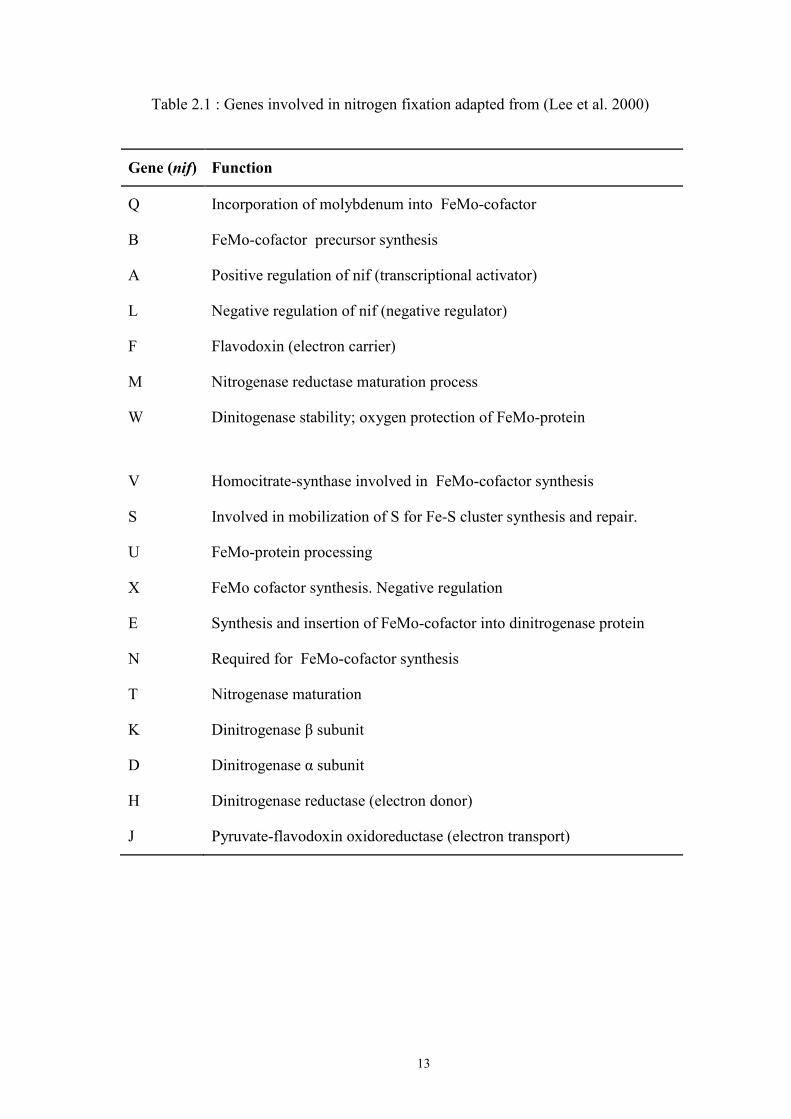
13
Table 2.1 : Genes involved in nitrogen fixation adapted from (Lee et al. 2000)
Gene (nif) Function
Q Incorporation of molybdenum into FeMo-cofactor
B FeMo-cofactor precursor synthesis
A Positive regulation of nif (transcriptional activator)
L Negative regulation of nif (negative regulator)
F Flavodoxin (electron carrier)
M Nitrogenase reductase maturation process
W Dinitogenase stability; oxygen protection of FeMo-protein
V Homocitrate-synthase involved in FeMo-cofactor synthesis
S Involved in mobilization of S for Fe-S cluster synthesis and repair.
U FeMo-protein processing
X FeMo cofactor synthesis. Negative regulation
E Synthesis and insertion of FeMo-cofactor into dinitrogenase protein
N Required for FeMo-cofactor synthesis
T Nitrogenase maturation
K Dinitrogenase β subunit
D Dinitrogenase α subunit
H Dinitrogenase reductase (electron donor)
J Pyruvate-flavodoxin oxidoreductase (electron transport)

14
2.10 Bioinformatics as a Fusion of Genomics and Computing
The nitrogenous bases "Adenine (A), Cytosine (C), Guanine (G), Thymine
(T)" are basically the four significant compounds that hold the key towards the
meaning of life. Understanding the sequential order of the bases mobilized
researchers to investigate the genetic information hidden within a biological system.
Looking at the bigger scale, study of genomics includes understanding how the
genetic and non-genetic makeup interacts and form the biological system of an
organism.
A genome can be defined as the genetic material of an organism and it is
made up from nucleic acids. It consists of coding and non-coding parts that together
hold the mechanisms on how the organism lives and maintains its existence. This
includes gene function, expression, regulation as well as its evolution. Recent
advancement in the next generation sequencing technologies has increased the
capability to mine biological information exponentially from various aspects. The
speed of sequencing and the amount of data produced allow more discoveries and
biological understanding to be made. The progress made at the level of data
resolutions (or sensitivity) itself was phenomenal. However, connecting the dots that
involve analysis and interpretation often face obstacles, especially in handling huge
amount of biological information made up of genomics sequences. With advances in
computing, most of the challenges faced can be answered through bioinformatics, a
hybrid intersection of the disciplines of Biology and Computation. Bioinformatics is
a way to use computing capabilities to systematically gather, manage, and make
connections of biological information that aids more insights into understanding life
and the scientific meaning behind it (Kelly 1989).
15
2.11 Next Generation Sequencing and Bioinformatics
Sequencing technology refers to the process of nucleotide identification in a
chain of nucleic acid polymers which can be either be referred to as DNA or RNA.
The development of DNA sequencing has opened a new window of understanding
for genomic sciences especially on genome organization and compositions among
different organisms. This has further enhanced by development of the “Next
Generation Sequencing” (NGS) platforms which make data generation grow
exponentially. This phenomenon triggered the expansion of bioinformatics as a
discipline to cope with large and complex datasets (Metzker 2005).
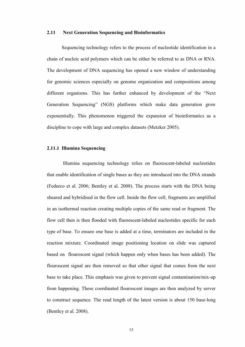
2.11.1 Illumina Sequencing
Illumina sequencing technology relies on fluorescent-labeled nucleotides
that enable identification of single bases as they are introduced into the DNA strands
(Fedurco et al. 2006; Bentley et al. 2008). The process starts with the DNA being
sheared and hybridised in the flow cell. Inside the flow cell, fragments are amplified
in an isothermal reaction creating multiple copies of the same read or fragment. The
flow cell then is then flooded with fluorescent-labeled nucleotides specific for each
type of base. To ensure one base is added at a time, terminators are included in the
reaction mixture. Coordinated image positioning location on slide was captured
based on flouroscent signal (which happen only when bases has been added). The
flouroscent signal are then removed so that other signal that comes from the next
base to take place. This emphasis was given to prevent signal contamination/mix-up
from happening. These coordinated flouroscent images are then analyzed by server
to construct sequence. The read length of the latest version is about 150 base-long
(Bentley et al. 2008).

16
2.11.2 Roche 454 Pyro Sequencing,
The principle of the Roche 454 sequencing technology involves ligating
nebulized fragments of DNA with adaptors (Ronaghi et al. 1996; Ronaghi et al.
1998). These ligated DNA molecules are then captured into beads enveloped in oil-
water emulsions. The fragments on the beads were amplified using PCR. The beads
containing the amplified DNA bound to them are then placed on a Picotiter plate. An
enzymatic combination of DNA polymerase, luciferase and ATP sulfurylase are also
packed into each well of the plate. The plate are placed into the sequencing machine
called GS FLX where light peaks are produced in a “pyrogram format” when
nucleotides are added. These light peaks are generated as a result of a reaction
involving luciferase and ATP. This technology can give read length of up to 1
kilobases (Droege and Hill 2008).
2.11.3 Ion Torrent Sequencing
The principle of the Ion Torrent technology is different from Illumina and
Roche 454 in a sense that it does not use optical signal to identify the bases. Instead
it relies on pH values. The primary principle relies on the fact that addition of a
dNTP molecule to a DNA fragment will involve the release of H+ ion (Rothberg et al.
2011). The process starts by flooding the slide with a single type of dNTP together
with buffers and polymerase and one NTP at a time. Each H+ ion released in each
well is measured as a decrease of pH value. The values of the pH changes allow the
machine to determine the profile of the base added. The sequences of changes are
progressively read to develop the sequencing reads. The average read of this method
is about 200 bases.

17
2.11.4 Pac Bio SMRT Sequencing
Pacific Biosciences (PacBio) developed a single molecule real time (SMRT)
sequencing method that uses parallel sequencing of single DNA molecules by
synthesis (Eid et al. 2009). It uses zero –mode waveguide (ZMW) which guides light
energy into a volume that is compact in all dimension compared to the wavelength of
the light (Levene et al. 2003). A single molecule of DNA polymerase with a single
molecule of DNA template was affixed at bottom of the ZMW. The ZMW that was
equipped with high-resolution camera records the activity of the fluorescence signal
of incorporation of nucleotide in a movie-sequential format. This method is capable
of generating reads maximum length over 40000bp with average of 14000bp.
2.12 Genome Assembly
The shotgun sequencing route was first introduced on the DNA sequencing
of phiX174 bacteriophage genome and it still remains an important technique for
genome sequence assembly (Sanger et al. 1977a; Sanger et al. 1977b). Shotgun
sequencing method involves obtaining random sequence reads from a genome
(which may ranging from a small plasmid to a complete chromosome of an organism)
and merging them together into contigs (a linear representation of nucleotides) using
overlaps as the basis of contigs construction. Paired end reads are then used to link
together distinct sequence contigs into super contigs using forward and reverse end
reads. Contigs that are joined together are called supercontigs, and a combination of
supercontigs becomes scaffold. Assemblies are measured by the size and accuracy of
their contigs and scaffolds. Assessement of assemblies usually consider contig
accuracy and size. N50 refers to contig size that sum of sorted length of 50 percent
of total assembly is reached.
18
Basically there are two standard methods of genome assembly, namely, the
Overlapping Consensus Layout (OCL) method and the De Bruijn method (Miller et
al. 2010). The OCL method usually works well with long reads. It relies on
analyzing the overlap of reads through graphing process. The ‘contiging’ or
assembly process require the potential reads to be joined by ‘make do’ where the
reads are joined exactly one time through overlap. Examples of current softwares
that use the OCL method are Newbler, Phast, and Celera Assembler (Miller et al.
2010).
The De bruijn method has the same principle of using overlapping
information (Compeau et al. 2011). However, the main difference between this and
the OCL method is in the process of contig forming. The algorithm permits paths of
the graphing process to accept potential reads that can be joined by different edges of
overlaps by one or more times (Miller et al. 2010). This characteristic is most
beneficial towards short reads that have less specificity in the sequence. Examples of
software using this method are IDBA-UD, Velvet and ABySS (Simpson et al. 2009).
2.13 Gene Finding
Upon genome assembly, the next process is to find biologically meaningful
regions associated with protein coding genes, RNA genes and regulatory regions.
The search for these often utilizes tools known as “gene finders”. In prokaryotes, the
gene finding process is associated with finding the longest transcribe-able coding
region that is uninterrupted by a stop codon. Since coding regions will be translated,
they are characterized by the fact that three successive bases in the correct frame

19
define a codon which will be translated into a specific amino acid in the final protein.
The gene finding process will avoid the intergenic region where a coding DNA
sequence (CDS) cannot be found.
Generally there are two types of gene finders and they are categorized as
intrinsic and extrinsic (Korf 2004). The intrinsic approach refers to an ab initio
method in which gene finders are trained just based on the assembled DNA
sequences. Statistical probabilities are tabulated based on specific signals (for
example translation initiation site recognition) that indicate the presence of a gene
nearby, Examples of software that use the intrinsic gene finding method are Prodigal,
Glimmer and Genemark. The extrinsic gene finding method refers to a process that
looks for evidences based on known similarity from other evidences. Usually, other
characterised genes from establish databases such as Uniprot, NCBI Refseq, are used
to find similar regions that have protein-coding sequences. Examples of software
that use the external gene finding approach are IPRSCAN and FASTA (Alves and
Buck 2007).
2.14 Post Annotation; Analysis, Comparative and Evolutionary Studies
The availability of NGS in the genomics era has impacted the value of
science tremendously. The ability to sequence in a high throughput fashion has not
only advanced our fundamental understanding of how genes and genomes are
assembled, it also has yielded in depth knowledge of the structure of evolutionary
trees, increased our understanding of genetics and development, and led to the
growth of new biotechnologies. The genomic information obtained enabled us to
understand the strategies of the microbes to survive under diverse environmental
20
conditions. Upon gathering genome assembly and annotation, post genome analysis
are crucial in determining novel features of the organism, relationship, susceptibility,
exploitability, uniqueness, pattern . These features can be tracked by comparing
features between one and another. The principle revolves around the idea of
analyzing conserved motif/sequence/features/genes could account for biological
function by comparing the entity to one another. These are the essence of
comparative genomics (Bejerano et al. 2004). The study of these conserved
motif/sequence/features/genes ancestory linage would give light to evolutionary
information (Alfoldi and Lindblad-Toh 2013).
21
CHAPTER 3: MATERIALS AND METHODS
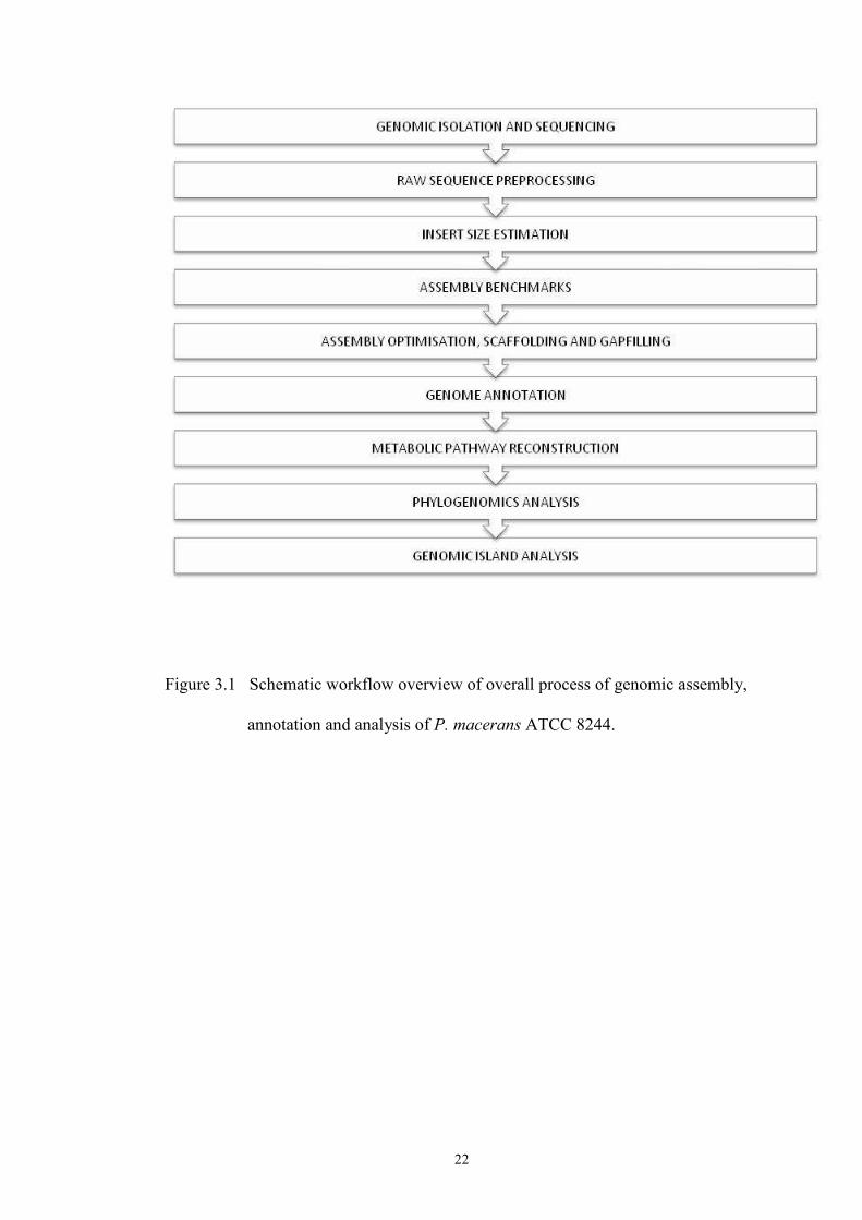
3.1 Schematic Workflow Of This Study
The overall process of genomic assembly, annotation and analysis of
Paenibacillus macerans ATCC 8244 is as portrayed in Figure 3.1 .
3.2 Bioinformatics Tools For Assembly, Annotation And Analysis
Various softwares were employed for the whole genome assembly,
annotation and analysis. All of the softwares are available as freewares. The specific
function of each software is described in all the subsections below which delineate
the all bioinformatic processes involved. Table 3.1 lists out the softwares involved as
well as their web addresses for downloading purposes.
22
Figure 3.1 Schematic workflow overview of overall process of genomic assembly,
annotation and analysis of P. macerans ATCC 8244.

23
Table 3.1: List of freewares used for the overall process of Assembly, Annotation
and Analysis of P. macerans ATCC 8244 genome
Software Version URL
NGS QC Toolkit 2.33 http://www.nipgr.res.in/ngsqctoolkit.html
VELVET 1.1 http://www.molecularevolution.org/software/genomics/velvet
BOWTIE 1.01 http://bowtie-bio.sourceforge.net/index.shtml
MIRA 3 https://sourceforge.net/projects/mira-assembler/
SOAP DENOVO r218 https://github.com/aquaskyline/SOAPdenovo2
ABYSS 1.3 http://www.bcgsc.ca/platform/bioinfo/software/abyss
SSPACE 2 https://omictools.com/sspace-tool
GAPFILLER 1.4 https://sourceforge.net/projects/gapfiller/
PRODIGAL 2.6 http://prodigal.ornl.gov/
ARAGORN 1.2.36 http://mbio-serv2.mbioekol.lu.se/ARAGORN/
RNAMMER 1.23 http://www.cbs.dtu.dk/services/RNAmmer/
BLAST+ 2.227 ftp://ftp.ncbi.nlm.nih.gov/blast/executables/blast+/
HMMER 3 http://www.ebi.ac.uk/Tools/hmmer/
ASGARD 1.5 https://sourceforge.net/projects/asgard-bio/
MUSCLE 3.3 http://www.drive5.com/muscle/
TRIMAI 1.2 http://trimal.cgenomics.org/
PHYML 3 http://www.atgc-montpellier.fr/phyml/binaries.php
PROTTEST 2 https://github.com/ddarriba/prottest
ORTHOMCL 2 http://orthomcl.org/common/downloads/
MCL 1.4 http://micans.org/mcl/
ISLANDVIEWER 2 http://www.pathogenomics.sfu.ca/islandviewer/
CRISPRFINDER 2014 http://crispr.i2bc.paris-saclay.fr/Server/
3.3 Genomic DNA isolation
The overall process of genomic DNA isolation was done as follows (kindly
executed by Mr Sim Kee Shin, Lab 414, School of Biological Sciences, Universiti
Sains Malaysia). A single colony of P. macerans ATCC 8244 was cultured into LB
medium (5 ml) and incubated at 37 ˚C with 180 rpm agitation for about 16 hours.
This P. macerans culture was used as an inoculum into a larger volume of LB
medium (200 ml) for DNA isolation. The culture was checked for contamination by
streaking it on an LB agar medium and observed for colonial consistency. The

24
isolation protocols were performed by using QIAGEN Genomic tips – 100G
according to the manufacturer’s protocol. The 200 ml overnight culture was chilled
on ice before centrifuging at 5,000 x G for 5 minutes at 4 ˚C. The pellet was
resuspended in 3.5 ml of Buffer B1 with 10 µl RNase A (100 mg/ml, Thermo
Scientific, USA), 100 µl of lysozyme (100 mg/ml, Sigma Aldrich, USA), and 100 µl
of Proteinase K (100 mg/ml, Sigma Aldrich, USA). The mixture was then incubated
at 37 ˚C in a water bath for at least 1 hour until the solution turned sticky or clear.
After this incubation, 1.2 ml of Buffer B2 was added and the mixture was further
incubated at 55 ˚C for at least 1 hour or until the solution became totally clear. The
lysate was centrifuged for 10 minutes at 5,000 x G at 4 ˚C to precipitate any non-
soluble particle. Then, the QIAGEN Genomic-tip 100G was equilibrated with 4 ml
of Buffer QBT and allowed to empty by gravitational flow. The column was loaded
with the cell lysate and the latter was allowed to flow out by gravitational flow. The
column was washed with 7.5 ml of Buffer QC and this process was repeated. The
genomic DNA was then eluted from the column by flowing through 5 ml of Buffer
QF. The genomic DNA was precipitated by adding 3.5 ml of isopropanol and
immediately spooled by using a sterile glass rod. The DNA was washed with 75 %
ethanol twice by spooling gently. The genomic DNA was rehydrated by dipping the
end of the glass rod bearing the DNA into an Eppendorf tube containing 1 ml of
nuclease free water.
The genomic DNA (gDNA) was subjected to agarose gel electrophoresis for
DNA integrity assessment. The absorbance value of OD230mm, OD260mm, and
OD280mm were used to assess the quality of DNA by determining the ratio of
OD260mm/ OD280mm and OD260mm/ OD230mm, respectively.