a functional magnetic resonance imaging study of … · 1the mind research network/lovelace...
TRANSCRIPT
A Functional Magnetic Resonance Imaging Studyof Cognitive Control and Neurosensory Deficits in
Mild Traumatic Brain Injury
Andrew R. Mayer,1,2,3* Faith M. Hanlon,1 Andrew B. Dodd,1 Josef M. Ling,1
Stefan D. Klimaj,1 and Timothy B. Meier1
1The Mind Research Network/Lovelace Biomedical and Environmental Research Institute,Albuquerque, New Mexico
2Department of Neurology, University of New Mexico School of Medicine, Albuquerque,New Mexico
3Department of Psychology, University of New Mexico, Albuquerque, New Mexico
r r
Abstract: Mild traumatic brain injury patients (mTBI) frequently report symptoms of increased distract-ability and sensory disturbances during mutisensory stimulation. These common post-concussive symp-toms could putatively result from dysfunction within the cognitive control network (CCN; top-down) orfrom unisensory cortex (bottom-up) itself. Functional magnetic resonance imaging (fMRI) and high-resolution structural data were therefore prospectively collected during a multisensory (audio-visual)cognitive control task from 46 mTBI patients within 3 weeks of injury and 46 matched healthy controls(HC), with a subset of participants returning at 4 months. Multisensory stimuli were presented at twofrequencies to manipulate cognitive and perceptual load. Patients self-reported more cognitive, emo-tional, somatic, vestibular and visual symptoms relative to HC, which improved, but did not entirelyresolve, over the 4 month follow-up period. There were no group differences in behavior or functionalactivation during cognitive control (incongruent – congruent trials). In contrast, patients exhibited abnor-mal activation within different regions of visual cortex that depended on whether attention was focusedon auditory or visual information streams. Patients also exhibited increased activation within bilateralinferior parietal lobules during higher cognitive/perceptual loads, suggesting a compensatory mecha-nism to achieve similar levels of behavioral performance. Functional abnormalities within the visual cor-tex and inferior parietal lobules were only partially resolved at 4 months post-injury, suggesting thatneural abnormalities may take longer to resolve than behavioral measures used in most clinical settings.In summary, current results indicate that abnormalities within unisensory cortex (particularly visualareas) following mTBI, which likely contribute to deficits commonly reported during multisensory stimu-lation. Hum Brain Mapp 00:000–000, 2015. VC 2015 Wiley Periodicals, Inc.
Key words: cognitive control; multisensory; auditory; visual; functional magnetic resonance imaging
r r
Contract grant sponsor: National Institutes of Health; Contractgrant numbers: R24-HD050836, R21-NS064464-01A1, R21-NS064464-01S1 (to A.M.).
*Correspondence to: Andrew Mayer, The Mind Research Net-work, Pete & Nancy Domenici Hall, 1101 Yale Blvd. NE, Albu-querque, New Mexico 87106. E-mail: [email protected]
Received for publication 18 May 2015; Revised 11 June 2015;Accepted 22 July 2015.
DOI: 10.1002/hbm.22930Published online 00 Month 2015 in Wiley Online Library(wileyonlinelibrary.com).
r Human Brain Mapping 00:00–00 (2015) r
VC 2015 Wiley Periodicals, Inc.
INTRODUCTION
Mild traumatic brain injury (mTBI) represents a majorhealth concern, accounting for a majority of the 1.7 millionhead injuries sustained each year (Faul et al., 2010). Impor-tantly, this number is likely underestimated given that manymTBI, and sports-related concussions in particular, go unre-ported (McCrea et al., 2013). Common patient complaints dur-ing the acute to sub-acute injury phase include increaseddistractibility and sensory overstimulation, which could arisefrom difficulties in processing conflicting information (i.e., oneform of cognitive control) or from neurosensory impairments(Fischer et al., 2014; Halterman et al., 2006; Hoffer, 2015). Fewstudies have prospectively examined (e.g., semi-acute tochronic injury stage) abnormalities within the cognitive controlnetwork (CCN) or within unisensory cortex (e.g., primary orsecondary auditory and visual cortices) in homogeneous sam-ples of mTBI patients, a necessary first step for understandingpotential physiological basis of self-reported symptoms.
As demonstrated by Hubel et al. (1959), there are multifac-eted interactions between top-down cognitive control and uni-sensory cortex (Talsma et al., 2010). The dorsal medialprefrontal cortex (dMFC), dorsolateral and ventrolateral pre-frontal cortex (lPFC), anterior insula, and the inferior parietallobes (IPL) of the CCN exhibit increased activation during theprocessing of conflicting stimuli (Ridderinkhof et al., 2004;Roberts and Hall, 2008), orchestrating changes in neural repre-sentations that ultimately affect behavior (Shenhav et al.,2013). One of these neural changes include the appearance ofattention related modulations (ARM) within unisensory cor-tex, including enhanced neural responses (i.e., up-regulation)for attended stimuli and suppressed responses (i.e., inhibition)for ignored stimuli (Baier et al., 2006; Mayer et al., 2009a;Weissman et al., 2004).
Studies examining cognitive control/working memory fol-lowing mTBI have reported both hyper-activation and hypo-activation of the CCN, with mixed findings likely driven by
the types of mTBI patients studied (e.g., acute vs. chronic,symptomatic vs. asymptomatic) and differences in taskrequirements (reviewed in Mayer et al., 2014). To date nostudies have examined the neural basis (i.e., structural or func-tional) of neurosensory deficits (e.g., visual, auditory or vestib-ular symptoms) during the acute to semi-acute phase of mTBIdespite suggestions that these deficits represent a major con-tributing factor for chronic post-concussive symptoms (Hoffer,2015; Pogoda et al., 2012). Early non-invasive imaging workexamining unisensory cortical functioning suggests gatingdeficits across the TBI spectrum (Arciniegas et al., 2000; Arci-niegas and Topkoff, 2004), and that post-concussive symp-toms result in part from sensory gating abnormalities (Kumaret al., 2005). SPECT (Stamatakis et al., 2002), PET (Kato et al.,2007; Nakashima et al., 2007) and fMRI (Kim et al., 2012) stud-ies also indicate hypometabolism or hypoperfusion in unisen-sory cortex in severe TBI patients, providing additionalevidence that cortical dysfunction may contribute to self-reported neurosensory symptoms. Finally, incoming informa-tion from sensory organs as well as top-down modulatory sig-nals from heteromodal cortex are often routed through thethalamus and/or brainstem prior to reaching unisensory corti-cal regions. Midbrain regions may be particularly susceptibleto injury due to the accumulation of shear stresses (Zhanget al., 2004), with previous studies implicating structural andfunctional abnormalities in these regions following mTBI (Luiet al., 2014; Mayer et al., 2009b).
The aims of the current study were therefore (1) toexamine the relationship between cognitive control andunisensory cortical functioning during the semi-acute stageof mTBI, (2) determine how these relate to self-reportedneurosensory deficits and (3) to track neurosensory symp-toms/neural abnormalities as a function of recovery over afour month period. We hypothesized that there would befunctional abnormalities in the CCN as well as in unisen-sory cortex (i.e., decreased ARM) following mTBI. We alsopredicted that decreased ARM would be associated withneurosensory deficits in patients, both of which wouldnormalize as a function of recovery.
METHODS
Participants
Forty-eight mTBI patients and 48 age and educationmatched controls were included in the current study.Inclusion criteria included age of 18–55 years, a closedhead injury with a self-reported alteration in mental status,Glasgow Coma Score of 13–15 upon initial presentation tomedical staff, a maximum of 30 minutes for loss of con-sciousness (if experienced) and a maximum of 24 hours ofpost-traumatic amnesia (if experienced). Patients and con-trols were excluded from the current study based on self-reported history of other neurological disease, recent alco-hol or other drug abuse, history of Axis I disorder, priorclosed head injuries with more than 5 minutes loss of
Abbreviations
A1 primary auditory cortexA2 secondary auditory cortexARM attention related modulationsBDI-II Beck Depression Inventory – Second EditionCCN cognitive control networkdMFC dorsal medial prefrontal cortexHC healthy controlIPL inferior parietal lobelPFC dorsolateral and ventrolateral prefrontal cortexmTBI mild traumatic brain injuryNSI Neurobehavioral Symptom InventoryPSC percent signal changeSTAI State-Trait Anxiety InventoryTOMM Test of Memory MalingeringV1 primary visual cortexV2 secondary visual cortexWTAR Wechsler Test of Adult Reading
r Mayer et al. r
r 2 r
consciousness, additional closed head injury in last year,strong preference for the left hand (score of less than 220on Edinburgh handedness scale), learning disorder, orAttention Deficit Hyperactivity Disorder. Two patientswere identified as outliers on frame-wise displacement(greater than three times the interquartile range on two ormore of six motion parameters) relative to their cohort,and were excluded from further analyses along with theirhealthy matches. All remaining participants performed atacceptable levels (above 70% accuracy) on the functionaltask, resulting in a total of 46 mTBI patients (24 males,28.9 6 9.8 years old, 13.2 6 2.6 years of education) and 46matched controls (24 males, 28.4 6 9.9 years old, 13.8 6 2.3years of education). Informed consent was obtainedaccording to institutional guidelines at the University ofNew Mexico Health Science Center.
Patients and healthy controls (HC) completed identicalprocedures. Patients were assessed with neuropsychologi-cal (mean day post-injury 5 13.7 6 5.0) and neuroimaging(mean day post-injury 5 13.8 6 5.0) measures within 21days of injury (Table I), with the imaging and clinical ses-sions typically conducted within a few days of each other(mean difference between assessments 5 1.2 6 1.5 days). Atthe time of assessment, seven of the mTBI participants wereprescribed medications for pain and other conditions asso-ciated with injury. Data from a subset of participants on thesame task has previously been reported (Mayer et al., 2012).
Neuropsychological Assessment
Composite indices were calculated for the following cogni-tive domains to reduce redundancy amongst similar neuro-psychological measures: attention (Trails A, Paced AuditorySerial Addition Test, Stroop [color-word and interferencescores], and Wechsler Adult Intelligence Scale-Third Edition[WAIS-III] digit span); working memory (WAIS-III letter num-ber sequence, arithmetic, and digits backward); processingspeed (grooved pegboard [dominant and non-dominanthand] and WAIS-III digit symbol coding); executive function(Wisconsin Card Sort [errors and perseverative errors], TrailsB, and Controlled Oral Word Association FAS Test); andmemory (California Verbal Learning Test-Second Edition:immediate recall, short-delay free recall, long-delay freerecall). Emotional, somatic and cognitive complaints wereassessed using the Neurobehavioral Symptom Inventory(NSI), the Beck Depression Inventory-Second Edition (BDI-II)and the State-Trait Anxiety Inventory (STAI). The NSI wasalso used to assess vestibular, visual and auditory symptomsof neurological deficits based on previously published guide-lines (Lew et al., 2011). The Wechsler Test of Adult Reading(WTAR) provided an estimate of overall premorbid intellec-tual functioning and the Test of Memory Malingering(TOMM) assessed effort.
One patient and one control did not complete the full neu-ropsychological battery. Additionally, one control’s TOMMdata was eliminated due to poor performance (t-score< 0) in
conjunction with normal performance on more demandingneuropsychological measures. Univariate tests were con-ducted to compare estimates of effort and premorbid intelli-gence, whereas multivariate tests were used to comparecomposite neuropsychological indices and self-reportedsymptoms given the known moderate correlational structures.Chi-square tests assessed for group differences in the occur-rence of neurosensory deficits.
Task
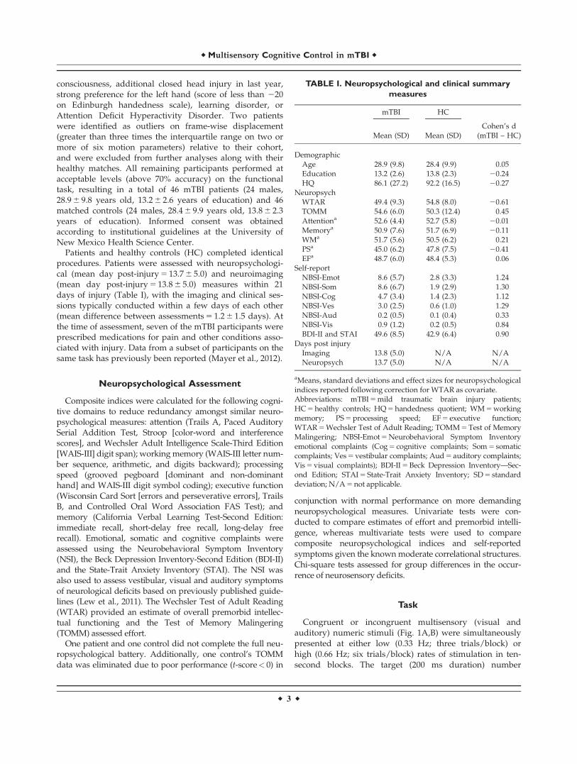
Congruent or incongruent multisensory (visual andauditory) numeric stimuli (Fig. 1A,B) were simultaneouslypresented at either low (0.33 Hz; three trials/block) orhigh (0.66 Hz; six trials/block) rates of stimulation in ten-second blocks. The target (200 ms duration) number
TABLE I. Neuropsychological and clinical summary
measures
mTBI HC
Mean (SD) Mean (SD)Cohen’s d
(mTBI – HC)
DemographicAge 28.9 (9.8) 28.4 (9.9) 0.05Education 13.2 (2.6) 13.8 (2.3) 20.24HQ 86.1 (27.2) 92.2 (16.5) 20.27
NeuropsychWTAR 49.4 (9.3) 54.8 (8.0) 20.61TOMM 54.6 (6.0) 50.3 (12.4) 0.45Attentiona 52.6 (4.4) 52.7 (5.8) 20.01Memorya 50.9 (7.6) 51.7 (6.9) 20.11WMa 51.7 (5.6) 50.5 (6.2) 0.21PSa 45.0 (6.2) 47.8 (7.5) 20.41EFa 48.7 (6.0) 48.4 (5.3) 0.06
Self-reportNBSI-Emot 8.6 (5.7) 2.8 (3.3) 1.24NBSI-Som 8.6 (6.7) 1.9 (2.9) 1.30NBSI-Cog 4.7 (3.4) 1.4 (2.3) 1.12NBSI-Ves 3.0 (2.5) 0.6 (1.0) 1.29NBSI-Aud 0.2 (0.5) 0.1 (0.4) 0.33NBSI-Vis 0.9 (1.2) 0.2 (0.5) 0.84BDI-II and STAI 49.6 (8.5) 42.9 (6.4) 0.90
Days post injuryImaging 13.8 (5.0) N/A N/ANeuropsych 13.7 (5.0) N/A N/A
aMeans, standard deviations and effect sizes for neuropsychologicalindices reported following correction for WTAR as covariate.Abbreviations: mTBI 5 mild traumatic brain injury patients;HC 5 healthy controls; HQ 5 handedness quotient; WM 5 workingmemory; PS 5 processing speed; EF 5 executive function;WTAR 5 Wechsler Test of Adult Reading; TOMM 5 Test of MemoryMalingering; NBSI-Emot 5 Neurobehavioral Symptom Inventoryemotional complaints (Cog 5 cognitive complaints; Som 5 somaticcomplaints; Ves 5 vestibular complaints; Aud 5 auditory complaints;Vis 5 visual complaints); BDI-II 5 Beck Depression Inventory—Sec-ond Edition; STAI 5 State-Trait Anxiety Inventory; SD 5 standarddeviation; N/A 5 not applicable.
r Multisensory Cognitive Control in mTBI r
r 3 r
stream (one, two, or three) was preceded by a multisen-sory cue (175 ms duration) word (“HEAR,” “LOOK” or“NONE”) indicating the modality for focused attention.During “HEAR” trials (attend-auditory) participantsresponded to aurally presented target stimuli via a right-handed button press while ignoring simultaneously pre-sented visual numbers. During “LOOK” trials (attend-vis-ual) participants responded to the visual targets (Englishnumerals “ONE,” “TWO” or “THREE”) while ignoring theauditory stream (spoken number words). “NONE” trialsare not reported in the current manuscript. The inter-blockintervals varied between 8, 10, and 12 seconds to decreasetemporal expectations and permit modeling of the baselineresponse. Block-order was pseudorandom, with a total of432 trials presented across six separate imaging runs. Asresponse time data has a tendency towards positive skew,the median reaction time was used as a measure of centraltendency. Two 2 3 2 3 2 [Group (mTBI vs. HC) 3 Congru-ency (Congruent vs. Incongruent) 3 Frequency (0.33 Hz vs.0.66 Hz)] mixed-measures ANCOVAs were conducted sep-arately on attend-auditory and attend-visual conditions.
MR Imaging
High-resolution multi-echo MPRAGE T1 [TR (repetitiontime) 5 2.53 s, 78 flip angle, number of excitations(NEX) 5 1, slice thickness 5 1 mm, FOV (field ofview) 5 256 mm, resolution 5 256 3 256, voxel size 5 1.0 3
1.0 3 1.0 mm] and T2 [echo time 5 77.0 ms, TR 5 1.55 s, flipangle 1528, NEX 5 1, slice thickness 5 1.5 mm,FOV 5 220 mm, matrix 5 192 3 192, voxel size 5 1.15 3
1.15 3 1.5 mm] sequences were collected at 3 Tesla Sie-mens TIM Trio scanner with a 12-channel head coil. Sus-ceptibility weighted images were collected with a gradientecho sequence [TR 5 28 ms; TE 5 20 ms; flip angle 158;bandwidth 5 120 Hz/Px; FOV 5 180 3 240 mm;matrix 5 177 3 256; slice thickness 5 1.5 mm] to bettercharacterize petechial hemorrhages on a subset of patients(N 5 21). A single-shot, gradient-echo echoplanar pulsesequence [TR 5 2,000 ms; TE 5 29 ms; flip angle 5 758;FOV 5 240 mm; matrix size 5 64 3 64; voxel size 5 3.75 3
3.75 3 4.55 mm] was collected during the multisensorytask.
Figure 1.
Representations of incongruent trials for A) attend-auditory (AA)
and B) attend-visual (AV) tasks as determined by the initial cue
(AA 5 “hear”; AV 5 “look”). Expected correct responses (CR),
inter-trial interval (ITI) and inter-block interval (IBI) are repre-
sented at the right of the timeline. The bottom row presents box-
and-whisker plots for reaction times (RT) in AA (Panel C) and AV
(Panel D). Data are presented separately for HC and mTBI, with
incongruent (IT; light gray) and congruent (CT; dark gray) trials at
the two stimulation frequencies (0.33 Hz 5 notched boxes; 0.66
Hz 5 un-notched boxes) used in the experiment.
r Mayer et al. r
r 4 r
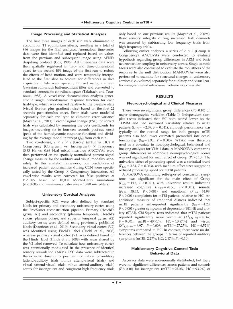
Image Processing and Statistical Analyses
The first three images of each run were eliminated toaccount for T1 equilibrium effects, resulting in a total of966 images for the final analyses. Anomalous time-seriesdata were first identified and replaced based on valuesfrom the previous and subsequent image using AFNI’sdespiking protocol (Cox, 1996). All time-series data werethen spatially registered in two- and three-dimensionalspace to the second EPI image of the first run to reducethe effects of head motion, and were temporally interpo-lated to the first slice to account for differences in sliceacquisition. Data were spatially blurred using a 6 mmGaussian full-width half-maximum filter and converted tostandard stereotaxic coordinate space (Talairach and Tour-noux, 1988). A voxel-wise deconvolution analysis gener-ated a single hemodynamic response function for eachtrial-type, which was derived relative to the baseline state(visual fixation plus gradient noise) based on the first 22seconds post-stimulus onset. Error trials were modelledseparately for each trial-type to eliminate error variance(Mayer et al., 2011). Percent signal change (PSC) for correcttrials was calculated by summing the beta coefficients forimages occurring six to fourteen seconds post-cue onset(peak of the hemodynamic response function) and divid-ing by the average model intercept (b0) from each run.
Two voxel-wise, 2 3 2 3 2 [Group (mTBI vs. HC) 3
Congruency (Congruent vs. Incongruent) 3 Frequency(0.33 Hz vs. 0.66 Hz)] mixed-measures ANCOVAs werethen performed on the spatially normalized percent signalchange measure for the auditory and visual modality sepa-rately. In this analytic framework, our predictions ofincreased patient abnormalities during CCN were specifi-cally tested by the Group 3 Congruency interaction. Allvoxel-wise results were corrected for false positives atP< 0.05 based on 10,000 Monte-Carlo simulations(P< 0.005 and minimum cluster size 5 1,280 microliters).
Unisensory Cortical Analyses
Subject-specific ROI were also defined by standardlabels for primary and secondary unisensory cortex usingthe FreeSurfer reconstruction pipeline. Primary (Heschl’sgyrus; A1) and secondary (planum temporale, Heschl’ssulcus, planum polare, and superior temporal gyrus; A2)auditory cortex were defined using previously publishedlabels (Destrieux et al., 2010). Secondary visual cortex (V2)was identified using Fischl’s label (Fischl et al., 2008)whereas primary visual cortex (V1) was defined based onthe Hinds’ label (Hinds et al., 2008) with areas shared bythe V2 label removed. To calculate how unisensory cortexwas attentionally modulated in the presence of identicalsensory stimulation (ARM), PSC data were subtracted inthe expected direction of positive modulation for auditory(attend-auditory trials minus attend-visual trials) andvisual (attend-visual trials minus attend-auditory trials)cortex for incongruent and congruent high frequency trials
only based on our previous results (Mayer et al., 2009a).Basic sensory integrity during increased task demandswas assessed by subtracting low frequency trials fromhigh frequency trials.
Following outlier analyses, a series of 2 3 2 (Group 3
Congruency) ANCOVAs were conducted to evaluatehypothesis regarding group differences in ARM and basicneurovascular coupling in unisensory cortex. Single-samplet-tests were also conducted to evaluate the robustness of theresponse to the null distribution. MANCOVAs were alsoperformed to examine for structural changes in unisensorycortices (i.e., volume) separately for auditory and visual cor-tex using estimated intracranial volume as a covariate.
RESULTS
Neuropsychological and Clinical Measures
There were no significant group differences (P> 0.10) onmajor demographic variables (Table I). Independent sam-ples t-tests indicated that HC both scored lower on theTOMM and had increased variability relative to mTBIpatients (t61.5 5 22.09, P 5 0.041), although performance wastypically in the normal range for both groups. mTBIpatients also had lower estimated premorbid intellectualfunctioning (t88 5 2.90, P 5 0.005). WTAR was thereforeused as a covariate in neuropsychological, behavioral andimaging analyses for Visit 1 data. A MANCOVA comparinggroup differences in composite neuropsychological scoreswas not significant for main effect of Group (P> 0.10). Theunivariate effect of processing speed was a statistical trend(F1,87 5 3.54, P 5 0.063), with medium effect sizes suggestingreduced processing speed for mTBI patients.
A MANOVA examining self-reported concussion symp-toms was significant for the main effect of Group(F3,87 5 14.4, P< 0.001), with univariate results indicatingincreased cognitive (F1,89 5 28.53, P< 0.001), somatic(F1,89 5 38.43, P< 0.001) and emotional (F1,89 5 34.98,P< 0.001) complaints for mTBI patients relative to HC. Anadditional measure of emotional distress indicated thatmTBI patients self-reported significantly (t89 5 24.28,P< 0.001) greater symptoms of depression (BDI-II) and anx-iety (STAI). Chi-Square tests indicated that mTBI patientsreported significantly more vestibular (X2
1,N590 5 10.67,P 5 0.001; mTBI 5 40.91%, HC 5 10.87%) and visual(X2
1,N590 5 6.97, P 5 0.008; mTBI 5 27.27%, HC 5 6.52%)symptoms compared to HC. In contrast, there were no dif-ferences between the groups in terms of reported auditorysymptoms (mTBI: 2.27%; HC: 2.17%; P> 0.10).
Multisensory Cognitive Control Task:
Behavioral Data
Accuracy data were non-normally distributed, but therewere no significant differences across patients and controls(P> 0.10) for incongruent (mTBI 5 95.0%; HC 5 93.9%) or
r Multisensory Cognitive Control in mTBI r
r 5 r
congruent (mTBI 5 98.1%; HC 5 97.7%) trials. Reactiontimes for attend-auditory trials (Fig. 1C) were significantfor the main effects of Congruency (F1,89 5 165.18,P< 0.001) and Frequency (F1,89 5197.98, P< 0.001), withfaster response times for congruent (mean 5 564.40 6 74.71)compared to incongruent (mean 5 644.38 6 90.37) trialsand for higher (0.66 Hz; mean 5 565.29 6 63.60) relative tolower (0.33 Hz; mean 5 643.47 6 96.67) frequency trials.Neither the main effect of Group nor any of interactionswere significant (P> 0.10).
The main effects of Congruency (F1,89 5 88.14, P< 0.001)and Frequency (F1,89 5 175.74, P< 0.001) were also signifi-cant for the attend-visual trials (Fig. 1D), with participantsresponding more quickly to congruent (mean 5 535.746 70.99) compared to incongruent (mean 5 578.08 6 76.01)trials, as well as to high (0.66 Hz; mean 5 527.49 6 64.24)compared to low (0.33 Hz; mean 5 586.35 6 81.80) fre-quency trials. No other main effects or interaction effectsapproached significance (P> 0.10).
Radiological Findings
Thirty-five mTBI patients had a CT scan as part of rou-tine emergency care. Of those patients with brain CT, threehad positive findings. A board certified radiologist blindedto participant diagnosis also reviewed all baseline struc-tural MRI sequences for both mTBI and HC. There wereno positive MRI findings among HC, but four mTBIpatients had positive findings on MRI scans. Thus, therewere seven mTBI patients with either a positive CT or apositive MRI scan in total. None of the patients with apositive CT finding also had a positive MRI finding.
Functional Imaging Data
Two MANOVAs were first conducted to investigate anypotential group differences in head motion (both rotationaland translational framewise displacements in imagespace), which could confound the interpretation of fMRIdata. However, results indicated that the multivariateeffect of Group was not significant (P> 0.10) for either ofthe MANOVAs, with small to medium effect sizes for allsix parameters (range from 0.23 to 0.46).
Functional Results: Patient Comparisons
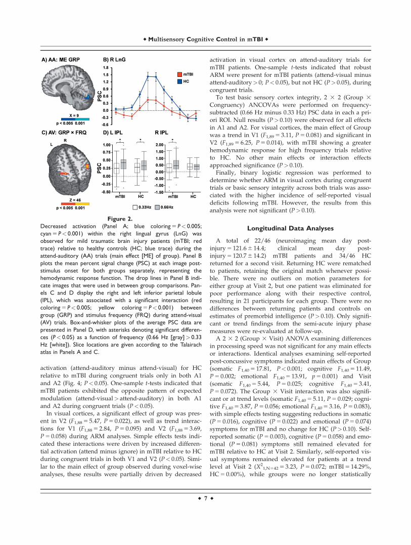
Voxel-wise, 2 3 2 3 2 (Group 3 Congruency 3 Fre-quency) mixed-measures ANCOVAs were performed sepa-rately for data in the attend-auditory and attend-visualconditions. For both attend-auditory and attend-visual tri-als, the Group 3 Congruency interaction was not signifi-cant following appropriate correction for false positives.During attend-auditory trials, mTBI patients exhibiteddecreased activation relative to HC within the right lingualgyrus (BAs 17/18) extending into the declive of the cerebel-lum (Fig. 2A; ml 5 1,353). In addition, the Group 3 Fre-
quency interaction (Fig. 2C) was significant in the left (BAs5/40; ml 5 2,998) and right (BA 40; ml 5 1,661) IPL duringattend-visual trials. Simple effects tests indicated increasedactivation in the right IPL for high relative to low frequencytrials in mTBI patients (P< 0.05) but not HC. In the left IPL,the interaction was driven by a greater difference in activa-tion between high and low frequency trials in mTBI relativeto HC (P< 0.05). The Group 3 Frequency interaction wasnot significant in the attend-auditory trials.
Functional Results: Task Comparisons
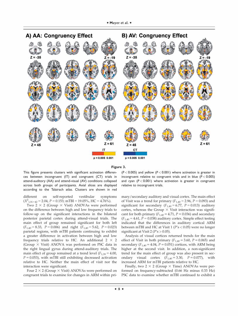
Functional results from the identical task have been pre-sented previously in a subset of mTBI patients (Mayeret al., 2012). Thus, the main effects associated with Con-gruency and Frequency are only briefly presented here toestablish that the task evoked expected patterns of activa-tion across both groups of participants. In both the attend-auditory and attend-visual conditions, incongruent trialsresulted in increased activation within the anterior insula,lPFC, the dMFC, posterior temporal sulcus, IPL, thalamusand sub-thalamic nuclei and cerebellum relative to congru-ent trials (Fig. 3). Activation was typically greater in vol-ume and more bilateral for incongruent trials during theattend-visual condition, whereas increased activation wasobserved in bilateral secondary/associative visual cortexfor congruent trials during the attend-auditory condition.
The main effects of frequency were in the expecteddirection (0.66 Hz> 0.33 Hz) and similar for both theattend-auditory and attend-visual trials. Specifically,increased activation was observed bilaterally for higherfrequency trials within primary and secondary auditorycortex, pre-SMA and SMA, DLPFC, posterior parietal cor-tex, basal ganglia and cerebellum (Lobules IV-VII) acrossboth the attend-visual and attend-auditory conditions.More lateralized motor-related activity was also observedin the left sensori-motor cortex. In addition, increaseddeactivation during the high relative to low frequency tri-als was observed within default-mode network for bothattend-auditory and attend-visual conditions.
Unisensory Cortex Analyses: Patient
Comparisons
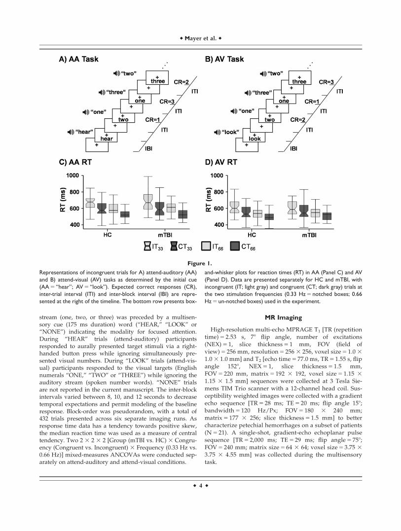
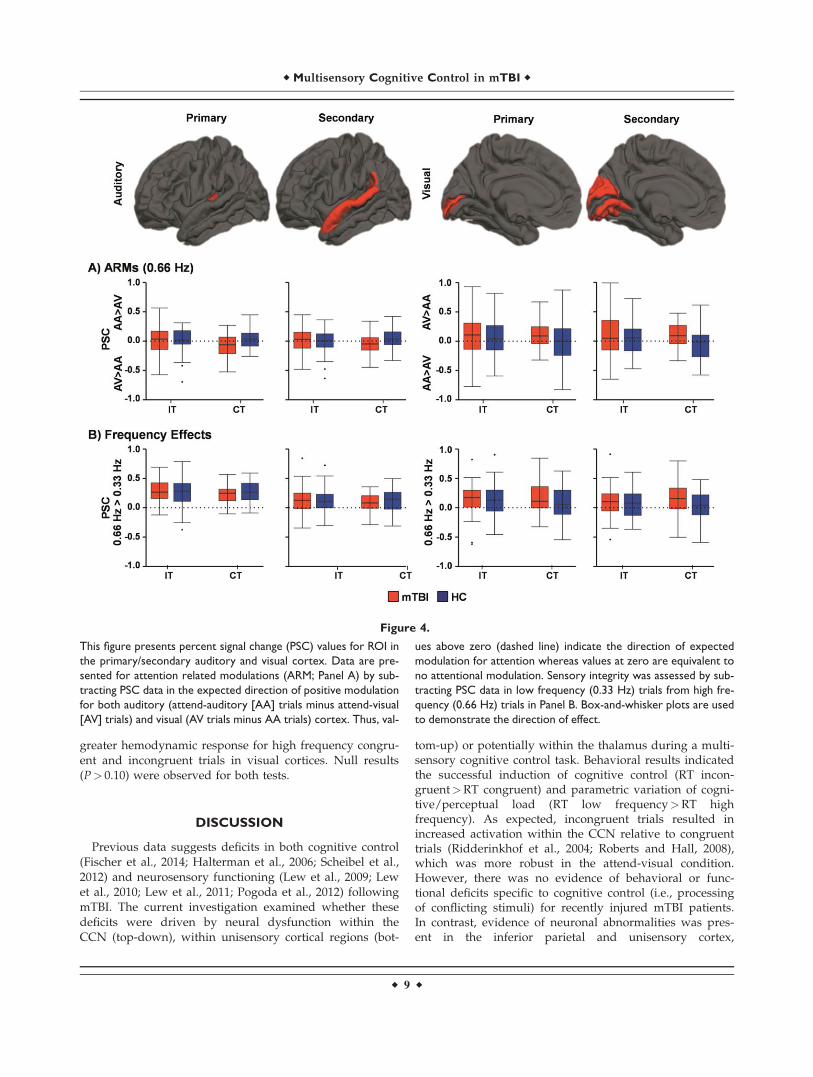
Results from two MANCOVAs (intracranial volume as acovariate) indicated that there were no significant differen-ces in either primary/secondary auditory or visual corticalvolume between the two groups (P> 0.10). Four 2 3 2(Group 3 Congruency) ANCOVAs examined for ARMduring high frequency trials in primary and secondary vis-ual (V1; V2) and auditory (A1; A2) cortices.
There was a non-significant effect of group in A1(F1,89 5 3.11, P 5 0.081), as well as non-significant Group 3
Congruency interactions within A1 (F1,89 5 2.85, P 5 0.095)and A2 (F1,89 5 3.40, P 5 0.069). Simple effects tests indi-cated that these trends resulted from increased differential
r Mayer et al. r
r 6 r
activation (attend-auditory minus attend-visual) for HCrelative to mTBI during congruent trials only in both A1and A2 (Fig. 4; P< 0.05). One-sample t-tests indicated thatmTBI patients exhibited the opposite pattern of expectedmodulation (attend-visual> attend-auditory) in both A1and A2 during congruent trials (P< 0.05).
In visual cortices, a significant effect of group was pres-ent in V2 (F1,88 5 5.47, P 5 0.022), as well as trend interac-tions for V1 (F1,88 5 2.84, P 5 0.095) and V2 (F1,88 5 3.69,P 5 0.058) during ARM analyses. Simple effects tests indi-cated these interactions were driven by increased differen-tial activation (attend minus ignore) in mTBI relative to HCduring congruent trials in both V1 and V2 (P< 0.05). Simi-lar to the main effect of group observed during voxel-wiseanalyses, these results were partially driven by decreased
activation in visual cortex on attend-auditory trials formTBI patients. One-sample t-tests indicated that robustARM were present for mTBI patients (attend-visual minusattend-auditory> 0; P< 0.05), but not HC (P> 0.05), duringcongruent trials.
To test basic sensory cortex integrity, 2 3 2 (Group 3
Congruency) ANCOVAs were performed on frequency-subtracted (0.66 Hz minus 0.33 Hz) PSC data in each a pri-ori ROI. Null results (P> 0.10) were observed for all effectsin A1 and A2. For visual cortices, the main effect of Groupwas a trend in V1 (F1,89 5 3.11, P 5 0.081) and significant inV2 (F1,89 5 6.25, P 5 0.014), with mTBI showing a greaterhemodynamic response for high frequency trials relativeto HC. No other main effects or interaction effectsapproached significance (P> 0.10).
Finally, binary logistic regression was performed todetermine whether ARM in visual cortex during congruenttrials or basic sensory integrity across both trials was asso-ciated with the higher incidence of self-reported visualdeficits following mTBI. However, the results from thisanalysis were not significant (P> 0.10).
Longitudinal Data Analyses
A total of 22/46 (neuroimaging mean day post-injury 5 121.6 6 14.4; clinical mean day post-injury 5 120.7 6 14.2) mTBI patients and 34/46 HCreturned for a second visit. Returning HC were rematchedto patients, retaining the original match whenever possi-ble. There were no outliers on motion parameters foreither group at Visit 2, but one patient was eliminated forpoor performance along with their respective control,resulting in 21 participants for each group. There were nodifferences between returning patients and controls onestimates of premorbid intelligence (P> 0.10). Only signifi-cant or trend findings from the semi-acute injury phasemeasures were re-evaluated at follow-up.
A 2 3 2 (Group 3 Visit) ANOVA examining differencesin processing speed was not significant for any main effectsor interactions. Identical analyses examining self-reportedpost-concussive symptoms indicated main effects of Group(somatic F1,40 5 17.81, P< 0.001; cognitive F1,40 5 11.49,P 5 0.002; emotional F1,40 5 13.91, p 5 0.001) and Visit(somatic F1,40 5 5.44, P 5 0.025; cognitive F1,40 5 3.41,P 5 0.072). The Group 3 Visit interaction was also signifi-cant or at trend levels (somatic F1,40 5 5.11, P 5 0.029; cogni-tive F1,40 5 3.87, P 5 0.056; emotional F1,40 5 3.16, P 5 0.083),with simple effects testing suggesting reductions in somatic(P 5 0.016), cognitive (P 5 0.022) and emotional (P 5 0.074)symptoms for mTBI and no change for HC (P> 0.10). Self-reported somatic (P 5 0.003), cognitive (P 5 0.058) and emo-tional (P 5 0.081) symptoms still remained elevated formTBI relative to HC at Visit 2. Similarly, self-reported vis-ual symptoms remained elevated for patients at a trendlevel at Visit 2 (X2
1,N542 5 3.23, P 5 0.072; mTBI 5 14.29%,HC 5 0.00%), while groups were no longer statistically
Figure 2.
Decreased activation (Panel A; blue coloring 5 P< 0.005;
cyan 5 P< 0.001) within the right lingual gyrus (LnG) was
observed for mild traumatic brain injury patients (mTBI; red
trace) relative to healthy controls (HC; blue trace) during the
attend-auditory (AA) trials (main effect [ME] of group). Panel B
plots the mean percent signal change (PSC) at each image post-
stimulus onset for both groups separately, representing the
hemodynamic response function. The drop lines in Panel B indi-
cate images that were used in between group comparisons. Pan-
els C and D display the right and left inferior parietal lobule
(IPL), which was associated with a significant interaction (red
coloring 5 P< 0.005; yellow coloring 5 P< 0.001) between
group (GRP) and stimulus frequency (FRQ) during attend-visual
(AV) trials. Box-and-whisker plots of the average PSC data are
presented in Panel D, with asterisks denoting significant differen-
ces (P< 0.05) as a function of frequency (0.66 Hz [gray]> 0.33
Hz [white]). Slice locations are given according to the Talairach
atlas in Panels A and C.
r Multisensory Cognitive Control in mTBI r
r 7 r
different on self-reported vestibular symptoms(X2
1,N542 5 2.04, P 5 0.153; mTBI 5 19.05%, HC 5 4.76%).Two 2 3 2 (Group 3 Visit) ANOVAs were performed
on the difference between high and low frequency trials tofollow-up on the significant interactions in the bilateralposterior parietal cortex during attend-visual trials. Themain effect of group remained significant for both left(F1,40 5 8.33, P 5 0.006) and right (F1,40 5 5.62, P 5 0.023)parietal regions, with mTBI patients continuing to exhibita greater difference in activation between high and lowfrequency trials relative to HC. An additional 2 3 2(Group 3 Visit) ANOVA was performed on PSC data inthe right lingual gyrus during attend-auditory trials. Themain effect of group remained at a trend level (F1,39 5 4.00,P 5 0.053), with mTBI still exhibiting decreased activationrelative to HC. Neither the main effect of visit nor theinteraction were significant.
Four 2 3 2 (Group 3 Visit) ANOVAs were performed oncongruent trials to examine for changes in ARM within pri-
mary/secondary auditory and visual cortex. The main effectof Visit was a trend for primary (F1,40 5 2.96, P 5 0.093) andsignificant for secondary (F1,40 5 6.77, P 5 0.013) auditorycortex, whereas the Group 3 Visit interaction was signifi-cant for both primary (F1,40 5 4.71, P 5 0.036) and secondary(F1,40 5 4.61, P 5 0.038) auditory cortex. Simple effect testingindicated that the differences in auditory cortical ARMbetween mTBI and HC at Visit 1 (P’s< 0.05) were no longersignificant at Visit 2 (P’s> 0.05).
Analysis of visual cortices returned trends for the maineffect of Visit in both primary (F1,40 5 3.60, P 5 0.065) andsecondary (F1,40 5 4.04, P 5 0.051) cortices, with ARM beinghigher at the second visit. In addition, a non-significanttrend for the main effect of group was also present in sec-ondary visual cortex (F1,40 5 3.30, P 5 0.077), withincreased ARM for mTBI patients relative to HC.
Finally, two 2 3 2 (Group 3 Time) ANOVAs were per-formed on frequency-subtracted (0.66 Hz minus 0.33 Hz)PSC data to examine whether mTBI continued to exhibit a
Figure 3.
This figure presents clusters with significant activation differen-
ces between incongruent (IT) and congruent (CT) trials in
attend-auditory (AA) and attend-visual (AV) conditions collapsed
across both groups of participants. Axial slices are displayed
according to the Talairach atlas. Clusters are shown in red
(P< 0.005) and yellow (P< 0.001) where activation is greater in
incongruent relative to congruent trials and in blue (P< 0.005)
and cyan (P< 0.001) where activation is greater in congruent
relative to incongruent trials.
r Mayer et al. r
r 8 r
greater hemodynamic response for high frequency congru-ent and incongruent trials in visual cortices. Null results(P> 0.10) were observed for both tests.
DISCUSSION
Previous data suggests deficits in both cognitive control(Fischer et al., 2014; Halterman et al., 2006; Scheibel et al.,2012) and neurosensory functioning (Lew et al., 2009; Lewet al., 2010; Lew et al., 2011; Pogoda et al., 2012) followingmTBI. The current investigation examined whether thesedeficits were driven by neural dysfunction within theCCN (top-down), within unisensory cortical regions (bot-
tom-up) or potentially within the thalamus during a multi-sensory cognitive control task. Behavioral results indicatedthe successful induction of cognitive control (RT incon-gruent>RT congruent) and parametric variation of cogni-tive/perceptual load (RT low frequency>RT highfrequency). As expected, incongruent trials resulted inincreased activation within the CCN relative to congruenttrials (Ridderinkhof et al., 2004; Roberts and Hall, 2008),which was more robust in the attend-visual condition.However, there was no evidence of behavioral or func-tional deficits specific to cognitive control (i.e., processingof conflicting stimuli) for recently injured mTBI patients.In contrast, evidence of neuronal abnormalities was pres-ent in the inferior parietal and unisensory cortex,
Figure 4.
This figure presents percent signal change (PSC) values for ROI in
the primary/secondary auditory and visual cortex. Data are pre-
sented for attention related modulations (ARM; Panel A) by sub-
tracting PSC data in the expected direction of positive modulation
for both auditory (attend-auditory [AA] trials minus attend-visual
[AV] trials) and visual (AV trials minus AA trials) cortex. Thus, val-
ues above zero (dashed line) indicate the direction of expected
modulation for attention whereas values at zero are equivalent to
no attentional modulation. Sensory integrity was assessed by sub-
tracting PSC data in low frequency (0.33 Hz) trials from high fre-
quency (0.66 Hz) trials in Panel B. Box-and-whisker plots are used
to demonstrate the direction of effect.
r Multisensory Cognitive Control in mTBI r
r 9 r
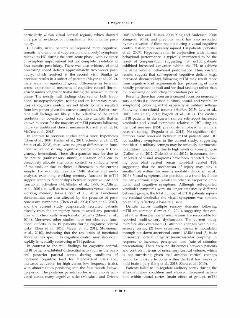
particularly within visual cortical regions, which showedonly partial evidence of normalization four months post-injury.
Clinically, mTBI patients self-reported more cognitive,somatic, and emotional (depression and anxiety) symptomsrelative to HC during the semi-acute phase, with evidenceof symptom improvement but not complete resolution atfour months post-injury. There was also evidence of mildprocessing speed deficits approximately two weeks post-injury, which resolved at the second visit. Similar toprevious results in a subset of patients (Mayer et al., 2012),there were no significant group differences in behavioracross experimental measures of cognitive control (incon-gruent minus congruent trials) during the semi-acute injuryphase. The mostly null findings observed on both tradi-tional neuropsychological testing and on laboratory meas-ures of cognitive control are not likely to have resultedfrom low power given the current sample size. Rather, cur-rent null findings are likely to be reflective of the rapidresolution of objectively tested cognitive deficits that isknown to occur for the majority of mTBI patients followinginjury on traditional clinical measures (Carroll et al., 2014;McCrea et al., 2013).
In contrast to previous studies and a priori hypotheses(Chen et al., 2007; Fischer et al., 2014; Scheibel et al., 2012;Smits et al., 2008), there were no group differences in func-tional activation during cognitive control (Group 3 Con-gruency interaction). These discrepancies may be due tothe nature (multisensory stimuli, utilization of a cue toproactively allocate attentional control) or difficulty levelof the task, or due to clinical differences in the currentsample. For example, previous fMRI studies and meta-analyses examining working memory function in mTBIsuggest complex relationships between cognitive load andfunctional activation (McAllister et al., 1999; McAllisteret al., 2001), as well as between continuous versus discreetworking memory tasks (Bryer et al., 2013). Functionalabnormalities are also affected by the presence of post-concussive symptoms (Chen et al., 2004; Chen et al., 2007),and the current study purposefully recruited patientsdirectly from the emergency room to avoid any potentialbias with chronically symptomatic patients (Mayer et al.,2014). Moreover, other studies have not observed func-tional deficits in mTBI patients during cognitive controltasks (Elbin et al., 2012; Mayer et al., 2012; Stulemeijeret al., 2010), indicating that the resolution of functionalabnormalities specific to cognitive control may also occurrapidly in typically recovering mTBI patients.
In contrast to the null findings for cognitive control,mTBI patients exhibited differential activation in the bilat-eral posterior parietal cortex during conditions ofincreased cognitive load for attend-visual trials (i.e.,increased activation for high versus low frequency trials),with abnormalities persisting into the four month follow-up period. The posterior parietal cortex is commonly acti-vated across many cognitive tasks (Macaluso and Driver,
2005; Nachev and Husain, 2006; Xing and Andersen, 2000;Zmigrod, 2014), and previous work has also indicatedhyper-activation of these regions during a visual cognitivecontrol task in more severely injured TBI patients (Scheibelet al., 2007). Hyper-activation in conjunction with normalbehavioral performance is typically interpreted to be theresult of compensation, suggesting that mTBI patientsexhibited increased activation within the IPL to achievethe same level of behavioral performance. Thus, currentresults suggest that self-reported cognitive deficits (e.g.,increased distractibility) following mTBI may result morefrom cognitive load requirements (i.e., processing of morerapidly presented stimuli and/or dual tasking) rather thanthe processing of conflicting information per se.
Recently there has been an increased focus on neurosen-sory deficits (i.e., increased auditory, visual, and vestibularsymptoms) following mTBI, especially in military settingsfollowing blast-related trauma (Hoffer, 2015; Lew et al.,2009; Lew et al., 2011; Pogoda et al., 2012). The civilianmTBI patients in the current sample self-report increasedvestibular and visual symptoms relative to HC using anidentical measure (NSI) previously employed in militaryresearch settings (Pogoda et al., 2012). No significant dif-ferences were observed between mTBI patients and HCfor auditory symptoms in the current study, suggestingthat blast in military settings may be uniquely detrimentalto auditory functioning due to high levels of acoustic noise(Gallun et al., 2012; Oleksiak et al., 2012). In contrast, simi-lar levels of visual symptoms have been reported follow-ing both blast related versus non-blast related TBI,suggesting that the mechanism of injury may play asmaller role within this sensory modality (Goodrich et al.,2013). Visual symptoms also persisted at a trend level intothe early chronic stage, similar to other self-reported emo-tional and cognitive symptoms. Although self-reportedvestibular symptoms were no longer statistically differentbetween groups, the total number of mTBI patients report-ing residual vestibular and visual symptoms was similar,potentially reflecting a base-rate issue.
Deficits across multiple sensory domains followingmTBI are common (Lew et al., 2011), suggesting that cen-tral rather than peripheral mechanisms are responsible forreported multi-sensory dysfunction. The current studytherefore also examined (1) atrophic changes within uni-sensory cortex, (2) how unisensory cortex is modulatedthrough top-down attentional control (ARM) and (3) basicunisensory cortical integrity (neurovascular coupling) inresponse to increased perceptual load (rate of stimuluspresentation). There were no differences between patientsand controls in terms of unisensory cortical volume, whichis not surprising given that atrophic cortical changeswould be unlikely to occur within the first few weeks ofmild brain injury (Ling et al., 2013; Zhou et al., 2013).
Patients failed to up-regulate auditory cortex during theattend-auditory condition and showed decreased activa-tion within visual cortex (main effect of group). mTBI
r Mayer et al. r
r 10 r
patients also exhibited an increased BOLD response in vis-ual cortex during increased perceptual load (stimulus rate)as well as attentional demands (ARM). However, the latterfinding was partially driven by a decreased response invisual cortex during attend-auditory trials, which was alsopresent as a main effect in voxel-wise analyses. Thus, cur-rent findings indicate different visual cortical abnormal-ities that were partially dependent on whether attentionwas focused on auditory or visual information streams.Similar to symptom self-report, there was only partialresolution of neuronal abnormalities in visual cortex atfour months post-injury, providing additional evidence oflonger-term neuronal changes in unisensory cortex follow-ing mTBI. Importantly, previous studies have observedstructural and functional abnormalities in the thalamus fol-lowing mTBI (Lui et al., 2014; Mayer et al., 2009b), andboth incoming sensory information and top-down modula-tory signals are routed through the thalamus prior toreaching unisensory cortex. Thus, we cannot rule-out thecontribution of thalamic and other mid-brain abnormalitiesto current results.
There are several limitations to the current study. First,our anatomical battery did not include more advancedstructural imaging (e.g., FLAIR, T2*) that may have pro-vided additional sensitivity for detecting lesions. Similarly,the current sample included patients both with (i.e., com-plicated mTBI) and without intracranial structural lesionsbased on CT and structural MRI scans, and evidence sug-gests that complicated patients may experience a moreprolonged recovery (Kashluba et al., 2008). Second, abnor-malities in the BOLD signal following mTBI may resultfrom impaired neuronal function, impaired neural controlof microvessels, direct damage to the vascular system,metabolic disruptions, changes in cerebral blood flow or acombination of these factors (Mayer et al., 2014; Meieret al., 2015). Standard EPI, when used alone, is not capableof disambiguating these various possible contributingsources. Similarly, it is not possible to determine whetherobserved differences in functional activation betweenpatients and HC are indicative of cortical dysfunction,compensatory activation or a neuro-protective response tobrain injury. Third, patients were not assessed with a for-mal neurological exam following injury, which could haveprovided additional information regarding neurosensorydysfunction. Finally, many of the hemodynamic abnormal-ities in unisensory cortex were at trend levels in spite ofour larger sample size, suggesting that neural evidence ofsensory dysfunction is likely to be subtle even during thesemi-acute injury phase when a larger number of patientsare more symptomatic.
In summary, understanding the central mechanismsunderlying self-reported neurosensory deficits followingmTBI is imperative, as these sensory deficits negativelyaffect everyday functioning and there are currently noaccepted treatments (Hoffer, 2015). Consistent with reportsfrom military cohorts (Pogoda et al., 2012), mTBI patients
self-reported more cognitive, somatic, emotional as well asvisual and vestibular symptoms during the semi-acutestage, which only partially resolved at four months post-injury. Evidence of multifaceted neural abnormalities inunisensory cortex was also observed, particularly withinvisual cortex. In contrast, current results provided no evi-dence of behavioral or functional deficits in cognitive con-trol during the semi-acute phase of mTBI. Rather, currentresults suggest a greater role for compensatory activationwithin the IPL as a function of increased cognitive load toachieve similar levels of behavioral performances. Func-tional abnormalities within the visual cortex and IPL per-sisted into the early chronic stage, suggesting that neuralabnormalities may lag behind the traditional behavioralmeasures that are used in most clinical settings.
ACKNOWLEDGMENT
Special thanks to Diana South and Cathy Smith for assis-tance with data collection.
REFERENCES
Arciniegas D, Olincy A, Topkoff J, McRae K, Cawthra E, FilleyCM, Reite M, Adler LE (2000): Impaired auditory gating andP50 nonsuppression following traumatic brain injury.J Neuropsychiatry Clin Neurosci 12:77–85.
Arciniegas DB, Topkoff JL (2004): Applications of the P50 evokedresponse to the evaluation of cognitive impairments after trau-matic brain injury. Phys Med Rehabil Clin N Am 15:177–203,viii.
Baier B, Kleinschmidt A, Muller NG (2006): Cross-modal process-ing in early visual and auditory cortices depends on expectedstatistical relationship of multisensory information. J Neurosci26:12260–12265.
Bryer EJ, Medaglia JD, Rostami S, Hillary FG (2013): Neuralrecruitment after mild traumatic brain injury is task depend-ent: A meta-analysis. J Int Neuropsychol Soc 19:751–762.
Carroll LJ, Cassidy JD, Cancelliere C, Cote P, Hincapie CA,Kristman VL, Holm LW, Borg J, Nygren-de BC, Hartvigsen J(2014): Systematic review of the prognosis after mild traumaticbrain injury in adults: Cognitive, psychiatric, and mortalityoutcomes: Results of the International Collaboration on MildTraumatic Brain Injury Prognosis. Arch Phys Med Rehabil 95:S152–S173.
Chen JK, Johnston KM, Frey S, Petrides M, Worsley K, Ptito A(2004): Functional abnormalities in symptomatic concussedathletes: An fMRI study. Neuroimage 22:68–82.
Chen JK, Johnston KM, Collie A, McCrory P, Ptito A (2007): Avalidation of the post concussion symptom scale in the assess-ment of complex concussion using cognitive testing and func-tional MRI. J Neurol Neurosurg Psychiatry 78:1231–1238.
Cox RW (1996): AFNI: Software for analysis and visualization offunctional magnetic resonance neuroimages. Comput BiomedRes 29:162–173.
Destrieux C, Fischl B, Dale A, Halgren E (2010): Automatic parcel-lation of human cortical gyri and sulci using standard anatom-ical nomenclature. Neuroimage 53:1–15.
Elbin RJ, Covassin T, Hakun J, Kontos AP, Berger K, Pfeiffer K,Ravizza S (2012): Do brain activation changes persist in
r Multisensory Cognitive Control in mTBI r
r 11 r
athletes with a history of multiple concussions who are asymp-
tomatic? Brain Inj 26:1217–1225.Faul M, Xu L, Wald MM, Coronado VG (2010): Traumatic brain
injury in the United States: Emergency department visits, hos-
pitalizations, and deaths. Centers for Disease Control and
Prevention.Fischl B, Rajendran N, Busa E, Augustinack J, Hinds O, Yeo BT,
Mohlberg H, Amunts K, Zilles K (2008): Cortical folding patterns
and predicting cytoarchitecture. Cereb Cortex 18:1973–1980.Fischer BL, Parsons M, Durgerian S, Reece C, Mourany L, Lowe
MJ, Beall EB, Koenig KA, Jones SE, Newsome MR, Scheibel
RS, Wilde EA, Troyanskaya M, Merkley TL, Walker M,
Levin HS, Rao SM (2014): Neural activation during response
inhibition differentiates blast from mechanical causes of
mild to moderate traumatic brain injury. J Neurotrauma 31:
169–179.Gallun FJ, Lewis MS, Folmer RL, Diedesch AC, Kubli LR,
McDermott DJ, Walden TC, Fausti SA, Lew HL, Leek MR
(2012): Implications of blast exposure for central auditory func-
tion: A review. J Rehabil Res Dev 49:1059–1074.Goodrich GL, Flyg HM, Kirby JE, Chang CY, Martinsen GL
(2013): Mechanisms of TBI and visual consequences in military
and veteran populations. Optom Vis Sci 90:105–112.Halterman CI, Langan J, Drew A, Rodriguez E, Osternig LR,
Chou LS, van DP (2006): Tracking the recovery of visuospatial
attention deficits in mild traumatic brain injury. Brain 129:
747–753.Hinds OP, Rajendran N, Polimeni JR, Augustinack JC, Wiggins G,
Wald LL, Diana RH, Potthast A, Schwartz EL, Fischl B (2008):
Accurate prediction of V1 location from cortical folds in a sur-
face coordinate system. Neuroimage 39:1585–1599.Hoffer ME (2015): Mild traumatic brain injury: Neurosensory
effects. Curr Opin Neurol 28:74–77.Hubel DH, Henson CO, Rupert A, Galambos R (1959): “Attention”
units in the auditory cortex. Science 129:1279–1280.Kashluba S, Hanks RA, Casey JE, Millis SR (2008): Neuropsycho-
logic and functional outcome after complicated mild traumatic
brain injury. Arch Phys Med Rehabil 89:904–911.Kato T, Nakayama N, Yasokawa Y, Okumura A, Shinoda J,
Iwama T (2007): Statistical image analysis of cerebral glucose
metabolism in patients with cognitive impairment following
diffuse traumatic brain injury. J Neurotrauma 24:919–926.Kim J, Whyte J, Patel S, Europa E, Slattery J, Coslett HB, Detre JA
(2012): A perfusion fMRI study of the neural correlates of
sustained-attention and working-memory deficits in chronic
traumatic brain injury. Neurorehabil Neural Repair 26:870–880.Kumar S, Rao SL, Nair RG, Pillai S, Chandramouli BA,
Subbakrishna DK (2005): Sensory gating impairment in devel-
opment of post-concussive symptoms in mild head injury. Psy-
chiatry Clin Neurosci 59:466–472.Lew HL, Garvert DW, Pogoda TK, Hsu PT, Devine JM, White
DK, Myers PJ, Goodrich GL (2009): Auditory and visual
impairments in patients with blast-related traumatic brain
injury: Effect of dual sensory impairment on Functional Inde-
pendence Measure. J Rehabil Res Dev 46:819–826.Lew HL, Weihing J, Myers PJ, Pogoda TK, Goodrich GL (2010):
Dual sensory impairment (DSI) in traumatic brain injury
(TBI)–An emerging interdisciplinary challenge. NeuroRehabili-
tation 26:213–222.Lew HL, Pogoda TK, Baker E, Stolzmann KL, Meterko M, Cifu
DX, Amara J, Hendricks AM (2011): Prevalence of dual sen-
sory impairment and its association with traumatic brain
injury and blast exposure in OEF/OIF veterans. J Head
Trauma Rehabil 26:489–496.Ling JM, Klimaj S, Toulouse T, Mayer AR (2013): A prospective
study of gray matter abnormalities in mild traumatic brain
injury. Neurology 81:2121–2127.Lui YW, Xue Y, Kenul D, Ge Y, Grossman RI, Wang Y (2014):
Classification algorithms using multiple MRI features in mild
traumatic brain injury. Neurology 83:1235–1240.Macaluso E, Driver J (2005): Multisensory spatial interactions: A
window onto functional integration in the human brain.Trends Neurosci 28:264–271.
Mayer AR, Franco AR, Canive J, Harrington DL (2009a): The
effects of stimulus modality and frequency of stimulus presen-
tation on cross-modal distraction. Cereb Cortex 19:993–1007.Mayer AR, Mannell MV, Ling J, Elgie R, Gasparovic C, Phillips
JP, Doezema D, Yeo RA (2009b): Auditory orienting and inhi-
bition of return in mild traumatic brain injury: A FMRI study.
Hum Brain Mapp 30:4152–4166.Mayer AR, Teshiba TM, Franco AR, Ling J, Shane MS, Stephen
JM, Jung RE (2011): Modeling conflict and error in the medial
frontal cortex. Hum Brain Mapp 33:2843–2855.Mayer AR, Yang Z, Yeo RA, Pena A, Ling JM, Mannell MV,
Stippler M, Mojtahed K (2012): A functional MRI study of mul-timodal selective attention following mild traumatic brain
injury. Brain Imaging Behav 6:343–354.Mayer AR, Bellgowan PS, Hanlon FM (2014): Functional magnetic
resonance imaging of mild traumatic brain injury. Neurosci
Biobehav Rev 49C:8–18.McAllister TW, Saykin AJ, Flashman LA, Sparling MB, Johnson
SC, Guerin SJ, Mamourian AC, Weaver JB, Yanofsky N (1999):
Brain activation during working memory 1 month after mild
traumatic brain injury: A functional MRI study. Neurology 53:
1300–1308.McAllister TW, Sparling MB, Flashman LA, Guerin SJ,
Mamourian AC, Saykin AJ (2001): Differential working mem-
ory load effects after mild traumatic brain injury. Neuroimage
14:1004–1012.McCrea M, Guskiewicz K, Randolph C, Barr WB, Hammeke TA,
Marshall SW, Powell MR, Woo AK, Wang Y, Kelly JP (2013):
Incidence, clinical course, and predictors of prolonged recov-
ery time following sport-related concussion in high school and
college athletes. J Int Neuropsychol Soc 19:22–33.Meier TB, Bellgowan PS, Singh R, Kuplicki R, Polansky M, Mayer
AR (2015): Recovery of cerebral blood flow following sports-related concussion. JAMA Neurol 72:530–538.
Nachev P, Husain M (2006): Disorders of visual attention and the
posterior parietal cortex. Cortex 42:766–773.Nakashima T, Nakayama N, Miwa K, Okumura A, Soeda A,
Iwama T (2007): Focal brain glucose hypometabolism in
patients with neuropsychologic deficits after diffuse axonal
injury. Am J Neuroradiol 28:236–242.Oleksiak M, Smith BM, St Andre JR, Caughlan CM, Steiner M
(2012): Audiological issues and hearing loss among Veterans
with mild traumatic brain injury. J Rehabil Res Dev 49:995–
1004.Pogoda TK, Hendricks AM, Iverson KM, Stolzmann KL, Krengel
MH, Baker E, Meterko M, Lew HL (2012): Multisensoryimpairment reported by veterans with and without mild trau-
matic brain injury history. J Rehabil Res Dev 49:971–984.Ridderinkhof KR, Ullsperger M, Crone EA, Nieuwenhuis S (2004):
The role of the medial frontal cortex in cognitive control. Sci-
ence 306:443–447.
r Mayer et al. r
r 12 r
Roberts KL, Hall DA (2008): Examining a supramodal network forconflict processing: A systematic review and novel functionalmagnetic resonance imaging data for related visual and audi-tory stroop tasks. J Cogn Neurosci 20:1063–1078.
Scheibel RS, Newsome MR, Steinberg JL, Pearson DA, Rauch RA,Mao H, Troyanskaya M, Sharma RG, Levin HS (2007): Alteredbrain activation during cognitive control in patients with mod-erate to severe traumatic brain injury. Neurorehabil NeuralRepair 21:36–45.
Scheibel RS, Newsome MR, Troyanskaya M, Lin X, Steinberg JL,Radaideh M, Levin HS (2012): Altered brain activation in mili-tary personnel with one or more traumatic brain injuries fol-lowing blast. J Int Neuropsychol Soc 18:89–100.
Shenhav A, Botvinick MM, Cohen JD (2013): The expected valueof control: An integrative theory of anterior cingulate cortexfunction. Neuron 79:217–240.
Smits M, Dippel DW, Houston GC, Wielopolski PA, Koudstaal PJ,Hunink MG, van der Lugt A (2008): Postconcussion syndromeafter minor head injury: Brain activation of working memoryand attention. Hum Brain Mapp 30:2789–2803.
Stamatakis EA, Wilson JT, Hadley DM, Wyper DJ (2002): SPECTimaging in head injury interpreted with statistical parametricmapping. J Nucl Med 43:476–483.
Stulemeijer M, Vos PE, van der Werf S, van DG, Rijpkema M,Fernandez G (2010): How mild traumatic brain injury mayaffect declarative memory performance in the post-acute stage.J Neurotrauma 27:1585–1595.
Talairach J, Tournoux P (1988): Co-Planar Stereotaxic Atlas of theHuman Brain. New York: Thieme.
Talsma D, Senkowski D, Soto-Faraco S, Woldorff MG (2010): Themultifaceted interplay between attention and multisensoryintegration. Trends Cogn Sci 14:400–410.
Weissman DH, Warner LM, Woldorff MG (2004): The neuralmechanisms for minimizing cross-modal distraction.J Neurosci 24:10941–10949.
Xing J, Andersen RA (2000): Models of the posterior parietal cor-tex which perform multimodal integration and represent spacein several coordinate frames. J Cogn Neurosci 12:601–614.
Zhang L, Yang KH, King AI (2004): A proposed injury thresholdfor mild traumatic brain injury. J Biomech Eng 126:226–236.
Zhou Y, Kierans A, Kenul D, Ge Y, Rath J, Reaume J, GrossmanRI, Lui YW (2013): Mild traumatic brain injury: Longitudinalregional brain volume changes. Radiology 267:880–890.
Zmigrod S (2014): The role of the parietal cortex in multisensoryand response integration: Evidence from transcranial directcurrent stimulation (tDCS). Multisens Res 27:161–172.
r Multisensory Cognitive Control in mTBI r
r 13 r