a field key to common genera of hypogeous and gasteriod basidiomycetes of
TRANSCRIPT

A FIELD KEY TO COMMON GENERA OF HYPOGEOUSAND GASTERIOD BASIDIOMYCETES OF NORTH
AMERICA
Steven L. MillerBotany Department
University of Wyoming
Laramie, Wyoming 82071
INTRODUCTIONTo honor the tenth anniversary of the North American Truffling
Society (NATS) in 1987, the author taught a course entitled "Truffles of thePacific Northwest" at Linn-Benton Community College near Corvallis,Oregon. As part of that course he assembled and distributed a field key tohypogeous fungi, including both ascomycetes (true truffles) andbasidiomycetes (false truffles), which would allow NATS members, most ofwhom did not have ready access to a microscope, to identify most of theircollections to genus. The key was extensively used and revised. In honor ofthe twentieth anniversary of NATS, the present paper is a field key tohypogeous and gasteroid basidiomycetes (and one additional subdivision thatmay be confusing in the field).
The task of producing a field key to hypogeous fungi is a difficultone. Many of the characters that serve to distinguish genera and speciesrequire the use of a microscope. In order to use only macroscopic charactersin this key, several not-so-common genera have been intentionally omitted.The key is admittedly artificial, but an attempt has been made to structure itso that omitted genera will key out to a related genus, and with the use of amicroscope and appropriate literature, should be readily identifiable.
This key is not intended as a substitute for a well rounded regimen ofecological, morphological, and microscopical study. However, it is hopedthat the key will allow interested persons that do not have access to amicroscope to identify most common genera of hypogeous basidiomycetes.
METHODS AND MATERIALSThere are many sources describing the best ways to collect fleshy
fungi (Miller 1978; Arora 1986) and there is no need to repeat thisinformation here, except to point out that truffles are fleshy fungi too, andwill become rotten and disgusting just like any mushroom if left in a plasticbag even for a few hours. Therefore collecting should be done with wax-paper bags or compartmentalized plastic fishing-tackle boxes. Althoughnotes on the fresh condition should be taken as soon after collecting aspossible, it is generally not advisable to cut into all of the sporocarps whilein the field unless sufficient time can be spent to study the specimen andassess the best way to cut it to reveal the necessary structures. If you dofollow that practice (admittedly, most of us can't always control ourexcitement), it is best to assess the fresh odor, taste, color etc. at the sametime.

2
The following key is best used when a full range of developmentalfeatures are present in a single collection, i.e. young, mature and oldspecimens all collected from a relatively close proximity. Young, immaturesporocarps will be difficult to identify, but may provide clues as to color,odor etc. Full utilization of the key also requires the use of a sharp knife orpreferably a razor blade, hand lens and various macrochemical reagents. Whenthe sporocarps are cut open to reveal detail in the gleba, a true vertical,median section should be used, i.e. cut vertically from the base through to theapex. This is important because the terminology of the columella, gleba, andperidium is based on a true, vertical, median view, and would otherwise beconfusing. In addition, mushroom buttons (from both poisonous andnonpoisonous species) often resemble gasteroid and hypogeous fungi and thebest way to tell the difference is to perform a true, vertical median section andlook for the embryonic mushroom form that appears in mushroom buttonsbut not in hypogeous or gasteroid fungi. An incorrect, transverse section of amushroom button will appear as concentric circles and not like any of thesporocarp or gleba types described below.
MORPHOLOGYMany of the morphological features of the fungi treated in this key
are continuous or intergrade, meaning that one sporocarp, columella or glebatype may overlap slightly or greatly with another. However, there aretypically limits on the amount of intergradation for any given genus whichcan be recognized.
Sporocarp form
Agaricoid sporocarps generally have a stipe, a cap and either gills(lamellae) or tubes (pores) where the spores are produced. This is the typicalmushroom morphology (Fig. 1). Gasteroid sporocarps can appear verysimilar, with stipe-like, cap-like and gill-like structures, or they can havesome degree of enclosure of the spore producing tissue either from the capsurface or by wrinkling, contorting or chambering of the gill-like structures,or both (Figs 2, 4). Hypogeous sporocarps typically lack any true stipe-like structure, are more or less spherical or globose, and have little, if anyexposure of the spore bearing tissue. A good analogy would be a golf-ball.
Stipe, stipe-columella, or columella
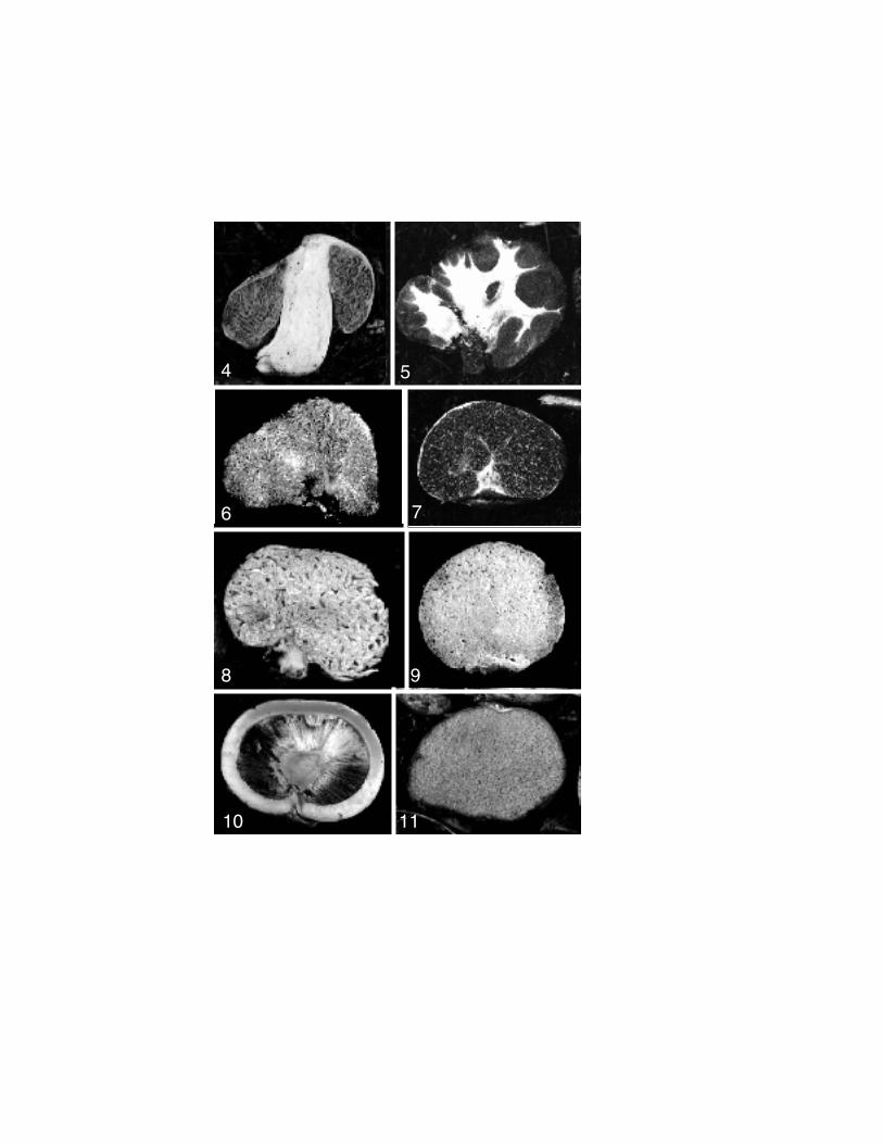
The columella (including stipe, stipe-columella, and columella) forthe purposes of this key is considered a reduction series in the amount andlocation of stipe (stalk) tissue (Figs 4-11). A stipe is by definition foundonly in agaricoid sporocarps (Fig. 1), however, the stipe-columella foundin some gasteroid fungi is indistinguishable from a stipe, and is likely to behomologous with it (Fig 4). The stipe-columella found in somegasteroid and hypogeous sporocarps is much reduced in extent, and no longerserves the function of elevating the sporocarp above the ground surface (Figs.2, 8). The columella is yet a further reduction in the amount and locationof sterile, stipe-like tissue, and is found almost exclusively in hypogeousfungi (Figs. 5-7, 9-10). Sporocarps of some hypogeous fungi lack columellatissue (Fig. 11).

3
To provide additional characteristics for the key, columella typeshave been further subdivided into several categories. A percurrentcolumella extends completely through the sporocarp as viewed in section(Figs. 4-5) and is a condition that can be either found in a stipe, stipe-columella or columella. A dendritic columella forms many branches from amore or less central mass of stipe-like tissue (Fig 5). A diffuse columellais similar to the dendritic form, but is much less evident in both branchingpattern and extent (Figs 6-7). A sterile base can either resemble a shortnonpercurrent stipe-columella (Fig 8) or a more reduced mass of stipe-liketissue at the base of a hypogeous sporocarp (Fig. 9). A central columellatypically has a large spherical mass of sterile tissue near the center of thesporocarp from which rays of spore bearing tissue or peridioles radiateoutward (Figs 10, 19). Other fungi exhibit no columella and possess nosterile stipe-like tissue when viewed in section (Figs 3, 11, 15, 17-19).
Gleba type
The gleba is a combination of sterile and spore-bearing tissuetypically found enclosed by protective tissues in fungi such as puffballs,some gasteroid fungi, and hypogeous fungi. For simplicity, the term hasbeen extended to gasteroid/agaricoid sporocarps, even though the spore-bearing tissue is exposed (Figs. 2, 12-13). The lamellate gleba can rangefrom gill-like, nearly indistinguishable from that of agaricoid sporocarps, towrinkled, contorted or chambered, but retaining the radial pattern of lamellae(Fig. 12). The lamellate gleba is typically exposed, at least to some extent.The labyrinthiform gleba may also retain a partial radial pattern but iscomposed of large to small chambers, typically irregularly bent and crumpled,formed by wrinkles, contortions or cross-walls of the glebal plates (Figs. 2,4, 5-6, 8, 13-14). There may also be some degree of exposure. Thelacunate gleba appears in section to be formed of large to small shallowholes or chambers, which are more or less identical in shape, and are typicallyonly rarely exposed (Figs. 3, 7, 9, 11, 15). The gleba may also appear solid(not necessarily implying tough) in section because the chambers are, orappear to be, filled or stuffed with material. The solid and rubbery glebais typically composed of minute chambers difficult to discern even with ahand lens, and the sporocarp is usually difficult to cut into (Fig. 16). In thegel-filled gleba, the chambers are filled at maturity with a gelatinous,usually highly colored material (Fig. 17). In the liquid-filled gleba, themature chambers are filled with a material that is more fluid, and appears tobleed or flow from the chambers (Fig. 18). This material is usually nothighly colored. A concern for correct interpretation of the gel-filled andliquid-filled gleba types is the degree of maturity of the specimen. Thesegleba types should also not be confused with exudation of a latex from thesterile tissue between chambers. The peridiate gleba (Fig. 19) is formedfrom distinct, separable packets of spores called peridioles that look likeseeds. This is a rare type of gleba restricted to only one or two genera and isusually associated with a central columella type.
Spore color
The color of spores in the gleba is an important characteristic in

4
identifying hypogeous and gasteroid fungi and should be interpreted carefully.Frequently, the color observed in the freshly cut section is not the color ofspores, rather it is the color of the sterile tissue comprising the glebalelements. A good example is the genus Hydnangium, which often appearspurplish or flesh colored in section. However, close examination of theglebal chambers with a hand lens reveals that the spores are whitish and thetissue is purplish.
KEY TO HYPOGEOUS AND GASTEROIDBASIDIOMYCETES BASED ON MACROSCOPIC AND
ECOLOGICAL CHARACTERISTICS1. Sporocarps globose to subglobose, rarely with a stipe; usually
hypogeous; gleba either: solid, hard, lacking chambers or composed of auniformly thickened layer lining a single, large, hollow chamber, usuallywith an orifice, grooves or openings to the inside or nearly hollow,stuffed with cottony mycelium young but powdery in age or composedof infolded partitions, the partitions either single or multiple andcompact, in some nearly becoming solid; texture firm, solid to plastic-like, usually very brittle. Hypogeous members of SubdivisionAscomycotina..............................(see Trappe and Castellano 1991)
1. Sporocarps globose, subglobose or like an enclosed mushroom, oftenwith a stipe, stipe-columella or sterile base; hypogeous to epigeous;gleba composed of numerous, small or moderately large, distinctchambers or lamellae-like, contorted tramal plates or thin to moderatelythick, radiating septae or tramal plates or numerous, tapering,centripetally arranged peridioles or with combinations of chambers andcontorted tramal plates; if powdery at maturity then young sporocarpschambered or with radiating tramal plates, the chamber walls thin,breaking down, dehiscent, if appearing solid, then mature chambersfilled with moist, gel-like material; texture soft bread-like, firm to toughand rubbery, or cartilaginous, usually not brittle. (Hypogeous andgasteroid members of Subdivision Basidiomycotina).....................2
2. Sporocarps globose, subglobose to elongated or flattened, occasionallywith a thick, matted cluster of basal rhizomorphs but not possessing anobvious external stipe, stipe-columella or well formed, large sterile base;gleba usually not exposed to the surface, but if gleba exposed, thenperidium entirely lacking or present in a large patch near the apex;hypogeous, subepigeous or occasionally epigeous; mostlyectomycorrhizal .........................................................................3
2. Sporocarps variously shaped but agaricoid or gasteroid, with a stipe,stipe-columella or sterile base that is evident before sectioning, oftenresembling a contorted mushroom, an unexpanded mushroom button, ora fully developed but unopened mushroom; with or without a small orlarge portion of the gleba exposed; hypogeous, subepigeous or frequentlyepigeous; ectomycorrhizal or saprotrophic .....................................46
3. Sporocarps when cut with true vertical section possessing a small or large

5
columella or small sterile base in the gleba or densely matted cluster ofrhizomorphs resembling a sterile base ............................................4
3. Sporocarps when cut with true vertical section not possessing a columellaor sterile base in the gleba..........................................................31
4. Gleba of centripetally or radially arranged tramal plates or peridiolesextending from central columella or sterile base or of chambers evidentat youth but becoming powdery at maturity.....................................5
4. Gleba composed of numerous, small or large chambers, occasionallyrequiring a hand lens to be visible .................................................9
5. Gleba of radially arranged, tapering black peridioles........ Pyrenogaster
5. Gleba of radiating tramal plates or chambers in youth but rapidlybecoming powdery at maturity.......................................................6
6. Gleba with persistent central columella ..........................................76. Gleba lacking a columella ............................................................87. Gleba brown, dark brown or black at full development; tramal plates
radiating centripetally from spherical central columella or sterilebase............................................................................ Radiigera
7. Gleba black at full development ; tramal plates and chambers pendant fromdisk of sterile tissue supported by a percurrent columella composed ofthick rhizomorphs; peridium thick, leathery, dehiscing ...... Sedecula
8. Thick outer peridium tomentose, and usually splitting into well formedstellate pattern in age; gleba brown to dark brown usually contained in aleathery inner peridium..................................................... Astraeus
8. Thick outer peridium mostly not splitting into stellate pattern in age, orif so, peridium not tomentose; gleba black, powdery at maturity and notcontained in an inner peridium..................................... Scleroderma
9. Gleba darkly or brightly colored, usually in the range of brown, pink,yellow, orange, green or blue-green..............................................10
9. Gleba typically white or off-white................................................2810. Gleba gray-brown, brown, red-brown to dark brown ........................1110. Gleba variously colored, occasionally olive to olive brown but not
brown.....................................................................................1911. Sporocarp lacking peridium; reaction to Melzer's reagent giving a
dextrinoid reaction ................................................. Protogautieria
11. Sporocarp with well developed peridium entirely covering gleba; reactionto Melzer's reagent various, but usually not dextrinoid.....................12
12. Gleba of chambers filled with gel-like material ................. Destuntzia
12. Gleba of small or large chambers, containing spores but not filled atmaturity with gel-like material ...................................................13
13. Columella well developed, dendritic or percurrent ...........................1413. Columella poorly developed or lacking; sterile base may be present....16

6
14. Sporocarps usually soft, fleshy, to firm but not cartilaginous orrubbery, columella or stipe-columella present ........... Gymnoglossum
14. Sporocarps usually cartilaginous, tough or rubbery; columella usuallydiffuse, never a percurrent stipe-columella or columella....................15
15. Sporocarps medium to large; peridium usually thin, evanescent, white orpale brown; gleba gray brown, red brown, brown or darkbrown.......................................................................... Gautieria
15. Sporocarps usually small; peridium thick, persistent, white; gleba pinkto red brown ......................................................... Hysterangium
16. Gleba strongly amyloid in Melzer's reagent....................................1716. Gleba not amyloid in Melzer's reagent, but may be slightly
dextrinoid................................................................................1817. Brown color of gleba usually from discoloring reaction of tramal tissue
and not from accumulation of spores; odor strong, oily, of maple syrupor slightly of rubber................. Martellia (including Gymnomyces )
17. Brown color of gleba usually from accumulation of spores in the glebalchambers; odor absent or different...................................... Martellia
18. Fresh sporocarps with strong, penetrating, farinaceous odor.......................................................................... Hymenogaster
18. Fresh sporocarps with no or different odor ..................... Octavianina
19. Gleba usually some shade of green including yellow green, olive green,blue green................................................................................20
19. Gleba some shade of pink, yellow, orange, or gray-blue...................2220. Columella derived from a stubby sterile base, percurrent or nearly
percurrent, dentritic; fleshy but not cartilaginous or gelatinous except inage.................................................................. Truncocolumella
20. Columella absent or simple or branched; glebal tissues cartilaginous orgelatinous................................................................................21
21. Sporocarps single or loosely clustered, habitat various; cartilaginous andrubbery, the peridium often cleanly separating from the gleba, associatedwith a variety of tree hosts....................................... Hysterangium
21. Sporocarps, tightly clustered, encased in heavy mycelium, hyphalstrands, soil and ectomycorrhizal rootlets, moderately gelatinousassociated with Eucalyptus ..................................... Chondrogaster
22. Fresh gleba when cut exuding a latex............................................2322. Fresh gleba when cut not exuding a latex.......................................2423. Gleba amyloid in Melzer's reagent.............................. Zelleromyces
23. Gleba not amyloid in Melzer's reagent; with large orange spores that arevisible with a hand-lens embedded in tissue... Endogone (Zygomycete)
24. Gleba blue-gray at maturity ............................................. Gautieria
24. Gleba not blue-gray at maturity, but some shade of pink, yellow or

7
orange....................................................................................2525. Gleba some shade of pink, associated with Eucalyptus .... Hydnangium
25. Gleba some shade of yellow or orange...........................................2626. Gleba amyloid in Melzer's reagent ...............................................2726. Gleba not amyloid in Melzer's reagent .......................... Sclerogaster
27. Columella or sterile base if present poorly developed............ Martellia
27. Small stipe-columella, columella or sterile base present.. Gymnomyces
28. Gleba amyloid in Melzer's reagent................................................2928. Gleba not amyloid in Melzer's reagent........................... Octavianina
29. Fresh sporocarps exuding latex when cut...................... Zelleromyces
29. Fresh sporocarps not exuding latex when cut..................................3030. Columella or sterile base if present poorly developed............ Martellia
30. Small stipe-columella, columella or sterile base present.. Gymnomyces
31. Gleba composed of empty or gel-filled chambers, or powdery at maturity,with or without a latex...............................................................32
31. Gleba appearing solid or with orange spores embedded in tissue visiblewith a hand lens, no evidence of chambers surrounded by pale coloredveins, with a latex .................................... Endogone (Zygomycete)
32. Gleba with chambers filled with thick, colored gel-like material, thesedelineated by pale colored veins....................................................33
32. Gleba composed of chambers that are not filled with a gel-like material atmaturity, chambers may appear empty, filled with thin, white latex orwith powdery content ................................................................34
33. Gleba composed of black gel-filled chambers surrounded by pale coloredveins; usually with a strong odor of latex paint............. Melanogaster
33. Gleba yellow, brown, olive, brown or red colored, if black or nearlyblack then the peridium is colored bright yellow; usually with no ordifferent odor.................................................................... Alpova
34. Gleba white when fresh and mature, and with a scant or profuse whitelatex ......................................................................................35
34. Gleba usually not white at maturity, with or without latex or if whitethen no latex is present..............................................................36
35. Gleba with copious latex in fresh specimens; chambers ≥ 0.5-3.0 mmbroad....................................................................... Leucogaster
35. Gleba with scant latex in fresh specimens, this drying rapidly into achalky, white surface, often obscuring the chambers; chambers ≤ 0.3-0.5mm broad................................................................ Leucophleps
36. Gleba strongly amyloid in Melzer's reagent....................................3736. Gleba not amyloid in Melzer's reagent...........................................4237. Gleba white or off-white at maturity.............................................38

8
37. Gleba more darkly colored at maturity...........................................3938. Sporocarp very light in weight; with a fruity or nauseous odor; and fresh
the gleba is dry ........................................................... Mycolevis
38. Sporocarp more dense; with various odors but usually not nauseous, andfresh gleba moist .......................................................... Martellia
39. Fresh sporocarps exuding a copious latex..................... Zelleromyces
39. Fresh sporocarps moist but not exuding a copious latex ..................4040. Fresh peridium quickly turning vinaceous in KOH........... Rhizopogon
40. Color of fresh peridium not affected by KOH..................................4141. Columella or sterile base if present poorly developed; brown color of
gleba usually from discoloring of tramal tissue, and not fromaccumulation of spores ................................................... Martellia
41. Small stipe-columella, or columella and sterile basepresent.................................................................. Gymnomyces
42. Gleba at first composed of chambers separated by pale colored veins, butat maturity gleba powdery .......................................... Scleroderma
42. Gleba not composed of chambers separated by pale colored veins, and isnot powdery at maturity.............................................................43
43. Gleba gray-brown, brown, red-brown to dark brown; usually with astrong, penetrating, farinaceous odor ......................... Hymenogaster
43. Gleba variously colored; with a variety of odors but not farinaceous....4444. Gleba some shade of pink (like Entoloma spore print color) at maturity,
found only in the southeastern and southwestern United States.............................................................................. Richoniella
44. Gleba usually some shade of white, yellow, green or brown but i f someshade of pink then not found in the southeast or southwest and glebabruises black, blue or red............................................................45
45. Gleba white to cinnamon brown at maturity, discoloring on exposure;peridium white to pink at maturity, also discoloring upon bruising; scantlatex may be present................................................... Octavianina
45. Gleba various colors, gleba infrequently discoloring on exposure;peridium various colors, frequently discoloring on bruising, latex usuallylacking but if present then from the southeastern United States withsterile veins present in gleba........................................ Rhizopogon
46. Gleba chocolate brown, dark purple brown, smoky gray, or black at fulldevelopment due to coloration of the spores in mass........................47
46. Gleba white, yellow, orange, green, vinaceous (pale purple brown) orolive yellow, yellow brown, red brown to rusty brown at fulldevelopment due to coloration of the spores in mass .......................55
47. Stipe or stipe columella tough, woody in mature specimens; found indeserts and arid grasslands and steppe ............................................48

9
47. Stipe or stipe columella fleshy, even in age; found in forests in theRockies westward and infrequently in deserts and steppe....................52
48. Peridium dehiscing cleanly around the circumference of margin; glebablack, lamellate, hanging free from disk of pileus............................49
48. Peridium not dehiscing cleanly, rather, breaking up and weatheringrandomly; gleba usually powdery at maturity..................................50
49. Stipe-columella with a persistent volva and annulus.. Gyrophragmium
49. Stipe-columella with volva but lacking an annulus ........... Montagnea
50. Resembling a large unopened Coprinus ("inky cap", or "shaggy-mane");annulus absent or poorly developed ..............................................51
50. Resembling a large, unopened Agaricus , often bruising red, or yellowwhen fresh; volva absent, annulus present............................ Longula
51. Sporocarps large, many reaching 25 cm or so; external portion of stipe-columella prominent with a bulbous base; gleba often maturing from thebase of the gleba upward ................................................... Podaxis
51. Sporocarps typically smaller; all parts of gleba maturing simultaneously;external portion of stipe-columella poorly developed, tapering to thebase........................................................................... Phellorina
52. Trama of gleba and stipe-columella colored pale orange to pale red,turning bright red in ETOH......................................... Brauniellula
52. Trama of gleba and stipe-columella white or very pale, not turning brightred in ETOH............................................................................53
53. Sporocarp robust, fleshy ............................................................5453. Sporocarp slender , fragile; not necessarily associated with conifers;
pileus typically acutely conical; often found on dung or otherdecomposing organic matter; resembling an enclosed Stropharia orPsilocybe ...................................................................... Weraroa
54. Hypogeous to subepigeous associated with Pinaceae; resembling anenclosed or partially enclosed Gomphidius ; gleba sublamellate and notpowdery at maturity; usually yellowish near the base of the stipe-columella............................................................. Gomphogaster
54. Hypogeous to subepigeous associated with Pinaceae in the PacificNorthwest; resembling a large unopened Agaricus button; gleba powderyat maturity; usually bruising vinaceous near the base of the stipe-columella.............................................................. Endoptychum
55. Spores in mass in the chambers or tramal plates white, pale yellow ororange at full development .........................................................56
55. Spores in mass in the chambers or tramal plates green, olive yellow,olive brown, vinaceous, yellow brown, red brown to rusty brown......59
56. Color of glebal trama pink to pale red; sterile base and/or small, non-percurrent columella present, easily lost or broken; gleba not amyloid inMelzer's reagent; found only in association with Eucalyptus

10
............................................................................. Hydnangium
56. Glebal and tramal characters not as above; may or may not be found withEucalyptus ..............................................................................57
57. Fresh gleba and/or peridium exuding latex when cut; resemblingcontorted or unopened Lactarius ................................. Arcangeliella
57. Neither fresh gleba or peridium exuding latex when cut....................5858. Percurrent stipe-columella or columella present; gleba loculate,
labyrinthiform, or sublamellate, may or may not be exposed; resemblingcontorted or unopened Russula..................... Macowanites (includingElasmomyces )
58. Sterile base or at most small, columella present; gleba usually loculateor at most labyrinthiform, not exposed........................ Gymnomyces
59. Spores in mass in the chambers or tramal plates some shade of green toolive brown or vinaceous ...........................................................60
59. Spores in mass in the chambers or tramal plates some shade of yellowbrown, red brown to rusty brown.................................................64
60. Gleba vinaceous at full development from coloration of spores in mass...................................................................... Truncocolumella
60. Gleba some shade of green, olive yellow to olive brown...................6161. Stipe-columella present, percurrent, simple unbranched....................6261. Columella present, usually branched, if percurrent then highly branched
and obviously delimited from the gleba ........................................6362. Gleba tubular, pendant from substantial pileus disk....... Gastroboletus
62. Gleba loculate, adhering to the stipe-columella, no substantial pileus diskpresent ......................................................... Rhopalogaster
63. Sporocarp obpyriforme, in median section with prolonged, tapering stalk-like columella, gleba found above, in the thick portion of the sporocarp,the columella dividing the gleba into sharply delimited sectors; found ineastern and southeastern NorthAmerica.................................................................. Phallogaster
63. Sporocarp subglobose to turbinate or slightly elongated, but usually notobpyriforme; columella typically branching from sterile base...................................................................... Truncocolumella
64. Sporocarp slender; pileus conic, parabolic to cylindric, acute to narrowlyobtuse.....................................................................................65
64. Sporocarp more robust; pileus subglobose, convex or broadlyconical....................................................................................67
65. Pileus surface filamentous under the hand-lens................................6665. Pileus surface cellular under the hand lens , at least not distinctly
filamentous or with velar patches; gleba lamellate; sporocarp soondeliquescent and slimy; found in pastures or grasslands............................................................................... Gastrocybe

11
66. Sporocarp fleshy, frail; brightly colored or drab; pileus surface often withevanescent, scattered patches or remnants of a partial veil; glebasublamellate to chambered; often bruising blue near the base; found inarid or humid regions growing on the ground, in woods and forests,grasslands, or more rarely on woody debris ........................... Weraroa
66. Sporocarp dry, tough; drab or dull colored (ie. ochre, tan); pileus surfaceglabrous; gleba lamellate; not bruising blue; found in arid or seasonallydry regions, usually not in forests ................... Galeropsis
67. Odor strong, penetrating, farinaceous; found only in association withEucalyptus ....................................................... Setchelliogaster
67. Odor not strong or farinaceous; found in a variety of habitats.............6868. Saprophytic on twigs or logs in the springtime, often appearing as
snowbanks melt, at high elevations or in mountains from the RockyMountains westward; reminiscent of an enclosed Pholiota........................................................................ Nivatogastrium
68. Not saprophytic on twigs or logs; found in grassland, steppe orforests.....................................................................................69
69. Sporocarps epigeous and nearly sessile; found in grassland or steppehabitats................................................................. Endoptychum
69. Sporocarps hypogeous, subepigeous, or rarely epigeous; in coniferforests.....................................................................................70
70. Sporocarps hypogeous, subglobose; peridium and stipe-columellabruising instantly blue; glebal plates grayish brown to dark brown fromspores; sterile base or stipe-columella present, i f stipe-columellapercurrent then fragile, the external portion easily lost or broken.............................................................................. Chamonixia
70. Sporocarps hypogeous, subepigeous or rarely epigeous; neither peridiumnor stipe-columella bruising blue; spore-bearing surface red brown orrusty brown from spores; sturdy, percurrent stipe or stipe-columellapresent....................................................................................71
71. Sporocarps hypogeous with thick, cobwebby to cottony, unbroken ortorn partial veil, spores commonly deposited on the inside surface of theveil; spore-bearing surface lamellate .............................. Cortinarius
71. Sporocarps hypogeous, semiepigeous or rarely epigeous, withcontinuous extension of the peridium protecting the gleba, occasionallyseparating from the stipe-columella to expose the gleba; glebalabyrinthiform or loculate, no spore deposit except in the chambersthemselves........................................................... Thaxterogaster
General
Arora, D. 1986. Mushrooms Demystified. Ten Speed Press, Berkeley. 959pp.
Miller, O. K., Jr. 1978. Mushrooms of North America. E. P. Dutton, New

12
York. 368 pp.Trappe, J. M. and M. A. Castellano. 1991. Keys to the genera of truffles
(Ascomycetes). McIlvainea 10: 47-65.
Alpova and Melanogaster
Beaton, G., D. N. Pegler and T. W. K. Young. 1985. GasteroidBasidiomycota of Victoria State, Australia: 5-7. Kew Bull. 40: 573-598.
Clemençon, H. 1977. Über Melanogaster microsporus und Alpovadiplophloeus. Schweiz. Ztschr. Pilzk. 55: 155-156.
Dodge, C.W. 1931. Alpova, a new genus of Rhizopogonaceae, with furthernotes on Leucogaster and Arcangeliella. Ann. Bot. Gard. 18: 457-463.
Fogel, R.D. 1977. A note on the nomenclatural problem associated withthe name Alpova luteus (Basidiomycetes, Melanogastraceae).Mycologia 69: 840-843.
Gross, G. 1980. Über einige Alpova -funde in den Bayerischen Alpen.Ztschr. Mykol. 46: 21-26.
Svrcek, M. 1966. Distribution of Melanogaster tuberiformis Corda in theCentral Bohemia. (Cz) Ceska Mykol. 20: 28.
Trappe, J.M. 1975b. A revision of the genus Alpova with notes onRhizopogon and the Melanogastraceae. Nova Hedwigia Beih. 51: 279-309.
Zeller, S. M. 1939. New or noteworthy Gasteromycetes. Mycologia 31:1-32.
Zeller, S. M. and C. W. Dodge. 1935. New species of Hydnangiaceae.Ann. Mo. Bot. Gard. 22: 365-373.
Zeller, S.M. 1939. Developmental morphology of Alpova. Oregon StateCollege Mon. Bot. #2, 19 pp.
Zeller, S.M., and Dodge, C.W. 1936. Melanogaster. Ann. Mo. Bot. Gard.23: 639-655.
Arcangeliella, Gymnomyces, Macowanites, Martellia, andZelleromyces
Beaton, B., D. N. Pegler and T. W. K. Young. 1984. GasteroidBasidiomycota of Victoria State, Australia 2. Russulales. KewBull. 39: 669-698.
Dodge, C.W. 1931. Alpova, a new genus of Rhizopogonaceae, with furthernotes on Leucogaster and Arcangeliella. Ann. Bot. Gard. 18: 457-463.
Miller, S.L., and Miller, O.K. Jr. 1986. Zelleromyces stephensii-aninteresting member of the gasteroid Russulales from Europe. Mycol.Helv. 2:59-66.
Miller, S. L. and T. Lebel . 1999. Hypogeous fungi from the southeasternUnited States II. The genus Zelleromyces. Mycotaxon (in press).
Pegler, D. N. and T. W. K. Young. 1979. The gasteroid Rusulales.Trans. Brit. Mycol. Soc. 72: 353-388.
Pfister, D. H. 1976. A new combination in the genus Gymnomyces.

13
Occ. Paps. Farlow Herb. Crypt. Bot. 9: 43.Singer, R. and A.H. Smith. 1960. Studies on secotiaceous fungi. IX.
The astrogastraceous series. Mem. Torrey Bot. Club 21: 1-112.Smith, A. H. 1962. Notes on astrogastraceous fungi. Mycologia 54:
626-639.Smith, A. H. 1963. New astrogastraceous fungi from the pacific
northwest. Mycologia 55: 421-441.Stewart, E.L., and Trappe, J.M. 1975. Gymnomyces monosporus sp. nov.
Mycotaxon 2: 209-213.Thiers, H. D. 1979. New and interesting hypogeous and secotioid fungi
from California. Sydowia Beih. 8: 381-390.Thiers, H.D. 1984b. The genus Arcangeliella in the western United States.
Sydowia 37: 296-308.Trappe, J.M., and Castellano, M.A. 1986. Newly described hypogeous
fungi and the mycorrhizae they form in vitro . I. Martellia medlocküsp. nov. (Russulaceae). Mycologia 78: 918-921.
Zeller, S. M. 1947. More notes on Gasteromycetes. Mycologia 39: 282-312.
Zeller, S. M. and C. W. Dodge. 1935. New species of Hydnangiaceae.Ann. Mo. Bot. Gard. 22: 365-373.
Zeller, S.M., and Dodge, C.W. 1919. Arcangeliella , Gymnomyces, andMacowanties in North America. Ann. Mo. Bot. Gard. 6: 49-59.
Zeller, S.M., and Dodge, C.W. 1936. Elasmomyces, Arcangeliella, andMacowanites. Ann. Mo. Bot. Gard. 23: 599-638.
Astreus, Pyrenogaster, Radiigera
DomÃnguez de Toledo, L. and M.A. Castellano. 1996. A revision of thegenera Radiigera and Pyrenogaster. Mycologia 88(5): 863-884.
Long, W. H. and D. J. Stouffer. 1948. Studies in the Gasteromycetes: XVI.The Geastraceae of the south-western United States. Mycologia 40:547-585.
Malençon, G. and L. Riousset. 1977. Pyrenogaster pityophilus G.Malençon et L. Riousset, noveau genre et nouvell espèce degastéromycète (Geastraceae). Bull. Soc. Mycol. France 93: 310.
Zeller, S. M. 1944. Representative of the Mesophelliaceae in NorthAmerica. Mycologia 36: 627-637.
Zeller, S. M. 1948. Notes on certain Gasteromycetes including two neworders. Mycologia 40: 639-668.
Zeller, S. M. 1949. Keys to the orders, families and genera of theGasteromycetes. Mycologia 41: 36-58.
Brauniellula and Gomphogaster
Pacioni, G. and R. Fogel. 1990. Brauniellula crassitunicata, a newsecotioid species of Gomphidiaceae (Boletales, Basidiomycotina).Mycologia 82: 617-621.
Miller, O. K., Jr. 1973. A new gastroid genus related to Gomphidius.

14
Mycologia 65: 226-229.Smith, A. H. 1965. New and unusual Basidiomycetes with comments on
hyphal and spore reactions with Melzer's solution. Mycopath.Mycol. Appl. 26: 385-402.
Smith, A. H. and R. Singer. 1958. Studies on secotiaceous fungi. VIII.A new genus in the Secotiaceae related to Gomphidius. Mycologia50: 927-938.
Chamonixia, Truncocolumella and Gastroboletus
Singer, R., and Both, E.E. 1977. A new species of Gastroboletus and itsphylogenetic significance. Mycologia 69: 59-72.
Singer, R., and Smith, A.H. 1964. Studies on secotiaceous fungi X.Additional data on Gastroboletus. Mycologia 56: 310-313.
Smith, A.H., and Singer, R. 1959. Studies on secotiaceous fungi IVGastroboletus, Truncocolumella, and Chamonixia. Brittonia 11: 205-223.
Thiers, H. D. 1989. Gastroboletus Revisited. Mem. NY Bot. Gard. 49:355-359.
Thiers, H.D., and Trappe, J.M. 1969. Studies in the genus Gastroboletus.Brittonia 21: 244-254.
Chondrogaster
Maire, R. 1925. Etudes mycologiques II. Rev. Mycol. Soc. France 40:293-317.
Destuntzia
Fogel, R. and J. M. Trappe. 1985. Destuntzia, a new genus in theHymenogastraceae (Basidiomycotina). Mycologia 77: 732-742.
Endogone
Trappe, J.M., and Gerdemann, J.W. 1972. Endogone flammicorona sp.nov. a distinctive segregate from Endogone lactiflua. Trans. Brit.Mycol. Soc. 59: 403-407.
Nicolson, T.H., and Gerdemann, J.W. 1968. Mycorrhizal Endogone species.Mycologia 60: 313-325.
Eckblad, F-.E. 1954. Studies in the hypogaean fungi of Norway I.Endogoneand Tuberales. Nytt. Mag. J. Bot. 3: 35-41.
Bonfante-Fasolo, P., and Scannerini, S. 1976. The ultrastructure of thezygospore in Endogone flammicorona Trappe and Gerdemann.Mycopathol. 59: 117-123.
Bucholtz, F. 1911. Über die Befruchtung von Endogone lactiflua Berk.Ann. Mycol. 9: 329-330.
Gerdemann, J.W., and Trappe, J.M. 1970. Endogone incrassata: aZygosporic species with hollow sporocarps. Mycologia 62: 1204-

15
1208.Gerdemann, J.W., and Trappe, J..M. The Endogonaceae in the Pacific
Northwest. Mycologia Mem. 5: 1-76.
Endoptychum, Gyrophragmium, Phellorina, and Podaxis
Long, W. H. 1946. The genus Phellorina. Lloydia 9: 132-138.Morse, Elizabeth, E. 1933. A study of the genus Podaxis. Mycologia 25:
1-33.Quadracia, L. 1977. Gyrophragmium delilei (Montagne) Specie nuova per la
flora italiana. Boll. Gruppo Micol. G. Bresadola 20(5-6): 133-139.Singer, R. and Smith, A.H. 1958b. Studies on secotiaceous fungi II.
Endoptychum depressum. Brittonia 10: 216-221.Zeller, S.M. 1943. North American species of Galeropsis,
Gyrophragmium, Longia, and Montagnea. Mycologia 35(4): 409-421.
Galeropsis, Gastrocybe, Nivatogastrium, Setchelliogaster andWeraroa
Beaton, G., D. N. Pegler and T. W. K. Young. 1985. GasteroidBasidiomycota of Victoria State, Australia: 5-7. Kew Bull. 40: 573-598.
Singer, R. 1960. Three new specie of secotiaceae from Patigonia.Persoonia 1: 385-391.
Singer, R. 1963. Notes on secotiaceous fungi; Galeropsis and Brauniella.Nederland. Akad. van Wetensch. Proc. Ser. C. Biol. Med. Sci. 66(1):106-117.
Singer, R. and A. H. Smith. 1958. Studies on secotiaceous fungi III.The genus Weraroa. Bull. Torr. Bot. club 85: 324-334.
Singer, R. P. Ponce De Leon. 1982. Galeropsidaceae west of the RockyMountains. Mycotaxon 14: 82-90.
Singer, R., and Smith, A.H. 1959. Studies on secotiaceous fungi. V.Nivatogastrium gen. nov. Brittonia 11: 224-228.
Singer, R., and Smith, A.H. 1959. Studies on secotiaceous, fungi. VI.Setchelliogaster Pouzar. Madroño 15: 73-79.
Singer, R., and Wright, Jorge E. 1959. A new species of the genus Weraroafrom South America. Darwinia. 11: 607-610.
Thiers, H. D. and R. Watling. 1971. Secotiaceous fungi from westernUnited States. Madroño 21: 1-48.
Watling, R. 1968. Observations on the Bolbitiaceae. IV. A new genusof gastromycetoid fungi. Mich. Botanist 7: 19-24.
Zeller, S.M. 1943. North American species of Galeropsis,Gyrophragmium, Longia, and Montagnea. Mycologia 35(4): 409-421.
Gautieria and Protogautieria
Fitzpatrick, H.M. 1913. A comparative study of the development of thefruit body in Phallogaster, Hysterangium, and Gautieria. Ann.

16
Mycol. 11: 119-149.Rauschert, R. 1975. Die Gattung Gautieria (Gasteromycetes) in der DDR.
Hercynia N.F., Leipzig 12(2): 217-227.Smith, A. H. 1965. New and unusual basidiomycetes with comments on
hyphal and spore wall reactions with Melzer's solution. MycopathMycol. Appl. 26: 385-402.
Smith, A.H., and Singer, R. 1959. Studies on secotiaceous fungi IVGastroboletus, Truncocolumella, and Chamonixia. Brittonia 11: 205-223.
Soehner, E. 1951. Bayerische Gautieria-Arten. Sydowia 5: 396-406.Stewart, E.L., and Trappe, J.M. 1975. Gautieria albida and Hymenogaster
monosporus sp. nov. Trans. Brit. Mycol. Soc. 65: 330-332.Stewart, E.L., and Trappe, J.M. 1985. The new genus Austrogautieria
(Basidiomycotina), segregate from Gautieria. Mycologia 77: 674-687.Zeller, S. M. 1939. New and noteworthy Gastromycetes. Mycologia 31:
6-1-32.Zeller, S.M., and Dodge, C.W. 1918. Gautieria in North America. Ann.
Mo. Bot. Gard. 5: 133-142.
Gymnoglossum
Cribb, J.W. 1957. The Gasteromycetes of Queensland--IV Gautieria,Hysterangium and Gymnoglossum . Pap. Dept. Bot.Univ.Queensland. 17: 153-159.
Smith, A.H. 1966. Notes on Dendrogaster, Gymnoglossum, Protoglossum,and species of Hymenogaster. Mycologia 58: 100-124.
Zeller, S. M. 1948. Notes on certain Gasteromycetes including two neworders. Mycologia 40: 639-668.
Hydnangium
Cribb, J.W. The Gasteromycetes of Queensland--V. Octaviania andHydnangium. Pap. Dept. Bot. Univ. Queensland. 25: 247-254.
Dodge, C.W., and Zeller, S.M. 1936. Hydnangium and related genera. Ann.Mo. Bot. Gard. 23: 565-598.
Hawker, L.E. 1952 Hypogeous fungi II. A new variety of Hydnangiumcarneum Wallr. from North Wales III. Three new British records.Trans. Brit. Mycol. Soc. 35: 279-284.
Singer, R. and A.H. Smith. 1960. Studies on secotiaceous fungi. IX.The astrogastraceous series. Mem. Torrey Bot. Club 21(3) : 1-112.
Zeller, S. M. 1939. New or noteworthy Gasteromycetes. Mycologia 31:1-32.
Zeller, S. M. 1941. Further notes on fungi. Mycologia 33: 196-214.Zeller, S. M. 1948. Notes on certain Gasteromycetes including two new
orders. Mycologia 40: 639-668.Zeller, S. M. and C. W. Dodge. 1935. New species of Hydnangiaceae.
Ann. Mo. Bot. Gard. 22: 365-373.Zeller, S.M., and Dodge, C.W. 1919. Arcangeliella , Gymnomyces, and

17
Macowanties in North America. Ann. Mo. Bot. Gard. 6: 49-59.
Hymenogaster
Dodge, C.W., and Zeller, S.M. 1934. Hymenogaster and related genera.Ann. Mo. Bot. Gard. 21: 625-708.
Fogel, R.D. 1985. Studies on Hymenogaster (Basidiomycotina): a re-evaluation of the subgenus Dendrogaster. Mycologia 77: 72-82.
Gross, G. 1969 Über Hymenogasterfunde mit Sporem von 35-35µ mittlererLaenge (On Hymenogaster collections with spores having an averagelenth of 25-35 u.). Z. Pilz. 35(3/4): 157-174.
Malençon, G. 1959. La sporogenese des Hymenogaster et sesenseignement. Bull. Mycol. France 75: 99-131.
Smith, A.H. 1966. Notes on Dendrogaster, Gymnoglossum, Protoglossum,and species of Hymenogaster. Mycologia 58: 100-124.
Soehner, E. 1962 Die Gattung Hymenogaster Vitt. Nova Hedwigia Beih. 2:1-113.
Hysterangium
Beaton, G., Pegler, D.N., and Young, T.W.K. 1985c. GasteroidBasidiomycota of Victoria State, Australia. 4. Hysterangium . KewBull. 40: 435-444.
Castellano, M. A. 1990. The Taxonomy of the genus Hysterangium(Basidiomycotina, Hysterangiaceae) with notes on its ecology. Ph. D.Thesis, Oregon State University, 237 pp.
Fischer, E. 1938. Hypogaeen studien. 1. Zur Kenntnis der GattungHysterangium. Schweiz. Bot. Gesell., 48: 29-44.
Soehner, E. 1952. Bayerische Hysterangium-Arten. Sydowia 6: 246-264.Zeller, S. M. 1939. New or notworthy Gasteromycetes. Mycologia 31:
1-32.Zeller, S.M., and Dodge, C.W. 1929. Hysterangium in North America.
Ann. Mo. bot. Gard. 16: 83-128.
Leucogaster and Leucophleps
Zeller, S.M., and Dodge, C.W. 1924. Leucogaster and Leucophlebs inNorth America. Ann. Mo. Bot. Gard. 11: 389-410.
Zeller, S. M. 1947. More notes on Gasteromycetes. Mycologia 39: 282-312.
Zeller, S. M. 1941. Further notes on fungi. Mycologia 33: 194-214.Dodge, C.W. 1931. Alpova, a new genus of Rhizopogonaceae, with further
notes on Leucogaster and Arcangeliella. Ann. Bot. Gard. 18: 457-463.Fogel, R.D. 1975. The genus Leucogaster (Basidiomycetes,
Leucogastraceae) Ph.D. Thesis, Oregon State University, 114 pp.Fogel, R.D. 1979. The genus Leucophleps (Basidiomycotina
Leucogastrales). Can. J. Bot. 57: 1718-1728.

18
Mycolevis
Fogel, R.D. 1976. Notes on distribution and spore ornamentation ofMycolevis siccigleba (Basidiomycetes, Cribbeaceae). Mycologia. 68:1097-1103.
Smith, A. H. 1965. New and unusual Basidiomycetes with comments onhyphal and spore reactions with Melzer's solution. Mycopath.Mycol. Appl. 26: 385-402.
Octavianina
Cribb, J.W. The Gasteromycetes of Queensland--V. Octaviania andHydnangium. Pap. Dept. Bot. Univ. Queensland. 25: 247-254.
Singer, R. and A.H. Smith. 1960. Studies on secotiaceous fungi. IX.The astrogastraceous series. Mem. Torrey Bot. Club 21(3) : 1-112.
Pegler, D. N. and T. W. K. Young. 1979. The gasteroid Russulales.Trans. Brit. Mycol. Soc. 72: 353-388.
Smith, A. H. 1962. Notes on astrogastraceous fungi. Mycologia 54:626-639.
Richoniella
Pegler, D.N. 1977. A new species of Richoniella (Hymenogastrales) fromGhana. Kew Bull. 32: 12.
Cribb, J. W. 1956. The Gasteromycetes of Queensland-III. Rhizopogon,Hymenogaster and Richoniella. Paps. Dept. Bot. Univ. Queensland3: 127-129.
Cunningham, G. H. 1940. Richoniella pumila, a rare Gasteromycete.New Zeal. J. Sci. Techn. 22: 62.
Dodge, C. W. and S. M. Zeller. 1934. Hymenogaster and related genera.Ann. Mo. Bot. Gard. 21: 683.
Rhizopogon
Gillis, William, T. 1959. Subterranean Elaphomyces and Rhizopogon inthe Michigan jack-pine region. Mycologia 51: 364-367.
Harrison, K.A., and Smith, A.H. 1968. Some new species and distributionrecords of Rhizopogon in North America. Can. J. Bot. 46: 881-899.
Hosford, D.R. 1975. Taxonomic studies of the genus Rhizopogon I. twonew species from the Pacific Northwest. Nova Hedwigia Beih 51: 163-169.
Hosford, D.R., and Trappe, J.M. 1980. Taxonomic studies on the genusRhizopogon, II. Notes and new records of species from Mexico andCaribbean Countries. Bol. Soc. Mex. Micol. 14: 3-15.
Miller, S.L. 1986. Hypogeous fungi from the southeastern United States I.The genus Rhizopogon. Mycotaxon 27: 193-218.
Smith, A.H. 1964. Rhizopogon; a curious genus of false truffles. Mich.Bot. 3: 13-19.

19
Smith, A.H. 1968. Further studies on Rhizopogon. I. J. Elisha MitchellSci. Soc. 84: 274-280.
Smith, A.H., and Zeller, S.M. 1966. A preliminary account of the NorthAmerican species of Rhizopogon. Mem. N.Y. Bot. Gard. 14: 1-178.
Trappe, J.M. 1975b. A revision of the genus Alpova with notes onRhizopogon and the Melanogastraceae. Nova Hedwigia Beih. 51: 279-309.
Zeller, S.M., and Dodge, C.W. 1918. Rhizopogon in North America.Ann. Mo. Bot. Gard. 5: 1-36.
Rhopalogaster and Phallogaster
Fitzpatrick, H.M. 1913. A comparative study of the development of thefruit body in Phallogaster, Hysterangium, and Gautieria. Ann.Mycol. 11: 119-149.
Johnston, J. R. 1902. On Cauloglossum transverarium Fries (Bosc.).Amer. Acad. Arts Sci. Proc. 38: 59-74.
Morgan, A. P. 1892. Description of a new phalloid. J. Cinn. Soc. Nat.Hist. 15: 171.
Peck, C. H. 1907. New extralimital species of fungi. Bull. NY StateMuseum 116: 31.
Thaxter, R. 1893. Contributions from the Cryptogamic Laboratory ofHarvard University. XIX. Note on Phallogaster saccatus. BotanicalGazette 18: 117-121.
Scleroderma
Guzmán, G. 1970. Monografía del género Scleroderma Pers. emend. Fr.Darwiniana 16: 233-407.
Sclerogaster
Coker, W. C. and J. N. Couch. 1928. Gasteromycetes of the EasternUnited States and Canada. Univ. of North Carolina Press, ChapelHill. 201 p..
Fogel, R.D. 1977. Additions to the hypogaeous mycoflora of Colorado II.Sclerogaster xerophilum (Basidiomycetes, Hymogastrales). Mycologia66: 980-986.
Hawker, L.E. 1951. Hypogeous fungi. I. A hypogeous Gasteromycete,Sclerogaster macrosporus . J. S. Afr. Bot. 38: 29-31.
Zeller, S. M. and C. W. Dodge. 1935. New species of Hydnangiaceae.Ann. Mo. Bot. Gard. 22: 365-373.
Sedecula
Zeller, S. M. 1941. Further notes on fungi. Mycologia 33: 196-214.
Thaxterogaster and Cortinarius

20
Bougher, N.L., and Malajczuk, N. 1986. An undescribed species ofhypogeous Cortinarius associated with Eucalyptus in westernAustralia. Trans. Brit. Mycol. Soc. 86: 301-304.
Halling, R.E. 1981. Thaxter's Thaxterogasters and other Chileanhypogeous fungi. Mycologia 73: 853-868.
Horak, E. 1979 (1980). Fungi, Basidiomycetes Agaricales yGasteromycetes Secotoides. Flora Criptogámica de Tierra del Fuego11: 1-524.
Singer, R. 1951. Thaxterogaster --a new link between Gastromycetes andAgaricales. Mycologia 43: 215-228.
Singer, R., and Smith, A.H. 1963. A revision of the genus Thaxterogaster.Madroño 17: 22-26.
Thiers, H. D. and A. H. Smith. Hypogeous Cortinarii. Mycologia 61: 526-536.
Watling, R. 1980. A hypogeous Cortinarius from Kashmir. Notes Roy.Bot. Gard. Edinburgh 38: 357-362.

21
Figures 1-3. Sporocarp types. 1. Agaricoid sporocarps, Russula sp . 2 .Gastroid sporocarps, Arcangeliella sp.. 3. Hypogeous sporocarps,Martellia sp.
Figures 4-11. Stipe-columella or columella types. 4. Percurrent stipe-columella, Thaxterogaster sp. 5. Dendritic columella, Truncocolumellasp. 6-7. Diffuse columella. 6. Gautieria sp. 7. Hymenogaster sp.8-9. Sterile base. 8. Hydnangium. 9. Gymnomyces. 10. Centralcolumella, Radiigera. 11. No columella, Rhizopogon.
Figures 12-19. Gleba types. 12. Lamellate gleba, Macowanites. 13. Labyrinthiform gleba, Macowanites. 14-16. Lacunate gleba. 14.Large lacunae, Gautieria. 15. Small lacunae, Rhizopogon. 16-18.Filled or stuffed lacunae. 16. Solid or rubbery gleba, Hysterangium.17. Gel-filled gleba, Melanogaster. 18. Liquid-filled, Leucogaster. 19.Peridiate gleba, Pyrenogaster.

1
2
3
4 5
3
9
10 11
6 7
8
