a double case: socket shield and pontic shield techniques
TRANSCRIPT

Case ReportA Double Case: Socket Shield and Pontic Shield Techniques onAesthetic Zone
Carlos Polis-Yanes ,1 Carla Cadenas-Sebastián,1 Claudia Oliver-Puigdomenech,1
Raul Ayuso-Montero,2 Antoni Marí-Roig,3,4 and José López-López 5
1School of Dentistry, University of Barcelona, University Campus of Bellvitge, Barcelona, Spain2School of Dentistry, University of Barcelona, Oral Health and Masticatory System Group (Bellvitge Biomedical ResearchInstitute) IDIBELL, University of Barcelona, University Campus of Bellvitge, L’Hospitalet de Llobregat, Barcelona, Spain3Department of Maxillofacial Surgery, University Hospital of Bellvitge, Catalonia, Spain4Oral Health and Masticatory System Group (Bellvitge Biomedical Research Institute) IDIBELL, University of Barcelona,L’Hospitalet de Llobregat, Barcelona, Spain5Department of Odontostomatology, Faculty of Medicine and Health Sciences (Dentistry), Odontological Hospital University ofBarcelona-University of Barcelona/Oral Health and Masticatory System Group (Bellvitge Biomedical Research Institute) IDIBELL,University of Barcelona, L’Hospitalet de Llobregat, Barcelona, Spain
Correspondence should be addressed to José López-López; [email protected]
Received 31 March 2020; Revised 9 September 2020; Accepted 14 October 2020; Published 30 October 2020
Academic Editor: Rui Amaral Mendes
Copyright © 2020 Carlos Polis-Yanes et al. This is an open access article distributed under the Creative Commons AttributionLicense, which permits unrestricted use, distribution, and reproduction in any medium, provided the original work isproperly cited.
When a tooth is extracted, a bone remodeling of the alveolar process occurs irretrievably. Various techniques have emerged overtime to maintain the thickness of the bone crest in fixed prosthetics on teeth and implants. The socket shield and pontic shieldtechniques are aimed at minimizing buccal bone remodeling, especially in the aesthetic area. We present a case of an aestheticsector rehabilitated with partial fixed denture using the socket shield and pontic shield techniques.
1. Introduction
When a tooth is extracted, the healing of the socket inev-itably involves reshaping of the periodontal alveolar pro-cess with horizontal and vertical bone loss. Postextractionimmediate implant placement has been proven to be areliable and safe process, with an osseointegration successrate similar to that of conventional implants, but it doesnot prevent bone remodeling after extraction [1]. It hasalso been suggested that various regenerative treatments,such as soft or hard tissue grafts, help to compensate thisalveolar process reduction [1, 2].
Among bone grafts that have been studied for immediateimplantology, we can find allogenic, xenogenic, and alloplas-
tic grafts. All of them are successful, and they have minimaldifferences in reabsorption [1].
The socket shield technique (SST) was published for thefirst time in 2010 as a feasible technique to avoid buccal bonereshaping after tooth extraction, in combination with imme-diate implant placement. A thin buccal and interproximallayer or fragment of the tooth needs to be maintained inorder to avoid buccal bone resorption [3].
2. Objectives
The purpose of this article is to present a clinical case of apatient who underwent immediate implant placement withthe SS technique in the superior aesthetic area.
HindawiCase Reports in DentistryVolume 2020, Article ID 8891772, 6 pageshttps://doi.org/10.1155/2020/8891772
3. Case Report
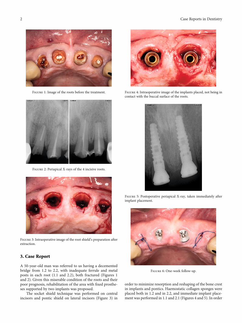
A 50-year-old man was referred to us having a decementedbridge from 1.2 to 2.2, with inadequate ferrule and metalposts in each root (1.1 and 2.2), both fractured (Figures 1and 2). Given this miserable condition of the roots and theirpoor prognosis, rehabilitation of the area with fixed prosthe-ses supported by two implants was proposed.
The socket shield technique was performed on centralincisors and pontic shield on lateral incisors (Figure 3) in
order to minimize resorption and reshaping of the bone crestin implants and pontics. Haemostatic collagen sponges wereplaced both in 1.2 and in 2.2, and immediate implant place-ment was performed in 1.1 and 2.1 (Figures 4 and 5). In order
Figure 1: Image of the roots before the treatment.
Figure 2: Periapical X-rays of the 4 incisive roots.
Figure 3: Intraoperative image of the root shield’s preparation afterextraction.
Figure 4: Intraoperative image of the implants placed, not being incontact with the buccal surface of the roots.
Figure 5: Postoperative periapical X-ray, taken immediately afterimplant placement.
Figure 6: One-week follow-up.
2 Case Reports in Dentistry

to keep anterior guidance, lateral incisors were planned incantilever, contactless during disclusion movements.
Two Biohorizons Tapered Internal 3:8 × 12mm implantswith 3.5mm diameter internal hexagonal connection (Bio-horizons®, Spain) with an insertion torque of 25N/cm wereplaced, as well as the healing abutments, because the patientdid not request provisional teeth. After one week, healingwas proper (Figure 6).
Three months after, the osseointegration of the implantswas corrected (Figure 7), but teeth 1.1 had a superficial expo-sure of the shield (Figure 8), which was reshaped with the
Figure 9: One-month follow-up, after high-speed handpiecereshaping of the shield exposure on 2.1.
Figure 10: Final bridge delivery.
Figure 11: Control X-ray after bridge placing.
Figure 12: Clinical lateral vision. A good maintenance of the buccalthickness is seen.
Figure 7: Three-month follow-up, after implant placement.
Figure 8: Shield exposure on 1.1 implant.
3Case Reports in Dentistry
high-speed handpiece using a diamond bur, without needinganesthesia. After one more month, the clinical appearancewas corrected (Figure 9) and a partial fixed denture wasscrewed at 30N/cm to the implant (Figures 10 and 11). Goodmaintenance of buccal volume and natural appearance isseen (Figure 12).
These partial extraction techniques have allowed us tomaintain the volume of the buccal crest without the need tofill the gap with bone grafts and give the gingiva a naturalappearance.
The authors want to emphasize that the value of thisstudy is limited, since it is a case report of a single patient.
4. Discussion
Already in 2009, Davarpana and Szmukler-Moncler [4] pre-sented a limited series of 5 cases with a follow-up between 12and 42 months, in which the implants were drilled andplaced through the roots of ankylosed teeth, leaving the frag-ments that remained stable in the socket. They found nocomplications derived from tooth remains in contact withthe implant, and the cases were successful throughout thefollow-up. They present the technique as an alternative totooth extraction when it can cause morbidity.
Several studies have been published regarding the socketshield technique since its first reference by Hürzeler et al. [3].Thus, Kan and Rungcharassaeng [5] and later Cherel and Eti-enne [6] used the SST in order to preserve the interdentalpapilla in between implants, in cases where adjacent teethneed to be restored.
Siormpas et al. [7] conducted a study on 46 patients withsingle implants, performing the “root-membrane” technique,similar to the TSS, and leaving the vestibular fragment at least1 millimeter above the bone level and in contact with the
implant surface. They obtained 100% success in a follow-upbetween 24 and 60 months and correct maintenance of thebuccal bone volume. They concluded that it is a predictabletechnique for the placement of implants in aesthetic areasin healthy adults.
Gluckman et al. [8] described the pontic shield tech-nique in which they performed a treatment similar toSST but, in these cases, filling the socket with xenogeneicgraft in the pontics. Among the presented series, 13 caseswere successful and one case required reshaping andadvancement flap due to shield exposure. They concludetheir study, describing the technique as a feasible optionin bone preservation in pontics and assuming the needfor more studies in this regard.
Afterwards, the same authors (in 2016 [9] and 2017 [10])carried out two consecutive studies. The first study definedpartial extraction techniques in three groups: (i) root sub-mergence when the entire root volume is kept under thegum to preserve the volume, being very important the factthat the teeth do not have apical pathology; (ii) socket shieldas a preparation of the root’s buccal surface and 1mm abovethe bony crest in anterior teeth and with simultaneous place-ment of immediate implants in the palatal surface of thesocket, having eliminated any possible apical pathologybefore; (iii) pontic shield, which means preparing the socketshield exactly the same as SST but recommends fillingthe socket with osteoconductive material. They also advisesoft tissue sealing and a three-month follow-up beforepressing with ovoid pontics. Table 1 shows the indicationsfor the different partial extraction techniques based on thisstudy and review. The study finishes by concluding thatpartial extraction techniques should be considered by clini-cians as a conservative strategy to maintain bone crest inoral rehabilitation [9].
Table 1: Partial extraction therapies (PET) and their indications, adapted from Siormpas et al. [7].
PET Clinical situation(s) indicated
Root submergence
(i) Unrestorable tooth crown or tooth indicated for extraction absence of apical pathology(ii) Healthy amputated pulp or endodontic therapy completed intention to preserve the alveolar ridge(ii) Planned removable full or partial prosthesis(iv) Planned pontic site beneath fixed prosthesis(v) Cantilever pontic site as an alternative to two adjacent implants(vi) Actively growing young patient planned for implant treatment later(vii) Ridge preservation in conjunction with other PET
Socket shield
(i) Unrestorable tooth crown or tooth indicated for extraction tooth root with or without apical pathology(ii) Intention to preserve the alveolar ridge, specifically to prevent buccopalatal collapse(iii) Immediate implant placement(iv) Ridge preservation in conjunction with other PET
Pontic shield
(i) Unrestorable tooth crown or tooth indicated for extraction tooth root with or without apical pathology(ii) Intention to preserve the alveolar ridge(iii) Planned pontic site(s) beneath fixed prosthesis(iv) Cantilever pontic site as an alternative to two adjacent implants(v) Ridge preservation in conjunction with other PET
Proximal socket shield
(i) Unrestorable tooth crown or tooth indicated for extraction tooth root with or without apical pathology(ii) Intention to preserve interdental papillae(iii) Planned immediate implant placement sites of two or more adjacent implants(iv) Papillae preservation in conjunction with other PET
4 Case Reports in Dentistry

In his second work, he focuses on the technical aspectsand the management of complications. The necessary mate-rials for partial extraction techniques described in this articleare presented in Table 2. They conclude by defining partialextraction techniques as a very promising set for the futuremanagement of bone ridges after the removal of nonpreser-vable teeth [10].
In 2017, Gharpure & Bhatavadekar wrote in a systematicreview reporting limited scientific support regarding TSS,with evidence of less osseointegration, some biological com-plications and short follow-up cases. They suggest the needof more studies to obtain scientific support [11]. Morerecently, Han et al. [12] presented a modified TSS with aseries of 40 implants maintaining the buccal shield at bonecrest level and with an approximate thickness of 1.5mm,without biological complications and with a 100% successrate. They conclude that modified TSS appears to be a suc-cessful procedure combined with immediate implantsbecause the root surface does not interfere with osseointe-gration and is beneficial for the aesthetics and maintenanceof bone.
Bramanti et al. [13] reported a sample of forty patientswho received an immediate single implant in the estheticzone with SST versus immediate implants with the conven-tional technique and filling the gap with bone graft. No clin-ical complications were recorded at 3, 6, and 36 months fromimplant placement. A lower rate of crestal bone resorptionwas recorded in the TSS group in all checks over time.
Based on the published studies and the case we present,we can confirm that complications arise more easily when
the tooth fragment remains above the bone crest. The fillingof the socket does not seem necessary when performing thistechnique since the blood clot achieves a regeneration ofthe socket and the shield prevents buccal reshaping.
5. Conclusion
The conclusion that we can draw from this case report andthe subsequent review of the literature is that partial extrac-tion techniques, such as the socket shield and the ponticshield, are procedures that should be considered in oral reha-bilitation in selected cases.
This kind of intervention is dependent on the surgicaltechnique of the operator, and its reproducibility must beassessed.
Conflicts of Interest
The authors declare that there is no conflict of interestregarding the publication of this paper.
References
[1] J. Majzoub, A. Ravida, T. Starch-Jensen, M. Tattan, andF. Suárez-López del Amo, “The Influence of Different GraftingMaterials on Alveolar Ridge Preservation: a SystematicReview,” Journal of Oral and Maxillofacial Research, vol. 10,no. 3, article e6, 2019.
[2] M. G. Araújo, C. O. Silva, A. B. Souza, and F. Sukekava,“Socket healing with and without immediate implant place-ment,” Periodontology 2000, vol. 79, no. 1, pp. 168–177, 2019.
[3] M. B. Hürzeler, O. Zuhr, P. Schupbach, S. F. Rebele,N. Emmanouilidis, and S. Fickl, “The socket-shield technique:a proof-of-principle report,” Journal of Clinical Periodontol-ogy, vol. 37, no. 9, pp. 855–862, 2010.
[4] M. Davarpanah and S. Szmukler-Moncler, “Unconventionalimplant treatment: I. Implant placement in contact with anky-losed root fragments. A series of five case reports,” ClinicalOral Implants Research, vol. 20, no. 8, pp. 851–856, 2009.
[5] J. Y. K. Kan and K. Rungcharassaeng, “Proximal socket shieldfor interimplant papilla preservation in the esthetic zone,” TheInternational Journal of Periodontics & Restorative Dentistry,vol. 33, no. 1, pp. e24–e31, 2013.
[6] F. Cherel and D. Etienne, “Papilla preservation between twoimplants: a modified socket-shield technique to maintain thescalloped anatomy? A case report,” Quintessence Interna-tional, vol. 45, no. 1, pp. 23–30, 2014.
[7] K. D. Siormpas, M. E. Mitsias, E. Kontsiotou-Siormpa,D. Garber, and G. A. Kotsakis, “Immediate implant placementin the esthetic zone utilizing the "root-membrane" technique:clinical results up to 5 years postloading,” The InternationalJournal of Oral & Maxillofacial Implants, vol. 29, no. 6,pp. 1397–1405, 2014.
[8] H. Gluckman, J. Du Toit, and M. Salama, “The pontic-shield:partial extraction therapy for ridge preservation and pontic sitedevelopment,” The International Journal of Periodontics &Restorative Dentistry, vol. 36, no. 3, pp. 417–423, 2016.
[9] H. Gluckman, M. Salama, and J. Du Toit, “Partial extractiontherapies (PET) part 1: maintaining alveolar ridge contour atpontic and immediate implant sites,” The International
Table 2: Instruments and materials required for PET [8].
Socket shield
(1) Long shank root resection bur(2) Extralarge round diamond head bur (to reduce the inner
aspect of shield into concavity)(3) End-cutting diamond head bur (to reduce coronal aspect of
shield) 4. Gingival protector(4) Irrigated surgical motor(5) Contra-angled surgical fast handpiece(6) Microperiotomes(7) Microforceps
Pontic shieldAs for socket-shield, plus:
(1) Socket grafting instruments: plugger, particulate graft spoon,crucible
(2) SM 69 blade (or other suitable microblade, mandatory forsplit thickness dissection of facial and palatal pouches to tuck CTGinto) 3. 6/0 nylon sutures
Root submergence
(1) Irrigated surgical motor(2) Contra-angled surgical fast handpiece(3) Extralarge round diamond head bur (for reducing coronal
aspect root into concavity)(4) SM 69 blade (or other suitable microblade, mandatory for
split thickness dissection of facial and palatal pouches to tuck CTGinto)
(5) 6/0 nylon sutures
5Case Reports in Dentistry

Journal of Periodontics & Restorative Dentistry, vol. 36, no. 5,pp. 681–687, 2016.
[10] H. Gluckman, M. Salama, and J. Du Toit, “Partial extractiontherapies (PET) part 2: procedures and technical aspects,”The International Journal of Periodontics & Restorative Den-tistry, vol. 37, no. 3, pp. 377–385, 2017.
[11] A. S. Gharpure and N. B. Bhatavadekar, “Current evidence onthe socket-shield technique: a systematic review,” The Journalof Oral Implantology, vol. 43, no. 5, pp. 395–403, 2017.
[12] C.-H. Han, K.-B. Park, and F. G. Mangano, “The modifiedsocket shield technique,” Journal of Craniofacial Surgery,vol. 29, no. 8, pp. 2247–2254, 2018.
[13] E. Bramanti, A. Norcia, M. Cicciù et al., “Postextraction dentalimplant in the aesthetic zone, socket shield technique versusconventional protocol,” The Journal of Craniofacial Surgery,vol. 29, no. 4, pp. 1037–1041, 2018.
6 Case Reports in Dentistry