a corticostriatal path targeting striosomes controls decision-making under conflict
DESCRIPTION
A Corticostriatal Path Targeting Striosomes Controls Decision-Making under ConflictTRANSCRIPT
Article
ACorticostriatal Path Targeting StriosomesControls
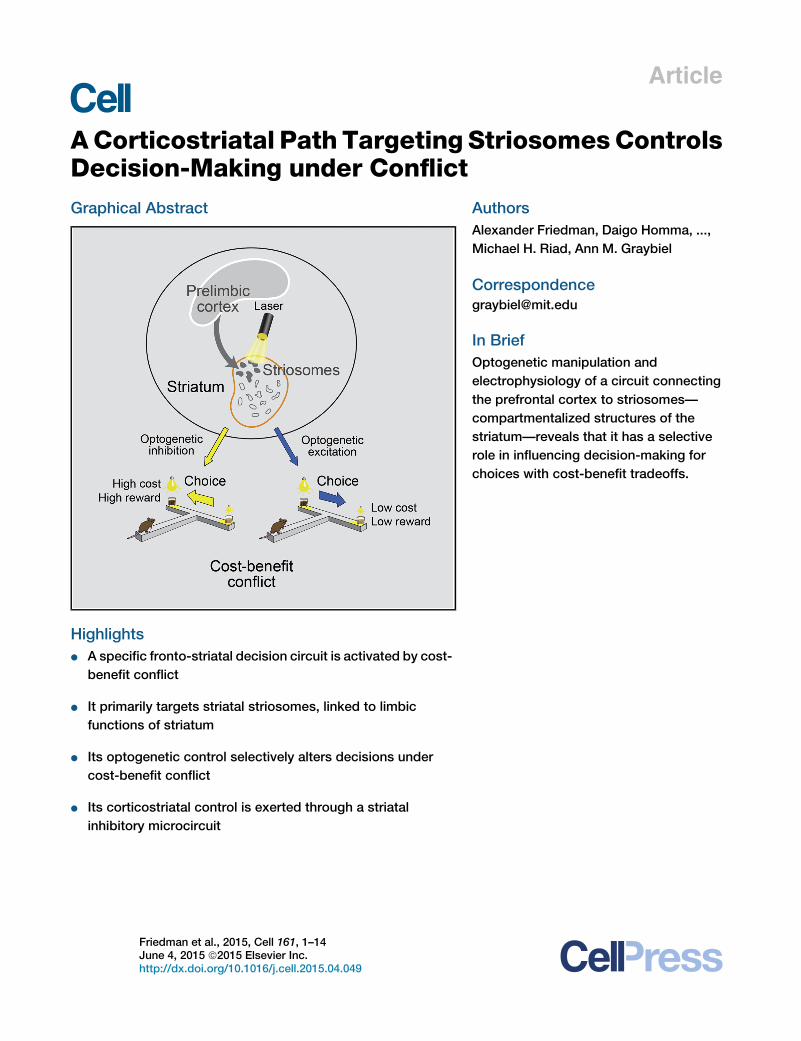
Decision-Making under ConflictGraphical Abstract
Highlights
d A specific fronto-striatal decision circuit is activated by cost-
benefit conflict
d It primarily targets striatal striosomes, linked to limbic
functions of striatum
d Its optogenetic control selectively alters decisions under
cost-benefit conflict
d Its corticostriatal control is exerted through a striatal
inhibitory microcircuit
Friedman et al., 2015, Cell 161, 1–14June 4, 2015 ª2015 Elsevier Inc.http://dx.doi.org/10.1016/j.cell.2015.04.049
Authors
Alexander Friedman, Daigo Homma, ...,
Michael H. Riad, Ann M. Graybiel
In Brief
Optogenetic manipulation and
electrophysiology of a circuit connecting
the prefrontal cortex to striosomes—
compartmentalized structures of the
striatum—reveals that it has a selective
role in influencing decision-making for
choices with cost-benefit tradeoffs.
Please cite this article in press as: Friedman et al., A Corticostriatal Path Targeting Striosomes Controls Decision-Making under Conflict, Cell(2015), http://dx.doi.org/10.1016/j.cell.2015.04.049
Article
A Corticostriatal Path Targeting StriosomesControls Decision-Making under ConflictAlexander Friedman,1 Daigo Homma,1 Leif G. Gibb,1 Ken-ichi Amemori,1 Samuel J. Rubin,1 Adam S. Hood,1
Michael H. Riad,1 and Ann M. Graybiel1,*1McGovern Institute for Brain Research and Department of Brain and Cognitive Sciences, Massachusetts Institute of Technology,
Cambridge, MA 02139, USA*Correspondence: [email protected]
http://dx.doi.org/10.1016/j.cell.2015.04.049
SUMMARY
A striking neurochemical form of compartmentaliza-tion has been found in the striatum of humans andother species, dividing it into striosomes and matrix.The function of this organization has been unclear,but the anatomical connections of striosomes indi-cate their relation to emotion-related brain regions,including the medial prefrontal cortex. We capital-ized on this fact by combining pathway-specific op-togenetics and electrophysiology in behaving ratsto search for selective functions of striosomes. Wedemonstrate that a medial prefronto-striosomal cir-cuit is selectively active in and causally necessaryfor cost-benefit decision-making under approach-avoidance conflict conditions known to evokeanxiety in humans. We show that this circuit hasunique dynamic properties likely reflecting striatalinterneuron function. These findings demonstratethat cognitive and emotion-related functions are,like sensory-motor processing, subject to encodingwithin compartmentally organized representationsin the forebrain and suggest that striosome-targetingcorticostriatal circuits can underlie neural processingof decisions fundamental for survival.
INTRODUCTION
Across the animal kingdom, neural mechanisms have evolved
to allow decision-making based on rewarding and aversive fea-
tures of the environment (Glimcher and Fehr, 2014). This funda-
mental capacity, critical to normal human life, is disabled in a
range of neuropsychiatric and neurologic disorders (Gleich-
gerrcht et al., 2010). As yet, the mechanisms underlying the
relation between decision-making and emotion-related circuit
function remain largely unknown (Aupperle and Paulus,
2010). Pioneering work, however, has shown that behavioral
reactions to value-based decision-making, based on the
perceived potential costs and benefits of taking a given action,
are differentially represented in regions of the medial prefrontal
cortex (Rangel and Hare, 2010; Rudebeck et al., 2006; Rush-
worth et al., 2011; Walton et al., 2002). These cortical regions
are interlinked with each other and with other downstream
parts of the limbic system (Salamone, 1994; Stopper et al.,
2014; Watabe-Uchida et al., 2012). They are also linked to
the striatum, part of the basal ganglia (Amemori and Graybiel,
2012; Donoghue and Herkenham, 1986; Eblen and Graybiel,
1995). These networks have been identified in human brain
imaging studies as regions of co-activation in relation to
emotional task performance (Aupperle et al., 2015; Etkin
et al., 2006; Gleichgerrcht et al., 2010). In rodents, potential ho-
mologs of these regions of the prefrontal and orbitofrontal cor-
tex have been identified (Milad et al., 2007) and have been
particularly intensively studied to identify sub-circuit functions
of these networks (Rangel and Hare, 2010; Rudebeck et al.,
2006; Walton et al., 2002).
Much anatomical work supports the view that such value-
related networks include corticostriatal circuits (Donoghue
and Herkenham, 1986; Eblen and Graybiel, 1995; Gerfen,
1984). A striking feature of a subset of these prefronto-striatal
and orbitofronto-striatal circuits is that they preferentially
target a distinctive set of distributed striatal microzones
known as striosomes (Graybiel and Ragsdale, 1978). These re-
gions are distinguishable from the much larger matrix tissue of
the striatum by their differential expression of most of the
neurotransmitter-related molecules expressed in the striatum
(Crittenden and Graybiel, 2011; Graybiel, 1990) and the birth-
dates of their neurons (Newman et al., 2015), as well as by
their differential inputs and direct and indirect output to parts
of the dopamine-containing midbrain (Fujiyama et al., 2011;
Prensa and Parent, 2001; Watabe-Uchida et al., 2012)
and the lateral habenula (Rajakumar et al., 1993; Stephen-
son-Jones et al., 2013), regions strongly implicated in the
control and modulation of motivation and reinforcement-
driven behavior (Hong and Hikosaka, 2013; Lak et al., 2014;
Stopper et al., 2014). By contrast, the large matrix compart-
ment of the striatum is divided into a mosaic of microzones
known as matrisomes, and these are linked by their outputs
to the classic sensorimotor zones of the basal ganglia (Flah-
erty and Graybiel, 1993).
The differential functions of striosomes, relative to matrix
and its matrisomes, have never been identified, largely for
technical reasons. Nor is it known how they and their cortico-
striatal pathways are recruited in different modes of decision-
making related to mood and motivation, despite the fact that
it has been known for years that cortical regions targeting strio-
somes are parts of value-related networks (Rudebeck et al.,
Cell 161, 1–14, June 4, 2015 ª2015 Elsevier Inc. 1
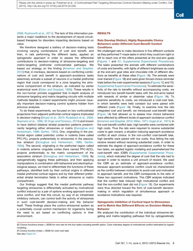
Figure 1. Decision-Making Tasks(A) The five decision-making tasks.
(B) Training timeline.
(C) Session of cost-benefit conflict task.
(D) Schematic of maze run.
(legend continued on next page)
2 Cell 161, 1–14, June 4, 2015 ª2015 Elsevier Inc.
Please cite this article in press as: Friedman et al., A Corticostriatal Path Targeting Striosomes Controls Decision-Making under Conflict, Cell(2015), http://dx.doi.org/10.1016/j.cell.2015.04.049
Please cite this article in press as: Friedman et al., A Corticostriatal Path Targeting Striosomes Controls Decision-Making under Conflict, Cell(2015), http://dx.doi.org/10.1016/j.cell.2015.04.049
2006; Rushworth et al., 2011). The lack of this information pre-
sents a major roadblock to the development of neural circuit-
based therapies for disorders impacting decisions based on
value.
We therefore designed a battery of decision-making tests
involving varying combinations of cost and benefit, and
then, in rats performing the different tasks, applied an
optogenetic approach in order to assess the differential
contributions to decision-making of striosome-targeting and
matrix-targeting prefrontal corticostriatal pathways. We
based our strategy on the finding (Amemori and Graybiel,
2012) that, in monkeys, decisions based on conflicting combi-
nations of cost and benefit in approach-avoidance tasks
selectively activate a subset of neurons in a medial prefrontal
region that could correspond to a zone targeting the strio-
some compartment of the striatum, as indicated by earlier
anatomical work (Eblen and Graybiel, 1995). These results in
the non-human primate suggested that in-depth analysis of
striosome-targeting and matrix-targeting circuits with multiple
methods feasible in rodent experiments might uncover caus-
ally important decision-making control systems hidden from
previous analyses.
To do these experiments, we focused on two corticostriatal
projections from adjacent prefrontal cortical regions implicated
in decision-making (Dwyer et al., 2010; Rudebeck et al., 2006;
Seamans et al., 1995; St Onge and Floresco, 2010) and known
to have distinct bilateral striatal projection patterns predomi-
nantly targeting either striosomes or matrix (Donoghue and
Herkenham, 1986; Gerfen, 1984). One, originating in the pre-
frontal region called prelimbic cortex in rodents (here called
PFC-PL), projects preferentially to striosomes in the associa-
tive striatum (Donoghue and Herkenham, 1986; Gerfen,
1984). The second, originating in the prefrontal region called
in rodents anterior cingulate cortex (here named PFC-ACC),
projects preferentially to the matrix compartment of the
associative striatum (Donoghue and Herkenham, 1986). By
optogenetically tagging these pathways, and then applying
manipulations in combination with behavioral and electrophys-
iological assays, we tried to differentiate the functions of these
pathways, identified both by their cortical origins in different
medial prefrontal cortical regions and by their different prefer-
ential striatal termination fields in either striosome or matrix
compartment.
Our findings suggest that the medial prefrontal pathway
targeting striosomes is differentially activated by motivational
conflict induced by a pair of options evoking approach-avoid-
ance conflict, and that this circuit, by way of an intrastriatal
inhibitory network, can control both the activity of striosomes
in such cost-benefit decision-making and the behavior
itself. These findings place the cortico-striosomal system as
a potentially crucial control mechanism for organisms facing
the need to act based on conflicting options in their
environment.
(E) Choice functions (mean ± SEM) for nine rats for the four tasks including bene
modeling.
(F) Choice function (mean ± SEM) for cost-cost task.
See also Figure S1.
RESULTS
Rats Develop Distinct, Highly Repeatable ChoiceBehaviors under Different Cost-Benefit ContextConditionsWe challenged rats to make decisions in five different contexts
as they performed T-maze tasks in which they could turn right or
left to reach one of the offers presented at the end-arm goals
(Figures 1 and S1; Supplemental Experimental Procedures).
The tasks presented the animals with different combinations
of costs and benefits, with lights of different brightness as costs
at the end-arm goal sites and chocolate milk of different dilu-
tions as benefits at these sites (Figure 1A). The animals were
over-trained (Figure 1B) and were given forced-choice reminder
trials before the main experimental sessions (Figures 1C and 1D;
Supplemental Experimental Procedures). To estimate the sensi-
tivity of the rats to benefits without accompanying costs, we
introduced two benefit-benefit tasks with the end-arms baited
with rewards of similar or dissimilar value (Figure 1A). To
examine sensitivity to costs, we introduced a cost-cost task,
in which benefits were held constant but were paired with
different costs (Figure 1A). Finally, to examine how the rats
integrated cost and benefit, we introduced cost-benefit tasks
(Figure 1A) that allowed us to assess how choice behaviors
were affected by different levels of approach-avoidance conflict
(Amemori and Graybiel, 2012; Miller, 1971; Vogel et al., 1971). In
the cost-benefit conflict task, high benefits were combined with
high costs. Thus the rats were required to receive substantial
costs to gain reward, a situation inducing approach-avoidance
conflict at each choice. In the non-conflict cost-benefit task,
high benefits were paired with low costs, thus letting the rats
receive reward without receiving substantial costs. In order to
estimate the degree of approach-avoidance conflict for these
two tasks, we applied logistic modeling and parameterized the
cost-benefit ratio (CBR) (Supplemental Experimental Proce-
dures), which indicated the degree of cost that the rats had to
accept in order to receive a unit amount of reward. We used
the CBR as an estimate of approach-avoidance conflict,
because approach-avoidance conflict could be characterized
as the conflict between motivation to avoid cost and motivation
to approach benefit, and the CBR corresponds to the ratio of
these two opponent motivations. This CBR analysis indicated
that the conflict task induced high motivational conflict and
that the non-conflict task did not (Figure 1E). Our experiments
were thus directed toward the form of cost-benefit decision-
making in which regulation of simultaneous approach or
avoidance motivations was required.
Optogenetic Inhibition of Cortical Input to Striosomesand to Matrix Has Different Effects on Decision-MakingBehaviorWe analyzed the contribution of the individual striosome-tar-
geting and matrix-targeting pathways first by optogenetically
fit option. Color indicates degree of motivational conflict estimated by logistic
Cell 161, 1–14, June 4, 2015 ª2015 Elsevier Inc. 3
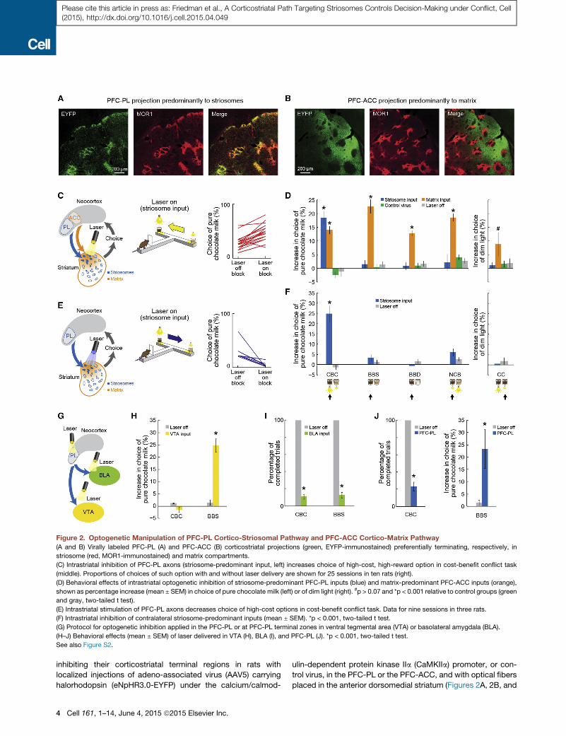
Figure 2. Optogenetic Manipulation of PFC-PL Cortico-Striosomal Pathway and PFC-ACC Cortico-Matrix Pathway
(A and B) Virally labeled PFC-PL (A) and PFC-ACC (B) corticostriatal projections (green, EYFP-immunostained) preferentially terminating, respectively, in
striosome (red, MOR1-immunostained) and matrix compartments.
(C) Intrastriatal inhibition of PFC-PL axons (striosome-predominant input, left) increases choice of high-cost, high-reward option in cost-benefit conflict task
(middle). Proportions of choices of such option with and without laser delivery are shown for 25 sessions in ten rats (right).
(D) Behavioral effects of intrastriatal optogenetic inhibition of striosome-predominant PFC-PL inputs (blue) and matrix-predominant PFC-ACC inputs (orange),
shown as percentage increase (mean ± SEM) in choice of pure chocolate milk (left) or of dim light (right). #p > 0.07 and *p < 0.001 relative to control groups (green
and gray, two-tailed t test).
(E) Intrastriatal stimulation of PFC-PL axons decreases choice of high-cost options in cost-benefit conflict task. Data for nine sessions in three rats.
(F) Intrastriatal inhibition of contralateral striosome-predominant inputs (mean ± SEM). *p < 0.001, two-tailed t test.
(G) Protocol for optogenetic inhibition applied in the PFC-PL or at PFC-PL terminal zones in ventral tegmental area (VTA) or basolateral amygdala (BLA).
(H–J) Behavioral effects (mean ± SEM) of laser delivered in VTA (H), BLA (I), and PFC-PL (J). *p < 0.001, two-tailed t test.
See also Figure S2.
Please cite this article in press as: Friedman et al., A Corticostriatal Path Targeting Striosomes Controls Decision-Making under Conflict, Cell(2015), http://dx.doi.org/10.1016/j.cell.2015.04.049
inhibiting their corticostriatal terminal regions in rats with
localized injections of adeno-associated virus (AAV5) carrying
halorhodopsin (eNpHR3.0-EYFP) under the calcium/calmod-
4 Cell 161, 1–14, June 4, 2015 ª2015 Elsevier Inc.
ulin-dependent protein kinase IIa (CaMKIIa) promoter, or con-
trol virus, in the PFC-PL or the PFC-ACC, and with optical fibers
placed in the anterior dorsomedial striatum (Figures 2A, 2B, and
Please cite this article in press as: Friedman et al., A Corticostriatal Path Targeting Striosomes Controls Decision-Making under Conflict, Cell(2015), http://dx.doi.org/10.1016/j.cell.2015.04.049
S2; Supplemental Experimental Procedures). The cortical inputs
to the striatum were differentially selective for striosomes in
cases with virus-labeled PFC-PL fibers, in accordance with prior
findings, and differentially selective for matrix in PFC-ACC
cases. However, also in accordance with prior work, the non-
preferred compartment always exhibited some labeling, varying
case by case (Eblen and Graybiel, 1995; Flaherty and Graybiel,
1991; Gerfen, 1984; Parthasarathy et al., 1992) (Figures S2A
and S2B).
Rats with the PFC-PL and PFC-ACC viral injections success-
fully performed all five decision-making tasks during the optoge-
netic experiments. In laser-on sessions, a 3-s pulse of yellow
light (590 nm, 2 mW) was delivered to the intrastriatal terminal
fields from the time of the click indicating trial start to goal-reach-
ing at trial end (Figure 2C). We compared results from blocks of
laser-on and baseline trials and from control experiments with
viral constructs lacking opsin. Image analysis suggested that
within the estimated regions of illumination, the relative density
of virally expressed EYFP label was �5.2 times higher in strio-
somes than in the matrix compartment in the PFC-PL cases
and �2.6 times higher in the matrix than in striosomes in the
PFC-ACC cases (Figures S2C–S2E; Supplemental Experimental
Procedures). We explicitly chose a block design due to our
finding (Figure S1G) that choices from trial to trial within a block
were not independent. Moreover, the optogenetic results were
not cumulative (Supplemental Experimental Procedures).
Optogenetic inhibition of the striosome-targeting and matrix-
targeting circuits had strikingly different effects on decision-
making behavior (Figures 2C and 2D). Intrastriatal inactivation
of the striosome-targeting PFC-PL pathway strongly affected
decision-making in the cost-benefit conflict task: the animals
ran more toward the high-cost option, an increase of >20%
over control levels (n = 10; Figure 2C). Yet the same intrastriatal
manipulation in these animals had almost no effect on their per-
formance of any of the other tasks, including the two other tasks
with cost components (Figure 2D). The increased choice of the
high-cost, high-reward option induced by inhibiting the PFC-
PL pathway to striosomes thus appeared specific to the cost-
benefit conflict context, in which the animals had to accept
substantial cost to gain reward and had to regulate their
approach and avoidance behaviors (Figure 2D).
By contrast, inhibition of the predominantly matrix-targeting
PFC-ACC pathway significantly affected the animals’ choices
in all tasks except the cost-cost task: the animals shifted their
choices toward the option with higher reward (n = 8; Figure 2D).
In the cost-cost task, there was an increase, not statistically sig-
nificant (p = 0.07), in the animals’ choice of the option with lower
cost, perhaps reflecting a partial overlap of the neural represen-
tations of higher reward and lower cost in this pathway. Thus the
predominantly non-striosome-targeting corticostriatal input to
the same dorsomedial striatal region did not exhibit context
selectivity comparable to that of the predominantly striosome-
projecting circuit.
We took advantage of the patterns of bilateral projection of the
PFC-PL and PFC-ACC to perform optogenetic inhibition within
the striatum contralateral to the side of virus injection, thus
avoiding possible optogenetic effects on corticofugal fibers of
passage traveling through the ipsilateral striatum (Figure S2H).
These contralateral manipulations (n = 3) gave the same results
as those of the main group of bilateral manipulations: for the
PFC-PL circuit, inhibition affected behavior selectively in the
cost-benefit conflict task (Figure 2F), and for the PFC-ACC cir-
cuit, inhibition affected behavior in all tasks (Figure S2G). These
results confirmed that the contrasting patterns of behavior
induced by the striosome-targeting and matrix-targeting path-
ways were not due principally to illumination of bundles of inter-
nal capsule fibers within the illuminated zones.
In three rats we applied C1V1-mediated intrastriatal excitation,
instead of halorhodopsin-mediated intrastriatal inhibition, to the
PFC-PL pathway terminals in the dorsomedial striatum (Figures
2E and S2F; Supplemental Experimental Procedures). The re-
sults of the excitatory manipulations were diametrically opposed
to those of the inhibitory manipulations: now the rats ran more to
the side with low cost and low benefit in the cost-benefit conflict
task.
Finally, to test whether the control of conflict decision-making
by the striosome-targeting PFC-PL circuit was unique to the
striatal terminals of the PFC-PL, we tested whether inhibiting
PFC-PL terminals in three other regions to which the PFC-PL
projects affected performance on the cost-benefit conflict and
benefit-benefit tasks (n = 3 per group; Figure 2G): we inhibited
PFC-PL terminals in the ventral tegmental area (Figure 2H),
in the basolateral amygdala (Figure 2I) and in the PFC-PL itself
(Figure 2J). In none of these experiments was the result of the
PL-PFC terminal inhibition similar to the effects of PFC-PL termi-
nal inhibition in the striatum (Figure 2D). Thus the effects of
optogenetic inhibition of PFC-PL projections to the dorsomedial
striatum were not generalized effects, but rather, were target-
specific.
Collectively, these results point to the PFC-PL projection pref-
erentially targeting striosomes as unique among PFC-PL circuits
tested in selectively affecting decision-making about approach-
ing or avoiding simultaneously presented, and motivationally
conflicting, cost and benefit options.
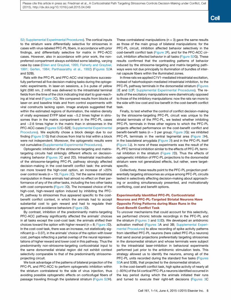
Experimentally Identified PFC-PL CorticostriatalNeurons and PFC-PL-Targeted Striatal Neurons HaveOpposite Firing Patterns during Maze Runs in theCost-Benefit Conflict TaskTo uncover mechanisms that could account for this selectivity,
we performed chronic tetrode recordings in the PFC-PL and
the striatum (Figures 3 and S3D). We developed an antidromic
activation method (Figures 3A and 3B; Supplemental Experi-
mental Procedures) to allow recording of spike activity patterns
from identified PFC-PL neurons (here called PFC-PLs neurons)
that send axonal projections preferentially targeting striosomes
in the dorsomedial striatum and whose terminals were subject
to the intrastriatal laser-inhibition in behavioral experiments
performed just prior to the antidromic stimulation tests. This
strategy allowed us to identify the neurons, among all of the
PFC-PL units recorded during the standard five tasks (Figures
S3A and S3B), that projected to the dorsomedial striatum.
In the cost-benefit conflict task, high spike activity of nearly all
(�93%) of the 54 cortical PFC-PLs neurons identified occurred in
the key period during which the animals initiated their runs
and turned to execute their right-left decisions (Figures 3C
Cell 161, 1–14, June 4, 2015 ª2015 Elsevier Inc. 5
Figure 3. Contrasting Activity of Putative Cortical PFC-PLs Neurons and Striosomal SPNs during Task Performance
(A) Antidromic stimulation protocol to identify PFC-PLs neurons.
(B) Cortical spikes aligned to striatal microstimulation onset.
(C) Spike activity of PFC-PLs neurons (top) and bursty spike activity heat maps (bottom) in cost-benefit conflict (left) and benefit-benefit (right) tasks. Inner T-maze
outline indicates click to first lick (i.e., in-run) time-period; outer outline includes 3 s before and after runs. Activity shown asmean Z scores and firing rates in color
scale from blue (low) to red (high). Heat map rectangles show bursts with lengths proportional to burst durations, with min-max normalized intra-burst firing rates
from yellow (low) to red (high).
(D) PFC-PLs spike activity (mean ± SEM) during click-to-turn period for all tasks (abbreviated as in Figure 1A). *p < 0.001 (two-tailed t test, difference between
CBC and each of other tasks).
(E) Orthodromic stimulation protocol for identification of putative striosomal neurons.
(F) Putative striosomal SPN activity aligned to PFC-PL microstimulation. Yellow and gray shading respectively indicates peak and inhibition time windows.
(G) Activity of putative striosomal SPNs (top) and associated burst activity heat maps (bottom).
(H) Average click-to-turn activity of putative striosomal SPNs in the five tasks. *p < 0.001 (two-tailed t test; difference between CBC and each of other tasks).
(legend continued on next page)
6 Cell 161, 1–14, June 4, 2015 ª2015 Elsevier Inc.
Please cite this article in press as: Friedman et al., A Corticostriatal Path Targeting Striosomes Controls Decision-Making under Conflict, Cell(2015), http://dx.doi.org/10.1016/j.cell.2015.04.049
Please cite this article in press as: Friedman et al., A Corticostriatal Path Targeting Striosomes Controls Decision-Making under Conflict, Cell(2015), http://dx.doi.org/10.1016/j.cell.2015.04.049
and S3A). The activity of these neurons peaked in series during
this period. In all the other task-versions, PFC-PLs task-related
activity was low during this period (Figure 3D), and instead, either
peaked at goal-reaching or did not exhibit a significant peak at all
(Figure S3A). Thus, the PFC-PLs neuron activity patterns, like op-
togenetic inhibition of the PFC-PL pathway, pointed to a unique
influence of this striosome-targeting pathway on performance of
the cost-benefit conflict task.
Identifying the activity of striosomal neurons themselves dur-
ing task performance would provide a critical test of the selec-
tivity of the effects of PFC-PLs neurons on striosomes. However,
chronic in vivo recording from striosomes has never before been
achieved. We attempted to do this by capitalizing on the prefer-
ential PFC-PL inputs to striosomes demonstrable anatomically
(Figures 2A and S2; Supplemental Experimental Procedures).
We applied electrical stimulation to the PFC-PL region that we
had identified as projecting predominantly to striosomes in the
dorsomedial striatum (Figures 3E and 3F), and we simulta-
neously recorded from large numbers of striatal projection
neurons (SPNs) (Figure S3E) in rats with arrays of tetrodes chron-
ically implanted in the dorsomedial striatum (Figure 3M). At the
end of each session, we determined for each SPN whether it
had short latency responses to the PFC-PL stimulation (Figures
3E, 3F, and 3I), then marked some of these tetrode tip locations
with lesions (Figure 3M) and in histologically prepared brain sec-
tions mapped these locations relative to immunostained strio-
somes (Figure 3N). We found a reliable orthodromic response
signature of putative striosomal SPNs so identified (Figure 3O;
Supplemental Experimental Procedures).
Given that the corticostriatal projection to SPNs is glutamater-
gic and excitatory, we expected that the activity of these putative
striosomal SPNs would match that of the antidromically identified
PFL-PLsneurons.We found theopposite.During the in-runperiod
duringwhich the PFC-PLs neurons were highly active (Figure 3C),
the striosomal neurons were largely quieted (Figure 3G). Yet
�98% of striosomal SPNs exhibited high activity during this
click-to-turn period in all of the other task-versions, again peaking
at different points during this period (Figures 3H and S3F). Thus,
the striosome-projecting PFC-PLs neurons and the striosomal
SPNs to which they mainly projected had almost perfectly com-
plementary patterns of activity during the maze runs.
To test the selectivity of these patterns, we analyzed the activ-
ity of the PFC-PL neuronal population as a whole, excluding the
54 antidromically identified PFC-PLs neurons. In sharp contrast
to the PFC-PLs cells, this PFC-PL population fired at goal-reach-
ing in every task (Figures 3J, S3B, and S3C). Thus, the PFC-PLs
in-run activity pattern was highly selective. We also analyzed the
activity of striatal SPNs excluded from the putative striosomal
(I) Method to determine time-window of short-latency orthodromic SPN activatio
demarcating start and end of time window.
(J and K) Average spike activity of non-PFC-PLs population recorded in PFC-PL
(L) Average click-to-turn activity of putative matrix SPNs.
(M) Four tetrode tracks and tip marked with micro-lesion (CD11, green) relative t
(N) Sample of tip-striosome measurements (Supplemental Experimental Proced
(white, left panel); distance to nearest striosome (light blue) shown (yellow line).
(O) Distribution of SPNs that respond to PFC-PL stimulation (left) was significant
See also Figure S3.
SPN population (Figures 3K, 3L, S3G, and S3H). In contrast to
the population of putative striosomal SPNs, the striatal neurons
identified as non-striosomal SPNs, which should mostly have
been matrix neurons, were active in all tasks (Figures 3L and
S3G; Supplemental Experimental Procedures), and their activity
tended to be selective for choices of higher reward (Figure S3H).
These activations were significantly different from baseline activ-
ity (two-tailed t test, p < 0.001), and their response patterns
for the five tasks were significantly different from those of
the putative striosomal SPNs (MANOVA and two-way ANOVA,
p < 0.001).
As shown in Figure S3, baseline activity was not uniform
across all tasks, being particularly low in the non-conflict cost-
benefit task and the cost-cost task and especially for PFC-PLs
neurons. To test the possibility that baseline firing rates might in-
fluence our optogenetic results, we compared baseline firing
rates during consecutive laser-off and laser-on trial blocks.
Despite the large changes in behavior induced by the optoge-
netic manipulation, baseline rates for both the putative strioso-
mal neurons and the putative matrix neurons were unaffected
(Supplemental Experimental Procedures).
Switching of Striosomal NeuronActivity PatternsCanBeInduced by Behavioral Task Switches Alone and byOptogenetic Inhibition of PFC-PLs Inputs PreferentiallyTargeting StriosomesIf, as these results suggested, the striosomal SPNswere uniquely
sensitive to the conflict context, then it should be possible to
demonstrate such sensitivity at a single-unit level by recording
for prolonged periods extending through consecutive perfor-
mance of benefit-benefit and cost-benefit conflict sessions. We
succeeded in doing this for ten SPNs recorded in two rats (Fig-
ure 4A). Strikingly, all of these putative striosomal SPNs, identi-
fied by post-performance template tests, switched their firing
patterns depending on the task being performed, from being
active during the click-to-turn period in the benefit-benefit task
to being nearly silent during the same click-to-turn period in the
succeeding cost-benefit task (Figures 4B-4E). The switch was
nearly immediate (Figure 4C). Such firing switches did not occur
when the two blocks were of the same task-type (Figure S4).
Thus, the spike-firing patterns of putative striosomal SPNs can
be dynamically determined by behavioral context alone.
Next we asked whether the complementarity of firing of the
striosomal SPNs and the PFC-PLs neurons reflected an inhibi-
tion of the striosomal SPNs by the PFC-PLs neurons. If so, opto-
genetic inhibition of the PFC-PLs input during the cost-benefit
conflict task might increase the run-period activity of striosomal
SPNs, thereby abolishing the unique lack of such activity in the
n (Supplemental Experimental Procedures). Larger squares show spike times
(J) and putative matrix SPNs (K) during cost-benefit conflict task.
o striosomes (MOR1, red).
ures). Tetrode tip in matrix (red, middle and right panels) along tetrode track
ly different from that of unresponsive SPNs (right; p < 0.001, chi-square test).
Cell 161, 1–14, June 4, 2015 ª2015 Elsevier Inc. 7
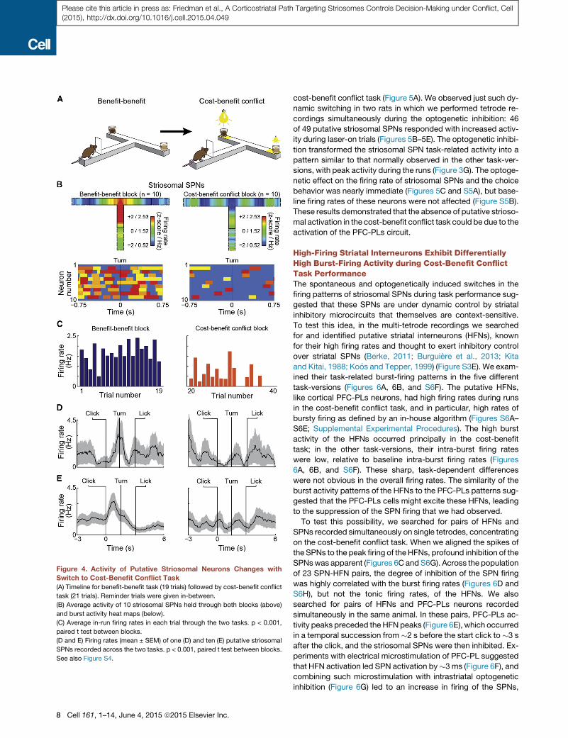
Figure 4. Activity of Putative Striosomal Neurons Changes with
Switch to Cost-Benefit Conflict Task
(A) Timeline for benefit-benefit task (19 trials) followed by cost-benefit conflict
task (21 trials). Reminder trials were given in-between.
(B) Average activity of 10 striosomal SPNs held through both blocks (above)
and burst activity heat maps (below).
(C) Average in-run firing rates in each trial through the two tasks. p < 0.001,
paired t test between blocks.
(D and E) Firing rates (mean ± SEM) of one (D) and ten (E) putative striosomal
SPNs recorded across the two tasks. p < 0.001, paired t test between blocks.
See also Figure S4.
8 Cell 161, 1–14, June 4, 2015 ª2015 Elsevier Inc.
Please cite this article in press as: Friedman et al., A Corticostriatal Path Targeting Striosomes Controls Decision-Making under Conflict, Cell(2015), http://dx.doi.org/10.1016/j.cell.2015.04.049
cost-benefit conflict task (Figure 5A). We observed just such dy-
namic switching in two rats in which we performed tetrode re-
cordings simultaneously during the optogenetic inhibition: 46
of 49 putative striosomal SPNs responded with increased activ-
ity during laser-on trials (Figures 5B–5E). The optogenetic inhibi-
tion transformed the striosomal SPN task-related activity into a
pattern similar to that normally observed in the other task-ver-
sions, with peak activity during the runs (Figure 3G). The optoge-
netic effect on the firing rate of striosomal SPNs and the choice
behavior was nearly immediate (Figures 5C and S5A), but base-
line firing rates of these neurons were not affected (Figure S5B).
These results demonstrated that the absence of putative strioso-
mal activation in the cost-benefit conflict task could be due to the
activation of the PFC-PLs circuit.
High-Firing Striatal Interneurons Exhibit DifferentiallyHigh Burst-Firing Activity during Cost-Benefit ConflictTask PerformanceThe spontaneous and optogenetically induced switches in the
firing patterns of striosomal SPNs during task performance sug-
gested that these SPNs are under dynamic control by striatal
inhibitory microcircuits that themselves are context-sensitive.
To test this idea, in the multi-tetrode recordings we searched
for and identified putative striatal interneurons (HFNs), known
for their high firing rates and thought to exert inhibitory control
over striatal SPNs (Berke, 2011; Burguiere et al., 2013; Kita
and Kitai, 1988; Koos and Tepper, 1999) (Figure S3E). We exam-
ined their task-related burst-firing patterns in the five different
task-versions (Figures 6A, 6B, and S6F). The putative HFNs,
like cortical PFC-PLs neurons, had high firing rates during runs
in the cost-benefit conflict task, and in particular, high rates of
bursty firing as defined by an in-house algorithm (Figures S6A–
S6E; Supplemental Experimental Procedures). The high burst
activity of the HFNs occurred principally in the cost-benefit
task; in the other task-versions, their intra-burst firing rates
were low, relative to baseline intra-burst firing rates (Figures
6A, 6B, and S6F). These sharp, task-dependent differences
were not obvious in the overall firing rates. The similarity of the
burst activity patterns of the HFNs to the PFC-PLs patterns sug-
gested that the PFC-PLs cells might excite these HFNs, leading
to the suppression of the SPN firing that we had observed.
To test this possibility, we searched for pairs of HFNs and
SPNs recorded simultaneously on single tetrodes, concentrating
on the cost-benefit conflict task. When we aligned the spikes of
the SPNs to the peak firing of theHFNs, profound inhibition of the
SPNswas apparent (Figures 6C and S6G). Across the population
of 23 SPN-HFN pairs, the degree of inhibition of the SPN firing
was highly correlated with the burst firing rates (Figures 6D and
S6H), but not the tonic firing rates, of the HFNs. We also
searched for pairs of HFNs and PFC-PLs neurons recorded
simultaneously in the same animal. In these pairs, PFC-PLs ac-
tivity peaks preceded the HFN peaks (Figure 6E), which occurred
in a temporal succession from �2 s before the start click to �3 s
after the click, and the striosomal SPNs were then inhibited. Ex-
periments with electrical microstimulation of PFC-PL suggested
that HFN activation led SPN activation by�3 ms (Figure 6F), and
combining such microstimulation with intrastriatal optogenetic
inhibition (Figure 6G) led to an increase in firing of the SPNs,
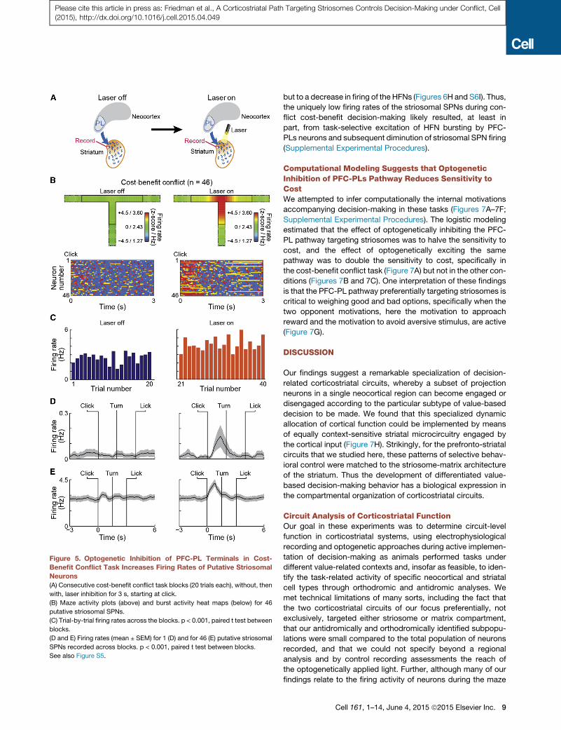
Figure 5. Optogenetic Inhibition of PFC-PL Terminals in Cost-
Benefit Conflict Task Increases Firing Rates of Putative Striosomal
Neurons
(A) Consecutive cost-benefit conflict task blocks (20 trials each), without, then
with, laser inhibition for 3 s, starting at click.
(B) Maze activity plots (above) and burst activity heat maps (below) for 46
putative striosomal SPNs.
(C) Trial-by-trial firing rates across the blocks. p < 0.001, paired t test between
blocks.
(D and E) Firing rates (mean ± SEM) for 1 (D) and for 46 (E) putative striosomal
SPNs recorded across blocks. p < 0.001, paired t test between blocks.
See also Figure S5.
Please cite this article in press as: Friedman et al., A Corticostriatal Path Targeting Striosomes Controls Decision-Making under Conflict, Cell(2015), http://dx.doi.org/10.1016/j.cell.2015.04.049
but to a decrease in firing of the HFNs (Figures 6H and S6I). Thus,
the uniquely low firing rates of the striosomal SPNs during con-
flict cost-benefit decision-making likely resulted, at least in
part, from task-selective excitation of HFN bursting by PFC-
PLs neurons and subsequent diminution of striosomal SPN firing
(Supplemental Experimental Procedures).
Computational Modeling Suggests that OptogeneticInhibition of PFC-PLs Pathway Reduces Sensitivity toCostWe attempted to infer computationally the internal motivations
accompanying decision-making in these tasks (Figures 7A–7F;
Supplemental Experimental Procedures). The logistic modeling
estimated that the effect of optogenetically inhibiting the PFC-
PL pathway targeting striosomes was to halve the sensitivity to
cost, and the effect of optogenetically exciting the same
pathway was to double the sensitivity to cost, specifically in
the cost-benefit conflict task (Figure 7A) but not in the other con-
ditions (Figures 7B and 7C). One interpretation of these findings
is that the PFC-PL pathway preferentially targeting striosomes is
critical to weighing good and bad options, specifically when the
two opponent motivations, here the motivation to approach
reward and the motivation to avoid aversive stimulus, are active
(Figure 7G).
DISCUSSION
Our findings suggest a remarkable specialization of decision-
related corticostriatal circuits, whereby a subset of projection
neurons in a single neocortical region can become engaged or
disengaged according to the particular subtype of value-based
decision to be made. We found that this specialized dynamic
allocation of cortical function could be implemented by means
of equally context-sensitive striatal microcircuitry engaged by
the cortical input (Figure 7H). Strikingly, for the prefronto-striatal
circuits that we studied here, these patterns of selective behav-
ioral control were matched to the striosome-matrix architecture
of the striatum. Thus the development of differentiated value-
based decision-making behavior has a biological expression in
the compartmental organization of corticostriatal circuits.
Circuit Analysis of Corticostriatal FunctionOur goal in these experiments was to determine circuit-level
function in corticostriatal systems, using electrophysiological
recording and optogenetic approaches during active implemen-
tation of decision-making as animals performed tasks under
different value-related contexts and, insofar as feasible, to iden-
tify the task-related activity of specific neocortical and striatal
cell types through orthodromic and antidromic analyses. We
met technical limitations of many sorts, including the fact that
the two corticostriatal circuits of our focus preferentially, not
exclusively, targeted either striosome or matrix compartment,
that our antidromically and orthodromically identified subpopu-
lations were small compared to the total population of neurons
recorded, and that we could not specify beyond a regional
analysis and by control recording assessments the reach of
the optogenetically applied light. Further, although many of our
findings relate to the firing activity of neurons during the maze
Cell 161, 1–14, June 4, 2015 ª2015 Elsevier Inc. 9
Figure 6. Sequence of Activity during Cost-
Benefit Decision-Making Recorded from
PFC-PLs Neurons, Striatal HFNs, and SPNs
(A) Average intra-burst activity (top) and heatmaps
(bottom) of HFNs during cost-benefit conflict (left)
and benefit-benefit (right) tasks.
(B) HFN intra-burst firing rates (mean ± SEM)
during click-to-turn periods. *p < 0.001 (two-tailed
t test; difference between CBC and other tasks).
(C) Activity of single SPN (mean ± SEM), aligned at
zero to activity peak of an HFN (inset) recorded
simultaneously by single tetrode. Inset zero in-
dicates time of start click.
(D) Firing rates of a simultaneously recorded
HFN-SPN pair, for phasic (burst, red) and tonic
(non-burst, blue) HFN activity, with correlation
coefficient (R) and slope for each. Dots show SPN
activity averaged across all 240 ms bins sorted for
HFN firing rates in 5-Hz steps.
(E) Sequence of peak excitation of PFC-PLs
neurons (green) and HFNs (red) and peak inhibition
of SPNs (blue) recorded in pairs as shown. Plots
aligned to start click (zero).
(F) HFN (n = 29, red) and simultaneously recorded
SPN (n=56, blue) responses toPFC-PLstimulation.
HFNs lead SPNs by �3 ms. *p = 0.001 (Wilcoxon
and two-sample Kolmogorov-Smirnov tests).
(G) Stimulation-inhibition protocol with SPN re-
cordings in consecutive blocks of PFC-PL elec-
trical stimulation then combined PFC-PL electrical
stimulation and intrastriatal optogenetic inhibition
of PFC-PL input.
(H) Putative striosomal SPN and HFN population
firing rates aligned to PFC-PL stimulation during
the stimulation-only block (black) and stimulation-
laser block (yellow). Laser illumination increased
SPN firing by 34% but decreased HFN firing by
32%. *p < 0.001 (two-tailed t test).
See also Figure S6.
Please cite this article in press as: Friedman et al., A Corticostriatal Path Targeting Striosomes Controls Decision-Making under Conflict, Cell(2015), http://dx.doi.org/10.1016/j.cell.2015.04.049
runs, we could not specify the time of decision-making, but
rather, had to rely on recording the rats’ behavioral responses
based on their decisions. Nor did we test decision-making
modes with other costs and other benefits, for example, costs
based on effort (Rangel and Hare, 2010; Rudebeck et al.,
2006; Salamone and Correa, 2012). Further, we used as costs
light stimuli, which can produce fear, and although our evidence
suggests that fear was not amain driver of the animals’ behavior,
this issue remains of interest. Despite these and other problems,
which we attempt to address in Supplemental Experimental Pro-
cedures, we found a striking internal consistency to the results
10 Cell 161, 1–14, June 4, 2015 ª2015 Elsevier Inc.
that we obtained across animals, across
behavioral, electrophysiological, and op-
togenetic findings, and across stimula-
tion-recording experiments.
Motivational Conflict Activates thePFC-PL Pathway PreferentiallyTargeting StriosomesOur findings demonstrate a rich reper-
toire of decision-making capabilities that
can be engaged in relation to environmental costs and benefits.
Yet, for the five tasks studied here, differing only in their cost and
benefit offers, the PFC-PL corticostriatal circuit preferentially tar-
geting striosomes was brought into play duringmaze runs only in
cost-benefit decision-making contexts involving approach-
avoidance conflict. Optogenetic inhibition of this pathway had
equally selective effects on cost-benefit conflict task perfor-
mance, increasing approach to high-cost options, yet scarcely
changing performance in the other four tasks, including the
non-conflict cost-benefit task in which simultaneous evaluation
of cost and benefit was still required, but in which the degree
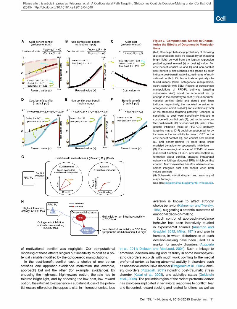
Figure 7. Computational Models to Charac-
terize the Effects of Optogenetic Manipula-
tions
(A–F) Choice probability (p: probability of choosing
diluted chocolate milk; p0: probability of choosing
bright light) derived from the logistic regression
plotted against reward (x) or cost (y) value. For
cost-benefit conflict (A and D) and non-conflict
cost-benefit (B and E) tasks, lines graded by color
indicate cost-benefit ratio (i.e., estimates of moti-
vational conflict). Circles indicate empirically ob-
tained means (filled: optogenetic manipulation,
open: control) with SEM. Results of optogenetic
manipulations of PFC-PL pathway targeting
striosomes (A–C) could be accounted for by
change in the sensitivity to cost (‘‘C’’) under moti-
vational conflict. Solid and dotted pink lines
indicate, respectively, the modeled behaviors for
optogenetic inhibition (halo) and excitation (C1V1)
of the striosome-targeting pathway. Changes in
sensitivity to cost were specifically induced in
cost-benefit conflict task (A), but not in non-con-
flict cost-benefit (B) or cost-cost (C) task. Opto-
genetic inhibition (halo) of PFC-ACC pathway
targeting matrix (D–F) could be accounted for by
increase in the sensitivity to reward (‘‘B’’) in the
cost-benefit conflict (D), non-conflict cost-benefit
(E), and benefit-benefit (F) tasks (blue lines:
modeled behaviors for optogenetic inhibition).
(G) Phenomenological model of PFC-PL strioso-
mal circuit function. PFC-PL provides context in-
formation about conflict, engages intrastriatal
network inhibiting striosomal SPNs in high-conflict
context. Matrix evaluates benefits, whereas strio-
somes integrate cost and benefit when both
values are high.
(H) Schematic circuit diagram and summary of
major findings.
See also Supplemental Experimental Procedures.
Please cite this article in press as: Friedman et al., A Corticostriatal Path Targeting Striosomes Controls Decision-Making under Conflict, Cell(2015), http://dx.doi.org/10.1016/j.cell.2015.04.049
of motivational conflict was negligible. Our computational
modeling of these effects singled out sensitivity to cost as a po-
tential variable modified by the optogenetic manipulations.
In the cost-benefit conflict task, a choice of one option
satisfies one approach-avoidance motivation (for example,
approach) but not the other (for example, avoidance). By
choosing the high-cost, high-reward option, the rats had to
tolerate bright light, and by choosing the low-cost, low-reward
option, the rats had to experience a substantial loss of the poten-
tial reward offered on the opposite site. In microeconomics, loss
Cell 161,
aversion is known to affect strongly
choice behavior (Kahneman and Tversky,
1984), suggesting a potential substrate of
emotional decision-making.
Such control of approach-avoidance
behavior has been intensively studied
in experimental animals (Amemori and
Graybiel, 2012; Miller, 1971) and also in
humans, in whom disturbances of such
decision-making have been used as a
marker for anxiety disorders (Aupperle
et al., 2011; Dickson and MacLeod, 2004). Such a linkage to
emotional decision-making and its frailty in some neuropsychi-
atric disorders accords with much work pointing to the medial
prefrontal cortex as having abnormal activity in disorders such
as obsessive-compulsive disorder (Fitzgerald et al., 2005), anxi-
ety disorders (Pizzagalli, 2011) including post-traumatic stress
disorder (Kasai et al., 2008), and addictive states (Goldstein
et al., 2009). The prelimbic region of the rodent prefrontal cortex
has also been implicated in behavioral responses to conflict, fear
and its control, reward seeking and related functions, as well as
1–14, June 4, 2015 ª2015 Elsevier Inc. 11
Please cite this article in press as: Friedman et al., A Corticostriatal Path Targeting Striosomes Controls Decision-Making under Conflict, Cell(2015), http://dx.doi.org/10.1016/j.cell.2015.04.049
in behavioral flexibility, all of which could be related to mood and
its control (Sangha et al., 2014; Seamans et al., 1995; Walton
et al., 2002).
Here, by comparing behavior and neural activity across perfor-
mance of multiple tasks, we found that a particular prefronto-
striatal circuit can be differentially engaged by conditions
inducing approach-avoidance conflict. Thus, the regulation of in-
ternal approach-avoidance drives could be an essential part of
the functional selectivity of the PFC-PL pathway.
Cost-Benefit Conflict Context Engages SelectiveStriatal Interneuronal Circuits to Control the Effects ofPFC-PL on the Activity of StriosomesOur physiological experiments demonstrated that PFC-PLs neu-
rons were selectively activated during runs in the cost-benefit
conflict task, that striatal HFNs, thought to correspond to inhib-
itory interneurons, were also selectively activated, but that puta-
tive striosomal SPNs were selectively inhibited during these
runs. Moreover, in simultaneous recordings from combinations
of cortical PFC-PLs neurons and striatal SPNs and HFNs, we
observed dynamic sequences in which activity peaks in the
PFC-PLs neurons preceded burst activity peaks of the HFNs,
which in turn preceded inhibition peaks of the SPNs. Finally,
when we electrically stimulated the PFC-PL but optogenetically
inhibited its striatal terminals, the inhibition increased the spike
firing of putative SPNs but decreased firing of the HFNs.
These results suggest that an inhibitory bursting interneuronal
system within the striatum is engaged when the PFC-PLs neu-
rons are activated, and that this intrastriatal system can sup-
press activity preferentially in striosomes, but only under highly
task-dependent conditions. This inversion of excitatory cortical
drive into inhibition supports evidence for fast dynamics of
high-firing interneurons (Berke, 2011; Burguiere et al., 2013;
Koos and Tepper, 1999). Our results, however, further suggest
that the dynamics of the corticostriatal and intrastriatal circuits
are task-selective and are crucial in underpinning the powerful
effects of optogenetic manipulation of the PFC-PL pathway to
striosomes.
By this account, optogenetic inhibition of the PFC-PL pathway
during the high conflict task released activity in the striosomes
during the time in which the animals greatly increased their
approaches to high-cost, high-benefit goals, and optogenetic
excitation of this pathway turned off the striosomes during the
time when the animals decreased their approaches to such
goals. If this simple interpretation is correct, then striosomal acti-
vation would itself be correlated with, and potentially causative
to, increased approaches to high-cost options (decreased cost
sensitivity), and striosomal shutdown would be related to, and
potentially causative to, decreased approach to high-cost op-
tions (increased cost sensitivity).
More generally, these findings suggest that the PFC-PL
pathway to striosomes is an important controller of behavior
elicited bymotivational conflict. It is remarkable that these selec-
tive effects were not apparent when we applied optogenetic in-
hibition to PFC-PL terminals in either the ventral tegmental area
or the basolateral amygdala, recipients of PFC-PL input, nor
even when we applied the inhibition to the PFC-PL itself. All of
these findings together, across physiology, behavior and opto-
12 Cell 161, 1–14, June 4, 2015 ª2015 Elsevier Inc.
genetics, point to a specialized function for the circuit preferen-
tially interconnecting the PFC-PL to striosomes.
ABroad Influence of thePFC-ACCCorticostriatal CircuitPreferentially Targeting the Striatal MatrixWith optogenetic inhibition of the PFC-ACC terminals in the
same dorsomedial region of the striatum, the rats increased their
approaches to higher benefits in all of the tasks. These results
suggest that the matrix, at least in the dorsomedial striatum, is
involved in evaluating the benefit of the goals and could have
less specific relation to cost-benefit integration. Logistic regres-
sion analysis suggested that the effect of inhibiting the PFC-ACC
terminals in the striatum could be consistently accounted for by
an increase in the sensitivity to the benefit component of the goal
(increase in approach motivation), regardless of the task condi-
tion. Thus within the striatum, the behavioral effects of discon-
necting the striosome and matrix compartments from their
respective preferential prefrontal inputs were strikingly different.
The Potential Functional Impact of Cortical Control ofStriosomesStriosomes are anatomically in a position to affect the dopamine-
containing neurons of the midbrain both directly (Fujiyama et al.,
2011; Prensa and Parent, 2001; Watabe-Uchida et al., 2012) and
indirectly via the lateral habenula (Rajakumar et al., 1993; Ste-
phenson-Jones et al., 2013), sites that affect mood, motivation,
and action (Hong and Hikosaka, 2013; Lak et al., 2014; Stopper
et al., 2014). Here, we did not address the potential effects of op-
togenetic manipulations of the PFC-PL pathway to striosomes
on the dopamine-containing neurons of the midbrain. Our find-
ings nevertheless raise the possibility that striosomes could
have an impact on decision-making via direct and indirect regu-
lation of dopaminergic signaling.
Our findings demonstrate that a medial prefrontal pathway,
here defined in rodents, can profoundly influence the activity of
striosomal projection neurons in the dorsomedial part of the
striatum. The exquisite selectivity of this control for situations
in which motivational conflict was induced by the cost-benefit
conflict context suggests that striosomes might influence not
only ongoing value-based decision-making but alsomood states
and state-dependent control of motivation. Striosomes, due to
their small size, have not yet be identified in fMRI studies of the
human brain and therefore cannot yet be seen in action. They
have already been found, however, in clinical studies including
post-mortem analysis, to be abnormal in neurologic and neuro-
psychiatric disorders involving changes in mood and loss of
normal motor control (Crittenden and Graybiel, 2011; Tippett
et al., 2007). In studies in experimental animals, their activity as
judged by gene expression patterns has been associated with
insistent, repetitive and stereotyped behaviors (Canales and
Graybiel, 2000; Crittenden and Graybiel, 2011).
Our experiments were limited to analysis of cortical inputs to
the dorsomedial associative striatum and likely do not expose
the full functionality of cortico-striosomal functions. Striosomes
exist in regions of the associative striatum that are not at all or
only weakly connected with the cortical regions that we manip-
ulated. What we emphasize here is that by manipulating the
striatal terminals of the PFC-PL circuit preferentially engaging
Please cite this article in press as: Friedman et al., A Corticostriatal Path Targeting Striosomes Controls Decision-Making under Conflict, Cell(2015), http://dx.doi.org/10.1016/j.cell.2015.04.049
striosomes, we obtained behavioral effects that were context-
dependent and that were not matched either by directly manip-
ulating the cortical region itself or by manipulating two other
regions targeted by the terminals of this cortical region. The
exquisitely specialized corticostriatal striosomal circuit identified
here could contribute to these and related disturbances of mood
and balanced decision-making under conditions of conflicting
motivational demands.
EXPERIMENTAL PROCEDURES
All experimental procedures were approved by the Committee on Animal Care
at the Massachusetts Institute of Technology. Adult Long-Evans rats (n = 55)
were trained to perform value-based decision-making tasks on a T-maze
composed of a running track and two end-arms with light focused on reward
feeders. To dissociate reward, cost, and motivational conflict, we used five
different types of decision-making tasks: cost-benefit conflict, benefit-benefit
with similar reward, benefit-benefit with dissimilar reward, non-conflict cost-
benefit and cost-cost (Figures 1A and 1B). We adjusted the cost and reward
scales according to the psychometric function of each individual rat. Tasks
were presented in random order, but only one cost-benefit conflict session
was given per week (Figure S1D). We performed simultaneous cortical and
striatal tetrode recordings and optogenetic manipulation to examine function-
ally corticostriatal circuits anatomically identified as preferentially targeting
striosomes (originating in PFC-PL) or matrix (originating in PFC-ACC) in the
dorsomedial striatum. For optogenetic experiments, virus encoding halorho-
dopsin or C1V1, or control virus, was injected into the PFC-PL or PFC-ACC.
Light (1.8–2.2 mW) was intrastriatally delivered for 3 s from trial-starting click
(Figure 1D). Compartment selectivity of the optogenetic manipulations was
estimated by densitometric analysis of EYFP tagged to the opsin (Figure S2).
Following each session, electrical microstimulation was delivered to the dor-
somedial striatum or PFC-PL to identify, respectively, PFC-PL neurons projec-
ting to the dorsomedial striatum (PFC-PLs neurons) and putative striosomal
SPNs (Figure 3). Within the striatum, we identified putative SPNs and HFNs
based on their spike waveforms, firing rates and spiking patterns (Figure S3E).
Burst activity was extracted by identifying spike trains with short interspike
intervals (Figure S6). Z scores of trial activity were calculated using the baseline
(11–3 s before the click) firing rate and SD and were plotted onto the maze
shape. Behavior and optogenetic effects were modeled using logistic regres-
sion. Significant differences were determined by two-tailed t tests (for opto-
genetic manipulation effects) and by chi-square tests (for spike activity in
histograms).More details are given in Supplemental Experimental Procedures.
SUPPLEMENTAL INFORMATION
Supplemental Information includes Supplemental Experimental Procedures
and six figures and can be found with this article online at http://dx.doi.org/
10.1016/j.cell.2015.04.049.
AUTHOR CONTRIBUTIONS
A.M.G. oversaw the project. A.F., D.H., L.G.G., and A.M.G designed the exper-
iments. A.F., D.H., and S.J.R. performed electrophysiological, optogenetic,
and behavioral experiments. A.F., D.H., L.G.G., A.S.H., and A.M.G. analyzed
data. D.H., L.G.G., M.H.R., and A.M.G. performed histological imaging and
analysis. K.A. performed modeling with input from A.F. and A.M.G. All authors
contributed to discussions about the data and their interpretation. A.M.G.
wrote the manuscript with input from A.F., D.H., L.G.G., K.A., and M.H.R.
ACKNOWLEDGMENTS
The authors thank Jannifer Lee, Dan Hu, Henry Hall, Yasuo Kubota, and Xiao-
jian Li for their help in many aspects of this work, Prof. Drazen Prelec for his
valuable comments, and the undergraduate students who assisted in these
experiments. This work was funded by NIH/NIMH (R01 MH060379 to
A.M.G.), the CHDI Foundation (A-5552 to A.M.G.), the Defense Advanced
Research Projects Agency and the U.S. Army Research Office (W911NF-10-
1-0059 to A.M.G.), the Bachmann-Strauss Dystonia and Parkinson Foundation
(to A.M.G.), the William N. and Bernice E. Bumpus Foundation (RRDA Pilot:
2013.1 to A.M.G.), and the Uehara Memorial Foundation and Japan Society
for the Promotion of Science (to D.H.).
Received: February 13, 2015
Revised: March 30, 2015
Accepted: April 10, 2015
Published: May 28, 2015
REFERENCES
Amemori, K., and Graybiel, A.M. (2012). Localized microstimulation of primate
pregenual cingulate cortex induces negative decision-making. Nat. Neurosci.
15, 776–785.
Aupperle, R.L., and Paulus, M.P. (2010). Neural systems underlying approach
and avoidance in anxiety disorders. Dialogues Clin. Neurosci. 12, 517–531.
Aupperle, R.L., Sullivan, S., Melrose, A.J., Paulus,M.P., and Stein,M.B. (2011).
A reverse translational approach to quantify approach-avoidance conflict in
humans. Behav. Brain Res. 225, 455–463.
Aupperle, R.L., Melrose, A.J., Francisco, A., Paulus, M.P., and Stein, M.B.
(2015). Neural substrates of approach-avoidance conflict decision-making.
Hum. Brain Mapp. 36, 449–462.
Berke, J.D. (2011). Functional properties of striatal fast-spiking interneurons.
Front. Syst. Neurosci. 5, 45.
Burguiere, E., Monteiro, P., Feng, G., and Graybiel, A.M. (2013). Optogenetic
stimulation of lateral orbitofronto-striatal pathway suppresses compulsive be-
haviors. Science 340, 1243–1246.
Canales, J.J., and Graybiel, A.M. (2000). Ameasure of striatal function predicts
motor stereotypy. Nat. Neurosci. 3, 377–383.
Crittenden, J.R., andGraybiel, A.M. (2011). Basal ganglia disorders associated
with imbalances in the striatal striosome and matrix compartments. Front.
Neuroanat. 5, 59.
Dickson, J.M., and MacLeod, A.K. (2004). Approach and avoidance goals and
plans: their relationship to anxiety and depression. Cognit. Ther. Res. 28,
415–432.
Donoghue, J.P., and Herkenham, M. (1986). Neostriatal projections from indi-
vidual cortical fields conform to histochemically distinct striatal compartments
in the rat. Brain Res. 365, 397–403.
Dwyer, D.M., Dunn, M.J., Rhodes, S.E., and Killcross, A.S. (2010). Lesions of
the prelimbic prefrontal cortex prevent response conflict produced by action-
outcome associations. Q. J. Exp. Psychol. (Hove) 63, 417–424.
Eblen, F., and Graybiel, A.M. (1995). Highly restricted origin of prefrontal
cortical inputs to striosomes in the macaque monkey. J. Neurosci. 15, 5999–
6013.
Etkin, A., Egner, T., Peraza, D.M., Kandel, E.R., and Hirsch, J. (2006).
Resolving emotional conflict: a role for the rostral anterior cingulate cortex in
modulating activity in the amygdala. Neuron 51, 871–882.
Fitzgerald, K.D., Welsh, R.C., Gehring, W.J., Abelson, J.L., Himle, J.A., Liber-
zon, I., and Taylor, S.F. (2005). Error-related hyperactivity of the anterior cingu-
late cortex in obsessive-compulsive disorder. Biol. Psychiatry 57, 287–294.
Flaherty, A.W., and Graybiel, A.M. (1991). Corticostriatal transformations in the
primate somatosensory system. Projections from physiologically mapped
body-part representations. J. Neurophysiol. 66, 1249–1263.
Flaherty, A.W., and Graybiel, A.M. (1993). Output architecture of the primate
putamen. J. Neurosci. 13, 3222–3237.
Fujiyama, F., Sohn, J., Nakano, T., Furuta, T., Nakamura, K.C., Matsuda, W.,
and Kaneko, T. (2011). Exclusive and common targets of neostriatofugal pro-
jections of rat striosome neurons: a single neuron-tracing study using a viral
vector. Eur. J. Neurosci. 33, 668–677.
Cell 161, 1–14, June 4, 2015 ª2015 Elsevier Inc. 13
Please cite this article in press as: Friedman et al., A Corticostriatal Path Targeting Striosomes Controls Decision-Making under Conflict, Cell(2015), http://dx.doi.org/10.1016/j.cell.2015.04.049
Gerfen, C.R. (1984). The neostriatal mosaic: compartmentalization of cortico-
striatal input and striatonigral output systems. Nature 311, 461–464.
Gleichgerrcht, E., Ibanez, A., Roca, M., Torralva, T., and Manes, F. (2010). De-
cision-making cognition in neurodegenerative diseases. Nat. Rev. Neurol. 6,
611–623.
Glimcher, P.W., and Fehr, E. (2014). Neuroeconomics: Decision Making and
the Brain, Second Edition (New York: Academic Press).
Goldstein, R.Z., Alia-Klein, N., Tomasi, D., Carrillo, J.H., Maloney, T., Woicik,
P.A., Wang, R., Telang, F., and Volkow, N.D. (2009). Anterior cingulate cortex
hypoactivations to an emotionally salient task in cocaine addiction. Proc. Natl.
Acad. Sci. USA 106, 9453–9458.
Graybiel, A.M. (1990). Neurotransmitters and neuromodulators in the basal
ganglia. Trends Neurosci. 13, 244–254.
Graybiel, A.M., and Ragsdale, C.W., Jr. (1978). Histochemically distinct com-
partments in the striatum of human, monkeys, and cat demonstrated by ace-
tylthiocholinesterase staining. Proc. Natl. Acad. Sci. USA 75, 5723–5726.
Hong, S., and Hikosaka, O. (2013). Diverse sources of reward value signals in
the basal ganglia nuclei transmitted to the lateral habenula in the monkey.
Front. Hum. Neurosci. 7, 778.
Kahneman, D., and Tversky, A. (1984). Choices, values, and frames. Am. Psy-
chol. 39, 341–350.
Kasai, K., Yamasue, H., Gilbertson, M.W., Shenton, M.E., Rauch, S.L., and
Pitman, R.K. (2008). Evidence for acquired pregenual anterior cingulate gray
matter loss from a twin study of combat-related posttraumatic stress disorder.
Biol. Psychiatry 63, 550–556.
Kita, H., and Kitai, S.T. (1988). Glutamate decarboxylase immunoreactive neu-
rons in rat neostriatum: their morphological types and populations. Brain Res.
447, 346–352.
Koos, T., and Tepper, J.M. (1999). Inhibitory control of neostriatal projection
neurons by GABAergic interneurons. Nat. Neurosci. 2, 467–472.
Lak, A., Stauffer, W.R., and Schultz, W. (2014). Dopamine prediction error re-
sponses integrate subjective value from different reward dimensions. Proc.
Natl. Acad. Sci. USA 111, 2343–2348.
Milad, M.R., Quirk, G.J., Pitman, R.K., Orr, S.P., Fischl, B., and Rauch, S.L.
(2007). A role for the human dorsal anterior cingulate cortex in fear expression.
Biol. Psychiatry 62, 1191–1194.
Miller, N.E. (1971). Selected Papers on Conflict, Displacement, Learned Drives
and Theory (Chicago, Illinois: Aldine Atherton).
Newman, H., Liu, F.C., and Graybiel, A.M. (2015). Dynamic ordering of early
generated striatal cells destined to form the striosomal compartment of the
striatum. J. Comp. Neurol. 523, 943–962.
Parthasarathy, H.B., Schall, J.D., and Graybiel, A.M. (1992). Distributed but
convergent ordering of corticostriatal projections: analysis of the frontal eye
field and the supplementary eye field in the macaque monkey. J. Neurosci.
12, 4468–4488.
14 Cell 161, 1–14, June 4, 2015 ª2015 Elsevier Inc.
Pizzagalli, D.A. (2011). Frontocingulate dysfunction in depression: toward bio-
markers of treatment response. Neuropsychopharmacology 36, 183–206.
Prensa, L., and Parent, A. (2001). The nigrostriatal pathway in the rat: a single-
axon study of the relationship between dorsal and ventral tier nigral neurons
and the striosome/matrix striatal compartments. J. Neurosci. 21, 7247–7260.
Rajakumar, N., Elisevich, K., and Flumerfelt, B.A. (1993). Compartmental origin
of the striato-entopeduncular projection in the rat. J. Comp. Neurol. 331,
286–296.
Rangel, A., and Hare, T. (2010). Neural computations associated with goal-
directed choice. Curr. Opin. Neurobiol. 20, 262–270.
Rudebeck, P.H., Walton, M.E., Smyth, A.N., Bannerman, D.M., and Rush-
worth, M.F. (2006). Separate neural pathways process different decision
costs. Nat. Neurosci. 9, 1161–1168.
Rushworth, M.F., Noonan, M.P., Boorman, E.D., Walton, M.E., and Behrens,
T.E. (2011). Frontal cortex and reward-guided learning and decision-making.
Neuron 70, 1054–1069.
Salamone, J.D. (1994). The involvement of nucleus accumbens dopamine in
appetitive and aversive motivation. Behav. Brain Res. 61, 117–133.
Salamone, J.D., and Correa, M. (2012). The mysterious motivational functions
of mesolimbic dopamine. Neuron 76, 470–485.
Sangha, S., Robinson, P.D., Greba, Q., Davies, D.A., and Howland, J.G.
(2014). Alterations in reward, fear and safety cue discrimination after inactiva-
tion of the rat prelimbic and infralimbic cortices. Neuropsychopharmacology
39, 2405–2413.
Seamans, J.K., Floresco, S.B., and Phillips, A.G. (1995). Functional differences
between the prelimbic and anterior cingulate regions of the rat prefrontal cor-
tex. Behav. Neurosci. 109, 1063–1073.
St Onge, J.R., and Floresco, S.B. (2010). Prefrontal cortical contribution to risk-
based decision making. Cereb. Cortex 20, 1816–1828.
Stephenson-Jones, M., Kardamakis, A.A., Robertson, B., and Grillner, S.
(2013). Independent circuits in the basal ganglia for the evaluation and selec-
tion of actions. Proc. Natl. Acad. Sci. USA 110, E3670–E3679.
Stopper, C.M., Tse, M.T., Montes, D.R., Wiedman, C.R., and Floresco, S.B.
(2014). Overriding phasic dopamine signals redirects action selection during
risk/reward decision making. Neuron 84, 177–189.
Tippett, L.J., Waldvogel, H.J., Thomas, S.J., Hogg, V.M., van Roon-Mom, W.,
Synek, B.J., Graybiel, A.M., and Faull, R.L. (2007). Striosomes and mood
dysfunction in Huntington’s disease. Brain 130, 206–221.
Vogel, J.R., Beer, B., and Clody, D.E. (1971). A simple and reliable conflict pro-
cedure for testing anti-anxiety agents. Psychopharmacology (Berl.) 21, 1–7.
Walton, M.E., Bannerman, D.M., and Rushworth, M.F. (2002). The role of rat
medial frontal cortex in effort-based decision making. J. Neurosci. 22,
10996–11003.
Watabe-Uchida, M., Zhu, L., Ogawa, S.K., Vamanrao, A., and Uchida, N.
(2012). Whole-brain mapping of direct inputs to midbrain dopamine neurons.
Neuron 74, 858–873.