a comprehensive review of the toxic effects of …
TRANSCRIPT

Comprehensive Review of Mercury in Dental Amalgam Fillings; www.iaomt.org; Page 1
A COMPREHENSIVE REVIEW OF THE TOXIC EFFECTS OF
MERCURY IN DENTAL AMALGAM FILLINGS ON THE ENVIRONMENT AND HUMAN HEALTH
The International Academy of Oral Medicine and Toxicology October 2020 Update
About the IAOMT: Representing a network of over 1,000 dentists, physicians, and other health professionals in more than 30 countries, the International Academy of Oral Medicine and Toxicology (IAOMT) has been researching the risks of dental mercury since our non-profit organization was founded in 1984. Our members have been expert witnesses for government bodies and health agencies around the world. We are an accredited member of the United Nations Environment Programme (UNEP)’s Global Mercury Partnership and were involved in the negotiations leading to UNEP’s Minamata Convention on Mercury. Brief Overview of Mercury Used in Dentistry: Millions of dentists around the world routinely use dental amalgam as a filling material in decayed teeth. Often referred to as “silver fillings,” all dental amalgams actually consist of 45-55% metallic mercury.1 Mercury is a neurotoxin that can cause harm to humans, especially children, pregnant women, and fetuses. Furthermore, the use of dental amalgam results in substantial quantities of toxic mercury released annually into the environment. Once in the environment, mercury pollution damages animals, plants, and the entire ecosystem, while creating “hotspots that last for centuries.”2 According to the United States Environmental Protection Agency (EPA), there are currently over 1,000 tons of mercury in the mouths of Americans, which is more than half of all the mercury being used in the U.S. today.3 While amalgams are currently used for 45% of all direct dental restorations worldwide,4 articles published in the Journal of the American Dental Association have established that these mercury fillings are used on 51.0% of White/Caucasian Americans, on 53.4% of Black/African Americans, on 72.9% of American Indians/Alaska Natives/Asians/Pacific Islanders,5 and on more than 75% of posterior restorations for new recruits to the U.S. Navy and Marines.6 Controversy has surrounded the use of mercury in dentistry since the 1800’s, when the hazardous material was first widely introduced as a filling component. The American Society of Dental Surgeons, the predecessor to the American Dental Association (ADA), made its members pledge not to use mercury because of its known toxicity,7 and in more recent years, government officials, scientists, dentists, consumers, and many others have raised serious concerns about the threats dental mercury poses to humans and to the environment at large. In 2013, the United Nations Environment Programme (UNEP)’s Intercessional Negotiating Committee formalized a global, legally-binding mercury treaty, which has now been signed by over 100 countries, including the U.S. Part of UNEP’s “Minamata Convention on Mercury” text includes initiatives with regards to dental mercury amalgam such as setting national objectives aimed at minimizing its use, promoting the use of cost-effective and clinically effective mercury-free alternatives for dental restoration, discouraging insurance policies that favor dental amalgam use over mercury-free dental restoration, and promoting the use of best environmental practices in dental facilities to reduce releases of mercury and its compounds to water and land.8
Many people do not realize that all silver-colored dental fillings contain approximately 50% mercury.
Comprehensive Review of Mercury in Dental Amalgam Fillings; www.iaomt.org; Page 2
A number of countries have taken action against the use of dental mercury amalgam fillings. In Norway and Sweden, dental amalgam is no longer in use.9 Bangladesh, the Czech Republic, Finland, Ireland, Nepal, and Slovakia are phasing it out.10 11 12 Denmark uses dental amalgam for only 5% of restorations, and Germany for about 10%.13 The government of Canada has recommended that dentists not use amalgam for children, pregnant women, and persons with kidney disorders.14 As part of the Minamata Convention on Mercury, the European Parliament voted in March 2017 to reduce dental mercury use. In addition to reporting “on the feasibility of a phase out of the use of dental amalgam in the long term, and preferably by 2030,”15 the new European Union regulation qualifies that dental amalgam not be used for children under 15 years and pregnant or breastfeeding women. Details in the U.S. Food and Drug Administration (FDA)’s public statements about dental amalgam on its website have changed over the years, including information about amalgam’s potentially harmful impact on pregnant women, fetuses, and children under the age of six. However, finally, in September 2020, the FDA advised that the following groups avoid getting dental amalgam whenever possible and appropriate: pregnant women and their developing fetuses; women who are planning to become pregnant; nursing women and their newborns and infants; children, especially those younger than six years of age; people with pre-existing neurological disease such as multiple sclerosis, Alzheimer’s disease or Parkinson’s disease; people with impaired kidney function; and people with known heightened sensitivity (allergy) to mercury or other components of dental amalgam.16 Meanwhile, scientific studies continue to demonstrate that the mercury used in dentistry poses serious risks to all populations and to the environment. Dental Amalgam Pollutes the Environment in a Variety of Ways:
Some 340 tonnes of mercury is used per year in dentistry,
of which about 70-100 tonnes (i.e. 20- 30%) likely enters the solid waste stream.17
--United Nations Environment Programme (UNEP), Global Mercury Assessment, 2013
1) Wastewater from Dental Offices After mercury is released into the environment, it can contaminate the food web and harm wildlife in the area for multiple generations. According to the United States Geological Survey, in 2010, dental amalgam was the leading end-use sector of mercury in the U.S.18 The use of mercury for dental amalgam in the U.S. has been estimated at 35.2 tons/year,19 and the discharge per dentist is an average of 250 milligrams/day (for an equivalent of 12 tons collectively released to the environment each year).20 For example, a 2002 New York Academy of Sciences report found over 40% of the mercury entering the New York/New Jersey harbor through wastewater was the result of discharges from dental offices.21
Additionally, in a 2014 document, the EPA recognized that “dental offices are the largest source of mercury discharges to POTWs [publicly-owned treatment works], contributing about half of the mercury received by POTWs.”22 This is dangerous because wastewater treatment facilities are designed to process human waste, not heavy metals. Thus, the mercury from dental discharges is separated out into sludge or biosolids.23 The sludge is usually incinerated, which releases mercury pollution into the atmosphere,24 and the biosolids are often used as fertilizer, which contaminates soil with mercury.25
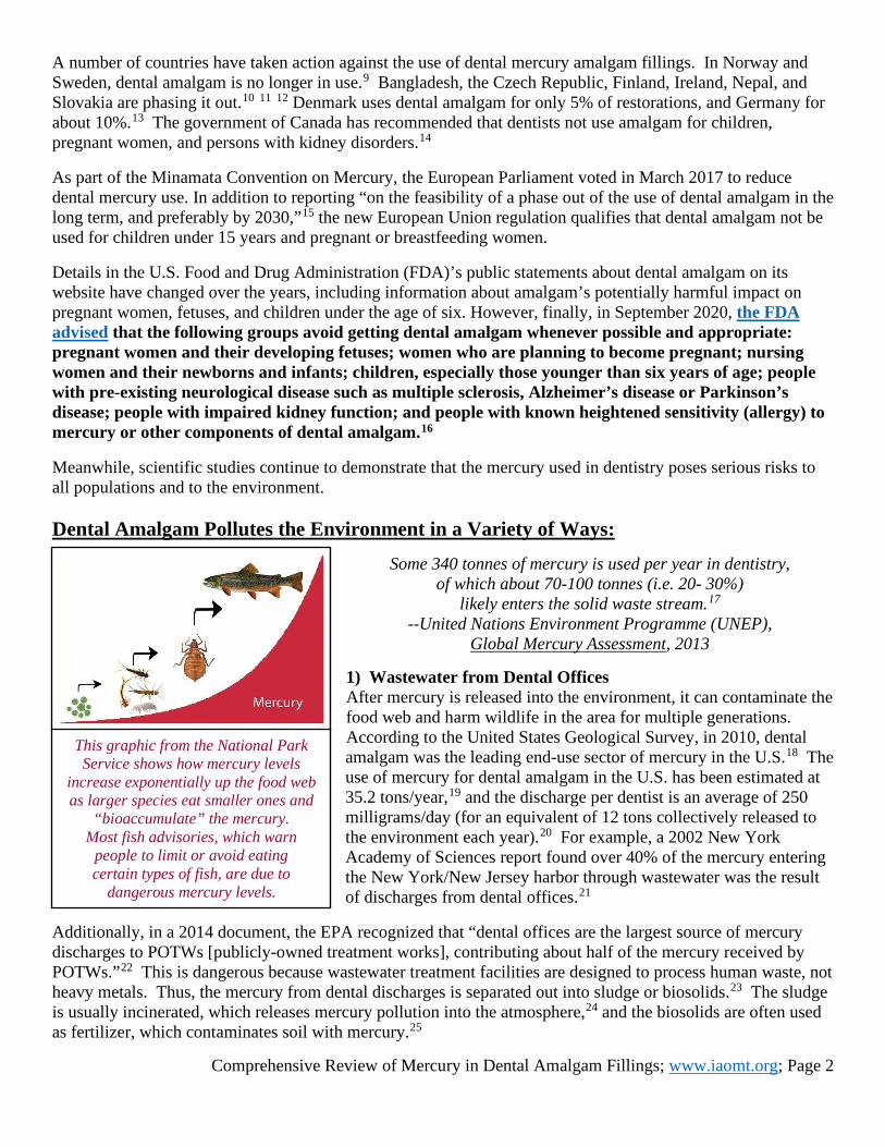
This graphic from the National Park Service shows how mercury levels
increase exponentially up the food web as larger species eat smaller ones and
“bioaccumulate” the mercury. Most fish advisories, which warn
people to limit or avoid eating certain types of fish, are due to
dangerous mercury levels.

Comprehensive Review of Mercury in Dental Amalgam Fillings; www.iaomt.org; Page 3
“Human waste is second only to direct release from dental offices
as a contributor of dental mercury to wastewater plants (AMSA 2001).”26 --Cited in Letter to the Editor by Larose & Basciano (IAOMT),
Journal of Dental Research, 2008 2) Human Waste Research has shown that amalgam fillings contribute to notable mercury levels in saliva, urine, and feces, and patients with dental amalgam excrete more than ten times more mercury in their feces than those without mercury fillings.27 Based on figures provided in scientific studies, the IAOMT has estimated that in the U.S., this amounts to over eight tons of mercury being flushed out to sewers, streams, and lakes per year.28 The same types of calculations were derived in Sweden in 1994, when researchers suggested that 100 kilograms (over 220 pounds) of mercury was being released to their country’s environment annually as a result of dental mercury excretion in feces and urine.29 Considering that dental mercury is released in feces and urine,30 and methylmercury (such as that taken in from fish consumption), is also released in feces and urine,31 the impact of human waste containing various forms of mercury is a pertinent factor in water pollution.
“Amalgam fillings not replaced before death will cause emissions to air, soil, and water upon cremation or burial.”32
--Hylander & Goodsite, Science of the Total Environment, 2006 3) Cremation and Burial
A 2013 assessment on mercury from UNEP reported: “Global emissions from use of mercury in dental amalgam resulting from cremation of human remains are estimated at 3.6 (0.9 – 11.9) tonnes in 2010.”33 With this consequential amount of mercury being released, it is apparent that cremation of bodies with amalgam fillings adds to air emissions and deposition onto land and into waterways. To illustrate this point, in 1992, the IAOMT applied scientific data to calculate that the cremation of 320,372 bodies in the U.S. during the preceding year added an estimated 2,800 pounds of mercury emissions into the atmosphere.34 Austria, Belgium, Germany, the Netherlands, Norway, Sweden, and Switzerland have applied measures to reduce mercury pollution from cremations.35 Although legislation has yet to be passed in the U.S.,36 Colorado, Maine, Minnesota, and Vermont have attempted to achieve regulations that would make removing amalgam fillings before cremation mandatory.37 Meanwhile, citizens in the U.S. have fought crematoriums in their neighborhoods by filing lawsuits38 and initiating protests.39
A variety of trends suggest that mercury releases from amalgam fillings in crematoriums will continue to increase.40 41 However, one alternative to cremation is a traditional burial, but burying an individual with amalgam fillings means that the mercury is deposited directly into the soil. This means that whether a person is cremated or buried, the mercury is released back to the environment.42
Human waste from people with dental amalgam mercury fillings
is another route of mercury pollution, especially because
treatment works cannot remove all of the mercury.
Rates of cremations are expected to rise over the next several decades, which
means that the levels of mercury released into the atmosphere from corpses with
dental amalgam fillings will also continue to increase.
Comprehensive Review of Mercury in Dental Amalgam Fillings; www.iaomt.org; Page 4
“Hg [mercury] vapor release to the atmosphere from dental vacuums
can be substantial and can exceed human exposure limits.”43 --Stone, Cohen, & Debban, Naval Institute for Dental and Biomedical Research, 2007
4) Mercury Vapor In offices with air/water separator tanks as part of the central vacuum system, mercury vapor has been found in air vented to the outside of the dental office.44 45 Dr. Paul G. Rubin of IAOMT has explained: “This mercury-containing material is discharged into waste streams via the dental office vacuum-pump system. This system also discharges large quantities of air, either into the atmosphere exterior to the office building or into the sewer system, depending on the type of equipment used.”46 Indoor air can also be dangerously polluted as a result of dental mercury. A study published in 2014 comparing air measurements at 42 dental sites in 17 countries found that mercury levels at most of the clinics were above safe limits. Their comparison included ten sites in the U.S., eight of which reportedly had levels higher than the EPA reference concentration in air. The authors noted that one of the two sites in the U.S. with mercury levels below the EPA reference level was from an office that had not placed mercury fillings in 20 years.
Amalgam Separators Can Reduce Dental Mercury Releases to the Environment: Amalgam separators can successfully reduce the amount of mercury discharge in wastewater from dental offices47 48 49 50 51 52 53 54 55 56 57 58 59 with reported capture efficiency rates ranging between 95-99%.60
Recently, the U.S. Environmental Protection Agency (EPA) utilized measures in the Clean Water Act to develop standards for dental offices/clinics to use amalgam separators so that dental mercury is not flushed down the drain and into the environment.61 EPA estimates about 103,000 dental offices use or remove amalgam in the U.S. and that almost all of these send their wastewater to POTWs [publicly owned treatment works]. 62 The new guidelines went into effect in July 2017, and the EPA has estimated that these new measures could reduce dental discharges of mercury by 5.1 tons annually.63 However, even with required standards, there should be enforced maintenance requirements for amalgam separators, as the Royal College of Dental Surgeons has done in Ontario, Canada.64 It must also be remembered that amalgam separators only contribute to solving the problem of dental mercury in wastewater and not the additional burdens placed by amalgam fillings on the environment and human health. Human Health Risks of Dental Amalgam Mercury: Mercury particulate can be discharged from dental amalgam fillings, and mercury vapor is continuously emitted from dental mercury amalgam fillings,65 66 67 68 69 70 71 72 73 74 75 76 77 78 79 80 81 82 83 84 85 86 87 88 89 90 which means that people are directly exposed to mercury as a result of their dental mercury amalgam fillings. The output of mercury is intensified by the number of amalgam fillings in the mouth91 92 93 94 95 96 97 98 99 and/or the number of amalgam surfaces in the mouth,100 101 102 103 104 105 106 107 108 109 110 111 the type of the amalgam filling (i.e. specific content of metals),112 113 and other factors such as chewing, teeth-grinding, brushing, dental treatments and procedures, and the consumption of hot liquids.114 115 116 117 118 119 120 121 122 123 124 125 126 127 128 129 130 131 132 133 Mercury is also known to be released during the placement, replacement, and removal of dental mercury amalgam fillings.134 135 136 137 138 139 140 141 142 143 144 145 146 147 148

Comprehensive Review of Mercury in Dental Amalgam Fillings; www.iaomt.org; Page 5
“And I think that there really is perhaps no place for mercury in children.”149
--Dr. Suresh Kotagal, pediatric neurologist at the Mayo Clinic; FDA Dental Products Panel, 2010
1) Pregnant Women and Children Authorities have issued distinct warnings about mercury’s use in children and pregnant women. For example, a 2005 World Health Organization (WHO) report identified harmful effects of mercury exposure, including areas of risk specifically linked to mercury in fetuses and children: “Adverse health effects from mercury exposure can be: tremors, impaired vision and hearing, paralysis, insomnia, emotional instability, developmental deficits during fetal development, and attention deficit and developmental delays during childhood.”150 Moreover, as stated at the top of page 2 in this document, international legislation has warned of the clear and present danger that the mercury in dental amalgam fillings poses to pregnant women and children. Also, 19 members of the U.S. Congress wrote a letter to the FDA in 2009 to express their concern about mercury used in amalgam fillings, with a focus on potential dangers to pregnant women and children,151 and when Representative Diane Watson of California proposed a Mercury Filling Disclosure and Prohibition Act (H.R. 2101{not enacted}), she explained: “It is, in fact, children who are at greatest risk from these fillings.”152
Fetal and infant exposure to mercury is known to have potentially serious health consequences, and the number of maternal amalgam fillings has been associated with mercury levels in cord blood;153 154 155 in the placenta;156 in the kidneys157 158 and liver159 of fetuses; in fetal hair;160 161 and in the brain162 and kidneys163 of infants. Additionally, mercury is excreted in breast milk of mothers with dental mercury amalgam fillings, and the mercury concentration in breast milk increases as the number of amalgam fillings in the mother increases.164 165 166 167 Significantly, a study published in 2018 by researchers in Norway involved over 72,000 pregnant women with data on the number of teeth containing dental amalgam fillings. The researchers discovered a “statistically significant association between the number of teeth filled with dental amalgam and the risk of perinatal death.” 168
Although two studies169 170 (commonly referred to as the “New England Children‘s Amalgam Trial” and the “Casa Pia Children’s Amalgam Trial”) have repeatedly been used to defend the use of amalgam in children, other researchers have since demonstrated that factors such as long term effects, genetic predisposition, and measurement errors must be taken into account. 171 172 173 174 175 176 Furthermore, researchers studying the same cohort (of the Children’s Amalgam Trials) have provided data that has identified potential risks to these subjects from mercury exposure based on gender, 177 178 179 genetic predisposition,180 181 182 and even gum-chewing.183 Risk assessments have also explored designating safe levels for children, who are smaller and still developing,184 especially since many dose levels are based on a one-size-fits-all scale for children and adults. In the meantime, scientific research continues to show that children are, in fact, at-risk for health impairments potentially caused by dental amalgam mercury fillings.185 186 187 188 189 190 191 192 193 194 In summary, authors of a study from 2011 cautioned: “Changes in dental practices involving amalgam, especially for children, are highly recommended in order to avoid unnecessary exposure to Hg [mercury].”195
A number of countries have banned dental amalgam fillings for children and
pregnant women, although this use of dental mercury is still allowed in the U.S.
Pregnant women, lactating women, and women of childbearing age should be aware
that mercury from their dental amalgam fillings can pose a risk to fetuses and children.
Comprehensive Review of Mercury in Dental Amalgam Fillings; www.iaomt.org; Page 6
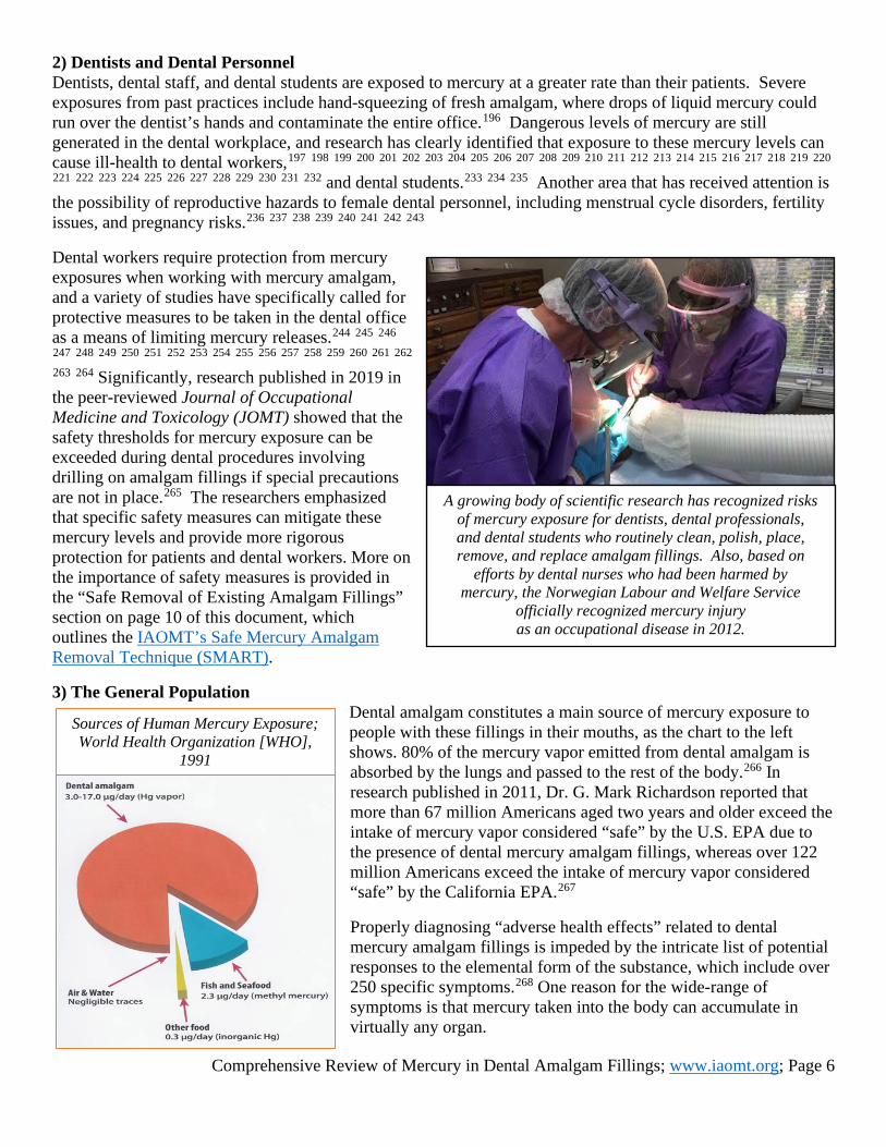
2) Dentists and Dental Personnel Dentists, dental staff, and dental students are exposed to mercury at a greater rate than their patients. Severe exposures from past practices include hand-squeezing of fresh amalgam, where drops of liquid mercury could run over the dentist’s hands and contaminate the entire office.196 Dangerous levels of mercury are still generated in the dental workplace, and research has clearly identified that exposure to these mercury levels can cause ill-health to dental workers,197 198 199 200 201 202 203 204 205 206 207 208 209 210 211 212 213 214 215 216 217 218 219 220 221 222 223 224 225 226 227 228 229 230 231 232 and dental students.233 234 235 Another area that has received attention is the possibility of reproductive hazards to female dental personnel, including menstrual cycle disorders, fertility issues, and pregnancy risks.236 237 238 239 240 241 242 243 Dental workers require protection from mercury exposures when working with mercury amalgam, and a variety of studies have specifically called for protective measures to be taken in the dental office as a means of limiting mercury releases.244 245 246 247 248 249 250 251 252 253 254 255 256 257 258 259 260 261 262 263 264 Significantly, research published in 2019 in the peer-reviewed Journal of Occupational Medicine and Toxicology (JOMT) showed that the safety thresholds for mercury exposure can be exceeded during dental procedures involving drilling on amalgam fillings if special precautions are not in place.265 The researchers emphasized that specific safety measures can mitigate these mercury levels and provide more rigorous protection for patients and dental workers. More on the importance of safety measures is provided in the “Safe Removal of Existing Amalgam Fillings” section on page 10 of this document, which outlines the IAOMT’s Safe Mercury Amalgam Removal Technique (SMART). 3) The General Population
Dental amalgam constitutes a main source of mercury exposure to people with these fillings in their mouths, as the chart to the left shows. 80% of the mercury vapor emitted from dental amalgam is absorbed by the lungs and passed to the rest of the body.266 In research published in 2011, Dr. G. Mark Richardson reported that more than 67 million Americans aged two years and older exceed the intake of mercury vapor considered “safe” by the U.S. EPA due to the presence of dental mercury amalgam fillings, whereas over 122 million Americans exceed the intake of mercury vapor considered “safe” by the California EPA.267 Properly diagnosing “adverse health effects” related to dental mercury amalgam fillings is impeded by the intricate list of potential responses to the elemental form of the substance, which include over 250 specific symptoms.268 One reason for the wide-range of symptoms is that mercury taken into the body can accumulate in virtually any organ.
A growing body of scientific research has recognized risks of mercury exposure for dentists, dental professionals, and dental students who routinely clean, polish, place, remove, and replace amalgam fillings. Also, based on
efforts by dental nurses who had been harmed by mercury, the Norwegian Labour and Welfare Service
officially recognized mercury injury as an occupational disease in 2012.
Sources of Human Mercury Exposure; World Health Organization [WHO],
1991

Comprehensive Review of Mercury in Dental Amalgam Fillings; www.iaomt.org; Page 7
Another reason for the wide-range of symptoms is that an array of co-existing factors influence each person’s reaction to dental mercury, including the presence of other health conditions, the number of amalgam fillings in the mouth, gender, genetic predisposition, dental plaque, selenium levels, exposure to lead, consumption of milk or alcohol, methylmercury levels from fish consumption, and the potential for mercury from dental amalgam fillings to be transformed into methylmercury within the human body.269
This is an abbreviated table of common symptoms of elemental mercury vapor inhalation270 271 272 273 274 275 276 277 278 279 to be considered by practitioners when evaluating the possible side effects of dental mercury amalgam:
Acrodynia or similar symptoms such as emotional instability, loss of appetite, general weakness, and skin changes (Magos and Clarkson, 2006)
Anorexia (Bernhoft, 2011) Cardiovascular problems/ labile pulse [frequent changes in heart rate]/tachycardia [abnormally rapid heartbeat] (Klassen, 2008)
Cognitive/neurological impairments/memory loss/decrease in mental function/difficulties with verbal and visual processing (Echeverria et al., 1998; Clarkson and Magos, 2006; Magos and Clarkson, 2006; Syversen and Kaur, 2012; USEPA, 2016)
Delusions/delirium/hallucination (Bernhoft, 2011; Syversen and Kaur, 2012)
Dermatological conditions/ dermographism [skin condition characterized by raised red marks]/dermatitis (Bernhoft, 2011; Klassen, 2008)
Endocrine disruption/enlargement of thyroid (Bernhoft, 2011; Klassen, 2008)
Erethism [symptoms such as irritability, abnormal responses to stimulation, and emotional instability] (Bernhoft, 2011; Clarkson et al., 2003; Clarkson and Magos, 2006; Magos and Clarkson, 2006)
Fatigue (Bernhoft, 2011; Echeverria et al., 1998)
Headaches (USEPA, 2016) Hearing loss (Rothwell and Boyd, 2008)
Immune system impairments (Bernhoft, 2011; Clarkson and Magos, 2006)
Insomnia (USEPA, 2016) Nerve response changes/peripheral neuropathy/decreased coordination/ decreased motor function/ polyneuropathy/ neuromuscular changes such as weakness, muscle atrophy, and twitching (Bernhoft, 2012; Clarkson et al., 2003; Clarkson and Magos, 2006; Echeverria et al., 1998; USEPA, 2016)
Oral manifestations/ gingivitis/metallic taste/oral lichenoid lesions/stomatitis/salivation (Bernhoft, 2011; Camisa et al., 1999; Clarkson et al., 2003; Clarkson and Magos, 2006; Klassen, 2008; Magos and Clarkson, 2006)
Psychological issues/mood changes related to anger, depression, excitability, irritability, mood swings, and nervousness (Echeverria et al., 1998; Klassen, 2008; Magos and Clarkson, 2006; USEPA, 2016)
Renal [kidney] problems/ proteinuria/nephrotic syndrome (Bernhoft, 2011; Clarkson et al., 2003; Clarkson and Magos, 2006; Klassen, 2008; USEPA, 2016; Syversen and Kaur, 2012)
Respiratory problems/ bronchial irritation/bronchitis/cough/ dyspnea [breathing difficulties]/ pneumonitis/respiratory failure (Bernhoft, 2011; Clarkson et al., 2003; Echeverria et al., 1998; Klassen, 2008; Magos and Clarkson, 2006; Syversen and Kaur, 2012; USEPA, 2016)
Shyness [excessive shyness]/social withdrawal (Magos and Clarkson, 2006; USEPA, 2016)
Tremors/mercurial tremors/ intention tremors (Bernhoft, 2011; Clarkson and Magos, 2006; Klassen, 2008; USEPA, 2016; Syversen and Kaur, 2012)
Weight loss (Bernhoft, 2011)

Comprehensive Review of Mercury in Dental Amalgam Fillings; www.iaomt.org; Page 8
While the symptoms of mercury exposure are individualized and have the potential to change over time, specific health conditions related to dental mercury exposure are also aptly documented in scientific literature, as the table below demonstrates.
Dental mercury amalgam fillings can potentially exacerbate and/or contribute to the conditions included below, as well as a myriad of other health outcomes:
Allergies280 281 282 Alzheimer’s
disease283 284 285 286 287
Amyotrophic lateral sclerosis (Lou Gehrig’s disease)288
Antibiotic resistance289 290 291 292
Autism spectrum disorders293 294 295 296
Autoimmune disorders/ immunodeficiency297 298 299 300 301 302 303 304 305
Cardiovascular problems306 307 308
Chronic fatigue, fatigue, and/or myalgic encephalomyelitis/chronic fatigue syndrome309 310 311 312 313 314 315 316
Dermatitis317 318 Fibromyalgia319 320 321 322
Gastrointestinal issues and/or irritable bowel syndrome323 324 325
Hearing loss326
Kidney disease327 328 329 330 331 332 333 334
Multiple sclerosis335 336 337 338
Oral lichenoid reaction339 340 341 342 343 344 345 346 347 348 349 350 351 352 353 354 355 and oral lichen planus356 357 358 359 360
Orofacial granulomatosis361 362
Parkinson’s disease363 364 365 366 367 368 369
Periodontal disease370 371
Psychological issues such as depression and anxiety372 373 374 375 376 377 378
Reproductive dysfunction379 380
Suicidal ideations381 382
Symptoms of chronic mercury poisoning383
Systemic lupus erythematosus384
Thyroiditis385 386 387 388 389
4) Genetic Predisposition The association of genetic predisposition with specific, adverse effects from mercury exposure has been examined in several studies. It has been found that mercury exposure from dental mercury amalgam particularly threatens individuals with genetic variants that can impact their response to mercury exposures such as those with CPOX4,390 391 392 APOE(3,4),393 394 395 396 and BDNF (brain-derived neurotropic factor) polymorphisms.397 398 399 For example, the researchers of a study published in 2006 linked the polymorphism CPOX4 (coproporphyrinogen oxidase, exon 4) to decreased visuomotor speed and indicators of depression in dental professionals. 400 Furthermore, the CPOX4 genetic variation was identified as a factor for neurobehavioral issues in a study of children with dental amalgams. The researchers noted, “…among boys, numerous significant interaction effects between CPOX4 and Hg [mercury] were observed spanning all 5 domains of neurobehavioral performance…These findings are the first to demonstrate genetic susceptibility to the adverse neurobehavioral effects of Hg [mercury] exposure in children.”401
Comprehensive Review of Mercury in Dental Amalgam Fillings; www.iaomt.org; Page 9
Another area of genetic susceptibility in relation to dental mercury risk that has merited attention is the APOE4 (apo-lipoprotein E4) genetic variation. A 2006 study found a correlation between individuals with APOE4 and chronic mercury toxicity.402 The same study found that removal of dental amalgam fillings resulted in “significant symptom reduction,” and one of the symptoms listed was memory loss. The symptom of memory loss is quite interesting, as APOE4 has also been associated with a higher risk for Alzheimer’s disease.403 404 405 Importantly, the authors of a study which found a connection between number of mercury fillings and neurotoxic effects for those with APOE genotype explained: “APO-E genotyping warrants investigation as a clinically useful biomarker for those at increased risk of neuropathology, including AD [Alzheimer’s disease], when subjected to long-term mercury exposures…An opportunity could now exist for primary health practitioners to help identify those at greater risk and possibly forestall subsequent neurological deterioration.”406 Research has also shown that dental mercury fillings can play a role in immune system problems for genetically predisposed patients. Whereas research on animals has established a connection between dental mercury and autoimmunity,407 408 research involving human subjects has confirmed that genetic susceptibility to reactions from dental mercury is potentially related to chronic fatigue syndrome,409 as well as amyotrophic lateral sclerosis, multiple sclerosis, and rheumatoid arthritis. 410 In addition, scientific data has linked mercury and genetic traits to autism,411 412 chemical sensitivities,413 and Kawasaki’s disease,414 and research has also suggested that genetic transporters could be involved in the toxicokinetics of mercury.415 Other than CPOX4, APOE, and BDNF polymorphisms, genetic traits that have been examined for association with health impairments caused by mercury exposure include metallothionein (MT) polymorphisms,416 417 catechol-O-methyltransferase (COMT) variants,418 PON1 variants,419 420 MTHFR mutations and other genetic aspects.421 422 423 The authors of one of these studies concluded: “It is possible that elemental mercury may follow the history of lead, eventually being considered a neurotoxin at extremely low levels.”424 5) Mercury and Metal Allergies In some genetically susceptible individuals, metals can also induce allergies.425 A study published in 2018 in the journal Dermatitis was conducted on 686 adults who were patch tested for allergies. The results demonstrated that “38.9% of patients had 1 or more positive patch-test reactions to a metal allergen, most commonly nickel (17.4%), mercury (12.3%), and palladium (9.2%)…Among patients with positive reactions to nickel, 34.5%, 15.1%, and 5.0% had positive reactions to 1, 2, or 3 additional metals, respectively.”426 That study involved individuals with suspected allergies; however, the statistics are relevant, as studies involving the general population and the prevalence of metal allergies are rare.427 However, a 1993 study reported that 3.9% of healthy subjects tested positive for metal reactions in general.428 If this figure is applied to the current U.S. population, this would mean that dental metal allergies potentially impact as many as 12.5 million Americans.
The ability of specific genetic variants to negatively impact the body’s reaction to dental mercury exposure has even achieved attention in the mainstream media. A January 2016 article by Greg Gordon of McClatchy News
included interviews with some of the researchers of the studies mentioned in this document.
Markedly, Dr. James Woods stated: “‘Twenty-five percent to 50 percent of people have these (genetic variants).’”
In the same article, Dr. Diana Echeverria discussed “‘a lifetime risk’” of neurological damage
related to this population, and she elaborated: “‘We’re not talking about a small risk.’”

Comprehensive Review of Mercury in Dental Amalgam Fillings; www.iaomt.org; Page 10
The number of affected individuals is likely much higher, though, because recent studies and reports tend to agree that metal allergies are on the rise.429 430 Part of this could be caused by increased exposure to metals, including ear/body piercings, because exposure to metals has been cited as a potential trigger for the development of allergies to them.431 Additionally, it has been hypothesized that contact with metals during an infection could increase chances of developing a metal allergy later in life.432 An issue is the wide-range of symptoms patients allergic to dental metals can exhibit. In a 2014 publication, Dr. Vera Stejskal wrote: “Metal-induced inflammation may be involved in the pathology of various autoimmune and allergic diseases, where abnormal fatigue, joint and muscle pain, cognitive impairment and other non-specific symptoms are often present.”433 Additionally, a gamut of health conditions has been linked to dental metal allergies, including autoimmunity,434 435 chronic fatigue syndrome,436 437 438 fibromyalgia,439 440 metallic pigmentation,441 multiple chemical sensitivities,442 443 multiple sclerosis,444 myalgic encephalitis,445 oral lichenoid lesions,446 447 448 449 450 orofacial granulomatosis,451 and even infertility in both women and men.452 Another issue with calculating the number of patients with adverse reactions to a metallic material is that the onset of symptoms can be delayed and therefore might not be associated with the implant or device. For example, researchers writing about dental amalgam fillings warned: “Sensitization appears most frequently after the amalgam has been present in the mouth for more than 5 years.”453 Furthermore, there may not be any local reaction to help the patient and doctor identify the metal as the culprit in ill health,454 and even if hypersensitivity reactions are noticed, they can be misdiagnosed as infection.455 Clinical screening for metal allergy has been recommended,456 and the importance of patients reporting reactions to metals to their doctors has also been emphasized in the scientific literature.457 458 459 460 461 462 In addition to reporting any rashes from jewelry, watches, or other metal exposures, it is essential for each patient to recognize the gamut of symptoms that can be related to the presence of a metal implant or device in their body. It is also vital for patients to remember that sensitization to metal can develop years after an implant or device has been placed and that adverse effects can occur with or without the sign of a rash or eruption on the skin or in the mouth. Unfortunately, in some reported cases, the only way to fully establish that a metal implant or device was causing health problems was to have it removed and then document the results. Researchers from Harvard School of Medicine wrote in 2016: “Paradoxically, a patient can sometimes only be diagnosed with metal allergy when the symptoms resolve upon replacement with an immunologically inert implant.”463 A few examples of conditions reportedly improved and/or cured as a result of removing dental metal allergens include amyotrophic lateral sclerosis,464 chronic fatigue syndrome, 465 dermatitis,466 fibromyalgia,467 multiple sclerosis,468 oral lichen planus, 469 470 471 oral lichenoid lesion, 472 473 474 orofacial granulomatosis,475 and other symptoms.476 In a 2011 report, Hosoki and Nishigawa suggested: “In principle, all restorations with allergy-positive metal elements need to be removed.”477
Many patients are not aware that reactions they have to jewelry and other metal accessories are a warning sign that they could have allergic reactions to dental amalgam fillings
and/or metal implants in their bodies.
Comprehensive Review of Mercury in Dental Amalgam Fillings; www.iaomt.org; Page 11
Safety Measures for Removal of Dental Amalgam Mercury Fillings:
Although individual response varies, in addition to the recovery situations listed above, research has documented the reduction of other health issues after the removal of amalgam fillings.478 479 480 481 482 483 484 485 486 487 488 However, it is important to note that removal of any dental material requires a number of precautions. This is because an unsafe removal process can cause serious injury to the patient, including the possibility of increased metal exposure. For example, if dental amalgam fillings are removed unsafely, patients can be exposed to increased levels of mercury.
To assist in mitigating the potential negative outcomes of mercury exposure to dental professionals, students, staff members, patients, and others, the IAOMT has developed safety recommendations for removal of existing dental mercury amalgam fillings.489 IAOMT’s Safe Mercury Amalgam Removal Technique (SMART) is located online at https://iaomt.org/safe-removal-amalgam-fillings/.490 The innovative recommendations build upon traditional safe amalgam removal techniques such as the use of masks, water irrigation, and high volume suction by supplementing these conventional strategies with a number of additional protective measures, the need for which have only recently been identified in scientific research. In addition to the dozens of studies that support each separate step of the recommendations, the overall technique has been supported by two studies published in peer-reviewed journals in 2019.491 492 IAOMT recommends that patients familiarize themselves with the recommendations to ensure protective strategies will be applied during amalgam removal.
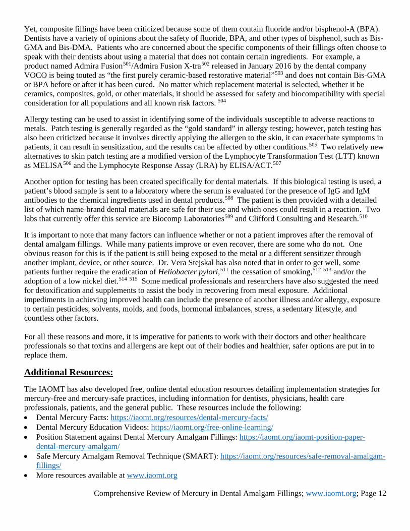
Alternatives to Amalgams as a Filling Material: Obviously, once amalgams have been removed, they must be replaced with a different dental filling material. Alternatives to amalgam include composite resin, glass ionomer, porcelain, and gold, among other options. When given the choice, most consumers opt for direct composite fillings because the white coloring matches the tooth better and the cost is considered moderate. In the past, a common argument against composite fillings was that they were not as durable as amalgam. However, recent studies have debunked this claim. Researchers of a study which was published in 2016 and conducted on over 76,000 patients for over ten years found that posterior amalgam fillings had a higher annual failure rate than composites.493 Two separate studies published in 2013 found that composite fillings performed as well as amalgam when comparing failure rates494 and replacement filling rates.495 Other research offers similar findings in support of composite filling durability.496 497 498 499 Research has further confirmed that composite resins present a lower risk for chemical exposures. In a 2016 publication co-authored by risk assessment specialist Dr. G. Mark Richardson, it was reported: “Relative risks of chemical exposures from dental materials decrease in the following order: Amalgam>Au (Gold) alloys>ceramics>composite resins.”500
Many patients choose dental filling materials that match the natural
coloring of teeth after they have their mercury amalgam fillings removed.
Comprehensive Review of Mercury in Dental Amalgam Fillings; www.iaomt.org; Page 12
Yet, composite fillings have been criticized because some of them contain fluoride and/or bisphenol-A (BPA). Dentists have a variety of opinions about the safety of fluoride, BPA, and other types of bisphenol, such as Bis-GMA and Bis-DMA. Patients who are concerned about the specific components of their fillings often choose to speak with their dentists about using a material that does not contain certain ingredients. For example, a product named Admira Fusion501/Admira Fusion X-tra502 released in January 2016 by the dental company VOCO is being touted as “the first purely ceramic-based restorative material”503 and does not contain Bis-GMA or BPA before or after it has been cured. No matter which replacement material is selected, whether it be ceramics, composites, gold, or other materials, it should be assessed for safety and biocompatibility with special consideration for all populations and all known risk factors. 504 Allergy testing can be used to assist in identifying some of the individuals susceptible to adverse reactions to metals. Patch testing is generally regarded as the “gold standard” in allergy testing; however, patch testing has also been criticized because it involves directly applying the allergen to the skin, it can exacerbate symptoms in patients, it can result in sensitization, and the results can be affected by other conditions.505 Two relatively new alternatives to skin patch testing are a modified version of the Lymphocyte Transformation Test (LTT) known as MELISA506 and the Lymphocyte Response Assay (LRA) by ELISA/ACT.507 Another option for testing has been created specifically for dental materials. If this biological testing is used, a patient’s blood sample is sent to a laboratory where the serum is evaluated for the presence of IgG and IgM antibodies to the chemical ingredients used in dental products.508 The patient is then provided with a detailed list of which name-brand dental materials are safe for their use and which ones could result in a reaction. Two labs that currently offer this service are Biocomp Laboratories509 and Clifford Consulting and Research.510 It is important to note that many factors can influence whether or not a patient improves after the removal of dental amalgam fillings. While many patients improve or even recover, there are some who do not. One obvious reason for this is if the patient is still being exposed to the metal or a different sensitizer through another implant, device, or other source. Dr. Vera Stejskal has also noted that in order to get well, some patients further require the eradication of Heliobacter pylori,511 the cessation of smoking,512 513 and/or the adoption of a low nickel diet.514 515 Some medical professionals and researchers have also suggested the need for detoxification and supplements to assist the body in recovering from metal exposure. Additional impediments in achieving improved health can include the presence of another illness and/or allergy, exposure to certain pesticides, solvents, molds, and foods, hormonal imbalances, stress, a sedentary lifestyle, and countless other factors. For all these reasons and more, it is imperative for patients to work with their doctors and other healthcare professionals so that toxins and allergens are kept out of their bodies and healthier, safer options are put in to replace them.
Additional Resources: The IAOMT has also developed free, online dental education resources detailing implementation strategies for mercury-free and mercury-safe practices, including information for dentists, physicians, health care professionals, patients, and the general public. These resources include the following: • Dental Mercury Facts: https://iaomt.org/resources/dental-mercury-facts/ • Dental Mercury Education Videos: https://iaomt.org/free-online-learning/ • Position Statement against Dental Mercury Amalgam Fillings: https://iaomt.org/iaomt-position-paper-
dental-mercury-amalgam/ • Safe Mercury Amalgam Removal Technique (SMART): https://iaomt.org/resources/safe-removal-amalgam-
fillings/ • More resources available at www.iaomt.org
Comprehensive Review of Mercury in Dental Amalgam Fillings; www.iaomt.org; Page 13
PHOTO CREDITS: Middle of page 1: Mercury Amalgam Fillings; IAOMT File Photo Bottom of page 2: Mercury (Hg) Biomagnification Graphic; National Park Service; https://npgallery.nps.gov/AssetDetail/ba40f463-9f49-4a28-bcd9-a1e65a0d1dee; Accessed October 2019 Top of page 3: Picture of a Honolulu city sign warning of sewage contamination, with Waikiki hotels and Diamond Head in background; Released under CC-BY-SA 2.5; Keith H.; https://commons.wikimedia.org/wiki/File:20060402_waikiki_sewage.jpg; Accessed October 2019 and cropped to fit page. (For more about mercury contamination in sewage, see https://www.ncbi.nlm.nih.gov/pmc/articles/PMC4764622/.) Bottom of page 3: Chimney Crematorium Peak Smoke Top, Stock Photo purchased by IAOMT from 123rf; https://www.123rf.com/photo_9682193_chimney-crematorium-peak-smoke-top.html?term=crematorium%2Bblack%2Bsmoke&vti=lsapn7cqoj9yci0ytl-1-11; Purchased March 2018 Bottom of page 4: Dental Office Warnings, Office of Environmental Health Hazard Assessment (OEHHA) of the California Environmental Protection Agency; https://oehha.ca.gov/media/downloads/proposition-65/presentation/dentalofficewarnings041514.pdf; Accessed October 2019 Top of page 5: Sri Lankan Woman and Child; Steve Evans; https://commons.wikimedia.org/wiki/File:Sri_Lankan_woman_and_child.jpg; Accessed October 2019 and slightly cropped to fit page; Originally posted at https://www.flickr.com/photos/64749744@N00/549738320; Shared in compliance with https://creativecommons.org/licenses/by-nc/2.0/. Bottom of page 5: Patient with Dentist in Alaska; Official Website of the United States Navy; 090309-F-3646G-199; https://www.navy.mil/view_image.asp?id=69543; Accessed October 2019 Top of page 6: Protective Equipment during Amalgam Removal; Courtesy of Dr. Jack Kall Bottom of page 6: Sources of Human Mercury Exposure based on 1991 World Health Organization [WHO] Report; Chart Made by the IAOMT from Statistics in http://www.inchem.org/documents/ehc/ehc/ehc118.htm Top of page 8: Quotes on Genetic Variants from: Gordon G. Dental group defends mercury fillings amid mounting evidence of risks. McClatchy News Service. January 5, 2016. Available from: http://www.mcclatchydc.com/news/nation-world/national/article53118775.html. Accessed October 2019. Middle of page 9: Pile of Old Jewelry; Retrieved from Pixabay; https://pixabay.com/photos/jewelry-junk-fake-gem-jewel-314473/; Accessed October 2019 Top of page 10: Before and After Photos of Teeth from Amalgam Removal; Courtesy of Dr. Michael Rehme from https://toothbody.com/the-inevitable-replacement-of-mercury-amalgam-fillings/
ENDNOTES: 1 World Health Organization. Mercury in Health Care [policy paper]. August 2005: 1. Available from WHO Web site: http://www.who.int/water_sanitation_health/medicalwaste/mercurypolpaper.pdf. Accessed October 2019. 2 Pirrone N, Mason R. Mercury Fate and Transport in the Global Atmosphere: Emissions, Measurements, and Models. New York, New York: Springer. 2009: 166. 3 United States Environmental Protection Agency. International Mercury Market Study and the Role and Impact of US Environmental Policy. 2004. 4 Heintze SD, Rousson V. Clinical effectiveness of direct Class II restorations—a meta-analysis. J Adhes Dent. 2012; 14(5):407-431. 5 Makhija SK, Gordan VV, Gilbert GH, Litaker MS, Rindal DB, Pihlstrom DJ, Gvist V. Dental practice-based research network restorative material: Findings from the characteristics associated with type of practitioner, patient and carious lesion. J Am Dent Assoc. 2011; 142: 622-632. 6 Simececk JW, Diefenderfer KE, Cohen ME. An evaluation of replacement rates for posterior resin-based composite and amalgam restorations in U.S. Navy and Marine recruits. J Am Dent Assoc. 2009; 140 (2): 207. 7 Health Canada. The Safety of Dental Amalgam. 1996: 3. Available from Health Canada Web site: http://www.hc-sc.gc.ca/dhp-mps/alt_formats/hpfb-dgpsa/pdf/md-im/dent_amalgam-eng.pdf. Accessed October 2019. 8 United Nations Environment Programme. Minamata Convention on Mercury: Text and Annexes. 2013: 48. Available from UNEP’s Minamata Convention on Mercury Web site: http://www.mercuryconvention.org/Portals/11/documents/Booklets/Minamata%20Convention%20on%20Mercury_booklet_English.pdf. Accessed October 2019. 9 UN Environment. Global Mercury Supply, Trade and Demand. Geneva, Switzerland: United Nations Environment Pro-gramme, Chemicals and Health Branch. 2017. Available from: http://wedocs.unep.org/bitstream/handle/20.500.11822/21725/global_mercury.pdf?sequence=1&isAllowed=y. Accessed March 2019. 10 UN Environment. Global Mercury Supply, Trade and Demand. Geneva, Switzerland: United Nations Environment Pro-gramme, Chemicals and Health Branch. 2017. Available from: http://wedocs.unep.org/bitstream/handle/20.500.11822/21725/global_mercury.pdf?sequence=1&isAllowed=y. Accessed March 2019. 11 Health Care without Harm. Four EU countries announce plans to phase out mercury fillings [press release]. August 28, 2019. Available from: https://noharm-europe.org/articles/news/europe/four-eu-countries-announce-plans-phase-out-mercury-fillings. Accessed October 2019.
Comprehensive Review of Mercury in Dental Amalgam Fillings; www.iaomt.org; Page 14
12 IPEN. Government of Nepal Bans Use of Mercury Dental Amalgam and Mercury-Based Equipment. September 20, 2019. Available from: https://ipen.org/news/government-nepal-bans-use-mercury-dental-amalgam-and-mercury-based-equipment. Accessed October 2019. 13 UN Environment. Global Mercury Supply, Trade and Demand. Geneva, Switzerland: United Nations Environment Pro-gramme, Chemicals and Health Branch. 2017. Available from: http://wedocs.unep.org/bitstream/handle/20.500.11822/21725/global_mercury.pdf?sequence=1&isAllowed=y. Accessed March 2019. 14 UN Environment. Global Mercury Supply, Trade and Demand. Geneva, Switzerland: United Nations Environment Pro-gramme, Chemicals and Health Branch. 2017. Available from: http://wedocs.unep.org/bitstream/handle/20.500.11822/21725/global_mercury.pdf?sequence=1&isAllowed=y. Accessed March 2019. 15 REGULATION (EU) 2017/852 OF THE EUROPEAN PARLIAMENT AND OF THE COUNCIL of 17 May 2017 on mercury, and repealing Regulation (EC) No 1102/2008. Official Journal of the European Union. Available from: https://eur-lex.europa.eu/legal-content/EN/TXT/PDF/?uri=CELEX:32017R0852&rid=2. Accessed March 2019. 16 United States Food and Drug Administration. FDA Issues Recommendations for Certain High-Risk Groups Regarding Mercury-Containing Dental Amalgam. September 24, 2020. Available from: https://www.fda.gov/news-events/press-announcements/fda-issues-recommendations-certain-high-risk-groups-regarding-mercury-containing-dental-amalgam.
This fact sheet was last updated on October 6, 2020. 17 United Nations Environment Programme. Global Mercury Assessment 2013: Sources, Emissions, Releases and Environmental Transport. Geneva, Switzerland: UNEP Chemicals Branch; 2013: 10. Available from: http://wedocs.unep.org/handle/20.500.11822/7984. Accessed October 2019. 18 Wilburn DR. Changing patterns in the use, recycling, and material substitution of mercury in the United States: U.S. Geological Survey Scientific Investigations Report 2013–5137. 2013. 32 p. Available from: http://pubs.usgs.gov/sir/2013/5137/. Accessed October 2019. 19 Vandeven JA, McGinnis SL. An assessment of mercury in the form of amalgam in dental wastewater in the United States. Water, Air, and Soil Pollution. 2005; 164(1-4):349. 20 Scarmoutzos L, Boyd M. OE: Environmental and Toxicological Concerns of Dental Amalgam and Mercury. Northboro, MA: MVS Solutions. Inc. and SolmeteX, Inc.; 2003: See Table 2 on pages 36-37. Available from MVS Solutions Web site: http://www.mvssolutions.com/mercury.pdf. Accessed October 2019. 21 de Cerreño AL, Panero M, Boehme S. Pollution Prevention and Management Strategies for Mercury in the New York/New Jersey Harbor. New York: New York Academy of Sciences; 2002: 35. 22 United States Environmental Protection Agency. Effluent Limitation Guidelines and Standards for the Dental Category Mercury in Dental Amalgam. EPA - 821-F-14-002. September 2014. Available from EPA Website: http://www.epa.gov/sites/production/files/2015-06/documents/dental-category-factsheet_proposed_rule_2014.pdf. Accessed October 2019. 23 Larose P. Position Paper: Environmental Committee. IAOMT Environmental Committee; 2011. Available from IAOMT Web site: https://iaomt.org/wp-content/uploads/article_02-03-Environment.pdf. Accessed October 2019. 24 Balogh S, Liang L. Mercury pathways in municipal wastewater treatment plants. Water, Air, and Soil Pollution. 1995; 80(1-4):1181-90. 25 Health & Environment Alliance and Health Care Without Harm. Chapter 2: Mercury pollution: where does it come from? Stay Healthy, Stop Mercury Campaign. 2007: 24. Available from Health and Environment Alliance Web site: http://www.env-health.org/IMG/pdf/mercury_chapter2.pdf. Accessed October 2019. 26 Cited as American Metropolitan Sewage Agencies. Mercury Pollution Prevention Program (submitted by Larry Walker Associates). 2001. in Larose P, Basciano M. Dental mercury and Norway. Journal of Dental Research. 2008; 87(5): 413. Available from http://jdr.sagepub.com/content/87/5/413.extract. Accessed October 2019. 27 Björkman L, Sandborgh-Englund G, Ekstrand J. Mercury in saliva and feces after removal of amalgam fillings. Toxicology and Applied Pharmacology. 1997; 144(1):156-62. Abstract available from http://www.sciencedirect.com/science/article/pii/S0041008X9798128X. Accessed October 2019. 28 Larose P. Position Paper; Environmental Committee. IAOMT Environmental Committee; 2011. Available from IAOMT Web site: https://iaomt.org/wp-content/uploads/article_02-03-Environment.pdf. Accessed October 2019. 29 Skare I, Engqvist A. Human exposure to mercury and silver released from dental amalgam restorations. Archives of Environmental Health: An International Journal. 1994; 49(5): 392-3. 30 Skare I, Engqvist A. Human exposure to mercury and silver released from dental amalgam restorations. Archives of Environmental Health: An International Journal. 1994; 49(5): 392-3. 31 Silbernagel SM, Carpenter DO, Gilbert SG, Gochfeld M, Groth E, Hightower JM, Schiavone FM. Recognizing and preventing overexposure to methylmercury from fish and seafood consumption: information for physicians. Journal of Toxicology. 2011. Available from http://www.hindawi.com/journals/jt/2011/983072/. Accessed October 2019. 32 Hylander LD, Goodsite ME. Environmental costs of mercury pollution. Science of the Total Environment. 2006; 368(1):366. 33 United Nations Environment Programme. Global Mercury Assessment 2013: Sources, Emissions, Releases and Environmental Transport. Geneva, Switzerland: UNEP Chemicals Branch; 2013: 10. Available from: http://wedocs.unep.org/handle/20.500.11822/7984. Accessed October 2019. 34 Ziff S, Ziff MF. Dental mercury—an environmental hazard: other sources of dental mercury environmental contamination. BioProbe Newsletter. September 1992; 8(5):4. Available from: http://www.keytoxins.com/hgbiblio-files/ziffs/bioprobe/Bioprobe_1992_Sept_Volume8_Issue5.pdf. Accessed October 2019. 35 Department for Environment, Food and Rural Affairs (Defra, UK). Mercury Emissions from Crematoria. Consultation on an Assessment by the Environment Agency’s Local Authority Unit. 2003: 2. 36 National Funeral Directors Association. What impact will the EPA Clean Air Act rule have on crematories? June 2013 Update. 37 Christiansen P, Larson M. Mercury removal prior to cremation: a collaboration of dentistry and mortuary science to prevent environmental contamination. Available from: http://www.thefreelibrary.com/Mercury+removal+prior+to+cremation%3A+a+collaboration+of+dentistry+and...-a0216339047. Accessed October 2019. 38 Culross M. Lawsuit alleges East Oakland air too polluted to allow crematorium. 5 KPIX CBS San Francisco. 2013 December 12. Available from: http://sanfrancisco.cbslocal.com/2013/12/12/lawsuit-alleges-east-oakland-air-too-polluted-to-allow-crematorium/. Accessed October 2019. 39 Chea T. Cremation pollution?: neighbors nervous. MSNBC and Associated Press. 2007 January 16. http://www.nbcnews.com/id/16656749/ns/us_news-environment/t/cremation-pollution-neighbors-nervous/#.XaTM3uhKg2w. Accessed October 2019. 40 Cited as American Dental Hygienists' Association. Dental characteristics of the older adult.

Comprehensive Review of Mercury in Dental Amalgam Fillings; www.iaomt.org; Page 15
in Christiansen P, Larson M. Mercury removal prior to cremation: a collaboration of dentistry and mortuary science to prevent environmental contamination. Available from: http://www.thefreelibrary.com/Mercury+removal+prior+to+cremation%3A+a+collaboration+of+dentistry+and...-a0216339047. Accessed October 2019. 41 Mari M, Domingo JL. Toxic emissions from crematories: a review. Environment international. 2010; 36(1):137. Available from: https://www.researchgate.net/profile/Montse_Mari/publication/26888045_Toxic_emissions_from_crematories_a_review/links/54353dc70cf2dc341dafb6d6.pdf. Accessed October 2019. 42 Hylander LD, Goodsite ME. Environmental costs of mercury pollution. Science of the Total Environment. 2006; 368(1):366. 43 Stone ME, Cohen ME, Debban BA. Mercury vapor levels in exhaust air from dental vacuum systems. Dental Materials. 2007; 23(5):527-32. Abstract available from: http://www.sciencedirect.com/science/article/pii/S0109564106000881. Accessed October 2019. 44 Rubin PG, Yu MH. Mercury vapor in amalgam waste discharged from dental office vacuum units. Archives of Environmental Health: An International Journal. 1996; 51(4):335-7. Abstract available from: http://www.tandfonline.com/doi/abs/10.1080/00039896.1996.9936036. Accessed October 2019. 45 Stone ME, Cohen ME, Debban BA. Mercury vapor levels in exhaust air from dental vacuum systems. Dental Materials. 2007; 23(5):527-32. Abstract available from: http://www.sciencedirect.com/science/article/pii/S0109564106000881. Accessed October 2019. 46 Rubin PG, Yu MH. Mercury vapor in amalgam waste discharged from dental office vacuum units. Archives of Environmental Health: An International Journal. 1996; 51(4):335-7. Abstract available from: http://www.tandfonline.com/doi/abs/10.1080/00039896.1996.9936036. Accessed October 2019. 47 United States Environmental Protection Agency. Dental effluent guidelines. Available from: https://www.epa.gov/eg/dental-effluent-guidelines. Last updated December 1, 2017. Accessed March 2019. 48 Adegbembo AO, Watson PA, Lugowski SJ. The weight of wastes generated by removal of dental amalgam restorations and the concentration of mercury in dental wastewater. Journal-Canadian Dental Association. 2002; 68(9):553-8. Available from: https://pdfs.semanticscholar.org/9759/35fac90f7abd015be12da55d5762a4616860.pdf. Accessed March 2019. 49 al-Shraideh M, al-Wahadni A, Khasawneh S, al-Shraideh MJ. The mercury burden in waste water released from dental clinics. SADJ: Journal of the South African Dental Association (Tydskrif van die Suid-Afrikaanse Tandheelkundige Vereniging). 2002; 57(6):213-5. Abstract available from: https://europepmc.org/abstract/med/12229075. Accessed March 2019. 50Alothmani O. Air quality in the endodontist’s dental surgery. New Zealand Endodontic Journal. 2009; 39: 12. Available at: http://www.nzse.org.nz/docs/Vol.%2039%20January%202009.pdf. Accessed March 2019. 51 Arenholt-Bindslev D. Dental amalgam—environmental aspects. Advances in Dental Research. 1992; 6(1):125-30. Available from: https://www.researchgate.net/publication/21864156_Dental_amalgam_-_Environmental_aspects. Accessed March 2019. 52 Arenholt-Bindslev D, Larsen AH. Mercury levels and discharge in waste water from dental clinics. Water, Air, and Soil Pollution. 1996; 86(1-4):93-9. Abstract available at: http://link.springer.com/article/10.1007/BF00279147. Accessed March 2019. 53 Batchu H, Rakowski D, Fan PL, Meyer DM. Evaluating amalgam separators using an international standard. The Journal of the American Dental Association. 2006; 137(7):999-1005. Abstract available from: https://www.ncbi.nlm.nih.gov/pubmed/16803827. Accessed March 2019. 54 Chou HN, Anglen J. An evaluation of amalgam separators. ADA Professional Product Review. 2012; 7(2): 2-7. 55 Fan PL, Batchu H, Chou HN, Gasparac W, Sandrik J, Meyer DM. Laboratory evaluation of amalgam separators. The Journal of the American Dental Association. 2002; 133(5):577-89. Abstract available from: https://www.ncbi.nlm.nih.gov/pubmed/12036162. Accessed March 2019. 56 Galligan C, Sama S, Brouillette N. Occupational Exposure to Elemental Mercury in Odontology/Dentistry. Lowell, MA: University of Massachusetts; 2012. Available from: https://www.uml.edu/docs/Occupational%20Exposure%20to%20Elemental%20Mercury%20in%20Dentistry_tcm18-232339.pdf. Accessed March 2019. 57 Hylander LD, Lindvall A, Uhrberg R, Gahnberg L, Lindh U. Mercury recovery in situ of four different dental amalgam separators. Science of the Total Environment. 2006; 366(1):320-36. Abstract available from: https://www.ncbi.nlm.nih.gov/pubmed/16182343. Accessed March 2019. 58 Khwaja MA, Nawaz S, Ali SW. Mercury exposure in the work place and human health: dental amalgam use in dentistry at dental teaching institutions and private dental clinics in selected cities of Pakistan. Reviews on Environmental Health. 2016. Available from: https://www.researchgate.net/publication/291390990_Mercury_exposure_in_the_work_place_and_human_health_Dental_amalgam_use_in_dentistry_at_dental_teaching_institutions_and_private_dental_clinics_in_selected_cities_of_Pakistan. Accessed March 2019. 59 Stone ME, Cohen ME, Berry DL, Ragain JC. Design and evaluation of a filter-based chairside amalgam separation system. Science of the Total Environment. 2008; 396(1):28-33. Abstract available from: https://europepmc.org/abstract/med/18394681. Accessed March 2019. 60 Vandeven J, McGinnis S. An assessment of mercury in the form of amalgam in dental wastewater in the United States. Water, Air and Soil Pollution. 2005; 164: 349-366. DCN 0469. Available from: http://www.ada.org/en/~/media/ADA/Member%20Center/FIles/topics_amalgamwaste_springer. Accessed October 2019. 61 United States Environmental Protection Agency. Effluent Limitation Guidelines and Standards for the Dental Category Mercury in Dental Amalgam. EPA - 821-F-14-002. September 2014. Available from: https://www.epa.gov/sites/production/files/2015-06/documents/dental-category-factsheet_proposed_rule_2014.pdf. Accessed October 2019. 62 United States Environmental Protection Agency. Dental effluent guidelines. Available from: https://www.epa.gov/eg/dental-effluent-guidelines. Last updated December 1, 2017. Accessed March 2019. 63 United States Environmental Protection Agency. Dental effluent guidelines. Available from: https://www.epa.gov/eg/dental-effluent-guidelines. Last updated December 1, 2017. Accessed March 2019. 64 Royal College of Dental Surgeons in Ontario. Amalgam waste disposal. Standard of Practice. November 2003. Available from: https://az184419.vo.msecnd.net/rcdso/pdf/standards-of-practice/RCDSO_Standard_of_Practice__Amalgam_Waste_Disposal.pdf. Accessed October 2019. 65 Barregård L. Biological monitoring of exposure to mercury vapor. Scandinavian Journal of Work, Environment & Health. 1993:45-9. Available from: http://www.sjweh.fi/download.php?abstract_id=1532&%3Bfile_nro=1&origin=publication_detail. Accessed March 2019. 66 Fredin B. Mercury release from dental amalgam fillings. Int J Risk Saf Med. 1994; 4(3): 197-208. Abstract available from: https://www.ncbi.nlm.nih.gov/pubmed/23511257. Accessed March 2019. 67 Gay DD, Cox RD, Reinhardt JW: Chewing releases mercury from fillings. Lancet. 1979; 1(8123):985-6.

Comprehensive Review of Mercury in Dental Amalgam Fillings; www.iaomt.org; Page 16
68 Goldschmidt PR, Cogan RB, Taubman SB. Effects of amalgam corrosion products on human cells. J Period Res. 1976; 11(2):108-15. Abstract available from: http://onlinelibrary.wiley.com/doi/10.1111/j.1600-0765.1976.tb00058.x/abstract. Accessed March 2019. 69 Hahn LJ, Kloiber R, Vimy MJ, Takahashi Y, Lorscheider FL. Dental" silver" tooth fillings: a source of mercury exposure revealed by whole-body image scan and tissue analysis. The FASEB Journal. 1989; 3(14):2641-6. Available from: http://www.fasebj.org/content/3/14/2641.full.pdf. Accessed March 2019. 70 Haley BE. Mercury toxicity: genetic susceptibility and synergistic effects. Medical Veritas. 2005; 2(2): 535-542. Available from: http://www.medicalveritas.com/images/00070.pdf. Accessed March 2019. 71 Hanson M, Pleva J. The dental amalgam issue. A review. Experientia. 1991; 47(1):9-22. Available from: https://www.researchgate.net/profile/Jaro_Pleva/publication/21157262_The_dental_amalgam_issue._A_review/links/00b7d513fabdda29fa000000.pdf. Accessed March 2019. 72 Krausß P, Deyhle M, Maier KH, Roller E, Weiß HD, Clédon P. Field study on the mercury content of saliva. Toxicological & Environmental Chemistry. 1997; 63(1-4):29-46. Abstract available from: http://www.tandfonline.com/doi/abs/10.1080/02772249709358515. Accessed March 2019. 73 Leistevuo J, Leistevuo T, Helenius H, Pyy L, Osterblad M, Huovinen P, Tenovuo J. Dental amalgam fillings and the amount of organic mercury in human saliva. Caries Res. 2001; 35(3):163-6. Abstract available from: http://www.karger.com/Article/Abstract/47450. Accessed March 2019. 74 Lönnroth EC, Shahnavaz H. Amalgam in dentistry. A survey of methods used at dental clinics in Norrbotten to decrease exposure to mercury vapour. Swed Dent J. 1995; 19(1-2): 55. Abstract available from: http://europepmc.org/abstract/med/7597632. Accessed March 2019. 75 Mahler DB, Adey JD, Fleming MA. Hg emission from dental amalgam as related to the amount of Sn in the Ag-Hg Phase. J Dent Res. 1994; 73(10):1663-8. Abstract available from: http://jdr.sagepub.com/content/73/10/1663.short. Accessed March 2019. 76 Molin M, Bergman B, Marklund SL, Schutz A, Skerfving S. Mercury, selenium, and glutathione peroxidase before and after amalgam removal in man. Acta Odontol Scand. 1990; 48(3): 189-202. Abstract available from: http://www.tandfonline.com/doi/abs/10.3109/00016359009005875?journalCode=iode20. Accessed March 2019. 77 Mortada WL, Sobh MA, El-Defrawi, MM, Farahat SE. Mercury in dental restoration: is there a risk of nephrotoxity? J Nephrol. 2002; 15(2): 171-176. Abstract available from: http://europepmc.org/abstract/med/12018634. Accessed March 2019. 78 Mutter J. Is dental amalgam safe for humans? The opinion of the scientific committee of the European Commission. Journal of Occupational Medicine and Toxicology. 2011; 6:2. Available from: https://www.ncbi.nlm.nih.gov/pmc/articles/PMC3025977/. Accessed March 2019. 79 Nimmo A, Werley MS, Martin JS, Tansy MF. Particulate inhalation during the removal of amalgam restorations. J Prosth Dent. 1990; 63(2):228-33. Abstract available from: http://www.sciencedirect.com/science/article/pii/002239139090110X. Accessed March 2019. 80 Nylander M, Friberg L, Lind B. Mercury concentrations in the human brain and kidneys in relation to exposure from dental amalgam fillings. Swed Dent J. 1987; 11(5): 179-187. Abstract available from: http://europepmc.org/abstract/med/3481133. Accessed March 2019. 81 Redhe O, Pleva J. Recovery of amyotrophic lateral sclerosis and from allergy after removal of dental amalgam fillings. Int J Risk & Safety in Med. 1994; 4(3): 229-236. Available from: https://www.researchgate.net/profile/Jaro_Pleva/publication/235899060_Recovery_from_amyotrophic_lateral_sclerosis_and_from_allergy_after_removal_of_dental_amalgam_fillings/links/0fcfd513f4c3e10807000000.pdf. Accessed March 2019. 82 Reinhardt JW. Side-effects: Mercury contribution to body burden from dental amalgam. Adv Dent Res. 1992; 6(1):110-3. Abstract available from: http://adr.sagepub.com/content/6/1/110.short. Accessed March 2019. 83 Richardson GM, Brecher RW, Scobie H, Hamblen J, Samuelian J, Smith C. Mercury vapour (Hg(0)): Continuing toxicological uncertainties, and establishing a Canadian reference exposure level. Regul Toxicol Pharmicol. 2009; 53(1):32-38. Abstract available from: http://www.sciencedirect.com/science/article/pii/S0273230008002304. Accessed March 2019. 84 Stock A. [Zeitschrift fuer angewandte Chemie, 29. Jahrgang, 15. April 1926, Nr. 15, S. 461-466, Die Gefaehrlichkeit des Quecksilberdampfes, von Alfred Stock (1926).] The Dangerousness of Mercury Vapor. Translated by Birgit Calhoun. Available from: http://www.stanford.edu/~bcalhoun/AStock.htm. Accessed March 2019. 85 Vahter M, Akesson A, Lind B, Bjors U, Schutz A, Berglund M. Longitudinal study of methylmercury and inorganic mercury in blood and urine of pregnant and lactating women, as well as in umbilical cord blood. Environ Res. 2000; 84(2):186-94. Abstract available from: http://www.sciencedirect.com/science/article/pii/S0013935100940982. Accessed March 2019. 86 Vimy MJ, Lorscheider FL. Intra-oral air mercury released from dental amalgam. J Den Res. 1985; 64(8):1069-71. Abstract available from: https://www.ncbi.nlm.nih.gov/pubmed/3860538. Accessed March 2019. 87 Vimy MJ, Lorscheider FL: Serial measurements of intra-oral air mercury; Estimation of daily dose from dental amalgam. J Dent Res. 1985; 64(8):1072-5. Abstract available from: http://jdr.sagepub.com/content/64/8/1072.short. Accessed March 2019. 88 Vimy MJ, Luft AJ, Lorscheider FL. Estimation of mercury body burden from dental amalgam computer simulation of a metabolic compartment model. J. Dent. Res. 1986; 65(12):1415-1419. Abstract available from: http://jdr.sagepub.com/content/65/12/1415.short. Accessed March 2019. 89 Weiner JA, Nylander M, Berglund F. Does mercury from amalgam restorations constitute a health hazard? Sci Total Environ. 1990; 99(1-2):1-22. Abstract available from: http://www.sciencedirect.com/science/article/pii/004896979090206A. Accessed March 2019. 90 Zahir F, Rizwi SJ, Haq SK, Khan RH. Low dose mercury toxicity and human health. Environ Toxicol Pharmacol. 2005; 20(2): 351-360. Available from: https://www.researchgate.net/profile/Soghra_Haq/publication/51515936_Low_dose_mercury_toxicity_and_human_health/links/00b7d51bd5115b6ba9000000.pdf. Accessed March 2019. 91 Bergdahl IA, Ahlqwist M, Barregard L, Björkelund C, Blomstrand A, Skerfving S, Sundh V, Wennberg M, Lissner L. Mercury in serum predicts low risk of death and myocardial infarction in Gothenburg women. Int Arch Occup Environ Health. 2013; 86(1): 71-77. Abstract available from: http://link.springer.com/article/10.1007/s00420-012-0746-8. Accessed March 2019. 92 Fakour H, Esmaili-Sari A. Occupational and environmental exposure to mercury among Iranian hairdressers. Journal of Occupational Health. 2014; 56(1):56-61. Abstract available from: https://www.jstage.jst.go.jp/article/joh/56/1/56_13-0008-OA/_article. Accessed March 2019. 93 Geer LA, Persad MD, Palmer CD, Steuerwald AJ, Dalloul M, Abulafia O, Parsons PJ. Assessment of prenatal mercury exposure in a predominately Caribbean immigrant community in Brooklyn, NY. J Environ Monit. 2012; 14(3):1035-1043. Available from: https://www.researchgate.net/profile/Laura_Geer/publication/221832284_Assessment_of_prenatal_mercury_exposure_in_a_predominately_Caribbean_immigrant_community_in_Brooklyn_NY/links/540c89680cf2df04e754718a.pdf. Accessed March 2019.

Comprehensive Review of Mercury in Dental Amalgam Fillings; www.iaomt.org; Page 17
94 Geier DA, Kern JK, Geier MR. A prospective study of prenatal mercury exposure from dental amalgams and autism severity. Neurobiolgiae Experimentals Polish Neuroscience Society. 2009; 69(2): 189-197. Abstract available from: http://www.ncbi.nlm.nih.gov/pubmed/19593333. Accessed March 2019. 95 Gibicar D, Horvat M, Logar M, Fajon V, Falnoga I, Ferrara R, Lanzillotta E, Ceccarini C, Mazzolai B, Denby B, Pacyna J. Human exposure to mercury in the vicinity of chlor-alkali plant. Environ Res. 2009; 109(4): 355-367. Abstract available from: http://www.sciencedirect.com/science/article/pii/S0013935109000188. Accessed March 2019. 96 Gul N, Khan S, Khan A, Nawab J, Shamshad I, Yu X. Quantification of Hg excretion and distribution in biological samples of mercury-dental-amalgam users and its correlation with biological variables. Environmental Science and Pollution Research. 2016; 23(20):20580-90. Abstract available from: https://link.springer.com/article/10.1007/s11356-016-7266-0. Accessed March 2019. 97 Krausß P, Deyhle M, Maier KH, Roller E, Weiß HD, Clédon P. Field study on the mercury content of saliva. Toxicological & Environmental Chemistry. 1997; 63, (1-4):29-46. Abstract available from: http://www.tandfonline.com/doi/abs/10.1080/02772249709358515#.VnM7_PkrIgs. Accessed March 2019. 98 Pesch A, Wilhelm M, Rostek U, Schmitz N, Weishoff-Houben M, Ranft U, et al. Mercury concentrations in urine, scalp hair, and saliva in children from Germany. J Expo Anal Environ Epidemiol. 2002; 12(4):252–8. Abstract available from: http://europepmc.org/abstract/med/12087431. Accessed March 2019. 99 Rothwell JA, Boyd PJ. Amalgam fillings and hearing loss. International Journal of Audiology. 2008; 47(12): 770-776. Abstract available from: http://www.tandfonline.com/doi/abs/10.1080/14992020802311224. Accessed March 2019. 100 Akerstrom M, Barregard L, Lundh T, Sallsten G. Relationship between mercury in kidney, blood, and urine in environmentally exposed individuals, and implications for biomonitoring. Toxicology and Applied Pharmacology. 2017; 320:17-25. Abstract available from: https://www.sciencedirect.com/science/article/pii/S0041008X17300637. Accessed March 2019. 101 Baek HJ, Kim EK, Lee SG, Jeong SH, Sakong J, Merchant AT, Im SU, Song KB, Choi YH. Dental amalgam exposure can elevate urinary mercury concentrations in children. International Dental Journal. 2016; 66(3):136-43. Abstract available from: https://onlinelibrary.wiley.com/doi/abs/10.1111/idj.12214. Accessed March 2019. 102 Barregard L, Fabricius-Lagging E, Lundh T, Molne J, Wallin M, Olausson M, Modigh C, Sallsten G. Cadmium, mercury, and lead in kidney cortex of living kidney donors: impact of different exposure sources. Environ Res. 2010; 110(1): 47-54. Available from: https://www.researchgate.net/profile/Johan_Moelne/publication/40024474_Cadmium_mercury_and_lead_in_kidney_cortex_of_living_kidney_donors_Impact_of_different_exposure_sources/links/0c9605294e28e1f04d000000.pdf. Accessed March 2019. 103 Dutton DJ, Fyie K, Faris P, Brunel L, Emery JH. The association between amalgam dental surfaces and urinary mercury levels in a sample of Albertans, a prevalence study. Journal of Occupational Medicine and Toxicology. 2013; 8(1):22. Available from: https://occup-med.biomedcentral.com/articles/10.1186/1745-6673-8-22. Accessed March 2019. 104 Dye BA, Schober SE, Dillon CF, Jones RL, Fryar C, McDowell M, et al. Urinary mercury concentrations associated with dental restorations in adult women aged 16–49 years: United States, 1999–2000. Occup Environ Med. 2005; 62(6):368–75. Abstract available from: http://oem.bmj.com/content/62/6/368.short. Accessed March 2019. 105 Eggleston DW, Nylander M. Correlation of dental amalgam with mercury in brain tissue. J Prosthet Dent. 1987; 58(6): 704-707. Abstract available from: http://www.sciencedirect.com/science/article/pii/0022391387904240. Accessed March 2019. 106 Gul N, Khan S, Khan A, Nawab J, Shamshad I, Yu X. Quantification of Hg excretion and distribution in biological samples of mercury-dental-amalgam users and its correlation with biological variables. Environmental Science and Pollution Research. 2016; 23(20):20580-90. Abstract available from: https://link.springer.com/article/10.1007/s11356-016-7266-0. Accessed March 2019. 107 McGrother CW, Dugmore C, Phillips MJ, Raymond NT, Garrick P, Baird WO. Epidemiology: Multiple sclerosis, dental caries and fillings: a case-control study. Br Dent J. 1999; 187(5): 261-264. Available from: http://www.nature.com/bdj/journal/v187/n5/full/4800255a.html. Accessed March 2019. 108 Pesch A, Wilhelm M, Rostek U, Schmitz N, Weishoff-Houben M, Ranft U, et al. Mercury concentrations in urine, scalp hair, and saliva in children from Germany. J Expo Anal Environ Epidemiol. 2002; 12(4):252–8. Abstract available from: http://europepmc.org/abstract/med/12087431. Accessed March 2019. 109 Richardson GM, Wilson R, Allard D, Purtill C, Douma S, Gravière J. Mercury exposure and risks from dental amalgam in the US population, post-2000. Sci Total Environ. 2011; 409(20):4257-4268. Abstract available from: http://www.sciencedirect.com/science/article/pii/S0048969711006607. Accessed March 2019. 110 Rothwell JA, Boyd PJ. Amalgam fillings and hearing loss. International Journal of Audiology. 2008; 47(12): 770-776. Abstract available from: http://www.tandfonline.com/doi/abs/10.1080/14992020802311224. Accessed March 2019. 111 Yin L, Yu K, Lin S, Song X, Yu X. Associations of blood mercury, inorganic mercury, methyl mercury and bisphenol A with dental surface restorations in the US population, NHANES 2003–2004 and 2010–2012. Ecotoxicology and Environmental Safety. 2016; 134:213-25. Abstract available from: https://www.sciencedirect.com/science/article/pii/S0147651316303475. Accessed March 2019. 112 Bahari, M., Oskoee, P.A., Oskoee, S.S., Pouralibaba, F. and Ahari, A.M. Mercury release of amalgams with various silver contents after exposure to bleaching agent. Journal of Dental Research, Dental Clinics, Dental Prospects. 2016; 10(2): 118-123. Available from: https://www.ncbi.nlm.nih.gov/pmc/articles/PMC4946001/. Accessed March 2019. 113 Bengtsson UG, Hylander LD. Increased mercury emissions from modern dental amalgams. BioMetals. 2017; 30(2):277-83. Available from: https://link.springer.com/article/10.1007/s10534-017-0004-3. Accessed March 2019. 114 Abraham JE, Svare CW, Frank CW. The effect of dental amalgam restorations on blood mercury levels. J Dent Res. 1984; 63(1):71-3. Abstract available from: http://jdr.sagepub.com/content/63/1/71.short. Accessed March 2019. 115 Björkman L, Lind B. Factors influencing mercury evaporation rate from dental amalgam fillings. Scand J Dent Res. 1992; 100(6):354–60. Abstract available from: http://onlinelibrary.wiley.com/doi/10.1111/j.1600-0722.1992.tb01086.x/abstract. Accessed March 2019. 116 Dunn JE, Trachtenberg FL, Barregard L, Bellinger D, McKinlay S. Scalp hair and urine mercury content of children in the Northeast United States: the New England Children's Amalgam Trial. Environmental Research. 2008; 107(1):79-88. Available from: http://www.ncbi.nlm.nih.gov/pmc/articles/PMC2464356/. Accessed March 2019. 117 Fredin B. Mercury release from dental amalgam fillings. Int J Risk Saf Med. 1994; 4(3): 197-208. Abstract available from: http://europepmc.org/abstract/med/23511257. Accessed March 2019.
Comprehensive Review of Mercury in Dental Amalgam Fillings; www.iaomt.org; Page 18
118 Gay DD, Cox RD, Reinhardt JW. Chewing releases mercury from fillings. Lancet. 1979; 313(8123):985-6. 119 Health Canada. The Safety of Dental Amalgam. 1996: 4. Available from Health Canada Web site: http://www.hc-sc.gc.ca/dhp-mps/alt_formats/hpfb-dgpsa/pdf/md-im/dent_amalgam-eng.pdf. Accessed March 2019. 120 Isacsson G, Barregård L, Seldén A, Bodin L. Impact of nocturnal bruxism on mercury uptake from dental amalgams. European Journal of Oral Sciences. 1997; 105(3):251-7. Abstract available from: http://onlinelibrary.wiley.com/doi/10.1111/j.1600-0722.1997.tb00208.x/abstract. Accessed March 2019. 121 Karahalil B, Rahravi H, Ertas N. Examination of urinary mercury levels in dentists in Turkey. Hum Exp Toxicol. 2005; 24(8):383-388. Abstract available from: http://het.sagepub.com/content/24/8/383.short. Accessed March 2019. 122 Krausß P, Deyhle M, Maier KH, Roller E, Weiß HD, Clédon P. Field study on the mercury content of saliva. Toxicological & Environmental Chemistry. 1997; 63(1-4):29-46. Abstract available from: http://www.tandfonline.com/doi/abs/10.1080/02772249709358515#.VnnujPkrIgs. Accessed March 2019. 123 Lönnroth EC, Shahnavaz H. Dental clinics--a burden to environment? Swed Dent J. 1996; 20(5):173. Abstract available from: http://europepmc.org/abstract/med/9000326. Accessed March 2019. 124 Martin MD, Naleway C, Chou HN. Factors contributing to mercury exposure in dentists. J Am Dent Assoc. 1995; 126(11):1502-1511. Abstract available from: http://www.sciencedirect.com/science/article/pii/S0002817715607851. Accessed March 2019. 125 Nimmo A, Werley MS, Martin JS, Tansy MF. Particulate inhalation during the removal of amalgam restorations. J Prosth Dent. 1990; 63(2):228-33. Abstract available from: http://www.sciencedirect.com/science/article/pii/002239139090110X. Accessed March 2019. 126 Oliveira MT, Constantino HV, Molina GO, Milioli E, Ghizoni JS, Pereira JR. Evaluation of mercury contamination in patients and water during amalgam removal. The Journal of Contemporary Dental Practice. 2014; 15(2):165. Abstract available from: https://europepmc.org/abstract/med/25095837. Accessed March 2019. 127 Richardson GM. Inhalation of mercury-contaminated particulate matter by dentists: an overlooked occupational risk. Human and Ecological Risk Assessment. 2003; 9(6): 1519-1531. Abstract available from: https://www.tandfonline.com/doi/abs/10.1080/10807030390251010. Accessed March 2019. 128 Sällsten G, Thoren J, Barregård L, Schütz A, Skarping G. Long-term use of nicotine chewing gum and mercury exposure from dental amalgam fillings. Journal of Dental Research. 1996; 75(1):594-8. Abstract available from: http://jdr.sagepub.com/content/75/1/594.short. Accessed March 2019. 129 Sandborgh-Englund G, Elinder CG, Langworth S, Schutz A, Ekstrand J. Mercury in biological fluids after amalgam removal. J Dent Res. 1998; 77(4):615-24. Abstract available from: https://www.researchgate.net/profile/Gunilla_Sandborgh-Englund/publication/51331635_Mercury_in_biological_fluids_after_amalgam_removal/links/0fcfd50d1ea80e1d3a000000.pdf. Accessed March 2019. 130 Svare CW, Peterson LC, Reinhardt JW, Boyer DB, Frank CW, Gay DD, et al. The effect of dental amalgams on mercury levels in expired air. J Dent Res. 1981; 60:1668–71. Abstract available from: http://jdr.sagepub.com/content/60/9/1668.short. Accessed March 2019. 131 Vimy MJ, Lorscheider FL. Clinical Science Intra-oral Air Mercury Released from Dental Amalgam. Journal of Dental Research. 1985; 64(8):1069-71. Abstract available from: http://jdr.sagepub.com/content/64/8/1069.short. Accessed March 2019. 132 Vimy MJ, Lorscheider FL. Serial measurements of intra-oral air mercury: estimation of daily dose from dental amalgam. Journal of Dental Research. 1985; 64(8):1072-5. Abstract available from: http://jdr.sagepub.com/content/64/8/1072.short. Accessed March 2019. 133 Warwick R, O Connor A, Lamey B. Mercury vapour exposure during dental student training in amalgam removal. Journal of Occupational Medicine and Toxicology. 2013; 8(1):27. 2015. Available from: https://occup-med.biomedcentral.com/articles/10.1186/1745-6673-8-27. Accessed March 2019. 134 Health Canada. The Safety of Dental Amalgam. 1996: 4. Available from Health Canada Web site: http://www.hc-sc.gc.ca/dhp-mps/alt_formats/hpfb-dgpsa/pdf/md-im/dent_amalgam-eng.pdf. Accessed March 2019. 135 Galligan C, Sama S, Brouillette N. Occupational Exposure to Elemental Mercury in Odontology/Dentistry. Lowell, MA: University of Massachusetts; 2012. Available from: https://www.uml.edu/docs/Occupational%20Exposure%20to%20Elemental%20Mercury%20in%20Dentistry_tcm18-232339.pdf. Accessed March 2019. 136 Gioda A, Hanke G, Elias-Boneta A, Jiménez-Velez B. A pilot study to determine mercury exposure through vapor and bound to PM10 in a dental school environment. Toxicology and Industrial Health. 2007; 23(2):103-13. Available from: https://www.researchgate.net/profile/Braulio_Jimenez-Velez/publication/5647180_A_pilot_study_to_determine_mercury_exposure_through_vapor_and_bound_to_PM10_in_a_dental_school_environment/links/56d9a95308aebabdb40f7bd3/A-pilot-study-to-determine-mercury-exposure-through-vapor-and-bound-to-PM10-in-a-dental-school-environment.pdf. Accessed March 2019. 137 Gul N, Khan S, Khan A, Nawab J, Shamshad I, Yu X. Quantification of Hg excretion and distribution in biological samples of mercury-dental-amalgam users and its correlation with biological variables. Environmental Science and Pollution Research. 2016; 23(20):20580-90. Abstract available from: https://link.springer.com/article/10.1007/s11356-016-7266-0. Accessed March 2019. 138 Karahalil B, Rahravi H, Ertas N. Examination of urinary mercury levels in dentists in Turkey. Hum Exp Toxicol. 2005; 24(8):383-388. Abstract available from: http://het.sagepub.com/content/24/8/383.short. Accessed March 2019. 139 Kasraei S, Mortazavi H, Vahedi M, Vaziri PB, Assary MJ. Blood mercury level and its determinants among dental practitioners in Hamadan, Iran. Journal of Dentistry (Tehran, Iran). 2010;7(2):55. Available from: https://www.ncbi.nlm.nih.gov/pmc/articles/PMC3184749/. Accessed March 2019. 140 Lönnroth EC, Shahnavaz H. Dental clinics--a burden to environment? Swed Dent J. 1996; 20(5):173. Abstract available from: http://europepmc.org/abstract/med/9000326. Accessed March 2019. 141 Manceau, A., Enescu, M., Simionovici, A., Lanson, M., Gonzalez-Rey, M., Rovezzi, M., Tucoulou, R., Glatzel, P., Nagy, K.L. and Bourdineaud, J.P. Chemical forms of mercury in human hair reveal sources of exposure. Environmental Science & Technology. 2016; 50(19): 10721-10729. Available from: https://www.researchgate.net/profile/Jean_Paul_Bourdineaud/publication/308418704_Chemical_Forms_of_Mercury_in_Human_Hair_Reveal_Sources_of_Exposure/links/5b8e3d9ba6fdcc1ddd0a85f9/Chemical-Forms-of-Mercury-in-Human-Hair-Reveal-Sources-of-Exposure.pdf. Accessed March 2019.

Comprehensive Review of Mercury in Dental Amalgam Fillings; www.iaomt.org; Page 19
142 Martin MD, Naleway C, Chou HN. Factors contributing to mercury exposure in dentists. J Am Dent Assoc. 1995; 126(11):1502-1511. Abstract available from: http://www.sciencedirect.com/science/article/pii/S0002817715607851. Accessed March 2019. 143 Nimmo A, Werley MS, Martin JS, Tansy MF. Particulate inhalation during the removal of amalgam restorations. J Prosth Dent. 1990; 63(2):228-33. Abstract available from: http://www.sciencedirect.com/science/article/pii/002239139090110X. Accessed March 2019. 144 Oliveira MT, Constantino HV, Molina GO, Milioli E, Ghizoni JS, Pereira JR. Evaluation of mercury contamination in patients and water during amalgam removal. The Journal of Contemporary Dental Practice. 2014; 15(2):165. Abstract available from: https://europepmc.org/abstract/med/25095837. Accessed March 2019. 145 Richardson GM. Inhalation of mercury-contaminated particulate matter by dentists: an overlooked occupational risk. Human and Ecological Risk Assessment. 2003; 9(6): 1519-1531. Abstract available from: https://www.tandfonline.com/doi/abs/10.1080/10807030390251010. Accessed March 2019. 146 Sandborgh-Englund G, Elinder CG, Langworth S, Schutz A, Ekstrand J. Mercury in biological fluids after amalgam removal. J Dent Res. 1998; 77(4):615-24. Abstract available from: https://www.researchgate.net/profile/Gunilla_Sandborgh-Englund/publication/51331635_Mercury_in_biological_fluids_after_amalgam_removal/links/0fcfd50d1ea80e1d3a000000.pdf. Accessed March 2019. 147 Warwick D, Young M, Palmer J, Ermel RW. Mercury vapor volatilization from particulate generated from dental amalgam removal with a high-speed dental drill–a significant source of exposure. Journal of Occupational Medicine and Toxicology. 2019 Dec;14(1):22. Available from: https://occup-med.biomedcentral.com/track/pdf/10.1186/s12995-019-0240-2. Accessed July 2019. 148 Warwick R, O Connor A, Lamey B. Mercury vapour exposure during dental student training in amalgam removal. Journal of Occupational Medicine and Toxicology. 2013; 8(1):27. 2015. Available from: https://occup-med.biomedcentral.com/articles/10.1186/1745-6673-8-27. Accessed March 2019. 149 United States Food and Drug Administration. Dental Products Panel Transcript. Center for Devices and Radiological Health Medical Devices Committee; December 15, 2010. Available from: https://wayback.archive-it.org/7993/20170404141643/https://www.fda.gov/downloads/AdvisoryCommittees/CommitteesMeetingMaterials/MedicalDevices/MedicalDevicesAdvisoryCommittee/DentalProductsPanel/UCM242363.pdf. Accessed March 2019. 150 World Health Organization. Mercury in Health Care [policy paper]. August 2005: 1. Available from WHO Web site: http://www.who.int/water_sanitation_health/medicalwaste/mercurypolpaper.pdf. Accessed October 2019. 151 Watson D, and 18 other members of Congress. Dear Acting Commissioner Dr. Joshua Sharfstein…[Congressional letter] Washington, D.C. 2009 May 14. Copy of letter available upon request to [email protected] 152 Watson, D (Congresswoman). Mercury in Dental Filling Disclosure and Prohibition Act. Los Angeles, CA. 2001 November 5. Available from: http://amalgamillness.com/Text_DCAct.html. Accessed October 2019. 153 Bedir Findik R, Celik HT, Ersoy AO, Tasci Y, Moraloglu O, Karakaya J. Mercury concentration in maternal serum, cord blood, and placenta in patients with amalgam dental fillings: effects on fetal biometric measurements. The Journal of Maternal-Fetal & Neonatal Medicine. 2016 ; 29(22):3665-9. Available from: https://www.researchgate.net/profile/Hueseyin_Celik/publication/295686928_Mercury_concentration_in_maternal_serum_cord_blood_and_placenta_in_patients_with_amalgam_dental_fillings_effects_on_fetal_biometric_measurements/links/5ac8859d4585151e80a57417/Mercury-concentration-in-maternal-serum-cord-blood-and-placenta-in-patients-with-amalgam-dental-fillings-effects-on-fetal-biometric-measurements.pdf. Accessed March 2019. 154 Björnberg KA, Vahter M, Petersson-Grawe K, Glynn A, Cnattingius S, Darnerud PO, Atuma S, Aune M, Becker W, Berglund M. Methyl mercury and inorganic mercury in Swedish pregnant women and in cord blood: influence of fish consumption. Environmental Health Perspectives. 2003; 111(4):637–41. Available from: http://www.ncbi.nlm.nih.gov/pmc/articles/PMC1241457/pdf/ehp0111-000637.pdf&sa=X&scisig=AAGBfm29zmnT2SVYZIpIJY1-xFZOaZbpMQ&oi=scholarr&ei=zFOKT7TVKJDa0QXU3cm3CQ&sqi=2&ved=0CCcQgAMoADAA. Accessed March 2019. 155 Palkovicova L, Ursinyova M, Masanova V, Yu Z, Hertz-Picciotto I. Maternal amalgam dental fillings as the source of mercury exposure in developing fetus and newborn. J Expo Sci Environ Epidemiol. 2008; 18(3):326-331. Available from: http://www.nature.com/jes/journal/v18/n3/full/7500606a.html. Accessed March 2019. 156 Ask K, Akesson A, Berglund M, Vahter M. Inorganic mercury and methylmercury in placentas of Swedish women. Environ Health Perspect. 2002; 110(5):523-6. Available from: http://www.ncbi.nlm.nih.gov/pmc/articles/PMC1240842/pdf/ehp0110-000523.pdf. Accessed March 2019. 157 Drasch G, Schupp I, Hofl H, Reinke R, Roider G. Mercury burden of human fetal and infant tissues. Eur J Pediatr. 1994; 153(8):607–10. Abstract available from: http://link.springer.com/article/10.1007/BF02190671. Accessed March 2019. 158 Lutz E, Lind B, Herin P, Krakau I, Bui TH, Vahter M. Concentrations of mercury, cadmium and lead in brain and kidney of second trimester fetuses and infants. J Trace Elem Med Biol. 1996; 10(2):61–7. Abstract available from: http://www.sciencedirect.com/science/article/pii/S0946672X96800137. Accessed March 2019. 159 Drasch G, Schupp I, Hofl H, Reinke R, Roider G. Mercury burden of human fetal and infant tissues. Eur J Pediatr. 1994; 153(8):607–10. Abstract available from: http://link.springer.com/article/10.1007/BF02190671. Accessed March 2019. 160 Lindow SW, Knight R, Batty J, Haswell SJ. Maternal and neonatal hair mercury concentrations: the effect of dental amalgam. Journal of Obstetrics and Gynecology. 2003; 23(S1):S48-S49. Available from: https://www.researchgate.net/profile/Robert_Knight4/publication/10864434_Maternal_and_neonatal_hair_mercury_concentrations_the_effect_of_dental_amalgam/links/543fc3110cf21227a11b7820.pdf. Accessed March 2019. 161 Razagui IB, Haswell SJ. Mercury and selenium concentrations in maternal and neonatal scalp hair. Biological Trace Element Research. 2001; 81(1):1-9. Abstract available from: https://link.springer.com/article/10.1385/BTER:81:1:01. Accessed March 2019. 162 Drasch G, Schupp I, Hofl H, Reinke R, Roider G. Mercury burden of human fetal and infant tissues. Eur J Pediatr. 1994; 153(8):607–10. Abstract available from: http://link.springer.com/article/10.1007/BF02190671. Accessed March 2019. 163 Drasch G, Schupp I, Hofl H, Reinke R, Roider G. Mercury burden of human fetal and infant tissues. Eur J Pediatr. 1994; 153(8):607–10. Abstract available from: http://link.springer.com/article/10.1007/BF02190671. Accessed March 2019. 164 Björnberg KA, Vahter M, Petersson-Grawe K, Glynn A, Cnattingius S, Darnerud PO, Atuma S, Aune M, Becker W, Berglund M. Methyl mercury and inorganic mercury in Swedish pregnant women and in cord blood: influence of fish consumption. Environmental Health Perspectives.

Comprehensive Review of Mercury in Dental Amalgam Fillings; www.iaomt.org; Page 20
2003; 111(4):637–41. Available from: http://www.ncbi.nlm.nih.gov/pmc/articles/PMC1241457/pdf/ehp0111-000637.pdf&sa=X&scisig=AAGBfm29zmnT2SVYZIpIJY1-xFZOaZbpMQ&oi=scholarr&ei=zFOKT7TVKJDa0QXU3cm3CQ&sqi=2&ved=0CCcQgAMoADAA. Accessed March 2019. 165 da Costa SL, Malm O, Dorea JG. Breast-milk mercury concentrations and amalgam surface in mothers from Brasilia, Brasil. Biol Trace Elem Res. 2005; 106(2): 145–51. Abstract available from: http://link.springer.com/article/10.1385/BTER:106:2:145. Accessed March 2019. 166 Oskarsson A, Schutz A, Schkerving S, Hallen IP, Ohlin B, Lagerkvist BJ. Total and inorganic mercury in breast milk in relation to fish consumption and amalgam in lactating women. Arch Environ Health. 1996; 51(3):234-51. Abstract available from: http://www.tandfonline.com/doi/abs/10.1080/00039896.1996.9936021. Accessed March 2019. 167 Nourouzi E, Bahramifar N, Ghasempouri SM. Effect of teeth amalgam on mercury levels in the colostrums human milk in Lenjan. Environ Monit Access. 2012; 184(1): 375-380. Available from: https://www.researchgate.net/profile/Seyed_Mahmoud_Ghasempouri/publication/51052927_Effect_of_teeth_amalgam_on_mercury_levels_in_the_colostrums_human_milk_in_Lenjan/links/00463522eee955d586000000.pdf. Accessed March 2019. 168 Björkman L, Lygre GB, Haug K, Skjærven R. Perinatal death and exposure to dental amalgam fillings during pregnancy in the population-based MoBa cohort. PloS One. 2018 ; 13(12):e0208803. Available from: https://journals.plos.org/plosone/article?id=10.1371/journal.pone.0208803. Accessed March 2019. 169 Bellinger DC, Trachtenberg F, Daniel D, Zhang A, Tavares MA, McKinlay S. A dose-effect analysis of children's exposure to dental amalgam and neuropsychological function: the New England Children's Amalgam Trial. J Am Dent Assoc. 2007; 138(9):1210-6. Abstract available from: http://jada.ada.org/article/S0002-8177(14)63190-1/abstract. Accessed October 2019. 170 DeRouen TA, Martin MD, Leroux BG, Townes BD, Woods JS, Leitão J, Castro-Caldas A, et al. Neurobehavioral effects of dental amalgam in children: a randomized clinical trial. JAMA. 2006; 295(15):1784-1792. Available from: https://jamanetwork.com/journals/jama/articlepdf/202707/joc60045.pdf. Accessed October 2019. 171 Geier DA, Carmody T, Kern JK, King PG, Geier MR. A dose-dependent relationship between mercury exposure from dental amalgams and urinary mercury levels: a further assessment of the Casa Pia Children’s Dental Amalgam Trial. Human & Experimental Toxicology. 2012; 31(1):11-7. Abstract available from: http://het.sagepub.com/content/31/1/11.short. Accessed October 2019. 172 Geier DA, Carmody T, Kern JK, King PG, Geier MR. A significant dose-dependent relationship between mercury exposure from dental amalgams and kidney integrity biomarkers A further assessment of the Casa Pia children’s dental amalgam trial. Human & Experimental Toxicology. 2012; 32(4):434-440. Abstract available from: http://het.sagepub.com/content/early/2012/08/09/0960327112455671.abstract. October 2019. 173 Geier DA, Carmody T, Kern JK, King PG, Geier MR. A significant relationship between mercury exposure from dental amalgams and urinary porphyrins: a further assessment of the Casa Pia children’s dental amalgam trial. Biometals. 2011; 24, (2):215-224. Abstract available from: http://link.springer.com/article/10.1007/s10534-010-9387-0. Accessed October 2019. 174 Guzzi G, Pigatto PD. Urinary mercury levels in children with amalgam fillings. Environ Health Perspect. 2008; 116(7):A286-7. Available from: http://www.ncbi.nlm.nih.gov/pmc/articles/PMC2453182/. Accessed October 2019. 175 Haley B. Response to the NIDCR funded Children’s Amalgam Testing publications in the JAMA 2006. Available from the IAOMT Web site: https://iaomt.org/wp-content/uploads/CAT_Haley_scientific_critique.pdf. Accessed October 2019. 176 Homme KG, Kern JK, Haley BE, Geier DA, King PG, Sykes LK, Geier MR. New science challenges old notion that mercury dental amalgam is safe. BioMetals. 2014; 27(1); 19-24. Abstract available at http://www.ncbi.nlm.nih.gov/pubmed/24420334. Accessed October 2019. 177 Woods JS, Heyer NJ, Echeverria D, Russo JE, Martin MD, Bernardo MF, Luis HS, Vaz L, Farin FM. Modification of neurobehavioral effects of mercury by a genetic polymorphism of coproporphyrinogen oxidase in children. Neurotoxicol Teratol. 2012; 34(5):513-21. Available from: http://www.ncbi.nlm.nih.gov/pmc/articles/PMC3462250/. Accessed October 2019. 178 Woods JS, Heyer NJ, Russo JE, Martin MD, Pillai PB, Bammler TK, Farin FM. Genetic polymorphisms of catechol-O-methyltransferase modify the neurobehavioral effects of mercury in children. Journal of Toxicology and Environmental Health. 2014; Part A, 77(6): 293-312. Available from: http://www.tandfonline.com/doi/full/10.1080/15287394.2014.867210. Accessed October 2019. 179 Woods JS, Martin MD, Leroux BG, DeRouen TA, Leitão JG, Bernardo MF, Luis HS, Simmonds PL, Kushleika JV, Huang Y. The contribution of dental amalgam to urinary mercury excretion in children. Environmental Health Perspectives. 2007; 115(10); 1527. Available from: http://www.ncbi.nlm.nih.gov/pmc/articles/PMC2022658/. Accessed October 2019. 180 Woods JS, Heyer NJ, Echeverria D, Russo JE, Martin MD, Bernardo MF, Luis HS, Vaz L, Farin FM. Modification of neurobehavioral effects of mercury by a genetic polymorphism of coproporphyrinogen oxidase in children. Neurotoxicol Teratol. 2012; 34(5):513-21. Available from: http://www.ncbi.nlm.nih.gov/pmc/articles/PMC3462250/. Accessed October 2019. 181 Woods JS, Heyer NJ, Russo JE, Martin MD, Pillai PB, Bammler TK, Farin FM. Genetic polymorphisms of catechol-O-methyltransferase modify the neurobehavioral effects of mercury in children. Journal of Toxicology and Environmental Health. 2014; Part A, 77(6): 293-312. Available from: http://www.tandfonline.com/doi/full/10.1080/15287394.2014.867210. Accessed October 2019. 182 Woods JS, Heyer NJ, Russo JE, Martin MD, Pillai PB, Farin FM. Modification of neurobehavioral effects of mercury by genetic polymorphisms of metallothionein in children. Neurotoxicology and Teratology. 2013; 39:36-44. Available from: http://europepmc.org/articles/pmc3795926. Accessed October 2019. 183 Dunn JE, Trachtenberg FL, Barregard L, Bellinger D, McKinlay S. Scalp hair and urine mercury content of children in the northeast United States: the New England children's amalgam trial. Environ Res. 2008; 107(1):79–88. Available from: http://www.ncbi.nlm.nih.gov/pmc/articles/PMC2464356/. Accessed October 2019. 184 Richardson GM. Assessment of mercury exposure risks from dental amalgam: final report. Medical Devices Bureau, Health Canada, Ottawa; 1995. 185 Al-Saleh I, Al-Sedairi A. Mercury (Hg) burden in children: The impact of dental amalgam. Sci Total Environ. 2011; 409(16):3003-3015. Abstract available from: http://www.sciencedirect.com/science/article/pii/S0048969711004359. Accessed October 2019. 186 Ask K, Akesson A, Berglund M, Vahter M. Inorganic mercury and methylmercury in placentas of Swedish women. Environ Health Perspect. 2002; 110(5):523-6. Available from: http://www.ncbi.nlm.nih.gov/pmc/articles/PMC1240842/pdf/ehp0110-000523.pdf. Accessed October 2019. 187 Berlin M. Mercury in dental amalgam: a risk analysis. SMDJ Seychelles Medical and Dental Journal, Special Issue. 2004;7(1): 154-158.

Comprehensive Review of Mercury in Dental Amalgam Fillings; www.iaomt.org; Page 21
188 Dunn JE, Trachtenberg FL, Barregard L, Bellinger D, McKinlay S. Scalp hair and urine mercury content of children in the northeast United States: the New England children's amalgam trial. Environ Res. 2008; 107(1):79–88. Available from: http://www.ncbi.nlm.nih.gov/pmc/articles/PMC2464356/. Accessed October 2019. 189 Geier DA, Carmody T, Kern JK, King PG, Geier MR. A significant dose-dependent relationship between mercury exposure from dental amalgams and kidney integrity biomarkers A further assessment of the Casa Pia children’s dental amalgam trial. Human & Experimental Toxicology. 2012; 32(4):434-440. Abstract available from: http://het.sagepub.com/content/early/2012/08/09/0960327112455671.abstract. Accessed October 2019. 190 Geier DA, Carmody T, Kern JK, King PG, Geier MR. A significant relationship between mercury exposure from dental amalgams and urinary porphyrins: a further assessment of the Casa Pia children’s dental amalgam trial. Biometals. 2011; 24, (2):215-224. Abstract available from: http://link.springer.com/article/10.1007/s10534-010-9387-0. Accessed October 2019. 191 Guzzi G, Pigatto PD. Urinary mercury levels in children with amalgam fillings. Environ Health Perspect. 2008; 116(7):A286-7. Available from: http://www.ncbi.nlm.nih.gov/pmc/articles/PMC2453182/. Accessed October 2019. 192 Haley BE. Mercury toxicity: genetic susceptibility and synergistic effects. Medical Vertias. 2005; 2(2): 535-542. Available from: https://childrenshealthdefense.org/wp-content/uploads/Haley-2005-Mercury-toxicity-Gene.pdf. Accessed October 2019. 193 Holmes, AS, Blaxill, MF, Haley, BE. Reduced levels of mercury in first baby haircuts of autistic children. Int J Toxicol. 2003. 22 (4): 277-85. Abstract available from: http://ijt.sagepub.com/content/22/4/277.short. Accessed October 2019. 194 Vahter M, Akesson A, Lind B, Bjors U, Schutz A, Berglund M. Longitudinal study of methylmercury and inorganic mercury in blood and urine of pregnant and lactating women, as well as in umbilical cord blood. Environ Res. 2000; 84(2):186-94. Abstract available from: http://www.sciencedirect.com/science/article/pii/S0013935100940982. Accessed October 2019. 195 Al-Saleh I, Al-Sedairi A. Mercury (Hg) burden in children: The impact of dental amalgam. Sci Total Environ. 2011; 409(16):3003-3015. Abstract available from: http://www.sciencedirect.com/science/article/pii/S0048969711004359. Accessed October 2019. 196 Buchwald H. Exposure of dental workers to mercury. American Industrial Hygiene Association Journal. 1972; 33(7): 492-502. Abstract available from: http://www.tandfonline.com/doi/abs/10.1080/0002889728506692#.Vnolb_krIgs. Accessed March 2019. 197 Aaseth J, Hilt B, Bjørklund G. Mercury exposure and health impacts in dental personnel. Environmental Research. 2018; 164:65-9. Abstract available from: https://www.sciencedirect.com/science/article/pii/S0013935118300847. Accessed March 2019. 198 Ahlbom A, Norell S, Rodvall Y, Nylander M. Dentists, dental nurses, and brain tumors. Br. Med. J. 1986; 292(6521):662. Available from: http://www.ncbi.nlm.nih.gov/pmc/articles/PMC1339649/pdf/bmjcred00224-0024.pdf. Accessed March 2019. 199 Akesson I, Schutz A, Attewell R, Skerfving S, Glantz PO. Status of mercury and selenium in dental personnel: impact of amalgam work and own fillings. Archives of Environmental Health: An International Journal. 1991; 46(2):102-9. Abstract available from: http://www.tandfonline.com/doi/abs/10.1080/00039896.1991.9937436. Accessed March 2019. 200 Anglen J, Gruninger SE, Chou HN, Weuve J, Turyk ME, Freels S, Stayner LT. Occupational mercury exposure in association with prevalence of multiple sclerosis and tremor among US dentists. The Journal of the American Dental Association. 2015; 146(9):659-68. Abstract available from: http://jada.ada.org/article/S0002-8177(15)00630-3/abstract. Accessed March 2019. 201 Bjørklund G, Hilt B, Dadar M, Lindh U, Aaseth J. Neurotoxic effects of mercury exposure in dental personnel. Basic & Clinical Pharmacology & Toxicology. 2018: 1-7. Abstract available from: https://onlinelibrary.wiley.com/doi/full/10.1111/bcpt.13199. Accessed March 2019. 202 Buchwald H. Exposure of dental workers to mercury. Am Ind Hyg Assoc J. 1972; 33(7):492-502. Abstract available from: http://www.tandfonline.com/doi/abs/10.1080/0002889728506692. Accessed March 2019. 203 Cooper GS, Parks CG, Treadwell EL, St Clair EW, Gilkeson GS, Dooley MA. Occupational risk factors for the development of systemic lupus erythematosus. J Rheumatol. 2004; 31(10):1928-1933. Available from: http://www.jrheum.org/content/31/10/1928.short. Accessed March 2019. 204 Duplinsky TG, Cicchetti DV. The health status of dentists exposed to mercury from silver amalgam tooth restorations. International Journal of Statistics in Medical Research. 2012; 1(1):1-15. Available from: http://www.lifescienceglobal.com/pms/index.php/ijsmr/article/download/433/pdf. Accessed March 2019. 205 Echeverria D, Aposhian HV, Woods JS, Heyer NJ, Aposhian MM, Bittner AC, Mahurin RK, Cianciola M. Neurobehavioral effects from exposure to dental amalgam Hgo: new distinctions between recent exposure and body burden. FASEBJ. 1998; 12(11):971-980. Available from: http://www.fasebj.org/content/12/11/971.long. Accessed March 2019. 206 Echeverria D, Heyer N, Martin MD, Naleway CA, Woods JS, Bittner AC. Behavioral effects of low-level exposure to Hg0 among dentists. Neurotoxicol Teratol. 1995; 17(2):161-8. Abstract available from: http://www.sciencedirect.com/science/article/pii/089203629400049J. Accessed March 2019. 207 Echeverria D, Woods JS, Heyer NJ, Rohlman D, Farin F, Li T, Garabedian CE. The association between a genetic polymorphism of coproporphyrinogen oxidase, dental mercury exposure and neurobehavioral response in humans. Neurotoxicol Teratol. 2006; 28(1):39-48. Abstract available from: http://www.sciencedirect.com/science/article/pii/S0892036205001492. Accessed March 2019. 208 Echeverria D, Woods JS, Heyer NJ, Rohlman DS, Farin FM, Bittner AC, Li T, Garabedian C. Chronic low-level mercury exposure, BDNF polymorphism, and associations with cognitive and motor function. Neurotoxicology and Teratology. 2005; 27(6):781-796. Abstract available from: http://www.sciencedirect.com/science/article/pii/S0892036205001285. Accessed March 2019. 209 El-Badry A, Rezk M, El-Sayed H. Mercury-induced oxidative stress may adversely affect pregnancy outcome among dental staff: a cohort study. The International Journal of Occupational and Environmental Medicine. 2018; 9(3 July):1181-3. Available from: http://www.theijoem.com/ijoem/index.php/ijoem/article/download/1181/985. Accessed March 2019. 210 Fabrizio E, Vanacore N, Valente M, Rubino A, Meco G. High prevalence of extrapyramidal signs and symptoms in a group of Italian dental technicians. BMC Neurol. 2007; 7(1):24. Available from: http://www.biomedcentral.com/1471-2377/7/24. Accessed March 2019. 211 Fell AKM, Eikeland R, Aaseth JO. A woman in her thirties with cough, tremor, agitation and visual disturbances. Tidsskr Nor Legeforen. 2016; 136(14-15):1233. Available from: https://www.researchgate.net/profile/Jan_Aaseth2/publication/306529761_En_kvinne_i_30-arene_med_hoste_tremor_uro_og_synsforstyrrelser/links/57c5cf8b08ae424fb2cf8219.pdf. Accessed March 2019. 212 Goodrich JM, Wang Y, Gillespie B, Werner R, Franzblau A, Basu N. Methylmercury and elemental mercury differentially associate with blood pressure among dental professionals. Int J Hyg Environ Health. 2013; 216(2):195-201. Available from: http://www.ncbi.nlm.nih.gov/pmc/articles/PMC3727420/. Accessed March 2019.
Comprehensive Review of Mercury in Dental Amalgam Fillings; www.iaomt.org; Page 22
213 Hilt B, Svendsen K, Syversen T, Aas O, Qvenild T, Sletvold H, Melø I. Occurrence of cognitive symptoms in dental assistants with previous occupational exposure to metallic mercury. Neurotoxicology. 2009; 30(6):1202-1206. Abstract available from: http://www.sciencedirect.com/science/article/pii/S0161813X09001119. Accessed March 2019. 214 Jesus LF, Moreira FR. Impact of exposure to low levels of mercury on the health of dental workers. Acta Scientiarum. Health Sciences. 2016; 38(2):219. Available from: https://www.redalyc.org/html/3072/307247622014/. Accessed March 2019. 215 Johnson KF. Mercury hygiene. Dental Clinics of North America. 1978; 22(3):477-89. Abstract available from: http://europepmc.org/abstract/med/277421. Accessed March 2019. 216 Kanerva L, Lahtinen A, Toikkanen J, Forss H, Estlander T, Susitaival P, Jolanki R. Increase in occupational skin diseases of dental personnel. Contact Dermatitis. 1999; 40(2):104-108. Abstract available from: http://onlinelibrary.wiley.com/doi/10.1111/j.1600-0536.1999.tb06000.x/abstract. Accessed March 2019. 217 Karahalil B, Rahravi H, Ertas N. Examination of urinary mercury levels in dentists in Turkey. Hum Exp Toxicol. 2005; 24(8):383-388. Abstract available from: http://het.sagepub.com/content/24/8/383.short. Accessed March 2019. 218 Lee JY, Yoo JM, Cho BK, Kim HO. Contact dermatitis in Korean dental technicians. Contact Dermatitis. 2001; 45(1):13-16. Abstract available from: http://onlinelibrary.wiley.com/doi/10.1034/j.1600-0536.2001.045001013.x/abstract?userIsAuthenticated=false&deniedAccessCustomisedMessage=. Accessed March 2019. 219 Lönnroth EC, Shahnavaz H. Amalgam in dentistry. A survey of methods used at dental clinics in Norrbotten to decrease exposure to mercury vapour. Swed Dent J. 1995; 19(1-2):55. Abstract available from: http://europepmc.org/abstract/med/7597632. Accessed March 2019. 220 Martin MD, Naleway C, Chou HN. Factors contributing to mercury exposure in dentists. J Am Dent Assoc. 1995; 126(11):1502-1511. Abstract available from: http://www.sciencedirect.com/science/article/pii/S0002817715607851. Accessed March 2019. 221 Ngim CH, Foo SC, Boey KW, Jeyaratnem J. Chronic neurobehavioural effects of elemental mercury in dentists. Br J Ind Med. 1992; 49(11):782-790. Available from: http://www.ncbi.nlm.nih.gov/pmc/articles/PMC1039326/pdf/brjindmed00023-0040.pdf. Accessed March 2019. 222 Nylander M, Friberg L, Eggleston D, Björkman L. Mercury accumulation in tissues from dental staff and controls in relation to exposure. Swed Dent J. 1989; 13(6):235-236. Abstract available from: http://europepmc.org/abstract/med/2603127. Accessed March 2019. 223 Oliveira MT, Constantino HV, Molina GO, Milioli E, Ghizoni JS, Pereira JR. Evaluation of mercury contamination in patients and water during amalgam removal. The Journal of Contemporary Dental Practice. 2014; 15(2):165. Abstract available from: https://europepmc.org/abstract/med/25095837. Accessed March 2019. 224 Parsell DE, Karns L, Buchanan WT, Johnson RB. Mercury release during autoclave sterilization of amalgam. J Dent Educ. 1996; 60(5):453-458. Abstract available from: http://www.jdentaled.org/content/60/5/453.short. Accessed March 2019. 225 Pérez-Gómez B, Aragonés N, Gustavsson P, Plato N, López-Abente G, Pollán, M. Cutaneous melanoma in Swedish women: occupational risks by anatomic site. Am J Ind Med. 2005; 48(4):270-281. Available from: https://www.researchgate.net/profile/Beatriz_Perez-Gomez/publication/227715301_Cutaneous_melanoma_in_Swedish_women_Occupational_risks_by_anatomic_site/links/0deec519b27246a598000000.pdf. Accessed March 2019. 226 Richardson GM, Brecher RW, Scobie H, Hamblen J, Samuelian J, Smith C. Mercury vapour (Hg(0)): Continuing toxicological uncertainties, and establishing a Canadian reference exposure level. Regul Toxicol Pharmicol. 2009; 53(1):32-38. Abstract available from: http://www.sciencedirect.com/science/article/pii/S0273230008002304. Accessed March 2019. 227 Richardson GM. Inhalation of mercury-contaminated particulate matter by dentists: an overlooked occupational risk. Human and Ecological Risk Assessment. 2003; 9(6):1519-1531. Abstract available from: http://www.tandfonline.com/doi/abs/10.1080/10807030390251010. Accessed March 2019. 228 Rojas M, Seijas D, Agreda O, Rodríguez M. Biological monitoring of mercury exposure in individuals referred to a toxicological center in Venezuela. Sci Total Environ. 2006; 354(2):278-285. Available from: https://www.researchgate.net/profile/David_Seijas/publication/7372790_Biological_monitoring_of_mercury_exposure_in_individuals_referred_to_a_toxicological_center_in_Venezuela/links/0c9605253f5d25bbe9000000.pdf. Accessed March 2019. 229 Shapiro IM, Cornblath DR, Sumner AJ, Sptiz LK, Uzzell B, Ship II, Bloch P. Neurophysiological and neuropsychological function in mercury-exposed dentists. Lancet. 1982; 319(8282):1447-1150. Available from: http://www.thelancet.com/journals/lancet/article/PIIS0140-6736(82)92226-7/abstract?cc=y=. Accessed March 2019. 230 Uzzell BP, Oler J. Chronic low-level mercury exposure and neuropsychological functioning. J Clin Exp Neuropsychol. 1986; 8(5):581-593. Abstract available from: http://www.tandfonline.com/doi/abs/10.1080/01688638608405177. Accessed March 2019. 231 Votaw AL, Zey J. Vacuuming a mercury-contaminated dental office may be hazardous to your health. The Dental Assistant. 1990; 60(1):27-9. Abstract available from: http://europepmc.org/abstract/med/1860523. Accessed March 2019. 232 Zahir F, Rizwi SJ, Haq SK, Khan RH. Low dose mercury toxicity and human health. Environ Toxicol Pharmacol. 2005; 20(2):351-360. Available from: https://www.researchgate.net/profile/Soghra_Haq/publication/51515936_Low_dose_mercury_toxicity_and_human_health/links/00b7d51bd5115b6ba9000000.pdf. Accessed March 2019. 233 de Oliveira MT, Pereira JR, Ghizoni JS, Bittencourt ST, Molina GO. Effects from exposure to dental amalgam on systemic mercury levels in patients and dental school students. Photomed Laser Surg. 2010; 28(S2):S-111. Available from: https://www.researchgate.net/profile/Jefferson_Pereira/publication/47369541_Effects_from_exposure_to_dental_amalgam_on_systemic_mercury_levels_in_patients_and_dental_school_students/links/02bfe50f9f8bf8946e000000.pdf. Accessed March 2019. 234 Warwick R, O Connor A, Lamey B. Mercury vapour exposure during dental student training in amalgam removal. Journal of Occupational Medicine and Toxicology. 2013; 8(1):27. 2015. Available from: https://occup-med.biomedcentral.com/articles/10.1186/1745-6673-8-27. Accessed March 2019. 235 White RR, Brandt RL. Development of mercury hypersensitivity among dental students. JADA. 1976; 92(6):1204-7. Abstract available from: http://www.sciencedirect.com/science/article/pii/S0002817776260320. Accessed March 2019. 236 El-Badry A, Rezk M, El-Sayed H. Mercury-induced oxidative stress may adversely affect pregnancy outcome among dental staff: a cohort study. The International Journal of Occupational and Environmental Medicine. 2018; 9(3 July):1181-3. Available from: http://www.theijoem.com/ijoem/index.php/ijoem/article/download/1181/985. Accessed March 2019.

Comprehensive Review of Mercury in Dental Amalgam Fillings; www.iaomt.org; Page 23
237 Gelbier S, Ingram J. Possible fetotoxic effects of mercury vapor: a case report. Public Health. 1989; 103(1):35-40. Available from: http://www.sciencedirect.com/science/article/pii/S0033350689801003. Accessed March 2019. 238 Lindbohm ML, Ylöstalo P, Sallmén M, Henriks-Eckerman ML, Nurminen T, Forss H, Taskinen H. Occupational exposure in dentistry and miscarriage. Occupational and environmental medicine. 2007; 64(2):127-33. Available from: http://www.ncbi.nlm.nih.gov/pmc/articles/PMC2078431/. Accessed March 2019. 239 Moienafshari R, Bar-Oz B, Koren G. Occupational exposure to mercury. What is a safe level?. Canadian Family Physician. 1999; 45:43. Available from: https://www.ncbi.nlm.nih.gov/pmc/articles/PMC2328063/pdf/canfamphys00035-0045.pdf. Accessed March 2019. 240 Olfert, SM. Reproductive outcomes among dental personnel: a review of selected exposures. Journal (Canadian Dental Association). 2006; 72(9), 821. Available from: http://www.cda-adc.ca/jcda/vol-72/issue-9/821.pdf. Accessed March 2019. 241 Rowland AS, Baird DD, Weinberg CR, Shore DL, Shy CM, Wilcox AJ. The effect of occupational exposure to mercury vapour on the fertility of female dental assistants. Occupat Environ Med. 1994; 51:28-34. Available from: http://oem.bmj.com/content/51/1/28.full.pdf. Accessed March 2019. 242 Sikorski R, Juszkiewicz T, Paszkowski T, Szprengier-Juszkiewicz T. Women in dental surgeries: reproductive hazards in exposure to metallic mercury. International Archives of Occupational and Environmental Health. 1987; 59(6):551-557. Abstract available from: http://link.springer.com/article/10.1007/BF00377918. Accessed March 2019. 243 Wasylko L, Matsui D, Dykxhoorn SM, Rieder MJ, Weinberg S. A review of common dental treatments during pregnancy: implications for patients and dental personnel. J Can Dent Assoc. 1998; 64(6):434-9. Abstract available from: http://europepmc.org/abstract/med/9659813. Accessed March 2019. 244 Aaseth J, Hilt B, Bjørklund G. Mercury exposure and health impacts in dental personnel. Environmental Research. 2018; 164:65-9. Abstract available from: https://www.sciencedirect.com/science/article/pii/S0013935118300847. Accessed March 2019. 245 Bjørklund G, Hilt B, Dadar M, Lindh U, Aaseth J. Neurotoxic effects of mercury exposure in dental personnel. Basic & Clinical Pharmacology & Toxicology. 2018: 1-7. Abstract available from: https://onlinelibrary.wiley.com/doi/full/10.1111/bcpt.13199. Accessed March 2019. 246 Buchwald H. Exposure of dental workers to mercury. Am Ind Hyg Assoc J. 1972; 33(7):492-502. Abstract available from: http://www.tandfonline.com/doi/abs/10.1080/0002889728506692. Accessed March 2019. 247 Galligan C, Sama S, Brouillette N. Occupational Exposure to Elemental Mercury in Odontology/Dentistry. Lowell, MA: University of Massachusetts; 2012. Available from: https://www.uml.edu/docs/Occupational%20Exposure%20to%20Elemental%20Mercury%20in%20Dentistry_tcm18-232339.pdf. Accessed March 2019. 248 Gioda A, Hanke G, Elias-Boneta A, Jiménez-Velez B. A pilot study to determine mercury exposure through vapor and bound to PM10 in a dental school environment. Toxicology and Industrial Health. 2007; 23(2):103-13. Available from: https://www.researchgate.net/profile/Braulio_Jimenez-Velez/publication/5647180_A_pilot_study_to_determine_mercury_exposure_through_vapor_and_bound_to_PM10_in_a_dental_school_environment/links/56d9a95308aebabdb40f7bd3/A-pilot-study-to-determine-mercury-exposure-through-vapor-and-bound-to-PM10-in-a-dental-school-environment.pdf. Accessed March 2019. 249 Jamil N, Baqar M, Ilyas S, Qadir A, Arslan M, Salman M, Ahsan N, Zahid H. Use of mercury in dental silver amalgam: an occupational and environmental assessment. BioMed Research International. 2016; 2016. Available from: http://downloads.hindawi.com/journals/bmri/2016/6126385.pdf. Accessed March 2019. 250 Jesus LF, Moreira FR. Impact of exposure to low levels of mercury on the health of dental workers. Acta Scientiarum. Health Sciences. 2016; 38(2):219. Available from: https://www.redalyc.org/html/3072/307247622014/. Accessed March 2019. 251 Johnson KF. Mercury hygiene. Dental Clinics of North America. 1978; 22(3):477-89. Abstract available from: http://europepmc.org/abstract/med/277421. Accessed March 2019. 252 Kanerva L, Lahtinen A, Toikkanen J, Forss H, Estlander T, Susitaival P, Jolanki R. Increase in occupational skin diseases of dental personnel. Contact Dermatitis. 1999; 40(2):104-108. Abstract available from: http://onlinelibrary.wiley.com/doi/10.1111/j.1600-0536.1999.tb06000.x/abstract. Accessed March 2019. 253 Kasraei S, Mortazavi H, Vahedi M, Vaziri PB, Assary MJ. Blood mercury level and its determinants among dental practitioners in Hamadan, Iran. Journal of Dentistry (Tehran, Iran). 2010;7(2):55. Available from: https://www.ncbi.nlm.nih.gov/pmc/articles/PMC3184749/. Accessed March 2019. 254 Khwaja MA, Abbasi MS, Mehmood FA, Jahangir SE. Study of high levels indoor air mercury contamination from mercury amalgam use in dentistry. Science Technology and Development. 2014;33(2):94-106. Available from: http://docsdrive.com/pdfs/std/std/2014/94-106.pdf. Accessed March 2019. 255 Lönnroth EC, Shahnavaz H. Amalgam in dentistry. A survey of methods used at dental clinics in Norrbotten to decrease exposure to mercury vapour. Swed Dent J. 1995; 19(1-2):55. Abstract available from: http://europepmc.org/abstract/med/7597632. Accessed March 2019. 256 Lönnroth EC, Shahnavaz H. Dental clinics--a burden to environment? Swed Dent J. 1996; 20(5):173. Abstract available from: http://europepmc.org/abstract/med/9000326. Accessed March 2019. 257 Martin MD, Naleway C, Chou HN. Factors contributing to mercury exposure in dentists. J Am Dent Assoc. 1995; 126(11):1502-1511. Abstract available from: http://www.sciencedirect.com/science/article/pii/S0002817715607851. Accessed March 2019. 258 Nimmo A, Werley MS, Martin JS, Tansy MF. Particulate inhalation during the removal of amalgam restorations. J Prosth Dent. 1990; 63(2):228-33. Abstract available from: http://www.sciencedirect.com/science/article/pii/002239139090110X. Accessed March 2019. 259 Parsell DE, Karns L, Buchanan WT, Johnson RB. Mercury release during autoclave sterilization of amalgam. J Dent Educ. 1996; 60(5):453-458. Abstract available from: http://www.jdentaled.org/content/60/5/453.short. Accessed March 2019. 260 Stonehouse CA, Newman AP. Mercury vapour release from a dental aspirator. Br Dent J. 2001; 190(10):558-60. Abstract available from: http://www.nature.com/bdj/journal/v190/n10/full/4801034a.html. Accessed March 2019. 261 Perim SI, Goldberg AF. Mercury in hospital dentistry. Special Care in Dentistry. 1984; 4(2):54-5. Abstract available from: http://onlinelibrary.wiley.com/doi/10.1111/j.1754-4505.1984.tb00146.x/abstract. Accessed March 2019. 262 Pleva J. Mercury from dental amalgams: exposure and effects. The International Journal of Risk & Safety in Medicine. 1992; 3(1):1-22. Abstract available from: http://europepmc.org/abstract/med/23510804. Accessed March 2019.
Comprehensive Review of Mercury in Dental Amalgam Fillings; www.iaomt.org; Page 24
263 Votaw AL, Zey J. Vacuuming a mercury-contaminated dental office may be hazardous to your health. The Dental Assistant. 1990; 60(1):27-9. Abstract available from: http://europepmc.org/abstract/med/1860523. Accessed March 2019. 264 Warwick D, Young M, Palmer J, Ermel RW. Mercury vapor volatilization from particulate generated from dental amalgam removal with a high-speed dental drill–a significant source of exposure. Journal of Occupational Medicine and Toxicology. 2019 Dec;14(1):22. Available from: https://occup-med.biomedcentral.com/track/pdf/10.1186/s12995-019-0240-2. Accessed July 2019. 265 Warwick D, Young M, Palmer J, Ermel RW. Mercury vapor volatilization from particulate generated from dental amalgam removal with a high-speed dental drill–a significant source of exposure. Journal of Occupational Medicine and Toxicology. 2019 Dec;14(1):22. Available from: https://occup-med.biomedcentral.com/track/pdf/10.1186/s12995-019-0240-2. Accessed July 2019. 266 Lorscheider FL, Vimy MJ, Summers AO. Mercury exposure from" silver" tooth fillings: emerging evidence questions a traditional dental paradigm. The FASEB Journal. 1995 Apr;9(7):504-8. 267 Richardson GM, Wilson R, Allard D, Purtill C, Douma S, Gravière, J. Mercury exposure and risks from dental amalgam in the US population, post-2000. Science of the Total Environment. 2011; 409(20): 4257-4268. Available from: https://www.researchgate.net/publication/51514541_Mercury_exposure_and_risks_from_dental_amalgam_in_the_US_population_post-2000/download. Accessed March 2019. 268 Rice KM, Walker Jr EM, Wu M, Gillette C, Blough ER. Environmental mercury and its toxic effects. Journal of Preventive Medicine and Public Health. 2014 Mar;47(2):74. Available from: https://www.ncbi.nlm.nih.gov/pmc/articles/PMC3988285/. Accessed October 2019. 269 Kall J, Just A, Aschner M. What is the risk? Dental amalgam, mercury exposure, and human health risks throughout the lifespan. Epigenetics, the Environment, and Children’s Health across Lifespans. David J. Hollar, ed. Springer. 2016. pp. 159-206 (Chapter 7). Abstract available from: http://link.springer.com/chapter/10.1007/978-3-319-25325-1_7. Accessed March 2019. 270 Bernhoft RA. Mercury toxicity and treatment: a review of the literature. Journal of Environmental and Public Health. 2011 Dec 22; 2012. Available from: http://downloads.hindawi.com/journals/jeph/2012/460508.pdf. Accessed March 2019. 271 Camisa C, Taylor JS, Bernat JR, Helm TN. Contact hypersensitivity to mercury in amalgam restorations may mimic oral lichen planus. Cutis. 1999; 63(3): 189-192. Abstract available from: https://europepmc.org/abstract/med/10190076. Accessed March 2019. 272 Clarkson TW, Magos L, Myers GJ. The toxicology of mercury—current exposures and clinical manifestations. New England Journal of Medicine. 2003; 349(18): 1731-1737. Available from: https://web.stanford.edu/dept/SUSE/projects/emsi/pages/workshopresources/Toxicology%20of%20Mercury.pdf. Accessed March 2019. 273 Clarkson TW, Magos L. The toxicology of mercury and its chemical compounds. Critical Reviews in Toxicology. 2006; 36(8): 609-662. Abstract available from: https://www.ncbi.nlm.nih.gov/pubmed/16973445. Accessed March 2019. 274 Echeverria D, Aposhian HV, Woods JS, Heyer NJ, Aposhian MM, Bittner AC, Mahurin RK, Cianciola M. Neurobehavioral effects from exposure to dental amalgam Hgo: new distinctions between recent exposure and Hg body burden. The FASEB Journal. 1998; 12(11): 971-980. Available from: https://www.researchgate.net/profile/Alvah_Bittner/publication/13578478_Neurobehavioral_effects_from_exposure_to_dental_amalgam_Hg_New_distinctions_between_recent_exposure_and_Hg_body_burden/links/551d93e80cf29dcabb0302eb/Neurobehavioral-effects-from-exposure-to-dental-amalgam-Hg-New-distinctions-between-recent-exposure-and-Hg-body-burden.pdf. Accessed March 2019. 275 Klassen CD, editor. Casarette & Doull’s Toxicology (7th Edition). New York: McGraw-Hill Medical; 2008: 949. 276 Magos L, Clarkson TW. Overview of the clinical toxicity of mercury. Annals of Clinical Biochemistry. 2006; 43(4): 257-268. Available from: https://journals.sagepub.com/doi/pdf/10.1258/000456306777695654. Accessed March 2019. 277 Rothwell JA, Boyd PJ. Amalgam dental fillings and hearing loss. International Journal of Audiology. 2008; 47(12): 770-776. Abstract available from: https://www.tandfonline.com/doi/abs/10.1080/14992020802311224. Accessed March 2019. 278 Syversen T, Kaur P. The toxicology of mercury and its compounds. Journal of Trace Elements in Medicine and Biology. 2012; 26(4): 215-226. Abstract available from: https://www.sciencedirect.com/science/article/pii/S0946672X12000077. Accessed March 2019. 279 United States Environmental Protection Agency (USEPA). Health effects of exposure to mercury: elemental (metallic) mercury effects. Available from: https://www.epa.gov/mercury/health-effects-exposures-mercury#metallic. Accessed March 2019. 280 Stejskal V, Danersund A, Lindvall A, Hudecek R, Nordman V, Yaqob A, Mayer W, Bieger W, Lindh U. Metal-specific lymphocytes: biomarkers of sensitivity in man. Neuroendocrinol Lett. 1999; 20(5): 289-298. Abstract available from: http://www.ncbi.nlm.nih.gov/pubmed/11460087. Accessed March 2019. 281 Prochazkova J, Sterzl I, Kucerkova H, Bartova J, Stejskal VDM. The beneficial effect of amalgam replacement on health in patients with autoimmunity. Neuroendocrinology Letters. 2004; 25: 3. Available from: http://www.nel.edu/pdf_/25_3/NEL250304A07_Prochazkova_.pdf. Accessed March 2019. 282 Zamm A. Dental mercury: a factor that aggravates and induces xenobiotic intolerance. Journal of Orthomolecular Medicine. 1991; (6)2. Available from: http://orthomolecular.org/library/jom/1991/pdf/1991-v06n02-p067.pdf. Accessed March 2019. 283 Bjørklund G, Tinkov AA, Dadar M, Rahman MM, Chirumbolo S, Skalny AV, Skalnaya MG, Haley BE, Ajsuvakova OP, Aaseth J. Insights into the potential role of mercury in Alzheimer’s disease. Journal of Molecular Neuroscience. 2019:1-23. Abstract available from: https://link.springer.com/article/10.1007/s12031-019-01274-3. Accessed March 2019. 284 Cariccio VL, Samà A, Bramanti P, Mazzon E. Mercury involvement in neuronal damage and in neurodegenerative diseases. Biological Trace Element Research. 2018; 18:1-6. Abstract available from: https://link.springer.com/article/10.1007/s12011-018-1380-4. Accessed March 2019. 285 Godfrey ME, Wojcik DP, Krone CA. Apolipoprotein E genotyping as a potential biomarker for mercury toxicity. Journal of Alzheimer’s Disease. 2003; 5(3): 189-195. Abstract available at http://www.ncbi.nlm.nih.gov/pubmed/12897404. Accessed March 2019. 286 Mutter J, Naumann J, Sadaghiani C, Schneider R, Walach H. Alzheimer disease: mercury as pathogenetic factor and apolipoprotein E as a moderator. Neuro Endocrinol Lett. 2004; 25(5): 331-339. Abstract available at http://www.ncbi.nlm.nih.gov/pubmed/15580166. Accessed March 2019. 287 Sun YH, Nfor ON, Huang JY, Liaw YP. Association between dental amalgam fillings and Alzheimer’s disease: a population-based cross-sectional study in Taiwan. Alzheimer's Research & Therapy. 2015; 7(1):1-6. Available from: http://link.springer.com/article/10.1186/s13195-015-0150-1/fulltext.html. Accessed March 2019. 288 Redhe O, Pleva J. Recovery of amyotrophic lateral sclerosis and from allergy after removal of dental amalgam fillings. Int J Risk & Safety in Med. 1994; 4(3): 229-236. Available from:

Comprehensive Review of Mercury in Dental Amalgam Fillings; www.iaomt.org; Page 25
https://www.researchgate.net/profile/Jaro_Pleva/publication/235899060_Recovery_from_amyotrophic_lateral_sclerosis_and_from_allergy_after_removal_of_dental_amalgam_fillings/links/0fcfd513f4c3e10807000000.pdf. Accessed March 2019. 289 Edlund C, Bjorkman L, Ekstrand J, Englund GS, Nord CE. Resistance of the normal human microflora to mercury and antimicrobials after exposure to mercury from dental amalgam fillings. Clinical Infectious Diseases. 1996; 22(6):944-50. Available from: http://cid.oxfordjournals.org/content/22/6/944.full.pdf. Accessed March 2019. 290 Leistevuo J, Leistevuo T, Helenius H, Pyy L, Huovinen P, Tenovuo J. Mercury in saliva and the risk of exceeding limits for sewage in relation to exposure to amalgam fillings. Archives of Environmental Health: An International Journal. 2002; 57(4):366-70. Abstract available from: https://www.tandfonline.com/doi/abs/10.1080/00039890209601423. Accessed March 2019. 291 Mutter J. Is dental amalgam safe for humans? The opinion of the scientific committee of the European Commission. Journal of Occupational Medicine and Toxicology. 2011; 6:5. Available from: http://www.biomedcentral.com/content/pdf/1745-6673-6-2.pdf. Accessed March 2019. 292 Summers AO, Wireman J, Vimy MJ, Lorscheider FL, Marshall B, Levy SB, Bennet S, Billard L. Mercury released from dental ‘silver’ fillings provokes an increase in mercury- and antibiotic- resistant bacteria in oral and intestinal flora of primates. Antimicrob Agents and Chemother. 1993; 37(4): 825-834. Available from http://aac.asm.org/content/37/4/825.full.pdf. Accessed March 2019. 293 Geier DA, Kern JK, Geier MR. A prospective study of prenatal mercury exposure from dental amalgams and autism severity. Neurobiolgiae Experimentals Polish Neuroscience Society. 2009; 69(2): 189-197. Abstract available from: http://www.ncbi.nlm.nih.gov/pubmed/19593333. Accessed March 2019. 294 Geier DA, Kern JK, Geier MR. The biological basis of autism spectrum disorders: Understanding causation and treatment by clinical geneticists. Acta Neurobiol Exp (Wars). 2010; 70(2): 209-226. Available from: https://www.ane.pl/pdf/7025.pdf. Accessed March 2019. 295 Khaled EM, Meguid NA, Bjørklund G, Gouda A, Bahary MH, Hashish A, Sallam NM, Chirumbolo S, El-Bana MA. Altered urinary porphyrins and mercury exposure as biomarkers for autism severity in Egyptian children with autism spectrum disorder. Metabolic Brain Disease. 2016; 31(6):1419-26. Available from: https://www.researchgate.net/profile/Nagwa_Meguid/publication/312976048_Altered_urinary_porphyrins_and_mercury_exposure_as_biomarkers_for_autism_severity_in_Egyptian_children_with_autism_spectrum_disorder/links/5a2a3b97a6fdccfbbf81bcaa/Altered-urinary-porphyrins-and-mercury-exposure-as-biomarkers-for-autism-severity-in-Egyptian-children-with-autism-spectrum-disorder.pdf. Accessed March 2019. 296 Mutter J, Naumann J, Schneider R, Walach H, Haley B. Mercury and autism: accelerating evidence. Neuro Endocrinol Lett. 2005: 26(5): 439-446. Abstract available from: http://www.ncbi.nlm.nih.gov/pubmed/16264412. Accessed March 2019. 297 Bartova J, Prochazkova J, Kratka Z, Benetkova K, Venclikova C, Sterzl I. Dental amalgam as one of the risk factors in autoimmune disease. Neuro Endocrinol Lett. 2003; 24(1-2): 65-67. Abstract available from: https://www.ncbi.nlm.nih.gov/pubmed/12743535. Accessed March 2019. 298 Eggleston DW. Effect of dental amalgam and nickel alloys on T-lymphocytes: preliminary report. J Prosthet Dent. 1984; 51(5):617-23. Abstract available from: http://www.sciencedirect.com/science/article/pii/0022391384904049. Accessed March 2019. 299 Hultman P, Johansson U, Turley SJ, Lindh U, Enestrom S, Pollard KM. Adverse immunological effects and autoimmunity induced by dental amalgam and alloy in mice. FASEB J. 1994; 8(14):1183-90. Available from: http://www.fasebj.org/content/8/14/1183.full.pdf. Accessed March 2019. 300 Lindqvist B, Mörnstad H. Effects of removing amalgam fillings from patients with diseases affecting the immune system. Medical Science Research. 1996; 24(5):355-356. 301 Prochazkova J, Sterzl I, Kucerkova H, Bartova J, Stejskal VDM. The beneficial effect of amalgam replacement on health in patients with autoimmunity. Neuroendocrinology Letters. 2004; 25(3): 211-218. Available from: http://www.nel.edu/pdf_/25_3/NEL250304A07_Prochazkova_.pdf. Accessed March 2019. 302 Rachmawati D, Buskermolen JK, Scheper RJ, Gibbs S, von Blomberg BM, van Hoogstraten IM. Dental metal-induced innate reactivity in keratinocytes. Toxicology in Vitro. 2015; 30(1):325-30. Abstract available from: http://www.sciencedirect.com/science/article/pii/S0887233315002544. Accessed March 2019. 303 Sterzl I, Procházková J, Hrdá P, Bártová J, Matucha P, Stejskal VD. Mercury and nickel allergy: risk factors in fatigue and autoimmunity. Neuro Endocrinol Lett. 1999; 20:221-228. Available from: http://www.melisa.org/pdf/nialler.pdf. Accessed March 2019. 304 Venclikova Z, Benada O, Bartova J, Joska L, Mrklas L, Prochazkova J, Stejskal V, Podzimek S. In vivo effects of dental casting alloys. Neuro Endocrinol Lett. 2006; 27:61. Abstract available from: http://europepmc.org/abstract/med/16892010. Accessed March 2019. 305 Weiner JA, Nylander M, Berglund F. Does mercury from amalgam restorations constitute a health hazard? Sci Total Environ. 1990; 99(1-2):1-22. Abstract available from: http://www.sciencedirect.com/science/article/pii/004896979090206A. Accessed March 2019. 306 Bergdahl IA, Ahlqwist M, Barregard L, Björkelund C, Blomstrand A, Skerfving S, Sundh V, Wennberg M, Lissner L. Mercury in serum predicts low risk of death and myocardial infarction in Gothenburg women. Int Arch Occup Environ Health. 2013; 86(1): 71-77. Abstract available from: http://link.springer.com/article/10.1007/s00420-012-0746-8. Accessed March 2019. 307 Houston MC. Role of mercury toxicity in hypertension, cardiovascular disease, and stroke. The Journal of Clinical Hypertension. 2011; 13(8):621-7. Available from: http://onlinelibrary.wiley.com/doi/10.1111/j.1751-7176.2011.00489.x/full. Accessed March 2019. 308 Siblerud RL. The relationship between mercury from dental amalgam and the cardiovascular system. Science of the Total Environment. 1990; 99(1-2): 23-35. Available from: http://www.sciencedirect.com/science/article/pii/004896979090207B. Accessed March 2019. 309 Björkman L, Sjursen TT, Dalen K, Lygre GB, Berge TL, Svahn J, Lundekvam BF. Long term changes in health complaints after removal of amalgam restorations. Acta Odontologica Scandinavica. 2017; 75(3):208-19. Abstract available from: https://www.tandfonline.com/doi/abs/10.1080/00016357.2016.1278262. Accessed March 2019. 310 Lindh U, Hudecek R, Danersund A, Eriksson S, Lindvall A. Removal of dental amalgam and other metal alloys supported by antioxidant therapy alleviates symptoms and improves quality of life in patients with amalgam-associated ill health. Neuroendocrinology Letters. 2002; 23(5-6):459. Available from: http://www.dr-jacques-imbeau.com/PDF/Removal%20of%20amalgam%20alleviates%20symptoms.pdf. Accessed March 2019. 311 Kern JK, Geier DA, Bjørklund G, King PG, Homme KG, Haley BE, Sykes LK, Geier MR. Evidence supporting a link between dental amalgams and chronic illness, fatigue, depression, anxiety, and suicide. Neuro Endocrinol Lett. 2014; 35(7): 537-52. Available from: https://www.researchgate.net/profile/David_Geier/publication/271536688_Evidence_supporting_a_link_between_dental_amalgams_and_chronic_illness_fatigue_depression_anxiety_and_suicide/links/54d3b2a40cf246475802a640.pdf. Accessed March 2019.
Comprehensive Review of Mercury in Dental Amalgam Fillings; www.iaomt.org; Page 26
312 Stejskal V. Metals as a common trigger of inflammation resulting in non-specific symptoms: diagnosis and treatment. The Israel Medical Association Journal: IMAJ. 2014; 16(12):753-8. Available from: http://www.melisa.org/wp-content/uploads/2015/01/Metals-as-a-Common-Trigger-of-Inflammation.pdf. Accessed March 2019. 313 Stejskal V, Danersund A, Lindvall A, Hudecek R, Nordman V, Yaqob A, Mayer W, Bieger W, Lindh U. Metal-specific lymphocytes: biomarkers of sensitivity in man. Neuroendocrinol Lett. 1999; 20(5): 289-298. Abstract available from: http://www.ncbi.nlm.nih.gov/pubmed/11460087. Accessed March 2019. 314 Stejskal V, Hudecek R, Stejskal J, Sterzl I. Diagnosis and treatment of metal-induced side-effects. Neuro Endocrinol Lett. 2006 Dec;27(Suppl 1):7-16. Available from http://www.melisa.org/pdf/Metal-induced-side-effects.pdf. Accessed March 2019. 315 Wojcik DP, Godfrey ME, Christie D, Haley BE. Mercury toxicity presenting as chronic fatigue, memory impairment and depression: diagnosis, treatment, susceptibility, and outcomes in a New Zealand general practice setting: 1994-2006. Neuro Endocrinol Lett. 2006; 27(4): 415-423. Abstract available from: http://europepmc.org/abstract/med/16891999. Accessed March 2019. 316 Yaqob A, Danersund A, Stejskal VD, Lindvall A, Hudecek R, Lindh U. Metal-specific lymphocyte reactivity is down-regulated after dental metal replacement. Neuroendocrinology Letters. 2006 Feb 1;27(1-2):189-97. Available from http://www.melisa.org/pdf/Yaqob_2006.pdf. Accessed March 2019. 317 Adachia A, Horikawab T, Takashimac T, Ichihashib M. Mercury-induced nummular dermatitis. Journal of the American Academy of Dermatology. 2000; 43(2):383-5. Abstract available from: https://www.sciencedirect.com/science/article/pii/S0190962200452746. Accessed March 2019. 318 Feuerman EJ. Recurrent contact dermatitis caused by mercury in amalgam dental fillings. International Journal of Dermatology. 1975; 14(9):657-60. Abstract available from: https://onlinelibrary.wiley.com/doi/abs/10.1111/j.1365-4362.1975.tb00158.x. Accessed March 2019. 319 Bjørklund G, Dadar M, Aaseth J. Delayed-type hypersensitivity to metals in connective tissue diseases and fibromyalgia. Environmental Research. 2018;161:573-9. Abstract available from: https://www.sciencedirect.com/science/article/pii/S0013935117317280. Accessed March 2019. 320 Stejskal V. Metals as a common trigger of inflammation resulting in non-specific symptoms: diagnosis and treatment. The Israel Medical Association Journal: IMAJ. 2014; 16(12):753-8. Available from: http://www.melisa.org/wp-content/uploads/2015/01/Metals-as-a-Common-Trigger-of-Inflammation.pdf. Accessed March 2019. 321 Stejskal V, Öckert K, Bjørklund G. Metal-induced inflammation triggers fibromyalgia in metal-allergic patients. Neuroendocrinology Letters. 2013 Jan 1;34(6):559-65. Available from http://www.melisa.org/wp-content/uploads/2013/04/Metal-induced-inflammation.pdf. Accessed March 2019. 322 Stejskal V, Hudecek R, Stejskal J, Sterzl I. Diagnosis and treatment of metal-induced side-effects. Neuro Endocrinol Lett. 2006 Dec;27(Suppl 1):7-16. Available from http://www.melisa.org/pdf/Metal-induced-side-effects.pdf. Accessed March 2019. 323 Björkman L, Sjursen TT, Dalen K, Lygre GB, Berge TL, Svahn J, Lundekvam BF. Long term changes in health complaints after removal of amalgam restorations. Acta Odontologica Scandinavica. 2017; 75(3):208-19. Abstract available from: https://www.tandfonline.com/doi/abs/10.1080/00016357.2016.1278262. Accessed March 2019. 324 Kristoffersen AE, Alræk T, Stub T, Hamre HJ, Björkman L, Musial F. Health complaints attributed to dental amalgam: a retrospective survey exploring perceived health changes related to amalgam removal. The Open Dentistry Journal. 2016;10:739. Available from: https://www.ncbi.nlm.nih.gov/pmc/articles/PMC5299553/. Accessed March 2019. 325 Stejskal V. Metals as a common trigger of inflammation resulting in non-specific symptoms: diagnosis and treatment. The Israel Medical Association Journal: IMAJ. 2014; 16(12):753-8. Available from: http://www.melisa.org/wp-content/uploads/2015/01/Metals-as-a-Common-Trigger-of-Inflammation.pdf. Accessed March 2019. 326 Rothwell JA, Boyd PJ. Amalgam fillings and hearing loss. International Journal of Audiology. 2008; 47(12): 770-776. Abstract available from: http://www.tandfonline.com/doi/abs/10.1080/14992020802311224#.VnH7tkorIgs. Accessed March 2019. 327 Barregard L, Fabricius-Lagging E, Lundh T, Molne J, Wallin M, Olausson M, Modigh C, Sallsten G. Cadmium, mercury, and lead in kidney cortex of living kidney donors: impact of different exposure sources. Environ, Res. Sweden, 2010; 110: 47-54. Available from: https://www.researchgate.net/profile/Johan_Moelne/publication/40024474_Cadmium_mercury_and_lead_in_kidney_cortex_of_living_kidney_donors_Impact_of_different_exposure_sources/links/0c9605294e28e1f04d000000.pdf. Accessed March 2019. 328 Boyd ND, Benediktsson H, Vimy MJ, Hooper DE, Lorscheider FL. Mercury from dental “silver” tooth fillings impairs sheep kidney function. Am J Physiol. 1991; 261(4 Pt 2):R1010-4. Abstract available from: http://ajpregu.physiology.org/content/261/4/R1010.short. Accessed March 2019. 329 Fredin B. The distribution of mercury in various tissues of guinea-pigs after application of dental amalgam fillings (a pilot study). Sci Total Environ. 1987; 66: 263-268. Abstract available from: http://www.sciencedirect.com/science/article/pii/0048969787900933. Accessed March 2019. 330 Mortada WL, Sobh MA, El-Defrawi, MM, Farahat SE. Mercury in dental restoration: is there a risk of nephrotoxity? J Nephrol. 2002; 15(2): 171-176. Abstract available from: http://europepmc.org/abstract/med/12018634. Accessed March 2019. 331 Nylander M., Friberg L, Lind B. Mercury concentrations in the human brain and kidneys in relation to exposure from dental amalgam fillings. Swed Dent J. 1987; 11(5): 179-187. Abstract available from: http://europepmc.org/abstract/med/3481133. Accessed March 2019. 332 Richardson GM, Wilson R, Allard D, Purtill C, Douma S, Gravière J. Mercury exposure and risks from dental amalgam in the US population, post-2000. Sci Total Environ. 2011; 409(20):4257-4268. Abstract available from: http://www.sciencedirect.com/science/article/pii/S0048969711006607. Accessed March 2019. 333 Spencer AJ. Dental amalgam and mercury in dentistry. Aust Dent J. 2000; 45(4):224-34. Available from: http://onlinelibrary.wiley.com/doi/10.1111/j.1834-7819.2000.tb00256.x/pdf. Accessed March 2019. 334 Weiner JA, Nylander M, Berglund F. Does mercury from amalgam restorations constitute a health hazard? Sci Total Environ. 1990; 99(1):1-22. Abstract available from: http://www.sciencedirect.com/science/article/pii/004896979090206A. Accessed March 2019. 335 Huggins HA, Levy TE. Cerebrospinal fluid protein changes in multiple sclerosis after dental amalgam removal. Altern Med Rev. 1998; 3(4): 295-300. Abstract available from: http://www.ncbi.nlm.nih.gov/pubmed/9727079. Accessed March 2019. 336 Prochazkova J, Sterzl I, Kucerova H, Bartova J, Stejskal VD. The beneficial effect of amalgam replacement on health in patients with autoimmunity. Neuro Endocrinol Lett. 2004; 25(3):211-218. Available from: http://www.nel.edu/pdf_/25_3/NEL250304A07_Prochazkova_.pdf. Accessed March 2019.

Comprehensive Review of Mercury in Dental Amalgam Fillings; www.iaomt.org; Page 27
337 Siblerud RL. A comparison of mental health of multiple sclerosis patients with silver/mercury dental fillings and those with fillings removed. Psychol Rep. 1992; 70(3c):1139-51. Abstract available from: https://journals.sagepub.com/doi/abs/10.2466/pr0.1992.70.3c.1139. Accessed March 2019. 338 Siblerud RL, Kienholz E. Evidence that mercury from silver dental fillings may be an etiological factor in multiple sclerosis. The Science of the Total Environment. 1994; 142(3): 191-205. Abstract available from: http://www.sciencedirect.com/science/article/pii/0048969794903271. Accessed March 2019. 339 Camisa C, Taylor JS, Bernat JR, Helm TN. Contact hypersensitivity to mercury in amalgam restorations may mimic oral lichen planus. Cutis. 1999; 63(3):189-92. Abstract available from: http://europepmc.org/abstract/med/10190076. Accessed March 2019. 340 Dunsche A, Kastel I, Terheyden H, Springer ING, Christopher E, Brasch J. Oral lichenoid reactions associated with amalgam: improvement after amalgam removal. British Journal of Dermatology. 2003; 148(1):70-76. Abstract available from: http://onlinelibrary.wiley.com/doi/10.1046/j.1365-2133.2003.04936.x/abstract?userIsAuthenticated=false&deniedAccessCustomisedMessage=. Accessed March 2019. 341 Gönen ZB, Asan CY, Etöz O, Alkan A. Oral leukoplakia associated with amalgam restorations. Journal of Oral Science. 2016;58(3):445-8. Available from: https://www.jstage.jst.go.jp/article/josnusd/58/3/58_16-0071/_pdf. Accessed March 2019. 342 Henriksson E, Mattsson U, Håkansson J. Healing of lichenoid reactions following removal of amalgam. A clinical follow-up. J Clin Periodontol. 1995; 22(4):287-94. Abstract available from: http://onlinelibrary.wiley.com/doi/10.1111/j.1600-051X.1995.tb00150.x/full. Accessed March 2019. 343 Ibbotson SH, Speight EL, Macleod RI, Smart ER, Lawrence CM. The relevance and effect of amalgam replacement in subjects with oral lichenoid reactions. British Journal of Dermatology. 1996; 134(3):420-423. Abstract available from: http://onlinelibrary.wiley.com/doi/10.1046/j.1365-2133.1996.25760.x/abstract. Accessed March 2019. 344 Laine J, Kalimo K, Forssell H, Happonen R. Resolution of oral lichenoid lesions after replacement of amalgam restorations in patients allergic to mercury compounds. JAMA. 1992; 267(21):2880. Abstract available from: http://onlinelibrary.wiley.com/doi/10.1111/j.1365-2133.1992.tb08395.x/abstract. Accessed March 2019. 345 Karatasli B, Karatasli G, Mete O, Erdem MA, Cankaya AB. Healing of oral lichenoid lesions following replacement of dental amalgam restorations with feldspathic ceramic inlay-onlay restorations: clinical results of a follow-up period varied from three months up to five years. BioMed Research International. 2018;2018. Available from: http://downloads.hindawi.com/journals/bmri/2018/7918781.pdf. Accessed March 2019. 346 Koch P, Bahmer FA. Oral lesions and symptoms related to metals used in dental restorations: a clinical, allergological, and histologic study. Journal of the American Academy of Dermatology. 1999; 41(3):422-30. Abstract available from: https://www.sciencedirect.com/science/article/pii/S0190962299701167. Accessed March 2019. 347 Laine J, Konttinen YT, Beliaev N, Happonen RP. Immunocompetent cells in amalgam‐associated oral lichenoid contact lesions. Journal of Oral Pathology & Medicine. 1999; 28(3):117-21. Abstract available from: https://onlinelibrary.wiley.com/doi/abs/10.1111/j.1600-0714.1999.tb02008.x. Accessed March 2019. 348 Lind PO, Hurlen B, Lyberg T, Aas E. Amalgam-related oral lichenoid reaction. Scand J Dent Res. 1986; 94(5):448-51. Abstract available from: http://onlinelibrary.wiley.com/doi/10.1111/j.1600-0722.1986.tb01786.x/abstract. Accessed March 2019. 349 Lynch M, Ryan A, Galvin S, Flint S, Healy CM, O’Rourke N, Lynch K, Rogers S, Collins P. Patch testing in oral lichenoid lesions of uncertain etiology. Dermatitis. 2015; 26(2):89-93. Available from: https://journals.lww.com/dermatitis/Fulltext/2015/03000/Patch_Testing_in_Oral_Lichenoid_Lesions_of.5.aspx?WT.mc_id=HPxADx20100319xMP&utm_source=TrendMD&utm_medium=cpc&utm_campaign=Dermatitis_TrendMD_0. Accessed March 2019. 350 Pawar RR, Mattigatti SS, Mahaparale RR, Kamble AP. Lichenoid reaction associated with silver amalgam restoration in a Bombay blood group patient: a case report. Journal of Conservative Dentistry: JCD. 2016; 19(3):289. Available from: https://www.ncbi.nlm.nih.gov/pmc/articles/PMC4872588/. Accessed March 2019. 351 Sharma R, Handa S, De D, Radotra BD, Rattan V. Role of dental restoration materials in oral mucosal lichenoid lesions. Indian Journal of Dermatology, Venereology, and Leprology. 2015 ; 81(5):478. Available from: http://www.ijdvl.com/article.asp?issn=0378-6323;year=2015;volume=81;issue=5;spage=478;epage=484;aulast=Sharma. Accessed March 2019. 352 Skoglund A. Value of epicutaneous patch testing in patients with oral, mucosal lesions of lichenoid character. European Journal of Oral Sciences. 1994;102(4):216-22. Abstract available from: https://onlinelibrary.wiley.com/doi/abs/10.1111/j.1600-0722.1994.tb01183.x. Accessed March 2019. 353 Skoglund A, Egelrud T. Hypersensitivity reactions to dental materials in patients with lichenoid oral mucosal lesions and in patients with burning mouth syndrome. European Journal of Oral Sciences. 1991; 99(4):320-8. Abstract available from: https://onlinelibrary.wiley.com/doi/abs/10.1111/j.1600-0722.1991.tb01035.x. Accessed March 2019. 354 Suter VG, Warnakulasuriya S. The role of patch testing in the management of oral lichenoid reactions. Journal of Oral Pathology & Medicine. 2016; 45(1):48-57. Abstract available from: https://onlinelibrary.wiley.com/doi/abs/10.1111/jop.12328. Accessed March 2019. 355 Wong L, Freeman S. Oral lichenoid lesions (OLL) and mercury in amalgam fillings. Contact Dermatitis. 2003; 48(2): 74-79. Abstract available from: http://onlinelibrary.wiley.com/doi/10.1034/j.1600-0536.2003.480204.x/abstract?userIsAuthenticated=false&deniedAccessCustomisedMessage=. Accessed March 2019. 356 Athavale PN, Shum KW, Yeoman CM, Gawkrodger DJ. Oral lichenoid lesions and contact allergy to dental mercury and gold. Contact Dermatitis. 2003; 49(5): 264-265. Abstract available from: http://onlinelibrary.wiley.com/doi/10.1111/j.0105-1873.2003.0225g.x/abstract?userIsAuthenticated=false&deniedAccessCustomisedMessage=. Accessed March 2019. 357 Finne K, Goransson K, Winckler L. Oral lichen planus and contact allergy to mercury. Int J Oral Surg. 1982; 11(4):236-9. Abstract available from: http://www.sciencedirect.com/science/article/pii/S0300978582800732. Accessed March 2019. 358 Laeijendecker R, Dekker SK, Burger PM, Mulder PG, Van Joost T, Neumann MH. Oral lichen planus and allergy to dental amalgam restorations. Archives of Dermatology. 2004; 140(12):1434-8. Available from: https://jamanetwork.com/journals/jamadermatology/fullarticle/480908. Accessed March 2019. 359 Lundstrom, IM. Allergy and corrosion of dental materials in patients with oral lichen planus. Int J Oral Surg. 1984; 13(1):16. Abstract available from: http://www.sciencedirect.com/science/article/pii/S0300978584800514. Accessed March 2019. 360 Smart ER, Macleod RI, Lawrence CM. Resolution of lichen planus following removal of amalgam restorations in patients with proven allergy to mercury salts: a pilot study. British Dental Journal. 1995; 178(3):108. Abstract available from: https://www.nature.com/articles/4808663. Accessed March 2019.
Comprehensive Review of Mercury in Dental Amalgam Fillings; www.iaomt.org; Page 28
361 Guttman‐Yassky E, Weltfriend S, Bergman R. Resolution of orofacial granulomatosis with amalgam removal. Journal of the European Academy of Dermatology and Venereology. 2003; 17(3):344-7. Abstract available from: https://onlinelibrary.wiley.com/doi/abs/10.1046/j.1468-3083.2003.00793.x. Accessed March 2019. 362 Tomka M, Machovcová A, Pelclová D, Petanová J, Arenbergerová M, Procházková J. Orofacial granulomatosis associated with hypersensitivity to dental amalgam. Oral Surgery, Oral Medicine, Oral Pathology, Oral Radiology, and Endodontology. 2011; 112(3):335-41. Available from: https://www.sciencedirect.com/science/article/pii/S1079210411002046. Accessed March 2019. 363 Bjørklund G, Stejskal V, Urbina MA, Dadar M, Chirumbolo S, Mutter J. Metals and Parkinson's disease: mechanisms and biochemical processes. Current Medicinal Chemistry. 2018; 25:1-7. Available from: https://www.detoxklinik.de/files/9215/2005/4819/2018_Parkinson_and_metals_reprint.pdf. Accessed March 2019. 364 Cariccio VL, Samà A, Bramanti P, Mazzon E. Mercury involvement in neuronal damage and in neurodegenerative diseases. Biological Trace Element Research. 2018; 18:1-6. Abstract available from: https://link.springer.com/article/10.1007/s12011-018-1380-4. Accessed March 2019. 365 Hsu YC, Chang CW, Lee HL, Chuang CC, Chiu HC, Li WY, Horng JT, Fu E. Association between history of dental amalgam fillings and risk of Parkinson’s Disease: a population-based retrospective cohort study in Taiwan. PloS One. 2016; 11(12):e0166552. Available from: https://journals.plos.org/plosone/article?id=10.1371/journal.pone.0166552. Accessed March 2019. 366 Mutter J. Is dental amalgam safe for humans? The opinion of the scientific committee of the European Commission. Journal of Occupational Medicine and Toxicology. 2011; 6:2. Available from: https://www.ncbi.nlm.nih.gov/pmc/articles/PMC3025977/. Accessed March 2019. 367 Ngim C, Devathasan G. Epidemiologic study on the association between body burden mercury level and idiopathic Parkinson's disease. Neuroepidemiology. 1989: 8(3):128-141. Abstract available from: http://www.karger.com/Article/Abstract/110175. Accessed March 2019. 368 Venclikova Z, Benada O, Bartova J, Joska L, Mrklas L, Prochazkova J, Stejskal V, Podzimek S. In vivo effects of dental casting alloys. Neuro Endocrinol Lett. 2006; 27:61. Abstract available from: http://europepmc.org/abstract/med/16892010. Accessed March 2019. 369 Seidler A, Hellenbrand W, Robra BP, Vieregge P, Nischan P, Joerg J, Oertel WH, Ulm G, Schneider E. Possible environmental, occupational, and other etiologic factors for Parkinson's disease: a case‐control study in Germany. Neurology. 1996; 46(5):1275-1284. Abstract available from: https://n.neurology.org/content/46/5/1275.short. Accessed March 2019. 370 Goldschmidt PR, Cogan RB, Taubman SB. Effects of amalgam corrosion products on human cells. J Period Res. 1976; 11(2):108-15. Abstract available from: http://onlinelibrary.wiley.com/doi/10.1111/j.1600-0765.1976.tb00058.x/abstract. Accessed March 2019. 371 Ziff MF. Documented side effects of dental amalgam. ADR. September 1992; 6(1):131-134. Abstract available from: https://journals.sagepub.com/doi/abs/10.1177/08959374920060010601. Accessed March 2019. 372 Echeverria D, Woods JS, Heyer NJ, Rohlman D, Farin F, Li T, Garabedian CE. The association between a genetic polymorphism of coproporphyrinogen oxidase, dental mercury exposure and neurobehavioral response in humans. Neurotoxicol Teratol. 2006; 28(1):39-48. Abstract available from: https://www.sciencedirect.com/science/article/pii/S0892036205001492. Accessed March 2019. 373 Lindh U, Hudecek R, Danersund A, Eriksson S, Lindvall A. Removal of dental amalgam and other metal alloys supported by antioxidant therapy alleviates symptoms and improves quality of life in patients with amalgam-associated ill health. Neuroendocrinology Letters. 2002; 23(5-6):459. Available from: http://www.dr-jacques-imbeau.com/PDF/Removal%20of%20amalgam%20alleviates%20symptoms.pdf. Accessed March 2019. 374 Kern JK, Geier DA, Bjørklund G, King PG, Homme KG, Haley BE, Sykes LK, Geier MR. Evidence supporting a link between dental amalgams and chronic illness, fatigue, depression, anxiety, and suicide. Neuro Endocrinol Lett. 2014; 35(7): 537-52. Available from: https://www.researchgate.net/profile/David_Geier/publication/271536688_Evidence_supporting_a_link_between_dental_amalgams_and_chronic_illness_fatigue_depression_anxiety_and_suicide/links/54d3b2a40cf246475802a640.pdf. Accessed March 2019. 375 Kidd RF. Results of dental amalgam removal and mercury detoxification using DMPS and neural therapy. Altern Ther Health Med. 2000; 6(4):49-55. Abstract available from: https://www.ncbi.nlm.nih.gov/pubmed/10895513. Accessed March 2019. 376 Siblerud RL. A comparison of mental health of multiple sclerosis patients with silver/mercury dental fillings and those with fillings removed. Psychol Rep. 1992; 70(3c):1139-1151. Abstract available from: https://journals.sagepub.com/doi/abs/10.2466/pr0.1992.70.3c.1139. Accessed March 2019. 377 Siblerud RL, Motl J, Kienholz E. Psychometric evidence that mercury from silver dental fillings may be an etiological factor in depression, excessive anger, and anxiety. Psychol Rep. 1994; 74(1):67-80. Abstract available from: https://journals.sagepub.com/doi/abs/10.2466/pr0.1994.74.1.67. Accessed March 2019. 378 Wojcik DP, Godfrey ME, Christie D, Haley BE. Mercury toxicity presenting as chronic fatigue, memory impairment and depression: diagnosis, treatment, susceptibility, and outcomes in a New Zealand general practice setting: 1994-2006. Neuro Endocrinol Lett. 2006; 27(4):415-423. Available from: http://www.nel.edu/userfiles/articlesnew/NEL270406A02.pdf. Accessed March 2019. 379 Podzimek S, Prochazkova J, Buitasova L, Bartova J, Ulcova-Gallova Z, Mrklas L, Stejskal VD. Sensitization to inorganic mercury could be a risk factor for infertility. Neuro Endocrinol Lett. 2005; 26(4), 277-282. Abstract available from: https://www.ncbi.nlm.nih.gov/pubmed/16136024. Accessed March 2019. 380 Rowland AS, Baird DD, Weinberg CR, Shore DL, Shy CM, Wilcox AJ. The effect of occupational exposure to mercury vapour on the fertility of female dental assistants. Occupat Environ Med. 1994; 51:28-34. Available from: http://oem.bmj.com/content/51/1/28.full.pdf. Accessed March 2019. 381 Guzzi G, Grandi M, Cattaneo C, Calza S, Minoia C, Ronchi A, Gatti A, Severi G. Dental amalgam and mercury levels in autopsy tissues: food for thought. Am J Forensic Med Pathol. 2006; 27(1):42-5. Abstract available from: https://www.ncbi.nlm.nih.gov/pubmed/16501347. Accessed March 2019. 382 Kern JK, Geier DA, Bjørklund G, King PG, Homme KG, Haley BE, Sykes LK, Geier MR. Evidence supporting a link between dental amalgams and chronic illness, fatigue, depression, anxiety, and suicide. Neuro Endocrinol Lett. 2014; 35(7): 537-52. Abstract available from: https://www.ncbi.nlm.nih.gov/pubmed/25617876. Accessed March 2019. 383 Wojcik DP, Godfrey ME, Christie D, Haley BE. Mercury toxicity presenting as chronic fatigue, memory impairment and depression: diagnosis, treatment, susceptibility, and outcomes in a New Zealand general practice setting: 1994-2006. Neuro Endocrinol Lett. August 2006; 27(4): 415-423. Abstract available from: http://www.ncbi.nlm.nih.gov/pubmed/16891999. Accessed March 2019. 384 Prochazkova J, Sterzl I, Kucerova H, Bartova J, Stejskal VD. The beneficial effect of amalgam replacement on health in patients with autoimmunity. Neuroendocrinology Letters. 2004 Jun 1;25(3):211-8. Available from: http://citeseerx.ist.psu.edu/viewdoc/download?doi=10.1.1.503.954&rep=rep1&type=pdf. Accessed October 2019.

Comprehensive Review of Mercury in Dental Amalgam Fillings; www.iaomt.org; Page 29
385 Bartova J, Prochazkova J, Kratka Z, Benetkova K, Venclikova Z, Sterzl I. Dental amalgam as one of the risk factors in autoimmune diseases. Neuro Endocrinol Lett. 2003; 24(1/2):65-67. Abstract available from: https://www.ncbi.nlm.nih.gov/pubmed/12743535. Accessed March 2019. 386 Hybenova M, Hrda P, Prochazkova J, Stejskal V, Sterzl I. The role of environmental factors in autoimmune thyroiditis. Neuroendocrinol. Lett. 2010; 31:283-9. Available from: http://www.melisa.org/wp-content/uploads/2016/09/the-role-of-environmental-facotirs-and-autoimmune-throiditis-2.pdf. Accessed March 20, 2019. 387 Prochazkova J, Sterzl I, Kucerova H, Bartova J, Stejskal VD. The beneficial effect of amalgam replacement on health in patients with autoimmunity. Neuroendocrinology Letters. 2004 Jun 1;25(3):211-8. Available from: http://www.melisa.org/wp-content/uploads/2016/09/the-role-of-environmental-facotirs-and-autoimmune-throiditis-2.pdf. Accessed March 2019. 388 Stejskal V, Hudecek R, Stejskal J, Sterzl I. Diagnosis and treatment of metal-induced side-effects. Neuro Endocrinol Lett. 2006 Dec;27(Suppl 1):7-16. Available from http://www.melisa.org/pdf/Metal-induced-side-effects.pdf. Accessed March 2019. 389 Sterzl I, Procházková J, Hrdá P, Bártová J, Matucha P, Stejskal VD. Mercury and nickel allergy: risk factors in fatigue and autoimmunity. Neuro Endocrinol Lett. 1999; 20:221-228. Available from: http://www.melisa.org/pdf/nialler.pdf. Accessed March 2019. 390 Echeverria D, Woods JS, Heyer NJ, Rohlman D, Farin FM, Li T, Garabedian CE. The association between a genetic polymorphism of coproporphyrinogen oxidase, dental mercury exposure and neurobehavioral response in humans. Neurotoxicology and Teratology. 2006; 28(1):39-48. Abstract available from: http://www.sciencedirect.com/science/article/pii/S0892036205001492. Accessed March 2019. 391 Parajuli RP, Goodrich JM, Chou HN, Gruninger SE, Dolinoy DC, Franzblau A, Basu N. Genetic polymorphisms are associated with hair, blood, and urine mercury levels in the American Dental Association (ADA) study participants. Environmental Research. 2015. Abstract available from: http://www.sciencedirect.com/science/article/pii/S0013935115301602. Accessed March 2019. 392 Woods JS, Heyer NJ, Echeverria D, Russo JE, Martin MD, Bernardo MF, Luis HS, Vaz L, Farin FM. Modification of neurobehavioral effects of mercury by a genetic polymorphism of coproporphyrinogen oxidase in children. Neurotoxicol Teratol. 2012; 34(5):513-21. Available from: http://www.ncbi.nlm.nih.gov/pmc/articles/PMC3462250/. Accessed March 2019. 393 Godfrey ME, Wojcik DP, Krone CA. Apolipoprotein E genotyping as a potential biomarker for mercury neurotoxicity. J Alzheimers Dis. 2003; 5(3):189-195. Abstract available from: http://www.ncbi.nlm.nih.gov/pubmed/12897404. Accessed March 2019. 394 Haley BE. The relationship of the toxic effects of mercury to exacerbation of the medical condition classified as Alzheimer’s disease. Medical Veritas. 2007; 4(2):1510–1524. Abstract available from: http://www.medicalveritas.com/images/00161.pdf. Accessed March 2019. 395 Mutter J, Naumann J, Sadaghiani C, Schneider R, Walach H. Alzheimer disease: mercury as pathogenetic factor and apolipoprotein E as a moderator. Neuro Endocrinol Lett. 2004; 25(5): 331-339. Abstract available from: http://www.ncbi.nlm.nih.gov/pubmed/15580166. Accessed March 2019. 396 Wojcik DP, Godfrey ME, Christie D, Haley BE. Mercury toxicity presenting as chronic fatigue, memory impairment and depression: diagnosis, treatment, susceptibility, and outcomes in a New Zealand general practice setting: 1994-2006. Neuro Endocrinol Lett. 2006; 27(4):415-423. Available from: http://europepmc.org/abstract/med/16891999. Accessed March 2019. 397 Echeverria D, Woods JS, Heyer NJ, Rohlman DS, Farin FM, Bittner AC, Li T, Garabedian C. Chronic low-level mercury exposure, BDNF polymorphism, and associations with cognitive and motor function. Neurotoxicology and Teratology. 2005; 27(6):781-796. Abstract available from: http://www.sciencedirect.com/science/article/pii/S0892036205001285. Accessed March 2019. 398 Heyer NJ, Echeverria D, Bittner AC, Farin FM, Garabedian CC, Woods JS. Chronic low-level mercury exposure, BDNF polymorphism, and associations with self-reported symptoms and mood. Toxicological Sciences. 2004; 81(2):354-63. Available from: http://toxsci.oxfordjournals.org/content/81/2/354.long. Accessed March 2019. 399 Parajuli RP, Goodrich JM, Chou HN, Gruninger SE, Dolinoy DC, Franzblau A, Basu N. Genetic polymorphisms are associated with hair, blood, and urine mercury levels in the American Dental Association (ADA) study participants. Environmental Research. 2015. Abstract available from: http://www.sciencedirect.com/science/article/pii/S0013935115301602. Accessed March 2019. 400 Echeverria D, Woods JS, Heyer NJ, Rohlman D, Farin FM, Li T, Garabedian CE. The association between a genetic polymorphism of coproporphyrinogen oxidase, dental mercury exposure and neurobehavioral response in humans. Neurotoxicology and Teratology. 2006; 28(1):39-48. Abstract available from: http://www.sciencedirect.com/science/article/pii/S0892036205001492. Accessed March 2019. 401 Woods JS, Heyer NJ, Echeverria D, Russo JE, Martin MD, Bernardo MF, Luis HS, Vaz L, Farin FM. Modification of neurobehavioral effects of mercury by a genetic polymorphism of coproporphyrinogen oxidase in children. Neurotoxicol Teratol. 2012; 34(5):513-21. Available from: http://www.ncbi.nlm.nih.gov/pmc/articles/PMC3462250/. Accessed October 2019. 402 Wojcik DP, Godfrey ME, Christie D, Haley BE. Mercury toxicity presenting as chronic fatigue, memory impairment and depression: diagnosis, treatment, susceptibility, and outcomes in a New Zealand general practice setting: 1994-2006. Neuro Endocrinol Lett. 2006; 27(4):415-423. Available from: http://www.nel.edu/userfiles/articlesnew/NEL270406A02.pdf. Accessed October 2019. 403 Breitner J, Kathleen A. Welsh KA, Gau BA, McDonald WM, Steffens DC, Saunders AM, Kathryn M. Magruder KM et al. Alzheimer's Disease in the National Academy of Sciences--National Research Council Registry of Aging Twin Veterans: III. Detection of Cases, Longitudinal Results, and Observations on Twin Concordance. Archives of Neurology. 1995; 52(8):763. Abstract available from: http://archneur.jamanetwork.com/article.aspx?articleid=593579. Accessed October 2019. 404 Haley BE. The relationship of the toxic effects of mercury to exacerbation of the medical condition classified as Alzheimer’s disease. Medical Veritas. 2007; 4(2):1510–1524. Available from: https://mercuryexposure.info/wp-content/uploads/2011/11/k2_attachments_Boyd_Haley_Can_mercurys_toxic_effects_exacerbate_the_medical_condition_classified_as_Alzheimers_Disease_1.pdf. Accessed October 2019. 405 Mutter J, Naumann J, Sadaghiani C, Schneider R, Walach H. Alzheimer disease: mercury as pathogenetic factor and apolipoprotein E as a moderator. Neuro Endocrinol Lett. 2004; 25(5): 331-339. Available from: http://www.keytoxins.com/hgbiblio-files/neurological/mutter_neuroendocrin_ltr_04_Hg_alzheimer's.pdf. Accessed October 2019. 406 Godfrey ME, Wojcik DP, Krone CA. Apolipoprotein E genotyping as a potential biomarker for mercury neurotoxicity. J Alzheimers Dis. 2003; 5(3):189-195. Available from: http://kemikaliedetektiven.se/wordpress/wp-content/uploads/2012/07/apo-E_PDF_copy.pdf. Accessed October 2019. 407 Hultman P, Johansson U, Turley SJ, Lindh U, Enestrom S, Pollard KM. Adverse immunological effects and autoimmunity induced by dental amalgam and alloy in mice. FASEB J. 1994; 8(14):1183-90. Abstract available from: http://www.fasebj.org/content/8/14/1183.full.pdf. Accessed October 2019.

Comprehensive Review of Mercury in Dental Amalgam Fillings; www.iaomt.org; Page 30
408 Weiner JA, Nylander M, Berglund F. Does mercury from amalgam restorations constitute a health hazard? Sci Total Environ. 1990; 99(1):1-22. Abstract available from: http://www.sciencedirect.com/science/article/pii/004896979090206A. Accessed October 2019. 409 Stejskal VDM, Cederbrant K, Lindvall A, Forsbeck M. MELISA—an in vitro tool for the study of metal allergy. Toxicology in vitro. 1994; 8(5):991-1000. Available from: http://www.melisa.org/pdf/MELISA-1994.pdf. Accessed October 2019. 410 Stejskal J, Stejskal VD. The role of metals in autoimmunity and the link to neuroendocrinology. Neuro Endocrinol Lett. 1999; 20(6):351-366. Available from http://www.melisa.org/pdf/neuroen.pdf. Accessed October 2019. 411 Bernard S, Enayati A, Redwood L, Roger H, Binstock T. Autism: a novel form of mercury poisoning. Med Hypotheses. 2001; 56(4):462-71. Available from: http://putchildrenfirst.org/media/3.2.pdf. Accessed October 2019. 412 Mutter J, Naumann J, Schneider R, Walach H, Haley B. Mercury and autism: accelerating evidence. Neuro Endocrinol Lett. 2005: 26(5):439-446. Available from: https://www.semanticscholar.org/paper/Mercury-and-autism%3A-accelerating-evidence-Mutter-Naumann/2ce424f8e1bd9f5df05a40231e61dd5ae3f2d9e6. Accessed October 2019. 413 Zamm A. Dental mercury: a factor that aggravates and induces xenobiotic intolerance. Journal of Orthomolecular Medicine. 1991; (6)2. Available from: http://www.orthomolecular.org/library/jom/1991/pdf/1991-v06n02-p067.pdf. Accessed October 2019. 414 Mutter J, Yeter D. Kawasaki's disease, acrodynia, and mercury. Curr Med Chem. 2008; 15(28):3000-10. Available from: http://www.toxcenter.org/artikel/Hg-ADHS-Acrodynie.pdf. Accessed October 2019. 415 Engström K, Ameer S, Bernaudat L, Drasch G, Baeuml J, Skerfving S, Bose-O’Reilly S, Broberg, K. Polymorphisms in genes encoding potential mercury transporters and urine mercury concentrations in populations exposed to mercury vapor from gold mining. Environmental Health Perspectives. 2013; 121(1): 85. Abstract available from: http://www.ncbi.nlm.nih.gov/pmc/articles/PMC3553430/. Accessed October 2019. 416 Parajuli RP, Goodrich JM, Chou HN, Gruninger SE, Dolinoy DC, Franzblau A, Basu N. Genetic polymorphisms are associated with hair, blood, and urine mercury levels in the American Dental Association (ADA) study participants. Environmental Research. 2015. Abstract available from: http://www.sciencedirect.com/science/article/pii/S0013935115301602. Accessed March 2019. 417 Woods JS, Heyer NJ, Russo JE, Martin MD, Pillai PB, Farin FM. Modification of neurobehavioral effects of mercury by genetic polymorphisms of metallothionein in children. Neurotoxicology and Teratology. 2013; 39:36-44. Available from: http://europepmc.org/articles/pmc3795926. Accessed March 2019. 418 Woods JS, Heyer NJ, Echeverria D, Russo JE, Martin MD, Bernardo MF, Luis HS, Vaz L, Farin FM. Modification of neurobehavioral effects of mercury by a genetic polymorphism of coproporphyrinogen oxidase in children. Neurotoxicol Teratol. 2012; 34(5):513-21. Available from: http://www.ncbi.nlm.nih.gov/pmc/articles/PMC3462250/. Accessed March 2019. 419 Austin DW, Spolding B, Gondalia S, Shandley K, Palombo EA, Knowles S, Walder K. Genetic variation associated with hypersensitivity to mercury. Toxicology International. 2014; 21(3):236. Abstract available from: http://www.ncbi.nlm.nih.gov/pmc/articles/PMC4413404/. Accessed March 2019. 420 Cardenas A, Rifas-Shiman SL, Agha G, Hivert MF, Litonjua AA, DeMeo DL, Lin X, Amarasiriwardena CJ, Oken E, Gillman MW, Baccarelli AA. Persistent DNA methylation changes associated with prenatal mercury exposure and cognitive performance during childhood. Scientific Reports. 2017; 7(1):288. Available from: https://www.nature.com/articles/s41598-017-00384-5. Accessed March 2019. 421 Andreoli V, Sprovieri F. Genetic aspects of susceptibility to mercury toxicity: an overview. International Journal of Environmental Research and Public Health. 2017; 14(1):93. Available from: https://www.mdpi.com/1660-4601/14/1/93/pdf. Accessed on March 2019. 422 Homme KG, Kern JK, Haley BE, Geier DA, King PG, Sykes LK, Geier MR. New science challenges old notion that mercury dental amalgam is safe. BioMetals. 2014; 27(1); 19-24. Available from: https://www.ncbi.nlm.nih.gov/pmc/articles/PMC3905169/. Accessed March 2019. 423 Parajuli RP, Goodrich JM, Chou HN, Gruninger SE, Dolinoy DC, Franzblau A, Basu N. Genetic polymorphisms are associated with hair, blood, and urine mercury levels in the American Dental Association (ADA) study participants. Environmental Research. 2015. Abstract available from: http://www.sciencedirect.com/science/article/pii/S0013935115301602. Accessed March 2019. 424 Heyer NJ, Echeverria D, Bittner AC, Farin FM, Garabedian CC, Woods JS. Chronic low-level mercury exposure, BDNF polymorphism, and associations with self-reported symptoms and mood. Toxicological Sciences. 2004; 81(2):354-63. Available from: http://toxsci.oxfordjournals.org/content/81/2/354.long. Accessed October 2019. 425 Stejskal J, Stejskal VD. The role of metals in autoimmunity and the link to neuroendocrinology. Neuroendocrinology Letters. 1999;20(6):353. Available from http://www.melisa.org/pdf/neuroen.pdf. Accessed October 2019. 426 Rastogi S, Patel KR, Singam V, Lee HH, Silverberg JI. Associations of nickel co-reactions and metal polysensitization in adults. Dermatitis. 2018 Nov 1;29(6):316-20. Abstract available from: https://journals.lww.com/dermatitis/Abstract/2018/11000/Associations_of_Nickel_Co_Reactions_and_Metal.4.aspx. Accessed October 2019. 427 Hosoki M, Nishigawa K. Book Chapter “Dental Metal Allergy” in Contact Dermatitis, edited by Young Suck Ro, ISBN 978-953-307-577-8, InTech, December 12, 2011. Available from: https://www.intechopen.com/download/pdf/25247. Accessed October 2019. 428 Cited as Inoue M. The Status Quo of Metal Allergy and Measures Against it in Dentistry. J.Jpn.Prosthodont.Soc. 1993; (37): 1127-1138. In Hosoki M, Nishigawa K. Dental metal allergy [book chapter]. Contact Dermatitis. [edited by Young Suck Ro, ISBN 978-953-307-577-8]. December 16, 2011. Page 91. Available from: https://www.intechopen.com/download/pdf/25247. Accessed October 2019. 429 Hosoki M, Nishigawa K. Book Chapter “Dental Metal Allergy” in Contact Dermatitis, edited by Young Suck Ro, ISBN 978-953-307-577-8, InTech, December 12, 2011. Available from: https://www.intechopen.com/download/pdf/25247. Accessed October 2019. 430 Kaplan M. Infections may trigger metal allergies. Nature. May 2, 2007. Available from: http://www.nature.com/news/2007/070430/full/news070430-6.html. Accessed October 2019. 431 Hosoki M, Nishigawa K. Book Chapter “Dental Metal Allergy” in Contact Dermatitis, edited by Young Suck Ro, ISBN 978-953-307-577-8, InTech, December 12, 2011. Available from: https://www.intechopen.com/download/pdf/25247. Accessed October 2019. 432 Kaplan M. Infections may trigger metal allergies. Nature. May 2, 2007. Available from: http://www.nature.com/news/2007/070430/full/news070430-6.html. Accessed October 2019. 433 Stejskal V. Metals as a common trigger of inflammation resulting in non-specific symptoms: diagnosis and treatment. The Israel Medical Association Journal: IMAJ. 2014 Dec;16(12):757. Available from http://www.melisa.org/wp-content/uploads/2015/01/Metals-as-a-Common-Trigger-of-Inflammation.pdf. Accessed October 2019.

Comprehensive Review of Mercury in Dental Amalgam Fillings; www.iaomt.org; Page 31
434 Prochazkova J, Sterzl I, Kucerova H, Bartova J, Stejskal VD. The beneficial effect of amalgam replacement on health in patients with autoimmunity. Neuro Endocrinol Lett. 2004; 25(3):211-218. Available from: http://www.nel.edu/pdf_/25_3/NEL250304A07_Prochazkova_.pdf. Accessed October 2019. 435 Sterzl I, Procházková J, Hrdá P, Bártová J, Matucha P, Stejskal VD. Mercury and nickel allergy: risk factors in fatigue and autoimmunity. Neuro Endocrinol Lett. 1999; 20:221-228. Available from: http://www.melisa.org/pdf/nialler.pdf. Accessed October 2019. 436 Stejskal VDM, Cederbrant K, Lindvall A, Forsbeck M. MELISA—an in vitro tool for the study of metal allergy. Toxicology in vitro. 1994; 8(5): 991-1000. Available from: http://www.melisa.org/pdf/MELISA-1994.pdf. Accessed October 2019. 437 Stejskal I, Danersund A, Lindvall A, Hudecek R, Nordman V, Yaqob A, Mayer W, Bieger W, Lindh U. Metal-specific lymphocytes: biomarkers of sensitivity in man. Neuroendocrinol Lett. 1999; 20(5):289-298. Available from: https://pdfs.semanticscholar.org/65ae/a028255f73a98a0944920506c9a6deda29ee.pdf. Accessed October 2019. 438 Sterzl I, Procházková J, Hrdá P, Bártová J, Matucha P, Stejskal VD. Mercury and nickel allergy: risk factors in fatigue and autoimmunity. Neuro Endocrinol Lett. 1999; 20:221-228. Available from: http://www.melisa.org/pdf/nialler.pdf. Accessed October 2019. 439 Stejskal V, Öckert K, Bjørklund G. Metal-induced inflammation triggers fibromyalgia in metal-allergic patients. Neuroendocrinology Letters. 2013; 34(6). Available from: http://www.melisa.org/wp-content/uploads/2013/04/Metal-induced-inflammation.pdf. Accessed October 2019. 440 Sterzl I, Procházková J, Hrdá P, Bártová J, Matucha P, Stejskal VD. Mercury and nickel allergy: risk factors in fatigue and autoimmunity. Neuro Endocrinol Lett. 1999; 20:221-228. Available from: http://www.melisa.org/pdf/nialler.pdf. Accessed October 2019. 441 Venclikova Z, Benada O, Bartova J, Joska L, Mrklas L, Prochazkova J, Stejskal V, Podzimek S. In vivo effects of dental casting alloys. Neuro Endocrinol Lett. 2006; 27:61. Available from: http://www.nel.edu/userfiles/articlesnew/NEL270706A07.pdf. Accessed October 2019. 442 Pigatto PD, Minoia C, Ronchi A, Brambilla L, Ferrucci SM, Spadari F, Passoni M, Somalvico F, Bombeccari GP, Guzzi G. Allergological and toxicological aspects in a multiple chemical sensitivity cohort. Oxidative Medicine and Cellular Longevity. 2013. Available from: http://downloads.hindawi.com/journals/omcl/2013/356235.pdf. Accessed October 2019. 443 Stejskal I, Danersund A, Lindvall A, Hudecek R, Nordman V, Yaqob A, Mayer W, Bieger W, Lindh U. Metal-specific lymphocytes: biomarkers of sensitivity in man. Neuroendocrinol Lett. 1999; 20(5):289-298. Available from: http://dr-jacques-imbeau.com/PDF/metal-specific%20lymphocytes.pdf. Accessed October 2019. 444 Prochazkova J, Sterzl I, Kucerova H, Bartova J, Stejskal VD. The beneficial effect of amalgam replacement on health in patients with autoimmunity. Neuro Endocrinol Lett. 2004; 25(3):211-218. Available from: http://www.nel.edu/pdf_/25_3/NEL250304A07_Prochazkova_.pdf. Accessed October 2019. 445 Stejskal I, Danersund A, Lindvall A, Hudecek R, Nordman V, Yaqob A, Mayer W, Bieger W, Lindh U. Metal-specific lymphocytes: biomarkers of sensitivity in man. Neuroendocrinol Lett. 1999; 20(5):289-298. Available from: http://dr-jacques-imbeau.com/PDF/metal-specific%20lymphocytes.pdf. Accessed October 2019. 446 Ditrichova D, Kapralova S, Tichy M, Ticha V, Dobesova J, Justova E, Eber M, Pirek P. Oral lichenoid lesions and allergy to dental materials. Biomedical Papers. 2007; 151(2): 333-339. Available from: https://mefanet.upol.cz/BP/2007/2/333.pdf. Accessed October 2019. 447 Laine J, Kalimo K, Forssell H, Happonen R. Resolution of oral lichenoid lesions after replacement of amalgam restorations in patients allergic to mercury compounds. JAMA. 1992; 267(21):2880. Abstract available from: http://onlinelibrary.wiley.com/doi/10.1111/j.1365-2133.1992.tb08395.x/abstract. Accessed October 2019. 448 Pang BK, Freeman S. Oral lichenoid lesions caused by allergy to mercury in amalgam fillings. Contact Dermatitis. 1995; 33(6):423-7. Abstract available from: http://onlinelibrary.wiley.com/doi/10.1111/j.1600-0536.1995.tb02079.x/abstract. Accessed October 2019. 449 Syed M, Chopra R, Sachdev V. Allergic reactions to dental materials-a systematic review. Journal of Clinical and Diagnostic Research: JCDR. 2015; 9(10):ZE04. Available from: http://www.ncbi.nlm.nih.gov/pmc/articles/PMC4625353/. Accessed October 2019. 450 Wong L, Freeman S. Oral lichenoid lesions (OLL) and mercury in amalgam fillings. Contact Dermatitis. 2003; 48(2):74-79. Abstract available from: http://onlinelibrary.wiley.com/doi/10.1034/j.1600-0536.2003.480204.x/abstract?userIsAuthenticated=false&deniedAccessCustomisedMessage=. Accessed October 2019. 451 Tomka M, Machovkova A, Pelclova D, Petanova J, Arenbergerova M, Prochazkova J. Orofacial granulomatosis associated with hypersensitivity to dental amalgam. Science Direct. 2011; 112(3):335-341. Abstract available from: https://www.sciencedirect.com/science/article/pii/S1079210411002046. Accessed October 2019. 452 Podzimek S, Prochazkova J, Buitasova L, Bartova J, Ulcova-Gallova Z, Mrklas L, Stejskal VD. Sensitization to inorganic mercury could be a risk factor for infertility. Neuro Endocrinol Lett. 2005; 26(4):277-282. Available from: http://www.melisa.org/pdf/Mercury-infertility.pdf. Accessed October 2019. 453 Djerassi E, Berova N. The possibilities of allergic reactions from silver amalgam restorations. Internat Dent J. 1969; 19(4):481-8. 454 Stejskal V, Hudecek R, Stejskal J, Sterzl I. Diagnosis and treatment of metal-induced side-effects. Neuro Endocrinol Lett. 2006 Dec;27(Suppl 1):11. Available from http://www.melisa.org/pdf/Metal-induced-side-effects.pdf. Accessed October 2019. 455 Schalock PC, Menné T, Johansen JD, Taylor JS, Maibach HI, Lidén C, Bruze M, Thyssen JP. Hypersensitivity reactions to metallic implants–diagnostic algorithm and suggested patch test series for clinical use. Contact Dermatitis. 2012 Jan 1;66(1):11. Available from https://onlinelibrary.wiley.com/doi/full/10.1111/j.1600-0536.2011.01971.x. Accessed October 2019. 456 Ko JW, Nicholson TA, Hoffler CE, Williams Jr G, Getz C. Metal allergy as a cause of implant failure in shoulder arthroplasty. Orthopedics. 2017 Oct 10;40(5):e844-8. Abstract available from https://www.ncbi.nlm.nih.gov/pubmed/28776630. Accessed October 2019. 457 Dry J, Leynadier F, Bennani A, Piquet P, Salat J. Intrauterine copper contraceptive devices and allergy to copper and nickel. Annals of Allergy. 1978 Sep;41(3):194. Abstract available from https://www.ncbi.nlm.nih.gov/pubmed/686515. Accessed October 2019. 458 Schalock PC, Menné T, Johansen JD, Taylor JS, Maibach HI, Lidén C, Bruze M, Thyssen JP. Hypersensitivity reactions to metallic implants–diagnostic algorithm and suggested patch test series for clinical use. Contact Dermatitis. 2012 Jan 1;66(1):11. Available from https://onlinelibrary.wiley.com/doi/full/10.1111/j.1600-0536.2011.01971.x. Accessed October 2019. 459 Stejskal VD, Danersund A, Lindvall A, Hudecek R, Nordman V, Yaqob A, Mayer W, Bieger W, Lindh U. Metal-specific lymphocytes: biomarkers of sensitivity in man. Neuroendocrinology Letters. 1999;20(5):289-98. Available from http://www.melisa.org/pdf/biomark.pdf. Accessed October 2019.

Comprehensive Review of Mercury in Dental Amalgam Fillings; www.iaomt.org; Page 32
460 Stejskal V, Öckert K, Bjørklund G. Metal-induced inflammation triggers fibromyalgia in metal-allergic patients. Neuroendocrinology Letters. 2013 Jan 1;34(6):559-65. Available from http://www.melisa.org/wp-content/uploads/2013/04/Metal-induced-inflammation.pdf. Accessed October 2019. 461 Sterzl I, Procházková J, Hrdá P, Bártová J, Matucha P, Stejskal VD. Mercury and nickel allergy: risk factors in fatigue and autoimmunity. Neuroendocrinology Letters. 1999;20(3):221-8. Available from http://www.melisa.org/pdf/nialler.pdf. Accessed October 2019. 462 Teo ZW, Schalock PC. Hypersensitivity reactions to implanted metal devices: facts and fictions. J Investig Allergol Clin Immunol. 2016 Jan 1;26(5):280. Available from https://pdfs.semanticscholar.org/698e/e81a0e73f24113646ef6e9d0ec9f34b7e135.pdf. Accessed October 2019. 463 Teo ZW, Schalock PC. Hypersensitivity reactions to implanted metal devices: facts and fictions. J Investig Allergol Clin Immunol. 2016 Jan 1;26(5):287. Available from https://pdfs.semanticscholar.org/698e/e81a0e73f24113646ef6e9d0ec9f34b7e135.pdf. Accessed October 2019. 464 Redhe O, Pleva J. Recovery of amyotrophic lateral sclerosis and from allergy after removal of dental amalgam fillings. Int J Risk & Safety in Med. 1994; 4(3): 229-236. Available from: https://www.researchgate.net/profile/Jaro_Pleva/publication/235899060_Recovery_from_amyotrophic_lateral_sclerosis_and_from_allergy_after_removal_of_dental_amalgam_fillings/links/0fcfd513f4c3e10807000000.pdf. Accessed October 2019. 465 Sterzl I, Prochazkova J, Hrda P, Matucha P, Stejskal VD. Mercury and nickel allergy: risk factors in fatigue and autoimmunity. Neuroendocrinol Lett. 1999; 20(3-4):221-228. Available from: http://www.melisa.org/pdf/nialler.pdf. Accessed October 2019. 466 Pigatto PDM, Brambilla L, Ferrucci S, Guzzi G. Systemic allergic contact dermatitis due to galvanic couple between mercury amalgam and titanium implant. Skin Allergy Meeting. 2010. 467 Stejskal V, Öckert K, Bjørklund G. Metal-induced inflammation triggers fibromyalgia in metal-allergic patients. Neuroendocrinology Letters. 2013; 34(6). Available from: http://www.melisa.org/wp-content/uploads/2013/04/Metal-induced-inflammation.pdf. Accessed October 2019. 468 Redhe O, Pleva J. Recovery of amyotrophic lateral sclerosis and from allergy after removal of dental amalgam fillings. Int J Risk & Safety in Med. 1994; 4(3): 232-233. Available from: https://www.researchgate.net/profile/Jaro_Pleva/publication/235899060_Recovery_from_amyotrophic_lateral_sclerosis_and_from_allergy_after_removal_of_dental_amalgam_fillings/links/0fcfd513f4c3e10807000000.pdf. Accessed October 2019. 469 Finne KAJ, Göransson K, Winckler L. Oral lichen planus and contact allergy to mercury. International Journal of Oral Surgery. 1982; 11(4):236-239. Abstract available from: http://www.sciencedirect.com/science/article/pii/S0300978582800732. Accessed October 2019. 470 Lind PO, Hurlen B, Lyberg T, Aas E. Amalgam-related oral lichenoid reaction. Scand J Dent Res. 1986; 94(5):448-51. Abstract available from: http://onlinelibrary.wiley.com/doi/10.1111/j.1600-0722.1986.tb01786.x/abstract. Accessed October 2019. 471 Lundstrom IM. Allergy and corrosion of dental materials in patients with oral lichen planus. Int J Oral Surg. 1984; 13(1):16. Abstract available from: http://www.sciencedirect.com/science/article/pii/S0300978584800514. Accessed October 2019. 472 Camisa C, Taylor JS, Bernat JR, Helm TN. Contact hypersensitivity to mercury in amalgam restorations may mimic oral lichen planus. Cutis. 1999; 63(3):189-92. Abstract available from: http://europepmc.org/abstract/med/10190076. Accessed October 2019. 473 Laine J, Kalimo K, Forssell H, Happonen R. Resolution of oral lichenoid lesions after replacement of amalgam restorations in patients allergic to mercury compounds. JAMA. 1992; 267(21):2880. Abstract available from: http://onlinelibrary.wiley.com/doi/10.1111/j.1365-2133.1992.tb08395.x/abstract. Accessed October 2019. 474 Pang BK, Freeman S. Oral lichenoid lesions caused by allergy to mercury in amalgam fillings. Contact Dermatitis. 1995; 33(6):423-7. Abstract available from: http://onlinelibrary.wiley.com/doi/10.1111/j.1600-0536.1995.tb02079.x/abstract. Accessed October 2019. 475 Tomka M, Machovkova A, Pelclova D, Petanova J, Arenbergerova M, Prochazkova J. Orofacial granulomatosis associated with hypersensitivity to dental amalgam. Science Direct. 2011; 112(3):335-341. Abstract available from: https://www.researchgate.net/profile/Milan_Tomka/publication/51230248_Orofacial_granulomatosis_associated_with_hypersensitivity_to_dental_amalgam/links/02e7e5269407a8c6d6000000.pdf. Accessed October 2019. 476 Stejskal I, Danersund A, Lindvall A, Hudecek R, Nordman V, Yaqob A, Mayer W, Bieger W, Lindh U. Metal-specific lymphocytes: biomarkers of sensitivity in man. Neuroendocrinol Lett. 1999; 20(5): 289-298. Available from: http://dr-jacques-imbeau.com/PDF/metal-specific%20lymphocytes.pdf. Accessed October 2019. 477 Hosoki M, Nishigawa K. Dental metal allergy [book chapter]. Contact Dermatitis. [edited by Young Suck Ro, ISBN 978-953-307-577-8]. December 16, 2011. Page 99. Available from: http://cdn.intechopen.com/pdfs/25247/InTech-Dental_metal_allergy.pdf. Accessed October 2019. 478 Björkman L, Brokstad KA, Moen K, Jonsson R. Minor changes in serum levels of cytokines after removal of amalgam restorations. Toxicology Letters. 2012; 211(2):120-5. Abstract available from: http://www.sciencedirect.com/science/article/pii/S0378427412008715. Accessed October 2019 479 Hanson, M. Health and amalgam removal: a meta-analysis of 25 studies. Tf-bladet Bull of the Swedish Association of Dental Mercury Patients. Tf-bladet no. 2 2004 and SOU 2003:53 appendix 10, Sw. Dept. of Health. 204-216. Available from: https://iaomt.org/wp-content/uploads/article_Hanson-effects-of-amal-removal.pdf. Accessed October 2019. 480 Huggins HA, Levy TE. Cerebrospinal fluid protein changes in multiple sclerosis after dental amalgam removal. Altern Med Rev. 1998; 3(4): 295-300. Available from: https://chiro.org/research/ABSTRACTS/Cerebrospinal_Fluid_Protein.pdf. Accessed October 2019. 481 Lindqvist B, Mörnstad H. Effects of removing amalgam fillings from patients with diseases affecting the immune system. Medical Science Research. 1996; 24(5):355-356. 482 Lygre GB, Sjursen TT, Svahn J, Helland V, Lundekvam BF, Dalen K, Björkman L. Characterization of health complaints before and after removal of amalgam fillings—3-year follow-up. Acta Odontologica Scandinavica. 2013; 71(3-4):560-9. Abstract available from: http://www.tandfonline.com/doi/abs/10.3109/00016357.2012.697577. Accessed October 2019. 483 Siblerud RL. A comparison of mental health of multiple sclerosis patients with silver/mercury dental fillings and those with fillings removed. Psychol Rep. 1992; 70(3pt 2):1136-51. Abstract available from: https://journals.sagepub.com/doi/abs/10.2466/pr0.1992.70.3c.1139. Accessed October 2019. 484 Siblerud RL, Kienholz E. Evidence that mercury from silver dental fillings may be an etiological factor in multiple sclerosis. The Science of the Total Environment. 1994; 142(3): 191-205. Available from: https://pdfs.semanticscholar.org/0dbf/ce13abd5be0972b38c19c50dc7a570f9446f.pdf. Accessed October 2019.
Comprehensive Review of Mercury in Dental Amalgam Fillings; www.iaomt.org; Page 33
485 Sjursen TT, Lygre GM, Dalen K, Helland V, Laegreid T, Svahn J, Lundekvam BF, Bjorkman L. Changes in health complaints after removal of amalgam fillings. Journal of Oral Rehabilitation. 2011; 38(11): 835-848. Available from: https://onlinelibrary.wiley.com/doi/pdf/10.1111/j.1365-2842.2011.02223.x. Accessed October 2019. 486 Wojcik DP, Godfrey ME, Christie D, Haley BE. Mercury toxicity presenting as chronic fatigue, memory impairment and depression: diagnosis, treatment, susceptibility, and outcomes in a New Zealand general practice setting: 1994-2006. Neuro Endocrinol Lett. August 2006; 27(4): 415-423. Available from: http://www.nel.edu/userfiles/articlesnew/NEL270406A02.pdf. Accessed October 2019. 487 Zamm AV. Candida albicans therapy. Is there ever an end to it? Dental mercury removal: an effective adjunct. J. Orthomol. Med. 1986; 1(4): 261-266. Available from: http://www.orthomolecular.org/library/jom/1986/pdf/1986-v01n04-p261.pdf. Accessed October 2019. 488 Zwicker JD, Dutton DJ, Emery JC. Longitudinal analysis of the association between removal of dental amalgam, urine mercury and 14 self-reported health symptoms. Environmental Health. 2014; 13(1):95. Available from: http://www.biomedcentral.com/content/pdf/1476-069X-13-95.pdf. Accessed October 2019. 489 IAOMT. Safe Removal of Amalgam Fillings. Available from: https://iaomt.org/safe-removal-amalgam-fillings/. Accessed October 2019. 490 IAOMT. Safe Removal of Amalgam Fillings. Available from: https://iaomt.org/safe-removal-amalgam-fillings/. Accessed October 2019. 491 Warwick D, Young M, Palmer J, Ermel RW. Mercury vapor volatilization from particulate generated from dental amalgam removal with a high-speed dental drill–a significant source of exposure. Journal of Occupational Medicine and Toxicology. 2019 Dec;14(1):22. Available from: https://occup-med.biomedcentral.com/track/pdf/10.1186/s12995-019-0240-2. Accessed October 2019. 492 Ramesh KK, Ramesh M, Krishnan R. Management and disposal of mercury and amalgam in the dental clinics of South India: A cross-sectional study. Journal of Pharmacy & Bioallied Sciences. 2019 May;11(Suppl 2):S151. Available from: https://www.ncbi.nlm.nih.gov/pmc/articles/PMC6555386/. Accessed October 2019. 493 Laske M, Opdam NJ, Bronkhorst EM, Braspenning JC, Huysmans MC. Longevity of direct restorations in Dutch dental practices. Descriptive study out of a practice based research network. Journal of Dentistry. 2016 Mar 1;46:12-7. Available from: https://repository.ubn.ru.nl/bitstream/handle/2066/201886/201886.pdf?sequence=1#page=21. Accessed March 2019. 494 McCracken MS, Gordan VV, Litaker MS, Funkhouser E, Fellows JL, Shamp DG, Qvist V, Meral JS, Gilbert GH. A 24-month evaluation of amalgam and resin-based composite restorations: Findings from The National Dental Practice-Based Research Network. The Journal of the American Dental Association. 2013; 144(6):583-93. Available from: http://www.ncbi.nlm.nih.gov/pmc/articles/PMC3694730/. Accessed March 2019. 495 Laccabue M, Ahlf RL, Simecek JW. Frequency of restoration replacement in posterior teeth for US Navy and Marine Corps personnel. Operative dentistry. 2014; 39(1):43-9. Abstract available from: http://www.jopdentonline.org/doi/abs/10.2341/12-406-C. Accessed March 2019. 496 Vieira AR, Silva MB, Souza KK, Arnôldo Filho VA, Rosenblatt A, Modesto A. A pragmatic study shows failure of dental composite fillings is genetically determined: a contribution to the discussion on dental amalgams. Frontiers in Medicine. 2017; 4:186. Available from: https://www.frontiersin.org/articles/10.3389/fmed.2017.00186/full. Accessed March 2019. 497 Pallesen U, van Dijken JW. A randomized controlled 30 years follow up of three conventional resin composites in Class II restorations. Dental Materials. 2015; 31(10):1232-44. Abstract available from: http://www.sciencedirect.com/science/article/pii/S0109564115003607. Accessed March 2019. 498 Opdam NJ, van de Sande FH, Bronkhorst E, Cenci MS, Bottenberg P, Pallesen U, Gaengler P, Lindberg A, Huysmans MC, van Dijken JW. Longevity of Posterior Composite Restorations: A Systematic Review and Meta-analysis. Journal of Dental Research. 2014; 93(10):943-9. Available from: http://www.ncbi.nlm.nih.gov/pmc/articles/PMC4293707/. Accessed March 2019. 499 Rodolpho PAD, Donassollo TA, Cenci MS, Loguércio AD, Moraes RR, Bronkhorst EM, Opdam NJ, Demarco FF. 22-Year clinical evaluation of the performance of two posterior composites with different filler characteristics. Dental Materials. 2011; 27(10):955-63. Available from: https://www.researchgate.net/profile/Rafael_Moraes6/publication/51496272_22-Year_clinical_evaluation_of_the_performance_of_two_posterior_composites_with_different_filler_characteristics/links/00b7d531750b429121000000.pdf. Accessed March 2019. 500 Richardson GM, Clemow SR, Peters RE, James KJ, Siciliano SD. Assessment of exposures and potential risks to the US adult population from wear (attrition and abrasion) of gold and ceramic dental restorations. Journal of Exposure Science and Environmental Epidemiology. 2016 Jan 1;26(1):70-7. Abstract available from: https://www.nature.com/articles/jes201517. Accessed March 2019. 501 See Admira Fusion on the VOCO website at http://www.voco.com/us/product/admira_fusion/index.html. Accessed March 2019. 502 See Admira Fusion X-tra on the VOCO website at http://www.voco.com/us/product/admira_fusion_xtra/index.html. Accessed March 2019 503 See Admira/Admira Fusion X-tra News on VOCO website at http://www.voco.com/en/company/news/Admira_Fusion-Admira_Fusion_x-tra/index.html. Accessed March 2019. 504 Kall J, Just A, Aschner M. What is the risk? Dental amalgam, mercury exposure, and human health risks throughout the lifespan. Epigenetics, the Environment, and Children’s Health across Lifespans. David J. Hollar, ed. Springer. 2016. pp. 159-206 (Chapter 7). Abstract available from: http://link.springer.com/chapter/10.1007/978-3-319-25325-1_7. Accessed March 2019. 505 Stejskal V, Hudecek R, Stejskal J, Sterzl I. Diagnosis and treatment of metal-induced side-effects. Neuro Endocrinol Lett. 2006 Dec;27(Suppl 1):7. Available from http://www.melisa.org/pdf/Metal-induced-side-effects.pdf. Accessed March 2019. 506 Stejskal VD, Cederbrant K, Lindvall A, Forsbeck M. MELISA—an in vitro tool for the study of metal allergy. Toxicology in vitro. 1994 Oct 1;8(5):991-1000. Available from http://www.melisa.org/pdf/MELISA-1994.pdf. Accessed March 2019. 507 ELISA/ACT Biotechnologies Website is https://www.elisaact.com/. Accessed March 2019. 508 Koral S. A practical guide to compatibility testing for dental materials. IAOMT. Available from: http://iaomt.org/practical-guide-compatibility-testing-dental-materials/. Accessed March 2019. 509 Biocomp Laboratories Website is https://biocomplabs.com/. Accessed March 2019. 510 Clifford Consulting and Research Website is http://www.ccrlab.com/. Accessed March 2019. 511 Hybenova M, Hrda P, Prochazkova J, Stejskal V, Sterzl I. The role of environmental factors in autoimmune thyroiditis. Neuroendocrinol. Lett. 2010 Jan 1;31:283-9. Available from http://www.melisa.org/wp-content/uploads/2016/09/the-role-of-environmental-facotirs-and-autoimmune-throiditis-2.pdf. Accessed October 2019. 512 Regland B, Zachrisson O, Stejskal V, Gottfries CG. Nickel allergy is found in a majority of women with chronic fatigue syndrome and muscle pain—and may be triggered by cigarette smoke and dietary nickel intake. Journal of Chronic Fatigue Syndrome. 2001 Jan 1;8(1):57-65. Available from http://www.melisa.org/pdf/cfs_nickel.pdf. Accessed October 2019.
Comprehensive Review of Mercury in Dental Amalgam Fillings; www.iaomt.org; Page 34
513 Yaqob A, Danersund A, Stejskal VD, Lindvall A, Hudecek R, Lindh U. Metal-specific lymphocyte reactivity is down-regulated after dental metal replacement. Neuroendocrinology Letters. 2006 Feb 1;27(1-2):189-97. Available from http://www.melisa.org/pdf/Yaqob_2006.pdf. Accessed October 2019. 514 Regland B, Zachrisson O, Stejskal V, Gottfries CG. Nickel allergy is found in a majority of women with chronic fatigue syndrome and muscle pain—and may be triggered by cigarette smoke and dietary nickel intake. Journal of Chronic Fatigue Syndrome. 2001 Jan 1;8(1):57-65. Available from http://www.melisa.org/pdf/cfs_nickel.pdf. Accessed October 2019. 515 Yaqob A, Danersund A, Stejskal VD, Lindvall A, Hudecek R, Lindh U. Metal-specific lymphocyte reactivity is down-regulated after dental metal replacement. Neuroendocrinology Letters. 2006 Feb 1;27(1-2):189-97. Available from http://www.melisa.org/pdf/Yaqob_2006.pdf. Accessed October 2019.