a comprehensive proteomic analysis of the accessory … · a comprehensive proteomic analysis of...
TRANSCRIPT

Animal Reproduction Science 98 (2007) 169–188
A comprehensive proteomic analysis of the accessorysex gland fluid from mature Holstein bulls
Arlindo A. Moura a,∗, David A. Chapman a,Hasan Koc b, Gary J. Killian a
a J.O. Almquist Research Center, Department of Dairy and Animal Science,The Pennsylvania State University, University Park, PA 16802, USA
b The Huck Institute of Life Sciences, Proteomics Core Facility, The Pennsylvania State University,University Park, PA 16802, USA
Received 25 October 2005; accepted 10 March 2006Available online 19 May 2006
Abstract
The expression of proteins in accessory sex gland fluid (AGF) of proven, high use mature Holstein bullswas evaluated. Thirty-seven bulls with documented fertility based on their non-return rates were studied.AGF was obtained by artificial vagina after bulls were surgically equipped with cannulae in the vasa defer-entia. Samples of AGF were evaluated by two-dimensional SDS-PAGE, gels stained with Coomassie blueand polypeptide maps analyzed by PDQuest software. A master gel generated by the software representingthe best pattern of spots in the AGF polypeptide maps was used as a reference for protein identification.Proteins were identified by Western blots and capillary liquid chromatography–nanoelectrospray ionizationtandem-mass spectrometry (CapLC–MS/MS). The product ion spectra were processed using Protein LynxGlobal Server 2.1 prior to database search with both PLGS and MASCOT (Matrix Science) software. Theentire NCBI database was considered for mass fingerprint matching. An average of 52 ± 5 spots was detectedin the AGF 2D gels, which corresponded to proteins potentially involved in capacitation (bovine seminalplasma protein—BSP-A1/A2 and A3, BSP 30 kDa, albumin); sperm membrane protection, prevention ofoxidative stress, complement-mediated sperm destruction and anti-microbial activity (albumin, clusterin,acidic seminal fluid protein—aSFP, 5′-nucleotidase—5′-NT, phospholipase A2—PLA2); acrosome reactionand sperm-oocyte interaction (PLA2, osteopontin); interaction with the extracellular matrix (tissue inhibitorof metalloproteinase 2, clusterin) and sperm motility (aSFP, spermadhesin Z13, 5′-NT). The 20 spots distin-guished in all gels were matched to proteins associated with these functions. Proteins identified by tandemmass spectrometry as ecto-ADP-ribosyltransferase 5 and nucleobindin, never described before in the acces-sory sex gland secretions, were also detected. In summary, we identified a diverse range of components in the
∗ Corresponding author. Tel.: +1 814 863 7434; fax: +1 814 863 0833.E-mail address: [email protected] (A.A. Moura).
0378-4320/$ – see front matter © 2006 Elsevier B.V. All rights reserved.doi:10.1016/j.anireprosci.2006.03.012
170 A.A. Moura et al. / Animal Reproduction Science 98 (2007) 169–188
accessory sex gland fluid of a select group of Holstein bulls with documented fertility. Known characteristicsof these proteins suggest that they play important roles in sperm physiology after ejaculation.© 2006 Elsevier B.V. All rights reserved.
Keywords: Accessory sex gland; Bull; Proteomics
1. Introduction
It is commonly accepted that sperm acquires the capacity to fertilize oocytes in vivo while inthe epididymis (Bedford, 1966; Amann and Griel, 1974), but there is also evidence that secretionsof the accessory sex glands (AG) influence sperm physiology and fertilization. Mouse embryossired by males without the AG have impaired development (Ying et al., 1998; Chow et al., 2003)and, from another prospective, secretions of the accessory sex glands are able to enhance thefertilizing capacity of bovine sperm collected from the cauda epididymis (Henault et al., 1995).Functions of sperm that may be affected by seminal plasma proteins include capacitation and theacrosome reaction (Einspanier et al., 1993; Yanagimachi, 1994; Aumuller et al., 1997; Viscontiand Kopf, 1998; Atlas-White et al., 2000; Manjunath and Therien, 2002), as well as motility(Elzanaty et al., 2002; Curi et al., 2003), DNA integrity (Chen et al., 2002) and interaction withthe oocyte (Riffo and Parraga, 1997; Yuan et al., 2003).
Although many seminal plasma proteins have been studied in the bull and some of themare secreted by the accessory sex glands, a broad description of the components synthesizedexclusively by the AG “in vivo” remains to be established. We previously utilized a techniqueto obtain fluid from the accessory sex glands and cauda epididymis by cannulation of the vasadeferentia of bulls (Henault et al., 1995). This approach enables the collection of a compositeof AG secretions “mixed” by a living animal and that had not been in contact with either germcells or other components from the testis and epididymis. Using this model, we recently identifiedfour proteins of the accessory sex gland fluid (bovine seminal plasma protein—BSP 30 kDa,osteopontin, phospholipase A2 and spermadhesin Z13) that are significantly related to fertility ofHolstein bulls (Moura et al., 2006). However, we also believe that a comprehensive approach toidentify proteins of accessory sex gland fluid, beyond these four associated with fertility scores, iscrucial to understanding the potential mechanisms by which epididymal sperm function is alteredafter ejaculation. Thus, we presently report a proteomic analysis of the accessory sex gland fluidfrom mature Holstein bulls and discuss how known attributes of the proteins may influence themale gamete and fertilization.
2. Materials and methods
2.1. Sample collection
Accessory sex gland fluid (AGF) was collected by artificial vagina from 37 mature Holsteinbulls that had been surgically altered by inserting indwelling catheters in the vasa deferentia(Henault et al., 1995). This procedure was approved by the Institutional Animal Care and UseCommittee of the Pennsylvania State University. Fluid from the accessory sex glands was cen-trifuged at 2400 × g for 15 min immediately after collection and stored in liquid nitrogen untiluse. All bulls were assigned a fertility score expressed as the percentage point deviation (PD) of itsnon-return rate (NRR) from the average NRR of all bulls in a given artificial insemination center,

A.A. Moura et al. / Animal Reproduction Science 98 (2007) 169–188 171
according to the method described by Killian et al. (1993). Frozen semen from each bull had beenused to inseminate numerous cows (from 269 to 77,321 cows/bull) through sales of semen byartificial insemination centers in the Northeastern United States. The average NRR of bulls usedin this study was 66.8 ± 1%. The percentage point deviation of their NRR was 1.7 ± 0.9, rangingfrom +7.7% to −18.1% (Moura et al., 2006).
2.2. Eletroctrophoresis
Samples of AGF were removed from liquid nitrogen, thawed at room temperature and cen-trifuged at 10,000 × g (60 min at 5 ◦C). The supernatant was then assayed for protein content(Lowry et al., 1951) using BSA as standards and aliquots, frozen at −80 ◦C. When used forelectrophoresis, samples were thawed at room temperature and subjected to 2D electrophoresis(Killian et al., 1993). Isoelectric focusing was carried out in tube gels (Bio Rad, Rockville Centre,NY, USA) containing a mixture of ampholytes with pH from 3 to 7 (0.4 ml) and 3 to 10 (0.1 ml;Serva, Heidelberg, Germany). Samples of AGF containing 500 �g of protein were brought to avolume of 100 �l using a solution of �-mercaptoethanol, urea and the same ampholytes used inthe gels. Gels were then subjected to 200 V for 15 min, 300 V for 30 min, 400 V for 30 min, 375 Vfor 16–18 h and 800 V during 1 h. Following focusing, gels were removed from the tubes andproteins were separated using gels with a linear gradient of acrylamide (10–17.5%). Standardsfrom 66 to 14 kDa were also used (Sigma Chemical Co., St. Louis, USA). Gels were stained withCoomassie brilliant blue R-250, destained in a solution of methanol, acetic acid and deionizeddistilled H2O and scanned using a GS-670 imaging densitometer (Bio Rad, Rockville Centre,NY). Images were analyzed using PDQuest 7.3.0 (Bio Rad, Rockville Centre, NY). A master gelwas generated by the software, which represented the best pattern of spots in the AGF polypeptidemaps. A few additional spots, consistently present in some gels, were also added to the masterso that they could be matched to all samples. Proteins in key regions of the gels were used aslandmarks and final matching of spots was achieved after several rounds of extensive comparisonsand by checking each spot in each gel with the respective pattern in the master. Protein quantitieswere given as PPM of the total integrated optical density of the spots, according to PDQuest.
2.3. Protein identification
Proteins separated by 2D SDS-PAGE and selected by PDQuest were subjected to in-gel trypsindigestion (Koc et al., 2001). Excised gel pieces were washed three times with 100 �l of ammoniumbicarbonate (25 mM) and dehydrated with 100 �l of acetonitrile (50%), and dried in a speed vac-uum. They were then incubated overnight at 37 ◦C with trypsin (12.5 ng/�l in 25 mM ammoniumbicarbonate). Peptides were then extracted twice with 25 �l of formic acid (5%) for 20 min. Theextracts were dried in a speed vacuum and resuspended in 10 �l of 5% acetonitrile with formicacid (0.1%). Tryptic digests were analyzed by capillary liquid chromatography–nanoelectrosprayionization-tandem mass spectrometry (CapLC–MS/MS). A Micromass Q-Tof API US mass spec-trometer coupled with a Waters CapLC HPLC unit was used for the analysis. As previouslydescribed (Abbas et al., 2005), the proteolytic digests (1–5 �l) were injected into solvent A (ace-tonitrile/water/formic acid, 5/95/0.1) supplied by the auxiliary pump of the capillary HPLC unitand trapped in a Waters Symmetry 300TM column (C-18, 5 �m film; 0.3 mm × 5 mm) for on-linedesalting and pre-concentration. After washing for 3 min with solvent A at 20 �l/min, trappedpeptides were then back-flushed with the gradient solvent flow on to the analytical column, aDionex PepMap fused silica capillary column (C-18 5 �m, 0.075 mm × 150 mm), using a 10-port

172 A.A. Moura et al. / Animal Reproduction Science 98 (2007) 169–188
switching valve. The analytical column was run with a gradient (5–42% solvent B; acetoni-trile/water/formic acid; 95/5/0.2; in 44 min. The mass spectrometry was calibrated using Glu-Fibproduct ion fragments as needed to maintain mass accuracy within10 ppm. The Q-Tof mass spec-trometer was operated to acquire MS/MS of tryptic peptides in data-dependent acquisition modefor precursor ion selection using charge-state recognition and intensity threshold as selectioncriteria using MassLynx 4.0 SP1. In order to carry out the tandem mass spectrometric data acqui-sition, a survey scan (2 s) over the m/z of 400–1500 was performed. From each survey scan, upto four most intense precursor ions based on the selection criteria were selected for tandem massspectrometry to obtain the production spectra resulting from collision-induced dissociation in thepresence of argon. The product ion spectra (6–8 s) collected were processed using Protein LynxGlobal Server 2.1 and were converted to peak list text files for database searching. In order toidentify the proteins, MS/MS ion searches were performed on the processed spectra against alocally maintained copy of the NCBI NR database using MASCOT Daemon and search engine(Matrix Science, Inc., Boston, MA). The searches were made with the assumption that therewas 1 maximum missed trypsin cleavage and that peptides were monoisotropic and oxidized atmethionine residues (variable modifications) and carbamidomethylated at cysteine residues (fixedmodification). Peptide mass tolerance and fragment mass tolerance were initially set to 1.2 and0.6 Da, respectively, for MS/MS ion searching. However peptide mass values were ensured to bewithin 0.1 Da (typically less than 0.05 Da) when manually reviewing MASCOT search results.
2.4. Western blots
Proteins separated by 2D SDS-PAGE were transferred to a nitrocellulose membrane(WestranTM S, Schleicher and Schuell Bioscience, Inc., Keene, NH, USA) using a Multiphor IINova Blot (Pharmacia, USA) at 208 A for 1 h. Blots were then blocked overnight at 4 ◦C with heatinactivated goat serum (10 ml), PBS-Tween 20 (200 ml PBS with 0.5% Tween 20) and BSA (3%,w/v), followed by incubation with the primary antibody (Cancel et al., 1997) in 100 ml of blockingbuffer for 2 h at room temperature. Membranes were then washed three times in PBS-Tween atroom temperature (20 min each), incubated (1:7500) with goat anti-rabbit IgG HRP (Sigma Co.,St. Loius, MO, USA) in 100 ml of blocking buffer for 1 h and again washed three times againin PBS-Tween (20 min each). Antibody reaction on the membranes was visualized with chemi-luminescence (ECLTM Western blotting detection reagents, Amersham Biosciences, USA) andexposition to a Kodak film (X: OMAT LS, Kodak Co., Rochester, NY, USA). Bovine seminal pro-teins BSP A1/A2 and A3 (Manjunath and Therien, 2002) were detected using antibodies isolatedfrom respective rabbit antiserum, kindly provided by Dr. P. Manjunath (Department of Medicine,University of Montreal, Canada). A column with protein-A Sepharose matrice (Sepharose CL-4B;Sigma Co., St. Loius, MO, USA) was washed initially with 50 nM PBS containing 0.15 M NaCl(pH 7.4) and adsorbed proteins were eluted with glycine–HCl (pH 2.5). Fractions with absorbanceat 280 nm were pooled, which contained basically immunoglobins, including the anti-BSP anti-bodies, and immediately adjusted to pH 7.4 with 0.1N NaOH. The antibody solution was thenaliquoted and stored in −20 ◦C until use. The blocking buffer dilutions used for the anti-BSPA1/A2, BSP A3 and BSP 30 kDa immunoblots were 1:10,000; 1:3,000 and 1:9,000, respectively.
3. Results
On average, 52 ± 5 spots were detected by PDQuest in the group forming the matchset of 37 AFG gels, with a maximum of 61 and minimum of 40 spots per gel. Based

A.A. Moura et al. / Animal Reproduction Science 98 (2007) 169–188 173
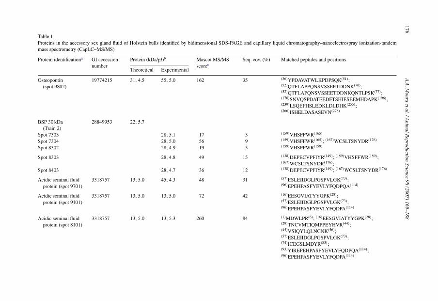
on the master image (Fig. 1A), which included all the proteins in the reference gel(Fig. 1B) and spots from other members of the match set, 36 spots were identified byCapLC–MS/MS and corresponded to 13 different proteins (Table 1). Intensity of spots relatedto these proteins accounted for 98.6% of the intensity of all spots shown on the master gel(Fig. 1A).
The large cluster of spots at 14 kDa (pI 5.8) was analyzed as a single unit by PDQuest (spot6203; Fig. 1A and B). Tandem mass spectrometry analysis of trypic peptides from that spotmatched to bovine seminal protein Pdc-109 and antibodies against BSP-A1/A2 and BSP-A3reacted with these respective proteins in that region (Fig. 2A and B). Collectively, the inten-sity of the BSP A1/A2 and A3 complex (spot 6203) represented 86.5% of all proteins stainedby Coomassie blue in the AGF maps. Another BSP protein was identified as BSP 30 kDa(spots 9601–9404, Train 3; Fig. 1A and B), but representing only 2.8% of the intensity ofall spots in the master gel. Only five spots of Train 3 (spots 7603 through 8403), from atotal of seven, were matched to BSP 30 kDa (Table 1) when analyzed by tandem mass spec-trometry. Three of those spots had low protein scores but Western blots confirmed that ananti-BSP 30 kDa antibody reacted with the entire region of the AGF gel delimited as Train 3(Fig. 3).
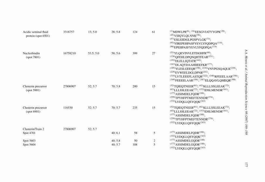
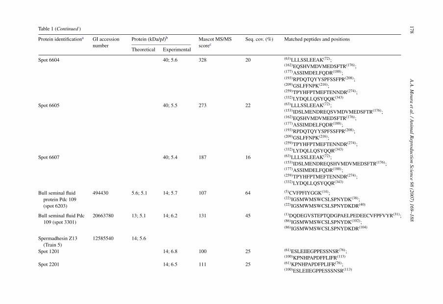
Twenty spots appeared consistently in every single member of the match set constructedby PDQuest (Fig. 1A). Based on their optical densities, this group of spots accounted for96.6% of all spots detected in the AGF master gel and had an average coefficient of varia-tion (CV) of 87.6 ± 9.1%. These spots matched to isoforms of clusterin at 70 kDa (spots 5901and 6901) and at 40 kDa (Train 2), albumin (spot 2901), nucleobindin (spot 7801), osteopon-tin (spot 9802), a 28 kDa acidic seminal fluid protein—aSFP (spot 6501), tissue inhibitor ofmetalloproteinase-2 (Train 4), 14 kDa spermadhesin Z13 (Train 5), isoforms of BSP 30 kDa(Train 3) and the complex formed by BSP A1/A2 and A3 at 14 kDa (Fig. 1A). Within this group,proteins with the lowest CV were BSP A1/A2 and A3 complex (35.8%), tissue inhibitor ofmetalloproteinase 2 (41.9%), low molecular weight spermadhesin Z13 isoforms (46.5%) andnucleobindin (52.1%). The highest CVs were associated with the expression of osteopontin(105.8%), 70 kDa clusterin isoforms (163.6%) and the acidic seminal fluid protein at 28 kDa(141%).
Although components of Train 1 (58 kDa; pI 6.0–6.2) were not present in every gel, everymember of the match set had at least one of those spots, which matched to type VII phospholipaseA2 (Fig. 1; Table 1). Proteins that were not present in all 37 maps of the accessory sex gland fluidincluded low molecular weigh isoforms of aSFP (spots 8101 and 9101), cathepsin L (spot 1601),ecto 5′-nucleotidase (spot 1703) and ecto-ADP-ribosyltransferase 5—ART 5 (spot 2702). Thoseisoforms of aSFP were detected in 32 gels and cathepsin L, in 28 samples. Nucleotidase and ART-5 had measurable expression only in 9 and 7 bulls, respectively. Both ART-5 and nucleobindinare two new proteins found in the accessory sex gland fluid and Fig. 4A and B shows the production spectrum of trypic peptides originated from them.
4. Discussion
We report a comprehensive protein profile of accessory sex gland fluid of Holstein sires. Thesebulls represent a population of tested, mature sires that have been extensively used for artificialinsemination of a large number of cows. Although such bulls were selected primarily for theirability to transmit desirable genetic characteristics associated with type and milk yield, they havealso been screened for fertility merit. Sires with poor semen quality that do not meet the standards

174 A.A. Moura et al. / Animal Reproduction Science 98 (2007) 169–188

A.A. Moura et al. / Animal Reproduction Science 98 (2007) 169–188 175
Fig. 2. Western blots of accessory sex gland fluid proteins incubated with anti-BSP A1/A2 (A) and A3 (B). Proteins wereseparated by 2D SDS-PAGE and transferred to nitrocellulose membranes.
of the artificial insemination industry are culled prior to entering commercial production of semen.A distinct advantage of using these bulls in a proteomic analysis is that their characteristics aretransmitted to a large population of offspring which enables an evaluation of the composite of AGproteins in a defined population of bulls of known fertility. To our knowledge, this is the first studyto report a proteomic analysis of the accessory gland fluid from bulls with these characteristics.
Fig. 1. Protein profile of the bull accessory sex gland fluid obtained by 2D SDS-PAGE and tandem massspectrometry–CapLC–MS/MS. (A) The master gel constructed by PDQuest (spots with asterisk (*) are those presentin every member of the AGF match set) and (B) the reference map (from bull #2) from which the master was mostlygenerated (letter a—aSFP: acidic seminal fluid protein; letter b—TMP-2: tissue inhibitor of metalloproteinase 2; letterc—spots 6203-1 and 6203-2 were analyzed separately by CapLC–MS/MS and matched to Pdc 109). Proteins were stainedby Coomassie blue and standards spot numbers (SSP) were given to spots by PDQuest.
176A
.A.M
ouraetal./A
nimalR
eproductionScience
98(2007)
169–188Table 1Proteins in the accessory sex gland fluid of Holstein bulls identified by bidimensional SDS-PAGE and capillary liquid chromatography–nanoelectrospray ionization-tandemmass spectrometry (CapLC–MS/MS)
Protein identificationa GI accessionnumber
Protein (kDa/pI)b Mascot MS/MSscorec
Seq. cov. (%) Matched peptides and positions
Theoretical Experimental
Osteopontin(spot 9802)
19774215 31; 4.5 55; 5.0 162 35 (36)YPDAVATWLKPDPSQK(51);(52)QTFLAPPQNSVSSEETDDNK(70);(52)QTFLAPQNSVSSEETDDNKQNTLPSK(77);(170)SNVQSPDATEEDFTSHIESEEMHDAPK(196);(239)LSQEFHSLEDKLDLDHK(255);(266)ISHELDASASEVN(278)
BSP 30 kDa(Train 2)
28849953 22; 5.7
Spot 7303 28; 5.1 17 3 (159)VHSFFWR(165)
Spot 7304 28; 5.0 56 9 (159)VHSFFWR(165); (167)WCSLTSNYDR(176)
Spot 8302 28; 4.9 19 3 (159)VHSFFWR(159)
Spot 8303 28; 4.8 49 15 (138)DEPECVPFIYR(149); (159)VHSFFWR(159);(167)WCSLTSNYDR(176);
Spot 8403 28; 4.7 36 12 (138)DEPECVPFIYR(149); (167)WCSLTSNYDR(176)
Acidic seminal fluidprotein (spot 9701)
3318757 13; 5.0 45; 4.3 48 31 (57)ESLEIIDGLPGSPVLGK(73);(96)EPEHPASFYEVLYFQDPQA(114)
Acidic seminal fluidprotein (spot 9101)
3318757 13; 5.0 13; 5.0 72 42 (16)EESGVIATYYGPK(28);(57)ESLEIIDGLPGSPVLGK(73);(96)EPEHPASFYEVLYFQDPA(114)
Acidic seminal fluidprotein (spot 8101)
3318757 13; 5.0 13; 5.3 260 84 (1)MDWLPR(6); (16)EESGVIATYYGPK(28);(29)TNCVMTIQMPPEYHVR(44);(45)VSIQYLQLNCNK(56);(57)ESLEIIDGLPGSPVLGK(73);(74)ICEGSLMDYR(83);(93)YIREPEHPASFYEVLYFQDPQA(114);(96)EPEHPASFYEVLYFQDPA(114)
A.A
.Moura
etal./Anim
alReproduction
Science98
(2007)169–188
177
Acidic seminal fluidprotein (spot 6501)
3318757 13; 5.0 28; 5.8 124 61 (1)MDWLPR(6); (16)EESGVIATYYGPK(28);(45)VSIQYLQLNNK(56);(57)ESLEIIDGLPGSPVLGK(73);(93)YIREPEHPASFYEVLYFQDPQA(114);(96)EPEHPASFYEVLYFQDPQA(114)
Nucleobindin(spot 7801)
16758210 53.5; 5.0 58; 5.6 399 27 (53)YLQEVINVLETDGHFR(68);(136)QFEHLDPQNQHTFEAR(151);(152)DLELLIQTATR(162);(163)DLAQYDAAHHEEFKR(177);(189)YLESLGEEQR(198); (218)VNVPGSQAQLK(228);(229)EVWEELDGLDPNR(241);(310)LVTLEEEFLASTQR(322); (348)RFEEELAAR(356);(349)FEEEELAAR(356); (387)ELQQAVLQMEQR(398)
Clusterin precursor(spot 5901)
27806907 52; 5.7 70; 5.8 200 15 (52)TQIEQTNEER(61); (62)KLLLSSLEEAK(72);(63)LLLSSLEEAK(72); (153)IDSLMENDR(161);(177)ASSIMDELFQDR(188);(259)TPYHFPTMEFTENNDR(274);(332)LYDQLLQSYQQK(343)
Clusterin precursor(spot 6901)
116530 52; 5.7 70; 5.7 235 15 (52)TQIEQTNEER(61); (62)KLLLSSLEEAK(72);(63)LLLSSLEEAK(72); (153)IDSLMENDR(161);(177)ASSIMDELFQDR(188);(259)TPYHFPTMEFTENNDR(274);(332)LYDQLLQSYQQK(343)
Clusterin/Train 2 27806907 52; 5.7Spot 4701 40; 6.1 58 5 (177)ASSIMDELFQDR(188);
(332)LYDQLLQSYQQK(343)
Spot 5603 40; 5.8 50 2 (177)ASSIMDELGQDR(188)
Spot 5604 40; 5.7 108 5 (177)ASSIMDELGQDR(188);(332)LYDQLLQSYQQR(343)
178A
.A.M
ouraetal./A
nimalR
eproductionScience
98(2007)
169–188Table 1 (Continued )
Protein identificationa GI accessionnumber
Protein (kDa/pI)b Mascot MS/MSscorec
Seq. cov. (%) Matched peptides and positions
Theoretical Experimental
Spot 6604 40; 5.6 328 20 (63)LLLSSLEEAK(72);(162)EQSHVMDVMEDSFTR(176);(177)ASSIMDELFQDR(188);(193)RPDQTQYYSPFSSFPR(208);(209)GSLFFNPK(216);(259)TPYHFPTMEFTENNDR(274);(332)LYDQLLQSYQQK(343)
Spot 6605 40; 5.5 273 22 (63)LLLSSLEEAK(72);(153)IDSLMENDREQSVMDVMEDSFTR(176);(162)EQSHVMDVMEDSFTR(176);(177)ASSIMDELFQDR(188);(193)RPDQTQYYSPFSSFPR(208);(209)GSLFFNPK(216);(259)TPYHFPTMEFTENNDR(274);(332)LYDQLLQSYQQR(343)
Spot 6607 40; 5.4 187 16 (63)LLLSSLEEAK(72);(153)IDSLMENDREQSHVMDVMEDSFTR(176);(177)ASSIMDELFQDR(188);(259)TPYHFPTMEFTENNDR(274);(332)LYDQLLQSYQQR(343)
Bull seminal fluidprotein Pdc 109(spot 6203)
494430 5.6; 5.1 14; 5.7 107 64 (5)CVFPFIYGGK(14);(22)IGSMWMSWCSLSPNYDK(38);(22)IGSMWMSWCSLSPNYDKDR(40)
Bull seminal fluid Pdc109 (spot 3301)
20663780 13; 5.1 14; 6.2 131 45 (1)DQDEGVSTEPTQDGPAELPEDEECVFPFVYR(31);(86)IGSMWMSWCSLSPNYDK(102);(86)IGSMWMSWCSLSPNYDKDR(104)
Spermadhesin Z13(Train 5)
12585540 14; 5.6
Spot 1201 14; 6.8 100 25 (61)ESLEIIEGPPESSNSR(76);(100)KPNHPAPDFFLIFR(113)
Spot 2201 14; 6.5 111 25 (61)KPNHPAPDFPLIFR(76);(100)ESLEIIEGPPESSSNSR(113)

A.A
.Moura
etal./Anim
alReproduction
Science98
(2007)169–188
179
Spot 2202 14; 6.3 154 33 (1)DSTDGLLVK(1); (61)KPNHPAPDFFLIFR(76);(100)ESLEIIEGPPESSNSR(113)
Spermadhesin Z13(spot 3601)
12585540 14; 5.6 29; 6.3 60 25 (61)KPNHPAPDFFLIFR76);(100)ESLEIIEGPPESSNSR(113)
TIMP-2/Train 4 27806163 25; 7.5Spot 502 26; 7.2 53 13 (54)EVDSGNDIYGNPIK(67);
(206)GAAPPKQEFLDIEDP(220)
Spot 1501 25; 7.1 246 28 (28)EVDSGNDIYGNPIK(41);(28)EVDSGNDIYGNPIKR(42);(90)AEGNMHITLCDFIVPWDTLSATQK(115);(180)GAAPPKQEFLDIEDP(194); (186)QEFLDIEDP(194)
Spot 1502 25; 6.8 150 8 (54)EVDSGNDIYGNPIK(67);(54)EVDSGNDIYGNPIKR(68)
Spot 2502 25; 6.7 155 13 (54)EVDSGNDIYGNPIK(67);(54)EVDSGNDIYGNPIKR(68);(206)GAAPPKQEFLDIEDP(220)
Cathepsin L(spot 1601)
27806673 38; 6.5 38; 6.9 68 12 (39)LYMNEEEWR(48); (58)IIDLHNQEYSEGK(70);(75)MAMNAFGDMTNEEFR(89)
Ecto-ADP-ribosyltransferase 5(spot 2702)
9998751 27; 7.6 43; 6.3 199 27 (64)EMADHALLR(72); (86)RPGLTLPPGFR(96);(123)TGGGSWESYMNHFPFK(138);(139)ALHFYLTR(146); (160)EPGQVVFR(167);(185)LGQFTSSSLDETVAR(199)
Abumin (spot 2901) 162648 71; 5.8 63; 6.4 178 7 (402)HLVDEPQNLIK(412);(421)LGEYGEQNALIVR(433); (548)KQTALVELLK(557);(569)TVMENFVAFVDK(580)
PLA2 (Train 1) 27807045 51; 6.1Spot 3801 58; 6.2 192 13 (42)IQALMAAANIGQSK(55);
(110)FLGTHWLVGK(119);(144)YPLIIFSHGLGAFR(157);(311)IPQPLFFINSER(322); (365)IIGYLFTLK(373)
180A
.A.M
ouraetal./A
nimalR
eproductionScience
98(2007)
169–188
Table 1 (Continued )
Protein identificationa GI accessionnumber
Protein (kDa/pI)b Mascot MS/MSscorec
Seq. cov. (%) Matched peptides and positions
Theoretical Experimental
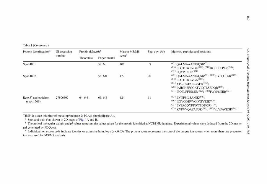
Spot 4801 58; 6.1 106 9 (42)IQALMAAANIGQSK(55);(110)FLGTHWLVGK(119); (211)RGEEEFPLR(219);(323)FQYPSNIIR(331)
Spot 4802 58; 6.0 172 20 (42)IQALMAAANIGQSK(55); (102)EYFLGLSK(109);(110)FLGTHWLVGK(119);(144)YPLIIFSHGLGAFR(157);(268)IAIIGHSFGGATVIQTLSEDQR(289);(311)IPQPLFFINSER(322); (323)FQYPSNIIR(331)
Ecto 5′-nucleotidase(spot 1703)
27806507 64; 6.4 63; 6.8 124 11 (134)EVNFPILSANIK(145);(163)ILTVGDEVVGIVGYTSK(179);(257)EVPAGQYPFIVTSDDGR(273);(274)KVPVVQAYAFGK(285); (537)VLYPAVEGR(545)
TIMP-2: tissue inhibitor of metalloproteinase 2; PLA2: phopholipase A2.a Spot and train # as shown in 2D maps of Fig. 1A and B.b Theoretical molecular weight and pI values represent the values given for the protein identified at NCBI NR database. Experimental values were deduced from the 2D master
gel generated by PDQuest.c Individual ion scores ≥48 indicate identity or extensive homology (p < 0.05). The protein score represents the sum of the unique ion scores when more than one precursor
ion was used for MS/MS analysis.

A.A. Moura et al. / Animal Reproduction Science 98 (2007) 169–188 181
Fig. 3. Western blot of accessory sex gland fluid proteins incubated with anti-BSP 30 kDa. Proteins were separated by2D SDS-PAGE and transferred to nitrocellulose membranes.
Fig. 4. Product ion spectrum of trypic peptides YLQEVINVLETDGHFR and LGQFTSSSLDETVAR matched by MAS-COT to nucleobindin (panel A) and ecto-ADP-ribosyltransferase 5 (panel B), respectively.
182 A.A. Moura et al. / Animal Reproduction Science 98 (2007) 169–188
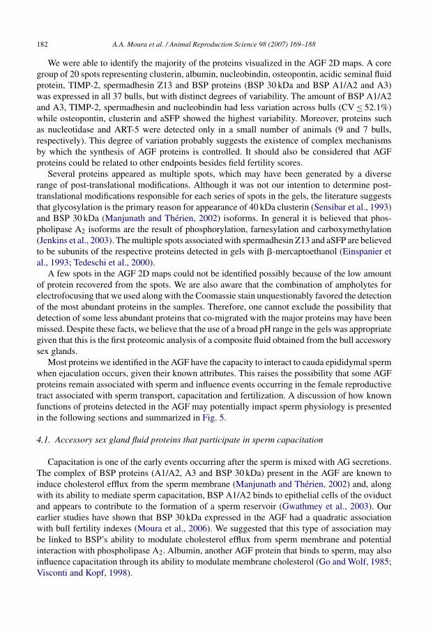
We were able to identify the majority of the proteins visualized in the AGF 2D maps. A coregroup of 20 spots representing clusterin, albumin, nucleobindin, osteopontin, acidic seminal fluidprotein, TIMP-2, spermadhesin Z13 and BSP proteins (BSP 30 kDa and BSP A1/A2 and A3)was expressed in all 37 bulls, but with distinct degrees of variability. The amount of BSP A1/A2and A3, TIMP-2, spermadhesin and nucleobindin had less variation across bulls (CV ≤ 52.1%)while osteopontin, clusterin and aSFP showed the highest variability. Moreover, proteins suchas nucleotidase and ART-5 were detected only in a small number of animals (9 and 7 bulls,respectively). This degree of variation probably suggests the existence of complex mechanismsby which the synthesis of AGF proteins is controlled. It should also be considered that AGFproteins could be related to other endpoints besides field fertility scores.
Several proteins appeared as multiple spots, which may have been generated by a diverserange of post-translational modifications. Although it was not our intention to determine post-translational modifications responsible for each series of spots in the gels, the literature suggeststhat glycosylation is the primary reason for appearance of 40 kDa clusterin (Sensibar et al., 1993)and BSP 30 kDa (Manjunath and Therien, 2002) isoforms. In general it is believed that phos-pholipase A2 isoforms are the result of phosphorylation, farnesylation and carboxymethylation(Jenkins et al., 2003). The multiple spots associated with spermadhesin Z13 and aSFP are believedto be subunits of the respective proteins detected in gels with �-mercaptoethanol (Einspanier etal., 1993; Tedeschi et al., 2000).
A few spots in the AGF 2D maps could not be identified possibly because of the low amountof protein recovered from the spots. We are also aware that the combination of ampholytes forelectrofocusing that we used along with the Coomassie stain unquestionably favored the detectionof the most abundant proteins in the samples. Therefore, one cannot exclude the possibility thatdetection of some less abundant proteins that co-migrated with the major proteins may have beenmissed. Despite these facts, we believe that the use of a broad pH range in the gels was appropriategiven that this is the first proteomic analysis of a composite fluid obtained from the bull accessorysex glands.
Most proteins we identified in the AGF have the capacity to interact to cauda epididymal spermwhen ejaculation occurs, given their known attributes. This raises the possibility that some AGFproteins remain associated with sperm and influence events occurring in the female reproductivetract associated with sperm transport, capacitation and fertilization. A discussion of how knownfunctions of proteins detected in the AGF may potentially impact sperm physiology is presentedin the following sections and summarized in Fig. 5.
4.1. Accessory sex gland fluid proteins that participate in sperm capacitation
Capacitation is one of the early events occurring after the sperm is mixed with AG secretions.The complex of BSP proteins (A1/A2, A3 and BSP 30 kDa) present in the AGF are known toinduce cholesterol efflux from the sperm membrane (Manjunath and Therien, 2002) and, alongwith its ability to mediate sperm capacitation, BSP A1/A2 binds to epithelial cells of the oviductand appears to contribute to the formation of a sperm reservoir (Gwathmey et al., 2003). Ourearlier studies have shown that BSP 30 kDa expressed in the AGF had a quadratic associationwith bull fertility indexes (Moura et al., 2006). We suggested that this type of association maybe linked to BSP’s ability to modulate cholesterol efflux from sperm membrane and potentialinteraction with phospholipase A2. Albumin, another AGF protein that binds to sperm, may alsoinfluence capacitation through its ability to modulate membrane cholesterol (Go and Wolf, 1985;Visconti and Kopf, 1998).

A.A. Moura et al. / Animal Reproduction Science 98 (2007) 169–188 183
Fig. 5. Schematic diagram of aspects of sperm physiology and fertilization that are potentially influenced by the proteinsidentified in the accessory sex gland fluid of mature Holstein bulls. After ejaculation, binding of BSP proteins and albumincontribute to capacitation. In the female reproductive tract, protection of sperm against oxidative stress, damage caused byhydrophobic molecules, protein precipitation and complement-induced attack could be mediated by AGF proteins suchas aSFP, clusterin, 5′-nucleotidase and albumin. When the sperm comes in contact with the cumulus layer, interactionwith and remodeling of the extracellular matrix (ECM) must be of importance. It is feasible thus that proteins such asTMIP-2, clusterin and possibly cathepsin L participate in these events. PLA2 secreted into the AGF acts during acrosomereaction, membrane fusion and has reported antimicrobial activity. Sperm-oocyte interaction also involves osteopontin.Sperm motility, a “sine qua non” for fertilization to occur in vivo, can be affected by aSFP, spermadhesin Z13 and5′-nucleotidase. The roles of nucleobindin and ADP-ribosyltransferase, two new proteins detected in the AGF, are stillunknown.
4.2. Proteins involved in protection of sperm
The accessory sex gland fluid contains proteins that intervene, either directly or indirectly,in mechanisms aimed to prevent damage to the sperm membrane, oxidative stress and immuneattack. Albumin absorbs lipid peroxides, which contributes to its protective effect on both spermmembrane and motility (Alvarez and Storey, 1995) and aSFP is known to inhibit oxidative stress(Einspanier et al., 1993; Schoneck et al., 1996). Bovine aSFP shares identity with proteins of thespermadhesin family (Romao et al., 1997). Binding of aSFP to ejaculated sperm occurs but itis lost after capacitation (Dostolova et al., 1994), suggesting that, unlike porcine spermadhesins(Caballero et al., 2004, 2005), bovine aSFP does not participate in sperm-oocyte interaction. TheaSFP isoforms we found in the AGF (pIs at 5 and 5.3), including the spot at 28 kDa, are similar tothe aSFPs that appear in the bovine seminal plasma at 13 kDa and a dimer at 26 kDa (Einspanieret al., 1994; Romao et al., 1997). However, the 45 kDa aSFP identified in the AGF has not beenpreviously described in the seminal plasma. It is possible that aSFP isoforms in AGF have differentcharacteristics from those observed in the seminal milieu.
The 70 and 40 kDa isoforms of clusterin in the AGF maps are similar to those reported in thecauda epididymis fluid of bulls (Ibrahim et al., 1999). Clusterin is a multifunctional constituent
184 A.A. Moura et al. / Animal Reproduction Science 98 (2007) 169–188
of the accessory sex gland secretions and it can prevent oxidative damage to the sperm (Reyes-Moreno et al., 2002), bind and agglutinate abnormal spermatozoa in bulls (Ibrahim et al., 1999)and humans (O’Bryan et al., 1990, 1994) and act like a chaperone, protecting sperm from the toxiceffects of protein precipitation (Humphreys et al., 1999; Wilson and Easterbrook-Smith, 2000).Clusterin has the ability to inhibit complement-induced sperm lysis (Jenne and Tchopp, 1989;O’Bryan et al., 1990; Ibrahim et al., 1999) and this is also one of the features of the 5′-nucleotidaseenzyme (Takayama et al., 2000) found in the AGF. Protection of sperm from oxidative damage,agglutination or lysis could be important for the spermatozoa once in the female reproductivetract.
4.3. Proteins involved in the acrosome reaction and sperm-oocyte interaction
Phospholipase A2 (PLA2) has a diverse range of functional features some of which relate tothe acrosome reaction and sperm-oocyte membrane fusion (Riffo and Parraga, 1997; Yuan et al.,2003). However, there is also evidence that PLA2 stimulates immune cells (Granata et al., 2005)and has antimicrobial activity in the seminal plasma (Weinrauch et al., 1996; Bourgeon et al.,2004). Osteopontin is another multifunctional protein identified in the AGF of bulls and it wasoriginally described in the extracellular matrix of bone tissues (Senger et al., 1979). Osteopontininteracts with the membrane of ejaculated bull sperm and also affects sperm-oocyte bindingand early embryonic development (our unpublished results). We previously reported that theexpression of both PLA2 and osteopontin was more pronounced in the accessory sex gland fluidof high fertility bulls than in low fertility bulls (Moura et al., 2006). A more in depth discussionof their attributes as related to male fertility was presented therein.
4.4. Proteins potentially associated with interaction and modulation of extracellular matrix(ECM) components
Interaction and remodeling of the ECM are some of the attributes of proteins such as TIMP-2,clusterin and probably cathepsin L, found in the accessory sex gland fluid. This group of AGFproteins may be important during mammalian fertilization when the sperm interact with andcross barriers established by the cumulus cells, zona pellucida and oocyte membrane. Metal-loproteinases (MMPs) released from the sperm during acrosome reaction facilitate sperm-eggfusion in hamsters (Dıaz-Perez and Meizel, 1992). TIMP-2 associates with bovine and humansperm membranes (McCauley et al., 2001; Bechman-Shaked et al., 2002) and inhibitors of MMPsand TIMPs interfere with gamete fusion in mice (Correa et al., 2000). Clusterin interacts withECM complexes in the testis (Sylvester et al., 1991; Bailey and Griswold, 1999) and a more recentstudy found that high molecular weight clusterin (70–73 kDa) inhibits certain types of metallo-proteinases (MT6-MMP/MMP-25) produced by neutrophils (Matsuda et al., 2003), a role sharedby other controllers of MMPs. The cathepsin L detected in AGF has the same molecular weightas a procathepsin L molecule found in the acrosome of epididymal spermatozoa (McDonald andEmerick, 1995). Cathepsins are cysteine peptidases involved in proteolysis of components usuallyfound as part of the ECM during growth, tissue invasion and remodeling (Dickinson, 2002).
4.5. Proteins associated with sperm motility
While several biochemical changes occur in the male gametes from emission until fertilization,sperm must also traverse the female reproductive tract to reach the oocyte. This is accomplished

A.A. Moura et al. / Animal Reproduction Science 98 (2007) 169–188 185
by a combination of contractions of the female reproductive tract and sperm motility. Someaccessory sex gland proteins have the potential to influence sperm motility, including BSP A1/A2(Sanchez-Luengo et al., 2004), aSFP (Schoneck et al., 1996) and phospholipase A2 (Bao et al.,2004). Interestingly, bovine seminal plasma aSFP shares a 50% homology with spermadhesinZ13 (Tedeschi et al., 2000), the second most abundant group of proteins that we were able todetect in the AGF 2D maps. We showed that this AGF spermadhesin had an inverse relationshipwith fertility (Moura et al., 2006) but the mechanism for its affect on sperm in the bull is unclear.The ecto 5′-nucleotidase (5′-NT) is another AGF component that can be grouped in this categorybecause antibodies against and inhibitors of this enzyme are associated with reductions in humanspermatozoa motility in vitro (Aumuller et al., 1997). This enzyme causes hydrolysis of nucleoside5-monophosphates (Thompson, 1985) and it has been shown as part of the bovine (Schiemann etal., 1994) and human spermatozoa (Takayama et al., 2000).
4.6. New proteins expressed in the accessory sex gland fluid
The ecto-ADP-ribosyltransferase 5 (ART 5) identified in the AGF belongs to a class ofenzymes that catalyze the transfer of an ADP-ribose group from NAD to arginine residues ontarget proteins, such as G proteins, rho, actin, CD44 and � integrins (Okazaki and Moss, 1999;Mueller-Dieckmann et al., 2002; Seman et al., 2004). ARTs (ADP-ribosyltransferases) are nor-mally expressed in immune cells and ART5, in the testis (Mueller-Dieckmann et al., 2002; Cordaand Di Girolamo, 2003), but none of these forms had been reported before in secretions ofaccessory sex glands. Another unique spot in the AGF two-dimensional gels was identified asnucleobindin, a protein with Ca2+ and DNA-binding motifs and conserved among species (Wendelet al., 1995). To our knowledge, this is the first identification of nucleobindin as a secreted com-ponent of the accessory sex gland fluid or seminal plasma. Interestingly, nucleobindin has beenfound secreted by bone cells and odontoblasts and as a structural element of their respectiveextracellular matrices (Petersson et al., 2004; Somogyi et al., 2004). These features resemblethose of osteopontin, another bone protein that was identified in the bull seminal plasma andaccessory sex gland fluid by our laboratory (Killian et al., 1993; Cancel et al., 1997; Moura et al.,2006).
5. Conclusion
Using a systematic proteomic analysis of 37 different 2D maps, we obtained a global view ofdifferent classes of proteins expressed in the accessory sex gland fluid of proven, high use Holsteinbulls with documented fertility history. The recognized characteristics of the AG proteins suggestthey modulate important sperm functions after ejaculation and in the female reproductive tract.Some of these proteins are described for the first time as part of the accessory sex gland fluid.These analyses and observations provide justification for studies to explore and better understandthe mechanisms by which accessory sex gland proteins influence male fertility.
Acknowledgements
This research has been supported by USDA grants 2003-34437-13460 and 2004-34437-15106.
186 A.A. Moura et al. / Animal Reproduction Science 98 (2007) 169–188
References
Abbas, A., Koc, H., Liu, F., Tien, M., 2005. Fungal degradation of wood: initial proteomic analysis of extracellular proteinsof Phanerochaete chrysosporium grown on oak substrate. Curr. Gen. 47, 49–56.
Alvarez, J.G., Storey, B.T., 1995. Differential incorporation of fatty acids into and peroxidative loss of fatty acids fromphospholipids of human spermatozoa. Mol. Reprod. Dev. 42, 334–346.
Amann, R.P., Griel Jr., L.C., 1974. Fertility of bovine spermatozoa from rete testis, cauda epididymis and ejaculatedsperm. J. Dairy Sci. 57, 212–219.
Atlas-White, M., Murphy, B.F., Baker, H.W.G., 2000. Localization of clusterin in normal sperm by immunogold electronmicroscopy. Pathology 32, 258–261.
Aumuller, G., Renneberg, H., Schiemann, P.J., Wilhelm, B., Seitz, J., Konrad, L., Wennemuth, G., 1997. The role ofapocrine released proteins in the post-testicular regulation of human sperm function. In: Ivell, R., Holstein, A.F.(Eds.), The Fate of the Male Germ Cell. Plenum Press, New York, pp. 193–219.
Bailey, R., Griswold, M.D., 1999. Clusterin in the male reproductive system: localization and possible function. Mol.Cell. Endocrinol. 151, 17–23.
Bao, S., Miller, D.J., Ma, Z., Wohltmann, M., Eng, G., Ramanadham, S., Moley, K., Turk, J., 2004. Male mice that do notexpress group VIA phospholipase A2 produce spermatozoa with impaired motility and have greatly reduced fertility.J. Biol. Chem. 279, 38194–38200.
Bechman-Shaked, O., Kraiem, Z., Gonen, Y., Goldman, S., 2002. Presence of metalloproteinases and tissue inhibitor ofmetalloproteinases in human sperm. J. Androl. 23, 702–708.
Bedford, J.M., 1966. Development of the fertilizing ability of spermatozoa in the epididymis of the rabbit. J. Exp. Zool.163, 319–330.
Bourgeon, F., Evrard, B., Brillard-Bourdet, M., Colleu, D., Jegou, B., Pineau, C., 2004. Involvement of semenogelin-derived peptides in the antibacterial activity of human seminal plasma. Biol. Reprod. 70, 768–774.
Caballero, I., Vazquez, J.M., Gil, M.A., Calvete, J.J., Roca, J., Sanz, L., Parrilla, I., Garcia, E.M., Rodriguez-Martinez,H., Martinez, E.A., 2004. Does seminal plasma PSP-I/PSP-II spermadhesin modulate the ability of boar spermatozoato penetrate homologous oocytes in vitro? J. Androl. 25, 1004–1012.
Caballero, I., Vazquez, J.M., Rodriguez-Martinez, H., Gill, M.A., Calvete, J.J., Sanz, L., Garcia, E.M., Roca, J., Mar-tinez, E.A., 2005. Influence of seminal plasma PSP-I/PSP-II spermadhesin on pig gamete interaction. Zygote 13,11–16.
Cancel, A.M., Chapman, D.A., Killian, G.J., 1997. Osteopontin is the 55-kilodalton fertility-associated protein in Holsteinbull seminal plasma. Biol. Reprod. 57, 1293–1301.
Chen, H., Cheung, M.P.L., Chow, P.H., Cheung, A.L.M., Liu, W., O, W.S., 2002. Protection of sperm DNA againstoxidative stress in vivo by accessory sex gland secretions in male hamsters. Reproduction 124, 491–499.
Chow, P.H., Jiang, H.Y., Poon, H.K., Lee, K.H., O, W.S., 2003. Embryos sired by males without accessory sex glandsinduce failure of uterine support: a study of VEGF, MMP and TGF expression in the golden hamster. Anat. Embryol.206, 203–213.
Corda, D., Di Girolamo, M., 2003. Functional aspects of protein mono-ADP-ribosylation. EMBO J. 22, 1953–1958.Correa, L.M., Cho, C., Myles, D.G., Primakoff, P., 2000. A role of TIMP-3-sensitive, Zn2+-dependent metalloprotease in
mammalian gamete membrane fusion. Dev. Biol. 225, 124–134.Curi, S.M., Ariagno, J.I., Chenlo, P.H., Mendeluk, G.R., Pugliese, M.N., Segovia, L.M.S., Repetto, H.E.H., Blanco, A.M.,
2003. Asthenozoospermia: analysis of a large population. Arch. Androl. 49, 343–349.Dıaz-Perez, E., Meizel, S., 1992. Importance of mammalian endoprotease activity during the acrosome reaction to sub-
sequent sperm-egg fusion: inhibitor studies with human sperm and zona-free hamster eggs. Mol. Reprod. Dev. 31,122–130.
Dickinson, D.P., 2002. Cysteine peptidases of mammals: their biological roles and potential effects in the oral cavity andother tissues in health and disease. Crit. Rev. Oral Biol. Med. 13, 238–275.
Dostolova, Z., Calvete, J.J., Sanz, L., Hettel, C., Riedel, D., Schoneck, C., Einspanier, R., Topfer-Petersen, E., 1994.Immunolocalization and quantitation of acidic seminal fluid protein (aSFP) in ejaculated, swim-up, and capacitatedbull spermatozoa. Biol. Chem. Hoppe-Seyler 375, 457–461.
Einspanier, R., Amselgruber, W., Sinowatz, F., Henle, T., Ropke, R., Schams, D., 1993. Localization and concentrationof a new bioactive acetic seminal fluid protein (asfp) in bulls (Bos taurus). J. Reprod. Fert. 98, 241–244.
Einspanier, R., Krause, I., Calvete, J., Topfer-Petersen, E., Klostermeyer, H., Karg, H., 1994. Bovine seminal plasmaasfp—localization of disulfide bridges and detection of 3 different isoelectric forms. FEBS Lett. 344, 61–64.
Elzanaty, S., Richthoff, J., Malm, J., Giwercman, A., 2002. The impact of epididymal and accessory sex gland functionon sperm motility. Hum. Reprod. 17, 2904–2911.
A.A. Moura et al. / Animal Reproduction Science 98 (2007) 169–188 187
Go, K.J., Wolf, D.P., 1985. Albumin-mediated changes in sperm sterol content during capacitation. Biol. Reprod. 32,145–153.
Granata, F., Petraroli, A., Boilard, E., Bezzine, S., Bollinger, J., Del Vecchio, L., Gelb, M.H., Lambeau, G., Marone, G.,Triggiani, M., 2005. Activation of cytokine production by secreted phospholipase A2 in human lung macrophagesexpressing the M-type receptor. J. Immunol. 174, 464–474.
Gwathmey, T.M., Ignotz, G.G., Suarez, S.S., 2003. PDC-109 (BSP-A1/A2) promotes bull sperm binding to oviductalepithelium in vitro and may be involved in forming the oviductal sperm reservoir. Biol. Reprod. 69, 809–815.
Henault, M.A., Killian, G.J., Kavanaugh, J.F., Griel Jr., L.C., 1995. Effect of accessory sex gland fluid from bulls ofdiffering fertilities on the ability of cauda epididymal sperm to penetrate zona-free bovine oocytes. Biol. Reprod. 52,390–397.
Humphreys, D.T., Carver, J.A., Easterbrook-Smith, S.B., Wilson, M.R., 1999. Clusterin has chaperone-like activity similarto that of small heat shock proteins. J. Biol. Chem. 274, 6875–6881.
Ibrahim, N.M., Troedsson, M.H., Foster, D.N., Loseth, K.J., Farris, J.A., Blaschuk, O., Crabo, B.G., 1999. Reproductivetract secretions and bull spermatozoa contain different clusterin isoforms that cluster cells and inhibit complement-induced cytolysis. J. Androl. 20, 230–240.
Jenkins, C.M., Ham, X., Yang, J., Mancuso, D.J., Sims, H.F., Muslin, A.J., Gross, R.W., 2003. Purification of recombinanthuman cPLA2� and identification of C-terminal farnesylation, proteolytic processing and carboxymathylation byMALDI-TOF–TOF analysis. Biochemistry 42, 11798–11807.
Jenne, D.E., Tchopp, J., 1989. Molecular characterization of a human complement cytolysis inhibitor found in bloodand seminal plasma: identity to sulfated glycoprotein 2, a constituent of rat testis fluid. Proc. Natl. Acad. Sci. 86,7123–7127.
Killian, G.J., Chapman, D.A., Rogowski, L.A., 1993. Fertility-associated proteins in bull seminal plasma. Biol. Reprod.49, 1202–1207.
Koc, E.C., Burkhart, W., Blackburn, K., Moyer, M.B., Schlatzer, D.M., Moseley, A., Spremulli, L.L., 2001. The largesubunit of the mammalian mitochondrial ribosome—analysis of the complement of ribosomal proteins present. J.Biol. Chem. 276, 43958–43969.
Lowry, O.H., Rosebrough, W.J., Farr, A.L., Randall, R.L., 1951. Protein measurement with Folin phenol reagent. J. Biol.Chem. 193 (265), 275.
Manjunath, P., Therien, I., 2002. Role of seminal plasma phospholipid-binding proteins in sperm membrane lipid modi-fication that occurs during capacitation. J. Reprod. Immunol. 53, 109–119.
Matsuda, A., Itoh, Y., Koshikawa, N., Akizawa, T., Yana, I., Seiki, M., 2003. Clusterin, an abundant serum factor, is apossible negative regulator of MT6-MMP/MMP-25 produced by neutrophils. J. Biol. Chem. 278, 36350–72003.
McCauley, T.C., Zhang, H.M., Bellin, M.E., Ax, R.L., 2001. Identification of a heparin-binding protein in bovine seminalfluid as tissue inhibitor of metalloproteinases-2. Mol. Reprod. Dev. 58, 336–341.
McDonald, J.K., Emerick, J.M., 1995. Purification and characterization of procathepsin L, a self-processing zymogen ofguinea pig spermatozoa that acts on a cathepsin D assay substrate. Arch. Biochem. Biophys. 323, 409–422.
Moura, A.A., Koc, H., Chapman, D.A., Killian, G.A., 2006. Identification of proteins in the accessory sex gland fluidassociated with fertility indexes of dairy bulls: a proteomic approach. J. Androl. 27, 201–211.
Mueller-Dieckmann, C., Scheuermann, T., Wursthorn, K., Schroder, J., Haag, F., Schulz, G.E., Koch-Nolte, F., 2002.Expression, purification, crystallization and preliminary X-ray analysis of rat ecto-ADP-ribosyltransferase 2 (ART2).Acta Crystall. Sect. D Biol. Crystall. 58, 1211–1213.
O’Bryan, M.K., Baker, H.W., Saunders, J.R., Kirszbaum, L., Walker, I.D., Hudson, P., Liu, D.Y., Glew, M.D., d’Apice, A.J.,Murphy, B.F., 1990. Human seminal clusterin (SP-40): Isolation and characterization. J. Clin. Invest. 85, 1477–1486.
O’Bryan, M.K., Mallidis, C., Murphy, B.F., Baker, H.W., 1994. Immunohistological localization of clusterin in the malegenital tract in humans and marmosets. Biol. Reprod. 50, 502–509.
Okazaki, I.J., Moss, J., 1999. Characterization of glycosylphosphatidylinositiol-anchored, secreted, and intracellularvertebrate mono-adp-ribosyltransferases. Ann. Rev. Nut. 19, 485–509.
Petersson, U., Somogyi, E., Reinholt, F.P., Karlsson, T., Sugars, R.V., Wendel, M., 2004. Nucleobindin is produced bybone cells and secreted into the osteoid, with a potential role as a modulator of matrix maturation. Bone 34, 949–960.
Reyes-Moreno, C., Boilard, M., Sullivan, R., Sirard, M.A., 2002. Characterization of secretory proteins from culturedcauda epididymal cells that significantly sustain bovine sperm motility in vitro. Mol. Reprod. Dev. 63, 500–509.
Riffo, M., Parraga, M., 1997. Role of phospholipase A2 in mammalian sperm-egg fusion: development of hamster oolemmafusibility by lysophosphatidylcholine. J. Exp. Zool. 79, 81–88.
Romao, M.J., Kolln, I., Dias, J.M., Carvalho, A.M., Romero, A., Varela, P.F., Sanz, L., Topfer-Petersen, E., Calvete,J.J., 1997. Crystal structure of acidic seminal fluid protein (asfp) at 1.9 A resolution: a bovine polypeptide of thespermadhesin family. J. Mol. Biol. 274, 650–660.
188 A.A. Moura et al. / Animal Reproduction Science 98 (2007) 169–188
Sanchez-Luengo, S., Aumuller, G., Albrecht, M., Sen, P.C., Rohm, K., Wilhelm, B., 2004. Interaction of PDC-109,the major secretory protein from bull seminal vesicles, with bovine sperm membrane Ca2+-ATPase. J. Androl. 25,234–244.
Schiemann, P.J., Aliante, M., Wennemuth, G., Fini, C., Aumuller, G., 1994. Distribution of endogenous and exogenous5′-nucleotidase on bovine spermatozoa. Histochemistry 101, 253–262.
Schoneck, C., Braun, J., Einspanier, R., 1996. Sperm viability is influenced in vitro by the bovine seminal protein aSFP:effects on motility, mitochondrial activity and lipid peroxidation. Theriogenology 45, 633–642.
Seman, M., Adriouch, S., Haag, F., Koch-Nolte, F., 2004. Ecto-ADP-ribosyltransferases (ARTs): emerging actors in cellcommunication and signaling. Curr. Med. Chem. 11, 857–872.
Senger, D.R., Wirth, D.F., Hynes, R.O., 1979. Transformed mammalian cells secrete specific proteins and phosphoproteins.Cell 16, 885–893.
Sensibar, J.A., Qian, Y., Griswold, M.D., Sylvester, S.R., Bardin, C.W., Cheng, C.Y., Lee, C., 1993. Localization andmolecular heterogeneity of sulfated glycoprotein-2 (clusterin) among ventral prostate, seminal vesicle, testis, andepididymis of rats. Biol. Reprod. 49, 233–242.
Somogyi, E., Petersson, U., Sugars, R.V., Hultenby, K., Wendel, M., 2004. Nucleobindin—a Ca2+-binding protein presentin the cells and mineralized tissues of the tooth. Calc. Tiss. Int. 74, 366–376.
Sylvester, S.R., Morales, C., Oko, R., Griswold, M.D., 1991. Localization of sulfated glycoprotein-2 (clusterin) onspermatozoa and in the reproductive tract of the male rat. Biol. Reprod. 45, 195–207.
Takayama, T., Matsubara, S., Shibahara, H., Minakami, H., Takizawa, T., Sato, I., 2000. Ultracytochemical localizationof 5′-nucleotidase activity in human ejaculated spermatozoa. Int. J. Androl. 23, 106–108.
Tedeschi, G., Oungre, E., Mortarino, M., Negri, A., Maffeo, G., Ronchi, S., 2000. Purification and primary structure of anew bovine spermadhesin. Eur. J. Biochem. 267, 6175–6179.
Thompson, L.F., 1985. Inosine 59-nucleotidase vs inosine and hypoxanthine as substrates for purine salvage in humanlymphoid cells. Proc. Soc. Exp. Biol. Med. 179, 432–436.
Visconti, P.E., Kopf, G.S., 1998. Regulation of protein phosphorylation during sperm capacitation. Biol. Reprod. 59, 1–6.Weinrauch, Y., Elsbach, P., Madsen, L.M., Foreman, A., Weiss, J., 1996. The potent anti-Staphylococcus aureus activity
of a sterile rabbit inflammatory fluid is due to a 14-kDa phospholipase A2. J. Clin. Invest. 97, 250–257.Wendel, M., Sommarin, Y., Bergman, T., Heinegard, D., 1995. Isolation, characterization, and primary structure of a
calcium binding 63-kDa bone protein. J. Biol. Chem. 270, 6125–6133.Wilson, M.R., Easterbrook-Smith, S.B., 2000. Clusterin is a secreted mammalian chaperone. TIBS 25, 95–98.Yanagimachi, R., 1994. Mammalian fertilization. In: Knobil, E., Neill, J.D. (Eds.), The Physiology of Reproduction.
Raven Press, New York, pp. 189–317.Ying, Y., Chow, P.H., O, W.S., 1998. Effects of male accessory sex glands and deoxyribonucleic acid synthesis in the first
cell cycle of golden hamster embryos. Biol. Reprod. 58, 659–663.Yuan, Y.Y., Chen, W.C., Shi, Q.X., Mao, L.M., Yu, Q., Fang, X., Roldan, E.R.S., 2003. Zona pellucida induces activation
of phospholipase A2 during acrosomal exocytosis in guinea pig spermatozoa. Biol. Reprod. 68, 904–913.