a comparative study of the diplococci occur
TRANSCRIPT

A COMPARATIVE STUDY OF THE DIPLOCOCCI OCCUR-RING IN EPIDEMIC CEREBRO-SPINAL MENIN-
GITIS AND POSTERIOR BASIC MENINGITIS.'
BY MARTHA WOLLSTEIN, M.D.
(From the Laboratories of the Rockefeller Institute for Medical Research,New York.)
PLATE XXVI.
As long ago as 898, Still (I) concluded from a study of eightcases of simple, posterior basilar meningitis occurring in infants,that the disease is a sporadic form of epidemic cerebro-spinal menin-gitis, and that the Gram negative diplococcus found in the meningealexudate of both forms is identical, such slight differences as greatervitality and lack of virulence being due to natural variation. This:view was generally accepted, and the observations made on caseswhich occurred in this country confirmed Still's findings withoutadding anything important to our knowledge of the subject. Ac-cording to the clinical view that posterior basilar meningitis, whennot of syphilitic origin, is the chronic stage of an acute cerebro-spinal meningitis (Holt) (2), there seemed no reason to doubt theidentity of the causative microorganism in the two affections.
During the past two years, since the recent epidemic of cerebro-spinal meningitis appeared in the British Isles, several observershave claimed that posterior basilar meningitis is a disease due to aspecific microorganism definitely and uniformly differing from themeningococcus causing the ordinary epidemic disease, the differ-ence consisting in a total lack of agglutination and opsonin reac-tions of the diplococcus from cases of posterior basic meningitiswith the serum from an epidemic case, and vice versa. Thus in.1907, Houston (3), after studying diplococci isolated from twocases of posterior basilar meningitis at the Great Ormond StreetHospital for Sick Children, which reacted with the blood from these
'Received for publication April , 1gog.
579
on April 12, 2019jem.rupress.org Downloaded from http://doi.org/10.1084/jem.11.4.579Published Online: 17 July, 1909 | Supp Info:
Diplococci Occurring in Meningitis.
children but not with that from epidemic cases in Belfast, whilecocci isolated from the Belfast cases failed to react with the bloodfrom the London cases, concluded that there are two races of men-ingococci differing in agglutination and opsonic reactions. Houstonstudied both reactions in the same slide, prepared according toWright's technique for the study of opsonins, thus using a dilutionof one to three. At the onset of meningitis the opsonic index forthe meningococcus is low, but it rises gradually. Agglutininsappear about the sixth day, and after that time the two reactionsmay be studied together.
At the meeting of the British Medical Association held at Sheffieldin July, I9o8, Houston and Rankin (4) reiterated the view that"Still's disease is due to a meningococcus which has much the samecultural characteristics as the coccus of epidemic cerebro-spinalfever, but differs from it entirely in its opsonic and agglutinatingproperties." Eve and Clements (5) expressed the same view evenmore forcibly, stating that the sporadic organism shows little or notendency to agglutinate or phagocyte in the patient's own blood orin the serum from an epidemic case and will not agglutinate in theFlexner-Jobling antimeningitis serum. Ker (6) was enabled topick out three cases of post-basic meningitis by means of the agglu-tination reactions, but one of these cases was very favorably influ-'enced by the Flexner-Jobling anti-serum.
Dr. Flexner having obtained, through the kindness of Dr. Hous-ton, three strains of diplococci from cases of posterior basilarmeningitis, kindly gave me the privilege of studying them in com-parison with a number of strains from epidemic meningitis casesisolated before and after this study was undertaken. In all, seven-teen cultures of diplococci from cases of meningitis were at mydisposal. Their histories were as follows:
Two were isolated from typical cases of epidemic meningitis,grown in the laboratory for one and two years.
Two were isolated from typical cases of epidemic meningitis,grown in the laboratory for three months.
Seven were isolated from typical cases of epidemic meningitis,during the course of this work.
One was isolated from the circulating blood of a patient withindefinite meningeal symptoms but without acute meningitis.
580
Martha Wollstein.
Three were obtained from Dr. Houston and regarded as posteriorbasilar strains.
Two were isolated from typical cases of epidemic meningitis, butwere not meningococci.
These cultures were divided into three groups for purposes ofstudy as follows: old, proved strains, recent strains and posteriorbasilar strains. The two which proved not to be meningococci willbe described later.
Morphology.-All were biscuit-shaped diplococci lying in singlepairs or in larger groups, and never forming chains. They werenot motile.
To Gram's stain they reacted negatively in smears from cerebro-spinal fluid, cultures, and in the sections of organs of inoculatedanimals.
Biology.-On sheep's-serum-agar slants the growth was profuseand typical: greyish-white, smooth, moist showing a slightly me-tallic sheen on drying, and when moist of a tenacious, mucoid con-sistency on removal with the needle. On human ascitic fluid agarand on glucose agar, the growth was abundant. On plain agar itwas very scant, but readily visible. In plain neutral bouillon aslight turbidity appeared within twenty-four hours and a small, flakyprecipitate formed in another day; no pellicle formed. The reac-tion of the bouillon became alkaline. In serum broth the precipitatewas more profuse; the reaction also became alkaline. Litmus milkremained unchanged.
The old, the recent and the posterior basilar strains showed noappreciable differences in any of these culture media. It was notpossible to tell one strain or group from another. The two strainsdesignated as not meningococcus were very different. They werealso Gram negative cocci occurring in pairs, but they were slightlylarger and less strictly biscuit-shaped. They grew profusely onplain agar, caused a marked clouding of serum-glucose agar (thealbumin precipitation of Libman) and a diffuse cloudiness of plainand serum bouillon with the formation of a thin pellicle and a thickprecipitate. Milk remained fluid. In serum-litmus water contain-ing dextrose, coagulation occurred within twenty-four hours; alsoin galactose. The other sugars remained unchanged. The facts
681

Diplococci Occurring in Meningitis.
that they were Gram negative diplococci and had failed to fermentlactose, mannite, saccharose, raffinose and levulose were their onlypoints in common with true meningococci. The latter showed, intheir reactions to sugars in the presence of serum-litmus water, thefirst points of differences encountered among the various strains;but again they were not race but individual variations. Dextrose,for instance, was fermented by all but the oldest strain, which hadlost the power it possessed in this direction eighteen months earlier.The next oldest strain fermented neither dextrose nor maltose,though it had produced acid in both a year before. All the otherstrains, thirteen in number, fermented maltose. One recent strain,three months old, failed to ferment dextrose at any time. The cul-ture isolated from the blood during life formed acid rapidly in bothmaltose and dextrose, coagulating the former after three weeks, thelatter not at all. The three posterior basilar strains fermentedmaltose and dextrose, one coagulating maltose only, one producinga sufficient amount of acid in both varieties of sugar to cause coagu-lation, and the third leaving both sugar media fluid. Arkwright (7)mentions the fact that four strains of meningococcus studied byhim fermented no sugars, and Wilson (8) observed the same phe-nomenon with one strain. The duration of laboratory cultivation,of these cultures is not stated. Arkwright gives it as his opinionthat the sporadic strains diverge more than the epidemic ones do, asregards the sugar reactions. With this view our observations donot agree. Galactose, lactose, mannite, saccharose, raffinose andlevulose were not affected by any one of these fifteen strains ofmeningococci.
Autolysis was apparent to a marked degree in stained cover slipsfrom all these meningococcus cultures more than twenty hours old,but it was not possible to pick out any one strain as being definitelymore or less resistant than another. Suspension of the fifteen cul-tures in salt solution kept at 4 C. showed no differences in theamount of autolysis present at the end of one, two and three days.The same was true of a series of suspensions kept at 370 C. Nordid the viability of the several strains differ appreciably on solid orin fluid media. One of the older strains invariably outlived all threeof the posterior basic strains when kept in the thermostat (370 C.)
682

Martha Wollstein.
on sheep's-serum-agar with or without calcium carbonate. Twenty-four hours after having been heated in a salt solution suspensionfor thirty minutes at 650 C. the posterior basic cocci were as wellpreserved as were the older and the recent strains. Evidently theautolytic ferment () was the same in all; at least the effects wereindistinguishable.
Agglutination.-It is much to be regretted that only one humanserum was obtainable for agglutination tests. The early adminis-tration of the Flexner-Jobling antimeningitis serum made all othercases under observation valueless for this purpose. The singlespecimen of human serum came from a seven months' old infant onthe second day of an attack of epidemic meningitis, before the injec-tion of the anti-serum. As was to be expected, it gave only negativeagglutination reactions with its own strain of meningococci andwith the older strains under observation.
The Flexner-Jobling antimeningitis serum remained the onlyavailable means for making agglutination tests with these cultures,normal horse serum being used as controls. A recent non-carbolizedspecimen was obtained. With it, the three posterior basilar strainsagglutinated in dilutions of one to twenty only, but five recent epi-demic strains did not agglutinate even in so low a dilution. On theother hand, the two oldest strains used for the inoculation of thehorses from the first were agglutinated by the serum in dilution ashigh as one to two hundred, as was the diplococcus isolated from thecerebro-spinal fluid from the case of the infant referred to above,the fluid having been withdrawn within thirty-six hours of the onsetof the meningitis and before any serum had been injected. Twofacts are brought out by these results: () that prolonged artificialcultivation was not the determining factor, and (2) that agglutina-tion reactions with the Flexner-Jobling anti-serum do not serve todifferentiate epidemic from posterior basilar strains of meningo-cocci. That all strains of meningococci are not agglutinated in thesame dilutions of a serum has been noted by Bruns and Hohn (o).The two non-meningococcus cultures were not agglutinated by thisserum in dilutions of one to five, nor was the Staphylococcus aureus.
Phagocytosis.-The final test which the English observers appliedto the differentiation of the two races of meningococci was that of
583
Diplococci Occurring in Meningitis.
phagocytosis. In this, as in the agglutination tests, I was handi-capped by the lack of human serum from patients and convalescents.The only specimen obtainable (vide supra) was taken on the secondday of the disease, when its opsonic content was low. The anti-meningitis horse serum remained as in the agglutination tests theonly available means of study until the inoculation of monkeys wasbegun. I am aware, therefore, that these tests are not comparablewith those upon which Houston based his conclusions. Neverthe-less, they are interesting and suggestive. The results with the mon-key's serum are given with the protocols of the cases.
Two parallel sets of experiments were made. In one Wright'stechnique for the study of opsonins, but with diluted serum accord-ing to the method of Klien (), was used; in the other the tech-nique devised by Neufeld and Hiine (2), also with diluted serum,and applied by Neufeld (3) to the standardization of antimenin-gitis serum was employed. It is based on the observation thatimmune serum favors phagocytosis because it contains bacteriotropicantibodies which are thermo-stable. The serum was used in dilu-tions as high as one to five thousand, and the results with bothmethods were fairly the same. Human leucocytes were used withthe Wright technique, those from the peritoneal cavity of a guinea-pig for the Neufeld method. The diplococcal suspensions requiredwere thick. The most striking result of both methods was thevariability in the degree of phagocytosis displayed by the same cor-puscles to different strains of meningococcus. A table on page 585gives the results of the first series, made with the Neufeld method.
By the Neufeld method no actual counts are made, but the amountof phagocytosis is estimated and compared in the series of preparedslides, a strain of diplococcus which phagocytes readily, but notspontaneously, being used as a comparison control throughout allthe experiments. The opsonic strength of the serum is measuredby the dilution at which specific phagocytosis exceeds that which isspontaneous and present in the salt solution control. In Wright'stechnique two sets of tubes were prepared, one being incubated oneand one-half hours, as in the Neufeld method, the other, fifteen min-utes only. In the slides prepared from these tubes the engulfedcocci were counted. The results were similar to those given in the
684

Martha Wollstein. 585
One and One-half Hours Incubation.
Strain.
Old { 20St. Luke'sLebanonA. S.SimonsWard
Recent- Fischer
JohnstonPriceT. C.Silverman
Post. MoseleyBasic Windsor
2-100
+++
++++++++++++
+++++
+++4+4-
Dilutions of anti-meningitis serum.
X-2O
+++
f+++
+++++
++
++++
1-500
4-
.+
-
1-!000
++
++4-
1-2000
+
++
_-
above table, showing that one of the recent strains was most sus-ceptible to phagocytosis, two other recent strains and an old strainbut slightly less so, while one of the posterior basilar strains was asresistant as two recent cultures, and less so than the other two post-basic strains, which were comparable with five recent strains in sus-ceptibility to phagocytosis in immune serum. Expressed in anotherway, the opsonic power of this serum was present in higher dilu-tions for some strains of meningococci than for others, exactly asthe agglutinins were present in higher dilutions for some strainsthan for others. Definite group distinctions are not deducible fromthese results.
Comparing the results obtained in the specimens incubated fifteenminutes with those incubated one and a half hours, we find thatphagocytosis occurs in the same serum dilutions in both, but thenumber of cocci taken up by the cells increases with the time ofexposure allowed them. Thus:
Serum Dilutions.
Wright's technique, z5' Incubation. I Wright's technique, go' incubation.
Strains. zoo aoo oo500 z,ooo ,o000 5,oo Salt sol. xoo 2 5o 1,ooo l 2000 5,ooo Salt sol.
720 4.0 2.5 2.2 1.5 1.O 0.1 0.5 8.o 8.o 3.0 2.5 1.5 1.0 1.0A.S. 1.9 1.5 1.2 1.0 0.8 o.2 0.2 4.0 3.2 2.5 1.5 1.2 0.3 0.4Johnston 1.4 0.5 0.1 0.1 0.0 0.0 0. 2.2 0.6 0.2 0.1 0.1 0.0 o0.Moseley 2.0 1.4 o.6 0.3 .0.2 0.1 0.2 2.9 1.7 1.0 0.4 0.2 0.2 0.5
1-5000
4-
4-
Salt ol.control.
+-4-4--4-4-4-4--4-
4-
Diplococci Occurring in Meningitis.
When normal horse serum was used the control diplococcusceased to be taken up by the leukocytes in a dilution greater thanone in two hundred, while in the immune serum this point was notreached until a dilution of one to two thousand was employed.-The phagocytic value of the immune serum for this diplococcus was,therefore, ten times the normal. The results with the other strainsin normal horse serum were similar.2
The three posterior basilar cultures were inoculated into the spinalcanal of monkeys and recovered from them by lumbar puncture.These cultures, after having been passed through an animal, did notagglutinate nor phagocyte to higher degrees than before.
The two non-meningococcus cultures showed no difference in thenumbers of organisms taken up by human or guinea-pig leukocytesin the presence of salt solution as compared with antimeningitishorse serum, diluted or in full strength. The phagocytic index ofthe serum toward the two cultures was practically nil. It has notbeen possible to identify these two strains of cocci, obtained in pureculture from two different adult cases of apparently typical menin-gitis. Von Lingelsheim (I4) encountered ten varieties of bacteriain his studies with material from patients suspected of having men-ingitis. These were meningococcus, Diplococcus crassus, staphylo-coccus, streptococcus, Streptococcus mucosus, Diplococcus mucosus,Diplococcus pharyngis flavus II, Micrococcus cinereus and Gramnegative, plump bacilli. From the nine varieties of cocci enumer-ated, our strains can be differentiated without much difficulty. FromDiplococcus intracellularis by its profuse growth on all media, lackof autolysis, fermentation of galactose, and its inability to affectmaltose. The cocci under consideration are Gram negative, whileDiplococcus crassus is still described by von Lingelsheim (4) asbeing Gram-doubtful; moreover, the latter ferments levulose, sac-charose, lactose and maltose, in addition to dextrose and galactose.The Gram positive staphylococcus, streptococcus and Streptococcusmucosus need no further differentiation, and the morphology and
I 'Dr. Alice Taylor of London very kindly sent serum from two cases of pos-terior basilar meningitis. Unfortunately the quantity was very small, and theresults of an opsonic test (Wright) with one of the posterior basilar strainswas entirely negative when. the serum reached us. There was not sufficientserum for study with other diplococci.
.586
Martha Wollstein.
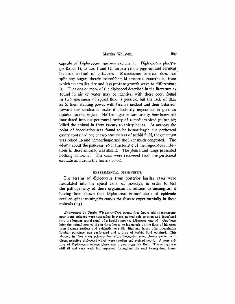
capsule of Diplococcus mucosus exclude it. Diplococcus pharyn-gis flavus II, as also I and III form a yellow pigment and fermentlevulose instead of galactose. Micrococcus cinereus does notsplit any sugar, therein resembling Micrococcus catarrhalis, fromwhich its smaller size and less profuse growth serve to differentiateit. That one or more of the diplococci described in the literature asfound in air or water may be identical with these cocci foundin two specimens of spinal fluid is possible, but the lack of dataas to their staining power with Gram's method and their behaviortoward the saccharids make it absolutely impossible to give anopinion on the subject. Half an agar culture twenty-four hours oldinoculated into the peritoneal cavity of a medium-sized guinea-pigkilled the animal in from twenty to thirty hours. At autopsy thepoint of inoculation was found to be hemorrhagic, the peritonealcavity contained one or two centimeters of turbid fluid, the momentumwas rolled up and hemorrhagic and the liver much congested. Theedema about the pancreas, so characteristic of meningococcus infec-tions in these animals, was absent. The pleura and lungs presentednothing abnormal. The cocci were recovered from the peritonealexudate and from the heart's blood.
EXPERIMENTAL MENINGITIS.
The strains of diplococcus from posterior basilar cases wereinoculated into the spinal canal of monkeys, in order to testthe pathogenicity of these organisms in relation to meningitis, ithaving been shown that Diplococcus intracelluiaris of epidemiccerebro-spinal meningitis causes the disease experimentally in theseanimals (15).
Experiment I: Strain Windsor.-Two twenty-four hours old sheep-serum-agar slant cultures were suspended in 2 c.c. normal salt solution and inoculatedinto the lumbar spinal canal of a healthy monkey (Macacus rhesus). One hourlater the animal seemed ill; in three hours he lay quietly on the floor of his cage,then became restless and evidently very ill. Eighteen hours after inoculationlumbar puncture was performed and a drop of turbid fluid obtained. Thisshowed in films many polymorphonuclear leucocytes, some closely packed withGram negative diplococci which were swollen and stained poorly. A pure cul-ture of Diplococcus intracellularis was grown from this fluid. The animal wasstill ill and very weak but improved throughout the next twenty-four hours.
587
Diplococci Occurring in Meningitis.
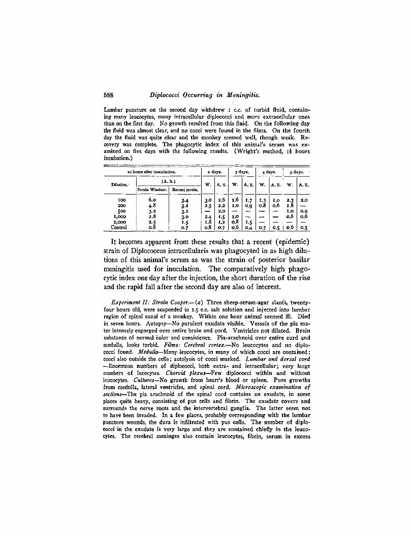
Lumbar puncture on the second day withdrew c.c. of turbid fluid, contain-ing many leucocytes, many intracellular diplococci and more extracellular onesthan on the first day. No growth resulted from this fluid. On the following daythe fluid was almost clear, and no cocci were found in the films. On the fourthday the fluid was quite clear and the monkey seemed well, though weak. Re-covery was complete. The phagocytic index of this animal's serum was ex-amined on five days with the following results. (Wright's method, Id hoursincubation.)
so hours after inoculation. 2 days. 3 days. 4 days. 9 days.
Dilution. (A. S.) W. A.S. W. A.S. W. A.S. W. A.S.Strain Windsor. Recent strain.
roo 6.o 3.4 3.0 2.6 i.6 1.7 1.3 1.o 2.3 2.0200 4.8 3.2 2.5 2.2 t.o o.g 0.8 0.6 1.8 -500o o
3.2 3.1 _ 2.0 -1.0 0.9 1,o00 2.8 3.0 2.4 1.5 I.1 -- - - o.8 0o.62,000 2.5 1.5 1.8 1.2 0.8 1.5 - - - -
Control o.8 0.7 o.8 0.7 o.6 0.4 0.7 0.5 0.6 0o.5
It becomes apparent from these results that a recent (epidemic)strain of Diplococcus intracellularis was phagocyted in as high dilu-tions of this animal's serum as was the strain of posterior basilarmeningitis used for inoculation. The comparatively high phago-cytic index one day after the injection, the short duration of the riseand the rapid fall after the second day are also of interest.
Experiment II: Strain Cooper.-(a) Three sheep-serum-agar slants, twenty-four hours old, were suspended in .5 c.c. salt solution and injected into lumbarregion of spinal canal of a monkey. Within one hour animal seemed ill. Diedin seven hours. Autopsy-No purulent exudate visible. Vessels of the pia ma-ter intensely engorged over entire brain and cord. Ventricles not dilated. Brainsubstance of normal olor and consistence. Pia-arachnoid over entire cord andmedulla, looks turbid. Films: Cerebral cortex.-No leucocytes and no diplo-cocci found. Medulla-Many leucocytes, in many of which cocci are contained;cocci also outside the cells; autolysis of cocci marked. Lumbar and dorsal cord-Enormous numbers of diplococci, both extra- and intracellular; very largenumbers of lucocytes. Choroid plexus-Few diplococci within and withoutleucocytes. Cultures-No growth from heart's blood or spleen. Pure growthsfrom medulla, lateral ventricles, and spinal cord. Microscopic examination ofsections-The pia arachnoid of the spinal cord contains an exudate, in someplaces quite heavy, consisting of pus cells and fibrin. The exudate covers andsurrounds the nerve roots and the intervertebral ganglia. The latter seem notto have been invaded. In a few places, probably corresponding with the lumbarpuncture wounds, the dura is infiltrated with pus cells. The number of diplo-cocci in the exudate is very large and they are contained chiefly in the leuco-cytes. The cerebral meninges also contain leucocytes, fibrin, serum in excess
588

Martha Wollstein.
and extravascular red corpuscles. The blood vessels are widely distended. Inthe depth of the sulci the cellular exudate is replaced by an inflammatory edemawith fibrin formation, Over the cerebellum the leucocytic exudate is heavy;within the cortex the perivascular lymph sheaths contain an excess of poly-nuclear leucocytes.
(b) Two sheep-serum-agar slants suspended in I c.c. of salt solution wereinoculated into the lumbar region of the spinal canal. The animal became verysick within two hours and died in nineteen hours. Autopsy-In the cervicalregion of the spinal cord the pia-arachnoid was edematous and hemorrhagic.Over the rest of the brain and cord there was marked congestion, but no visibleexudate. Films and cultures as in Experiment (a). Microscopic examinationof sections-The exudate in the spinal meninges is less in quantity than in thepreceding monkey and it is almost or entirely leucocytic in character. Theexudate in the cerebral meninges is also less and consists of a mixture of poly-nuclear leucocytes and of red corpuscles. The number of diplococci in the leu-cocytes in the spinal exudate is large; in the cerebral, small.
(c) One solid culture, twenty-four hours old, was suspended in I c.c. ofsalt solution and injected into the lumbar region of the spinal cord of a monkey.The animal was weak and ill during the night. Lumbar puncture on the fol-lowing morning withdrew very turbid fluid, showing many leucocytes crowdedwith diplococci; also some extracellular organisms. On the second morning0.5 c.c. of turbid fluid was withdrawn, showing numerous leucocytes and largenumbers of well staining diplococci. Cultures grew well and were pure. Onthe third day there was some improvement in the animal's condition. Thefluid withdrawn by lumbar puncture was still turbid, containing leucocytes butvery few diplococci. The following day no diplococci were found and on thefifth day the spinal fluid was clear. The monkey was then well. Examinationof the blood showed that while before inoculation the meningococcus (strainCooper) was not taken up by the leucocytes in dilutions exceeding to 20o,
that on the first and second days after injection they were phagocyted in dilu-tions of I to 2,ooo, on the fourth day to I to ,ooo, and o the seventh day notover I to 200. A recent strain (A.S.) again parallel with the one used for in-oculation. In the slides prepared from the serum on the seventh day aggluti-nation of the diplococci was apparent. In the earlier specimens it had beenabsent.
(d) Two-thirds of one serum-agar-culture was injected into the lumbarregion of the spinal canal of a healthy monkey. No symptoms of illness de-veloped, but on the day following the inoculation the spinal fluid was slightlyturbid, containing leucocytes with intracellular diplococci in small numbers, andstill fewer outside the cells. The cocci grew in pure cultures. The phagocyticindex of the serum before the beginning of the experiment was nil in dilutionsover to oo. It was apparent in dilutions of to 2,000 after twenty-two hoursand had fallen to what it was before inoculation on the fifth day, the recentstrain (A.S.) running a parallel curve with the one used for inoculation.
Experiment III: Strain Moseley.-One and a half sheep-serum-agar cul-tures, twenty-four hours old, were injected into the lumbar region of the spinalcanal of a monkey. He became ill within two hours, and on the following
689

Diplococci Occurring in Meningitis.
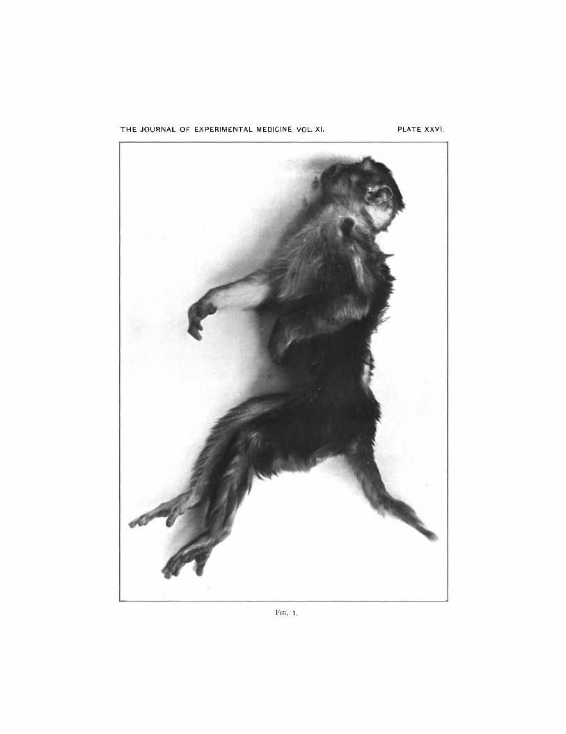
morning was very sick, lying on the floor in his cage. Two cubic centimeters ofturbid fluid were withdrawn by lumbar puncture and in it many leucocytes con-taining well staining diplococci were found; a small number of autolyzed diplo-cocci were also present. Cultures from this fluid grew well. On the followingmorning opisthotonos was marked; fibrillary twitchings and general convulsionswere produced by disturbance. The pupils were irregularly contracted and re-acted slowly. The animal was perfectly rigid unless disturbed by a touch or aloud noise, when a general convulsion came on. The cerebro-spinal fluid wasvery turbid, and contained many well-staining diplococci inside leucocytes andoutside them as well. On the third morning the monkey was very quiet, lyingon his side. No convulsions and no twitchings occurred. The eyes were lessirregular. The fluid was still turbid, with many diplococci in the leucocytes.The condition changed but slightly during the following day; the animal madeattempts to sit up but soon resumed the recumbent position.- On the morningof the fifth day it died. The symptoms of the first days were very much likethose of a human patient with meningitis and the clinical picture on the secondday was most suggestive. The photograph illustrates the condition at this time.
At the autopsy, encephalitis of the right frontal lobe was found.This portion of the cerebrum was very bright red in color, raisedabove the level of the adjoining lobes, translucent in appearance,studded with punctate hemorrhages. The layer of gray matter wasdouble the thickness of that in the opposite hemisphere. The re-mainder of the brain and cord showed a moderate congestion of thevessels of the pia mater. Cultures from the cerebrum, cerebellumand spinal cord showed no growth. The microscopic examinationof the sections of the spinal cord and brain of this animal indicatesthat it had recovered almost completely from the meningeal infec-tion and that it succumbed to the cerebral lesion. There are pres-ent small remains of the exudate in the meninges consisting ofleucocytes and proliferated mononuclear cells.
The chief interest centers in the encephalitis which proved to bean infarction affecting a large part of the hemisphere. The tissuein the infarcted area is necrotic, the contained blood vessels arenecrotic, and nuclear staining is generally absent. Around someof the blood vessels, now thrombosed, are collections of leucocytesalso undergoing necrosis, and in various parts are punctiformhemorrhages and beginning calcium salt deposits. The membranesover the encephalitic focus are inflamed; they show an accumula-tion of leucocytes and pus cells. The larger branches of the veinsare closed wholly or partially by thrombi containing fibrin and trans-
590
Martha Wollstein.
formed, fused, red corpuscles. In places a heavier exudate liesbetween the membranes and the encephalitic tissue of which thelarger part is composed of proliferated pial cells of large size, whichhave taken up extravasated red corpuscles that are undergoingdecolorization.
This case is of unusual interest because of its typical clinicalpicture, its prolonged course compared with the cases described byDr. Flexner in previous experiments, and the exceedingly severeand unusual lesion found at autopsy. It should be added thathemorrhagic encephalitis as a complication of cerebro-spinal menin-gitis in human beings is of occasional occurrence (6).
CONCLUSIONS.
The study carried out and recorded in this paper did not leadto the finding of any reliable criteria of difference between strainsof Diplococcus intracellularis obtained from typical cases of epi-demic meningitis and several cultures obtained from cases of pos-terior basic meningitis.
The successful experiments made with monkeys show that thediplococcus obtained from cases of posterior basic meningitis iscapable of setting up rapidly and acutely fatal forms of meningitisand in producing organic lesions of the cerebral tissues of greatseverity.
This study would, therefore, suggest that the antimeningitis.serum should be as useful in cases of posterior basic meningitis so-called, as it has been in epidemic meningitis, especially if it were-employed early in the disease.
BIBLIOGRAPHY.I. Still, Jour. of Path. and Bact., 898, v, 47.2. Holt, Diseases of Infancy and Childhood, 4th edition, New York, 9o8.3. Houston, British Med. Jour., 1907, ii, 144.4. Houston and Rankin, Idem, 1908, ii, 1340.5. Eve and Clements, Idem, 908, ii, 92.6. Ker, Idemn, 1908, ii, 1340.7. Arkwright, Lancet, 1908, ii, 475.8. Wilson, Lancet, 1908, ii, 477.9. Flexner, Jour. of Exper. Med., 1907, ix, o05.
io. Bruns and Hohn, Klin. Jahrbuch, 19o8, xviii, 285.
591
592 Diplococci Occurring in Meningitis.
ii. Klien, Johns Hopkins Hospital Bull., 1907, xviii, 285.12. Neufeld und Hine, Arb. a. d. k. Gsndhtsamte., 1907, xxv, 64.I3. Neufeld, Med. Klin., 9go8, iv, II58.14. von Lingelsheim, Arb. iiber die iibertragbare Genickstarre in Preussen im
Jahre I9o5, Jena, 9o6.I5. Flexner, Jour. of Exper. Med., 1907, ix, 142.
I6. Albrecht and Ghon, Wien. klin. Woch., Igoi, xiv, 988. Maschke, Berl. klin.Woch., 19o8, xlv, 56i.
EXPLANATION OF PLATE XXVI.The photograph which was kindly made by Dr. Learning shows the monkey
of Experiment III in the condition of opisthotonos. The hair was cut awayfrom the back of the neck in order to show the degree of curvature and retrac-tion of the head.
THE JOURNAL OF EXPERIMENTAL MEDICINE, VOL. XI.
FIGc. .
PLATE XXVI.