a clinico epidemiological study on facial
TRANSCRIPT

A CLINICO EPIDEMIOLOGICAL STUDY ON FACIAL
HYPERPIGMENTATION IN A TERTIARY CARE
HOSPITAL IN KANCHIPURAM DISTRICT,
TAMILNADU
Dissertation submitted in partial fulfilment of the
requirements for the degree of
M.D. (DERMATOLOGY, VENEREOLOGY &
LEPROSY)
BRANCH XX DEPARTMENT OF DERMATOLOGY
Reg. No: 201730451
KARPAGA VINAYAGA INSTITUTE OF MEDICAL
SCIENCES AND RESEARCH CENTRE,
KANCHIPURAM 603308
THE TAMILNADU DR. M.G.R.
MEDICALUNIVERSITY, CHENNAI
MAY 2020

II
CERTIFICATE
Certified that this dissertation titled “A CLINICO
EPIDEMIOLOGICAL STUDY ON FACIAL
HYPERPIGMENTATION IN A TERTIARY CARE
HOSPITAL IN KANCHIPURAM DISTRICT,
TAMILNADU” is a bonafide work done by Dr. MAHROOF
THAMARASSERY, post graduate student of the Department of
Dermatology, Venereology and Leprology, Karpaga Vinayaga
Institute of Medical Sciences and Research Centre, Kanchipuram
603308, during the academic year 2017 – 2020. This work has
not previously formed the basis for the award of any degree.
Prof. Dr. R. Baskaran MD, DD
Professor & HOD
Department of Dermatology, Venereology
& Leprology
Karpaga Vinayaga Institute of
Medical Sciences and Research Centre
Kanchipuram 603308
Dr. Sufala Sunil Viswasrao MD
Principal,
Karpaga Vinayaga Institute of Medical
Sciences and Research Centre
Kanchipuram
603308

III
DECLARATION
The dissertation entitled “A CLINICO EPIDEMIOLOGICAL
STUDY ON FACIAL HYPERPIGMENTATION IN A
TERTIARY CARE HOSPITAL IN KANCHIPURAM
DISTRICT, TAMILNADU” is a bonafide work done by Dr.
Mahroof Thamarassery at Department of Dermatology,
Venereology and Leprology, Karpaga Vinayaga Institute of
Medical Sciences and Research Centre Kanchipuram 603308,
during the academic year 2017 – 2020 under the guidance of Prof. Dr.
R. Baskaran M.D, DD (Dermatology), Professor & HOD,
Department of Dermatology, Karpaga Vinayaga Institute of Medical
Sciences and Research Centre Kanchipuram. This dissertation is
submitted to The Tamil Nadu Dr. M.G.R. Medical University,
Chennai towards partial fulfillment of the rules and regulations for the
award of M.D Degree in Dermatology, Venereology and Leprology
(BRANCH – XX).
Prof. Dr. R. Baskaran MD, DD
Professor & HOD
Department of Dermatology,
Venereology
& Leprology -Karpaga Vinayaga
Institute of
Medical Sciences and Research
Centre

IV
DECLARATION
I, Dr. Mahroof Thamarassery solemnly declare that this dissertation
titled “A CLINICO EPIDEMIOLOGICAL STUDY ON FACIAL
HYPERPIGMENTATION IN A TERTIARY CARE HOSPITAL
IN KANCHIPURAM DISTRICT, TAMILNADU” is a bonafide
work done by me at Karpaga Vinayaga Institute of Medical Sciences
and Research Centre, Kanchipuram 603308, during the academic year
2017-2020 under the guidance and supervision of Prof. Dr. R.
Baskaran M.D, DD, Professor & HOD, Department of Dermatology,
Karpaga Vinayaga Institute of Medical Sciences and Research Centre
Kanchipuram. This dissertation is submitted to The Tamil Nadu Dr.
M.G.R. Medical University, Chennai towards partial fulfilment of the
rules and regulations for the award of M.D Degree in Dermatology,
Venereology and Leprology
(BRANCH – XX).
PLACE:…………………..
DATE:…………………… Dr. Mahroof Thamarassery

V
SPECIAL ACKNOWLEDGEMENT
I sincerely thank Dr. R. ANNAMALAI, Managing Director,
Karpaga Vinayaga Institute of Medical Sciences for his kindness
in helping us with all available resources.

VI
ACKNOWLEDGEMENT
My sincere thanks to Dr. Sufala Sunil Viswasrao, MD, Principal,
Karpaga Vinayaga Institute of Medical Sciences and Research Centre,
Kanchipuram for allowing me to do this dissertation and utilize the
Institutional facilities.
I am grateful to my guide Prof. Dr. R. Baskaran M.D, DD
(Dermatology) Professor & HOD, Department of Dermatology,
Karpaga Vinayaga Institute of Medical Sciences and Research Centre
Kanchipuram, for his advice, guidance, motivation and
encouragement for my study.
I sincerely thank Dr. G. Srinivasan, MD, DV, Professor, Department
of Dermatology, Venereology & Leprology, Karpaga Vinayaga
Institute of Medical Sciences and Research Centre for his constant
encouragement for the study.

VII
I am deeply indebted to Dr. Dhanalakshmi K, Associate Professor,
Department of Dermatology, Venereology & Leprology, Karpaga
Vinayaga Institute of Medical Sciences and Research Centre for her
time devoted to my study.
I am also grateful to Dr. Jyothilakshmi, MD, Assistant Professor and
Dr. Sudarvizhi, DDVL, Senior Resident, Department of
Dermatology, Venereology & Leprology, Karpaga Vinayaga Institute
of Medical Sciences and Research Centre for constant inspiration
during the study.
I extend my gratitude to Dr. Roseline Fatima William, Professor and
HOD, Dr. Geetha M, Associate Professor, Dr. Karthik, Assistant
Professor and Ms. Gladius Jennifer, Assistant Professor, Department
of Community Medicine.

VIII
I am thankful to my colleagues for their support throughout the study.
I am also grateful to all paramedical staff for rendering timely help to
complete my study.
I am thankful to all my family members and specially thank my
spouse who has been standing by me for all helps needed in
completion of this work.
Last but not the least I am profoundly grateful to all patients for their
cooperation and participation in this study. They have been the
principal source of knowledge which I have gained during the course
of my clinical research.
Nothing would have been possible of this work without blessings
from God almighty. I am thankful to the God almighty for all the
blessings for successfully completing this work.

IX

X
CERTIFICATE – II
This is to certify that this dissertation work titled “A CLINICO
EPIDEMIOLOGICAL STUDY ON FACIAL
HYPERPIGMENTATION IN A TERTIARY CARE
HOSPITAL IN KANCHIPURAM DISTRICT, TAMILNADU”
of the candidate Dr. Mahroof Thamarassery, with registration
Number _201730451_ for the award of M.D. in the branch of
Dermatology, Venereology And Leprosy.
I personally verified the urkund.com website for the purpose of
plagiarism check. I found that the uploaded thesis file contains from
introduction to conclusion pages and result shows _1_ percentage
of plagiarism in the dissertation.
Guide and Supervisor
Sign with Seal

XI
S NO TITLE PAGE NO
1 INTRODUCTION 1
2 AIM OF THE STUDY 3
3 REVIEW OF LITERATURE 4
4 MATERIALS AND METHODOLOGY 33
5 RESULTS 36
6 DISCUSSION 73
7 SUMMARY 77
8 CONCLUSION 79
9 RECOMMENDATIONS 81
10 LIMITATIONS OF THE STUDY 82
11 REFERENCES 83
12 ABBREVIATIONS 98
13 CLINICAL IMAGES 101
14 ANNEXURES
14.1 PROFORMA 108
14.2 CONSENT FORM 110
14.3 ETHICAL COMMITTEE CERTIFICATE 112
14.4 MASTER CHART 113
14.5 KEY TO MASTER CHART 117

1
INTRODUCTION
Hyperpigmentary disorders of face are of major concern in both fair
skinned and dark skinned individuals in our country.1,2 A significant
proportion of individuals are affected by facial hypermelanoses due to
various clinical entities.1 It is a common complaint among patients
consulting with dermatologists.3 Facial hyperpigmentation, causes
cosmetic disfigurement with considerable psychological impact.2,4,5,6
Awareness of an individual of his embarrassing facial pigmentation can
decrease his self-confidence which can in turn decrease his productivity.7
Several more or less well-defined clinical syndromes leading to facial
hyperpigmentation can be recognized, but many transitional forms defy
classification.8
The causes of the pigmentation are often obscure.8 Common causes
are Melasma, Post Inflammatory Hyperpigmentation (PIH), Periorbital
Hyperpigmentation (POH), Facial Acanthosis Nigricans (FAN),
Lentigines, Freckles, Erythema Dyschromicum Perstans (EDP), Lichen
Planus Pigmentosus (LPP), Riehl’s Melanosis (RM), Erythromelanosis
Peribuccale Pigmentaire of Brocq (EPP), Poikiloderma of Civatte,
Erythromelanosis Follicularis of Face and Neck, Nevus of Ota, Seborrheic
Melanosis, Frictional Melanosis, Actinic Keratosis, Morphea, Systemic

2
Lupus Erythematosus rash, Topical Steroid Abuse, Drug Induced
Pigmentation
The differences in skin colour in various races and ethnicity are
related to the number, size, shape, distribution and degradation of melanin-
laden organelles called melanosomes. These are produced by melanocytes
and are transferred to the surrounding epidermal keratinocytes.
Face, the most visible area of the body serves as one’s identity and
therefore any pigmentary disturbance of the facial skin is of particular
cosmetic importance.
Studies have not been conducted in the southern part of the country
in connection with the facial hyperpigmentation considering all diseases
together. This study, conducted in the dermatology outpatient department
of the tertiary care centre, Karpaga Vinayaga Institute of Medical Sciences
and Research Centre, Kanchipuram, Tamilnadu will throw a light into the
details of facial hyperpigmentation.

3
AIM OF THE STUDY
To describe the clinical profile of patients presenting with facial
hyperpigmentation to Dermatology outpatient department of
Karpaga Vinayaga Institute of Medical Sciences, Kanchipuram
To determine the risk factors associated with facial
hyperpigmentation among the study participants.

4
REVIEW OF LITERATURE
The major determinant of normal skin color is the activity of
melanocytes, i.e. the quantity and quality of melanin pigment production
and not the density of melanocytes.9
Figure 1. Structure of melanocyte
To understand the pathology of hyperpigmentation, an appreciation of the
structure and function of the melanocyte is required (Figure 1). The
melanocyte is a neural crest-derived cell10, and during embryogenesis

5
precursor cells i.e., melanoblasts migrate along a dorsolateral pathway via
the mesenchyme to reach the epidermis and hair follicles. 11 Additional
sites of melanocyte migration include the uveal tract of the eye, the
leptomeninges and cochlea of the inner ear. In the hair, melanocytes
migrate to the basal layer of the hair matrix and the outer root sheath. 11
There are two populations of melanocytes in the skin, one in the
interfollicular epidermis and the second in the hair follicle.12 The
melanocytes maintain their position within the basal layer throughout life
in a normal individual.13 There is approximately one melanocyte per five
or six basal keratinocytes. 14
Epidermal melanin unit: this is the organization of a single epidermal
melanocyte surrounded by several epidermal keratinocytes.15
The dendrites of the melanocyte spread among the keratinocytes to
reach as high as mid stratum spinosum.16 One melanocyte can transfer the
melanin laden melanosomes to 30 to 40 keratinocytes.17 This association
is called epidermal melanin unit.18 The interaction of melanocyte with
keratinocytes is through cadherins and not through desmosomes.19
Melanocytes proliferate, migrate, and undergo maturation during
early to mid anagen phase. Melanogenesis and melanin transfer to

6
keratinocytes occurs throughout anagen phase.20 Melanocytes finally
undergo apoptosis during late catagen phase. 14
During embryogenesis, melanin producing melanocytes are found
diffusely throughout the dermis.21 They first appear in the head and neck
region at approximately 10 weeks of gestation. 22
Pigment production by melanocytes is increased on exposure to
agents that increase intracytoplasmic cAMP which includes cholera toxin,
forskolin and dibutyryl cAMP.11 Various other factors influencing
melanogenesis include Melanocyte Stimulating Hormone (MSH), basic
fibroblast growth factor (bFGF), endothelin-1 and UV light. α-MSH is the
major biologically active form of MSH in humans.23
An increase in the intracellular concentration of cAMP leads to an
increase in tyrosinase activity and increased eumelanin production.24
Ultraviolet exposure to melanocytes causes an increase in the size of
the melanocytes and increased tyrosinase activity.25 Following exposure to
UVA irradiation, an immediate pigmentary darkening can be observed,
which occurs within minutes and fades over 6–8 hours which is due to
oxidation of pre-existing melanin or melanin precursors.26 Delayed tanning
occurs within 48 to 72 hours of exposure to UVB and UVA radiation and

7
this represents new pigment production via an increase in tyrosinase
activity. 11
Main function of melanocytes is synthesizing melanin in specialized
organelles, the melanosomes, and transferring it to the keratinocytes
nearby so as to provide protection from UV radiation.27,28
Steps involved in melanogenesis includes transcription of proteins
required for melanogenesis; melanosome biogenesis; sorting of
melanogenic proteins into the melanosomes; transport of melanosomes to
the tips of melanocyte dendrites and transfer of melanosomes to
keratinocytes.29 Any deviation in this normal chain will result in
hypopigmentation.
Types of melanin
There are two types of melanin:
Eumelanin and Pheomelanin 30
Eumelanin
o Eumelanin is brown-black or dark which is an insoluble
polymer.
o It is the major type of melanin seen in individuals with
dark skin and hair.
o It has better ability to provide photoprotection.

8
Pheomelanin
o A red-yellow soluble polymer.
o Sulphur containing melanin.
o Formed by the conjugation of cysteine or glutathione.
o Pheomelanin is predominantly found in individuals
with red hair and skin phototypes I and II. 31
Figure 2. Steps involved in synthesis of melanin pigments -eumelanin
and pheomelanin

9
Melanogenesis is a complex process with several steps.32 (Figure 2).
The starting material for the production of melanin, is the amino acid
tyrosine. The rate limiting step in synthesis of both eumelanin and
pheomelanin is the first step, (the catalytic step) oxidation of tyrosine by
enzyme tyrosinase.33 This step is also called Raper–Mason pathway.34
Tyrosinase enzyme is also called tyrosine oxidase, DOPA oxidase,
monophenol, DOPA: oxygen oxidoreductase.14 Tyrosinase is a copper-
requiring enzyme. Rare copper deficiency disorders can cause
hypopigmentation as seen in Menkes Kinke hair disease.35
Tyrosinase also catalyzes additional steps in the biosynthesis of
melanin, e.g. the oxidation of dihydroxyindole. The activity of tyrosinase
is enhanced by DOPA and is stabilized by tyrosinase-related protein 1
(TRP1).
Competitive inhibitors of tyrosinase activity include hydroquinone
36 and L-phenylalanine.11 Hydroquinone is therefore used to treat disorders
of hyperpigmentation such as melasma.
P-protein is a transmembrane protein which is involved in the
transport of small molecules across the membrane of melanosomes.37 P-
protein regulates processing and trafficking of tyrosinase, possibly via
control of pH or glutathione content within intracellular compartments.38

10
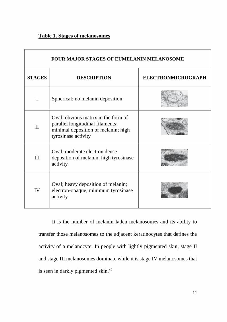
Stages of melanosome formation
The transformation of a non melanized melanosome to a fully
melanized melanosome can be divided into four stages 11. (Table 1).
Melanin pigment is not present in the initial two stages of
melanosome synthesis, whereas there is presence of intraluminal
proteinaceous fibrils which begin in Stage I and completes in Stage II.
Melanin synthesis starts to begin in Stage II melanosomes. Melanin starts
to get deposited on the fibrils and in later stages completes to mask all
structures in the melanosome.39
11
Table 1. Stages of melanosomes
FOUR MAJOR STAGES OF EUMELANIN MELANOSOME
STAGES DESCRIPTION ELECTRONMICROGRAPH
I Spherical; no melanin deposition
II
Oval; obvious matrix in the form of
parallel longitudinal filaments;
minimal deposition of melanin; high
tyrosinase activity
III
Oval; moderate electron dense
deposition of melanin; high tyrosinase
activity
IV
Oval; heavy deposition of melanin;
electron-opaque; minimum tyrosinase
activity
It is the number of melanin laden melanosomes and its ability to
transfer those melanosomes to the adjacent keratinocytes that defines the
activity of a melanocyte. In people with lightly pigmented skin, stage II
and stage III melanosomes dominate while it is stage IV melanosomes that
is seen in darkly pigmented skin.40

12
Melanosome transfer
Once the process of melanization is complete, the melanosome
migrates via microtubules into the dendrites prepared for transferring into
the neighbouring keratinocytes which might be within the epidermis or to
the anagen hair matrix.41 In addition to microtubules, proteins such as
kinesin and dynein are involved in the movement of melanosomes. Recent
work by Hideya Ando et al. shows that melanosomes are transferred from
melanocytes to keratinocytes through the processes of packaging, release,
uptake, and dispersion.42,40 (Figure 3 & 4)
Figure 3. Process of melanosome transfer from melanocyte to
keratinocyte

13
Figure 4. Steps involved in transfer of melanosomes to keratinocytes
Melanosomes are packed in globules enclosed by the
melanocyte plasma membrane
Melanosomes released into the extracellular space from melanocytic dendrites
Melanosomes are phagocytosed by keratinocytes
Membrane surrounding melanosomes gradually degraded
Melanocytes dispersed around the perinuclear area of the
keratinocyte.

14
Several chromophores are responsible for human skin color out of
which melanin is the most important one and others being haemoglobin (in
both the oxygenated and reduced state) and carotenoids.8,43 The thickness
of the stratum corneum, the dermal vasoconstriction or vasodilatation and
the occasional presence of endogenous or exogenous pigments may also
modify the skin color.44
DISORDERS PRESENTING WITH FACIAL
HYPERPIGMENTATION
Various diseases responsible for facial hyperpigmentation are:
1. Melasma
2. Post Inflammatory Hyperpigmentation (PIH)
3. Periorbital Hyperpigmentation (POH)
4. Facial Acanthosis Nigricans (FAN)
5. Lentigines
6. Freckles
7. Erythema Dyschromicum Perstans (EDP)

15
8. Lichen Planus Pigmentosus (LPP)
9. Riehl’s Melanosis (RM)
10. Erythromelanosis Peribuccale Pigmentaire of Brocq (EPP)
11. Poikiloderma of Civatte
12. Erythromelanosis Follicularis of Face and Neck
13. Nevus of Ota
14. Seborrheic Melanosis
15. Frictional Melanosis
16. Actinic Keratosis
17. Morphea
18. Systemic Lupus Erythematosus rash
19. Topical Steroid Abuse
20. Drug Induced Pigmentation
16
MELASMA
Melasma is a common acquired disorder of pigmentation
characterized by symmetric, brownish macules with well defined irregular
borders. Melasma most commonly affects the face. It is most prevalent
among young to middle aged females with more darkly pigmented skin.
Males can also present with melasma. Known exacerbating factors include
pregnancy, use of oral contraceptives and sun exposure. 11
Exact pathology of melasma is still unknown. It is hypothesised that
UV radiation exposure induces hyper functional melanocytes within
involved skin to produce more amounts of melanin pigment. 45 Hormonal
role in development of melasma has been investigated. 46 Studies have
shown increased expression of oestrogen and progesterone receptors in the
lesional skin compared to uninvolved skin. 47
Clinical features
Melasma can be clinically classified into 3 varieties based on area of
involvement (Figure 5). Areas of hyperpigmentation are symmetrical in
three classic patterns and other rare patterns on upper limbs and chest is
also possible.

17
Figure: 5 Clinical types of melasma
Clinical classification of melasma:
a) Centrofacial (most common), involving the forehead,
cheeks, nose, upper lip (sparing the philtrum) and chin;
b) Malar, affecting the cheeks and nose; and
c) Mandibular, along the jawline.
Less common sites include the extensor aspect of the forearms and
mid upper chest. (d & e)
Lesions often first appear or are accentuated following exposure to
UV irradiation or during pregnancy.48

18
On histopathological examination there is
Increased deposition of melanin in all layers of the epidermis.
Epidermal melanocytes are normal to increased in number,
and they are enlarged with prominent dendrites.
Increased number of melanophages.
Lesional melanocytes contain an increased number of
melanosomes. 49
Histologically, melasma can been classified as three variants:
epidermal, dermal, and mixed based upon the predominant layer of
melanin deposition. 50
For successful treatment of melasma, strict sun protection –
including hats, clothing & broad spectrum sunscreens are necessary. 51The
most common topical regimen is a combination of hydroquinone (2–4%),
tretinoin (0.05–0.1%) and a corticosteroid.
The mechanisms of action of hydroquinone include:
(1) Competes with tyrosine as a substrate for tyrosinase, the initial
enzyme in the melanin biosynthetic pathway that converts tyrosine to
dopaquinone.
(2) Selective damage to melanosomes and melanocytes. 52

19
POST INFLAMMATORY HYPERPIGMENTATION (PIH)
PIH is an excess of melanin pigment after inflammation or injury of
the skin. Occurs anywhere on the skin surface, including the mucous
membranes and the nail unit. PIH is common and has significant cosmetic
and psychosocial consequences.53
PIH can be epidermal or dermal. In the epidermal form of PIH, there
is increased melanin production with or without increased transfer to
keratinocytes. Inflammatory mediators like prostaglandins E2 and D2
which enhances pigment production may play a role in the same.
In dermal hyperpigmentation, melanin gets trapped in the dermis via
damaged basement membrane, where it is phagocytosed and subsequently
resides within melanophages (dermal macrophages). Histopathological
examination shows deposition of melanin in free form and in
melanophages in upper dermis and in perivascular region.54
Clinically PIH presents as asymptomatic hyperpigmented macules
with varying color ranging from tan to dark brown (seen in epidermal
melanin depostion) or grey–blue to grey–brown (dermal melanin). PIH
gets exacerbated by continued inflammation, trauma or exposure to
ultraviolet radiation. Various causes of PIH is summarised in Table 2.

20
T
Table 2. Disorders associated with Post Inflammatory Hyperpigmentation.

21
Mainstay of treatment of post inflammatory hyperpigmentation
includes treatment of underlying disease, strict sun protection using broad
spectrum sunscreens, topical hydroquinone (2–4%), topical azelaic acid
and α-hydroxy acids.11,55
PERIORBITAL HYPERPIGMENTATION
Periorbital hyperpigmentation (POH), is also called periorbital
melanosis, periocular hyperpigmentation, infraorbital darkening, dark
circles, infraorbital discoloration, or idiopathic cutaneous hyperchromia of
the orbital region.56
POH presents as homogenous ill-defined hyperpigmentation around
eyes usually bilaterally symmetrical. POH affect an individual’s emotional
well-being and it influences one’s quality of life. 57 Data regarding
periorbital hyperpigmentation is scarce. Periorbital hyperpigmentation is
considered to have a genetic basis. 57 An Indian study revealed POH was
most prevalent in age group of 16-25 years; commonest cause of POH was
constitutional followed by post inflammatory pigmentation.58 Risk factors
associated with periorbital hyperpigmentation according to the same study
were inadequate sleep, frequent usage of cosmetics, frequent rubbing of
eye and lack of correction of errors of refraction.59 The study concluded

22
that there is a strong association of POH with psychological stress, history
of atopy and family history. 58
Various causes for periorbital hyperpigmentation are:
Hereditary
Nevus of ota
Hori nevus
Post inflammatory hyperpigmentation
Superficial location of vasculature
Periorbital edema
Pigmentary demarcation lines. 57
Treatment options of periorbital hyperpigmentation include topical
depigmenting agents, like hydroquinone, azelaic acid, kojic acid, retinoic
acid and procedures like chemical peels, surgical corrections, fillers, botox
injections and laser therapy. 57,55

23
FACIAL ACANTHOSIS NIGRICANS (FAN)
Acanthosis Nigricans (AN) presents as dark, velvety, and thickened
skin, symmetrically distributed over the neck, axillae, and other flexural
regions of the body. Acanthosis Nigricans can also involve the face. There
is evidences for association between metabolic syndrome and facial AN.
Veysey and Ratnavel in 2005 reported a case of facial AN, who had obesity
and hyperinsulinemia. 60
Saumya pande et al concluded from their multicentric study that
Facial Acanthosis Nigricans could be considered a morphological marker
of metabolic syndrome as there were significant association between male
patients and positive OGTT, increased WHR, and BMI and FAN.61
LENTIGINES
Lentigines are benign pigmented macules in which there is an
increased number of melanocytes. The term ‘lentiginosis’ is applied either
when lentigines are present in large numbers or when they occur in a
distinctive distribution.62 Chronic as well as acute sun exposure has
important role in the pathogenesis of solar lentigines.63

24
Types of Lentigines:
Generalized lentigines
Lentigines usually being multiple can also appear singly or in small
crops at irregular intervals from infancy onwards. Their pathogenesis is not
exactly known and in majority of cases no genetic role can be found out.64
Unilateral lentiginosis (Zosteriform lentiginosis)
Such lentigines occur on one side of the body. Such lentigines can
be zosteriform and therefore occur in dermatomal or Blaschko linear
pattern
Eruptive lentiginosis
In eruptive lentiginosis, there is widespread occurrence of several
100 lentigines in short span of time without any systemic abnormalities. 65
FRECKLES
Freckles or ephelides are common and starts appearing around age
of 5 years as light‐brown pigmented macules on the light exposed skin of
fair skinned people.
Associations of freckles: Cutaneous disorders associated with
freckles are hereditary symmetrical dyschromatosis, xeroderma
pigmentosum and cutaneous malignant melanoma. 8
Fractional non-ablative 2940 nm Erb:YAG laser has given
satisfactory results in studies. 66

25
ERYTHEMA DYSCHROMICUM PERSTANS (EDP)
Erythema Dyschromicum Perstans is also known as Ashy
dermatosis or Dermatosis cenicienta (Cinderella dermatosis). Ramirez first
described erythema dyschromicum perstans (EDP) in 1957. 11
EDP is commonly seen in Latin America and Asia. Though it can
occur in males and females, causes greater concern in women. May affect
any age group, characteristically lesions start in the first or second decade
of life. EDP has been reported in children younger than 10 years in India.
67
Exact etiology of EDP is unknown. A cell mediated immune reaction
to ingested particles, or agents inducing contact irritation or to
microorganisms is said to be responsible for the pigmentary disturbance in
EDP. There are reports of association of EDP with the ingestion of
ammonium nitrate, oral X-ray contrast media, and medications like
benzodiazepines, penicillin; exposure to various pesticides, fungicides or
toxins and even endocrinopathies like thyroid disease; and whipworm and
HIV infections. 11
Clinically patients develop oval or irregular macules, slate-grey to
blue–brown in color in a symmetric pattern initially over trunk and later
spreading to the neck, upper limbs and face. In the initial stages

26
erythematous borders might be present. Though usually asymptomatic
mild itching might be present over lesions. Disease might rarely resolve
spontaneously.
Topical treatment is generally ineffective. Oral corticosteroids,
antibiotics, antimalarials, Isoniazid and Griseofulvin, as well as UV light
therapy might be effective. Clofazimine and Dapsone are also tried by
some workers with satisfactory results.68,69 Recently topical Tacrolimus
was found effective in ashy dermatosis.70
LICHEN PLANUS PIGMENTOSUS (LPP)
Lichen planus pigmentosus is an uncommon variant of lichen planus
affecting young to middle-aged population with skin phototypes III–V with
slight female preponderance.71 Bhutani et al found that there is presence of
colloid bodies in biopsy specimen of both LPP and LP. 72
Clinically LPP presents as irregular or oval, brown to grey–brown
macules in sun exposed areas like forehead, temples and neck or over the
intertriginous areas. Soles, nails and oral mucosa are usually spared.
Usually asymptomatic, mild pruritus might be present. Early lesions do not

27
have an erythematous border in contrast to Erythema Dischromicum
Perstans.
The etiology of LPP is unknown. UV rays are said to be one inciting
factor due to the involvement of disease over photo exposed areas. Kanwar
et al in 2003 reported topical application of mustard oil and amla oil as
triggering factors. 73 LPP is the result of an hypersensitivity reaction to an
unknown antigen showing a lichenoid inflammation, leading to melanin
incontinence which causes superficial dermal pigmentation.71
RIEHL’S MELANOSIS (RM)
Riehl’s Melanosis (RM) is said to be a pigmented contact dermatitis
to substances present in cosmetics and textile items. RM favours sites of
application of cosmetics. Lesions may be reticulated. Brown to grey
colored due to dermal melanin deposits. Biopsy reveals vacuolar
degeneration of basal layer and lichenoid infiltrate in early lesions.
Recently a lichenoid immune reaction caused by intrinsic as well as
extrinsic factors were postulated to be a cause of Riehl’s melanosis.
Repeated contact with low levels of allergens in cosmetic agents and
textile products produce a cytotoxic reaction characterized by vacuolar
basal cell degeneration and pigment incontinence. 74

28
RM is characterized by diffuse, patchy or reticular pigmentation,
often with satellite perifollicular pigmented macules and scaly follicular
hyperkeratosis. 2
Sites of involvement depend on the allergen responsible. Lesions
due to cosmetics begin on forehead and temples spreading over to rest of
the face and even extends to chest, neck, scalp, hands, and forearms. While
those due to textiles more often involve anterior aspect of thighs and
axillae.
IPL and Qs Nd:YAG lasers are some of the treatment options of
Riehl’s melanosis.75
NEVUS OF OTA (NOO)
Nevus of Ota is a bluish black dermal melanocytosis that affects the
sclera and the skin around the eye, distributed along the first and the second
branches of the trigeminal nerve. Extracutaneous lesions may be present in
the uveal tract, nasopharynx, tympanum and palate. 76
Nevus of Ota was first reported by Hulke in 1860 and later named
after Masao Ota. In 1939 he used the term nevus ‘fuscocaeruleus
ophthalmomaxillaris. Incidence of NOO is 0.014% to 0.034%. Male-

29
female ratio is 1:4.8. Most commonly seen among Asians and very rare in
other populations.
Associations of Nevus of Ota:
NOO is rarely associated with Nevus of Ito. Bilateral NOO is
associated with extensive Mongolian spots. Other associations are Sturge–
Weber syndrome and Klippel–Trenaunay syndrome.
Classification of NOO
Tanino has classified Nevus of Ota based on the extent and
distribution as: 77
Type I. Mild type
o IA. Orbital
o IB. Zygomatic
o IC. Forehead
o ID. Ala nasi alone
Type II. Moderate type: over the upper and lower eyelids,
periocular, zygomatic, cheek and temple regions.
Type III. Involves scalp, forehead, eyebrow and nose.
Type IV. Bilateral type: Both sides are involved.

30
The basic pathophysiology of NOO is embryonic failure of
migration of melanocytes from the neural crest to dermo-epidermal
junction which remains inactive and gets activated under hormonal
influence. The same is said to be the cause of nevus of lto, blue nevus and
mongolion spots. The blue to blue–grey pigmentation of Nevus of Ota is
due to melanin-producing melanocytes in the upper dermis whereas the
normal location of melanocytes are within the basal layer of the epidermis,
hair bulb, and outer root sheath of hair follicles. 78
Genetic studies have found somatic activating mutations in GNA11
and GNAQ gene which encode for ɑ subunits of G proteins. The same gene
mutations are also studied in melanocytic neoplasms of the central nervous
system.79 NOO may extend over time and persist for life. Treatment: Q-
switched ruby, alexandrite and Nd:YAG lasers, though post treatment
hypopigmentation is commonly reported.80
Facial hyperpigmentation in general
Upon reviewing previous literatures regarding facial
hyperpigmentation, in the five studies conducted across India during 2012-
2016 four studies dealt with facial hyperpigmentation alone and one with
both hypo and hyperpigmentation of face 81. Three of the studies analysed

31
100 3 cases each and two studies analysed 118 1 and 158 82 cases of facial
hyperpigmentation.
Youngest patient with facial hyperpigmentation in three of the
studies was 13 years old while in one study it was 12 years and the oldest
patient of the studies was 76 year old. All workers unanimously reported
that majority of patients with facial hyperpigmentation were females (63%
to 80%) and melasma was the most common cause of facial
hyperpigmentation ranging from 36% to 55%. Two studies revealed that
second most common cause of facial hyperpigmentation was Reihl’s
melanosis (both 35%)1,59 and post inflammatory hyperpigmentation (22%
& 25% each) were the second most common cause of facial
hyperpigmentation in another two studies.44,82 Whereas one study found
that second most common cause of facial hyperpigmentation was
periocular hyperpigmentation.3
While two studies reported history of 4% to 6% of melasma patients
taking oral contraceptive pills44,82, analysis by Shahana in 2013
interestingly showed nearly double fold increased association of oral
contraceptive pills with melasma (12.7%)59 compared to the other two
studies.

32
Cosmetic usage was found to be associated with facial
hyperpigmentation in majority of the studies. Revathy1 in 2016 reported in
a study in Bangalore that 31.4% cases of facial hyperpigmentation were
associated with cosmetic usage while Hassan et al. in 2013 observed
32.73% association82 whereas Kavya M from Bangalore in 2014 reported
only 22% association44 of cosmetic usage and facial hyperpigmentation.
Prolonged sun exposure was another commonly associated factor
with facial hyperpigmentation. Shahana 59 reported from Telangana that
54% of patients with facial hyperpigmentation had prolonged sun
exposure. Hassan et al. derived from their study in Jammu & Kashmir in
2013 that all cases of Lichen planus pigmentosus and Riehl’s melanosis,
80% cases of drug‑induced hypermelanoses, 80% cases of ephelides,
65.75% cases of melasma and 40% cases of post inflammatory
hyperpigmentation had exacerbation of facial pigmentation on sun
exposure. 82

33
MATERIALS & METHODOLGY
Study design: Cross sectional study
Study setting: The dermatology outpatient department of the
tertiary care centre, Karpaga Vinayaga Institute of Medical Sciences,
Chinnakolambakkam, Kanchipuam district of Tamilnadu
Study duration: 12 months. January 2018 to December 2018.
Study population: Patients with age 13 years and above including
both genders presenting to the outpatient department of dermatology,
Karpaga Vinayaga Institute of Medical Sciences with facial
hyperpigmentation.
Sample size: 100 cases
N = 4pq/d2
p = unknown so considered 50% prevalence;
q = 1-P;
d = clinical variation = 10%
N = 4*50*50 /100 = 100 individuals
Sampling: Convenience sampling technique

34
Inclusion criteria:
All males and females of age 13 years or above with non-
elevated hyperpigmented skin lesion(s) on face whether
treated/treating or have not taken any treatment yet.
Those who are willing to give consent.
Exclusion criteria:
Facial hyperpigmentation due to sequela of trauma.
Elevated skin lesions
Age less than 13 years
Those who are not willing to give consent.
Study instruments:
Pre tested validated structured questionnaire
Data collection: Primary data was collected by principle
investigator by interview method. Meticulous history taking, general
physical examination, systemic examination along with wood’s lamp
examination was done with consent from the patient. Histopathological
examination was done only in relevant cases were clinical diagnosis was
in doubt. Relevant details were recorded and tabulated.

35
Data analysis:
Software: SPSS version 20
Statistical analysis: Descriptive statistics; mean, median, standard
deviation and percentages were calculated. The association between
variables were calculated by chi square test and correlation coefficient at
5% level of significance.
Ethical issues:
Participants were made aware about the nature and purpose of the
study. Willingness and signature of the participants were taken on a
consent form. Participants were also informed that all the data provided
will be kept confidential and will be used only for the study purpose.
Written consents were obtained from all the subjects who participated in
the study before the study was started. Institutional ethics committee of
Karpaga Vinayaga Institute of Medical Sciences & Research Centre
reviewed the study proposal for ethical consideration and approval was
given.

36
RESULTS
Table 3. Age and Sex distribution of study participants:
Age Group
Sex
Total
Male Female
13-20 2 7 9
21-30 13 15 28
31-40 2 20 22
41-50 4 23 27
51-60 3 6 9
61-70 1 1 2
71-80 1 1 2
81-90 1 0 1
Total 27 73 100

37
Figure 6. Age and Sex distribution of study participants
In the study 100 patients were included in which females (73 cases)
outnumbered males (27 cases). Male to female ratio was 1:2.7. Youngest
patient was 13 years old and eldest patient was 81 years old. Mean age was
37.5 years.
The most common age group was 21 to 30 which had total 28
patients including 13 males and 15 females.
2
13
2
43
1 1 1
7
15
20
23
6
1 10
0
5
10
15
20
25
13-20 21-30 31-40 41-50 51-60 61-70 71-80 81-90Male Female

38
Table 4. Sex wise distribution of various diseases:
Diseases Male Female Total
MELASMA 5 36 41
POST INFLAMMATORY
HYPERPIGMENTATION - ACNE 10 9 19
PERIORBITAL
HYPERPIGMENTATION 1 9 10
FRECKLES 2 4 6
FACIAL ACANTHOSIS NIGRICANS 2 3 5
NEVUS OF OTA 0 4 4
DRUG INDUCED PIGMENTATION 3 0 3
RIEHL’S MELANOSIS 0 2 2
SEBORRHEIC MELANOSIS 0 2 2
MORPHEA 0 2 2
TOPICAL STEROID ABUSE 1 1 2
FRICTIONAL MELANOSIS 1 0 1
ACTINIC KERATOSIS 1 0 1
SLE RASH 0 1 1
SUN TAN 1 0 1
Total 27 73 100

39
Figure 7. Sex wise distribution of various diseases
Sex wise distribution showed 36 female and 5 male Melasma cases,
10 male and 9 female cases of Post inflammatory hyperpigmentation from
facial acne, 9 female and 1 male case of Periorbital hyperpigmentation, 4
female and 2 male cases of Freckles, 3 female and 2 male cases of Facial
Acanthosis Nigricans, 4 female cases of Nevus of Ota, 3 male cases of drug
induced pigmentation, 2 female cases of Riehl’s melanosis, 2 female cases
of seborrheic melanosis, 2 female cases of facial Morphea, 1 male and 1
female case of topical steroid abuse, 1 male case of frictional melanosis, 1
male case of actinic keratosis, 1 female case of SLE rash and one male case
of sun tan.
POSTINFLAMMAT
ORYHYPERPIGMENTATION -ACNE
PERIORBITA
LHYPERPIGMENTATI
ON
MELASMA
FACIAL
ACANTHOSI
SNIGRICANS
FRECKLES
RIEHL’S
MELANOSIS -RM
NEVUS OFOTA
SEBORRHEICMELANOSIS
FRICTIONALMELANOSIS
ACTINIC
KERATOSIS
MORPHEA
SLERASH
TOPICAL
STEROID
ABUSE
DRUGINDUC
EDPIGMENTATI
ON
SUNTAN
Female 9 9 36 3 4 2 4 2 0 0 2 1 1 0 0
Male 10 1 5 2 2 0 0 0 1 1 0 0 1 3 1
0
5
10
15
20
25
30
35
40
45
No
. of
Cas
es
Sex wise distribution of diseases

40
Figure 8. Occupation of the participants
In the study majority of the patients were unemployed falling to
45%. This falls in line with the Indian scenario where males go for work
more often than females and that in our study majority of patients consisted
female gender. The next majority were unskilled workers which fell to
36%. Professional workers were only 10% of the total 100 cases.
Professional10%
Semi-Professional
4%
Semi-Skilled Worker
5%
Unskilled Worker
36%
Unemployed45%
OCCUPATION

41
CLINICAL PROFILE OF DISEASES
In the study, while analysing the different causes of facial
hyperpigmentation the proportion of various diseases were as follows:
Table 5. Proportion of Diseases in the study
Diagnosis Percentage
MELASMA 41
POST INFLAMMATORY HYPERPIGMENTATION -
ACNE 19
PERIORBITAL HYPERPIGMENTATION 10
FRECKLES 6
FACIAL ACANTHOSIS NIGRICANS 5
NEVUS OF OTA 4
DRUG INDUCED PIGMENTATION 3
RIEHL’S MELANOSIS -RM 2
SEBORRHEIC MELANOSIS 2
MORPHEA 2
TOPICAL STEROID ABUSE 2
FRICTIONAL MELANOSIS 1
ACTINIC KERATOSIS 1
SLE RASH 1
SUN TAN 1
Total 100

42
Among the 100 cases of facial hyperpigmentation, melasma formed
the largest group (41 cases). Second largest group was post inflammatory
hyperpigmentation from acne (19 cases) followed by periorbital
hyperpigmentation which constituted 10 cases.

43
Table 6. Relation of facial hyperpigmentation with pregnancy:
Diagnosis Not
applicable Increases Decreases
No
change
Periorbital
hyperpigmentation 8 1 0 0
Melasma 20 4 0 12
Other diseases 36 0 0 19
When the female patients were asked whether the facial
pigmentation increased during pregnancy, four melasma (11%) patients
and one female (11.11%) with periorbital pigmentation noted increased
pigmentation during pregnancy whereas 20 female melasma patients
replied that they did not notice any visible change during pregnancy.
Females with other diseases did not notice any changes associated with
pregnancy.

44
Table 7. Relation of Facial hyperpigmentation with sun exposure
Aggravation of pigmentation on
sun exposure Percent
Yes 61.0
No 39.0
Total 100.0
Relation between sun exposure and aggravation of facial
hyperpigmentation was studied which showed the following results: 61
percentage of patients with facial hyperpigmentation remarked they noted
visible increase in the pigmentation while exposed to sun where as 39
percentage did not feel any such change.

45
Table 8. Details of treatment taken for the facial hyperpigmentation
Treatment taken Percent
No 66.0
Topical only 24.0
Topical + oral 9.0
Oral only 1.0
Total 100.0
While analysing the history of treatment taken so far, 66% had not
taken any treatment whereas 34% took some treatment in which 24% had
taken topical treatment and 9% took topical treatment along with oral
medicine for the facial hyperpigmentation.

46
Table 9. Details of home remedies taken by the participants
Home remedies used Percent
No 65.0
Yes 35.0
Total 100.0
Among the 100 patients, 35 patients had tried some form of home
remedies whereas majority of patients (65%) did not attempt any form of
home remedies. The home remedies included making a paste from
crushing various leaves and other objects and applying on face.

47
Table 10. Details of topical steroid usage for facial
hyperpigmentation by the participants:
Topical Steroid Usage Percent
No 82.0
Yes, plain steroid 8.0
Combination steroids 8.0
Do not know 2.0
Total 100.0
Since topical steroid abuse was a common problem faced by
dermatologists over the country, an analysis about the history of topical
steroid application and its complication was made.83
On analysing the cases, 16 cases gave history of application of
topical steroids out of which 8 cases had applied plain steroid and 8 had
applied combination steroids. Most common plain steroid was
Betamethasone valearate and combination steroid was Betamethasone
valearate with Gentamicin and Miconazole nitrate. Easy availability and
low price were the factors which led them to usage of Betamethasone
valearate. Majority of the patients were provided with topical steroids by
pharmacists themselves and most common indication was skin lightening.

48
Table 11. Details of usage of vegetables and other agents on face
Vegetable application Percent
No 77.0
Yes 23.0
Total 100.0
Another interesting finding in the study was the application of raw
vegetables and other agents on face. Twenty three percentage of the
patients admitted that they had tried applying numerous raw vegetables on
face in single and combination with other agents and other vegetables.
Various combination of vegetables and other agents were tomato
alone, tomato + garlic, tomato + lemon + ice, tomato + potato + aloe vera,
tomato + potato + lemon + onion + aloe vera + gram flour paste, tomato +
sugar scrub + cumin seeds, aloe vera, lemon + honey, lemon + curd, milk
+ sugar.
There was one patient who has been applying mint leaves (puthina),
fuller’s earth (multani mitti), green gram powder, green gram flour in
alternating combinations since 6 months.

49
Table 12. Details of turmeric application on face.
Sex
Turmeric application
Total
No Yes
Female 21 52 73
Male 22 5 27
Total 43 57 100
Figure 9. Details of turmeric application on face.
21
52
22
5
0
10
20
30
40
50
60
No Yes
TURMERIC APPLICATION ON FACE
Female Male

50
It is a common practice of people of Tamilnadu to apply turmeric on
face. In the study an attempt was made to study turmeric usage habit of the
participants.
It was found that 57% of the patients including both males & females
had the habit of application of topical turmeric either for their present skin
problem or otherwise. When females alone were considered, 52% of
females had history of topical application of turmeric. The reason for
application of turmeric on face included social practices, for general skin
health and some applied specifically in the belief that it will help for
lightening the skin pigmentation. Some of the previous studies had shown
statistically significant positive effects on skin health from turmeric
usage.84 Turmeric application on face obscured wood’s lamp findings in
some patients for which they were asked to wash face properly for re-
examining.

51
Table 13. Details of cosmetic usage on face
Diagnosis
Usage of Cosmetics
Total
p Value
No
Yes
0.034
Cases %
POST INFLAMMATORY
HYPERPIGMENTATION -
ACNE
8 11 58 19
PERIORBITAL
HYPERPIGMENTATION 4 6 60 10
MELASMA 8 33 80 41
FACIAL ACANTHOSIS
NIGRICANS 2 3 60 5
FRECKLES 0 6 100 6
RIEHL’S MELANOSIS 0 2 100 2
NEVUS OF OTA 1 3 75 4
SEBORRHEIC MELANOSIS 0 2 100 2
MORPHEA 0 2 100 2
SLE RASH 0 1 100 1
TOPICAL STEROID ABUSE 0 2 100 2
p value is <0.05 (0.034) which is statistically significant.

52
Figure 10. Details of cosmetic usage on face
Majority (71%) of the patients had used some form of cosmetics on
face. Various cosmetics that were documented in the study were:
Adapalene Cream, Aloe Vera Cream, Ayur Sunscreen, Blackberry
Facewash, Blackrose Hair Dye, Fair & Lovely fairness cream, Gokul
Powder, Gokul Sandal, HHlite Cream, J&J Talcum Powder, Kasturi
Manjal, Lakme Facewash, Neem Facewash, Olay fairness cream, Ponds
Age Miracle, Ponds Baby Cream, Ponds Facewash, Ponds Powder, Ponds
White Beauty, Skinlite, Vicco Turmeric,
‘Fair and Lovely’ topped the list followed by ‘Ponds age miracle’.
Interestingly, one patient in the study regularly used ‘Fair and Lovely’
fairness cream on a daily basis for 25 years.
Yes71%
No29%
COSMETIC USAGE
Yes No

53
Figure 11. Relation of frequent rubbing of face with facial
hyperpigmentation
Frequent rubbing of face being one of the risk factor of facial
hyperpigmentation, was analysed in the study which showed 100% of
frictional melanosis (1 case), 40% of periorbital hyperpigmentation, 21%
of acne pigmentation and 20% of facial acanthosis nigricans had the habit
of frequent rubbing of the face.
4 4
1 1
6
15
0
4
0
2
4
6
8
10
12
14
16
PERIORBITALHYPERPIGMENTATION
POST INFLAMMATORYHYPERPIGMENTATION -
ACNE
FRICTIONAL MELANOSIS FACIAL ACANTHOSISNIGRICANS
Yes No

54
Table 14. Drug history of the participants
Disease Nil
Oral
Contracep
tive Pills
(Females)
Thyroid
drugs
Leprosy
treatment Total
p
Value
PERIORBITAL
HYPERPIGME
NTATION
9 0 1 0 10
0.002
MELASMA 32 5 3 1 41
FRECKLES 2 4 0 0 6
DRUG
INDUCED
PIGMENTATI
ON
0 0 0 3 3
Total 9 4 4 17
While considering drug history, only 17 patients gave positive
history of drug intake. Details of antidiabetic and antihypertensive drugs
were not included in the study. In 9 patients of periorbital pigmentation,
one patient was on thyroid medication. In melasma patients, out of the total
41 patients, 78% (32 patients) did not give any drug history whereas 14%
(5 of 36 cases) of the female melasma patients gave history of oral
contraceptive pill usage and 3 patients (7%) and 1 patient (2.5%) gave
history of thyroid drugs and leprosy drugs respectively.
55
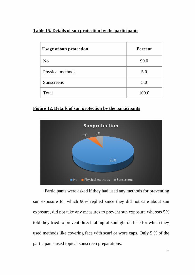
Table 15. Details of sun protection by the participants
Usage of sun protection Percent
No 90.0
Physical methods 5.0
Sunscreens 5.0
Total 100.0
Figure 12. Details of sun protection by the participants
Participants were asked if they had used any methods for preventing
sun exposure for which 90% replied since they did not care about sun
exposure, did not take any measures to prevent sun exposure whereas 5%
told they tried to prevent direct falling of sunlight on face for which they
used methods like covering face with scarf or wore caps. Only 5 % of the
participants used topical sunscreen preparations.
90%
5% 5%
Sunprotection
No Physical methods Sunscreens

56
Table 16. Details of sun exposure
Daily duration of sun exposure Percent
<3 hours 65.0
3-5 hours 31.0
6 or more hours 4.0
Total 100.0
Figure 13. Details of sun exposure
Sun exposure details were collected which showed that all the cases
were exposed to sunlight. Majority (65%) having exposed to sun daily for
less than 3 hours per day and 31% of cases getting exposed to sun daily for
3 to 5 hours per day as part of their routine activities including work. 4%
of people were exposed to sun for 6 or more hours per day. It was mostly
outside workers like agricultural workers, painters who were exposed to
sun as a part of their outside jobs.
65%
31%
4%
Sun exposure duration
<3 hours 3-5 hours 6 or more hours

57
Figure 14. Details of comorbidities of the participants
While assessing the comorbidities, diabetes peaked the diseases
(9%) followed by thyroid disorder (8%). Six patients had anemia, 5
patients had hypertension and another 5 had leprosy.
9
5
8
2
5
6
0
1
2
3
4
5
6
7
8
9
10
Diabetes Hypertension Thyroid TB Leprosy Anemia
Comorbidities

58
Table 17. Comorbidities and association with facial
hyperpigmentation
Diseases Diabetes
Mellitus
Hyper
tension Thyroid
Tuber
culosis Leprosy Anemia
POST
INFLAMMATORY
HYPERPIGMENT
ATION - ACNE
0 0 1 0 0 1
PERIORBITAL
HYPERPIGMENT
ATION
0 1 1 0 0 0
MELASMA 5 3 6 2 1 2
FACIAL
ACANTHOSIS
NIGRICANS
2 0 0 0 0 1
NEVUS OF OTA 0 0 0 0 1 0
FRICTIONAL
MELANOSIS 1 0 0 0 0 0
ACTINIC
KERATOSIS 0 0 0 0 0 1
DRUG INDUCED
PIGMENTATION 1 1 0 0 3 1

59
Five melasma patients, two patients with facial acanthosis nigricans,
one patient with frictional melanosis, and one patient with drug induced
pigmentation had diabetes mellitus.
One patient with periorbital pigmentation, three melasma patients
and one patient with drug induced pigmentation had hypertension. Eight
cases had thyroid diseases (1 post inflammatory hyperpigmentation
patient, 1 periorbital hyperpigmentation, 6 melasma patients). Past history
of pulmonary tuberculosis was present with two melasma patients.
History of leprosy was present in one melasma patient, one patient
with Nevus of Ota, and three patients with drug induced pigmentation.
Anemia was present with one case each of post inflammatory
hyperpigmentation from acne, facial acanthosis nigricans, actinic keratosis,
drug induced pigmentation and two patients with melasma.

60
Table 18. Details of consanguinity and facial hyperpigmentation
Diseases
Consanguinity
Not present
Yes
Total No. of
cases %
PERIORBITAL
HYPERPIGMENTATION 9 1 10 10
MELASMA 35 6 14.5 41
FACIAL ACANTHOSIS
NIGRICANS 4 1 20 5
MORPHEA 1 1 50 2
While the consanguinity history was elicited it was found that
parents of 9% of cases had consanguineous marriage. History of
consanguineous marriage was found in the following diseases: Melasma,
periorbital hyperpigmentation, facial acanthosis nigricans and morphea.
Top in the list was melasma patients in which 14.5% (6 of 41 cases) had
consanguineous parents and 10% (1 of 10 cases) of periorbital
hyperpigmentation, 20 % (1 of 4 cases) of facial acanthosis nigricans and
50% of facial morphea (1 of 2 cases) had consanguineous parents.

61
Table 19. Wood’s lamp findings of facial hyperpigmentation
Type Percent
Epidermal (Accentuation present) 55
Dermal (No accentuation) 13
Mixed 15
Indeterminate 15
Not done 2
Total 100
Figure 15. Wood’s lamp findings of facial hyperpigmentation
55
1315 15
2
0
10
20
30
40
50
60
E P I D E R M A L D E R M A L M I X E D I N D E T E R M I N A T E N O T D O N E
WOOD'S LAMP FINDINGS

62
Ninety eight percentage of the patients were subjected to wood’s
lamp examination; 2% could not be done due to technical reasons. 55% of
patients had epidermal pigmentation, 13% had dermal pigmentation and
15% had mixed dermal and epidermal pigmentation. 15% of patients
lacked specific findings since they had other applications on face (turmeric
etc.) which produced distinct fluorescence due to which wood’s lamp
examination was inconclusive.

63
Figure 16. Analysis of Fitzpatrick skin type in the study
Analyzing the skin type based on Fitzpatrick classification 85, 58%
of patients had Type V skin followed by 28% having type IV skin. 11%
patients fell to VI skin type. Only one patient in the study had Type II skin.
There were no patients with type I Fitzpatrick skin.
0
1
2
28
58
11
0 10 20 30 40 50 60 70
I Pale white skin
II Fair skin, blue eYes
III Darker white skin
IV Light brown skin
V Brown skin
VI Dark brown or black skin
Skin type

64
MELASMA SUMMARISED
Since melasma formed bulk of the disease further details of melasma
patients are summarised.
Figure 17. Sex distribution of melasma patients
Burden of melasma was more in females than males as evident from
the graph. Females formed 88% (36 cases) whereas males were only 12%
(5 cases).
Male12%
Female88%
Melasma sex wise
Male
Female
65
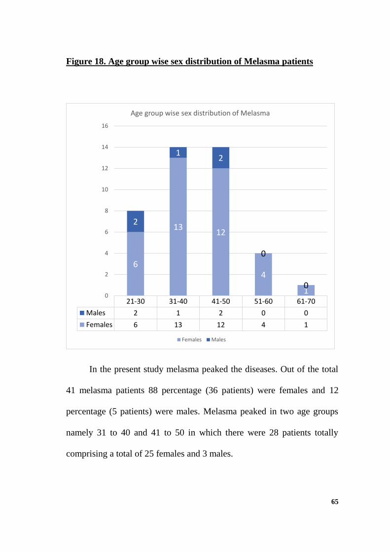
Figure 18. Age group wise sex distribution of Melasma patients
In the present study melasma peaked the diseases. Out of the total
41 melasma patients 88 percentage (36 patients) were females and 12
percentage (5 patients) were males. Melasma peaked in two age groups
namely 31 to 40 and 41 to 50 in which there were 28 patients totally
comprising a total of 25 females and 3 males.
21-30 31-40 41-50 51-60 61-70
Males 2 1 2 0 0
Females 6 13 12 4 1
6
1312
4
1
2
12
0
00
2
4
6
8
10
12
14
16
Age group wise sex distribution of Melasma
Females Males

66
Figure 19. History of pregnancy exacerbation of melasma
Melasma is known to have exacerbation during pregnancy in certain
patients. 86 In our study only 11% (4 cases) of females with melasma
reported aggravation of the disease during pregnancy.
Thirty three percentage (12 females) did not notice any visible
changes when they became pregnant while having the disease. Remaining
56% (20 cases) did not have the disease when they became pregnant.
Not applicable
2056%Increases
411%
No change12
33%
Pregnancy Exacerbation of Melasma

67
Figure 20. Relation of aggravation of melasma with exposure to sun
light
Majority of the patients (71%) said they noted visible darkening of
the pigmentation while exposed to sun light whereas only 12% did not
notice such visible changes.
No, 12, 29%
Yes, 29, 71%
Exacerbation of melasma in sunexposure

68
Table 20. Relation of melasma and psychological stress
Psychological
stress Cases %
p Value
Yes 25 61
0.015
No 16 39
Figure 21. Relation of melasma and psychological stress
Sixty one percentage of the people with melasma had psychological
stress due to various factors (not related to the disease), prior to the onset
of the disease. This is statistically significant as p value is <0.05.
61%
39%
PSYCHOLOGICAL STRESS IN MELASMA PATIENTS
Yes no

69
Figure 22. Comorbidities in melasma patients
Out of 41 melasma patients 6 patients had hypothyroidism, 5 patients
had diabetes mellitus, 3 had hypertension and 2 were anemic.
0
1
2
3
4
5
6
Anemia DiabetesMellitus
Hypertension Thyroid
2
5
3
6
Comobidities in melasma

70
Table 21. Usage of sun protection in melasma patients
Usage of sun protection in melasma Percent
Yes 17 %
No 83%
Total 100%
Awareness of importance of sun protection is less in melasma
patients which is evident from the data that only 17% (7 patients) were
using sun protection whereas majority of melasma patients (83%, 34
patients) did not use any form of sun protection.

71
Figure 23. Consanguinity in melasma patients
History of consanguineous marriage was present with parents of 6
cases (14.6%) out of the 41 melasma patients.
6
35
0 5 10 15 20 25 30 35 40
Yes
No
Consanguinity in melasma

72
Figure 24. Fitzpatrick skin type in melasma patients
Majority of melasma patients (59%, 24 cases) had Fitzpatrick skin
type V. Next majority had type IV (27%, 11 cases) Fitzpatrick skin. 5
patients (12%) had type VI skin. Only one patient had type III skin (2%).
Melasma was not observed in patients with type I & II skin.
Other observations
No melasma patients in the study had extrafacial disease.
Majority of the patients had symmetrical involvement of face
(37 patients, 90%).
0, 0% 0, 0% 1, 2%
11, 27%
24, 59%
5, 12%
Fitzpatrick Skin type in melasma
Fitzpatrick I
Fitzpatrick II
Fitzpatrick III
Fitzpatrick IV
Fitzpatrick V
Fitzpatrick VI

73
DISCUSSION
Number of cases included in the present study were 100 which
correlated with the studies by TN Revathi and Shahana in which both
studied 100 patients of facial hyperpigmentation. 3,59
In the study, females outnumbered males which correlated with all
other studies reviewed.1,3,59 The most common age group in the present
study was 21 to 30 whereas this age group was the second most common
age group in the studies by Kavya 87 and Revathi 1. Youngest patient was
13 years old. The youngest patient in the study by Revathi was also 13
years old. 1
Forty five percentage of the participants were unemployed and 36%
were unskilled workers in the study.
Most common cause of facial hyperpigmentation in the present study
was melasma which correlated well with five previous studies, all of which
concluded that melasma was the most common cause of facial
hyperpigmentation.1,3,59,81,87
Second most common cause of facial hyperpigmentation in the
present study was post inflammatory hyperpigmentation which correlated
with previous two studies. 82,87

74
Four melasma patients and one female patient with periorbital
pigmentation had increased pigmentation during pregnancy. Previous
studies by Ana Carolina Handel et al also said that melasma increased
during pregnancy.88
Seventy one percentage of patients with facial hyperpigmentation
noted increase in the pigmentation on exposure to sunlight. Various causes
said to be responsible for increased pigmentation on sun exposure include
ultraviolet (UV) radiation induced immediate pigment-darkening reaction,
due to photo-oxidation of preformed melanin, followed by delayed
tanning.89,90
Thirty four percentage of the participants had already tried some
form of treatment for their facial hyperpigmentation including topical and
oral treatments. Thirty five percentage had tried some form of home
remedies by mixing and making a paste (like vegetables and leaves
crushed) and applying on face for the pigmentation. Twenty three
percentage of the patients had tried applying numerous raw vegetables on
face. History of topical turmeric usage was present in 57% of participants
including males and females.
Sixteen percentage of cases had used topical steroids in combination
and singly in belief of lightening the facial pigmentation. A study by

75
Santwana Mahar et al showed 11.77% patients misused topical
corticosteroids91 whereas study by Sendrasoa et al showed as high as
49.8% had misused topical corticosteroids.92
Majority (71%) of the patients had used some form of cosmetics on
face. This correlated with the study by Mestawet Getachew et al who
concluded that 80.1% had used some form of cosmetics on face in their
study.93 Association between cosmetic usage and facial hyperpigmentation
was statistically significant in the study as p value was <0.05 (0.034).
Drug history was present in 17% of the participants. Association
between facial hyperpigmentation and drug intake was statistically
significant as p value was <0.05 (0.002).
Only 5% had the practice of using topical sunscreen creams on face
which was comparable to the findings by Ahamed et al showing 8.3% of
sun screen usage.94
Diabetes (9%) followed by thyroid disorder (8%) were the two most
common comorbidities noted in patients with facial hyperpigmentation.
Consanguinity history was present in 9% of patients with facial
hyperpigmentation.
Wood’s lamp examination of the cases showed majority had
epidermal pigmentation (55%), 13% had dermal pigmentation and 15%

76
had mixed dermal and epidermal pigmentation which correlated with the
study by Kavya & Nataraj where 55% had epidermal and15% had dermal
pigmentation.44
Fitzpatrick skin classification showed 58% of patients had Type V
skin followed by 28% having type IV skin.
On summarising the findings in melasma patients, females had
higher incidence of melasma. This correlated with all other previous
studies reviewed.2,50,81,87,95 Eleven percentage (4 cases) of females with
melasma reported aggravation of the disease during pregnancy. This
correlated with the findings by Arun Achar and Sanjay K Rathi where only
13.6% of females (out of 312 cases) had pregnancy exacerbation of
melasma. 86 Majority of the patients (71%) said they noted visible
darkening of the pigmentation while exposed to sun light. Sixty one
percentage of the people with melasma had statistically significant
association between psychological stress due to various factors and the
disease as p value was <0.05. Six melasma patients had hypothyroidism, 5
were diabetic, 3 had hypertension and 2 were anemic. Only 17% (7
patients) of the melasma patients were using sun protection. History of
consanguineous marriage was present with parents of 14.6% (6 cases) of
the 41 melasma patients. No studies in literature were found which
investigated association between melasma and consanguinity.

77
SUMMARY
In this one year study, females dominated over males in availing
dermatology services for facial hyperpigmentation. Most common age
group was 21 to 30 years.
Majority of the patients were unemployed and next majority were
unskilled workers working under sunlight like agricultural workers.
Melasma was the most common disease in the study. Second most
common disease was post inflammatory hyperpigmentation from acne.
Majority of the patients had aggravation of the pigmentation on exposure
to sunlight.
Thirty four percentage of the participants had tried some form of
treatment for their facial hyperpigmentation. Some patients (35%) had
tried some form of home remedies for their pigmentation, in which 23%
had tried applying raw vegetables on face. More than half of the
participants (57%) including males and females were applying turmeric
on face. Majority of the patients had used some form of cosmetics on
face.

78
Topical steroid abuse on face was present in sixteen percentage of cases.
Sunscreen usage was very low in patients as only 5% had the practice of
using topical sunscreen creams on face.
Diabetes mellitus and thyroid disorder was the two most common
comorbid illness noted in the study.
Wood’s lamp examination of the cases showed majority had epidermal
pigmentation and 13% had dermal pigmentation.
Majority of the participants had Fitzpatrik skin type V followed by type
IV skin.
In the melasma group of patients, females formed majority (36 cases)
whereas males were only 5 cases.
Some female melasma patients (11.11%) noted aggravation of the
pigmentation during pregnancy. Majority of the melasma patients noted
visible darkening of the pigmentation while exposed to sun light. Only
few (17%) of the melasma patients were using sun protection.
Consanguinity history of 14.6% was present in melasma patients.

79
CONCLUSION
1. Females dominated over males in availing dermatology services for
facial hyperpigmentation.
2. Melasma was the most common disease and second most common
was Post Inflammatory Hyperpigmentation from acne.
3. Topical steroid abuse on face was present in 16% of cases.
4. More than half of the participants (57%) including males and
females had the habit of applying turmeric on face.
5. Sunscreen usage was very low in general as overall only 5% had the
practice of using topical sunscreens. Whereas 17% of melasma
patients used topical sunscreen agents.
6. Association between cosmetic usage and facial hyperpigmentation
was statistically significant as p value was <0.05.
7. Association between facial hyperpigmentation and drug intake was
statistically significant as p value was <0.05.
8. More than half of the total participants (61%) and nearly three
fourths of the melasma patients (71%) noted visible aggravation of
pigmentation on exposure to sunlight.

80
9. There was statistically significant association between
psychological stress due to various factors and melasma (p value =
<0.05)
10. Consanguinity history of 14.6% was present in melasma patients.
11. Majority of the participants had Fitzpatrik skin type V.
12. Wood’s lamp examination showed that majority of the participants
(55%) had epidermal pigmentation.

81
RECOMMENDATIONS
The higher incidence of consanguinity in melasma patients (14.5%)
is to be investigated further. Structured large scale studies are
required in this regard to find association between consanguinity and
melasma.
Decreased usage of sun protection in melasma patients point out the
lack of awareness on sun protection in melasma. Therefore actions
are required to increase public awareness regarding importance of
sun protection in melasma.
Topical steroid abuse on face was noted in the study. Actions are
required to condemn topical steroid abuse in the public.

82
LIMITATIONS OF THE STUDY
Since the study focussed on overall causes of facial
hyperpigmentation, detailed evaluation on individual diseases were
not possible. Therefore only most relevant details of various diseases
were included in the study.
Only few studies in literature dealt with all causes of facial
hyperpigmentation as a whole due to which comparing results were
difficult.
In some observations, only generalized results could be included as
discussing the results individually with reference to each disease was
difficult.
Being a hospital based study, results cannot be extrapolated to the
general population.
Sample size was less which could give varying results when
generalized.
Diagnosis was based on history and clinical examination. Utility of
investigations was limited to Wood’s lamp.
83
REFERENCES
1. Revathi TN. A study of dermatoscopic features in facial melanosis
and its clinical co-relation – an observational study. Int J
Dermatology Cosmetol. 2016;1(1):17–26.
2. Khanna N, Rasool S. Facial melanoses: Indian perspective. Indian J
Dermatology, Venereol Leprol. 2011;77(5):552.
3. Revathi TN. Clinical profile of facial hyper pigmentation. Int J
Dermatology Cosmetol. 2016;1(1):31–6.
4. Darji K, Varade R, West D, Armbrecht ES, Guo MA. Psychosocial
impact of postinflammatory hyperpigmentation in patients with
acne vulgaris. J Clin Aesthet Dermatol. 2017;10(5):18–23.
5. Nouveau S, Agrawal D, Kohli M, Bernerd F, Misra N. Skin
Hyperpigmentation in Indian Population : Insights and Best
Practice. Indian J Dermatol. 2016;61(5):487–95.
6. Tiwari S, John J. Cross sectional study of psychiatric morbidity in
patients with melasma. Indian J Psychiatry. 2017;59(3):359–65.
7. Katlein França JK. Psychosocial impact of acne and
postinflammatory hyperpigmentation. An Bras Dermatol.
2017;92(4):505–9.

84
8. A.V. Anstey. Disorders of Skin Colour. 8th ed. Tony Burns,
Stephen Breathnach, Neil Cox CG, editor. Rook’s Textbook of
Dermatology. Wiley-Blackwell; 2010. 58.1-58.59.
9. Yamaguchi Y, Brenner M, Hearing VJ. The Regulation of Skin
Pigmentation. J Biol Chem [Internet]. 2007 Sep 21;282(38):27557–
61. Available from: http://www.jbc.org/content/282/38/27557.short
10. Shakhova O, Sommer L, Biology D. Neural crest-derived stem
cells. Stemb [Internet]. :1–20.
11. Bolognia JL, Orlow SJ. Disorders of Hyperpigmentation. In:
Bolognia JL, Jorizzo JL, Schaffer J V, editors. Dermatology. 3rd ed.
Elsevier Inc; 2009.
12. Zocco M, Blanpain C. Identifying the niche controlling melanocyte
differentiation. GENES Dev. 2017;31:721–3.
13. Ali SA, Naaz I. Biochemical aspects of mammalian melanocytes
and the emerging role of melanocyte stem cells in dermatological
therapies. Int J Health Sci (Qassim). 2018;12(1).
14. GOLDSMITH LA, KATZ SI, GILCHREST BA, editors.
Fitzpatrick’s Dermatology in General Medicine. 8th ed. McGraw
Hill; 2012.
15. Nordlund JJ. The melanocyte and the epidermal melanin unit: an

85
expanded concept. Dermatol Clin. 2007 Jul;25(3):271–81, vii.
16. Thingnes J, Lavelle TJ, Hovig E, Omholt SW. Understanding the
Melanocyte Distribution in Human Epidermis : An Agent-Based
Computational Model Approach. 2012;7(7):1–10.
17. Berens W, Bossche K Van Den, Yoon T, Westbroek W, Julio C,
Out CJ, et al. Different Approaches for Assaying Melanosome
Transfer Valencia. Pigment cell Res. 2005;18(5):370–81.
18. Cichorek M, Wachulska M, Stasiewicz A, Tymińska A. Skin
melanocytes : biology and development. Postep Dermatologii I
Alergol. 2013;30–41.
19. Garcia MA, Nelson WJ, Chavez N. Cell-Cell Junctions Organize
Structural and Signaling Networks To Regulate Epithelial Tissue
Homeostasis. Cold Spring Harb Perspect Biol. 2018;10(4).
20. Slominski A, Wortsman J, Plonka PM, Schallreuter KU, Paus R,
Tobin DJ. Hair follicle pigmentation. J Invest Dermatol. 2005
Jan;124(1):13–21.
21. Yamaguchi Y, Hearing VJ. Melanocytes and Their Diseases.
2014;1–18.
22. Mort RL, Jackson IJ, Patton EE. The melanocyte lineage in
development and disease. Development. 2015;142(7):1387–1387.

86
23. D’Agostino G, Diano S. Alpha-melanocyte stimulating hormone:
production and degradation. J Mol Med (Berl) [Internet].
2010/07/10. 2010 Dec;88(12):1195–201. Available from:
https://www.ncbi.nlm.nih.gov/pubmed/20617297
24. Otreba M, Rok J, Buszman E, Wrzesniok D. Regulation of
melanogenesis: the role of cAMP and MITF. Postepy Hig Med
Dosw (Online). 2012 Jan;66:33–40.
25. Baldea I, Mocan T, Cosgarea R. The role of ultraviolet radiation
and tyrosine stimulated melanogenesis in the induction of oxidative
stress alterations in fair skin melanocytes. Exp Oncol. 2009
Dec;31(4):200–8.
26. Maeda K, Hatao M. Involvement of Photooxidation of Melanogenic
Precursors in Prolonged Pigmentation Induced by Ultraviolet A. J
Invest Dermatol [Internet]. 2004 Feb 1 [cited 2019 Jul
21];122(2):503–9. Available from:
https://www.sciencedirect.com/science/article/pii/S0022202X15306
76X
27. Hirobe T. Structure and function of melanocytes: microscopic
morphology and cell biology of mouse melanocytes in the
epidermis and hair follicle. Histol Histopathol. 1995 Jan;10(1):223–

87
37.
28. Ali SA, Naaz I, Zaidi KU, Ali AS. Recent Updates in Melanocyte
Function: The Use of Promising Bioactive Compounds for the
Treatment of Hypopigmentary Disorders. Mini Rev Med Chem.
2017;17(9):785–98.
29. D’Mello SAN, Finlay GJ, Baguley BC, Askarian-Amiri ME.
Signaling Pathways in Melanogenesis. Int J Mol Sci. 2016 Jul
15;17(7):1144.
30. Thody AJ, Higgins EM, Wakamatsu K, Ito S, Burchill SA, Marks
JM. Pheomelanin as well as eumelanin is present in human
epidermis. J Invest Dermatol. 1991 Aug;97(2):340–4.
31. Tsatmali M, Ancans J, Thody AJ. Melanocyte function and its
control by melanocortin peptides. J Histochem Cytochem.
2002;50(2):125–33.
32. Ebanks JP, Wickett RR, Boissy RE. Mechanisms regulating skin
pigmentation: the rise and fall of complexion coloration. Int J Mol
Sci [Internet]. 2009 Sep 15;10(9):4066–87. Available from:
https://www.ncbi.nlm.nih.gov/pubmed/19865532
33. Videira IF dos S, Lima Moura DF, Vasconcelos Magina SBLM.
Mechanisms regulating melanogenesis. An Bras Dermatol.

88
2013;88(1):76–83.
34. Sugumaran M, Barek H. Critical analysis of the melanogenic
pathway in insects and higher animals. Int J Mol Sci.
2016;17(10):1–24.
35. Choudhary S, Gadegone R, Koley S. Menkes kinky hair disease.
Indian J Dermatol [Internet]. 2012 Sep 1;57(5):407–9. Available
from: http://www.e-ijd.org/article.asp?issn=0019-5154
36. Zolghadri S, Bahrami A, Hassan Khan MT, Munoz-Munoz J,
Garcia-Molina F, Garcia-Canovas F, et al. A comprehensive review
on tyrosinase inhibitors. J Enzyme Inhib Med Chem [Internet].
2019 Dec;34(1):279–309. Available from:
https://www.ncbi.nlm.nih.gov/pubmed/30734608
37. Yang JY, Yang MQ, Dunker AK, Deng Y, Huang X. Investigation
of transmembrane proteins using a computational approach. BMC
Genomics. 2008;9 Suppl 1(Suppl 1):S7–S7.
38. Park S, Morya VK, Nguyen DH, Singh BK, Lee H-B, Kim E-K.
Unrevealing the role of P-protein on melanosome biology and
structure, using siRNA-mediated down regulation of OCA2. Mol
Cell Biochem. 2015 May;403(1–2):61–71.
39. Raposo G, Marks MS. Melanosomes- Dark organelles enlighten

89
endosomal membrane transport. Nat Rev Mol Cell Biol. 2007
Oct;8(10):786–97.
40. Bastonini E, Kovacs D, Picardo M. Skin Pigmentation and
Pigmentary Disorders: Focus on Epidermal/Dermal Cross-Talk.
Ann Dermatol. 2016 Jun;28(3):279–89.
41. Choi HR, Park S-H, Choi JW, Kim D-S, Park KC. A simple assay
method for melanosome transfer. Ann Dermatol. 2012/02/02. 2012
Feb;24(1):90–3.
42. Ando H, Niki Y, Ito M, Akiyama K, Matsui MS, Yarosh DB, et al.
Melanosomes are transferred from melanocytes to keratinocytes
through the processes of packaging, release, uptake, and dispersion.
J Invest Dermatol. 2012;132(4):1222–9.
43. Ortonne JP. Normal and abnormal skin color. Ann Dermatol
Venereol. 2012 Dec;139 Suppl 4:S125-9.
44. Kavya M, Hv N. Clinico-Epidemiological Study of Facial
Hypermelanoses. Sch J Appl Med Sci. 2014;2(5B):1621–6.
45. Sanchez NP, Pathak MA, Sato S, Fitzpatrick TB, Sanchez JL,
Mihm Jr. MC. Melasma: A clinical, light microscopic,
ultrastructural, and immunofluorescence study. J Am Acad
Dermatol. 1981 Jun 1;4(6):698–710.

90
46. Tamega A de A, Miot HA, Moco NP, Silva MG, Marques MEA,
Miot LDB. Gene and protein expression of oestrogen-beta and
progesterone receptors in facial melasma and adjacent healthy skin
in women. Int J Cosmet Sci. 2015 Apr;37(2):222–8.
47. Jang YH, Lee JY, Kang HY, Lee E-S, Kim YC. Oestrogen and
progesterone receptor expression in melasma: an
immunohistochemical analysis. J Eur Acad Dermatol Venereol.
2010 Nov;24(11):1312–6.
48. Sonthalia S, Sarkar R. Etiopathogenesis of melasma. Pigment Int.
2015;2(1):21–7.
49. Kang WH, Yoon KH, Lee E-S, Kim J, Lee KB, Yim H, et al.
Melasma: histopathological characteristics in 56 Korean patients.
Br J Dermatol. 2002 Feb;146(2):228–37.
50. Ogbechie-Godec OA, Elbuluk N. Melasma: an Up-to-Date
Comprehensive Review. Dermatol Ther (Heidelb). 2017;7(3):305–
18.
51. Boukari F, Jourdan E, Fontas E, Montaudié H, Castela E, Lacour J-
P, et al. Prevention of melasma relapses with sunscreen combining
protection against UV and short wavelengths of visible light: A
prospective randomized comparative trial. J Am Acad Dermatol.

91
2014 Oct 22;72.
52. Palumbo A, D’Ischia M, Misuraca G, Prota G. Mechanism of
inhibition of melanogenesis by hydroquinone. Biochim Biophys
Acta - Gen Subj. 1991 Jan 23;1073(1):85–90.
53. DAVIS EC, D V, CALLENDER. A Review of the Epidemiology ,
Clinical Features , and Treatment Options in Skin of Color YEAR
STUDY POPULATION PREVALENCE RANK. J Clin Aesthet
Dermatol. 2010;3(7).
54. Cestari TF, Dantas LP. Acquired hyperpigmentations. An Bras
Dermatol. 2014;89(1):11–25.
55. Perez-Bernal A, Munoz-Perez MA, Camacho F. Management of
facial hyperpigmentation. Am J Clin Dermatol. 2000;1(5):261–8.
56. Sarkar R, Das A. Periorbital hyperpigmentation: What lies beneath?
Indian Dermatol Online J [Internet]. 2018 Jul 1;9(4):229–30.
Available from: http://www.idoj.in/article.asp?issn=2229-5178
57. Sarkar R, Ranjan R, Garg S, Garg VK, Sonthalia S, Bansal S.
Periorbital Hyperpigmentation: A Comprehensive Review. J Clin
Aesthet Dermatol. 2016;9(1):49–55.
58. Sheth P, Shah H, Dave J. Periorbital hyperpigmentation: A study of
its prevalence, common causative factors and its association with

92
personal habits and other disorders. Indian J Dermatol.
2014;59(2):151.
59. Mohammed Shahana, A Padma PMK. Study of clinical patterns of
facial pigmentation. Int J Biomed Res. 2015;6(11):869–73.
60. Veysey, Ratnavel. Facial acanthosis nigricans associated with
obesity. Clin Exp Dermatol. 2005;
61. Saumya Panda, Anupam Das KL, , Manas Chatterjee , Tanmay
Padhi , Sanjay Rathi , Sandipan Dhar NS, Abstractrjee P, Sarma N,
Hansda S. Facial Acanthosis Nigricans: A Morphological Marker of
Metabolic Syndrome. Indian J Dermatol. 2017;62(5):468–47.
62. Stefanaki I; CA. Benign Melanocytic proliferations and
melanocytic neavi. In: Griffiths CEM, Barker J, Bleiker T,
Chalmers R, Creamer D, editors. Rooks textbook of Dermatology.
9th ed. 2016.
63. Bastiaens M, Hoefnagel J, Westendorp R, Vermeer B-J, Bouwes
Bavinck JN. Solar lentigines are strongly related to sun exposure in
contrast to ephelides. Pigment cell Res. 2004 Jun;17(3):225–9.
64. Scarcella G, Wied M, Canova M, Nielsen E. Treatment of solar
lentigines using a combination of picosecond laser and biophotonic
treatment. Clin Case Reports. 2018;6(May):1868–70.

93
65. Kim MS, Youn SH, Na CH, Kim JK, Shin BS. Generalized eruptive
lentiginosis in a healthy elderly man. Ann Dermatol. 2014/09/26.
2014 Oct;26(5):649–50.
66. Tian B. Treatment of Freckles Using a Fractional Nonablative
2940nm Erb:YAG Laser in a Series of Asian Patients. J Clin
Aesthet Dermatol. 2017/08/01. 2017 Aug;10(8):28–30.
67. Keisham C, Sarkar R, Garg V, Chugh S. Ashy dermatosis in an 8-
year-old Indian child. Indian Dermatol Online J. 2013 Jan
1;4(1):30–2.
68. Micciantuono SR, Cardona L, Pinzon-plazas M, Rendon M, Derm
COS. Erythema Dyschromicum Perstans: Successful Treatment
With Clofazimine Under a Single-Patient Investigational New Drug
Study. Cosmet Dermatology. 2010;22(6):299–302.
69. Bahadir S, Cobanoglu U, Cimsit G, Yayli S, Alpay K. Erythema
dyschromicum perstans: response to dapsone therapy. Int J
Dermatol. 2004 Mar;43(3):220–2.
70. Mahajan V, Chauhan P, Mehta K, Sharma A. Erythema
dyschromicum perstans: Response to topical tacrolimus. Indian J
Dermatol. 2015 Sep 1;60(5):525.
71. Mathews I, Thappa D, Singh N, Gochhait D. Lichen planus

94
pigmentosus: A short review. Pigment Int. 2016;3(1):5.
72. Bhutani LK, Bedi TR, Pandhi RK, Nayak NC. Lichen planus
pigmentosus. Dermatologica. 1974;149(1):43–50.
73. Kanwar AJ, Dogra S, Handa S, Parsad D, Radotra BD. A study of
124 Indian patients with lichen planus pigmentosus. Clin Exp
Dermatol. 2003 Sep;28(5):481–5.
74. Shenoi SD, Rao R. Pigmented contact dermatitis. :5–7.
75. Smucker JE, Kirby JS. Riehl melanosis treated successfully with Q-
switch Nd:YAG laser. J Drugs Dermatol. 2014 Mar;13(3):356–8.
76. Nam J-H, Kim H-S, Choi YJ, Jung HJ, Kim W-S. Treatment and
Classification of Nevus of Ota: A Seven-Year Review of a Single
Institution’s Experience. Ann Dermatol. 2017 Aug;29(4):446–53.
77. Maheshwari R, Desai V, Sunil Kumar M, Gaurav I. Unilateral
nevus of ota: A case report of oculodermal melanocytosis! J Dent
Allied Sci. 2016 Jan 1;5(1):39–42.
78. Sekar S, Kuruvila M, Pai HS. Nevus of Ota: A series of 15 cases.
Indian J Dermatol Venereol Leprol. 2008;74(2).
79. Klaasen A, Ku B, Grunsven IACHVE, Dijk MRCF Van,
Reifenberger G. Activating mutations of the GNAQ gene : a

95
frequent event in primary melanocytic neoplasms of the central
nervous system. Acta Neuropathol. 2010;119:317–23.
80. Shah V V, Bray FN, Aldahan AS, Mlacker S, Nouri K. Lasers and
nevus of Ota: a comprehensive review. Lasers Med Sci. 2016
Jan;31(1):179–85.
81. Hassan I, Aleem S, Bhat YJ, Anwar P. A clinico ‑ epidemiological
study of facial melanosis. 2017;2(1):8–14.
82. Iffat Hassan, Samia Aleem, Yasmeen Jabeen Bhat PA. A clinico ‑
epidemiological study of facial melanosis. Pigment Int.
2017;2(1):8–14.
83. Sharma R, Abrol S, Wani M. Misuse of topical corticosteroids on
facial skin . A study of 200 patients . 2017;5–8.
84. Vaughn AR, Branum A, Sivamani RK. Effects of Turmeric
(Curcuma longa) on Skin Health: A Systematic Review of the
Clinical Evidence. Phytother Res. 2016 Aug;30(8):1243–64.
85. Sachdeva S. Fitzpatrick skin typing: Applications in dermatology.
Indian J Dermatol Venereol Leprol. 2009;75(1):93.
86. Rathi S, Achar A. Melasma: A clinico-epidemiological study of 312
cases. Indian J Dermatol. 2011;56(4):380.

96
87. Kavya M, Hv N. Clinico-Epidemiological Study of Facial
Hypermelanoses. 2014;2:1621–6.
88. Handel AC, Miot LDB, Miot HA. Melasma : a clinical and
epidemiological review. An Bras Dermatol. 2014;89(5):771–82.
89. Ortonne JP. The effects of ultraviolet exposure on skin melanin
pigmentation. J Int Med Res. 1990;18 Suppl 3:8C-17C.
90. Del Bino S, Duval C, Bernerd F. Clinical and Biological
Characterization of Skin Pigmentation Diversity and Its
Consequences on UV Impact. Int J Mol Sci. 2018 Sep;19(9).
91. Mahar S, Mahajan K, Agarwal S, Kar HK, Bhattacharya SK.
Topical Corticosteroid Misuse: The Scenario in Patients Attending
a Tertiary Care Hospital in New Delhi. J Clin Diagn Res. 2016
Dec;10(12):FC16–20.
92. Sendrasoa FA, Ranaivo IM, Andrianarison M, Raharolahy O,
Razanakoto NH, Ramarozatovo LS, et al. Misuse of Topical
Corticosteroids for Cosmetic Purpose in Antananarivo, Madagascar.
Biomed Res Int. 2017;2017:9637083.
93. Getachew M, Tewelde T. Cosmetic Use and Its Adverse Events
among Female Employees of Jimma University, Southwest
Ethiopia. Ethiop J Health Sci. 2018 Nov;28(6):717–24.

97
94. Al Robaee AA. Awareness to sun exposure and use of sunscreen by
the general population. Bosn J basic Med Sci. 2010 Nov;10(4):314–
8.
95. Pawar S, Gokhale N. A Clinico-Epidemiological Study of Melasma
in Pune Patients. J Pigment Disord. 2015;2(11):10–2.

98
ABBREVIATIONS
AK - Actinic Keratosis
AN - Acanthosis Nigricans
BFGF - Basic Fibroblast Growth Factor
BMI - Body Mass Index
CAMP - Cyclic AMP
DLE - Discoid Lupus Erythematosus
DM - Diabetes Mellitus
DOPA - Dihydroxyphenylalanine
EDP - Erythema Dyschromicum Perstans
EGF - Epidermal Growth Factor
EPPB - Erythromelanosis Peribuccale Pigmentaire of Brocq
FAN - Facial Acanthosis Nigricans
FM - Frictional Melanosis
GCR - Glucocorticoid Receptor
HN - Hori Nevus
99
HTN - Hypertension
IFN α - Interferon α
IPL - Intense Pulsed Light
LP - Lichen Planus
LPP - Lichen Planus Pigmentosus
MSH - Melanocyte Stimulating Hormone
NB UVB - Narrow Band Ultraviolet B
NOO - Nevus Of Ota
OCP - Oral Contraceptive Pills
OGTT - Oral Glucose Tolerance Test
PC - Poikiloderma Of Civatte
PCOS - Polycystic Ovarian Syndrome
PG - Prostaglandin
PIH - Post Inflammatory Hyperpigmentation
PMLE - Polymorphous Light Eruption
POH - Periorbital Hyperpigmentation

100
Qs
Nd:YAG
- Q Switched Neodymium-Doped Yttrium Aluminum
Garnet
RAR - Retinoic Acid Receptors
RM - Riehl's Menalosis
SLE - Systemic Lupus Erythematosus
SM - Seborrheic Melanosis
TCS - Topical Corticosteroid
TGF α - Transforming Growth Factor α
TNF α - Tumor Necrosis Factor α
TRP - Tyronsinase Related Protein
UV - Ultra Violet
UVA - Ultra Violet A
WHR - Waist To Hip Ratio

101
CLINICAL IMAGES
Image 1. Nevus of Ota
Image 2. Female patient with centrofacial melasma

102
Image 3. Young male patient with melasma
Image 4. Post inflammatory hyperpigmentation from acne

103
Image 5. Periorbital hyperpigmentation
Image 6. Facial Acanthosis Nigricans

104
Image 7. Freckles
Image 8. Drug (Clofazimine) induced pigmentation

105
Image 9. Riehl’s melanosis
Image 10. Facial morphea. (Note: Biopsy site visible)

106
Image 11. Topical steroid abuse
Image 12. Frictional melanosis

107
Image 13. Wood’s lamp examination of melasma
Image 14. Turmeric application masking Wood’s lamp features

108
ANNEXURE 1 - PROFORMA

109

110
ANNEXURE 2 - CONSENT

111
CONSENT (TAMIL)

112
ANNEXURE 3 - ETHICAL COMMITTEE CERTIFICATE

113
ANNEXURE 4 - MASTER CHART S
No
.
age
sex
edu
occ
u
inco
mar
ital
du
rin
g_p
reg
exac
_in
_su
n
Rx_
so_
far
home_remedies
alt_
rx
TCS
if_
TCS
turm
eic
oils
nee
m
oth
er_
veg
cosm
etic
s
hai
r_d
ye
freq
_ru
bb
ng
psy
ch_
stre
ss
dru
g_h
x
if_
lep
rosy
sun
_p
rote
c
sun
_ex
po
dal
y_d
ura
inse
ctb
ite
t2d
m
htn
thyr
tb lep
anem
ia
con
san
gun
ity
smo
k
alch
o
die
t
bev
erag
e
pal
lor
skin
typ
e
atro
ph
y
site
s
oth
ersi
tes
sym
met
rica
l
wo
od
s
Dia
g
2nd
dia
g
1 2 3 4 5 6 7 8 9 10 11 12 13 14 15 16 17 18 19 20 21 22 23 24 25 26 27 28 29 30 31 32 33 34 35 36 37 38 39 40 41 42 43 44 45 46 47
1 13 2 4 7 1 1 1 1 4 1 1 1 1 2 1 1 1 1 1 1 1 1 1 1 2 1 1 1 1 1 1 1 1 1 1 1 2 2 1 5 11,2,4,5,6,7,10,121 2 3 16 02 65 2 5 7 7 2 1 1 2 1 1 1 1 1 1 1 1 1 1 1 2 1 1 1 1 2 1 3 2 1 1 1 1 1 1 1 1 2 0 5 1 1,6,7,8,13 1 2 1 3 03 33 2 4 7 1 2 1 2 4 2 1 1 1 2 1 1 2 2 1 1 2 1 1 2 2 1 1 1 1 1 1 1 1 1 1 1 2 4 1 4 1 6,7,8 1 2 3 3 04 47 1 2 1 2 2 1 2 1 2 1 1 1 1 1 1 2 1 3 3 1 1 1 3 1 1 1 4 1 1 1 1 1 1 5 5 2 4 0 4 1 3,6,7,8,12 1 2 2 19 05 53 2 5 7 1 2 1 2 1 1 1 1 1 1 5 2 2 2 2 1 2 1 1 1 2 1 1 1 1 1 1 1 1 1 1 1 2 4 0 5 1 1,2,6,7 1 1 1 3 06 41 2 5 7 1 2 4 2 1 2 2 1 1 2 1 1 1 2 1 1 2 2 1 1 1 1 1 1 1 3 1 1 1 1 1 1 1 4 0 4 11,2,3,4,5,6,7,81 2 1 3 07 39 2 5 6 6 2 1 2 1 1 1 1 1 3 1 1 1 2 2 1 2 1 1 1 1 1 1 1 1 1 1 1 1 1 1 1 2 4 0 4 1 6,7 1 2 1 3 08 44 2 6 6 1 2 1 1 2 1 1 1 1 2 1 1 1 2 1 1 1 1 1 1 2 1 1 1 1 1 1 1 1 1 1 1 2 5 0 4 1 1,2,6,7,8 1 2 1 3 09 80 1 7 7 8 2 1 1 1 1 1 1 1 1 1 1 1 1 1 1 1 1 1 1 2 1 1 1 1 1 1 1 2 1 1 6 2 1 1 5 2 7 1 2 1 20 010 55 2 5 7 1 2 1 2 1 1 1 1 1 1 2,5 2 2 2 2 1 2 1 1 1 2 1 1 1 1 1 1 1 1 2 1 1 2 4 0 6 1 1,2,4,5,6,7,8 1 1 1 3 011 51 1 7 6 6 2 1 2 1 1 1 1 1 1 1 1 1 1 1 1 1 7 3 1 1 1 1 1 1 1 1 2 1 1 1 1 2 4 1 5 1 1,2,8,10 1 2 6 24 012 18 1 2 7 1 1 1 1 1 1 1 1 1 2 1 1 1 1 1 1 1 1 1 1 1 1 1 1 1 1 1 1 1 1 1 1 2 2 0 5 1 10 1 2 1 1 013 50 2 5 7 1 2 1 2 1 1 1 1 1 3 2 1 1 1 1 1 2 1 1 1 2 1 1 1 1 1 1 1 1 1 1 1 2 4 1 5 1 2,4,5,6,7,8 1 2 1 2 014 27 1 4 7 4 1 4 2 1 2 2 1 1 2 1 1 1 2 1 1 2 2 1 1 1 1 1 1 1 1 1 1 1 1 1 1 1 4 0 5 1 1,2,6,7 1 2 1 9 015 48 2 4 7 1 2 1 2 2 2 1 1 1 2 2 2 2 2 1 1 2 1 1 1 1 2 1 1 1 1 1 1 1 1 1 1 2 4 0 4 11,2,3,4,5,6,7,8,131 2 1 12 016 23 1 3 7 1 1 1 1 1 1 1 1 1 1 1 1 1 1 1 1 1 1 1 1 1 1 1 1 1 1 1 1 1 1 1 1 2 2 0 5 1 1,2,6,7 1 2 1 1 017 42 1 2 1 3 2 1 1 1 1 1 1 1 1 1 1 1 2 2 1 1 1 1 1 1 2 1 1 1 1 1 1 1 2 1 4 2 4 0 4 1 6,7 1 2 1 3 018 30 2 4 7 1 2 2 2 2 1 1 1 1 1 1 1 1 2 1 1 1 5 1 1 2 1 1 1 1 3 1 1 1 2 1 1 2 4 1 5 12,3,5,6,7,8,10 1 2 1 3 019 43 2 6 7 1 2 1 2 1 2 1 1 1 2 1 1 1 2 1 1 1 1 1 1 2 1 1 1 1 1 1 1 1 1 1 1 2 4 0 5 1 6,7 1 2 1 3 020 29 1 4 7 4 1 4 2 1 2 2 1 1 2 1 1 1 2 1 1 2 2 1 1 1 1 1 1 1 1 1 1 1 1 1 1 1 4 0 5 1 1,2,6,7 1 2 1 9 021 60 2 7 6 1 2 4 2 1 1 1 1 1 1 1 1 1 1 1 1 1 1 1 1 2 1 1 3 1 1 1 1 1 1 1 1 2 4 0 6 11,2,3,6,7,8,10,11,13,141 2 6 4 022 35 2 6 7 1 2 4 2 1 1 1 3 2 2 1 1 1 2 1 1 2 1 1 1 2 1 1 1 1 1 1 1 1 1 1 1 2 3 0 5 1 6,7 1 2 1 3 023 28 2 2 1 1 1 1 2 2 2 2 1 1 2 1 2 2 2 1 1 2 1 1 2 1 2 1 1 1 1 1 1 1 1 1 1 2 5 0 5 1 6,7,8 1 2 1 3 024 40 2 5 5 6 2 4 1 3 2 1 1 1 1 1 1 1 2 2 1 2 1 1 1 2 1 1 1 1 1 1 1 1 1 1 1 2 4 0 5 1 1,2,6,7 1 2 3 3 025 37 2 7 6 6 2 1 1 1 1 1 1 1 2 1 1 1 1 1 3 2 1 1 1 1 2 1 1 1 3 1 1 1 1 1 1 2 1 0 5 1 6,7,8 1 2 1 3 0

114
26 42 2 6 7 1 2 1 2 1 1 1 1 1 1 1 1 1 1 1 1 1 5 1 1 2 1 1 1 3 3 1 1 1 1 1 1 2 3 0 5 1 1,2,8 1 2 3 3 027 36 2 5 6 7 2 4 2 2 1 1 1 1 2 1 1 1 2 2 1 2 1 1 1 2 2 1 1 1 1 1 1 1 1 1 1 2 4 0 5 1 6,7,8 1 2 3 3 028 37 2 7 6 6 2 1 1 1 1 1 1 1 2 1 1 1 1 1 3 2 1 1 1 1 2 1 1 1 3 1 1 1 2 1 1 2 1 0 5 1 6,7,8 1 2 1 3 0
29 50 2 6 6 6 2 4 1 4 2 2 1 1 2 1 1 1 2 1 1 1 1 1 1 1 2 1 1 1 1 1 1 1 1 1 1 2 4 0 6 11,2,4,5,6,7,12 1 2 1 16 030 43 2 6 6 6 2 4 1 1 1 1 1 1 2 1 1 1 2 1 1 1 1 1 1 1 2 1 1 1 1 1 1 1 1 1 1 2 4 0 5 1 10,12 1 2 1 18 031 23 1 2 7 1 1 1 1 1 1 1 1 1 2 1 1 1 1 1 1 1 1 1 1 1 1 1 1 1 1 1 1 1 1 1 1 2 2 0 5 1 1,2,6,7 1 2 1 1 032 47 2 7 5 6 2 4 2 1 1 1 1 1 1 1 1 1 2 2 1 2 6 1 1 2 1 1 1 1 1 3 1 1 1 1 1 2 4 0 4 11,2,3,4,5,6,7,8,10,111 2 3 3 033 29 2 4 7 1 2 2 2 1 1 1 3 4 2 1 1 1 2 1 1 1 1 1 1 2 2 1 1 1 1 1 1 1 1 1 1 2 4 1 6 1 4,5 1 2 5 2 0
34 50 2 6 7 1 2 1 2 1 1 1 1 1 1 1 1 1 1 1 1 1 5 1 1 2 1 1 1 3 3 1 1 1 1 1 1 2 3 0 4 1 1,2,8 1 2 3 3 035 26 2 1 1 5 2 2 2 2 2 1 2 1 1 1 1 2 2 1 1 2 2 1 1 1 3 1 1 1 1 1 1 2 2 1 1 2 4 1 4 1 6,7,8 1 2 1 3 036 42 2 5 6 6 2 1 2 1 2 1 1 1 1 1 1 2 1 1 1 1 1 1 1 2 1 1 1 1 1 1 1 1 1 1 1 2 2 0 5 1 4,6,7 1 2 3 3 037 15 2 2 7 1 1 1 1 1 2 1 1 1 2 1 1 1 2 1 3 1 1 1 1 2 1 1 1 1 1 1 1 1 1 1 1 2 3 0 5 1 4,5 1 2 1 2 038 27 2 2 7 1 2 1 2 1 1 1 1 1 2 1 1 1 2 1 1 1 1 1 1 2 1 1 1 1 3 1 1 1 1 1 1 2 3 0 4 1 6,7,8 1 2 4 1 0
39 35 2 6 6 6 2 1 2 1 1 1 1 1 2 1 1 1 2 1 1 2 1 1 1 1 3 1 1 1 1 1 1 2 2 1 1 2 3 1 5 11,2,6,7,8,10,111 2 2 4 040 30 2 4 7 1 2 1 2 1 1 1 1 1 2 1 2 2 2 2 1 2 1 1 1 2 1 1 1 1 1 1 1 1 1 1 1 2 2 1 5 11,2,6,7,8,10,111 2 1 4 041 29 2 5 7 1 2 4 2 1 2 2 1 1 2 1 1 1 2 1 1 2 2 1 1 1 1 1 1 1 1 1 1 1 1 1 1 1 4 0 5 1 1,2,6,7 1 2 1 9 042 22 1 2 7 1 1 1 2 1 1 1 1 1 1 1 1 1 2 1 1 1 1 1 1 1 1 1 1 1 1 1 1 1 1 1 1 1 4 0 5 1 1,2,6,7,8 1 2 1 1 043 32 1 5 6 6 2 1 2 1 2 1 1 1 1 1 1 2 1 1 1 1 1 1 1 2 1 1 1 1 1 1 1 1 1 1 1 2 2 0 3 1 4,6,7 1 2 3 3 044 14 2 5 7 1 1 1 1 1 2 1 1 1 1 1 1 2 2 1 3 1 1 1 1 2 1 1 1 1 1 1 1 1 1 1 1 2 4 1 4 1 4,5 2 2 6 2 045 36 2 5 7 1 2 1 1 1 2 1 1 1 2 1 1 2 1 1 1 2 1 1 1 2 1 1 1 1 1 1 1 1 1 1 1 2 4 1 5 1 4,5 2 2 4 2 446 41 2 5 7 1 2 4 2 1 2 2 1 1 2 1 1 1 2 1 1 2 2 1 1 1 1 1 1 1 1 1 1 1 1 1 1 1 4 0 5 1 1,2,6,7 1 2 1 9 047 25 1 2 7 1 1 1 1 1 1 1 1 1 1 1 1 1 1 1 1 1 1 1 1 1 1 1 1 1 1 1 1 1 1 1 1 2 4 0 4 1 1,2,6,7 1 1 1 1 048 55 2 5 6 6 2 1 2 2 1 1 1 1 2 1 1 2 2 2 1 2 1 1 1 1 2 1 1 1 1 1 1 1 2 1 1 1 4 0 4 1 4,5 1 2 1 2 049 15 2 2 7 1 1 1 1 1 2 1 1 1 1 1 1 1 1 1 3 1 1 1 1 2 1 1 1 1 1 1 1 1 1 1 1 2 3 0 3 1 4,5 1 2 1 2 050 43 2 4 2 5 2 1 2 4 1 1 1 1 2 1 1 1 2 2 1 2 1 1 1 2 1 1 1 1 1 1 1 1 2 2 1 1 4 0 4 2 1 3 1 4 21 0

115
51 33 2 5 6 6 2 1 2 4 1 1 3 3 2 1 1 1 2 1 1 2 1 1 1 2 1 1 1 1 1 1 1 1 1 1 1 2 3 0 5 1 6,7,8 1 2 5 22 052 46 2 6 6 6 2 4 1 4 2 2 1 1 2 1 1 1 2 1 1 1 1 1 1 1 2 1 1 1 1 1 1 1 1 1 1 2 4 0 5 1 1,4 1 2 1 16 053 43 2 6 6 6 2 4 1 1 1 1 1 1 2 1 1 1 2 1 1 1 1 1 1 1 2 1 1 1 1 1 1 1 1 1 1 2 4 0 5 1 10,12 1 2 1 18 0
54 28 1 5 6 6 1 1 2 1 1 1 1 1 1 1 1 1 1 1 1 1 1 1 1 1 1 1 1 1 1 1 1 1 1 4 4 2 4 0 5 1 6,7,8 1 2 3 3 055 23 1 2 7 1 1 1 1 1 1 1 1 1 1 1 1 1 1 1 1 1 1 1 1 1 1 1 1 1 1 1 1 1 1 1 1 2 4 0 5 1 1,2,6,7 1 1 1 1 056 53 1 5 6 5 2 1 1 1 1 1 1 1 1 2 1 1 2 2 3 2 1 1 1 3 2 3 1 1 1 1 1 1 1 1 1 2 4 1 5 11,2,3,4,5,6,7,8,121 2 2 4 057 19 1 2 1 1 1 1 1 1 2 1 1 1 1 1 1 1 1 1 3 1 1 1 1 1 2 1 1 1 1 1 1 1 1 1 1 1 4 0 4 1 4,5,6,7 1 2 6 1 258 27 2 5 7 1 2 4 2 1 2 2 1 1 2 1 1 1 2 1 1 1 1 1 1 1 1 1 1 1 1 1 1 1 1 1 1 1 4 0 5 1 2,6,7 1 2 1 9 0
59 40 2 4 7 1 2 1 2 2 2 1 1 1 2 2 2 2 2 1 1 2 1 1 1 1 2 1 1 1 1 1 1 1 1 1 1 2 4 0 5 1 1,2,6,7 1 2 1 12 060 34 2 5 6 1 2 1 1 1 1 1 3 3 2 1 1 1 2 2 1 2 1 1 3 2 2 1 1 1 1 1 1 2 2 1 1 2 4 1 4 1 6,7,8,9 1 2 3 3 061 55 2 3 7 1 2 4 1 1 1 1 1 1 1 1 1 1 2 1 1 1 1 1 1 1 1 1 1 1 1 1 1 1 1 1 1 2 2 0 5 1 8 1 1 3 3 062 50 2 5 7 1 2 1 2 1 1 1 1 1 3 2 1 1 2 1 1 2 1 1 1 2 1 1 1 1 1 1 1 1 1 1 1 2 4 1 4 1 4,5 1 2 1 2 063 58 1 7 6 1 2 1 2 1 1 1 1 1 1 1 1 1 1 1 1 1 1 1 1 2 1 1 3 1 1 1 1 1 1 1 1 2 4 0 6 1 1,2,6,7 1 2 6 4 0
64 31 2 5 6 1 2 1 1 1 1 1 3 3 2 1 1 1 2 2 1 2 1 1 1 2 2 1 1 1 1 1 1 1 1 1 1 2 4 1 5 1 6,7,8,9 1 2 3 3 065 43 2 5 6 6 2 2 2 2 1 1 4 2 2 1 1 1 2 2 1 2 1 1 1 2 2 1 2 1 1 1 1 1 1 1 1 2 4 0 5 1 6,7 1 2 2 3 066 24 2 4 2 6 2 1 2 1 1 1 1 1 2 1 1 2 2 1 1 1 1 1 1 2 1 1 1 1 1 1 1 1 1 1 1 2 3 0 6 1 1,2,6,7 1 2 4 1 067 40 2 5 6 6 2 4 2 2 1 1 4 2 2 1 1 1 2 2 1 2 2 1 1 2 2 1 2 1 1 1 1 1 1 1 1 2 4 0 5 1 6,7 1 2 2 3 068 45 1 5 6 6 2 1 1 1 2 1 1 1 1 1 1 1 1 1 2 1 1 1 1 1 2 1 1 1 1 1 1 1 1 1 1 1 4 0 4 1 4,5 1 2 6 2 069 47 2 6 6 7 2 4 2 1 1 1 1 1 2 1 1 1 2 1 1 2 1 1 1 1 1 1 1 1 1 1 1 1 1 1 1 1 4 0 6 1 3,6,7,8 1 2 3 3 070 24 1 2 7 1 1 1 2 1 1 1 1 1 1 1 1 1 1 1 1 1 1 1 1 1 1 1 1 1 1 1 1 1 1 1 1 1 4 0 5 1 6,7,8 1 2 1 1 071 38 2 5 6 6 2 4 2 2 1 1 4 2 2 1 1 1 2 2 1 2 2 1 1 2 2 1 2 1 1 1 1 1 1 1 1 2 4 1 5 1 1,2,6,7,8 1 2 2 3 072 26 2 5 6 7 2 1 2 2 2 2 1 1 2 1 2 2 2 1 1 2 1 1 2 2 2 1 1 1 1 1 1 1 1 1 1 2 4 0 5 1 6,7,8 1 2 1 3 073 46 2 5 6 6 2 4 2 2 1 1 4 2 2 1 1 1 2 2 1 2 6 1 1 2 2 1 2 1 1 2 1 1 1 1 1 2 4 1 5 1 6,7,8 1 2 3 3 074 43 2 6 7 1 2 1 1 2 2 1 2 1 1 1 1 1 2 2 1 1 5 1 1 2 1 1 1 3 3 1 1 1 1 1 1 2 3 0 4 1 4,5,6,7,8 1 2 1 2 375 31 2 5 7 1 2 4 2 1 2 2 1 1 2 1 1 1 2 1 1 1 1 1 1 1 1 1 1 1 1 1 1 1 1 1 1 1 4 0 5 1 1,6,7 1 2 1 9 0

116
76 49 2 5 6 6 2 4 2 2 1 1 4 2 2 1 1 1 2 1 1 1 2 1 1 2 2 1 1 1 1 1 1 1 1 1 1 2 4 0 5 1 6,7,8 1 2 2 3 077 28 1 2 2 4 2 1 2 2 1 1 4 2 1 1 1 1 2 2 1 1 1 1 1 1 1 1 1 1 1 1 1 1 1 1 1 2 4 0 5 1 6,7 1 2 2 3 078 46 2 5 6 7 2 1 2 2 2 2 1 1 2 1 2 2 2 1 1 2 7 3 2 2 2 1 1 1 1 1 2 1 1 1 1 2 5 0 5 1 6,7,8 1 2 1 3 0
79 39 2 2 7 1 1 1 1 1 1 1 1 1 2 1 1 1 2 1 1 1 1 1 1 1 1 1 1 1 1 1 1 1 1 1 1 2 2 0 4 1 1,2,6,7 1 2 1 3 080 35 2 4 2 6 2 1 2 1 1 1 1 1 2 1 1 2 2 1 1 1 1 1 1 2 1 1 1 1 1 1 1 1 1 1 1 2 3 0 4 1 1,2,6,7 1 2 4 1 081 31 1 4 5 5 2 1 1 2 2 1 3 2 1 1 1 1 2 2 1 1 1 1 1 1 1 1 1 1 1 1 1 1 1 1 1 2 5 0 5 1 6,7 1 2 6 23 082 30 2 2 1 4 2 1 1 4 1 1 3 3 1 1 1 1 2 1 1 1 1 1 1 2 2 1 1 1 1 1 1 1 1 1 1 2 4 0 4 2 2 1 1 2 21 083 70 2 2 5 5 2 1 1 2 2 1 4 2 2 1 1 1 2 2 1 1 1 1 1 1 1 1 1 1 1 1 1 1 1 1 1 2 4 0 5 1 1,2,6,7 1 2 6 23 0
84 27 2 5 7 1 2 1 1 1 1 1 1 1 2 1 1 1 2 1 1 2 1 1 1 2 1 1 1 1 1 1 1 2 1 1 1 2 1 1 4 1 4,5 1 2 6 1 285 65 1 7 6 6 2 1 2 1 1 1 1 1 1 1 1 1 1 1 1 1 7 3 1 1 1 1 1 1 1 1 2 2 1 1 1 2 4 1 5 1 1,2,8,10 1 2 6 24 086 45 2 6 7 1 2 1 1 1 1 1 1 1 2 1 1 2 2 1 1 1 1 1 1 2 1 1 1 1 1 1 2 1 1 1 1 2 1 0 5 1 4 1 1 2 16 087 41 2 5 6 5 2 1 2 2 2 2 1 1 2 1 2 2 2 1 1 2 1 3 2 2 2 1 1 1 1 1 1 1 1 1 1 2 5 0 4 1 6,7,8 1 2 1 3 088 38 2 6 6 6 2 4 1 1 1 1 1 1 2 1 1 1 2 1 1 1 1 1 1 1 1 1 1 1 1 1 1 1 1 1 1 2 4 0 6 1 1,2,6,7 1 2 1 3 0
89 18 2 7 7 1 1 1 2 2 2 1 3 2 2 2 1 1 2 1 1 1 1 1 1 1 1 1 1 1 1 1 1 1 1 1 1 2 5 0 4 1 1,2,6,7 2,4 2 1 1 090 52 2 5 7 1 2 1 2 1 1 1 1 1 1 2,5 2 2 2 2 1 2 1 1 1 2 1 1 1 1 1 1 1 1 1 1 1 2 4 0 6 1 1,2,6,7,8 1 1 1 3 091 37 2 4 5 6 1 1 2 4 2 1 1 1 2 1 1 1 2 1 3 2 1 1 1 1 3 1 1 1 1 1 1 1 1 1 1 2 4 0 5 1 1,2,7,8,12 1 1 1 1 092 18 2 2 7 1 1 1 2 1 1 1 1 1 1 1 1 1 2 1 3 1 1 1 3 2 1 1 1 1 1 1 1 1 1 1 1 2 3 0 4 1 6 1 1 1 1 093 15 2 2 1 1 1 1 1 1 1 1 1 1 1 1 1 1 2 1 1 1 1 1 3 1 2 1 1 1 1 1 1 1 1 1 1 2 3 0 5 1 2,7 1 1 1 1 1694 24 1 5 6 6 1 1 2 2 2 1 4 3 3 1 1 2 2 1 3 2 1 1 1 1 2 1 1 1 1 1 1 1 1 2 1 2 2 0 5 1 1,2,6,7 1 1 2 1 095 22 2 1 1 1 1 1 1 4 1 1 1 1 1 1 1 1 2 1 1 1 1 1 1 1 1 1 1 1 1 1 1 1 1 1 1 1 4 0 2 1 1,2,6,7 1 1 2 1 096 42 1 6 6 6 2 1 1 1 1 1 1 1 1 1 1 1 2 1 1 1 1 1 3 1 3 1 1 1 1 1 1 1 1 1 6 2 4 0 6 11,2,3,4,5,6,7,8,91 2 1 3 097 23 1 2 1 1 1 1 1 1 2 1 1 1 1 2 1 2 1 1 1 1 1 1 1 1 1 1 1 1 1 1 1 1 1 1 1 2 4 0 4 1 6,8 1 1 2 1 098 81 1 6 6 1 2 1 2 1 1 1 1 1 1 1 1 1 1 1 1 1 7 3 1 1 2 1 3 3 1 1 2 1 1 3 1 2 2 0 6 1 1,2,5,8 1 1 1 24 099 22 1 2 1 7 1 1 2 1 1 1 1 1 1 1 1 1 1 1 1 1 1 1 1 1 1 1 1 1 1 1 1 1 1 1 1 1 2 0 5 1 1,2 1 2 1 25 0
100 30 2 4 7 1 2 2 2 2 1 1 1 1 1 1 1 1 2 1 1 1 1 1 1 2 1 1 1 1 1 1 1 1 1 1 1 2 4 1 5 1 2,5,6,7,8,10 1 2 1 3 0

117
VITAL STATISTICS
4. 1. Male 2. Female
8. Education:
1. Professional or honours
2. Graduate or
postgraduate
3. post-high school
diploma
4. High School Certificate
5. Middle School
certificate
6. Primary School
certificate
7. Illiterate
9. Occupation
1. Professional
2. Semi-Professional
3. Clerical, Shop Owner,
Farmer
4. Skilled Worker
5. Semi-Skilled Worker
6. Unskilled Worker
7. Unemployed
10. Monthly income
1. Not working
2. ≥ 42,876
3. 21,438-42,875
4. 16,078-21,437
5. 10719-16,077
6. 6,431-10,718
7. 2,165-6,430
8. ≤2164
11. Marital status:
1. Single
2. Married
3. Other
12. FHP during pregnancy
1. Not applicable
2. Increases
3. Decreases
4. No change
13. FHP during menstruation
1. Increases
2. Decreases
3. No change
4. Not applicable
14. Exacerbation in sun
exposure
1. No
2. Yes
15. Treatment?
1. No
2. Topical only
3. Oral only
4. Topical + oral
16. Home remedies
1. No
2. Yes
17. alternative treatments
1. No
2. Ayurveda
3. Homeo
4. Unani
5. Siddha
6. Naturals
18. H/o application of topical
steroid
1. No
2. Do not know
3. Yes, plain steroid
4. Combination steroids
19. If steroid applied
1. Not applicable
2. Self-medication
(Friends said)
3. Doctor prescribed
4. Pharmacist adviced
20. Application of turmeric
1. No
2. Yes
3. Yes, but not for this
condition
21. Local application of oils
1. No
2. Coconut oil
3. Mustard oil
4. Amala oils
5. Others [ ]
22. Local application of
Neem
1. No
2. Yes
23. Local appln of vegetables
1. No
2. Yes
24. Cosmetic usage
1. No
2. Talcum powder
3. Steroid combination
4. Fairness cream
5. Compact lotion
6. Foundation
7. Eye liner
8. Lip liner
9. Kumkum
10. Bleaching
11. Facial
12. Hair dye
25. hair dye?
1. Not applicable
2. Self
3. Doctor prescribed
26. frequent rubbing
1. No
2. occasional
3. most often
27. dermographism
1. No
2. Yes
28. H/o psychological stress?
1. No
2. Yes
29. Drug history -
1. Nil
2. OCPs
3. Phenytoin
4. Amiodarone
5. Thyroid drugs
6. ATT
7. Leprosy treatment
8. Minocycline
9. Psoralens
10. Psychotic medications
11. Others:
30. If leprosy treatment taken
1. Not applicable
2. Monotherapy
3. MDT
4. Details Not kNown
31. Usage of sun protection
1. No
2. physical methods
3. sunscreen
4. systemic
32. Sun exposure
1. Daily
2. occasionally
3. 2 to 3 days a week
33. If daily, duration per day
1. <3 hours
2. 3-5 hours
3. 6 or more hours
34. H/o recent insect bite on
face
1. Not applicable
ANNEXURE 5 - KEY TO MASTER CHART

118
2. > 6 months back
3. 2-5 months back
4. <1 month
35. Diabetes mellitus
1. No
2. <1 year
3. 1-5 yrs
4. 5-10 years
5. >10 years
36. Hypertension
1. No
2. <1 year
3. 1-5 yrs
4. 5-10 years
5. >10 years
37. Thyroid disorder
1. No
2. Hyper
3. Hypo
4. Autoimmune
38. Tuberculosis
1. NO
2. Pulmonary
3. Extrapulmonary
39. Leprosy
1. No
2. Yes
40. Anemia
1. No
2. Yes
41. consanguinity?
1. No
2. Yes
42. Smoking
1. No
2. <1yr
3. 1-5 years
4. 5-10 years
5. >10 years
6. Ex-smoker (1 year)
43. Alcoholism
1. No
2. <1yr
3. 1-5 years
4. 5-10 years
5. >10 years
6. Ex alcoholic (1year)
44. Diet
1. Veg
2. Mixed
45. Beverages (Tea, coffe)
1. No, only water
2. Tea
3. Coffee
4. Both tea & coffee
5. Milk only
46. PICCLE
1. Pallor
2. Icterus
3. Cyanosis
4. Clubbing
5. LNE
6. Edema
7. Nil
48. Skin atrophy?
1. No
2. Yes
49. Pattern of pigmentation
1. Diffuse
2. Reticular
3. Mottled
50. Borders
1. Ill defined
2. Well defined
51. Site/(s) of pigmentation
1. Forehead right
2. Forehead left
3. Glabellar
4. Periorbital right
5. Periorbital left
6. Cheek right
7. Cheek left
8. Nose
9. Philtrum
10. Upperlip
11. Lowerlip
12. Chin
13. Ear right
14. Ear left
15. Perioral
52. Other sites
1. Nil
2. neck
3. upper limb
4. body
5. groin
6. genitalia
7. lower limb
53. Signs of inflammation
1. No
2. Yes
54. Symmetrical
1. No
2. Yes
55. lesions present on sun-
exposed regions
1. No
2. Yes
56. sparing of sun-protected
sites
1. No
2. Yes
57. Wood’s lamp
1. Epidermal
2. Dermal
3. Mixed
4. Indeterminate
5. Not done
6. No relevant findings
58. DIAGNOSIS
1. POST INFLAMMATORY
HYPERPIGMENTATION
a. ACNE SCAR
2. PERIORBITAL
HYPERPIGMENTATION
3. MELASMA
4. FACIAL ACANTHOSIS
NIGRICANS
5. DPN
6. SEBORRHOEIC
KERATOSIS
7. EPHELIDES
8. LENTIGINES
9. FRECKLES
10. ERYTHEMA
DYSCHROMICUM
PERSTANS (EDP)
11. LPP
12. RIEHL’S MELANOSIS
13. ERYTHROMELANOSIS
PERIBUCCALE
PIGMENTAIRE OF
BROCQ (EPP)
14. POIKILODERMA OF
CIVATTE
15. ERYTHROMELANOSIS
FOLLICULARIS OF FACE
AND NECK
16. NEVUS OF OTA
17. NEVUS OF ITO
18. SEBORRHEIC MELAN
19. FRICTIONAL
MELANOSIS
20. ACTINIC KERATOSIS
21. MORPHEA
22. SLE RASH
23. TOPICAL STEROID
ABUSE

119
24. DRUG INDUCED
PIGMENTATION