a case report: adhesive capsulitis and physical therapy
TRANSCRIPT

University of North DakotaUND Scholarly Commons
Physical Therapy Scholarly Projects Department of Physical Therapy
2019
A Case Report: Adhesive Capsulitis and PhysicalTherapy InterventionHaley BrennerUniversity of North Dakota
Follow this and additional works at: https://commons.und.edu/pt-grad
Part of the Physical Therapy Commons
This Scholarly Project is brought to you for free and open access by the Department of Physical Therapy at UND Scholarly Commons. It has beenaccepted for inclusion in Physical Therapy Scholarly Projects by an authorized administrator of UND Scholarly Commons. For more information,please contact [email protected].
Recommended CitationBrenner, Haley, "A Case Report: Adhesive Capsulitis and Physical Therapy Intervention" (2019). Physical Therapy Scholarly Projects.668.https://commons.und.edu/pt-grad/668

A Case Report: Adhesive Capsulitis and Physical Therapy Intervention
by
Haley Brenner Bachelor of Science in Physical Therapy
University of North Dakota, 2018
A Scholarly Project Submitted to the Graduate Faculty of the
Department of Physical Therapy
School of Medicine
University of North Dakota
in partial fulfillment of the requirements for the degree of
Doctor of Physical Therapy
Grand Forks, North Dakota May, 2019

(
This Scholarly Project, submitted by Haley Brenner in partial fulfillment of the requirements for the Degree of Doctor of Physical Therapy from the University of North Dakota, has been read by the Advisor and Chairperson of Physical Therapy under whom the work has been done and is hereby approved.
{Chairperson, Ph
ii

(
/ '
Title
Department
Degree
PERMISSION
A Case Report: Adhesive Capsulitis and Physical Therapy Intervention
Physical Therapy
Doctor of Physical Therapy
In presenting this Scholarly Project in partial fulfillment of the requirements for a graduate degree from the University of North Dakota, I agree that the Department of Physical Therapy shall make it freely available for inspection. I further agree that permission for extensive copying for scholarly purposes may be granted by the professor who supervised my work or, in her absence, by the Chairperson of the department. It is understood that any copying or publication or other use of this Scholarly Project or part thereof for financial gain shall not be allowed without my written permission. It is also understood that due recognition shall be given to me and the University of North Dakota in any scholarly use which may be made of any material in this Scholarly Project.
Signature ~ rl-
Date ]- J 2 - I Q
iii

(
( \
TABLE OF CONTENTS
LIST OF FIGURES ......................................................................... v
LIST OF TABLES ........................................................................ vi
ACKNOWLEDGEMENTS ............................................................... vii
ABSTRACT ................................................................................ viii
CHAPTER I.
II.
Ill.
IV.
v.
BACKGROUND AND PURPOSE ......................... 1
CASE DESCRIPTION .......................................... 3
Examination, Evaluation and Diagnosis .................... 3
Prognosis and Plan of Care .................................... 6
INTERVENTION ............................................... 7
OUTCOMES ................................................... 10
DISCUSSION .................................................... 12
Reflective Practice ............................................. 13
APPENDIX ................................................................................. 15
REFERENCES ............................................................................. 16
iv

( \
LIST OF FIGURES
1. Figure 1 ............................................................... · ...................... 8
v

LIST OF TABLES
1. Table 1 ...................................................................................... 4
2. Table 2 ....................................................................................... 5
3. Table 3 ..................................................................................... 10
(
(
vi

(
(
(
ACKNOWLEDGEMENTS
I would like to thank my clinical instructor, faculty of the University of North
Dakota physical therapy department, and my classmates for the continual
support and proofreading for the preparation of this case report. I would also like
to thank Nathan Mertens for agreeing to be photographed to further demonstrate
the mobilizations done during this case report and Jayla Greene for assisting in
the photography.
vii

ABSTRACT
Background and Purpose
The purpose of this case report is to describe the physical therapy
interventions for a patient with adhesive capsulitis.
Case Description
The patient was a 68-year-old right-handed male who presented with left
shoulder pain and limited range of motion (ROM) following a fall 7 months prior.
The patient had a past medical history of type II diabetes mellitus. The diagnosis
of adhesive capsulitis was determined following radiographs, mechanism of
injury, past medical history, and physical therapy examination and evaluation.
Intervention
The patient was seen for a total of 8 physical therapy sessions over the
span of 6 weeks. Interventions included a home exercise program, instruction in
heat/ice use, mobilizations, therapeutic exercises, ROM, stretching, and upper
body ergometer use. Outcome measures included ROM measurements, pain
ratings, strength tests, the Shoulder Pain and Disability Index (SPADI), and the
Patient Specific Functional Scale (PSFS).
Outcomes
Following 6 weeks of physical therapy intervention and home exercise
program, the patient demonstrated increased shoulder ROM,_decreased pain, _
and improved function based on the improved SPADI and PSFS scores
viii

Discussion
Rationale for treatment was based on textbook information for shoulder
interventions and research articles. The treatment was altered based on patient's
response.
Conclusion
This case report is in concordance with the current research that shows
strengthening, mobilizations, a home exercise program and stretching are
appropriate combinations of interventions for individuals with adhesive capsulitis.
This patient returned to his prior level of function following the above treatment
regimen.
ix

(
CHAPTER I
BACKGROUND AND PURPOSE
Adhesive capsulitis is a nonspecific chronic inflammatory reaction of
tissues in the glenohumeral joint which causes synovial thickening. This
thickening results in limited range of motion (ROM) most commonly seen in
shoulder abduction and external rotation. Other signs and symptoms include
severe pain at night and pain upon palpation of the anterolateral aspect of the
shoulder. The onset can be insidious or occur after an injury. Secondary
adhesive capsulitis can be the result of an existing shoulder pathology such as a
dislocation, fracture, osteoarthritis, or a neurological condition leading to
muscular imbalances. 1 Risk factors include diabetes, trauma,
hypertriglyceridemia, and thyroid disease.2
The prevalence rate is reported to be 2-5.3% of the population with
individuals between ages 40-70 most commonly affected. Studies1 have shown
that adhesive capsulitis affects 20% of people with diabetes. Diabetes can alter
the collagen formation and delay the healing process following traumatic events
or surgery.
Individuals with adhesive capsulitis generally progress through 4 stages:
Prefreezing (1-3 months), Freezing (3-9 months), Frozen (9-14 months), and
Thawing (12-14 months). Interventions for adhesive capsulitis can include
physical therapy, corticosteroid injections, NSAIDs, and surgery.3 Rawat et al4
referred to Sahrmann's theory that rotator cuff muscle weakness is often seen in
1

( patients with adhesive capsulitis. Strengthening of the rotator cuff muscles can
restore proper alignment which can decrease pain and improve movement
strategies of the shoulder. A systematic review by Page et al5 found that
glucocorticoid injections are more effective than manual therapy and exercises in
the short term for decreasing pain, but manual therapy and exercise are more
effective than sham ultrasound to increase range of motion.
The purpose of this case report was to describe the physical therapy
interventions that were done with a patient with adhesive capsulitis and to
describe the outcomes following these interventions. Interventions included
strengthening, stretching, manual therapy, upper body ergometer, and instruction
in a home exercise program.
2

/ f
CHAPTER II
CASE DESCRIPTION
A 68-year-old right-handed male patient was seen in physical therapy 7
months after he fell on the ice. He presented with left shoulder pain and limited
range of motion during the initial evaluation. The patient had a past medical
history of type II diabetes mellitus, hyperlipidemia, hypertension, and obesity. The
diagnosis of adhesive capsulitis was determined following radiographs,
mechanism of injury, past medical history, and physical therapy examination and
evaluation. The patient's chief complaints were decreased ROM, pain, and
decreased ability to independently put on his jacket. His goals for therapy were to
decrease pain and improve movement of his arm.
Examination, Evaluation and Diagnosis
Upon palpation, patient had tenderness along the left biceps tendon and
coracoid process. Upon observation, patient had a forward head and rounded
shoulders. Examination and evaluation procedure was done according to
Dutton's Orthopaedic Examination, Evaluation, and lnteNention. 3 The
patient's cervical range of motion was within normal limits and painfree. The
initial evaluation included shoulder ROM into flexion, abduction, internal
rotation, functional internal rotation, external rotation, and functional external
rotation (Table 1). The main limitations in range of motion included abduction
and external rotation, which is consistent with the typical presentation of
3

(
adhesive capsulitis. \ Functional external rotation was decreased which was
also evident in the patient's inability to effectively put on his jacket.
Table 1. Shoulder Range of Motion Initial Evaluation
Right Left
Flexion 155 155
Abduction 170 135, pain
Internal Rotation 65 (passive) 70 (passive)
Functional T12 T12, pain
internal rotation
External rotation 90 (passive) 53, pain
Functional T4 C7, pain
external rotation
4

(
(
Table 2. Shoulder Strength Initial Evaluation
Right Left
Flexion 5/5 5/5
Abduction 5/5 4, pain
Internal Rotation 5/5 5/5
External Rotation 5/5 5/5
Positive special tests performed on the patient's left shoulder included
Neer's Impingement, Speeds, O'Brien, and empty can tests. Negative tests
included Hawkin's Kennedy, coracoid impingement, crossarm, drop-arm, and
apprehension tests. The patient's positive special tests did not clearly indicate
what was causing the pain and limited ROM. With the patient's history of
diabetes, decreased active and passive ROM most limited in external rotation,
and his fall on the ice 7 months prior, the diagnosis of adhesive capsulitis was
supported.
5

(
Prognosis and Plan of Care
The patient was seen for a total of 8 visits throughout 6 weeks.
Throughout the 8 visits, the patient was provided with a home exercise program
which was updated according to patient's tolerance. Other interventions included
upper body ergometer, shoulder strengthening exercises, stretching, and manual
therapy. Progress notes were completed every fifth visit which consisted of
goniometric and strength measurements, reassessment of goals and patient
reported pain. A Shoulder Pain and Disability Index (SPADI) was given to
measure the functional improvements at initial and final evaluation. The patient
was provided a home exercise program with the following exercises: shoulder
strengthening into flexion, external rotation, and abduction. Stretches included
abduction and external rotation. Exercises during therapy sessions included
various shoulder strengthening and stretching activities. Goniometric
measurements were used to determine progress and ROM improvements.
Manual muscle testing was performed to evaluate increases in shoulder strength.
Shoulder mobilizations were performed at each physical therapy session to
improve ROM. The patient's prognosis was deemed fair. This prognosis was
determined because of his history of diabetes, limited ROM, and SPADI score.
6

(
CHAPTER Ill
INTERVENTION
Patient was seen for a total of 8 visits during a 6-week period. The first
session following the evaluation included manual therapy with the patient in
supine. Mobilizations included glenohumeral distraction grade II, glenohumeral
inferior glide grade 11-IV, glenohumeral posterior glide grade II-IV and
glenohumeral posterior glide with external rotation grade II and IV. The
glenohumeral distraction, inferior glide, and posterior glide were done according
to Dutton's Orthopaedic Examination, Evaluation, and Jnte/Vention.3 The
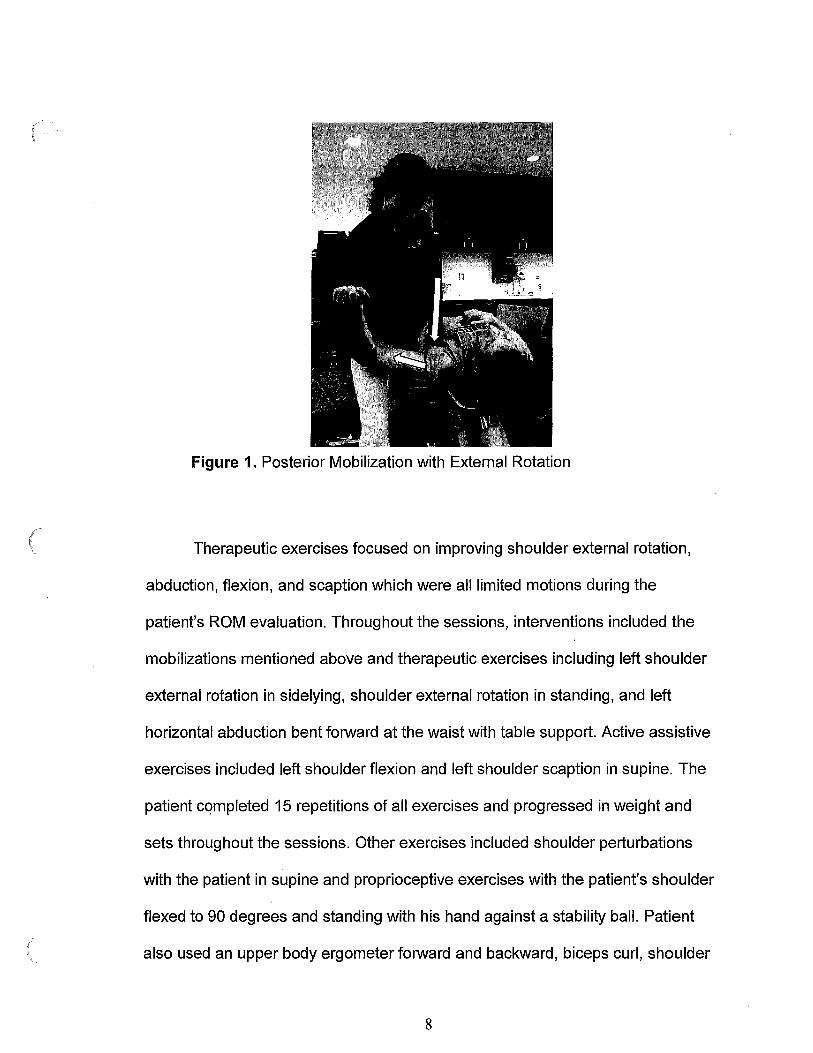
glenohumeral posterior glide with external rotation mobilization, illustrated in
Figure 1, was performed as described by Johnson et al6 which showed a
posterior mobilization with external rotation rather than the traditional anterior
mobilization to increase shoulder external rotation.
7
(
(
Figure 1. Posterior Mobilization with External Rotation
Therapeutic exercises focused on improving shoulder external rotation,
abduction, flexion, and scaption which were all limited motions during the
patient's ROM evaluation. Throughout the sessions, interventions included the
mobilizations mentioned above and therapeutic exercises including left shoulder
external rotation in sidelying, shoulder external rotation in standing, and left
horizontal abduction bent forward at the waist with table support. Active assistive
exercises included left shoulder flexion and left shoulder scaption in supine. The
patient completed 15 repetitions of all exercises and progressed in weight and
sets throughout the sessions. Other exercises included shoulder perturbations
with the patient in supine and proprioceptive exercises with the patient's shoulder
flexed to 90 degrees and standing with his hand against a stability ball. Patient
also used an upper body ergometer forward and backward, biceps curl, shoulder
8

(
scaption in standing, wall push-ups, shoulder flexion using an exercise ball to roll
up the wall, triceps extension, external rotation stretch in supine using a dowel,
external rotation stretch with palmar surface of hand on· wall, shoulder flexion ball
taps with 5-pound ball clockwise and counterclockwise on a grid on the wall, and
shoulder stretching in supine including flexion, scaption, external rotation, and
internal rotation.
During session 5, an examination was completed to assess how the
patient's pain, strength, and ROM improved over the previous 4 sessions of
physical therapy. The patient reported he could reach behind his back and above
his head with greater ease and, over the previous 2 days, his pain was at 0/10 at
the lowest and 3/10 at the highest. Sleeping, movement in general, and reaching
behind his back all continued to increase his pain.
The patient met his short-term goals. Those short-term goals included the
patient will: understand the biomechanical stressors of the shoulder joint in order
to make modifications to activities to reduce further risk of injury, be independent
with a short-term home exercise program, and report pain at worst of 5/10 in
order to perform ADLs.
9

(
(
CHAPTER IV
OUTCOMES
During the patient's last session, a reassessment was done before
discharge. The patient reported he felt he had made 60% improvement since the
initial evaluation and reported he was able to lay on his left arm without
numbness or pain and was sleeping normally. Patient was consistent with his
home exercise program and felt he could independently continue his exercises at
home. He reported his pain at 0/10 at the best and 1-2/1 0 at the worst. The
patient demonstrated increased ROM and strength before discharge (Table 3).
Table 3. Shoulder Range of Motion at Discharge
Right Left
Flexion 155 160
Abduction 170 160, no pain
Internal Rotation 65 (passive) 70 (passive)
Functional T12 T12, pain
Internal Rotation
External Rotation 90 (passive) 76
Functional T4 T3
External Rotation
The patient scored 4.7% disability on the SPADI which demonstrated an
improvement from the initial 17% disability. Patient met all his long-term goals
10

(
including: improve score on SPADI by 13 points to correlate with clinically
significant change, patient will report pain at worst of 2/10 or less, patient will
demonstrate 75 degrees or more of shoulder external rotation without pain in
order to manage his coat independently, and patient will demonstrate shoulder
abduction strength of 5/5 without pain.
11

(
( ;
CHAPTERV
DISCUSSION
According to Page et al5 using a combination of manual therapy and
strengthening increased function and range of motion patients with adhesive
capsulitis. Page et al concluded that patients also demonstrated improved
function, decreased pain, and increased range of motion following m~nual
therapy and exercise when compared with sham ultrasound. The authors also
found that glucocorticoid injections were more effective than manual therapy for
pain management. This evidence supports the interventions implemented in this
case study.
The glenohumeral posterior glide with external rotation mobilization was
performed on this patient rather than the traditional anterior mobilization.
Johnson, et al6 concluded greater improvements in range of motion were seen in
patients that had the posterior glide versus anterior glide done. Further research
should be done with a larger sample size to determine if a posterior mobilization
with external rotation should be the preferred mobilization for patients that need
increased external rotation. For the purpose of this case report, the patient
demonstrated improvements in function and ROM but we were not able to
determine which part of the intervention led to the increased functionality.
During the examination and evaluation of this patient, it was unknown if
there was a specific special test that could have been used to determine if a
patient has adhesive capsulitis. Carbone et aF found 96.4% patients with
adhesive capsulitis had a positive coracoid pain test. Only 11% of patients with
12

(
rotator cuff tears had a positive coracoid pain test, and 2% of patients that were
asymptomatic had a positive coracoid pain test. This test may be beneficial in the
future to determine adhesive capsulitis in patients. The coracoid pain test is one
of few special tests that has high specificity and sensitivity to determine if a
patient has adhesive capsulitis. Further research should be completed to
determine if the coracoid pain test alone could further support a diagnosis of
adhesive capsulitis. Also, further research should be done to determine if the
coracoid pain test can be used in differential diagnosis to separate adhesive
capsulitis from other shoulder pathologies.
This case report is in concordance with the current research that shows
strengthening, mobilizations, a home exercise program and stretching are an
appropriate combination of interventions for individuals with adhesive
capsulitis. 1 .4.5•6 This patient returned to his prior level of function following the
above treatment regimen.
Reflective Practice
Upon review of my clinical practice with this patient, I found that it would
have been beneficial to know about the coracoid pain test to have an easier test
to further determine whether the patient had adhesive capsulitis. In retrospect, I
should have reviewed and changed his home exercise program more often. The
mobilizations were very beneficial to him that we did in physical therapy, so
focusing primarily on the mobilizations during therapy and less time on the
exercises would have been more efficient. The client had time available outside
13

/~
\ of physical therapy to do his exercises. With that in mind, rather than having him
do his full exercises every session, I could have either given him new exercises
to keep him motivated or focused more on manual therapy.
After reflecting on my clinical practice, I evaluated my strengths and
weaknesses by creating a Johari Window.8 The Johari Window examines the
known and unknown factors to the patient and physical therapist. The model
helps to determine outside factors that niay be influential in treatment planning.
One unknown factor and question that I wish I would have asked sooner is if the
patient had any exercise equipment at home. For his first home exercise
program, I gave him theraband to perform shoulder strengthening exercises. In
therapy, we used 1-3 lb. weights to do other strengthening exercises. It wasn't
until the 4th visit that the patient told me he had weights at home. If I would have
asked for that information sooner, I could have given him more exercises to do
and performed other interventions during physical therapy. I also did not know
right away if the patient would consistently perform his home exercises.
However, after the third visit when he demonstrated the exercises without error, I
realized it was not an issue.
Overall, my patient increased in function and range of motion. There are
many factors that influenced his final goal. He was motivated, and hardworking.
He also had a supportive family, functional goals that he wanted to achieve, and
time outside of physical therapy to do his exercises and stretching. All of these
factors and attributes of my patient aided in his recovery.
( \
14

/~
(
APPENDIX
( \

REFERENCES
1. Agarwal S, Raza S, Moiz JA, Anwer S, Alghadir AH. Effects of two
different mobilization techniques on pain, range of motion and functional
disability in patients with adhesive capsulitis: A comparative study. Journal
of Physical Therapy Science. 2016;28(12):3342-
3349. https://jlc.jst.go.jp/DN/JLC/20036036132?from=SUMMON. doi:
1 0.1589/jpts.28.3342.
2. Malik S, Pirotte A. Shoulder. In: Sherman SC. eds. Simon's Emergency
Orthopedics, 7e New York, NY: McGraw-Hill;
2014. http://ezproxylr.med.und.edu:2085/content.aspx?bookid=1132§i
onid=64416458. Accessed May 22, 2018.
3. Chapter 16. The Shoulder. In: Dutton M. eds. Dutton's Orthopaedic
Examination, Evaluation, and Intervention, 3e New York, NY: McGraw-Hill;
2012. http://accessphysiotherapy.mhmedical.com.ezproxy.undmedlibrary.
org/content.aspx?bookid=4 75§ionid=40791212. Accessed December
05, 2017.
4. Rawat P, Eapen C, Seema KP. Effect of rotator cuff strengthening as an
adjunct to standard care in subjects with adhesive capsulitis: A
randomized controlled trial. J Hand Ther. 2017;30(3):241.e8. Accessed
Dec 5, 2017. doi: 10.1016/j.jht.2016.10.007.
( \
16

(
(
5. Page MJ, GreenS, Kramer S, et al. Manual therapy and exercise for
adhesive capsulitis (frozen shoulder). In: The cochrane library. John Wiley
& Sons, Ltd;
14. http://ezproxylr.med.und.edu:2170/doi/1 0.1 002/14651858.CD011275/f
ull. Accessed Mar 12,2018. 10.1002/14651858.CD011275.
6. Johnson AJ, Godges JJ, Zimmerman GJ, Ounanian LL. The effect of
anterior versus posterior glide joint mobilization on external rotation range
of motion in patients with shoulder adhesive capsulitis. J Orthop Sports
Phys Ther. 2007;37(3):88-99. Accessed Dec 5, 2017. doi:
1 0 .2519/jospt.2007 .2307.
7. Carbone S, Gumina S, Vestri AR, Postacchini R. Coracoid pain test: A
new clinical sign of shoulder adhesive capsulitis. International
Orlhopaedics.
8. Shenton AK. Viewing information needs through a johari
window. Reference SeJVices Review. 2007;35(3):487-
496. https://www.emeraldinsight.com/doi/abs/1 0.1108/0090732071077 433
z. Accessed Jun 5, 2018. doi: 10.1108/00907320710774337.
17

A Case Report: Adhesive Capsulitis and Physical Therapy Intervention Haley Brenner SPT, Peggy Mohr PhD, PT
Department of Physical Therapy, University of North Dakota School of Medicine and Health Sciences, Grand Forks, North Dakota 58202-9037
Abstract
Purpose: The purpose of this case report was to describe the physical therapy interventions for a patient with adhesive capsulitis. Case Description: A 68-year-old right-handed male patient who presented with left shoulder pain and limited range of motion (ROM) following a fall 7 months prior. The patient had a past medical history of type l1 diabetes mellitus. The diagnosis of adhesive capsulitis was determined following radiographs, mechanism of injury, past medical history, and physical therapy examination and evaluation.
Plan of Care: The patient was seen for a total of 8 physical therapy sessions over the span of 6 weeks. Interventions included a home exercise program, instruction in heat/ice use, mobilizations, therapeutic exercises, ROM, stretching, and upper body ergometer use. Outcome measures included ROM measurements, pain ratings, strength tests, the Shoulder Pain and Disability Index (SPADI), and Patient Specific Functional Scale (PSFS).
Outcomes: Following 6 weeks of physical therapy intervention and home exercise program, the patient demonstrated increased shoulder ROM, decreased pain, and improved function based on the improved SPADI and PSFS scores.
Discussion: Rationale for treatment was based on textbook information for shoulder interventions and research articles. The treatment was altered based on patient's response.
Conclusion: This case report is in concordance with the current research that shows strengthening, mobilizations, a home exercise program and stretching are an appropriate combinations of interventions for individuals with adhesive capsulitis. This patient returned to his prior level of function following the above treatment regimen.
Introduction
Studies have shown that adhesive capsulitis affects 20% of people with diabetes. Diabetes can alter the collagen formation and delay the healing process following traumatic events or surgery.
The etiology of adhesive capsulitis is not completely understood, but it is most likely caused by a nonspecific chronic inflammatory reaction of tissues in the glenohumeral joint causing synovial thickening. This thickening leads to a decrease in shoulder range of motion.
Individuals with adhesive capsulitis generally progress through 4 stages:
-I. Prefreezing (l-3months)
-2. Freezing (3-9 months)
-3. Frozen (9-14 months)
-4. Thawing (12-14 months)
Case Description
A 68-year-old, right-handed, male patient presented with left shoulder pain and limited range of motion following a fall 7 months prior. The patient had a past medical history of type 11 diabetes mellitus. The diagnosis of adhesive capsulitis was determined following radiographs, mechanism of injury, past medical history, and physical therapy examination and evaluation.
Plan of Care
Patient was seen for a total of 8 visits throughout 6 weeks
Patient was given a home exercise program with the following exercises: shoulder strengthening in flexion, external rotation, and abduction. Stretches included abduction and external rotation.
Exercises during therapy sessions included various shoulder strengthening and stretching activities.
Goniometric measurements were used to determine progress and ROM improvements.
Manual muscle testing was performed to evaluate increases in shoulder strength.
Shoulder mobilizations were performed at each physical therapy session to improve ROM.
The mobilization in Figure 1. was performed as described by Johnson et al which showed a posterior mobilization with external rotation rather than the traditional anterior mobilization increases shoulder external rotation.
Figure 1. Posterior mobilization with external rotation
Outcomes
Patient reached 5/5 strength (Manual Muscle Test Grading System 0-5) in shoulder flexion, abduction, internal rotation, and external rotation at final discharge.
As shown in Table 1., the patient demonstrated an increase of 30 degrees in shoulder abduction and 23 degrees improvement in shoulder external rotation from initial evaluation to final evaluation.
Table 1. Left Shoulder ROM
lnitial evaluation Final evaluation
llnciJide'r nexion Shoulder abduction 135° 165°
Patient improved on SPADI from 17% disability to 4. 7% disability.
Patient reported 60% improvement in functional activities from initial evaluation to discharge.
Patient met all goals prior to discharge.
Patient felt he could continue his HEP independently and was given progressions.
Taxonomy: ICF
ScHooL OF MEDICINE f. &.. HEALTH SciENCES l
Discussion
Research indicated using a combination of manual therapy and strengthening to increase function and range of motion.
According to Page et al, patients with adhesive capsulitis demonstrated improved function, decreased pain, and increased range of motion following manual therapy and exercise when compared with sham ultrasound. The authors also found that glucocorticoid injections are more effective than manual therapy for pain management.
Conclusion
Tiris case report is in concordance with the current research that shows strengthening, mobilizations, a home exercise program and stretching are an appropriate combinations of interventions for individuals with adhesive capsulitis. This patient returned to his prior level of function following the above treatment regimen.
Ackn.owledgements
I would like to thank my clinical instructor, faculty of the University ofNorth Dakota physical therapy department, and my classmates for the continual support and proofreading for the preparation of this case report. I would also like to thank Nathan Mertens for agreeing to be photographed to further demonstrate the mobilizations done during this case report and Jayla Greene for assisting in the photography.
Bibliography
Kelley MJ, McClure PW, Leggin BG. Frozen shoulder: Evidence and a proposed model guiding rehabilitation. The Journal of orthopaedic and sports physical therapy. 2009;39(2):135~148. http://www.ncbi.nlm.nih.gov/pubmed/19194024. doi: 1 0.2519/jospt.2009.2916. Johnson AJ, Godges JJ, Zimmerman GJ, Ounanian LL. The effect of anterior versus posterior glide joint mobilization on external rotation range of motion in patients with shoulder adhesive capsulitls. J Orthop Sports Phys Ther. 2007;37(3):88-99. Accessed Dec 5, 2017. dol: 10.2519/jospt.2007.2307. Rawat P, Eapen C, Seema KP. Effect of rotator cuff strengthening as an adjunct to standard care in subjects with adhesive capsu!itis: A randomized controlled trial. J Hand Ther, 2017;30(3):241.e8, Accessed Dec 5, 2017. doi: 10.1 016/j.jht.2016.1 0.007. Carbone S, Gumina S, Vestri AR, Postacchini R. Coracoid pain test: A new clinical sign of shoulder adhesive capsulitis. International Orthopaedics. 201 0;34(3):385. https://ezproxylr. med, und. edu:2243/pmc/articles/PMC2899298/. Accessed Mar 11,2018. doi: 10.1007/s00264-009..0791-4. a!, Tveita EK , et. Responsiveness of the shoulder pain and disability index in patients with adhesive capsulitis. ~ PubMed -NCBI. https://ezproxylr.med.und.edu:2243/pubrned/19055757. Accessed Mar 11, 2018. Page MJ, Green S, Kramer S, et al. Manual therapy and exercise for adhesive capsulitis (frozen shoulder). In: The cochrane library. John Wiley & Sons, Ltd; 2014, http://ezproxylr.med. und. ed u:2170/doi/1 o. 1 002/14651858. CD011275/futl. Accessed Mar 12, 2018. 10.1002/14651858.CD011275.