a case of squamous cell carcinoma arising from a suprapubic
TRANSCRIPT

CASE REPORT Open Access
A case of squamous cell carcinoma arising from asuprapubic cystostomy tractHiroki Ito1*, Masayuki Arao1, Hanako Ishigaki1, Noboru Ohshima2, Ayako Horita3, Ikuo Saito3 and Kotaro Hirai1
Abstract
Background: Patients with spinal cord injury and a chronic indwelling urinary catheter are known to have anincreased risk of bladder malignancy. However, squamous cell carcinoma (SCC) of the epidermis around asuprapubic cystostomy is relatively rare. Here, we report a case of lower abdominal SCC arising from thesuprapubic cystostomy tract.
Case presentation: A 58-year-old man with a complete spinal cord injury was referred to our hospital with a chiefcomplaint of an abdominal mass. Abdominal enhanced computed tomography (CT) showed a 7-cm masssurrounding the suprapubic cystostomy and bilateral inguinal and para-aortic lymph nodes metastasis.Histopathological examination of percutaneous biopsy specimens was performed. The diagnosis was stage IV(cT4N1M1) epidermal SCC, which was treated with palliative external radiation therapy.
Conclusion: The SCC in this case was thought to arise from mechanical stimulus of the suprapubic cystostomy.Physicians and patients should pay careful attention to any signs of neoplasms with long-term indwellingcatheters, such as skin changes around the suprapubic cystostomy site. This case presentation is only the fourthreport of SCC arising from the suprapubic cystostomy tract in the literature. In cases of unresectable tumors andcontraindications to chemotherapy, palliative radiotherapy may lead to disease remission and symptom relief.
BackgroundPatients with spinal cord injury and a chronic indwellingurinary catheter are known to have an increased risk ofbladder malignancy. This condition has been attributedto chronic inflammation and mechanical stimuli fromthe catheter. Chronic bladder irritation or infection isoften associated with squamous metaplasia [1]. Kaufmanet al. [2] reported that squamous metaplasia is morelikely in patients with indwelling catheters placed formore than 10 years than in those with indwelling cathe-ters for less than 10 years (80% vs 42%). In particular,the clinical significance of keratinizing squamous meta-plasia in the setting of indwelling urinary cathetersremains unclear, but some studies have linked this entityto the development of invasive squamous cell carcinoma(SCC) [3].This can be considered in light of the fact that epider-
mal SCC is the second most common type of skin
cancer and most cases are caused by exposure to thesun’s harmful ultraviolet rays or to mechanical stimuli.We report here a case of SCC that developed in theskin around a suprapubic cystostomy.
Case PresentationA 58-year-old man with a complete spinal cord injurywas referred to our hospital in August 2010 with a chiefcomplaint of a severely inflamed abdominal mass. Thespinal cord injury had resulted in the absence of sensa-tion below the waist and chronic neurogenic bladder.The patient also had a history of an intracranial hemor-rhage, from 4 years prior to his presentation, which hadseverely impaired some higher cognitive functions.For bladder management, the patient had undergone a
percutaneous cystostomy with placement of an indwel-ling catheter, about 35 years before his presentation.This was done due to the patient’s difficulty performingclean, intermittent catheterization on a regular basis.The suprapubic cystostomy catheter was changed oncea month at another urology clinic.
* Correspondence: [email protected] of Urology, National Hospital Organization Sagamihara NationalHospital, Kanagawa, JapanFull list of author information is available at the end of the article
Ito et al. BMC Urology 2011, 11:20http://www.biomedcentral.com/1471-2490/11/20
© 2011 Ito et al; licensee BioMed Central Ltd. This is an Open Access article distributed under the terms of the Creative CommonsAttribution License (http://creativecommons.org/licenses/by/2.0), which permits unrestricted use, distribution, and reproduction inany medium, provided the original work is properly cited.

The physical examination revealed an abdominalmass surrounding a suprapubic cystostomy (Figure 1a).The skin around the mass was erythematous, edema-tous, and a foul-smelling, purulent discharge was pre-sent (Figure 1b). Blood analysis revealed the followingabnormal values: albumin 2.3 g/dl, hemoglobin 8.6 g/dl, elevated white blood cells to 11,200/μl, and C-reac-tive protein 11.89 mg/dl. Urinalysis revealed leukocytes(< 100/HPF) and hematuria (50-99/HPF). Urine cytolo-gic evaluation was class a and atypical squamous cellswere seen on microscopic examination. Enhancedchest and abdominal CT (Figure 2) showed a mass (72mm × 63 mm) surrounding the suprapubic cystostomyand enlarged bilateral inguinal and para-aortic lymphnodes. Chest and anterior mediastinal lesions showedno specific findings. A cystoscopy could not be per-formed because the patient had a lower-extremity con-tracture deformity.After the local inflammation of the abdominal mass
was resolved with intravenous antibiotics, a percuta-neous biopsy was performed under local anesthesia.The histopathological examination of the biopsy speci-mens from the tumor suggested SCC (Figure 3). Thus,
stage IV (cT4N1M1) epidermal SCC was diagnosedand subsequently treated with palliative external radia-tion therapy. A dose of 56 Gy was administered over 5weeks to the pelvic area including the primary tumorand inguinal metastatic lymph nodes. The primarytumor and metastatic lymph nodes responded partiallyto this therapy. The period after radiotherapy wasuneventful. The patient has remained asymptomaticduring the subsequent 6 months.
Figure 1 Abdominal Mass surrounding a suprapubiccystostomy. (a) The mass(50 mm diameter) was noted around thesuprapubic catheter. (b) Closer view of the abdominal masssurrounding a suprapubic cystostomy.
Figure 2 An enhanced abdominal CT showed a mass (72 mm ×63 mm) surrounding suprapubic.
Figure 3 Microscopic findings (hematoxylin and eosin stain):Well differentiated squamous cell carcinoma were found. Frontformation(arrow head) were observed at the border betweencarcinoma cells and normal epithelial cells subcutaneously.
Ito et al. BMC Urology 2011, 11:20http://www.biomedcentral.com/1471-2490/11/20
Page 2 of 4
DiscussionThe most common bladder tumors in patients withspinal cord injury are SCC (33-46.9%), urothelial carci-noma (31.3-55%), and adenocarcinoma (9.4-10%) [4-6].In the literature SCC is more common in patients withindwelling urethral and suprapubic catheters than otherforms of bladder management. The incidence of SCC ofthe bladder in patients with indwelling catheters formore than 10 years is 10% [7]. In a study of 48 patients,the mean time between spinal cord injury and the firstbladder malignancy diagnosis was 22.6 years [8].In the present case, we were unable to perform a
cystoscopy and assess the bladder mucosa for a possibleorigin of the tumor. However, we concluded that theSCC developed not from the bladder but from the epi-dermis around the suprapubic catheter. This conclusionwas based on the absence of gross hematuria during fol-low-up, and a class II urine cytologic evaluation. Inaddition, front formation [9] was observed subcuta-neously at an obvious border between normal epithelialcells and carcinoma cells (Figure 3 arrow). These find-ings indicated the origin of SCC was squamous epithe-lial cells. To our knowledge, the present case is only thefourth report of SCC arising from the suprapubiccystostomy tract in the literatures [7,10,11].We believe that this case of SCC was caused by
chronic exposure of the cystostomy site to the mechani-cal stimuli from the indwelling suprapubic catheter.However, the risk of SCC would not have been elimi-nated by intermittent catheterization, as demonstratedby reported cases bladder malignancies in patients whoperform intermittent catheterization [4,6,8].The only acceptable treatment for deeply invasive but
localized SCC arising from a suprapubic cystostomytract is radical cystectomy and urinary diversion [11]. Inthis case, the patient’s tumor was not localized and hadmetastasized to the inguinal and para-aortic lymphnodes. In such cases, chemotherapy is considered, butwas contraindicated in this patient due to his poor per-formance status. Thus, the patient underwent externalradiation therapy as a palliative treatment. This treat-ment led to partial disease remission and good palliationof symptoms and it would appear that palliative radia-tion therapy (a total of 56 Gy) has a role to play in thepalliation of metastatic SCC, with good relief ofsymptoms.We have presented here a rare case of epidermal
SCC in a patient with a suprapubic cystostomy. Physi-cians and patients should pay close attention to anysuspicious signs associated with such long-termcystostomy sites, including skin changes. In this case,other urologists who were changing the catheter oncea month had noticed the abdominal mass for 6
months before referring the patient to our clinicadmission, but they had considered the cause to behyperplasia due to benign granulation. The slowgrowth of the mass may have made early diagnosisdifficult. This case clearly demonstrates that chronicindwelling catheters may cause malignancy of not onlythe bladder but also the epidermis. Thus, early detec-tion and treatment of SCC arising from a suprapubiccystostomy tract are crucial.
ConclusionsThis case presentation is only the fourth report of SCCarising from the suprapubic cystostomy tract in the lit-erature. In this case of unresectable metastatic SCC, pal-liative external radiation therapy led to partial diseaseremission and good relief of symptoms. It is crucial topay attention to any suspicious signs, including skinchanges around a suprapubic cystostomy, especially inthe presence of a long-term indwelling catheter.
ConsentWritten informed consent was obtained from the patientfor publication of this case report and any accompany-ing images. A copy of the written consent is availablefor review from the Editor-in-Chief of the journal.
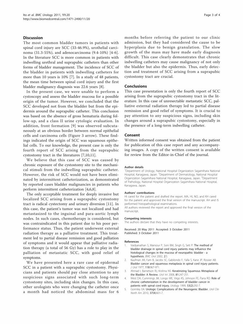
Author details1Department of Urology, National Hospital Organization Sagamihara NationalHospital, Kanagawa, Japan. 2Department of Dermatology, National HospitalOrganization Sagamihara National Hospital, Kanagawa, Japan. 3Departmentof Pathology, National Hospital Organization Sagamihara National Hospital,Kanagawa, Japan.
Authors’ contributionsHI cared for the patient and drafted the report. MA, HI, NO, and KH caredfor the patient and approved the final version of the manuscript. AH and ISperformed histopathological examinations.All authors reviewed the report and approved the final version of themanuscript.
Competing interestsThe authors declare that they have no competing interests.
Received: 20 May 2011 Accepted: 3 October 2011Published: 3 October 2011
References1. Vaidyanathan S, Mansour P, Soni BM, Singh G, Sett P: The method of
bladder drainage in spinal cord injury patients may influence thehistological changes in the mucosa of neuropathic bladder - ahypothesis. BMC Urol 2002, 2:5.
2. Kaufman JM, Fam B, Jacobs SC, Gabilondo F, Yalla S, Kane JP, Rossier AB:Bladder cancer and squamous metaplasia in spinal cord injury patients.J Urol 1977, 118:967-971.
3. Ahmad I, Barnetson RJ, Krishna NS: Keratinizing Squamous Metaplasia ofthe Bladder: A Review. Urol Int 2008, 81:247-251.
4. West DA, Cummings JM, Longo WE, Virgo KS, Johnson FE, Parra RO: Role ofchronic catheterization in the development of bladder cancer inpatients with spinal cord injury. Urology 1999, 53(2):292-7.
5. Gormley EA: Urologic Complications of the Neurogenic Bladder. Urol ClinNorth Am 2010, 37(4):601-7.
Ito et al. BMC Urology 2011, 11:20http://www.biomedcentral.com/1471-2490/11/20
Page 3 of 4

6. Kalisvaart JF, Katsumi HK, Ronningen LD, Hovey RM: Bladder cancer inspinal cord injury patients. Spinal Cord 2010, 48(3):257-61.
7. Stroumbakis N, Choudhury MS, Hernandez-Graulau JM: Squamous cellcarcinoma arising from suprapubic cystostomy site without bladderinvolvement. Urology 1993, 41(6):568-70.
8. Pannek J: Transitional cell carcinoma in patients with spinal cord injury:a high risk malignancy? Urology 2002, 59(2):240-4.
9. Sakuragi N: Diagnosis, Treatment and Management of GynecologicDiseases: Tumor and Kind Tumor. Acta Obstetrica et GynaecologicaJaponica 2009, 61(4):89-101.
10. Stokes S, Wheeler JS Jr, Reyes CV: Squamous cell carcinoma arising from asuprapubic cystostomy tract with extension into the bladder. J Urol 1995,154(3):1132-3.
11. Schaafsma RJ, Delaere KP, Theunissen PH: Squamous cell carcinoma ofsuprapubic cystostomy tract without bladder involvement. Spinal Cord1999, 37(5):373-4.
Pre-publication historyThe pre-publication history for this paper can be accessed here:http://www.biomedcentral.com/1471-2490/11/20/prepub
doi:10.1186/1471-2490-11-20Cite this article as: Ito et al.: A case of squamous cell carcinoma arisingfrom a suprapubic cystostomy tract. BMC Urology 2011 11:20.
Submit your next manuscript to BioMed Centraland take full advantage of:
• Convenient online submission
• Thorough peer review
• No space constraints or color figure charges
• Immediate publication on acceptance
• Inclusion in PubMed, CAS, Scopus and Google Scholar
• Research which is freely available for redistribution
Submit your manuscript at www.biomedcentral.com/submit
Ito et al. BMC Urology 2011, 11:20http://www.biomedcentral.com/1471-2490/11/20
Page 4 of 4