a case of marfan syndrome with massive haemoptysis from
TRANSCRIPT

CASE REPORT Open Access
A case of Marfan syndrome with massivehaemoptysis from collaterals of the lateralthoracic arteryYuki Yabuuchi1* , Hitomi Goto1, Mizu Nonaka1, Hiroaki Tachi1, Tatsuya Akiyama1, Naoki Arai1, Hiroaki Ishikawa1,Kentaro Hyodo1, Kenji Nemoto1, Yukiko Miura1, Isano Hase1, Shingo Usui2, Shuji Oh-ishi1, Kenji Hayashihara1,Takefumi Saito1 and Tatsuya Chonan3
Abstract
Background: Marfan Syndrome (MFS) is a heritable connective tissue disorder with a high degree of clinicalvariability including respiratory diseases; a rare case of MFS with massive intrathoracic bleeding has been reportedrecently.
Case presentation: A 32-year-old man who had been diagnosed with MFS underwent a Bentall operationwith artificial valve replacement for aortic dissection and regurgitation of an aortic valve in 2012. Warfarin wasstarted postoperatively, and the dosage was gradually increased until 2017, when the patient was transportedto our hospital due to sudden massive haemoptysis. Computed tomography (CT) with a maximum intensityprojection (MIP) revealed several giant pulmonary cysts with fluid levels in the apex of the right lung with anabnormal vessel from the right subclavian artery. Transcatheter arterial embolization was performed withangiography and haemostasis was achieved, which suggested that the bleeding vessel was the lateralthoracic artery (LTA) branch. CT taken before the incident indicated thickening of the cystic wall adjacent tothe thorax; therefore, it was postulated that the bleeding originated from fragile anastomoses between theLTA and pulmonary or bronchial arteries. It appears that the vessels exhibited inflammation that beganpostoperatively, which extended to the cysts.
Conclusion: We experienced a case of MFS with massive haemoptysis from the right LTA. We have to beaware of the possibility that massive haemoptysis could be induced in MFS with inflamed pulmonary cysts.
Keywords: Giant pulmonary cysts, Chronic intrathoracic inflammation, Non-bronchial systemic arteries
BackgroundIt has been reported that 16% of patients with MarfanSyndrome (MFS) have pulmonary disease, and that thesepulmonary diseases contribute to 10% of deaths in MFS[1, 2]. Pneumothorax in MFS arising from the labile wallof pulmonary cysts is well-known and one of the severecomplications of MFS [1, 2]. On the other hand, a rarecase of MFS with massive intrathoracic bleeding has re-cently been reported, which caused hemopneumothoraxand haemoptysis [3, 4]. We report here a case of MFS
with massive haemoptysis associated with pulmonarycysts; it was suggested that anastomoses between thebranches of pulmonary and lateral thoracic artery (LTA)were the origin of the bleeding.
Case presentationThe patient was a 32-year-old male ex-smoker. He wasdiagnosed as MFS at the age of 26 based on an aorticdissection (Stanford type A), aortic regurgitation andmarfanoid habitus. A Bentall operation with an artificialvalve replacement for aortic dissection and a regurgita-tion of the aortic valve was performed. Thereafter, hewas required to take warfarin for the artificial heart valveand was followed at a local hospital. His adherence to
© The Author(s). 2020 Open Access This article is distributed under the terms of the Creative Commons Attribution 4.0International License (http://creativecommons.org/licenses/by/4.0/), which permits unrestricted use, distribution, andreproduction in any medium, provided you give appropriate credit to the original author(s) and the source, provide a link tothe Creative Commons license, and indicate if changes were made. The Creative Commons Public Domain Dedication waiver(http://creativecommons.org/publicdomain/zero/1.0/) applies to the data made available in this article, unless otherwise stated.
* Correspondence: [email protected] of Respiratory Medicine, National Hospital Organization, IbarakiHigashi National Hospital, 825, Terunuma, Tokai-mura, Ibaraki, Naka-gun319-1113, JapanFull list of author information is available at the end of the article
Yabuuchi et al. BMC Pulmonary Medicine (2020) 20:4 https://doi.org/10.1186/s12890-019-1033-1
medication was poor, which went unnoticed, resulting inthe dose of warfarin gradually being increased to 6.5 mgper day in 2017, when he was admitted to the emergency
unit of our hospital suffering from a massive haemopty-sis and dyspnoea.The patient had a history of left pneumothorax at the
age of 16; however, the details of this are unknown sincehis mother died in his childhood and the father has beenmissing since then. On presentation, the patient wasfully conscious with a height of 198 cm, body weight of77 kg, blood pressure of 105/56 mmHg, pulse rate of 90bpm, body temperature of 36.9 °C, and a saturation ofpercutaneous oxygen (SpO2) of 94% in room air. Thephysical examination revealed coarse crackles on auscul-tation, especially in the right upper lung areas. He hadcharacteristic features such as a tall height, arachnodac-tyly, dolichocephaly and retrognathia, which are compat-ible with MFS.
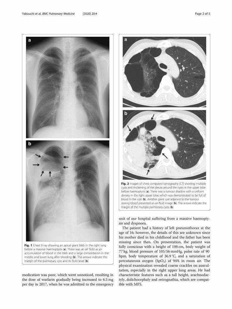
Fig. 1 Chest X-ray showing an apical giant bleb in the right lungbefore a massive haemoptysis (a). There was an air fluid as anaccumulation of blood in the bleb and a large consolidation in themiddle and lower lung after bleeding (b). The arrows indicate themargin of the pulmonary cyst and its fluid level (b)
Fig. 2 Images of chest computed tomography (CT) showing multiplecysts and thickening of the pleura around the cysts in the upper lobebefore haemoptysis (a). There was a tumour shadow with a uniformdensity in the right upper lobe, which was demonstrated to be full ofblood in the cyst (b). Another giant cyst adjacent to the tumourstoring blood presented an air fluid image (b). The arrows indicate themargin of the multiple pulmonary cysts (b)
Yabuuchi et al. BMC Pulmonary Medicine (2020) 20:4 Page 2 of 5

Laboratory data demonstrated anaemia (blood haemo-globin: 10.0 g/dL), an elevated white blood cell count(12,300 /μL), a prothrombin time of 32.5 s (internationalnormalized ratio PT-INR: 5.21), and an active partialthromboplastin time of 61.2 s. Serum tumour markers,antibodies of connective tissue disease and markers ofinfection such as Aspergillus and Mycobacterium wereall negative. Pathological bacteria were not detected insputum culture. A chest X-ray showed a right apical pul-monary cyst with an air-fluid level inside and consolida-tion in the right lower lung field (Fig. 1b). A chestcomputed tomography (CT) scan indicated multiple cys-tic lesions in the upper lobes (Fig. 2b) and a maximumintensity projection (MIP) detected an abnormal vesseloriginating from the right subclavian artery, which ex-tended into the cyst (Fig. 3).
An emergency bronchoscopy revealed continuousbleeding from the apical segmental bronchus of the rightlung (B1) (Fig. 4), which appeared to have been stoppedby topical application of epinephrine and thrombin.Vitamin K and haemostatic medications were infused,which was followed by a transfusion of four units ofblood. After emergent therapy, the patient was trans-ferred to the cardiovascular unit of another hospital. Anangiography was performed and the abnormal vessel wasradiographically diagnosed as the LTA (Fig. 5a, b).Moreover, the bleeding was stopped successfully byembolization of the artery, which confirmed that the ab-normal vessel responsible for bleeding was the LTA. Thepatient’s condition improved, and warfarin was restartedwith strict control of PT-INR. The patient has sincebeen stable with no signs of re-bleeding.
Fig. 3 Coronal slice images of a maximum intensity projection (MIP) showing an abnormal vessel from the right subclavian artery extending into thecyst (Fig. 3). The vessel was later revealed by angiography to be the lateral thoracic artery. The apical giant cyst had pleural adhesions resulting frominflammation due to an invasive operation, which induced the vessel to grow abnormally
Fig. 4 Bronchoscopic images showing bleeding from the right upper lobe bronchus (Fig. 4). A large amount of blood flowed into the righttruncus intermedius and left main bronchus
Yabuuchi et al. BMC Pulmonary Medicine (2020) 20:4 Page 3 of 5

Discussion and conclusionA case of MFS with massive haemoptysis was presented;the bleeding plausibly originated from the extended col-laterals of the right LTA. It is suggested that intratho-racic inflammation induced by surgical interventiontogether with fragile abnormal vessels due to MFSevoked the massive bleeding.There have been two reports that have presented MFS
with massive intrathoracic bleeding [3, 4]. In the presentcase, it is suggested that massive bleeding occurred on
the basis of chronic inflammation of pulmonary cysts. Itappears that pleural adhesions and inflamed cysts arecommon between the past case [3] and the present case,with the difference in the responsible arteries, i.e. thebronchial arteries in the former and the LTA in the lat-ter case. This is the first case of MFS we are aware ofthat showed massive haemoptysis caused by collateralsof the LTA. It might be argued that the role of the col-laterals was not confirmed pathologically. However, thebleeding was stopped by embolization of the LTA, whichstrongly suggests that bleeding occurred from collateralsof the LTA.It has been reported that, in chronic inflammatory dis-
orders, angiogenic growth factors are released from theinflammatory site, which promotes neovascularizationand remodelling of pulmonary vessels, and hence facili-tates anastomoses between the pulmonary and systemiccirculations [5]. It has been reported that these collat-erals, which connect the pulmonary and systemic circu-lations, mostly originate from bronchial arteries [6].However, in the present case, the non-bronchial arterieswere the counterpart of the pulmonary circulation [6].Pulmonary cystic diseases such as bullae and blebs areoften accompanied by chronic inflammation, which facil-itates the proliferation of branches and anastomoses ascompensation for decreased blood supply [7].Arterial blood under increased systemic arterial pres-
sure is prone to extravasation into the surrounding tis-sue, resulting in massive haemoptysis [5, 6]. In our case,the patient had previously had multiple thoracic surger-ies on the numerous existing pulmonary cysts that wereprobably related to MFS. Pleural effusion occurred afterthese operations, which could facilitate chronic pleuralinflammation and pleurodesis, as seen by CT scan (Fig.2a). As a result, a collateral grew abnormally into thecyst and likely anastomosed with the pulmonary artery.Systemic arterial pressure from the lateral thoracic arteryexerted pressure on the pulmonary circulation, which inturn induced the rupture of the vessel under the influ-ence of an overdose of warfarin. Furthermore, the collat-erals may have been prone to rupture due to theconnective tissue disorder responsible for MFS.We cannot completely rule out the possibility of my-
cotic infection, which could have induced an aneurysm.However, serum tests including Aspergillus precipitatingantibody, Aspergillus galactomannan antigen and beta-glucan were all negative and fungi were not detected incultured bloody phlegm obtained from the right upperlobe bronchus by bronchoscopy. Therefore, the origin ofbleeding was unlikely to be related to a mycotic infection.Here, a case of MFS was presented with massive
haemoptysis, which occurred from the collaterals of theLTA. In the post-cardiac operative period, MFS patientsneed to be aware of collateral vessels in the lung.
Fig. 5 Angiography showing abnormal vessels from the lateral thoracicartery (LTA) into the right lung (a). The arrow indicates the abnormalvessels. Embolization of the LTA was performed (b), resultingin haemostasis
Yabuuchi et al. BMC Pulmonary Medicine (2020) 20:4 Page 4 of 5

AbbreviationsB1: Apical segmental bronchus; CT: Computed tomography; LTA: Lateralthoracic artery; MFS: Marfan syndrome; MIP: Maximum intensity projection;PT-INR: Prothrombin time-international normalized ratio; SpO2: Percutaneousoxygen saturation
AcknowledgementsWe would like to acknowledge and thank Yuki Kawahara, MD, MotoakiHiguchi, MD, Yoshiro Chiba, MD, Department of Mito Saiseikai generalHospital, Ibaraki, Japan for conducting the transcatheter arterial embolization.
Authors’ contributionsYY and TS treated the patient. HG, MN, HT, TA, NA, HI, KH1 and KNsupervised the bronchoscopy and subsequent treatment. YY drafted theinitial manuscript and submitted the final manuscript. TC revised themanuscript. YM, IH, SU, SO and KH2 critically reviewed the manuscript. Allauthors read and approved the final manuscript.
FundingNot applicable.
Availability of data and materialsNot applicable.
Ethics approval and consent to participateThis study was approved by the Ibaraki Higassi National Hospital ethicalcommittee.
Consent for publicationWritten informed consent was obtained from the patient and patient’s familyfor publication of this case report and accompanying images. A copy of thewritten consent is available for review by the Editor of this journal.
Competing interestsThe authors declare that they have no competing interests.
Author details1Department of Respiratory Medicine, National Hospital Organization, IbarakiHigashi National Hospital, 825, Terunuma, Tokai-mura, Ibaraki, Naka-gun319-1113, Japan. 2Department of Clinical Research, National HospitalOrganization, Ibaraki Higashi National Hospital, Ibaraki, Japan. 3Department ofMedicine, Nikko Memorial Hospital, Ibaraki, Japan.
Received: 2 July 2019 Accepted: 17 December 2019
References1. Hao W, Fang Y, Shen Y, Wang H, Lin M, Tan L. Marfan syndrome with
pneumothorax: case report and review of literature. J Thorac Dis. 2017;9:1100–3.
2. Wood JR, Bellamy D, Child AH, Citron KM. Pulmonary disease in patientswith Marfan syndrome. Thorax. 1984;39:780–4.
3. Sakai T, Kimura D, Hatanaka R, Yamada Y, Tsushima T, Fukuda I. A caseof Marfan syndrome successfully treated by elective surgery forpersistent air leakage after onset of hemopneumothorax. J Jpn SurgAssoc. 2010;71:369–73.
4. Centeno J, Trivedi P, Iftikhar A, Sanso L. Massive hemopysis in a patient withMarfan’s syndrome. Am J Respir Crit Care Med. 2018;197:A6727.
5. Larici AR, Franchi P, Occhipinti M, Contegiacomo A, Ciello AD, Calandriello L,Storto ML, Marano R, Bonomo L. Diagnosis and management ofhaemoptysis. Diagn Interv Radiol. 2014;20:299–309.
6. Yoon W, Kim JK, Kim YH, Chung TW, Kang HK. Bronchial and non-bronchialsystemic artery embolization for life-threatening haemoptysis: acomprehensive review. Radiographics. 2002;22:1395–409.
7. Bruzzi JF, Jardin MR, Delhaye D, Teisseire A, Khalil C, Remy J. Multi-detectorrow CT of haemoptysis. Radiographics. 2006;26:3–22.
Publisher’s NoteSpringer Nature remains neutral with regard to jurisdictional claims inpublished maps and institutional affiliations.
Yabuuchi et al. BMC Pulmonary Medicine (2020) 20:4 Page 5 of 5