a 50-year-old man with chronic low back pain · spinal pain is pain in either or both regions and...
TRANSCRIPT

CLINICIAN’S CORNERCLINICAL CROSSROADSCONFERENCES WITH PATIENTS AND DOCTORS
A 50-Year-Old ManWith Chronic Low Back PainJames P. Rathmell, MD, Discussant
DR LIBMAN: Mr S is a 50-year-old man with chronic low backpain. In the mid-1970s he developed persistent right leg painand was diagnosed by myelogram as having a herniated disk.L5-S1diskectomywasperformedin1977withmodestimprove-ment in his leg pain. He developed low back pain, which wastreatedwithphysical therapyandnonopioidandopioiddrugs.Over the next decade, his intermittent back and right leg paincaused him to modify his daily activities. It worsened in 1994after he fell out of a bathtub. He was evaluated in a local painunit andreceived local injectionswith limitedbenefit. In1996,Mr S underwent repeat diskectomy, which improved his rightlegpainbutnothisbackpain.Followingsurgery,hehadacrushinjuryofhisrightfoot,whichslowedhisrecovery.Between1996and 2002, he had facet blocks, epidural injections, and physi-cal therapy, all of which were ineffective. Since 2003, he hasbeen followed up at a pain unit. He takes methadone withoxycodone-acetaminophen for breakthrough pain with mod-est relief, but he wants better treatment options.
His back pain is a constant dull ache, sometimes throb-bing and radiating to both legs. It worsens with sitting andstanding. There are no other musculoskeletal or neuro-logic symptoms.
Mr S also has hypertension, gastroesophageal reflux dis-ease, seasonal allergies, depression, anemia, and hyperlip-idemia. In the 1990s, he underwent tonsillectomy and ad-enoidectomy for obstructive sleep apnea.
He takes clonazepam, 1 mg 3 times per day; cyclobenza-prine, 10 mg by mouth 3 times per day; methadone, 40 mgevery morning, 30 mg at noon, and 40 mg at bedtime;naproxen, 500 mg twice per day; and oxycodone-acetaminophen, 5 mg/325 mg (one tablet) 4 times per dayas needed. He also takes atorvastatin, fenofibrate, lisinopril/hydrochlorothiazide, omeprazole, ranitidine, sertraline, andverapamil.
Mr S, a former restaurant worker, is now receiving dis-ability benefits and lives with his longtime female partner.He does not smoke cigarettes or use alcohol but occasion-ally uses marijuana for pain control. There is no other his-tory of drug use.
He is 5 ft, 7 in tall, weighs 209 lb [94 kg], and has a bloodpressure of 108/80 mm Hg and a heart rate of 72/min. Per-
tinent physical findings include mild paravertebral tender-ness in the lumbar region, 4/5 motor strength in his rightlower extremity, and 1�/4� right ankle jerk. He has painon straight leg raising on the right side at 60°.
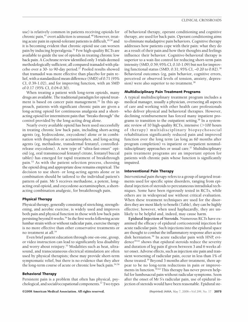
Magnetic resonance imaging of the lumbosacral spine withandwithoutcontrast(performedin2005) isshowninFIGURE1.Degenerative disk changes are noted at multiple lumbar lev-els, which are similar to those seen on a magnetic reso-nance imaging study obtained several years earlier.
MR S: HIS VIEWAbout 30 years ago, I developed pain in my leg, which Ithought was a groin pull. I was athletic at the time. I limped
CME available online at www.jamaarchivescme.comand questions on p 2096.
Mr S, a 50-year-old man, has long-standing low back pain.His pain began more than 20 years earlier with a lumbardisk herniation and has persisted despite diskectomy. Hehas undergone numerous treatments, but he remains dis-abled with ongoing pain. His treatment course is usedto frame the epidemiology and pathophysiology under-lying acute and chronic lumbosacral and radicular pain.The roles of neuropathic pain medications, chronic opi-oid therapy, physical therapy, spinal manipulation, andmultidisciplinary pain treatment programs are re-viewed. The indications for and outcomes associated withinterventional pain treatments, including epidural ste-roid injection, facet blocks and radiofrequency treat-ment for facet-related pain, intradiskal electrothermaltherapy, spinal cord stimulation, and intrathecal drug de-livery, are discussed. Clinicians are given an evidence-based approach to using available treatment options forlow back pain.JAMA. 2008;299(17):2066-2077 www.jama.com
This conference took place at the Anesthesia Grand Rounds of the Beth Israel Dea-coness Medical Center, Boston, Massachusetts, on January 24, 2007.Author Affiliation: Dr Rathmell is Director of the Center for Pain Medicine, De-partment of Anesthesia and Critical Care, Massachusetts General Hospital, andAssociate Professor, Department of Anaesthesia, Harvard Medical School, Bos-ton, Massachusetts.Corresponding Author: James P. Rathmell, MD, Center for Pain Medicine, Mas-sachusetts General Hospital, 15 Parkman St, WACC 333, Boston, MA 02114([email protected]).Clinical Crossroads at Beth Israel Deaconess Medical Center is produced and ed-ited by Tom Delbanco, MD, Howard Libman, MD, Eileen E. Reynolds, MD, AmyN. Ship, MD, and Anjala V. Tess, MD. Risa B. Burns, MD, is series editor.Clinical Crossroads Section Editor: Margaret A. Winker, MD, Deputy Editor.
2066 JAMA, May 7, 2008—Vol 299, No. 17 (Reprinted) ©2008 American Medical Association. All rights reserved.

for a year and a half before somebody suggested I see a backdoctor. That’s how I found out that I had a herniated disk.I was in the hospital the next day for a myelogram, and thefollowing day I had surgery.
I still had leg pain and went for another consult. And thedoctor’s famous words were, “If it didn’t work the first time,we can try it again.” And I said, “No, thank you,” and wenton to adopt a better lifestyle.
I took it upon myself to do a physical therapy regimento develop some way to control the pain. I tried to buildup my legs, back, and stomach muscles. I lived with thepain. Ten years later, the pain got worse, and it started to
go down to the other side of my left leg. In 1994, I fellout of my bathtub, which set off the pain in both legs andmy back.
Time-release medications and long-acting medications,like Oxycontin or a patch, are helpful. But at times the painis so acute that I need a short, quick-acting narcotic—to blockit for an hour or two.
Physical therapy has come far. They know more abouthow the muscles and bones are interacting. If I had themoney, I would have a massage twice a day. Acupuncture,because it looks at your whole body and not just the back,gives you an overall boost of energy.
Figure 1. Most Recent Magnetic Resonance Imaging (MRI) Study of Mr S’s Lumbosacral Spine (April 2005)
L 4
L 5
L 3
L 2
L 1
C L4-5 level
D L5-S1 level
A, Sagittal T2-weighted image near midline demonstrating advanced degenerative disk disease at the L3-4, L4-5, and L5-S1 levels. Note presence of a Schmorl node (black arrowhead). White lines indicate levels of axial images. B, Axial T2-weighted image at the L3-4 level demonstrating degenerative disk changes and a diffuse disk bulge (yellow arrowheads) resulting in mild stenosis of the central spinal canal. C, Axial T2-weighted image at the L4-5 level demonstrating degenerative disk changes and right-sided postoperative changes with indentation of the thecal sac (white arrowhead). D, Axial T2-weighted image at the L5-S1 level demonstrating degenerative disk changes and prior right hemilaminotomy (blue arrowhead). Overall, there were no significant changes in this MRI study compared with an earlier study performed in September 2003.
A Sagittal T2-weighted magnetic resonance image
Axial T2-weighted magnetic resonance images
B L3-4 level
CLINICAL CROSSROADS
©2008 American Medical Association. All rights reserved. (Reprinted) JAMA, May 7, 2008—Vol 299, No. 17 2067

Dealing with doctors at any level, you have to treadvery lightly. If you say, “I need pain medication,” it seemslike a bell goes off. They think, “This person just wantspain medication, nothing else.”
AT THE CROSSROADS:QUESTIONS FOR DR RATHMELLWhat is known about the epidemiology and pathogenesisof chronic low back pain? What about facet and epiduralinjections, physical therapy, acupuncture, and other alter-native care approaches? Would cognitive, behavioral, andpsychological therapies help? In patients for whom sur-gery is not indicated, how should treatment be ap-proached? What is the role of neuropathic pain medica-tions and short- and long-acting opioid medications? Whatabout newer treatments such as spinal cord stimulation andintrathecal drug delivery? What does the future hold in thisfield?
DR RATHMELL: Mr S is a middle-aged man disabled bychronic low back pain. He has had extensive evaluation andtreatment, including spinal surgery and injections to re-duce his pain. However, he is left with significant pain. It isdifficult to recommend what more can be done to help him.However, understanding of low back pain has advanced andhas made treatments available that reduce or eliminate painassociated with specific spine disorders.
Definitions
Low back pain, a nonspecific term, refers to pain centeredover the lumbosacral junction. To be precise in theapproach to diagnosis and treatment, pain primarily overthe axis of the spinal column is differentiated from thatwhich refers primarily to the leg (FIGURE 2). Lumbar spi-nal pain is pain inferior to the tip of the twelfth thoracicspinous process and superior to the tip of the first sacralspinous process.1 Sacral spinal pain is inferior to the firstsacral spinous process and superior to the sacrococcygealjoint.1 Lumbosacral spinal pain is pain in either or bothregions and constitutes “low back pain.” Other patientspresent with sciatica, or pain predominantly localized inthe leg. The proper term is radicular pain because stimu-lation of the nerve roots or the dorsal root ganglion of aspinal nerve evokes the pain.
Pain is a normal physiologic process and serves as asignal of actual or impending tissue injury. Pain from tis-sue injury is usually well localized and associated withsensitivity in the region. Pain signals are carried towardthe central nervous system via the sensory nerves. Thistype of pain is termed nociceptive pain1 or physiologicalpain.2 In contrast, persistent pain following injury to thenervous system, neuropathic pain,1,2 has unique character-istics: spontaneous pain (pain without any stimulus),hyperalgesia (more pain than expected from a painfulstimulus), and allodynia (pain following a nonpainfulstimulus).3
Mr S describes a deep ache in his low back with inter-mittent radiation of pain to his legs; thus, he has both lum-bosacral pain and radicular pain, likely with mixed etiol-ogy (ie, both nociceptive and neuropathic).
Figure 2. Distribution of Lumbosacral and Radicular Pain
S2 S1 L5
L5
L5
L4
L3
L2
L1
S2 S1
L AT E R A LL AT E R A L M E D I A L
S1
L4
L3
L2
L1
S3L4
L3L2L1
S4S5
A N T E R I O R V I E W P O S T E R I O R V I E W
L1
L5
Lumbar spinal pain
Sacral spinal pain
Radicular pain
P O S T E R I O R V I E WA Lumbosacral spinal pain
B Lumbar (L4) radicular pain and lumbar and sacral dermatomes(right leg)
A, “Low back pain” is more precisely termed lumbosacral spinal pain, which en-compasses both lumbar spinal pain (L) and sacral spinal pain (S). Lumbosacralspinal pain is pain in either or both regions and constitutes “low back pain.”B, Radicular pain is caused by stimulation of a spinal nerve and describes pain thatis referred to the lower extremity along the corresponding dermatome.
CLINICAL CROSSROADS
2068 JAMA, May 7, 2008—Vol 299, No. 17 (Reprinted) ©2008 American Medical Association. All rights reserved.

EpidemiologyLow back pain, ranked fifth among the most common prob-lems that lead patients to seek medical attention, ac-counted for nearly 15 million physician visits in a 1990 USsurvey.3 Most episodes of acute low back pain, with or with-out radicular pain, resolve without treatment. Overall, 60%to 70% of those affected recover by 6 weeks and 80% to 90%by 12 weeks.4 However, recovery after 12 weeks is slow anduncertain; fewer than half of patients disabled for longer than6 months return to work. The return-to-work rate for thoseabsent for 2 years is near zero.5 Back pain is the most com-mon reason for limitation of activity in younger adults andis the most frequent cause of absences from work.6 Low backpain is frequently recurrent; most patients experience morethan 1 episode.4 Risk factors for developing chronic low backpain include older age, female sex, low socioeconomic sta-tus and lower education level, higher body mass index, to-bacco use, lower perceived general health status, physicalactivity (eg, bending, lifting, twisting), repetitive tasks, jobdissatisfaction, depression, spinal anatomic variations, andimaging abnormalities.6
Pathophysiology
The basic unit of the spine is the functional spinal unit and com-prises 2 adjacent vertebral bodies with 2 posterior facet joints,an intervertebral disk, and the surrounding ligamentous struc-tures (FIGURE 3). The intervertebral disk absorbs energy anddistributes weight evenly from one spinal segment to thenext while allowing movement of the protective bony ele-ments.7 Lifting, bending, twisting, or whole body vibrationcan damage elements of the spine. With injury and aging,progressive degenerative changes appear in each element ofthe functional spinal unit, along with the onset of charac-teristic symptoms (FIGURE 4). The earliest change in the lum-bar facet joints is synovitis, which progresses to degrada-tion of the articular surfaces, capsular laxity and subluxation,and, finally, enlargement of the articular processes (facet hy-pertrophy). Progressive degeneration also occurs within theintervertebral disks, starting with loss of hydration of thenucleus pulposus, followed by appearance of tears withinthe annulus fibrosis (internal disk disruption).
Lumbosacral pain can arise from the facet joints or theannulus fibrosis.8 With internal disruption of the annu-lus, some of the gelatinous central nucleus pulposus canextend beyond the disk margin as a disk herniation (her-niated nucleus pulposus [HNP]). When HNP extends tothe region adjacent to the spinal nerve, it incites anintense inflammatory reaction.9 Patients with HNP typi-cally present with acute radicular pain. Hypertrophy ofthe facet joints and calcification of the ligamentous struc-tures can reduce the size of the intervertebral foraminaand/or central spinal canal (spinal stenosis), with onset ofradicular pain and/or neurogenic claudication (intermit-tent, unilateral or bilateral pain in the buttock, thigh, orleg that is brought on by walking or prolonged standing).
Initial Evaluation and TreatmentIn first evaluating a patient with low back pain, several fea-tures in the history—“red flag” conditions—require promptinvestigation, including new-onset or worsening back painafter trauma, infection, or previous cancer. Patients with pro-gressive neurologic deficits (typically worsening numb-ness or weakness) or bowel or bladder dysfunction also war-rant immediate radiologic imaging to rule out a compressivelesion.10
If no red flag condition is apparent, diagnosis and treat-ment rely on location and duration of symptoms and de-termining if the pain is acute or chronic and primarily ra-dicular or lumbosacral in nature. There is no clear point intime when acute back pain becomes chronic, but one well-accepted definition is acute low back pain is present for lessthan 3 months, while chronic low back pain is present for alonger time.1
Acute Radicular Pain. Herniated nucleus pulposus typi-cally causes acute radicular pain, with or without radicu-lopathy (radiculopathy would be indicated by numbness,weakness, or loss of deep tendon reflexes referable to a spe-cific spinal nerve). In elderly patients and those with ex-tensive lumbar spondylosis, acute radicular symptoms causedby narrowing of 1 or more intervertebral foramina can oc-cur.11 Initial treatment is symptomatic, and following HNP,symptoms resolve without specific treatment in about 90%of patients.12 Trials comparing advice to stay active vs stayin bed show no difference in pain or functional out-comes.13 For those with persistent pain after HNP, lumbardiskectomy may be indicated. A controlled trial of surgicalvs nonoperative treatment showed significant improve-ment in both groups over 2 years but remained inconclu-sive about the superiority of either approach.14
Chronic Radicular Pain. Persistent leg pain in the dis-tribution of a spinal nerve may occur in patients with diskherniation with or without subsequent surgery. In those withpersistent pain, a search for a reversible cause of spinal nervecompression is warranted. In some individuals, after sur-gery, scarring around the nerve root at the operative site canbe seen on magnetic resonance imaging,15 and electrodiag-nostic studies show a pattern suggesting chronic radicu-lopathy.16 This patient group has characteristics similar tothose with other nerve injuries, and initial managementshould consist of pharmacologic treatment for neuro-pathic pain.17
Acute Lumbosacral Pain. Most patients presenting withacute onset of lumbosacral pain without radicular symptomshave no obvious abnormal findings18 and radiologic imagingis unlikely to be helpful.19 Traumatic sprain of the muscles andligaments of the lumbar spine or the zygapophyseal joints andearly internal disk disruption are significant causes of acutelumbosacral pain. Similar to acute radicular pain, this type ofpain is best managed symptomatically. Advice for patients tostay active results in improved functional status and pain re-duction at 3 to 4 weeks relative to advice to stay in bed.13
CLINICAL CROSSROADS
©2008 American Medical Association. All rights reserved. (Reprinted) JAMA, May 7, 2008—Vol 299, No. 17 2069

Chronic Lumbosacral Pain. There are many causes ofchronic lumbosacral pain, and identification of the ana-tomic cause cannot be made with certainty in up to 90% ofcases.10 The structures most commonly implicated includethe sacroiliac joint, lumbar facets, and lumbar interverte-bral disks.20 In chronic low back pain, the incidence of in-ternal disk disruption has been estimated to be 39% (range,29%-49%); facet joint pain, 15% (range, 10%-20%); and sac-roiliac joint pain, 15% (range, 7%-23%).20 The gold stan-dard for diagnosing sacroiliac and facet joint pain is injec-tion of local anesthetic at the site.21 However, the use of
uncontrolled local anesthetic blocks for diagnostic pur-poses is plagued by placebo response.22 For patients achiev-ing significant short-term pain relief with diagnostic blocks,randomized controlled trials (RCTs) suggest that radiofre-quency treatment can provide pain reduction for 3 to 6months in those with facet-related pain. Pain from degen-erating intervertebral disks is also a source of chronic axialback pain.20 Diagnostic diskography may identify sympto-matic disks prior to management with therapies such as in-tradiskal electrothermal therapy (IDET) or surgical fusion.Overall, the evidence regarding treatment of chronic lum-
Figure 3. Normal Anatomy of the Functional Spinal Unit (L4-5) and Associated Neural Structures
L4
L5
L4
L5
Cauda equina
Dura mater
Intervertebraldisk
Intervertebraldisk (cut)
Spinal nerve
Spinal nerves
Nucleuspulposus
Nucleuspulposus
Posteriorlongitudinal ligament
Anterior longitudinalligament
Posteriorlongitudinalligament
Posteriorlongitudinalligament
Anteriorlongitudinalligament
Annulusfibrosus
Annulusfibrosus
Spinal canal
Ligamentumflavum
Facet jointcapsule
Superior articularprocess of L4
Inferior articularprocess of L5
Facet (zygapophyseal)joint capsule
Ligamentum flavum
Gray ramuscommunicans
Gray ramuscommunicans
S U P E R I O R V I E W
S A G I T TA L S E C T I O N ,
M E D I A L S U R FA C E
S U P E R O L AT E R A L V I E W
P O S T E R I O R V I E W O F FA C E T J O I N T
( C A P S U L E R E M O V E D )
Intervertebralforamen
et jointsule
C A P S U L E R E M O V E(
Superior articularprocess of L5
Superior articularprocess of L4
Inferior articularprocess of L4
Inferior articularprocess of L5
Cauda equinaLigamentum flavum
Anteriorlongitudinalligament
Sinuvertebralnerve (sensoryinnervation ofintervertebraldisk)
Posteriorprimary ramus
Medial branch of posteriorprimary ramus (sensoryinnervation of facet joint)
Nupu
Anfib
,
M E D I A L S U R FA C E
Posterior
All
L4
L5
The basic unit of the spine, the functional spinal unit, is composed of 2 adjacent vertebral bodies with 2 posterior facet joints, an intervertebral disk, and surroundingligamentous structures. See online interactive supplement at http://jama.ama-assn.org/cgi/content/full/299/17/2066/DC1.
CLINICAL CROSSROADS
2070 JAMA, May 7, 2008—Vol 299, No. 17 (Reprinted) ©2008 American Medical Association. All rights reserved.

Figure 4. Progressive Degenerative Changes of the Functional Spinal Unit (L4-5) Associated With Repetitive Mechanical Stress and Aging
Irritation of spinal nerve bynucleus pulposus
Internal disk disruption(lumbosacral pain)
Herniated nucleus pulposus(radicular pain)
Disk degeneration
Nucleuspulposus
Advanced degenerative changes (type of associated pain)
Early degenerative changes (type of associated pain)A
B
Osteophyte
Loss of nucleuspulposus
Enlarged capsulesurrounding facethypertrophy
Facet hypertrophy
S U P E R O L AT E R A L
V I E W
S U P E R I O R V I E WS U P E R I O R V I E W
S U P E R I O R V I E W S A G I T TA L S E C T I O N , M E D I A L S U R FA C E
Thickening andcalcification ofligamentum flavum
Thickening and calcificationof the posteriorlongitudinal ligamentand ligamentum flavum
P O S T E R I O R V I E W O F FA C E T J O I N T
( C A P S U L E R E M O V E D )
Disk bulge
Facet hypertrophy
Facet hypertrophy
Annulus fibrosis
Foraminal stenosis(radicular pain)
Facet hypertrophy(lumbosacral pain)
L4
L5
F h h
Fa
Foraminal stenosis
Schmorl node
Spinal canalSpinal canal
Diskinnervation
Central canal stenosis(neurogenic claudication)
Dura
Lateral recessstenosis
(radicular pain)
Lateral recess stenosis
Patterns of pain associated with specific degenerative changes are shown in red. A, Early degenerative changes of the functional spinal unit include loss of hydration ofthe nucleus pulposus accompanied by mild loss of height of the intervertebral disk. Internal disk disruption (left) begins with radial and/or concentric fissures thatextend from the periphery of the nucleus pulposus into the annulus fibrosus. Extension of these fissures or of material from the nucleus pulposus to the peripheralportion of the annulus fibrosis can produce lumbosacral pain mediated by the sinuvertebral nerve. Extension of material from the nucleous pulposus posterolaterallyoutside the annulus fibrosis (herniated nucleus pulposus, right) can produce an intense inflammatory reaction surrounding the spinal nerve leading to radicular pain.B, Advanced degenerative changes include complete loss of hydration of the nucleus pulposus, marked loss of height of the intervertebral disk, osteophyte formation,and thickening of ligaments. Central canal stenosis results from the combined effects of facet hypertrophy and thickening of the ligamentum flavum and posteriorlongitudinal ligaments. These degenerative changes can produce neurogenic claudication. Progressive degeneration of the disk or facets can produce chronic lumbo-sacral pain. Facet hypertrophy can produce stenosis of the lateral recess of the spinal canal and the intervertebral foramen, which may result in radicular pain.
CLINICAL CROSSROADS
©2008 American Medical Association. All rights reserved. (Reprinted) JAMA, May 7, 2008—Vol 299, No. 17 2071

bosacral pain is conflicting, precluding strong conclusionsfrom being drawn.
Patients with prior lumbar surgery and either recurrentor persistent low back pain, often termed failed back sur-gery syndrome, need mention.23 Knowing the type of sur-gery performed, the indications for and results of the sur-gery, and the time course and characteristics of any changesin the pattern and severity of postoperative pain is essen-tial. Recurrent pain or progressive symptoms signal the needfor further diagnostic evaluation. Mr S’s back pain began asacute radicular pain from a disk herniation, but his pain per-sisted, worsening after diskectomy. Now the pattern and se-verity of his pain are stable, suggesting that further diag-nostic evaluation is of questionable benefit.
Medical Therapies
Numerous pharmacologic agents and minimally invasivetreatments are beneficial in treating specific types of pain.There is no consensus on the order in which these thera-pies should be initiated for persistent low back pain; the gen-eral approach to each therapy is shown in the TABLE.
Neuropathic Pain Medications. Treatments of neuro-pathic pain such as chronic lumbar radicular pain are ex-trapolated from RCTs examining common forms of neuro-
pathic pain: diabetic neuropathy, and postherpeticneuralgia.42,43 Tricyclic antidepressants (eg, nortriptyline, de-sipramine) and newer selective norepinephrine reuptake in-hibitors (eg, venlafaxine, duloxetine) are effective in the treat-ment of neuropathic pain.42,43 Antiepileptic drugs (eg,gabapentin, pregabalin) also treat neuropathic pain.42,43 De-cisions regarding pharmacologic treatment of neuropathicpain may be based on an analysis of the number neededto treat derived from treatment of diabetic neuropathy andpostherpetic neuralgia. Across various neuropathic pain con-ditions, the numbers needed to treat (95% confidence in-terval [CI]) for tricyclic antidepressants ranged from 2.1 (1.8-2.6) to 3.1 (2.2-5.5); for selective norepinephrine reuptakeinhibitors, 5.1 (3.9-7.4); for gabapentin, 3.8 (3.1-5.1); andfor pregabalin, 3.7 (3.2-4.4).43 If Mr S has not already re-ceived a trial of neuropathic pain medications, a trial to re-duce his chronic radicular pain would be worthwhile.
Long-term Opioid Therapy. Long-term opioid therapyin the management of non–cancer-related pain remains con-troversial.44-46 Advocates point to long-term efficacy and im-provement in function in patients with chronic painful con-ditions, including low back pain. Opponents cite difficultiesin prescribing these drugs long-term.26 While aberrant drug-related behavior (eg, losing prescriptions, escalating drug
Table. Rational Sequence for Application of Medical Therapies in Treating Low Back Pain and Level of Evidence Supporting Each Treatmenta
Type of Pain Initial Therapy Therapy for Persistent PainTreatment Costs andInsurance Coverageb
Acute radicularpain
Seven- to 10-day course of an oral analgesic(NSAID or acetaminophen with or withoutopioid analgesic) with a muscle relaxant forthose with superimposed muscle spasm(level 1).24
Two to 6 weeks after onset of acute radicularpain, consider lumbar epidural steroidinjection to speed resolution of radicularsymptoms (level 2).25
Oxycodone-acetaminophen �muscle relaxant � 7-10days: $; variable coverage
Lumbar epidural steroidinjection: $$; covered bymost insurers
Chronic radicularpain
Chronic radicular pain may respond totreatment with chronic opioids, butneuropathic pain is less responsive toopioids than nociceptive pain (level 2).26,27
Consider evaluation for a trial of spinal cordstimulation (level 2).28-33
Medication (see text): $;variable coverage
Spinal cord stimulation: $$$$;covered by most insurers
Acutelumbosacralpain
Seven- to 10-day course of an oral analgesic(NSAID or acetaminophen with or withoutopioid analgesic) with a muscle relaxant forthose with superimposed muscle spasm(level 1).24
Two to 6 weeks after onset of chronic radicularpain, consider referral for physical therapyfor stretching, strengthening, and aerobicexercise in conjunction with patienteducation (level 1).24,34
Physical therapy (2-3�/wk for3-4 wk): $$; single coursecovered by most insurers
Chroniclumbosacralpain
Consider diagnostic medial branch blocks ofthe nerves to the facet joints. If �50% painrelief with the diagnostic blocks, considerradiofrequency treatment (level 2).35,36
Consider a formal multidisciplinary pain programthat incorporates medical management,behavioral therapy, and physical therapy(level 1).37
Consider cognitive-behavioral therapy (level 1).38
If no response to diagnostic facet blocks andMRI evidence of early degenerative diskdisease affecting a single intervertebral disk,consider diagnostic provocative diskography(level 3).39 If diskography is concordant (painis reproduced at anatomically abnormallevel[s] and no pain at an adjacentanatomically normal level), considertreatment with IDET at symptomatic level(s)(level 2).40
Radiofrequency treatment: $$;covered by most insurers
Multidisciplinary pain program:$$$; variable coverage butmany exclude chronic painprograms
Cognitive-behavioral therapy:$$; variable coverage
IDET: $$$; variable coverage
Abbreviations: IDET, intradiskal electrothermal therapy; MRI, magnetic resonance imaging; NSAID, nonsteroidal anti-inflammatory drug.aLevels of evidence are defined by the Oxford Centre for Evidence-Based Medicine41: level 1, high-quality randomized controlled trials (RCTs) or systematic reviews of RCTs; level
2, low-quality RCTs, cohort studies, or systematic reviews of cohort studies; level 3, case-control studies or systematic reviews of case-control studies; level 4, case series; level5, expert opinion.
bRelative costs represent the average US cost for a single course of treatment for 1 patient with low back pain: $, �$500; $$, $500-$2000; $$$, $2000-$10 000; and $$$$,�$10 000.
CLINICAL CROSSROADS
2072 JAMA, May 7, 2008—Vol 299, No. 17 (Reprinted) ©2008 American Medical Association. All rights reserved.
use) is relatively common in patients receiving opioids forchronic pain,47 overt addiction is unusual.48 However, treat-ing acute pain in opioid-tolerant patients is difficult,49,50 andit is becoming evident that chronic opioid use can worsenpain by inducing hyperalgesia.51 Few high-quality RCTs areavailable to guide the use of opioids in treating chronic lowback pain. A Cochrane review identified only 3 trials deemedmethodologically sufficient; all compared tramadol with pla-cebo over a 30- to 90-day period.27 Pooled results showedthat tramadol was more effective than placebo for pain re-lief, with a standardized mean difference (SMD) of 0.71 (95%CI, 0.39-1.02), and for improving function, with an SMDof 0.17 (95% CI, 0.04-0.30).
When treating a patient with long-term opioids, manydrugs are available. The traditional paradigm for opioid treat-ment is based on cancer pain management.52 In this ap-proach, patients with significant chronic pain are given along-acting opioid for continuous analgesia and a short-acting opioid for intermittent pain that “breaks through” thecontrol provided by the long-acting drug alone.
Nearly every available opioid has been used successfullyin treating chronic low back pain, including short-actingagents (eg, hydrocodone, oxycodone) alone or in combi-nation with ibuprofen or acetaminophen, and long-actingagents (eg, methadone, transdermal fentanyl, controlled-release oxycodone). A new type of “ultra-fast-onset” opi-oid (eg, oral transmucosal fentanyl citrate, fentanyl buccaltablet) has emerged for rapid treatment of breakthroughpain.53 As with the patient selection process, choosingthe opioid drug and appropriate dose remains empirical. Thedecision to use short- or long-acting agents alone or incombination should be tailored to the individual patient’spattern of pain. Mr S receives methadone, a potent, long-acting oral opioid, and oxycodone-acetaminophen, a short-acting combination analgesic, for breakthrough pain.
Physical Therapy
Physical therapy, generally consisting of stretching, strength-ening, and aerobic exercise, is widely used and improvesboth pain and physical function in those with low back painpersisting beyond 6 weeks.34 In the first weeks following acutelumbar strain with or without radicular pain, exercise therapyis no more effective than other conservative treatments orno treatment at all.54
Even brief patient education through one-on-one, group,or video instruction can lead to significantly less disabilityand worry about reinjury.55 Modalities such as heat, ultra-sound, and transcutaneous electrical stimulation are oftenused by physical therapists; these may provide short-termsymptomatic relief, but there is no evidence that they alterthe long-term course of acute or chronic low back pain.54,56
Behavioral Therapy
Persistent pain is a problem that often has physical, psy-chological, and social/occupational components.57 Two types
of behavioral therapy, operant conditioning and cognitivetherapy, are used for back pain. Operant conditioning aimsto eliminate maladaptive pain behaviors. Cognitive therapyaddresses how patients cope with their pain: what they doas a result of their pain and how their thoughts and feelingsinfluence their behavior. Cognitive-behavioral therapy issuperior to a wait-list control for reducing short-term painintensity (SMD, 0.59; 95% CI, 0.10-1.09) but not for improv-ing functional status (SMD, 0.31; 95% CI, −0.20 to 0.82).38
Behavioral outcomes (eg, pain behavior, cognitive errors,perceived or observed levels of tension, anxiety, depres-sion) were also superior to no treatment.38
Multidisciplinary Pain Treatment Programs
A typical multidisciplinary treatment program includes amedical manager, usually a physician, overseeing all aspectsof care and working with other health care professionalswho deliver physical and behavioral therapies. However,declining reimbursement has forced many inpatient pro-grams to transition to the outpatient setting.58 In a system-atic review of 10 high-quality RCTs, intensive (�100 hoursof therapy) mul t id i sc ip l inary biopsychosoc ia lrehabilitation significantly reduced pain and improvedfunction over the long term (as long as 60 months afterprogram completion) vs inpatient or outpatient nonmul-tidisciplinary approaches or usual care.37 Multidisciplinarypain treatment programs are an important option forpatients with chronic pain whose function is significantlyimpaired.
Interventional Pain Therapy
Interventional pain therapy refers to a group of targeted treat-ments used for specific spine disorders, ranging from epi-dural injection of steroids to percutaneous intradiskal tech-niques. Some have been rigorously tested in RCTs, whileothers are in widespread use without critical evaluation.When these treatment techniques are used for the disor-ders they are most likely to benefit (Table), they can be highlyeffective; however, when used haphazardly, they are un-likely to be helpful and, indeed, may cause harm.
Epidural Injection of Steroids. Numerous RCTs have ex-amined the efficacy of epidural corticosteroid injection foracute radicular pain. Such injections into the epidural spaceare thought to combat the inflammatory response after acutedisk herniation.59 In acute radicular pain with HNP, evi-dence59-61 shows that epidural steroids reduce the severityand duration of leg pain if given between 3 and 6 weeks af-ter onset. Adverse effects, such as injection site pain and tran-sient worsening of radicular pain, occur in less than 1% ofthose treated.59 Beyond 3 months after treatment, there ap-pear to be no long-term reductions in pain or improve-ments in function.59,62 This therapy has never proven help-ful for lumbosacral pain without radicular symptoms. Soonafter the onset of Mr S’s radicular pain, use of epidural in-jection of steroids would have been reasonable. Epidural ste-
CLINICAL CROSSROADS
©2008 American Medical Association. All rights reserved. (Reprinted) JAMA, May 7, 2008—Vol 299, No. 17 2073

roid injections should be considered for future exacerba-tions of his radicular pain.
Facet Blocks and Radiofrequency Treatment. Pain fromthe lumbar facet joints affects up to 15% of patients withchronic low back pain.63 These patients typically have re-ferred pain, with maximal pain located directly over the facetjoints and pain on palpation over the facets; radiographicfindings are variable, but some degree of facet arthropathyis typically present.64 Case series and studies lacking ad-equate comparator groups or blinding suggest that the intra-articular injection of anesthetics and corticosteroids leadsto intermediate-term pain relief (1-3 months) in patients withan active inflammatory process.63 Radiofrequency denerva-tion delivers energy through an insulated, small-diameterneedle positioned adjacent to the sensory nerve to the facetjoint, creating a small area of tissue coagulation that dener-vates the facet joint. Two systematic reviews concluded thatthere is moderate evidence that radiofrequency denerva-tion provides better pain relief than sham intervention.35,36
The quality of the 6 available RCTs was deemed adequate,but they were conducted in a technically heterogeneous man-ner (eg, varying inclusion criteria, differing treatment pro-tocols); thus, their findings could not be easily combined.Approximately half of patients treated reported at least 50%pain reduction. Pain typically returned 6 and 12 months af-ter treatment, and denervation could be repeated.65 Ad-verse events were uncommon; in 1% of treated patients, painat the treatment site lasted 2 weeks or less.66 Mr S has re-ceived facet injections but had no pain relief; this lack ofresponse and the reproduction of his pain during subse-quent diskography suggests that his ongoing lumbosacralpain arises from his intervertebral disks.
Intradiskal Electrothermal Therapy. Intervertebral disksare estimated to be involved in 39% of chronic low back paincases.20 Provocative diskography is a controversial diagnos-tic test in which needles are placed into the intervertebraldisks; radiographic contrast is then introduced to try to re-produce the patient’s typical pain and determine the offend-ing disk. This test has been used to select patients for sur-gical fusion, but its ability to predict outcome isquestionable.39 Mr S had diskography some years ago, pre-sumably in preparation for lumbar fusion surgery. How-ever, numerous disks produced his pain, and no specific lev-els could be identified to target the fusion.
Diskography has also been used to select patients for IDET,which is used to treat chronic diskogenic lumbosacral pain.A steerable thermal resistance wire is placed along the pos-terior anulus fibrosus and thermal energy is applied to de-stroy penetrating nociceptive fibers and to change the cross-linking of glycosaminoglycans, thereby stiffening theintervertebral disk.67 Clinical study results are mixed; onehigh-quality RCT showed that 40% of patients achievedgreater than 50% pain relief while 50% of patients had noappreciable benefit (number needed to treat to achieve 75%relief of pain=5),68 while a second high-quality RCT showed
no effect.69 A meta-analysis of 17 studies showed a 50% re-duction in pain and improvement in sitting and standingtolerance in 40% to 50% of patients receiving IDET at a singlelevel with concordant diskography and well-preserved diskheight.40 For Mr S, the numerous disks involved and his ad-vanced disk degeneration suggest that IDET is unlikely tobenefit him.
Spinal Cord Stimulation. Based on the theory that non-noxious sensory input interferes with the perception of pain,direct activation of the dorsal columns of the spinal cord isused to treat chronic back pain. A pacemaker-like im-planted pulse generator is connected to a small electrodearray positioned within the dorsal epidural space. This sys-tem is implanted in a simple, brief surgical procedure.28 Ob-servational trials29-31 and a recent RCT28 support that spinalcord stimulation is effective in patients with chronic radicu-lar pain following prior lumbar surgery. Trials are of low qual-ity but suggest that half of patients report at least 50% on-going pain relief 5 years after device implantation.31 Use ofspinal cord stimulation for chronic lumbosacral pain has beenless satisfactory, but results have improved, with new dual-lead systems and electrode arrays providing a broad area ofstimulation.28,29 Spinal cord stimulation is less expensive andmore effective than reoperation in the management of per-sistent postoperative radicular pain; at a mean 3.1-year follow-up, 13 of 21 patients (62%) crossed over from reoperationvs 5 of 19 patients (26%) who crossed over from spinal cordstimulation to reoperation (P� .025).33 In the most recentRCT, 32% of patients had at least 1 complication, with leaddisplacement requiring reoperation in 10% and infection orwound breakdown in 8%.28 Observational studies29-31 and2 recent high-quality RCTs28,33 suggest that spinal cord stimu-lation has the most favorable outcomes in unilateral radicu-lar pain. Based on these data,28 Mr S is a candidate for spi-nal cord stimulation for his persistent lumbosacral pain.
Intrathecal Drug Delivery. Direct application of mor-phine to the spinal cord produces spinally mediated anal-gesia, and small, programmable pumps are now availablethat can be implanted in the abdominal wall to deliver pre-cise, continuous drug infusions to patients with chronic non–cancer-related pain.70 Intrathecal drug delivery is usually re-served for patients with severe pain that does not respondto conservative management. In cancer-related pain, painrelief was similar and opioid-related adverse effects were fewerthan with orally administered opioids.71 While morphine iscurrently the only opioid that is approved for intrathecal useby the US Food and Drug Administration, other drugs arealso used, singly and in combination. An RCT of intrathe-cally delivered ziconotide for severe chronic pain demon-strated a mean pain reduction of 50% vs a 20% reductionin those receiving placebo (P� .001). However, adverse ef-fects were common, with 97% of treated patients experi-encing 1 or more adverse effect vs 73% of those receivingplacebo, with the most common being central nervous sys-tem adverse effects.72 Intrathecal drug delivery in noncan-
CLINICAL CROSSROADS
2074 JAMA, May 7, 2008—Vol 299, No. 17 (Reprinted) ©2008 American Medical Association. All rights reserved.

cer pain has not been subject to controlled trials and re-mains controversial, but observational studies suggest itrelieves pain in some patients whose chronic low back painfails to respond to more conservative management.70,73
Other Therapeutic Approaches
Acupuncture. A meta-analysis of 33 RCTs comparing acu-puncture with sham interventions in treating back pain foundthat for short-term pain relief, acupuncture was signifi-cantly more effective than sham treatment (SMD, 0.54; 95%CI, 0.35-0.73 in 7 trials). Acupuncture was also more ef-fective than no treatment (SMD, 0.69; 95% CI, 0.40-0.98in 8 trials). For acute low back pain, data were “sparse andinconclusive.”74 The effectiveness of acupuncture in com-parison with other treatments has not been studied, nor hasthe frequency and duration of acupuncture required to pro-duce durable pain reduction.75
Spinal Manipulation. Generally, spinal manipulation in-volves the hands being applied to the patient to deliver aforceful load to specific body tissues to reduce pain and/orimprove range of movement.76 Mechanisms include in-crease of joint movement, changes in joint kinematics, in-crease of pain threshold and muscle strength, and releaseof endogenous analgesic peptides.77 Available data are con-flicting and the methodologic quality is low, with lack ofblinding and/or meaningful comparator groups. However,spinal manipulation generally results in more rapid recov-ery if applied within 3 weeks of onset of acute low back pain.78
The outcomes for treatment of chronic low back pain areless clear, and the conclusions of systematic reviews are in-consistent. One systematic review concluded that spinal ma-nipulation for chronic low back pain provides benefits simi-lar to a prescription nonsteroidal anti-inflammatory drug;it is more effective in the short-term than placebo and gen-eral practitioner care and in the long term vs physicaltherapy.78 However, in a review of 16 systematic reviews, spi-nal manipulation was ineffective in treating any condition,including low back pain.76 Nonetheless, the authors under-scored the low quality of the available evidence and the needfor additional clinical trials.76
Experimental Treatment Options
Tumor necrosis factor � is an important mediator of sci-atica in animal models of disk herniation,79 but while earlyuncontrolled trials of the anti–tumor necrosis factor � agentsinfliximab80 and etanercept81 showed faster pain reduc-tion, a subsequent RCT showed no benefit of infliximab overplacebo.82 Trials examining periradicular application are cur-rently under way.83
The progressive loss of proteoglycan within the nucleuspulposus, a characteristic finding in degenerative disk dis-ease, has suggested a possible role for growth factors. Al-though some growth factors (human transforming growth fac-tor �1, tissue inhibitor of matrix metalloproteinase 1) may up-regulate the anabolism or inhibit the catabolism of
proteoglycans within isolated intervertebral disk cells,84,85 theirshort eliminationhalf-livesprecludedirectdelivery.Adenoviral-mediated delivery of growth factors increases proteoglycan syn-thesis.86,87 Intradiskal injection of the genes (in rabbits) thatencode growth factors could be used to regenerate or slow de-generation of disks. Clinical trials have not yet begun.
RECOMMENDATIONS FOR MR SMr S’s chronic low back pain is associated with degenera-tive disk disease; diskography points to several lumbar in-tervertebral disks. He underwent treatments for facet-related pain with little or no success. He continues to takechronic opioid therapy and reports reasonable pain reliefwith moderate doses of methadone (a long-acting opioid)in combination with oxycodone-acetaminophen, a short-acting combination analgesic.
Additional treatment depends entirely on Mr S’s lifestyleand goals; there is no clear path to certain pain reductionor improvement in his functional status. If his overall levelof function is poor or declining, enrollment in a multidis-ciplinary program offers the best hope of long-term im-provement; weight reduction may also improve his level offunction and his back pain. Given the duration of his chronicpain, he is unlikely to have dramatically improved analge-sia or functional status. Treatment with spinal cord stimu-lation or intrathecal drug delivery are reasonable ap-proaches, with stronger evidence supporting the use of spinalcord stimulation, but selecting patients for these therapiesis difficult. It is essential that Mr S understand that he shouldnot ignore exacerbations of pain; changing patterns oftensignal the need for further diagnostic evaluation. These canoften be successfully managed, even in the context of long-standing pain.
QUESTIONS AND DISCUSSIONQUESTION: Would a personal trainer be of benefit for Mr S?
DR RATHMELL: Why not use a personal trainer? Well, who’sgoing to pay for a personal trainer? Mr S mentions the ben-efits he got from massage. He says, “I used to get massages,but I couldn’t afford it.” So, even when treatments prove use-ful, who will pay for it in our health care reimbursementsystem? Evidence from trials and meta-analyses shows theefficacy of multidisciplinary pain treatment programs.37 Butthese programs are becoming less available because of theexpense and the difficulty in gathering all of the needed prac-titioners in one place to coordinate their patients’ care.
QUESTION: How are we training medical students or resi-dents to be more empathetic to patients with chronic pain?One of Mr S’s biggest complaints about physicians is thatthe first thing we think is that this man wants drugs.
DR RATHMELL: When adopting a reasonable approach tochronic opioid therapy for chronic noncancer pain, thereare no easy answers. It is hard to decide who should andshould not receive opioids; there is no objective measureof pain. I think that young physicians see such widely vary-
CLINICAL CROSSROADS
©2008 American Medical Association. All rights reserved. (Reprinted) JAMA, May 7, 2008—Vol 299, No. 17 2075

ing approaches during their education that they do not knowwhat is right.
As pain specialists, what we do now is rely more on pri-mary care physicians to help us make decisions about theuse of chronic opioid therapy. Because of the long-term re-lationships they develop with their patients, primary carephysicians are best equipped to observe their patients’ be-haviors over time.
To train young physicians to be empathic, I think the onlyapproach is to show them how to discuss their own phi-losophy—why they do what they do—with each patient theycare for.
QUESTION: What are your thoughts on epidural steroidsas a maintenance therapy for patients—getting them everycouple of months?
DR RATHMELL: Epidural steroids speed resolution of painin patients with acute radicular pain—sciatica—associatedwith lumbar intervertebral disk herniations.59 They do nototherwise appear to change the long-term outcome of thisgroup of patients.
Epidural steroids have never shown benefit in treatingchronic lumbosacral pain or chronic, stable radicular pain.In patients with intermittent, acute exacerbations of radicu-lar pain, epidural steroid injection is reasonable, for in-stance, in patients with chronic back pain who present withnew radicular pain, and in spinal stenosis patients, particu-larly those with lateral recess stenosis and periodic exacer-bation of radicular pain. Whenever radicular symptoms flair,one suspects inflammation of 1 or more spinal nerves. Un-der these circumstances, the use of repeated epidural ste-roid injections makes sense. I would use this treatment onlyafter the radicular symptoms recurred and had persisted forat least 2 weeks, because most improve without treatment.
QUESTION: Lifestyle changes remain a big issue; peopleneed to take some ownership of their own disease process.When they receive opioids in the pain clinic, aren’t we justconditioning them to return, get an injection, and get theirmedication without taking responsibility for their illness?
DR RATHMELL: Mr S also could do much to help himself.Counseling him on how to improve his own overall healthwill not worsen his back pain. The pain may not improve,but it’s unlikely to get worse, and he may find himself moreinvolved in life. The best programs address all of a patient’smedical conditions. Mr S’s cholesterol is high; an exerciseprogram, dietary changes, and medication might be useful.Exercise programs don’t reduce chronic low back pain, butthey help improve function. Mr S also has fear avoidancebehavior; he fears going out with friends because activityincreases his pain. Behavioral management strategies are ahelpful part of many exercise programs.
Financial Disclosures: None reported.Funding/Support: This Clinical Crossroads was made possible in part by a grantfrom the Florence and Richard Koplow Charitable Foundation.Role of the Sponsor: The funding organization did not participate in the collec-tion, analysis, and interpretation of the data or in the preparation, review, or ap-proval of the manuscript.
Additional Information: An online interactive supplement is available athttp://jama.ama-assn.org/cgi/conent/full/299/17/2066/DC1.Additional Contributions: We thank the patient for sharing his story and for pro-viding permission to publish it.
REFERENCES
1. Merskey H, Bogduk N, eds. Classification of Chronic Pain: Descriptions of ChronicPain Syndromes and Definitions of Pain Terms. 3rd ed. Seattle, WA: IASP Press;1994.2. Backonja MM. Defining neuropathic pain. Anesth Analg. 2003;97(3):785-790.3. Hart LG, Deyo RA, Cherkin DC. Physician office visits for low back pain: fre-quency, clinical evaluation, and treatment pattern from a US national survey. Spine.1995;20(1):11-19.4. Andersson GB. Epidemiological features of chronic low-back pain. Lancet. 1999;354(9178):581-585.5. Scientific approach to the assessment and management of activity-relatedspinal disorders: a monograph for clinicians: report of the Quebec Task Force onSpinal Disorders. Spine. 1987;12(7)(suppl):S1-S59.6. Rubin DI. Epidemiology and risk factors for spine pain. Neurol Clin. 2007;25(2):353-371.7. Pope MH, DeVocht JW. The clinical relevance of biomechanics. Neurol Clin.1999;17(1):7-41.8. Schwarzer AC, Aprill CN, Derby R, Fortin J, Kline G, Bogduk N. The prevalenceand clinical features of internal disk disruption in patients with chronic low backpain. Spine. 1995;20(17):1878-1883.9. Olmarker K. Radicular pain—recent pathophysiologic concepts and therapeu-tic implications. Schmerz. 2001;15(6):425-429.10. Koes BW, van Tulder MW, Thomas S. Diagnosis and treatment of low backpain. BMJ. 2006;332(7555):1430-1434.11. Chad DA. Lumbar spinal stenosis. Neurol Clin. 2007;25(2):407-418.12. Saal JA, Saal JS. Nonoperative treatment of herniated lumbar intervertebraldisk with radiculopathy. Spine. 1989;14(4):431-437.13. Hagen KB, Jamtvedt G, Hilde G, Winnem MF. The updated Cochrane reviewof bed rest for low back pain and sciatica. Spine. 2005;30(5):542-546.14. Weinstein JN, Lurie JD, Tosteson TD, et al. Surgical vs nonoperative treat-ment for lumbar disk herniation: the Spine Patient Outcomes Research Trial (SPORT)observational cohort. JAMA. 2006;296(20):2451-2459.15. BenDebba M, Augustus van Alphen H, Long DM. Association between peri-dural scar and activity-related pain after lumbar diskectomy. Neurol Res. 1999;21(suppl 1):S37-S42.16. Wilbourn AJ, Aminoff MJ. The electrodiagnostic examination in patients withradiculopathies. Muscle Nerve. 1998;21(12):1612-1631.17. Chen H, Lamer TJ, Rho RH, et al. Contemporary management of neuro-pathic pain for the primary care physician. Mayo Clin Proc. 2004;79(12):1533-1545.18. Deyo RA, Weinstein JN. Low back pain. N Engl J Med. 2001;344(5):363-370.19. Jarvik JG, Deyo RA. Diagnostic evaluation of low back pain with emphasis onimaging. Ann Intern Med. 2002;137(7):586-597.20. Bogduk N, McGuirk B. Causes and sources of chronic low back pain. In: BogdukN, McGuirk B, eds. Medical Management of Acute and Chronic Low Back Pain:An Evidence-Based Approach. Amsterdam, the Netherlands: Elsevier Science BV;2002:115-126. Pain Research and Clinical Management Vol 13.21. Schwarzer AC, Aprill CN, Bogduk N. The sacroiliac joint in chronic low backpain. Spine. 1995;20(1):31-37.22. Bogduk N. Diagnostic blocks: a truth serum for malingering. Clin J Pain. 2004;20(6):409-414.23. Onesti ST. Failed back syndrome. Neurologist. 2004;10(5):259-264.24. Chou R, Qaseem A, Snow V, et al. Diagnosis and treatment of chronic lowback pain: a joint clinical practice guideline from the American College of Physi-cians and the American Pain Society. Ann Intern Med. 2007;147(7):478-491.25. Armon C, Argoff CE, Samuels J, Backonja MM; Therapeutics and TechnologyAssessment Subcommittee of the American Academy of Neurology. Assessment:use of epidural steroid injections to treat radicular lumbosacral pain. Neurology.2007;68(10):723-729.26. Ballantyne JC, Mao J. Opioid therapy for chronic pain. N Engl J Med. 2003;349(20):1943-1953.27. Deshpande A, Furlan A, Mailis-Gagnon A, Atlas S, Turk D. Opioids for chroniclow-back pain. Cochrane Database Syst Rev. 2007;3(3):CD004959.28. Kumar K, Taylor RS, Jacques L, et al. Spinal cord stimulation versus conven-tional medical management for neuropathic pain: a multicentre randomised con-trolled trial in patients with failed back surgery syndrome. Pain. 2007;132(1-2):179-188.29. Taylor RS, Van Buyten JP, Buchser E. Spinal cord stimulation for chronic backand leg pain and failed back surgery syndrome: a systematic review and analysisof prognostic factors. Spine. 2005;30(1):152-160.
CLINICAL CROSSROADS
2076 JAMA, May 7, 2008—Vol 299, No. 17 (Reprinted) ©2008 American Medical Association. All rights reserved.

30. Mailis-Gagnon A, Furlan AD, Sandoval JA, Taylor R. Spinal cord stimulationfor chronic pain. Cochrane Database Syst Rev. 2004;(3):CD003783.31. Turner JA, Loeser JD, Deyo RA, Sanders SB. Spinal cord stimulation for pa-tients with failed back surgery syndrome or complex regional pain syndrome: asystematic review of effectiveness and complications. Pain. 2004;108(1-2):137-147.32. North RB, Kidd DH, Olin J, et al. Spinal cord stimulation for axial low backpain: a prospective, controlled trial comparing dual with single percutaneouselectrodes. Spine. 2005;30(12):1412-1418.33. North RB, Kidd DH, Farrokhi F, Piantadosi SA. Spinal cord stimulation versusrepeated lumbosacral spine surgery for chronic pain: a randomized, controlled trial.Neurosurgery. 2005;56(1):98-106.34. Foye PM, Sullivan WJ, Sable AW, et al. Industrial medicine and acute mus-culoskeletal rehabilitation, 3: work-related musculoskeletal conditions: the role forphysical therapy, occupational therapy, bracing, and modalities. Arch Phys MedRehabil. 2007;88(3)(suppl 1):S14-S17.35. Geurts JW, van Wijk RM, Stolker RJ, Groen GJ. Efficacy of radiofrequency pro-cedures for the treatment of spinal pain: a systematic review of randomized clini-cal trials. Reg Anesth Pain Med. 2001;26(5):394-400.36. Slipman CW, Bhat AL, Gilchrist RV, et al. A critical review of the evidence forthe use of zygapophysial injections and radiofrequency denervation in the treat-ment of low back pain. Spine J. 2003;3(4):310-316.37. Guzman J, Esmail R, Karjalainen K, et al. Multidisciplinary rehabilitation forchronic low back pain: systematic review. BMJ. 2001;322(7301):1511-1516.38. Ostelo R, van Tulder MW, Vlaeyen JW, et al. Behavioural treatment for chroniclow back pain. Cochrane Database Syst Rev. 2005;(1):CD002014.39. Carragee EJ, Lincoln T, Parmar VS, Alamin T. A gold standard evaluation ofthe “diskogenic pain” diagnosis as determined by provocative diskography. Spine.2006;31(18):2115-2123.40. Appleby D, Andersson G, Totta M. Meta-analysis of the efficacy and safetyof intradiskal electrothermal therapy (IDET). Pain Med. 2006;7(4):308-316.41. Oxford Centre for Evidence-Based Medicine. Levels of evidence. http://www.cebm.net/index.aspx?o=1025. Accessed December 1, 2007.42. Dworkin RH, O’Connor AB, Backonja M, et al. Pharmacologic managementof neuropathic pain: evidence-based recommendations. Pain. 2007;132(3):237-251.43. Finnerup NB, Otto M, McQuay HJ, Jensen TS, Sindrup SH. Algorithm for neu-ropathic pain treatment: an evidence based proposal. Pain. 2005;118(3):289-305.44. Furlan AD, Sandoval JA, Mailis-Gagnon A, Tunks E. Opioids for chronic non-cancer pain: a meta-analysis of effectiveness and side effects. CMAJ. 2006;174(11):1589-1594.45. Morley-Forster PK, Clark AJ, Speechley M, Moulin DE. Attitudes toward opi-oid use for chronic pain: a Canadian physician survey. Pain Res Manag. 2003;8(4):189-194.46. Kalso E, Edwards JE, Moore RA, McQuay HJ. Opioids in chronic non-cancerpain: systematic review of efficacy and safety. Pain. 2004;112(3):372-380.47. Martell BA, O’Connor PG, Kerns RD, et al. Systematic review: opioid treat-ment for chronic back pain: prevalence, efficacy, and association with addiction.Ann Intern Med. 2007;146(2):116-127.48. Michna E, Ross EL, Hynes WL, et al. Predicting aberrant drug behavior in pa-tients treated for chronic pain: importance of abuse history. J Pain Symptom Manage.2004;28(3):250-258.49. Mackenzie JW. Acute pain management for opioid dependent patients.Anaesthesia. 2006;61(9):907-908.50. Mitra S, Sinatra RS. Perioperative management of acute pain in the opioid-dependent patient. Anesthesiology. 2004;101(1):212-227.51. Pud D, Cohen D, Lawental E, Eisenberg E. Opioids and abnormal pain per-ception: new evidence from a study of chronic opioid addicts and healthy subjects.Drug Alcohol Depend. 2006;82(3):218-223.52. Mercadante S. Opioid titration in cancer pain: a critical review. Eur J Pain. 2007;11(8):823-830.53. Portenoy RK, Messina J, Xie F, Peppin J. Fentanyl buccal tablet (FBT) for reliefof breakthrough pain in opioid-treated patients with chronic low back pain: a ran-domized, placebo-controlled study. Curr Med Res Opin. 2007;23(1):223-233.54. Koes B, Van Tulder M. Acute low back pain. Am Fam Physician. 2006;74(5):803-805.55. Von Korff M, Moore JE, Lorig K, et al. A randomized trial of a lay person-ledself-management group intervention for back pain patients in primary care. Spine.1998;23(23):2608-2615.56. van Tulder M, Koes B. Chronic low back pain. Am Fam Physician. 2006;74(9):1577-1579.57. Siddall PJ, Cousins MJ. Persistent pain as a disease entity: implications for clini-cal management. Anesth Analg. 2004;99(2):510-520.58. Covington E. Chronic pain management in spine disorders. Neurol Clin. 2007;25(2):539-566.59. McLain RF, Kapural L, Mekhail NA. Epidural steroid therapy for back and legpain: mechanisms of action and efficacy. Spine J. 2005;5(2):191-201.
60. Wilson-MacDonald J, Burt G, Griffin D, Glynn C. Epidural steroid injection fornerve root compression: a randomised, controlled trial. J Bone Joint Surg Br. 2005;87(3):352-355.61. Arden NK, Price C, Reading I, et al; WEST Study Group. A multicentre RCT ofepidural corticosteroid injections for sciatica: the WEST study. Rheumatology(Oxford). 2005;44(11):1399-1406.62. Koes BW, Scholten R, Mens JM, et al. Epidural steroid injections for low backpain and sciatica: an updated systematic review of randomized clinical trials. PainDigest. 1999;9:241-247.63. Cohen SP, Raja SN. Pathogenesis, diagnosis, and treatment of lumbar zyg-apophysial (facet) joint pain. Anesthesiology. 2007;106(3):591-614.64. Cohen SP, Hurley RW, Christo PJ, et al. Clinical predictors of success and fail-ure for lumbar facet radiofrequency denervation. Clin J Pain. 2007;23(1):45-52.65. Schofferman J, Kine G. Effectiveness of repeated radiofrequency neurotomyfor lumbar facet pain. Spine. 2004;29(21):2471-2473.66. Kornick C, Kramarich SS, Lamer TJ, Sitzman BT. Complications of lumbar facetradiofrequency denervation. Spine. 2004;29(12):1352-1354.67. Freeman BJ, Walters RM, Moore RJ, Fraser RD. Does intradiskal electrother-mal therapy denervate and repair experimentally induced annular tears in an ani-mal model? Spine. 2003;28(23):2602-2608.68. Pauza KJ, Howell S, Dreyfuss P, Peloza JH, Dawson K, Bogduk N. A random-ized, placebo-controlled trial of intradiskal electrothermal therapy for the treat-ment of diskogenic low back pain. Spine J. 2004;4(1):27-35.69. Freeman BJ, Fraser RD, Cain CM, Hall DJ, Chapple DC. A randomized, double-blind, controlled trial: intradiskal electrothermal therapy versus placebo for thetreatment of chronic diskogenic low back pain. Spine. 2005;30(21):2369-2377.70. Prager JP. Neuraxial medication delivery: the development and maturity of aconcept for treating chronic pain of spinal origin. Spine. 2002;27(22):2593-2605.71. Smith TJ, Coyne PJ, Staats PS, et al. An implantable drug delivery system (IDDS)for refractory cancer pain provides sustained pain control, less drug-related tox-icity, and possibly better survival compared with comprehensive medical manage-ment (CMM). Ann Oncol. 2005;16(5):825-833.72. Rauck RL, Wallace MS, Leong MS, et al; Ziconotide 301 Study Group. A ran-domized, double-blind, placebo-controlled study of intrathecal ziconotide in adultswith severe chronic pain. J Pain Symptom Manage. 2006;31(5):393-406.73. Turner JA, Sears JM, Loeser JD. Programmable intrathecal opioid delivery sys-tems for chronic noncancer pain: a systematic review of effectiveness andcomplications. Clin J Pain. 2007;23(2):180-195.74. Manheimer E, White A, Berman B, Forys K, Ernst E. Meta-analysis: acupunc-ture for low back pain. Ann Intern Med. 2005;142(8):651-663.75. NIH Consensus Development Panel on Acupuncture. Acupuncture. JAMA.1998;280(17):1518-1524.76. Ernst E, Canter PH. A systematic review of systematic reviews of spinalmanipulation. J R Soc Med. 2006;99(4):192-196.77. Meeker WC, Haldeman S. Chiropractic: a profession at the crossroads of main-stream and alternative medicine. Ann Intern Med. 2002;136(3):216-227.78. Bronfort G, Haas M, Evans RL, Bouter LM. Efficacy of spinal manipulation andmobilization for low back pain and neck pain: a systematic review and best evi-dence synthesis. Spine J. 2004;4(3):335-356.79. Igarashi T, Kikuchi S, Shubayev V, et al. Exogenous tumor necrosis factor �mimics nucleus pulposus-induced neuropathology: molecular, histologic, and be-havioral comparisons in rats. Spine. 2000;25(23):2975-2980.80. Karppinen J, Korhonen T, Malmivaara A, et al. Tumor necrosis factor-� mono-clonal antibody, infliximab, used to manage severe sciatica. Spine. 2003;28(8):750-753.81. Genevay S, Stingelin S, Gabay C. Efficacy of etanercept in the treatment ofacute and severe sciatica: a pilot study. Ann Rheum Dis. 2004;63(9):1120-1123.82. Korhonen T, Karppinen J, Paimela L, et al. The treatment of disk-herniation-induced sciatica with infliximab: one-year follow-up results of FIRST II, a random-ized controlled trial. Spine. 2006;31(24):2759-2766.83. Cohen SP. Efficacy of epidural etanercept in the treatment of sciatica. http://www.clinicaltrials.gov/ct2/show/NCT00364572?term=sciatica&rank=8. Ac-cessed December 1, 2007.84. Thompson JP, Oegema TR Jr, Bradford DS. Stimulation of mature canine in-tervertebral disk by growth factors. Spine. 1991;16(3):253-260.85. Nagase H, Woessner JF Jr. Matrix metalloproteinases. J Biol Chem. 1999;274(31):21491-21494.86. Nishida K, Kang JD, Gilbertson LG, et al. Modulation of the biologic activityof the rabbit intervertebral disk by gene therapy: an in vivo study of adenovirus-mediated transfer of the human transforming growth factor �1 encoding gene.Spine. 1999;24(23):2419-2425.87. Wallach CJ, Sobajima S, Watanabe Y, et al. Gene transfer of the catabolic in-hibitor TIMP-1 increases measured proteoglycans in cells from degenerated hu-man intervertebral disks. Spine. 2003;28(20):2331-2337.
CLINICAL CROSSROADS
©2008 American Medical Association. All rights reserved. (Reprinted) JAMA, May 7, 2008—Vol 299, No. 17 2077