78 yo female left breast us: there is a predominantly
TRANSCRIPT
• 78 yo Female
• Left breast US: there is a predominantly solid oval mass with slightly ill-defined margins noted at the 7-8 o'clock position measuring 43 x 24 x 23 mm
• Multiple other nodules noted
• Multicentric tumours cannot be excluded


Question 8.1
A. Benign
B. Malignant
C. Indeterminate
What is your diagnosis?

Diagnosis
Left breast 7-8 o’clock mass: core biopsy:
• Mucin with rare epithelial cells
Comment: The clinical information of 43mm mass and multiple smaller nodules is noted. Repeat biopsy suggested for further assessment.

US VAB


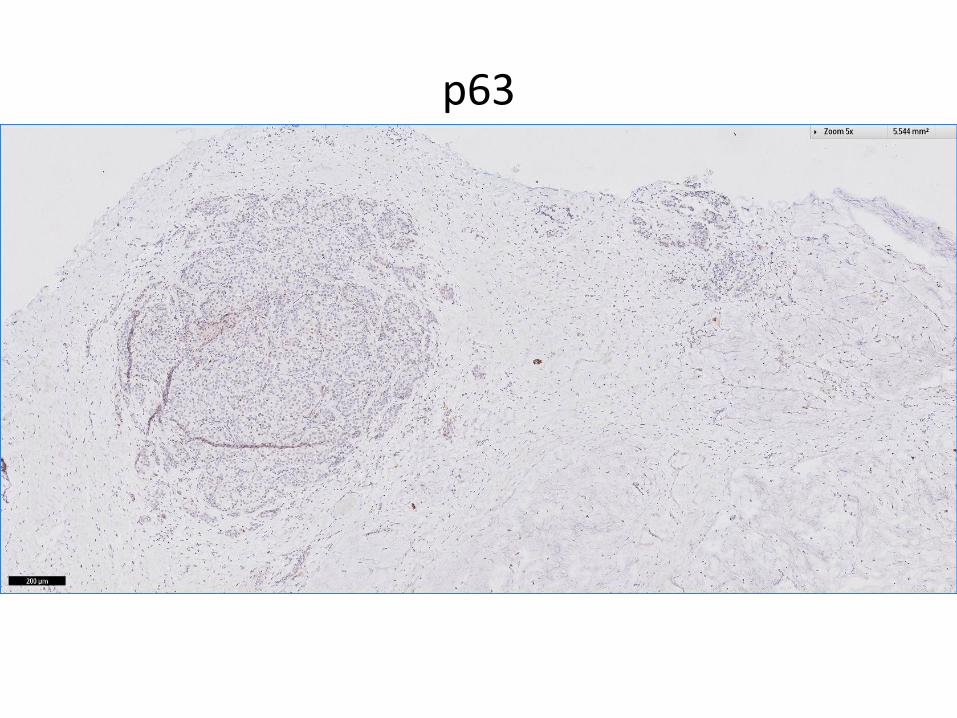
p63

SMMS

ER
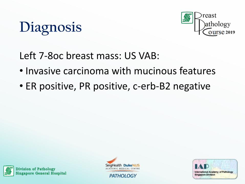
Diagnosis
Left 7-8oc breast mass: US VAB:
• Invasive carcinoma with mucinous features
• ER positive, PR positive, c-erb-B2 negative

Mucocele-like Lesions (MLL)
• Mucin-containing cysts with extravasated mucinresulting from rupture
• ~50% associated with ADH/ DCIS/ invasive carcinoma
• Cysts and dilated ducts contain luminal mucin; lined by attenuated to cuboidal epithelium
• Epithelium may show spectrum of proliferative changes e.g. UDH, ADH, DCIS

MLL• Extravasated mucin in stroma resulting from rupture
• Calcifications may be present within mucin
• Strips/ clusters of epithelial cells may become detached and float freely• Important not to overinterpret as carcinoma
• Multiple levels may be required
• Features favouring MLL with detached epithelium• Linear configuration of epithelial cells
• No nuclear atypia
• Presence of associated myoepithelial cells (may need IHC to demonstrate)

MLL
• Distinction of MLL and mucinous carcinoma is often not possible on core needle biopsy
• Presence of epithelial atypia would warrant excision
• If extravasated mucin is extensive, and imaging shows a mass, excision should be considered
• MLL with areas of mucinous carcinoma have been reported > excision + histological examination of entire lesion is necessary for final classification in many cases