761034orig1s000 - food and drug administration · 1.1 introduction tecentriq (atezolizumab) is a...
TRANSCRIPT

CENTER FOR DRUG EVALUATION AND RESEARCH
APPLICATION NUMBER:
761034Orig1s000
PHARMACOLOGY REVIEW(S)

MEMORANDUM
Tecentriq (atezolizumab)
Date: April 27, 2016To: File for BLA 761034From: John K. Leighton, PhD, DABT
Director, Division of Hematology Oncology ToxicologyOffice of Hematology and Oncology Products
I have examined pharmacology/toxicology supporting review and labeling for Tecentriq conducted by Dr. Ricks and secondary memorandum and labeling provided by Dr. Palmby. In his memorandum, Dr. Palmby recommends a Post-Marketing Commitment (PMC) to address the effect of atezolizumab on the magnitude of primary and recall response to antigen challenge. I concur with Dr. Palmby’s conclusion that Tecentriq may be approved for the proposed indication and the recommendation for a PMC.
Reference ID: 3923282

---------------------------------------------------------------------------------------------------------This is a representation of an electronic record that was signedelectronically and this page is the manifestation of the electronicsignature.---------------------------------------------------------------------------------------------------------/s/----------------------------------------------------
JOHN K LEIGHTON04/27/2016
Reference ID: 3923282

MEMORANDUM
Date: April 26, 2016From: Todd R. Palmby, PhD
Pharmacology/Toxicology SupervisorDivision of Hematology Oncology Toxicology (DHOT)Office of Hematology and Oncology Products (OHOP)
To: File for BLA 761034 Tecentriq (atezolizumab)Re: Approvability for Pharmacology and ToxicologyIndication: Treatment of patients with locally advanced or metastatic urothelial
carcinoma who have disease progression during or following platinum-containing chemotherapy or have disease progression within 12 months of neoadjuvant or adjuvant treatment with platinum-containing chemotherapy
Non-clinical pharmacology and toxicology literature and original reports for studies to support BLA 761034 for Tecentriq (atezolizumab) indicated for the treatment of patients with locally advanced or metastatic urothelial carcinoma who have disease progression during or following platinum-containing chemotherapy or have disease progression within 12 months of neoadjuvant or adjuvant treatment with platinum-containing chemotherapy were reviewed by Tiffany Ricks, PhD. Studies conducted by the Applicant with atezolizumab, or with murine chimeric anti-PD-L1 antibodies, for which reports were submitted to this BLA include pharmacology, pharmacokinetics and general toxicology. A detailed evaluation of the nonclinical data submitted to the BLA can be found in Dr. Ricks’ review.
Atezolizumab is a humanized IgG1 monoclonal antibody that binds to human PD-L1, thereby inhibiting the interaction of PD-L1 with PD-1 and B7-1 receptors. In order to decrease antibody-dependent cell-mediated cytotoxicity (ADCC), the Applicant modified amino acid 298 in the heavy chain from asparagine to alanine. This modification results in a non-glycosylated antibody, demonstrated to have no Fc-effector function (Fcγ), but retains binding to the neonatal Fc receptor (FcRn). The Applicant generated mouse IgG2a/human chimeric antibodies for murine in vivo studies in order to reduce immunogenicity, which was observed in prior mouse studies. Based on the submitted pharmacology data, the Established Pharmacologic Class (EPC) of “programmed death-ligand 1 (PD-L1) blocking antibody” was determined to be both clinically meaningful and scientifically valid for atezolizumab. Atezolizumab would be the first product in this class to be approved in the US. Based on its mechanism of action, atezolizumab treatment may increase the inflammatory response and enhance the severity of some infections in patients.
General toxicology studies evaluated atezolizumab in mice for two weeks and in Cynomolgus monkeys for up to 26 weeks. Atezolizumab was immunogenic in
Reference ID: 3922707

mice, resulting in significantly reduced exposures by Week 3. The major toxicological finding in mice was minimal sciatic neuropathy (vacuolation and lymphocytic infiltration). In Cynomolgus monkeys, IV administration of atezolizumab caused multi-organ arteritis/periarteritis and irregular menstruation, including an increase in mean menstrual cycle length and a corresponding lack of newly formed corpora lutea. The immune-mediated findings were consistent with the role of PD-L1 in regulating and maintaining peripheral tolerance. The mechanism of the effects on menstruation in monkeys is not clear, but these effects were reversible. Safety pharmacology assessments were incorporated into the repeat-dose toxicology study in monkeys, but no effects were observed on the respiratory, neurological or cardiovascular systems. Administration of atezolizumab to patients at the recommended dose of 1200 mg resulted in immune-related adverse events.
Since it is a monoclonal antibody and is not expected to interact with DNA, genetic toxicology studies were not conducted with atezolizumab.
The Applicant submitted a non-product specific literature-based assessment to characterize the potential risk of reproductive and developmental toxicity. Data in the scientific literature demonstrate that interference with PD-L1 leads to a loss of fetal tolerance and an increased risk of immune-mediated abortion. This assessment provides evidence that atezolizumab can cause fetal harm if administered to a pregnant woman.
Atezolizumab maintains binding to the FcRn receptor, so fetal exposure may occur if a patient is treated during pregnancy, although it is not recommended. It is unclear whether fetal exposure to atezolizumab would occur at levels sufficient to cause adverse effects on the developing immune system. Nevertheless, if a pregnant patient receives treatment with atezolizumab that does not result in loss of the fetus, there is a potential risk of developing immune-mediated disorders or altering the normal immune response in the offspring due to the mechanism of action.
Recommendation: I concur with Dr. Ricks’ conclusion that submitted pharmacology and toxicology data support the approval of BLA 761034 for Tecentriq (atezolizumab) for the proposed indication. The Pharmacology/Toxicology team recommends that the Applicant conduct a pharmacology study to further characterize the effect of atezolizumab on the immune response as a Post-Marketing Commitment (PMC). We recommend that the Applicant conduct an animal study that will measure the effect of PD-L1 inhibition on the magnitude of the primary (1st vaccination) and recall (2nd vaccination) antibody responses to antigen challenge (e.g. KLH). This PMC is currently being discussed with the Applicant. There are no outstanding non-clinical issues that would preclude the approval of Tecentriq for the proposed indication.
Reference ID: 3922707

---------------------------------------------------------------------------------------------------------This is a representation of an electronic record that was signedelectronically and this page is the manifestation of the electronicsignature.---------------------------------------------------------------------------------------------------------/s/----------------------------------------------------
TODD R PALMBY04/26/2016
Reference ID: 3922707

1
DEPARTMENT OF HEALTH AND HUMAN SERVICESPUBLIC HEALTH SERVICE
FOOD AND DRUG ADMINISTRATIONCENTER FOR DRUG EVALUATION AND RESEARCH
PHARMACOLOGY/TOXICOLOGY BLA REVIEW AND EVALUATION
Application number: 761034
Supporting document/s: 1, 53
Applicant’s letter date: January 12, 2016
CDER stamp date: January 12, 2016
Product: Tecentriq (atezolizumab)
Indication: Adult patients with locally advanced or
metastatic urothelial carcinoma who have
disease progression during or following
platinum-containing chemotherapy
or have disease progression
within 12 months of treatment with a platinum-
containing neoadjuvant or adjuvant
chemotherapy regimen
Applicant: Genentech, Inc.
1 DNA Way
South San Francisco, CA
Review Division: Division of Hematology Oncology Toxicology
(Division of Oncology Product 1)
Reviewer: Tiffany K. Ricks, PhD
Supervisor/Team Leader: Todd Palmby, PhD
Division Director: John Leighton, PhD, DABT (DHOT)
Geoffrey Kim, MD (DOP1)
Project Manager: Kim Robertson
Reference ID: 3920269
(b) (4)

2
Disclaimer
Except as specifically identified, all data and information discussed below and necessary for approval of BLA 761034 are owned by Genentech Inc. or are data for which Genentech Inc. has obtained a written right of reference.Any information or data necessary for approval of BLA 761034 that Genentech Inc. does not own or have a written right to reference constitutes one of the following: (1) published literature, or (2) a prior FDA finding of safety or effectiveness for a listed drug, as reflected in the drug’s approved labeling. Any data or information described or referenced below from reviews or publicly available summaries of a previously approved application is for descriptive purposes only and is not relied upon for approval of BLA 761034.
Reference ID: 3920269
BLA # 761034 Tiffany K. Ricks, PhD
3
TABLE OF CONTENTS
1 EXECUTIVE SUMMARY...........................................................................................61.1 INTRODUCTION .....................................................................................................61.2 BRIEF DISCUSSION OF NONCLINICAL FINDINGS .......................................................61.3 RECOMMENDATIONS .............................................................................................8
2 DRUG INFORMATION..............................................................................................92.1 DRUG ..................................................................................................................92.2 RELEVANT INDS, NDAS, BLAS AND DMFS..........................................................102.3 DRUG FORMULATION ..........................................................................................102.4 COMMENTS ON NOVEL EXCIPIENTS ......................................................................102.5 COMMENTS ON IMPURITIES/DEGRADANTS OF CONCERN ........................................102.6 PROPOSED CLINICAL POPULATION AND DOSING REGIMEN .....................................102.7 REGULATORY BACKGROUND ...............................................................................11
3 STUDIES SUBMITTED...........................................................................................113.1 STUDIES REVIEWED ............................................................................................113.2 STUDIES NOT REVIEWED.....................................................................................123.3 PREVIOUS REVIEWS REFERENCED.......................................................................13
4 PHARMACOLOGY .................................................................................................134.1 PRIMARY PHARMACOLOGY ..................................................................................134.2 SECONDARY PHARMACOLOGY .............................................................................234.3 SAFETY PHARMACOLOGY ....................................................................................23
5 PHARMACOKINETICS/ADME/TOXICOKINETICS ...............................................246 GENERAL TOXICOLOGY......................................................................................26
6.1 SINGLE-DOSE TOXICITY ......................................................................................266.2 REPEAT-DOSE TOXICITY .....................................................................................27
7 GENETIC TOXICOLOGY........................................................................................438 CARCINOGENICITY...............................................................................................439 REPRODUCTIVE AND DEVELOPMENTAL TOXICOLOGY.................................4310 SPECIAL TOXICOLOGY STUDIES....................................................................4511 INTEGRATED SUMMARY AND SAFETY EVALUATION..................................4712 APPENDIX/ATTACHMENTS ..............................................................................50
Reference ID: 3920269

BLA # 761034 Tiffany K. Ricks, PhD
4
Table of Tables
Table 1: Drug product composition of atezolizumab ......................................................10Table 2: Binding of MPDL3280A and chimeric antibodies to human, Cynomolgus monkey, and murine PD-L1............................................................................................14Table 3: Inhibitory activity of MPDL3280A and murine chimeric antibodies...................14Table 4: Anti-tumor activity of MPDL3280A in a syngeneic mouse model of MC38.OVA colorectal cancer.............................................................................................................16Table 5: Anti-tumor activity of anti-PD-L1 antibody in a syngeneic mouse model of CT26 colorectal cancer.............................................................................................................18Table 6: Anti-tumor activity of anti-PD-L1 antibody in a syngeneic mouse model of melanoma.......................................................................................................................20Table 7: Anti-tumor activity of anti-PD-L1 in a syngeneic mouse model of MC38 colorectal cancer.............................................................................................................21Table 8: Blood and urine collection times.......................................................................30Table 9: Summary of hematology parameters (% change relative to controls) ..............35Table 10: Summary of macroscopic findings in Cynomolgus monkeys at scheduled necropsy .........................................................................................................................35Table 11: Summary of organ weights in Cynomolgus monkeys at end of dosing necropsy (% change relative to control) .........................................................................36Table 12: Summary of histological findings in Cynomolgus monkeys at scheduled necropsy .........................................................................................................................36Table 13: Immunogenicity results from Cynomolgus monkeys receiving MPDL3280A weekly.............................................................................................................................39Table 14: Individual menstrual cycle lengths in days .....................................................40Table 15: Mean toxicokinetic parameters in monkeys administered intravenous MPDL3280A ...................................................................................................................43Table 16: Summary of Biotin-MPDL3280A cross-reactivity with human tissues ............47
Reference ID: 3920269

BLA # 761034 Tiffany K. Ricks, PhD
5
Table of Figures
Figure 1: ADCC activity of atezolizumab ........................................................................15Figure 2: Anti-tumor activity of anti-PD-L1 antibody in a syngeneic mouse model of MC38.OVA colorectal cancer .........................................................................................16Figure 3: Anti-tumor activity of anti-PD-L1 antibody in a syngeneic mouse model of CT26 colorectal cancer...................................................................................................18Figure 4: Anti-tumor activity of anti-PD-L1 antibody in a syngeneic mouse model of melanoma.......................................................................................................................19Figure 5: Anti-tumor activity of anti-PD-L1 in a syngeneic mouse model of MC38 colorectal cancer.............................................................................................................21Figure 6: Individual serum concentration-time profiles following a single IV dose of MPDL3280A in Cynomolgus monkeys ...........................................................................25Figure 7: Observed and predicted serum concentration-time profiles............................26Figure 8: Body weights of monkeys administered weekly IV doses of MPDL3280A......34Figure 9: Group mean menstrual cycle length................................................................41Figure 10: Group mean of individual treatment cycle length ..........................................42
Reference ID: 3920269
BLA # 761034 Tiffany K. Ricks, PhD
6
1 Executive Summary1.1 IntroductionTecentriq (atezolizumab) is a humanized, Fc-engineered, IgG1 monoclonal antibody targeting programmed death-ligand 1 (PD-L1). The Applicant submitted nonclinical study reports and relevant literature evaluating the pharmacology and toxicology of atezolizumab to support BLA 761034. The proposed indication is for treatment of patients with locally advanced or metastatic urothelial carcinoma who have disease progression during or following platinum-containing chemotherapy
or have disease progression within 12 months of treatment with a platinum-containing neoadjuvant or adjuvant chemotherapy regimen.
1.2 Brief Discussion of Nonclinical FindingsPD-L1 is a transmembrane protein that negatively regulates immune responses through its interactions with PD-1 and B7-1 receptors (Francisco et al. 2010, Keir et al. 2008). PD-L1 is constitutively expressed on B cells, dendritic cells, macrophages, bone marrow-derived mast cells, and T cells. PD-L1 is also expressed on a wide range of non-hematopoietic cells and can be upregulated on many cell types upon stimulation. Binding of PD-L1 to its receptors attenuates lymphocyte activation to maintain immune tolerance and prevent immune-mediated tissue damage. In the tumor microenvironment, tumor cells and tumor infiltrating immune cells can express PD-L1 and inhibit T-cell proliferation and function. Blocking PD-L1 releases inhibition of the immune response, resulting in enhanced immune surveillance and responses, including immune-mediated anti-tumor activity.
In vitro pharmacology studies demonstrated that atezolizumab binds to human, murine, and Cynomolgus monkey PD-L1 and inhibits the interaction of PD-L1 with PD-1 and B7-1 receptors. Atezolizumab contains mutation in the Fc domain to prevent glycosylation at that site, thereby limiting binding to human Fcγ receptors and preventing antibody-dependent cell-mediated cytotoxicity (ADCC). Atezolizumab retains binding to neonatal Fc receptors, despite the mutation in the Fc domain. In vitro, soluble or plate-bound atezolizumab did not stimulate cytokine release from unstimulated human peripheral blood mononuclear cells (PBMCs). For in vivo pharmacology studies, the Applicant generated murine/human chimeric antibodies of atezolizumab to minimize immunogenicity in mice and evaluated PD-L1 blockade in syngeneic mouse models of melanoma and colorectal cancer. Anti-PD-L1 antibody reduced tumor growth by ≥ 80%. Based on the submitted pharmacology data, the Established Pharmacologic Class (EPC) of “programmed death-ligand 1 (PD-L1) blocking antibody” was determined to be both clinically meaningful and scientifically valid for atezolizumab.
The Applicant also evaluated murine/human chimeric antibodies of atezolizumab and an independently derived anti-mouse PD-L1 antibody in a mouse model of chronic lymphocytic choriomeningitis virus (LCMV) CL-13. Administration of anti-PD-L1 before and during the peak of viral infection and acute T-cell response resulted in ~40 to 100% mortality. Anti-PD-L1 was not lethal when administered at later time points or in three
Reference ID: 3920269
(b) (4)
(b) (4)

BLA # 761034 Tiffany K. Ricks, PhD
7
other mouse models of acute viral infection. As reported in the literature, PD-1-/- mice succumb to M. tuberculosis infection with marked lung inflammation and bacterial loads and an increase in circulating pro-inflammatory cytokines (Lazar-Molnar et al. 2010). Based on its mechanism of action, atezolizumab may increase the inflammatory response and enhance the severity of some infections.
General toxicology studies evaluated atezolizumab in mice for two weeks and in Cynomolgus monkeys for up to 26 weeks. Atezolizumab was immunogenic in mice, resulting in significantly reduced exposures by Week 3. The major toxicological finding was minimal sciatic neuropathy (vacuolation and lymphocytic infiltration), observed at the end of dosing and recovery period. In Cynomolgus monkeys, atezolizumab caused irregular menstruation and multi-organ arteritis/periarteritis. The immune-mediated findings were consistent with the role of PD-L1 in regulating and maintaining peripheral tolerance. In humans, administration of atezolizumab at the recommended dose of 1200 mg resulted in immune-related adverse events. In the 26-week monkey study, females administered 50 mg/kg IV atezolizumab experienced irregular menstruation during the dosing period, including an increase in mean menstrual cycle length compared to controls and a corresponding lack of newly formed corpora lutea. These effects were reversible.
The Applicant submitted a non-product specific literature-based assessment to characterize the potential risk of reproductive and developmental toxicity. PD-L1 is expressed in the human placenta at the fetal/maternal interface throughout pregnancy (Holets et al. 2006, Petroff et al. 2003). In mouse models of allogenic pregnancy, loss of PD-L1 activity increased the rate of fetal resorption and reduced litter size (D'Addio et al. 2011, Guleria et al. 2005). Anti-PD-L1 antibody reduced fetal antigen-specific regulatory T cells (Tregs) and increased accumulation of fetal antigen-specific effector T cells in maternal lymphoid organs (D'Addio et al. 2011, Taglauer et al. 2009). Collectively, the scientific literature demonstrates that PD-L1 blockade leads to a loss of fetal tolerance and an increased risk of immune-mediated abortion.
It is unclear whether atezolizumab crosses the placental barrier at levels that would cause adverse effects on developing offspring. The allogenic pregnancy models did not evaluate offspring for teratogenicity or adverse developmental effects. Data from knockout mice demonstrate that syngeneic PD-1-/- and PD-L1-/- fetuses develop normally with no reported malformations (Keir et al. 2008, Okazaki and Honjo 2006, Okazaki and Honjo 2007). Depending on the genetic background, PD-1-/- and PD-L1-/-
mice develop late onset autoimmune phenotypes. Inhibition of the PD-L1/PD-1 pathway can exacerbate disease in mouse models of autoimmunity. Additionally, single nucleotide polymorphisms (SNPs) in the gene encoding human PD-1 are associated with several autoimmune diseases. It is unknown whether exposure to atezolizumab during pregnancy may increase the risk of offspring developing immune-mediated disorders or experiencing adverse effects on the normal immune response; however, this potential risk cannot be dismissed based on its mechanism of action.
Reference ID: 3920269

BLA # 761034 Tiffany K. Ricks, PhD
8
The submitted nonclinical pharmacology and toxicology studies support approval of Tecentriq for the proposed indication. The Applicant provided data demonstrating that atezolizumab binds to PD-L1 and blocks its interaction with PD-1 and B7-1 receptors. Consequently, atezolizumab relieves inhibition of immune responses, including anti-tumor activity and peripheral tolerance. Atezolizumab had no effects on cardiovascular, respiratory, or neurological systems in monkeys. Toxicological effects of atezolizumab administration in animals were limited to irregular menstruation in female monkeys and immune-mediated effects, including tissue damage and multi-organ inflammation. Other effects of PD-L1 blockade observed in animals, which may be relevant to patients receiving atezolizumab, consisted of enhanced inflammatory response and severity of infections and a risk of embryo-fetal toxicity, primarily an increased risk of immune-mediated abortion.
1.3 Recommendations1.3.1 ApprovabilityRecommended for approval for the proposed indication.1.3.2 Additional Non Clinical RecommendationsTreatment with atezolizumab may result in enhanced immune-mediated toxicity following vaccination and recall responses. To investigate this potential, the following post-marketing commitment (PMC) is recommended by the Pharmacology/Toxicology team.
“The PMC is to conduct an animal study that will measure the effect of PD-L1 inhibition on the magnitude of the primary (1st vaccination) and recall (2nd vaccination) antibody responses to antigen challenge (e.g. KLH). This study will evaluate the effect of PD-L1 inhibition on the primary immune response once steady state plasma levels have been achieved and will reassess the magnitude of the recall response after a suitable period in the presence or absence of continued dosing. The study should include, if possible, an evaluation of cytokine production by T cells at appropriate timepoints.”
Reference ID: 3920269
(b) (4)

BLA # 761034 Tiffany K. Ricks, PhD
9
1.3.3 LabelingThis Pharmacology/Toxicology review contains the content for labeling Tecentriq (atezolizumab). Based on pharmacology data submitted in the BLA, the EPC of “programmed death-ligand 1 (PD-L1) blocking antibody” was determined to be both clinically meaningful and valid for atezolizumab.
In section 8.2 Lactation, the Division recommended that patients should not breastfeed during treatment and for 5 months after the last dose due to the potential for serious adverse reactions in breastfed infants from atezolizumab. This was included to be consistent with labels for US FDA-approved products inhibiting PD-1. In section 8.3 Females and Males of Reproductive Potential, the Division recommended that females of reproductive potential should use effective contraception during treatment and for 5 months after the last dose due to the potential risk to a fetus. The elimination half-life of atezolizumab in patients receiving the recommended dose is 27 days. The duration of 5 months was determined based on approximately 5 half-lives of atezolizumab, and rounded to the nearest whole month.
The following pharmacokinetic data was used to calculate the animal to human exposure ratios for atezolizumab. Based on population analysis of a dose escalation clinical trial in patients with advanced solid tumors, the steady-state AUC is 6409 day*µg/mL at the recommended human dose of 1200 mg every 3 weeks. The elimination half-life in patients is 27 days. In repeat-dose toxicology studies, Cynomolgus monkeys received doses up to 50 mg/kg weekly, or 150 mg/kg over a 3-week period. Following single and repeat dosing in monkeys, the AUC was calculated from data collected over a 3-day period. Due to the long half-life of 12 to 24 days in monkeys, the measured AUC underestimated the total exposure of atezolizumab in monkeys over the period of three weeks between repeat-dosing. Using data from a pharmacokinetic study in monkeys, the Applicant estimated the AUC in monkeys to be 40,500 day*µg/mL based on a clearance of 3.7 mL/day/kg and a dose of 150 mg/kg over a 3-week interval (Study No. 08-0598).
2 Drug Information2.1 Drug
CAS Registry Number 1380723-44-3Generic Name AtezolizumabCode Names MPDL3280A, RO5541267, PRO#303280Chemical Name Humanized anti-PD-L1 IgG1 monoclonal antibodyMolecular Formula C6434H9878O1996N1702S42Molecular Weight 144.356 kDaPharmacological Class programmed death-ligand 1 (PD-L1) blocking
antibody
Reference ID: 3920269
(b) (4)


BLA # 761034 Tiffany K. Ricks, PhD
12
PharmacologyStudy number
Study title eCTD location
09-0426 In vitro binding and biological activity of MPDL3280A (rhuMAb PD-L1)
4.2.1.1.
15-0984 In vitro binding affinity of MPDL3280A 4.2.1.1.08-1033 Evaluation of the anti-tumor efficacy of anti-PD-L1
monoclonal antibody in the syngeneic MC38.OVA colorectal cancer model in C57BL/6 mice
4.2.1.1.
08-1734 Evaluation of the anti-tumor efficacy of anti-PD-L1 monoclonal antibody in the syngeneic CT26 colorectal cancer model in Balb/c mice
4.2.1.1.
09-2165 Evaluation of the anti-tumor efficacy of anti-PD-L1 monoclonal antibody in the syngeneic Cloudman S91 melanoma model in DBA/2 mice
4.2.1.1
10-1883 Evaluation of the anti-tumor efficacy of anti-PD-L1 monoclonal antibody in the syngeneic MC38 colorectal model in C57BL/6 mice
4.2.1.1.
08-0559A, 08-559B, 08-1160
Therapeutic efficacy and dose titration of anti-PD-L1 mAb in the lymphocytic choriomeningitis virus infection model
4.2.1.1.
08-1309 Evaluation of the combined effects of adenovirus expressed interferon-alpha (IFN-a) and anti-PD-L1 mAb in mice infected with lymphocytic choriomeningitis virus (LCMV)
4.2.1.1.
09-2500, 09-2500B, 09-2501, 09-2501A
Evaluation of the host response to Armstrong and CL-13 lymphocytic choriomeningitis virus (LCMV) infection in mice following administration of a single dose of anti-PD-L1 antibody at different times during infection
4.2.1.1.
10-1394 Studies to address mechanism of anti-PD-L1 enhanced pathology in lymphocytic choriomeningitis virus (LCMV) infection: comparison between clone-13 and Armstrong strains
4.2.1.1.
PharmacokineticsStudy number
Study title eCTD location
08-0598 A single dose pharmacokinetic study of MPDL3280A administered by intravenous injection to Cynomolgus monkeys
4.2.2.7.
3.2 Studies Not Reviewed Pharmacology15-0984 In vitro binding affinity of MPDL3280A 4.2.1.1.
Reference ID: 3920269

BLA # 761034 Tiffany K. Ricks, PhD
14
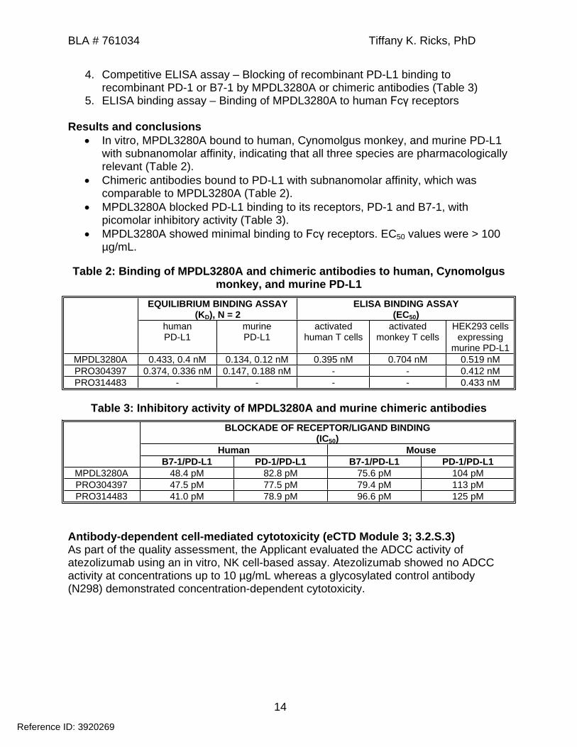
4. Competitive ELISA assay – Blocking of recombinant PD-L1 binding to recombinant PD-1 or B7-1 by MPDL3280A or chimeric antibodies (Table 3)
5. ELISA binding assay – Binding of MPDL3280A to human Fcγ receptors
Results and conclusions In vitro, MPDL3280A bound to human, Cynomolgus monkey, and murine PD-L1
with subnanomolar affinity, indicating that all three species are pharmacologically relevant (Table 2).
Chimeric antibodies bound to PD-L1 with subnanomolar affinity, which was comparable to MPDL3280A (Table 2).
MPDL3280A blocked PD-L1 binding to its receptors, PD-1 and B7-1, with picomolar inhibitory activity (Table 3).
MPDL3280A showed minimal binding to Fcγ receptors. EC50 values were > 100 µg/mL.
Table 2: Binding of MPDL3280A and chimeric antibodies to human, Cynomolgus monkey, and murine PD-L1
EQUILIBRIUM BINDING ASSAY(KD), N = 2
ELISA BINDING ASSAY(EC50)
humanPD-L1
murinePD-L1
activated human T cells
activated monkey T cells
HEK293 cells expressing
murine PD-L1MPDL3280A 0.433, 0.4 nM 0.134, 0.12 nM 0.395 nM 0.704 nM 0.519 nMPRO304397 0.374, 0.336 nM 0.147, 0.188 nM - - 0.412 nMPRO314483 - - - - 0.433 nM
Table 3: Inhibitory activity of MPDL3280A and murine chimeric antibodiesBLOCKADE OF RECEPTOR/LIGAND BINDING
(IC50)Human Mouse
B7-1/PD-L1 PD-1/PD-L1 B7-1/PD-L1 PD-1/PD-L1MPDL3280A 48.4 pM 82.8 pM 75.6 pM 104 pMPRO304397 47.5 pM 77.5 pM 79.4 pM 113 pMPRO314483 41.0 pM 78.9 pM 96.6 pM 125 pM
Antibody-dependent cell-mediated cytotoxicity (eCTD Module 3; 3.2.S.3)As part of the quality assessment, the Applicant evaluated the ADCC activity of atezolizumab using an in vitro, NK cell-based assay. Atezolizumab showed no ADCC activity at concentrations up to 10 µg/mL whereas a glycosylated control antibody (N298) demonstrated concentration-dependent cytotoxicity.
Reference ID: 3920269

BLA # 761034 Tiffany K. Ricks, PhD
15
Figure 1: ADCC activity of atezolizumab
[Excerpted from Applicant’s submission]
Study title: Evaluation of the anti-tumor efficacy of anti-PD-L1 monoclonal antibody in the syngeneic MC38.OVA colorectal model in C57BL/6 mice
Study No.: 08-1033 EStudy report date: February 11, 2011
Study report location: eCTD 4.2.1.1.Conducting laboratory: Genentech, Inc.
South San Francisco, CAGLP: No
The aim of this study was to assess the anti-tumor activity of murine chimeric antibody, PRO314483, in a syngeneic immunocompetent mouse model of MC38.OVA colorectal cancer. As shown in Table 2 and Table 3, binding of PRO314483 to PD-L1 was comparable to MPDL3280A. Cells were mixed 50:50 with Matrigel and implanted subcutaneously (SC) into the right flank of female C57BL/6 mice (10/group). Once tumors reached a mean volume of ~150 mm3, mice were treated with 10 mg/kg PRO314483 administered as an intraperitoneal (IP) injection three times weekly for one, two, or three cycles. Control anti-gp120 antibody was administered as an IP injection three times weekly for three cycles. Study endpoints were changes in tumor volume and body weight. If tumor volume decreased below the limit of detection (8 mm3), it was considered a complete response. If tumor volume decreases by ≥ 50% of its initial volume, it was considered a partial response.
Results and conclusions All mice receiving 10 mg/kg anti-PD-L1 had complete responses, which were
sustained through the end of study on Day 55. A dose of 10 mg/kg anti-PD-L1 antibody had no effect on body weights.
Reference ID: 3920269

BLA # 761034 Tiffany K. Ricks, PhD
16
Figure 2: Anti-tumor activity of anti-PD-L1 antibody in a syngeneic mouse model of MC38.OVA colorectal cancer
[Excerpted from Applicant’s submission]
Table 4: Anti-tumor activity of MPDL3280A in a syngeneic mouse model of MC38.OVA colorectal cancer
Reference ID: 3920269
BLA # 761034 Tiffany K. Ricks, PhD
17
[Excerpted from Applicant’s submission]
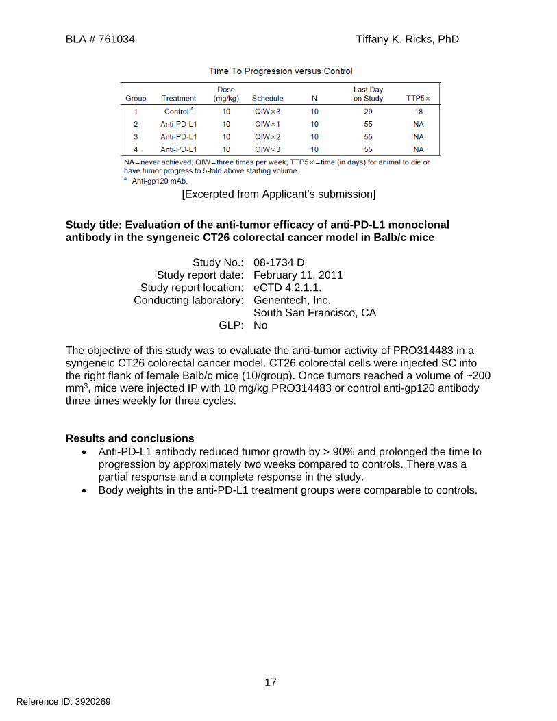
Study title: Evaluation of the anti-tumor efficacy of anti-PD-L1 monoclonal antibody in the syngeneic CT26 colorectal cancer model in Balb/c mice
Study No.: 08-1734 DStudy report date: February 11, 2011
Study report location: eCTD 4.2.1.1.Conducting laboratory: Genentech, Inc.
South San Francisco, CAGLP: No
The objective of this study was to evaluate the anti-tumor activity of PRO314483 in a syngeneic CT26 colorectal cancer model. CT26 colorectal cells were injected SC into the right flank of female Balb/c mice (10/group). Once tumors reached a volume of ~200 mm3, mice were injected IP with 10 mg/kg PRO314483 or control anti-gp120 antibody three times weekly for three cycles.
Results and conclusions Anti-PD-L1 antibody reduced tumor growth by > 90% and prolonged the time to
progression by approximately two weeks compared to controls. There was a partial response and a complete response in the study.
Body weights in the anti-PD-L1 treatment groups were comparable to controls.
Reference ID: 3920269

BLA # 761034 Tiffany K. Ricks, PhD
18
Figure 3: Anti-tumor activity of anti-PD-L1 antibody in a syngeneic mouse model of CT26 colorectal cancer
[Excerpted from Applicant’s submission]
Table 5: Anti-tumor activity of anti-PD-L1 antibody in a syngeneic mouse model of CT26 colorectal cancer
[Excerpted from Applicant’s submission]
Reference ID: 3920269
BLA # 761034 Tiffany K. Ricks, PhD
19
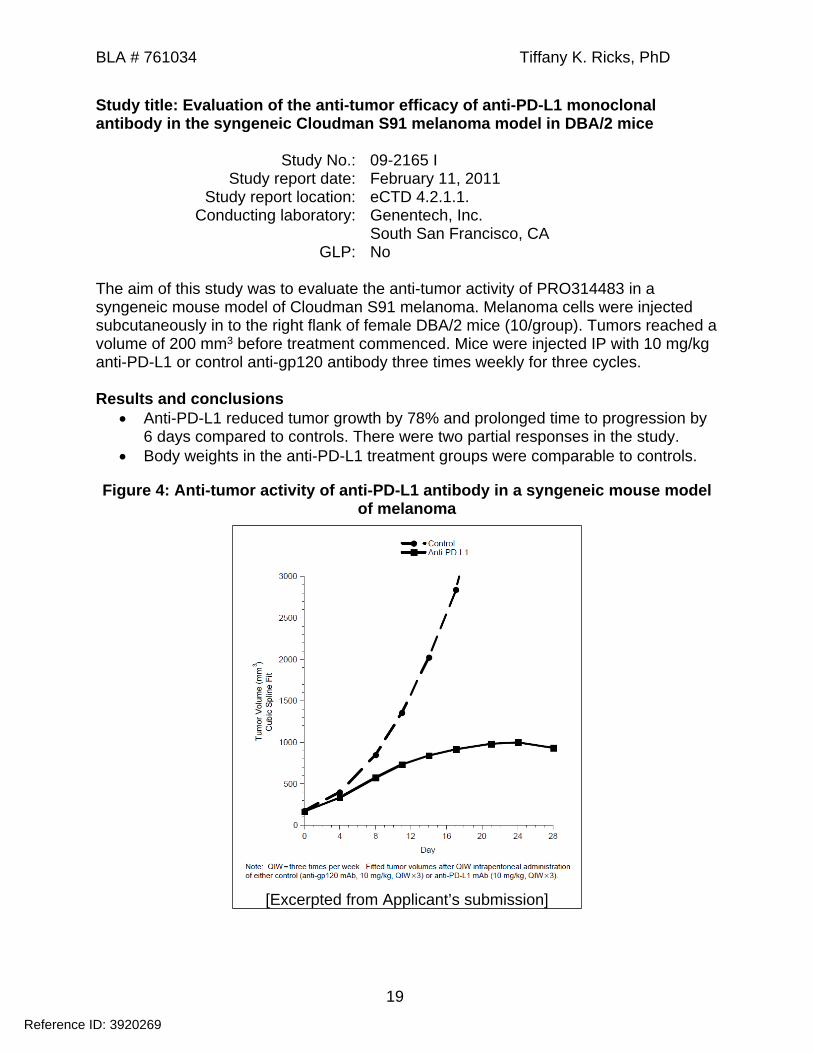
Study title: Evaluation of the anti-tumor efficacy of anti-PD-L1 monoclonal antibody in the syngeneic Cloudman S91 melanoma model in DBA/2 mice
Study No.: 09-2165 IStudy report date: February 11, 2011
Study report location: eCTD 4.2.1.1.Conducting laboratory: Genentech, Inc.
South San Francisco, CAGLP: No
The aim of this study was to evaluate the anti-tumor activity of PRO314483 in a syngeneic mouse model of Cloudman S91 melanoma. Melanoma cells were injected subcutaneously in to the right flank of female DBA/2 mice (10/group). Tumors reached a volume of 200 mm3 before treatment commenced. Mice were injected IP with 10 mg/kg anti-PD-L1 or control anti-gp120 antibody three times weekly for three cycles.
Results and conclusions Anti-PD-L1 reduced tumor growth by 78% and prolonged time to progression by
6 days compared to controls. There were two partial responses in the study. Body weights in the anti-PD-L1 treatment groups were comparable to controls.
Figure 4: Anti-tumor activity of anti-PD-L1 antibody in a syngeneic mouse model of melanoma
[Excerpted from Applicant’s submission]
Reference ID: 3920269

BLA # 761034 Tiffany K. Ricks, PhD
20
Table 6: Anti-tumor activity of anti-PD-L1 antibody in a syngeneic mouse model of melanoma
[Excerpted from Applicant’s submission]
Study title: Evaluation of the anti-tumor efficacy of anti-PD-L1 monoclonal antibody in the syngeneic MC38 colorectal model in C57BL/6 mice
Study No.: 10-1883Study report date: February 11, 2011
Study report location: eCTD 4.2.1.1.Conducting laboratory: Genentech, Inc.
South San Francisco, CAGLP: No
The aim of this study was to examine the anti-tumor activity of PRO314483 in a syngeneic mouse model of MC38 colorectal cancer. Colorectal cells were mixed 50:50 with Matrigel and injected SC into the right flank of female C57BL/6 mice (10/group). Tumors reached a mean volume of ~200 mm3 before treatment commenced. Mice were administered doses of 10 mg/kg PRO314483 by IP injection three times weekly for one, two, or three cycles. Control anti-gp120 antibody was administered according to the same dosing schedule for three cycles.
Results and conclusions Anti-PD-L1 reduced tumor growth by 76, 98, and 103% following treatment of
one, two, and three cycles, respectively. Time to progression was also prolonged with each increase in treatment length.
Reference ID: 3920269

BLA # 761034 Tiffany K. Ricks, PhD
21
Three complete and partial responses were reported after two and three treatment cycles.
There were no treatment-related effects on body weight.
Figure 5: Anti-tumor activity of anti-PD-L1 in a syngeneic mouse model of MC38 colorectal cancer
[Excerpted from Applicant’s submission]
Table 7: Anti-tumor activity of anti-PD-L1 in a syngeneic mouse model of MC38 colorectal cancer
Reference ID: 3920269

BLA # 761034 Tiffany K. Ricks, PhD
22
[Excerpted from the Applicant’s submission]
Study title: In vitro cytokine release study with anti-PD-L1 antibody in human PBMCs
Study No.: 081827Study report date: June 30, 2009, March 23, 2011 (amendment)
Study report location: eCTD 4.2.3.7.Conducting laboratory: Genentech, Inc.
South San Francisco, CAGLP: No
The aim of this study was to test whether MPDL3280A induces cytokine release from unstimulated human PBMCs. Briefly, human PBMCs isolated from three donors were incubated with soluble or immobilized MPDL3280A on tissue culture plates at concentrations ranging from 0.25 to 250 µg/mL. Anti-CD3 (1 µg/mL) and LPS (50 ng/mL) were positive controls. Anti-hFGFR3 (250 µg/mL) and medium only were negative controls. After incubating cells for 24 or 48 h at 37oC, supernatants were analyzed for cytokine secretion using a Luminex multiplex assay. The cytokine profiling kit tested for GM-CSF, TNFα, IFNγ, IL-1β, IL-2, IL-4, IL-6, IL-8, IL-10, and IL-12.
Results and conclusions Soluble and immobilized MPDL3280A did not induced cytokine release from
unstimulated human PBMCs at concentrations up to 250 µg/mL. Anti-CD3 induced secretion of GM-CSF, IFNγ, IL-10, IL-1β, IL-2, and TNFα. LPS stimulated secretion of IL-10, IL-1β, IL-6, and TNFα. High concentrations of IL-8 were detected in all wells containing PBMCs,
including negative controls.
Reference ID: 3920269

BLA # 761034 Tiffany K. Ricks, PhD
23
Chronic LCMV infection studies conducted with anti-PD-L1 antibodiesThe Applicant conducted a series of in vivo pharmacology studies to evaluate PD-L1 blockade in a mouse model of chronic LCMV CL-13 infection. The data were previously reviewed under IND Key results and conclusions are summarized below.
Study number Study Title08-559A, 08-0559B, 08-1160
Therapeutic efficacy and dose titration of anti-PD-L1 mAb in the lymphocytic choriomeningitis virus infection model
09-2500, 09-2500 B, 09-2501, 09-2501 A
Evaluation of the host response to Armstrong and CL-13 lymphocytic choriomeningitis virus (LCMV) infection in mice following administration of a single dose of anti-PD-L1 antibody at different times during the infection
10-1394 Studies to address mechanism of anti-PD-L1 enhanced pathology in lymphocytic choriomeningitis virus (LCMV) infection: comparisons between Clone-13 and Armstrong strains
08-1309 A Evaluation of the combined effects of adenovirus expressed interferon-alpha (IFN-a) and anti-PD-L1 mAb in mice infected with lymphocytic choriomeningitis virus (LCMV)
Results and conclusionsThe Applicant tested two human/murine chimeric antibodies of atezolizumab (PR0314483 and PRO304497; Table 2 and Table 3) and an independently derived anti-PD-L1 antibody in mice infected with LCMV CL-13. The LCMV CL-13 strain produces high, sustained viral titers in multiple tissues and has an enhanced replication capacity. The infection peaks on Day 7 and persists for several weeks due to an impaired T-cell response. A single dose of 10 mg/kg anti-PD-L1 on Day 2, before the peak of infection, resulted in a mortality rate of ~40% by Day 7. Treatment at the peak of infection on Day 7 resulted in 60 to 100% mortality by Day 10. In contrast, anti-PD-L1 treatment starting on Day 14 or 17 was not lethal, and there was no reported toxicity. PD-L1 blockade markedly reduced viral titers and increased splenic CD8+ T-cell function compared to controls. Collectively, these studies demonstrated that PD-L1 blockade causes immunopathology and death when administered during the acute phase of LCMV CL-13 infection and T-cell response.
The Applicant conducted additional mechanistic studies to better understand anti-PD-L1-mediated immunopathology and whether the effects were specific to LCMV CL-13 infection. Anti-PD-L1 treatment increased levels of T cell-derived cytokines, TNF-α and IFNγ. Depletion of CD8+ T cells prevented anti-PD-L1-induced immunopathology and death. PD-L1 blockade was not lethal in three other mouse models of acute viral infection.
4.2 Secondary PharmacologyNo data or information submitted
4.3 Safety PharmacologySafety pharmacology studies were incorporated into repeat-dose toxicology studies conducted in Cynomolgus monkeys and reviewed in the General Toxicology section. The studies included evaluation of ECG, blood pressure, respiratory rate, and
Reference ID: 3920269
(b) (4)


BLA # 761034 Tiffany K. Ricks, PhD
26
Figure 7: Observed and predicted serum concentration-time profiles
[Excerpted from Applicant’s submission]
Dose Level (mg/kg)
T1/2, beta(day)
Cmax(µg/mL)
AUCinf(day·µg/mL)
All animalsa
0.5 8.3 8.6 625 7.3 123 830
20 10.1 610 4680Animals with unaffected serum concentration-time curves (combined)b
5/20 11.5 Not calculated 4080a Non-compartmental analysesb Two compartmental analyses, included animals 2001, 3002, and 3501
6 General Toxicology6.1 Single-Dose ToxicityNo data or information submitted
Reference ID: 3920269


BLA # 761034 Tiffany K. Ricks, PhD
30
Hematology See table belowCoagulation and clinical chemistry
See table below
Urinalysis See table belowImmunophenotyping See table belowCytokine analysis See table belowImmunogenicity See table belowToxicokinetics See table below
Table 8: Blood and urine collection times
Reference ID: 3920269

BLA # 761034 Tiffany K. Ricks, PhD
31
[Excerpted from Applicant’s submission]
Summary of results and conclusionsCynomolgus monkeys received IV doses of 0, 5, 15, and 50 mg/kg MPDL3280A or SC doses of 0, 15, and 50 mg/kg weekly for 8 weeks followed by a 12-week recovery period. All animals survived to scheduled necropsy. The major toxicological finding was minimal to mild multifocal arteritis/periarteritis in multiple organs including the heart, aorta, kidney, liver, pancreas, epididymis, GI tract, female reproductive organs, and tongue. Arteritis/periarteritis was noted in animals administered ≥ 15 mg/kg MPDL3280A SC and 50 mg/kg IV. The Applicant characterized the findings as increased thickening of the tunica adventitia and intima of medium-sized arteries by spindle cells with large nuclei and associated mixed cell infiltrates. These microscopic findings were primarily observed within the interstitium of parenchymal organs and within the submucosa or muscularis of tubular organs (GI and female reproductive tract). No adverse findings were observed following a 12-week recovery period. ATAs were detected in 50 out of 56 monkeys receiving test article and 7 out of 16 animals in the vehicle control group. Although ATAs were present in the majority of treated animals, mean treatment exposure was maintained during the dosing period.
Reference ID: 3920269


BLA # 761034 Tiffany K. Ricks, PhD
33
Observations and ScheduleMortality Twice dailyClinical signs Twice daily, detailed evaluations weeklyBody weights Pre-test, weeklyFood consumption Not performedOphthalmology Pre-test, last week of dosing, last week of recoveryPhysical and neurological evaluations
Pre-test, Week 27 of dosing period, end of recovery period
ECG Pre-test, Week 11, 20, and 27 of dosing period, last week of recoveryRespiratory rate Pre-test, Week 11, 20, and 27 of dosing period, last week of recoveryHematology Pre-test, Day 3, 60, 116, 183, and 185
Last week of recoveryCoagulation and clinical chemistry
Pre-test, Day 3, 60, 116, 183, and 185Last week of recovery
Urinalysis Pre-test, Week 27, last week of recoveryMenstrual cycle Pre-test, DailyTesticular and semen evaluation
Pre-test, Week 26 of dosing, end of recovery period
Testosterone analysis Pre-test, Week 27 of dosing, end of recovery periodImmunophenotyping Pre-test, Day 3, 60, 116, and 185
Recovery Day 35 and end of recovery periodCell types: Total T cells, CD4+ T cells, CD8+ T cells, B cells, NK cells
Cytokine analysis Pre-test, Day 2, 58, 114, and 184Recovery Day 35 and end of recovery periodCytokines: IL-2, IL-4, IL-6, IL-8, IL-10, TNF-α, IFN-γ, GM-CSF, IL-1β
Immunogenicity Day 15, 57, 113, 183, 225, and 267Toxicokinetics Day 1 (0, 0.25 and 24 h post-dose), Day 3, 15, 57, 113, 183, 184, 186, 190,
225, and 267
MortalityAll animals survived to scheduled necropsy.Clinical Signs
One male in the 5 and 15 mg/kg dose groups experienced infusion-related reactions after dosing on Day 113 and 141, respectively. Clinical signs included severe hypoactivity, staggered movements, and increased heart rate (15 mg/kg male). Animals recovered after receiving glucose and sodium chloride.
Body Weights Body weight gain in males was slightly reduced at all dose levels compared to
controls. There were no test article-related effects in female monkeys.
Reference ID: 3920269

BLA # 761034 Tiffany K. Ricks, PhD
34
Figure 8: Body weights of monkeys administered weekly IV doses of MPDL3280A
[Excerpted from Applicant’s submission]
OphthalmoscopyUnremarkablePhysical and Neurological EvaluationUnremarkableRespiratory RateUnremarkableECGUnremarkable
Reference ID: 3920269

BLA # 761034 Tiffany K. Ricks, PhD
35
Hematology Females administered 50 mg/kg MPDL3280A showed elevated leukocyte counts,
correlating with microscopic findings of arteritis/periarteritis in multiple organs.
Table 9: Summary of hematology parameters (% change relative to controls)5
mg/kg/week15
mg/kg/week50
mg/kg/weekParameters Day Males Females Males Females Males FemalesLeukocytes 60
116--
--
--
--
--
36.454.5*
Neutrophils 3116183
-↓32.8
-
---
-↓41.3
-
↓31.6↓22.4↓37.5
-↓25.8
-
↓37.640.5↓35.8
Lymphocytes 360
116183185
-----
-----
-----
-----
-----
25.867.657.839.957.6
Eosinophils 60116
--
--
--
--
--
123.1326.7
Significant finding, *p < 0.05; (-) no test article-related change; (↓) decrease
Clinical Chemistry There was a ~2.5 to 3-fold increase in C reactive protein in female monkeys
administered 50 mg/kg compared to controls, correlating with microscopic findings of multi-organ arteritis/periarteritis.
UrinalysisUnremarkableGross Pathology
Table 10: Summary of macroscopic findings in Cynomolgus monkeys at scheduled necropsy
Males(mg/kg/week)
Females(mg/kg/week)
Grp10
Grp25
Grp315
Grp450
Grp10
Grp25
Grp315
Grp450
No. of animals examined 3/2R 3/2R 3/2R 3/2R 3/2R 3/2R 3/2R 3/2RCecum
Abnormal contents 1Colon
Abnormal contents 1General comment
Cachexia 1Lymph node, mesenteric
Enlargement 1
Reference ID: 3920269

BLA # 761034 Tiffany K. Ricks, PhD
36
Organ Weights Organ weight changes were observed in the ovary, thymus, and
thyroid/parathyroid without microscopic correlates.
Table 11: Summary of organ weights in Cynomolgus monkeys at end of dosing necropsy (% change relative to control)
5mg/kg
15 mg/kg
50 mg/kg
Organs Males Females Males Females Males FemalesOvary A
R/BW--
↓35.9-
↓35.9↓41.7
Ovary (cysts excluded)
AR/BW
--
↓27.3-
↓35.9↓41.7
Thymus AR/BW
--
--
--
26.541.9
--
44.731.0
Thyroid/ Parathyroid
AR/BW
--
↓43.8↓35.2
--
↓35.8↓26.1
--
↓28.5↓32.7
There were no statistically significant findings.(-) no test article-related change; (↓) decrease
HistopathologyAdequate Battery: yes
Peer Review: yes
Histological Findings
Table 12: Summary of histological findings in Cynomolgus monkeys at scheduled necropsy
Males(mg/kg/week)
Females(mg/kg/week)
Grp10
Grp25
Grp315
Grp450
Grp10
Grp25
Grp315
Grp450
No. of animals examined 3/2R 3/2R 3/2R 3/2R 3/2R 3/2R 3/2R 3/2RAdrenal
Infiltrate, mononuclear cellsMinimal……………………… 1
Cecum Balantidium coli
Minimal……………………… 1Cervix
Arteritis/periarteritisMinimal……………………… 1
Colon Arteritis/periarteritis
Slight………………………… Balantidium coli
Minimal……………………… Increased lymphocytes
Slight…………………………
1
1
1
Reference ID: 3920269

BLA # 761034 Tiffany K. Ricks, PhD
37
Males(mg/kg/week)
Females(mg/kg/week)
Grp10
Grp25
Grp315
Grp450
Grp10
Grp25
Grp315
Grp450
No. of animals examined 3/2R 3/2R 3/2R 3/2R 3/2R 3/2R 3/2R 3/2RDuodenum
Arteritis/periarteritisMinimal……………………… 1
Femur, marrow Arteritis/periarteritis
Minimal……………………… 1Gall bladder
Arteritis/periarteritisMinimal……………………… 1
Heart Arteritis/periarteritis
Minimal………………………Slight…………………………
11
Ileum Arteritis/periarteritis
Minimal……………………… Increased lymphocytes
Slight…………………………
1
1Jejunum
Arteritis/periarteritisMinimal……………………… 1
Kidney Arteritis/periarteritis
Moderate……………………. Fibrosis
Minimal……………………… Congestion
Minimal………………………
1
1
1RLarynx
Arteritis/periarteritisMinimal……………………… 1
Lung Infiltrate, mononuclear cells
Minimal……………………… 1RLymph node, mesenteric
Arteritis/periarteritisMinimal……………………… 1
Mammary gland Vacuolation, epithelium
glandsSlight………………………… 1R
Mandibular salivary gland Arteritis/periarteritis
Minimal……………………… 1
Reference ID: 3920269

BLA # 761034 Tiffany K. Ricks, PhD
38
Males(mg/kg/week)
Females(mg/kg/week)
Grp10
Grp25
Grp315
Grp450
Grp10
Grp25
Grp315
Grp450
No. of animals examined 3/2R 3/2R 3/2R 3/2R 3/2R 3/2R 3/2R 3/2RMuscle, skeletal
Degeneration/necrosisSlight…………………………
Infiltration of inflammatory cells Slight…………………………
1R
1ROvary
Corpus luteum, newPresent………………………
Corpus luteum, oldPresent………………………
2/1R
1
2
1/1R
1R
3/1R
1R
3/1RPancreas
Arteritis/periarteritisMinimal………………………
Infiltrate, mononuclear cellsMinimal……………………… 1
1
Parathyroid Infiltrate, lymphocytes
Minimal……………………… 1 1Pituitary
VacuolationMinimal……………………… 1R 2R
Rectum Arteritis/periarteritis
Minimal……………………… 1Skin/subcutis
Arteritis/periarteritisMinimal……………………… 1
Sternum, marrow Arteritis/periarteritis
Minimal……………………… Infiltrate, lymphocytes
Minimal……………………… 1
1
Stomach Arteritis/periarteritis
Minimal……………………… 1 1Thyroid
Infiltrate, lymphocytesMinimal……………………… 1R
Urinary bladder Arteritis/periarteritis
Minimal……………………… 1
Reference ID: 3920269

BLA # 761034 Tiffany K. Ricks, PhD
39
Males(mg/kg/week)
Females(mg/kg/week)
Grp10
Grp25
Grp315
Grp450
Grp10
Grp25
Grp315
Grp450
No. of animals examined 3/2R 3/2R 3/2R 3/2R 3/2R 3/2R 3/2R 3/2RUterus
Arteritis/periarteritisMinimal………………………
Menstrual phasePresent………………………
Early follicular phasePresent………………………
Mid follicular phasePresent………………………
Late follicular phasePresent………………………
Mid luteal phasePresent………………………
Late luteal phasePresent………………………
1/1R
1/1R
1
1R
1
1R
2
1R
1R
2
1
1
1
2R
2
Vagina Arteritis/periarteritis
Minimal……………………… Pigment
Slight…………………………
1 1
1R
Special EvaluationImmunogenicity
Methods: A bridging ELISA was used to detect anti-therapeutic antibodies (ATAs) throughout the dosing and recovery period. According to the Applicant, the relative sensitivity of the assay was 51.2 ng/mL. The assay was able to detect 400 ng/mL ATAs in the presence of up to 31.3 µg/mL MPDL3280A. Drug interference was observed at higher concentrations of MPDL3280A.
Results: A total of 21 out of 30 monkeys were positive for ATAs. The results in Table 13 suggest that the number of ATA-positive monkeys decreased with increasing MPDL3280A dose; however, Cmax values for MPDL3280A were greater than 31.3 µg/mL at doses of 15 and 50 mg/kg/dose on Days 1 and 182. This indicates that MPDL3280A likely interfered with the ELISA used to detect ATAs in these groups. The number of ATA-positive monkeys in the 15 and 50 mg/kg/dose groups in Table 13 may not be representative.
Table 13: Immunogenicity results from Cynomolgus monkeys receiving MPDL3280A weekly
5mg/kg/dose
15mg/kg/dose
50mg/kg/dose
Total no. of ATA-positive monkeys 10/10 8/10 3/10
Day -8 1/10Day 15 10/10 8/10Day 57 10/10 5/10 2/10
Day 113 10/10 4/10 2/10
Reference ID: 3920269

BLA # 761034 Tiffany K. Ricks, PhD
40
Day 183 10/10 4/10 1/10Day 225 4/4 2/4Day 267 4/4 3/4 2/4
Cytokine analysis Methods: The Applicant used a multiplex immunoassay (Luminex) to measure
serum levels of IL-2, IL-4, IL-6, IL-8, IL-10, TNF-α, IFN-γ, GM-CSF, and IL-1β. The lower and upper limit of quantification was 11.43 and 20850 pg/mL.
Results: Unremarkable
Immunophenotyping Methods: Changes in peripheral lymphocyte subpopulations (CD4+ T cells,
CD8+ T cells, B cells, and NK cells) were measured by flow cytometry analysis. Results: Females administered 50 mg/kg MPDL3280A demonstrated an increase
in total T cells (54-56%), CD4+ T cells (40-45%), and B cells (67-82%) compared to controls starting on Day 60 until the end of dosing. No test article-related changes were noted in males compared to controls or pre-test values.
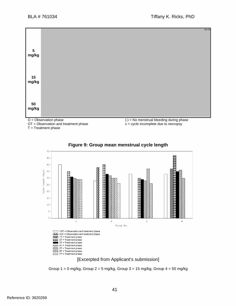
Menstrual cycle Methods: Menstruation was checked by daily vaginal swabs. Results: Females administered 50 mg/kg MPDL3280A experienced irregular
menstruation during the dosing period, including an increase in mean menstrual cycle length compared to controls (Figure 9 and Figure 10). Findings correlated with a lack of newly formed corpora lutea at terminal necropsy.
o Extended menstrual cycles were noted for Animal 18178F (67 days, Week 7-15), Animal 18196F (79 days, Week 4-14), and Animal 18227 (79 days, Week 11-20). Subsequent menstrual cycles were comparable to control animals and within the normal range for Cynomolgus monkeys.
o Animal 18159F showed menstrual bleeding during the pre-dose phase followed by amenorrhea until Week 25 after end of dosing. Regular menstrual cycles were observed for the remainder of study.
o Animal 18187F experienced menstrual bleeding prior to the pre-dose observation phase but not during the pre-dose, dosing, or recovery periods.
o Animal 18232F (15 mg/kg group) experienced menstrual bleeding during the pre-dose period followed by amenorrhea through the end of study.
The Applicant did not consider this finding to be test article-related; however, it is difficult to exclude a causal relationship, given the irregular menstrual cycles and amenorrhea observed in the 50 mg/kg dose group.
Table 14: Individual menstrual cycle lengths in daysAnimal # 1O 1OT 2OT 1T 2T 3T 4T 5T 6T 7T
0 mg/kg
Reference ID: 3920269
(b) (4)
BLA # 761034 Tiffany K. Ricks, PhD
41
5 mg/kg
15 mg/kg
50 mg/kg
O = Observation phaseOT = Observation and treatment phaseT = Treatment phase
(-) = No menstrual bleeding during phasex = cycle incomplete due to necropsy
Figure 9: Group mean menstrual cycle length
[Excerpted from Applicant’s submission]
Group 1 = 0 mg/kg, Group 2 = 5 mg/kg, Group 3 = 15 mg/kg, Group 4 = 50 mg/kg
Reference ID: 3920269
(b) (4)

BLA # 761034 Tiffany K. Ricks, PhD
42
Figure 10: Group mean of individual treatment cycle length
[Excerpted from Applicant’s submission]
Group 1 = 0 mg/kg, Group 2 = 5 mg/kg, Group 3 = 15 mg/kg, Group 4 = 50 mg/kg
Testicular and semen evaluation Methods: Collected semen was analyzed for test article-related effects on sperm
count, motility, and morphology. Males were also assessed for changes in testicular volume, homogeneity, and echogenicity by ultrasound.
Results: Unremarkable
Testosterone analysis Methods: Serum testosterone levels were measured in males using a
radioimmunoassay (RIA). Results: Unremarkable
Toxicokinetics Following a single dose, Cmax and AUC increased with increasing dose and were
generally dose proportional across the dose range tested. Following repeat dosing, systemic exposure in the 5 mg/kg dose group was
greatly reduced due to formation of ATAs. Cmax and AUC increased in monkeys receiving 15 and 50 mg/kg MPDL3280A and were dose proportional at these dose levels.
Systemic exposure was greater on Day 182 compared to Day 1 in animals receiving 15 and 50 mg/kg antibody, indicating accumulation following repeat dosing.
After the last dose, terminal half-life ranged from 11.8 to 23.5 days for recovery animals.
Reference ID: 3920269

BLA # 761034 Tiffany K. Ricks, PhD
43
Table 15: Mean toxicokinetic parameters in monkeys administered intravenous MPDL3280A
Sex Dose (mg/kg/week)
Cmax(µg/mL)
Cmax/D(µg/mL)/D
AUC(0-t)(day∙µg/mL)
AUC(0-t)/D(day∙µg/mL)/D
5 139 27.8 263 52.615 351 23.4 758 50.5Male50 1290 25.8 2880 57.65 107 21.4 224 44.8
15 251 16.7 629 41.9
Day1
Female50 1110 22.2 2690 53.85 7.3 1.5 NA NA
15 1220 81.3 4250 283Male50 4060 81.2 10100 2025 116 23.2 378 75.6
15 1350 90.0 2810 187
Day 182
Female50 3300 66.0 6740 135
Note: Limited data were available for the 5 mg/kg dose level due to anti-therapeutic antibody formation in all animals. Female means in the 5 mg/kg group were calculated from 2 of 3 animals. Following a single dose, AUC was estimated from Day 0 to 3. For TK analysis on Day 182, AUC was estimated from Day 182 to 185.NA – not applicable
Dosing Solution AnalysisSample formulations were analyzed on Day 1, Week 7, 13, 19, 25, and 27 of the dosing phase and were within the acceptance criteria of ± 10% of the intended concentration.
7 Genetic ToxicologyNot conducted
8 CarcinogenicityNot conducted
9 Reproductive and Developmental ToxicologyThe Applicant did not conduct reproductive and developmental toxicology studies with atezolizumab. The current nonclinical literature demonstrates that the PD-L1/PD-1 pathway plays a role in fetal/maternal tolerance, and inhibition of PD-L1 is associated with an increased risk of fetal rejection. The Division agreed with the Applicant’s request to provide a non-product specific literature-based assessment of the reproductive risks associated with blockade of the PD-L1 pathway to inform appropriate labeling for atezolizumab.
During pregnancy, the maternal immune system must maintain the ability to defend against infection while also maintaining tolerance for a fetus expressing and shedding foreign, paternally-inherited antigens. Several mechanisms protect a developing fetus from the maternal immune system, including expression of non-classical MHC molecules on fetal trophoblast cells, clonal deletion and anergy of fetal antigen-specific
Reference ID: 3920269
BLA # 761034 Tiffany K. Ricks, PhD
44
T cells, and the presence of immunomodulatory molecules, including PD-L1 (Guleria and Sayegh 2007, Petroff and Perchellet 2010).
PD-L1 is expressed in the human placenta throughout pregnancy. Expression is low during the first trimester and markedly increases during the second and third trimesters, corresponding to the onset of maternal blood flow (Holets et al. 2006, Petroff et al. 2003). PD-L1 expression is primarily located on fetal trophoblast populations in direct contact with maternal blood and tissue at the fetal/maternal interface. PD-1 receptor has been detected on Tregs and CD4+ and CD8+ T cells within the decidua (Petroff and Perchellet 2010). The spatial and temporal regulation of PD-L1 and PD-1 receptor at the fetal/maternal interface suggests an important role for these immunomodulatory proteins in maintaining fetal tolerance.
Nonclinical studies have demonstrated that loss of PD-L1 activity reduces embryo-fetal survival. In an allogenic pregnancy model (CBA x B6), administration of a PD-L1 blocking antibody to pregnant mice increased the rate of fetal resorption (86%) compared to isotype control (~18%), resulting in a corresponding decrease in litter size (Guleria et al. 2005). Similarly, allogenic pregnancies of PD-L1-/- mice resulted in a mean litter size of 2.7 whereas heterozygous or WT females produced mean litter sizes of 8.5 and 9, respectively. D’Addio et al. assessed the effects of PD-L1 blockade in Th1.1 B6 females mated with Bm12 males and associated changes in fetal antigen-specific T cells (D'Addio et al. 2011). Administration of a PD-L1 blocking antibody increased fetal rejection (34%) compared to isotype control (1.4%) and reduced mean litter size from 8.5 in the isotype control group to 5.8 in the anti-PD-L1 group. PD-L1 blockade reduced fetal antigen-specific Tregs and increased fetal antigen-specific effector T cells in the spleen and lymph nodes.
In another allogenic pregnancy model, Taglauer et al. assessed the role of PD-1 receptor on fetal antigen-specific T cells using a transgenic mouse model, in which T cells expressing the OT-I T-cell receptor recognize ovalbumin (OVA)-derived peptide (Taglauer et al. 2009). B6 WT females were mated with OVA-Tg males. On gestation Day 10.5, pregnant mice were injected with OT-I expressing splenocytes, which recognize parentally-inherited, OVA antigen. Three days post-transfer, proliferating PD-1+ OT-I cells, including CD8+ T cells, were detected in the uterus draining lymph nodes and spleen. Adoptive transfer of PD-1-/- OT-I splenocytes resulted in greater accumulation of OT-I CD8+ T cells in the uterus draining lymph nodes. In vitro analysis of PD-1-/- T cells suggested that T-cell accumulation was due to reduced apoptosis rather than an increase in cell proliferation. Collectively, these nonclinical studies demonstrate that the PD-L1/PD-1 pathway controls the accumulation of fetal antigen-specific T cells during pregnancy, and inhibition of PD-L1 leads to a loss of fetal tolerance and an increased risk of immune-mediated abortion.
It is unclear if atezolizumab crosses the placental barrier at levels that would have adverse effects on developing offspring. The allogenic pregnancy models did not evaluate offspring for teratogenicity or adverse developmental effects. Data from knockout mice demonstrate that syngeneic PD-1-/- and PD-L1-/- fetuses develop
Reference ID: 3920269


BLA # 761034 Tiffany K. Ricks, PhD
46
Study title: Tissue cross-reactivity of MPDL3280A with human and Cynomolgus monkey tissues ex vivo
Study no.: 08-1174Study report location: eCTD 4.2.3.7.
Conducting laboratory and location: Genentech, Inc.South San Francisco, CA
Date of study initiation: October 27, 2008GLP compliance: Yes
QA statement: YesDrug, lot #, and % purity: Biotin-MPDL3280A, from antibody lot #
729339, not provided
The objective of this study was to assess the tissue cross-reactivity of MPDL3280A with a panel of human and Cynomolgus monkey tissues.
MethodsSpecies of tissues Human and Cynomolgus monkeyPositive and negative control samples
HEK293 cells expressing human PD-L1Wild type HEK293 cells
Number of tissue donors 3/speciesTissues Adrenal
BladderBloodBone marrowBreastCerebellumCerebral cortexColonEndothelium (aorta)EyeFallopian tubeGI tract (small intestine)
HeartKidney (glomerulus)Kidney (tubule)LiverLungLymph nodeOvaryPancreasParathyroidPituitaryPlacentaProstate
SkinSpinal cordSpleenStriated muscleTestisThymusThyroidThymusTonsilUreterUterus (cervix)Uterus (endometrium)
Tissue fixation/embedding Cryosections in OCT compound or equivalent were mounted on slides and fixed in acetone.
Test and control article Biotin-MPDL3280A and Biotin-human IgG antibodyTest article concentrations 0.25 µg/mL (optimal), 1.25 µg/mLIHC methods Steps were performed at room temperature.
Washed slides 3X with PBS, incubated in 1X Morphosave for 15 min
Incubated with glucose oxidase solution for 1 h Blocked with avidin and biotin for 15 min followed by 2% gamma
globulin blocking solution for 30 min Incubated with primary antibody for 1 h Incubated with streptavidin-HRP for 30 min followed by DAB
substrate for detection for 5 minNote: Slides were washed with 1X TBST in between each step.
Tissue counterstain Mayer’s hematoxylinTissue validation Anti-CD31 antibody (3 µg/mL) to detect endothelial cellsMicroscopy Light microscopy
Reference ID: 3920269

BLA # 761034 Tiffany K. Ricks, PhD
47
Results and conclusions HEK293 cells expressing PD-L1 showed moderate intensity membrane staining
in the presence of 0.25 and 1.25 µg/mL Biotin-MPDL3280A but not in the presence of Biotin-IgG isotype control. Biotin-MPDL3280A did not bind to wild type HEK293 cells.
Positive CD31 staining was reported in all human and monkey tissues tested, indicating staining of endothelial cells, platelets, or megakaryocytes and validation of tissues in the immunohistochemistry assay.
No specific tissue staining was detected with Biotin-IgG isotype control. Membrane staining was reported only in syncytiotrophoblasts of human placenta.
Cytoplasmic staining was observed in human lymph node, thymus, and tonsils. Staining of Cynomolgus monkey tissues was reported only in the lymph node (1-
3+ cytoplasmic staining of sinusoidal cells; rare to frequent).
Table 16: Summary of Biotin-MPDL3280A cross-reactivity with human tissuesDonor
1Donor
2Donor
30.25
µg/mL1.25
µg/mL0.25
µg/mL1.25
µg/mL0.25
µg/mL1.25
µg/mLLymph node- sinusoidal cell, cytoplasmic Very rare……………………. 1+ 1+ 1+ 1+ neg negPlacenta- syncytiotrophoblasts, apical cytoplasm and membrane Frequent…………………….- chorionic plate, cytoplasm Frequent…………………….
3+
3+
3+
3+
3+
3+
3+
3+
3+
3+
3+
3+Thymus- thymic cortex, cytoplasm Rare………………………… Occasional………………..... Frequent…………………….- medulla, cytoplasm Rare………………………… Occasional………………….
2+
2+
3+
3+
2+
2+
2+
2+
2-3+
2-3+
2-3+
2-3+Tonsil- sinusoidal cells, cytoplasm Very rare……………………. neg 1-2+ 1-2+ 1-2+ 1-2+ 1-2+
Staining intensity: 1+ = minimal, 2+ = mild, 3+ = moderate, 4+ = marked, neg = negative Frequency of stained cells of particular type: very rare <25%, rare 25-50%, occasional >50-75%, frequent 76-100%
11 Integrated Summary and Safety EvaluationPharmacology
Atezolizumab is a humanized IgG1 monoclonal antibody that binds to PD-L1 and blocks its interaction with PD-1 and B7-1 receptors. Atezolizumab contains a mutation in the Fc domain to prevent glycosylation, limit binding to Fcγ receptors, and prevent Fc-mediated depletion of PD-L1 expressing cells (e.g., ADCC). Atezolizumab retains binding to
Reference ID: 3920269
BLA # 761034 Tiffany K. Ricks, PhD
48
neonatal Fc receptors in the presence of the mutation affecting Fcγ receptor binding. In pharmacology studies, atezolizumab bound to HEK293 cells exogenously expressing human or murine PD-L1 (EC50 = 0.4 and 0.1 nM) and to activated human and Cynomolgus monkey T cells (EC50 = 0.4 and 0.7 nM). In a competitive binding ELISA assay, atezolizumab blocked recombinant PD-L1 binding to human and murine PD-1 (IC50 = 82.8 and 104 pM) and B7-1 (IC50 = 48.4 and 75.6 pM) receptors with subnanomolar inhibitory activity. Atezolizumab showed minimal binding to human Fcγ receptors and did not stimulate cytokine release from human PBMCs at concentrations up to 250 µg/mL or ADCC activity at concentrations up to 10 µg/mL.
The Applicant conducted in vivo pharmacology studies to evaluate the effect of PD-L1 blockade in syngeneic tumor models and a mouse model of chronic viral infection. To minimize immunogenicity in mice, the Applicant generated two mouse IgG2a chimeric antibodies (PRO304397 and PRO314483), containing the binding region of atezolizumab. In vitro, the chimeric antibodies bound to PD-L1 with subnanomolar affinity, which was comparable to atezolizumab. In syngeneic tumor models of melanoma and colorectal cancer, a dose of 10 mg/kg anti-PD-L1 reduced tumor growth by ≥ 80% and prolonged time to progression. There were no treatment-related effects on body weight or reported clinical signs.
The Applicant evaluated the effect of PD-L1 blockade in a mouse model of chronic LCMV CL-13 infection using the murine/human chimeric antibodies and an independently derived anti-mouse PD-L1 antibody. LCMV CL-13 is highly virulent and produces an infection that persists for several weeks due to impaired T-cell response. The peak of viral infection and acute T-cell response occur at one week post-infection. A dose of 10 mg/kg anti-PD-L1 caused ~40% mortality when given prior to the peak of infection on Day 2 and 60 to 100% mortality when administered at the peak of infection on Day 7. In contrast, all mice receiving anti-PD-L1 starting on Day 14 or 17 survived to scheduled necropsy with no reported toxicity. Anti-PD-L1 enhanced virus-specific CD8+ T-cell function and reduced viral titers. Similarly, PD-L1-/- mice succumb to LCMV infection during the acute phase, suggesting that the PD-L1 pathway is required to control the acute immune response and prevent immune-mediated damage in the chronic LCMV CL-13 model (Barber et al. 2006). These findings were not observed in other models of acute viral infection.
In a mouse model of bacterial infection, PD-1-/- mice infected with M. tuberculosis showed a significant decrease in survival (Lazar-Molnar et al. 2010). The lungs from these mice showed a dose-dependent increase in bacterial infection and focal areas of necrotizing inflammation. Pro-inflammatory cytokines were markedly increased systemically and in the lungs. In contrast, infected wild type mice survived to the end of study with less severe lung inflammation and bacterial loads. Collectively, animal models demonstrate that blocking the PD-L1/PD-1 pathway may increase the inflammatory response and enhance the severity of some infections.
Safety pharmacology studies were incorporated into repeat-dose toxicology studies conducted in Cynomolgus monkeys. There were no test article-related effects on blood
Reference ID: 3920269
BLA # 761034 Tiffany K. Ricks, PhD
49
pressure, ECG, respiratory rate, or neurological parameters at doses up to 50 mg/kg/week administered for up to 26 weeks.
General Toxicology
The Applicant conducted repeat-dose toxicology studies in mice and Cynomolgus monkeys. In a pilot study, mice received doses up to 50 mg/kg/week IV atezolizumab for three doses. The major toxicological finding was minimal sciatic neuropathy (vacuolation and lymphocytic infiltration) at doses ≥ 10 mg/kg observed at the end of dosing and recovery period. All mice were positive for ATAs, which significantly reduced drug exposure by Day 17.
In GLP-compliant toxicology studies, Cynomolgus monkeys received atezolizumab at doses up to 50 mg/kg/week given SC for 8 weeks or administered IV for up to 26 weeks. The major toxicological finding was minimal to mild multifocal arteritis/ periarteritis in multiple organs including the heart, aorta, kidney, liver, pancreas, epididymis, GI tract, skin, tongue, and female reproductive organs. Arteritis/periarteritis was noted in animals administered ≥ 15 mg/kg SC and 50 mg/kg IV atezolizumab. The findings were reversible following a 3-month recovery period. In the 26-week study, female monkeys administered 50 mg/kg IV atezolizumab experienced irregular menstruation during the dosing period, including an increase in mean menstrual cycle length compared to controls and a corresponding lack of newly formed corpora lutea. Effects on menstruation occurred at an AUC that was approximately 6 times the estimated AUC in patients administered atezolizumab at the recommended dose of 1200 mg every 3 weeks. During the study, the majority of animals were positive for ATAs. At end of dosing (Day 182), mean exposure was markedly reduced in males receiving 5 mg/kg atezolizumab and was increased at doses ≥ 15 mg/kg for both genders compared to Day 1.
Reproductive and Developmental Toxicology
The Applicant did not conduct reproductive and developmental toxicology studies with atezolizumab. Atezolizumab was immunogenic in mice after three weekly doses, resulting in reduced exposures by Week 3. In addition, the scientific literature demonstrates that the PD-L1/PD-1 pathway plays a role in fetal/maternal tolerance and inhibiting this pathway increases the risk of embryo-fetal death in vivo. The Pharmacology/Toxicology team agreed with the Applicant that a non-product specific literature-based assessment was appropriate to characterize the potential risk of embryo-fetal toxicity.
PD-L1 is expressed in the human placenta throughout pregnancy and primarily located on fetal trophoblast cells in direct contact with maternal blood and tissue at the fetal/maternal interface (Holets et al. 2006, Petroff et al. 2003). PD-1 receptor has been detected on T-cell populations within the decidua (Petroff and Perchellet 2010). In allogenic pregnancy models, administration of anti-PD-L1 blocking antibodies to pregnant mice increased the rate of fetal resorption and caused a corresponding
Reference ID: 3920269

BLA # 761034 Tiffany K. Ricks, PhD
50
decrease in surviving pups (D'Addio et al. 2011, Guleria et al. 2005). Similarly, allogenic pregnancies of PD-L1-/- mice resulted in reduced litter sizes compared to wild type and heterozygous controls (Guleria et al. 2005). Blocking the PD-L1/PD-1 pathway reduces fetal antigen-specific Tregs and increases accumulation of fetal-antigen specific T effector cells in maternal lymphoid organs (D'Addio et al. 2011, Taglauer et al. 2009). Collectively, nonclinical studies demonstrate that PD-L1 blockade leads to a loss of fetal tolerance and an increased risk of immune-mediated abortion.
The allogenic pregnancy models did not evaluate offspring for teratogenicity or adverse developmental effects. Data from knockout mice, however, demonstrate that syngeneic PD-L1-/- and PD-1-/- mice develop normally with no malformations. Depending on the genetic background, inhibition of the PD-L1/PD-1 pathway by genetic deletion or blockade results in autoimmune phenotypes and exacerbation of disease in mouse models of autoimmunity. In humans, SNPs located in the gene encoding for PD-1 are associated with autoimmune diseases, including SLE, type 1 diabetes, and rheumatoid arthritis. It is unknown if atezolizumab crosses the placental barrier at concentrations that would cause adverse effects in developing offspring. The potential risk of offspring developing immune-mediated disorders or alterations in the normal immune response cannot be dismissed based on the mechanism of action of atezolizumab.
Special Toxicology Studies
The Applicant evaluated atezolizumab for cross-reactivity to a panel of normal human and Cynomolgus monkey tissues. Atezolizumab bound to the cell membrane of syncytiotrophoblasts in human placenta and within the cytoplasm of human and monkey lymph nodes and human thymus and tonsils. In a hemolysis assay, atezolizumab had no effect on human or Cynomolgus monkey blood at a concentration of 125 mg/mL.
12 Appendix/AttachmentsNone
ReferencesBarber, D. L., E. J. Wherry, D. Masopust, B. Zhu, J. P. Allison, A. H. Sharpe, G. J.
Freeman & R. Ahmed (2006) Restoring function in exhausted CD8 T cells during chronic viral infection. Nature, 439, 682-7.
D'Addio, F., L. V. Riella, B. G. Mfarrej, L. Chabtini, L. T. Adams, M. Yeung, H. Yagita, M. Azuma, M. H. Sayegh & I. Guleria (2011) The link between the PDL1 costimulatory pathway and Th17 in fetomaternal tolerance. J Immunol, 187, 4530-41.
Francisco, L. M., P. T. Sage & A. H. Sharpe (2010) The PD-1 pathway in tolerance and autoimmunity. Immunol Rev, 236, 219-42.
Guleria, I., A. Khosroshahi, M. J. Ansari, A. Habicht, M. Azuma, H. Yagita, R. J. Noelle, A. Coyle, A. L. Mellor, S. J. Khoury & M. H. Sayegh (2005) A critical role for the programmed death ligand 1 in fetomaternal tolerance. J Exp Med, 202, 231-7.
Guleria, I. & M. H. Sayegh (2007) Maternal acceptance of the fetus: true human tolerance. J Immunol, 178, 3345-51.
Reference ID: 3920269

BLA # 761034 Tiffany K. Ricks, PhD
51
Holets, L. M., J. S. Hunt & M. G. Petroff (2006) Trophoblast CD274 (B7-H1) is differentially expressed across gestation: influence of oxygen concentration. Biol Reprod, 74, 352-8.
Keir, M. E., M. J. Butte, G. J. Freeman & A. H. Sharpe (2008) PD-1 and its ligands in tolerance and immunity. Annu Rev Immunol, 26, 677-704.
Lazar-Molnar, E., B. Chen, K. A. Sweeney, E. J. Wang, W. Liu, J. Lin, S. A. Porcelli, S. C. Almo, S. G. Nathenson & W. R. Jacobs, Jr. (2010) Programmed death-1 (PD-1)-deficient mice are extraordinarily sensitive to tuberculosis. Proc Natl Acad Sci U S A, 107, 13402-7.
Okazaki, T. & T. Honjo (2006) The PD-1-PD-L pathway in immunological tolerance. Trends Immunol, 27, 195-201.
Okazaki, T. & T. Honjo (2007) PD-1 and PD-1 ligands: from discovery to clinical application. Int Immunol, 19, 813-24.
Petroff, M. G., L. Chen, T. A. Phillips, D. Azzola, P. Sedlmayr & J. S. Hunt (2003) B7 family molecules are favorably positioned at the human maternal-fetal interface. Biol Reprod, 68, 1496-504.
Petroff, M. G. & A. Perchellet (2010) B7 family molecules as regulators of the maternal immune system in pregnancy. Am J Reprod Immunol, 63, 506-19.
Taglauer, E. S., T. M. Yankee & M. G. Petroff (2009) Maternal PD-1 regulates accumulation of fetal antigen-specific CD8+ T cells in pregnancy. J Reprod Immunol, 80, 12-21.
Reference ID: 3920269
---------------------------------------------------------------------------------------------------------This is a representation of an electronic record that was signedelectronically and this page is the manifestation of the electronicsignature.---------------------------------------------------------------------------------------------------------/s/----------------------------------------------------
TIFFANY RICKS04/20/2016
TODD R PALMBY04/21/2016
Reference ID: 3920269