7 calcium channels - anukarri.anu.edu.au/corry/papers/2006_chapter07_corry... · 2011-05-24 · 7...
TRANSCRIPT

SVNY290-Chung July 25, 2006 11:34
7 Calcium Channels
Ben Corry and Livia Hool
7.1 Introduction
Ion channels underlie the electrical activity of cells. Calcium channels have a uniquefunctional role, because not only do they participate in this activity, they form themeans by which electrical signals are converted to responses within the cell. Calciumconcentrations in the cytoplasm of cells are maintained at a low level, and calciumchannels activate quickly such that the opening of ion channels can rapidly change thecytoplasmic environment. Once inside the cell, calcium acts as a “second messenger”prompting responses by binding to a variety of calcium sensitive proteins. Calciumchannels are known to play an important role in stimulating muscle contraction, inneurotransmitter secretion, gene regulation, activating other ion channels, control-ling the shape and duration of action potentials and many other processes. Sincecalcium plays an integral role in cell function, and since excessive quantities canbe toxic, its movement is tightly regulated and controlled through a large variety ofmechanisms.
The importance of calcium channels is highlighted by the fact many naturallyoccurring mutations in voltage-gated calcium channel proteins are known to un-derlie human disorders including childhood absence epilepsy, familial hemiplegicmigraine, spinocerebellar ataxia type 6 (a severe movement disorder), hypokalemicperiodic paralysis and X-linked congenital stationary night blindness (French andZamponi, 2005). Mutations in mice have also been shown to cause a variety of dis-orders including seizures, atrophy, and lethargy. Knockout mice, deficient in variouscalcium channel pore-forming subunits (see below) are often not viable as calciumchannels play an integral role in muscle function. The knockout mice exhibit ab-normalities in cardiac muscle contraction, paralysis of the diaphragm, deafness, andepilepsy. Mutations of Ca2+ release channel genes are known to be involved incardiac arrhythmias and malignant hypothermia (Priori and Napolitano, 2005).
The history of calcium channels dates back to soon after the discovery byHodgkin and Huxley that Na+ and K+ currents were responsible for the actionpotential in squid giant axon. Fatt and Katz (1953) identified a form of electricalexcitability in crab muscle that occurred in the absence of Na+. In 1958, this phe-nomenon was correctly explained to be due to a “calcium spike,” an influx of calciumduring the upstroke of the action potential (Fatt and Ginsborg, 1958). Calcium spikeswere also measured using radiolabeled calcium in squid axon (Hodgkin and Keynes,1957) and in response to lowering intracellular calcium concentrations (Hagiwara
241

SVNY290-Chung July 25, 2006 11:34
Ben Corry and Livia Hool
and Naka, 1964). The fact that Ca2+ passed through pathways other than Na+ andK+ channels was confirmed with the use of Na+ and K+ channel blockers. Ca2+
currents are generally difficult to measure, as calcium channels are much less denselydistributed than Na+ or K+ channels. Final verification of the hypothesis that Ca2+
passed through independent channels, and detailed investigations to their physiologyhad to wait until the development of the patch clamp and giga-seal techniques thatallowed for recordings to be made from small cells and for single-channel currentsto be recorded. Over the past four decades inward Ca2+ currents have been observedin every excitable cell, a huge variety of calcium channels have been identified andtheir essential biological roles are beginning to be well recognized and characterized.
In this chapter we outline the variety of different calcium channels that areutilized in biological organisms, highlighting their differences, nomenclature andfunction, and recent research. Next we give a more detailed discussion of the role ofcalcium in stimulating muscle contraction, neurotransmitter secretion and control-ling electrical excitability. We then discuss a variety of properties of these channelsexamining such questions as how calcium channels discriminate between differention types and how ions migrate through them. Included in this discussion is an ex-amination of recent theoretical investigations into the nature of ion permeation andselectivity. Finally, we discuss the mechanisms by which ion flow is controlled, re-viewing our knowledge of channel gating voltage- and calcium-dependent channelinactivation and the regulation of calcium channels by mechanisms such as thiolgroup oxidation or reduction.
It is the nature of a topic as large as this that we cannot hope to cover all theareas of current investigation. Instead we have highlighted some areas and focusedour attention particularly on the family of voltage-gated calcium channels. Calcium-release, mitochondrial or nuclear channels are no less interesting and significantunderstanding of their structure and function at both a physiological and molecularlevel has been determined in recent years. We hope that these may form the focus ofanother review chapter in the near future. The reader is referred to a number of recentreviews for further information into voltage-gated (Jones, 1998, 2003; Catterall,2000; Sather and McCleskey, 2003) calcium release (Sutko and Airey, 1996; Georgeet al., 2005; Wehrens et al., 2005) and store-operated channels (Parekh and Putney,2005).
7.2 Types of Ca2+ Channels
Calcium channels come in a range of different shapes and sizes, are located indifferent regions of the cell or different organs, carry out diverse roles and respondto different stimuli. Broadly, calcium channels are distinguished as either voltage-activated or responding to the binding of calcium or other agonists that releasecalcium from intracellular stores. Here we introduce the different types of Ca2+
channels, noting the names applied to them and the functions they fulfil.
242

SVNY290-Chung July 25, 2006 11:34
7. Calcium Channels
7.2.1 Voltage-Gated Channels
Many cell types, notably excitable cells express voltage-gated Ca2+ channels thatplay an integral role in calcium influx and regulate intracellular processes suchas contraction, secretion, neurotransmission, and gene expression discussed in moredetail below. They are members of a gene superfamily of transmembrane ion channelproteins that includes voltage-gated K+ and Na+ channels. Ca2+ channels sharestructural similarities with K+ and Na+ channels in that they possess a pore-forming�1 subunit in four repeats of a domain with six transmembrane-spanning segmentsthat include the voltage sensing S4 segment and the pore forming (P) region. The�1 subunit is large (190–250 kDa) and incorporates the majority of the known sitesregulated by second messengers, toxins, and drugs. This subunit is usually complexedwith at least three auxiliary subunits, �2, �, � and � , with the �2 and � subunits alwayslinked by a disulfide bond (Fig. 7.1).
Nomenclature for the Ca2+ channels has evolved over the decades since theearly electrophysiology studies and has had to incorporate diverse biochemical,pharmacological, and physiological properties of the channels with the more recentidentification of channel genes. Initially, Ca2+ channels were classified accordingto their ability to be activated by a large or small depolarization and whether theresponse was rapid or slow and persistent. For example, L-type Ca2+ channels area member of the HVA or high voltage activation type because the channels are ac-tivated by strong depolarizations typically to 0 or +10 mV and are long-lasting inthat they are slow to inactivate. In addition, they are blocked by the lipid-soluble1,4-dihydropyridines such as nifedipine and BAY K 8644. T-type Ca2+ channels onthe other hand are transiently activated at low membrane potentials and are insensitive
αααα1
αααα2
δδδδγγγγPore
S4 Voltage-sensor
Outside
Inside
AID
I II III IV
N
N
CC
N
C
N C
ββββ
CN
S
Fig. 7.1 Subunits of Cav channels showing the four homologous domains of the �1 subunit, andauxiliary �, � , and �2� subunits. Predicted alpha-helices are depicted as cylinders. AID = alphainteracting domain where the � subunit is proposed to interact with the � subunit (see text).
243

SVNY290-Chung July 25, 2006 11:34
Ben Corry and Livia Hool
to dihydropyridines and fall into the class of low voltage activated (LVA) channels.Taking into consideration the functional and pharmacological differences, Tsien andcolleagues (Tsien et al., 1988) devised a nomenclature based on single letters thatcontinues to be used today to describe the electrophysiological characteristics of ex-pressed channels studied in their native state. In addition to the L-type (long-lastingcurrents) and T-type (transient currents) channels mentioned above, a number ofother channel types were found with single-channel conductances between T- andL-type that were resistant to the dihydropyridines. These channels were predominantin neurons and could be blocked by the �-Conotoxin GVIA from cone snails. Theywere named N-type (Nowycky et al., 1985b). A unique Ca2+ channel was identifiedin Purkinje cells by Linas’ group that exhibited sensitivity to the venom of funnelweb spider (FTX) but was insensitive to �-Conotoxins and dihydropyridines (Llinaset al., 1989). The channel was named P-type. Another type of channel slightly lesssensitive to �-Conotoxins, termed Q-type, was identified in cerebellar granule neu-rons that otherwise has very similar physiological characteristics to P-type channels(Randall and Tsien, 1995). An R-type Ca2+ channel that is resistant to �-Conotoxin,�-Agatoxin IVA, and dihydropyridines but sensitive to �-Agatoxin IIIA was char-acterized as a rapid transient current with an activation potential slightly positive toT-type channels (Randall and Tsien, 1997).
This nomenclature required revision as the molecular identities of calciumchannel genes were rapidly discovered. Channel types were classified according todistinct hybridization patterns of mRNA on Northern blot analysis and grouped asA, B, C, or D (Snutch et al., 1990). Genes subsequently identified were classified Ethrough I except the skeletal muscle isoform that was referred to as �1S (Birnbaumeret al., 1994). In 2000, Ertel et al. (Ertel et al., 2000) suggested a nomenclature basedon that used to classify K+ channels with Ca as the permeating ion followed bythe physiological regulator “v” for voltage in subscript and the gene subfamily inorder of discovery. The Cav1 subfamily comprises the L-type Ca2+ currents, Cav2subfamily comprises the P/Q, N- and R-type Ca2+ currents, and Cav3 subfamily theT-type Ca2+ currents as shown in Fig. 7.2. Detailed listings of voltage-dependentCa2+ channel nomenclature as approved by the Nomenclature Committee of theInternational Union of Pharmacology including structure–function relationships areavailable (Catterall et al., 2003, 2005). Table 7.1 lists the nomenclature of Ca2+
channels as the classifications have evolved.Although the main characteristics that define channel subtypes such as ion
selectivity, voltage-dependence, and drug binding sites reside in the alpha subunit,the auxiliary subunits also play important regulatory roles. The first � subunit to beidentified (now termed Cav�1a) was cloned from skeletal muscle and observed as a54 kDa protein (Takahashi et al., 1987). To date four genes have been cloned encodinghuman � subunits (Cav�1–4) and there are several splice variants produced from thegenes (for review, see Birnbaumer et al., 1998; Dolphin, 2003). Cav� subunits areintracellularly located and bind the � subunit with high affinity via the � interactiondomain (AID) on the I–II linker (see Fig. 7.1; Pragnell et al., 1994). Cav� subunitspromote functional expression of Cav� subunits, localization, and insertion of the
244

SVNY290-Chung July 25, 2006 11:34
7. Calcium Channels
HVA
LVA
α1S Cav1.1
α1c Cav1.2
α1D Cav1.3
α1F Cav1.4
α1A Cav2.1
α1B Cav2.2
α1E Cav2.3
α1G Cav3.1
α1H Cav3.2
α1I Cav3.3
L
P/Q
N
R
T
Fig. 7.2 Family tree of mammalian voltage-gated Ca2+ channels and nomenclature. Adaptedfrom Jones (2003).
channel complex in the plasma membrane (Chien et al., 1995; Bichet et al., 2000). Inaddition, Cav� subunits can modify the kinetics and amplitude of current, includingincreasing the channel open probability and altering the inactivation rate (Colecraftet al., 2002).
The �2� subunit is highly glycosylated and encoded by a single gene. The �segment spans the plasma membrane and connects to the extracellular �2 protein viaa disulfide bridge formed between a number of cysteines present on both proteins.To date four genes have been identified that encode �2�1–4 subunits (Ellis et al.,1988; Klugbauer et al., 1999; Qin et al., 2002). The �2 protein has been shown toinfluence channel stimulation and the � protein alters voltage-dependent activationand inactivation kinetics (Felix et al., 1997).
In some tissues a fourth auxiliary subunit is expressed, the � subunit. At leasteight genes are know to encode for � subunits (Cav�1–8), the first known subunitbeing cloned from skeletal muscle with a mass of 25 kDa (Jay et al., 1990). Co-expression studies reveal the subunit can modulate peak current and activation/inactivation kinetics (Singer et al., 1991).
7.2.2 Ca Release Channels
One of the critical roles served by Ca2+ channels in cell function is the maintenanceof intracellular calcium levels and the initiation of calcium-dependent cellular pro-cesses. This is particularly poignant in cardiac muscle where calcium influx throughplasma membrane Cav1.2 channels is a requirement for the release of calcium from
245

SVNY290-Chung July 25, 2006 11:34
Ben Corry and Livia Hool
Table 7.1
HVA (high voltage activated, large conductance, persistent)Tsien class: L-typeSnutch/Birnbaumer class: �1S �1C �1D �1F
IUP classification: Cav1.1 Cav1.2 a,b,c Cav1.3 Cav1.4Gene: CACNA1S CACNA1C CACNA1D CACNA1FHuman chromosome: 1q31-32 12p13.3 3p14.3 Xp11.23Tissue: skeletal muscle heart, smooth
muscle, brain,adrenal
brain, pancreas,kidney, cochlea,ovary
retina
HVA (high voltage activated, rapid inactivating)Tsien class: P/Q-typeSnutch/Birnbaumer class: �1A �1B �1E
IUP class: Cav2.1a,b Cav2.2 a,b Cav2.3 a,bGene: CACNA1A CACNA1B CACNA1EHuman chromosome: 19p13 9q34 1q25-31Tissue: brain, cochlear,
pituitarybrain, NS brain, cochlea,
retina, heart,pituitary
LVA (low voltage activated)Tsien class: T-typeSnutch/Birnbaumer class: �1G �1H �1I
IUP class: Cav3.1 Cav3.2 Cav3.3Gene: CACNA1G CACNA1H CACNA1IHuman chromosome: 17q22 16p13.3 22q12.3-13-2Tissue: brain, NS brain, heart,
kidney, liverbrain
Tsien class (Tsien et al., 1988); Snutch/Birnbaumer class (Snutch et al., 1990; Birnbaumer et al., 1994); IUP class (Ertelet al., 2000; Catterall et al., 2003, 2005).
IUP = International Union of Pharmacology; NS = nervous system.
intracellular sarcoplasmic reticulum stores that then initiates contraction. This pro-cess is known as excitation–contraction coupling and will be discussed in detail in
Au: Pleaseprovide a tablecaption.
Section 7.3. There are, broadly speaking, three families of protein, known as Ca-release channels, that are responsible for the release of calcium from intracellularstores. They are inositol triphosphate receptors (IP3R), ryanodine receptors (RyR),and calcium-release-activated calcium (CRAC) channels. IP3Rs and RyRs serve torelease calcium into the cytoplasm from the sarcoplasmic reticulum and endoplasmicreticulum of muscle but they are also expressed in may other cell types includingneurons where they play a role in calcium-dependent neurotransmission. In mus-cle the activation of IP3Rs and RyRs is an important step in the process of musclecontraction, allowing intracellular calcium levels to increase and bind to troponinC that then switches on the contractile apparatus. During the course of a singlemuscle contraction, free intracellular calcium increases from resting concentrations(typically 10–300 nM) to micromolar levels. Equally important to the release of cal-cium, however, is the reuptake of calcium into intracellular stores to allow relaxation.The sarcoplasmic reticulum is capable of accumulating millimolar concentrations
246

SVNY290-Chung July 25, 2006 11:34
7. Calcium Channels
Ca2+
ATP
RyR
IP3R
SR
Mit
SERCA
MCU
MTP
NCE
Cav
Cav
SOC
IP3R
IP3R
IP3R
RyR SERCA
nucleus
ER
RyRSERCA
Ca2+ ATPase
NAADP
NCE
Ca2+
3 Na+
-tubule
Golgi
Fig. 7.3 Channels and transporters responsible for regulating calcium in a cell. Mit = mito-chondria, ER = endoplasmic reticulum, SR = sarcoplasmic reticulum, SERCA = sarcoplasmicendoplasmic reticulum ATPase, NCE = Na+/Ca2+ exchanger, SOC = store operated channel,MTP = mitochondrial transition pore, MCU = mitochondrial calcium uniporter, NAADP = nico-tinic acid adenine dinucleotide phosphate-sensitive calcium channel depicted here on lysosomes.
of calcium and this is returned after release by way of ATP-dependent pumps knownas SERCA pumps (sarcoplasmic reticulum, endoplasmic reticulum Ca2+ ATPase).The SERCA family of genes encode five isoforms that are all sensitive to the in-hibitor thapsigargin (Lytton et al., 1991). Like the plasma membrane Ca2+ ATPaseand Na+/K+ ATPase, SERCA utilize the energy produced from the hydrolysis ofATP to drive the pumping of calcium back into sarcoplasmic reticulum stores. Othermeans of removing calcium from the cytoplasm and restoring resting intracellu-lar calcium also exist such as sarcolemmal Na+/Ca2+ exchange and mitochondrialcalcium uptake (Fig. 7.3).
Three receptor types or isoforms of RyR and IP3Rs have been isolated and com-binations of isoforms are co-expressed in different muscle types (Nakagawa et al.,1991; Fill and Copello, 2002). RyRs are localized within membranes of organelleswith stores of calcium (such as the sarcoplasmic reticulum), but IP3Rs have beenidentified in the plasma membrane, endoplasmic reticulum, Golgi apparatus, andnuclear membranes (Khan et al., 1992; Leite et al., 2003). Both RyR and IP3Rs formtetrameric complexes with the channel region located at the C-terminal end of theprotein. There is good evidence that they appear to share common calcium storesand can interact with each other (McGeown, 2004). Activation of both receptortypes is Ca2+-dependent and varies with isoform. RyRs are activated at micromo-lar concentrations of calcium while IP3Rs are generally activated at submicromolar
247

SVNY290-Chung July 25, 2006 11:34
Ben Corry and Livia Hool
concentrations (Bezprozvanny et al., 1991). Therefore the local release of calciumby one receptor can be amplified by activating a nearby receptor. The dependenceof activation on calcium concentration, however, is often bell-shaped so that highconcentrations of calcium also have inhibitory effects on channel function. This isthe case for type I IP3Rs where it has been proposed that the bell-shaped-dependencesupports oscillations in cytosolic calcium, but not for type III IP3Rs where an in-crease in cytosolic calcium causes a further increase in calcium that is suited tosignal initiation (Hagar et al., 1998). In type I IP3Rs the Ca2+-binding sites liewithin the inositol triphosphate (InsP3)-binding core, and it has been proposed thatcalcium binding is negatively regulated by a conformational constraint imposed byInsP3-binding (Bosanac et al., 2002). Both IP3Rs and RyRs favor the conductanceof divalent cations over monovalent cations exhibiting single-channel conductancesof between 30 and 120 pS.
IP3Rs mediate calcium release in response to activation of G protein or tyrosinekinase-coupled plasma membrane receptors. Agonist binding leads to stimulationof phospholipase C resulting in production of diacylglycerol, an activator of proteinkinase C, and IP3. IP3 is a highly diffusible second messenger that binds to IP3Rson membranes of intracellular organelles. There is evidence, at least in neurons,that IP3Rs may be directly activated by G�� subunit following activation of a Gi
protein (Zeng et al., 2003). IP3Rs can be phosphorylated by many kinases. Proteinkinase A phosphorylates IP3Rs at serine 1589 and 1755 (Ferris et al., 1991a) as doescGMP-dependent protein kinase (Komalavilas and Lincoln, 1994). Protein kinase Cand calcium calmodulin-dependent protein kinase also phosphorylate the receptorat distinct sites regulating calcium release in a spatiotemporal manner contributingto formation of calcium microdomains (Ferris et al., 1991b). ATP augments calciumrelease from IP3Rs by stabilizing open channel states thereby allowing positivecontrol of calcium release when SERCA have utilized the local ATP during reuptakeor when the cell is experiencing metabolic inhibition (Mak et al., 1999). CyclicADP-ribose (cADPR) stimulates the association of ryanodine with RyR increasingchannel opening (Guse et al., 1999). IP3 does not bind to RyRs but caffeine andheparin potentiate RyR channel opening.
In nonexcitable cells the predominant mechanism for release of store-operatedcalcium (SOC) is by CRAC channels. The best characterized SOC influx channelis the CRAC channel expressed in T lymphocytes that is essential to the immuneresponse including the regulation of gene expression and cell proliferation. The in-crease in intracellular calcium occurs in a biphasic manner typically with an initialtransient increase due to binding of an agonist such as IP3 and release of internalstores. The depletion of intracellular calcium stores then triggers release of calciumvia CRAC channels on the plasma membrane and is entirely dependent on extracel-lular calcium. Recent evidence indicates that a transmembrane protein with an EFhand motif near the amino terminus located in the lumen of the ER termed STIM-1is the sensor that migrates from the calcium store to the plasma membrane to acti-vate CRAC channels (Zhang et al., 2005). The pore properties and biophysics of theCRAC channel are similar to the calcium transport protein CaT1 (Yue et al., 2001).
248

SVNY290-Chung July 25, 2006 11:34
7. Calcium Channels
CRAC channels have a low single-channel conductance (0.5 pS) with high selectiv-ity for divalent cations and typically exhibit inward rectification. They favor openingwhen the cell is hyperpolarized (Zweifach and Lewis, 1993).
7.2.3 Mitochondrial and Nuclear Ion Channels
Many organelles other than the sarcoplasmic reticulum and endoplasmic reticulumare able to take up calcium and impose strict control over its movement in order toregulate organelle function. As mentioned above, the Golgi complex expresses IP3Rs.It also contains SERCA-type pumps known as secretory pathway Ca2+-ATPase thatare located in close proximity to the nuclear membrane and hence may be involvedin calcium signaling with the nucleus (Wootton et al., 2004). Calcium uptake intoGolgi may occur by thapsigargin-sensitive and thapsigargin-insensitive mechanisms.Interestingly, only the cis-Golgi express IP3Rs unlike the trans-Golgi suggesting thatthe role of the trans-Golgi in vesicle packaging and posttranslational modificationhas differential requirements for calcium compared with the cis-Golgi that appearsto play a greater contribution to spatial and temporal calcium signals (Vanoevelenet al., 2004). Calcium release channels that do not involve IP3 or cADPR have beenidentified on lysosome-related organelles. A nicotinic acid adenine dinucleotidephosphate (NAADP)-sensitive calcium channel has been located on lysosomes inpancreatic � cells that requires proton exchange to assist with calcium loading(Yamasaki et al., 2004).
It is well established that the mitochondria can accumulate calcium. Control ofmitochondrial calcium is necessary for ATP production (McCormack et al., 1990)and the shaping of intracellular calcium signals (Jouaville et al., 1995). Calcium over-load is associated with the processes mediating necrosis and apoptosis (programmedcell death) (Halestrap et al., 2000). In addition, several of the mitochondrial enzymesrequire calcium as a cofactor in higher concentration in the mitochondrial matrix thanthe mitochondrial cytosol, therefore transport of calcium across the mitochondriamust be carefully regulated (Gunter et al., 2004). Calcium accumulation mainlyoccurs through the mitochondrial calcium uniporter (MCU; see Fig. 7.3) locatedon the inner mitochondrial membrane. Recently, the MCU was identified as a cal-cium channel with extremely high calcium affinity (K0.5 of 19 mM) and biophysicalcharacteristics that included an inwardly rectifying current suited to calcium uptakeinto mitochondria and inhibition by ruthenium red (Kirichok et al., 2004). Calciummay exit the mitochondria via the permeability transition pore (MPTP) that is morefrequently associated with the collapse of the membrane potential and influx of pro-teins that lead to cell death (Halestrap et al., 1998, 2000). PTP opening is thought tooccur when a protein, cyclophilin D binds to the matrix side of the PTP. Calcium isrequired for the binding of cyclophilin D. Calcium efflux from the mitochondria alsooccurs via the Na+/Ca2+ exchanger that, similar to the plasma membrane exchangertransports one calcium ion out of the mitochondria in exchange for three sodiumions.
249

SVNY290-Chung July 25, 2006 11:34
Ben Corry and Livia Hool
Although it is recognized that regulation of calcium within the nucleus is vitalfor many functions including gene transcription, little is understood about the reg-ulatory processes involved. The nuclear membrane consists of an envelope with aluminal space capable of storing calcium. It also possesses the components necessaryto regulate calcium levels such as SERCA, IP3Rs, and RyRs (Gerasimenko et al.,1996). Both IP3Rs and RyRs are located on inner and outer nuclear membranes. Thenucleus can generate its own IP3 but it is unclear whether it physically associateswith other calcium stores such as ER and SR. However, there is evidence that the nu-cleus contains its own stores (termed nucleoplasmic reticulum) that may physicallyassociate with the mitochondria suggesting a cooperative role on regulating calciumsignals (Lui et al., 2003).
7.3 Roles of Ca2+ Channels
Calcium is essential in electrical activity in that it shapes the long plateau phase ofthe ventricular action potential and the upstroke and duration of the smooth mus-cle and atrial pacemaker action potentials. However, the influx of calcium throughvoltage-dependent calcium channels is also an essential component in the initiationof intracellular processes. Here we discuss in detail the role of calcium in mus-cle contraction, neurotransmitter secretion, and in controlling electrical excitability,highlighting recent research.
7.3.1 Muscle Contraction
Calcium is the switch that initiates the contraction of muscle fibers. These fibersare formed from many overlapping strands, comprising a thick filament made frommyosin and a thin filament comprised of actin and tropomyosin. When Ca2+ isreleased from intracellular stores, it binds to troponin present on the thin filaments,and allosterically modulates the tropomyosin to unblock a series of myosin bindingsites. The myosin can then be powered by hydrolyzing ATP to move along thesebinding sites causing muscle contraction until the cytoplasmic Ca2+ is removed andthe myosin binding sites are once more blocked.
In the heart calcium influx across the plasma membrane through L-type Ca2+
channels is essential for triggering calcium release from the sarcoplasmic reticulum, aprocess known as calcium-induced calcium-release. This occurs as a result of a closephysical association between dihydropyridine receptors (L-type Ca2+ channels) andtransverse or T-tubules that communicate directly with RyRs on the sarcoplasmicreticulum (Fig. 7.3). Because the activation kinetics of L-type Ca2+ channels inskeletal muscle is 100-fold slower than in cardiac muscle, the process of calciuminflux is too slow to initiate contraction. In skeletal muscle, therefore, a depolarizationof the plasma membrane is the trigger for release of calcium from SR stores ratherthan the influx of calcium itself. Therefore, L-type Ca2+ channels serve as the voltagesensor used to initiate the release of Ca2+ from intracellular store for initiating
250

SVNY290-Chung July 25, 2006 11:34
7. Calcium Channels
contraction as calcium influx across the plasma membrane is not a requirement(Franzini-Armstrong and Protasi, 1997). In smooth muscle, calcium influx throughL-type Ca2+ channels can release calcium from the SR to trigger contraction. Inaddition, a hormone-induced calcium release can initiate contraction through releaseof calcium from internal stores via binding to IP3Rs.
The characteristics of the contractile force in muscle can also be controlled byintracellular calcium. The contractile force can be altered either by increasing theduration or amplitude of the calcium transient (the rise in intracellular calcium) oraltering the sensitivity of the myofilaments to calcium. Stretch increases myofilamentcalcium sensitivity as it enhances actin–myosin interaction (Fukuda et al., 2001b)while acidosis reduces calcium sensitivity and enhances the length-dependence oftension (Fukuda et al., 2001a). Cytosolic calcium must be removed to allow for therelaxation of muscle fibers. The majority of cytosolic calcium uptake occurs by theSR Ca2+-ATPase, with the remaining calcium being extruded from the cell either byNa+/Ca2+ exchange or a small amount being taken up by the mitochondria via theMCU. Na+/Ca2+ exchange is voltage-dependent and reversible with high intracel-lular calcium favoring calcium efflux; while a positive membrane potential and highintracellular Na+ favors Na+ efflux (Dipolo and Beauge, 2006). Under physiologi-cal conditions, Na+/Ca2+ exchange works mostly to extrude calcium driven by theintracellular calcium transient. It is also a secondary active transport process drivenby the Na+/K+ ATPase that extrudes three Na+ ions in exchange for 2 K+ ionswhile hydrolysing one molecule of ATP. If the Na+/K+ ATPase is partially inhibited,calcium influx through Na+–Ca2+ exchange is increased. This is the basis for theincreased force of muscle contractions induced by cardiac glycosides in the heart.
In principle, the Na+/Ca2+ exchanger could be a trigger of intracellular calciumrelease and muscle contraction, but the influx is too slow and too small to do so(Sipido et al., 1997). A contributing factor to this is the fact that Na+/Ca2+ exchangersare not positioned near the SR junctional cleft (Scriven et al., 2000). Promiscuouscalcium entry through tetrodotoxin-sensitive Na+ channels has also been proposedto trigger contraction, also termed slip-mode conductance (Santana et al., 1998).However, this remains controversial since a number of groups have been unable totrigger contraction by this means (Chandra et al., 1999; DelPrincipe et al., 2000).T-type Ca2+ channels are also not located near SR junctions and the current passingthrough them is less than for L-type channels, therefore any contribution they maketo triggering contraction is thought to be small (Sipido et al., 1998; Zhou and January,1998).
IP3Rs are the main trigger of calcium release from SR and ER in smoothmuscle. Generally, calcium release from IP3Rs stimulates further calcium releasein a cooperative fashion, but with high calcium concentrations inhibiting channelfunction. This creates the classical oscillatory pattern of calcium release typicallyseen in innervated smooth muscle such as that resulting in peristalsis in the bowelor agonist mediated calcium release pathways.
RyRs inactivate one of two ways. They either do not reopen until they recover(Schiefer et al., 1995; Sham et al., 1998), or they relax to a lower open probability
251

SVNY290-Chung July 25, 2006 11:34
Ben Corry and Livia Hool
but can still be reactivated at high calcium levels, a process known as adaptationand also common to IP3Rs (Gyorke and Fill, 1993). Adaptation of RyRs enables agraded calcium-induced calcium-release for contraction in muscle.
7.3.2 Neurotransmitter Release and Neuronal Plasticity
Another important physiological process involving calcium is neurotransmission atnerve terminals. Within the presynaptic terminal of neuron hormones, neurotransmit-ters, or other peptides are stored in vesicles. When an action potential depolarizes themembrane, the vesicles fuse with the plasma membrane and the contents are releasedinto the extracellular space where they can bind with receptors and initiate chemicalactivity in nearby cells. The process of neurotransmission is calcium-dependent inthat secretion requires extracellular calcium and is inhibited by extracellular Mg2+
(Douglas, 1968). In addition, intracellular calcium modulates the activity of a num-ber of ion channels involved in this process. Intracellular calcium influences theinactivation rate and inhibition of L-type Ca2+ channels (see above) and activateslarge conductance K+ channels BK(Ca) that terminate neurotransmitter release inthe presynaptic terminal by hyperpolarizing the membrane (Storm, 1990; Sah, 1996).BK(Ca) are expressed in virtually all excitable cells, have long and large unitary con-ductances typically 100–250 pS and can be blocked by charybdotoxin, iberiotoxin,and tetraethylammonium compounds. The BK(Ca) channels are derived from theslo family of genes originally cloned from the Drosophila mutant slowpoke. Inter-mediate (IK) and small (SK) conductance channels of K(Ca) family have also beencloned and characterized (Vergara et al., 1998). Calcium-dependent chloride chan-nels also contribute to membrane potential hyperpolarization along with the BK(Ca)channels in neurons (Mayer, 1985).
The release of neurotransmitters is steeply calcium dependent and a de-crease in calcium channel activity contributes to presynaptic inhibition (Dunlapand Fischbach, 1978). An example of presynaptic inhibition is the block of trans-mission of sensory fibers that occurs with opiates such as morphine. This occurs as aresult of rapid coupling of G protein �� subunits to Cav2 family of calcium channelscausing a slowing of activation of the channels (Dolphin, 1998). Several modulatorytransmitters such as acetylcholine, norepinephrine, adenosine, and GABA contributeto depression of calcium channels at presynaptic terminals through the G protein-dependent mechanism.
Both presynaptic and postsynaptic calcium stores are involved in long-term de-pression in the developing hippocampus (Caillard et al., 2000). However, presynapticAu: Please
include thisref. in thereference list.
RyRs appear to be important in determining the strength of synaptic transmissioninduced by NMDA receptors (Unni et al., 2004). Activation of presynaptic NMDA,kainate, and nicotinic acetylcholine receptors can lead to calcium-induced calcium-release from RyR stores similar to that recorded in muscle that acts to inhibit orregulate secretion. Although still controversial, calcineurin may interact with IP3Rsand RyRs to modulate intracellular calcium release and synaptic plasticity (Bultyncket al., 2003).
252
SVNY290-Chung July 25, 2006 11:34
7. Calcium Channels
Calcium can also play a role in building synaptic connections. Rapid changesin synaptic strength are mediated by posttranslational modifications of preexistingprotein but enduring changes are dependent on gene expression and the synthesis ofa variety of proteins required for synaptic transmission. One of the key mediatorsof gene expression involved in synaptic plasticity is the transcription factor cAMPresponse element binding protein (CREB). This transcription factor plays an im-portant role in behavioral adaptations to changes in the environment and complexprocesses such as learning and memory. It is also important in proliferation anddifferentiation in developing vertebrates. Calcium influx through L-type Ca2+ chan-nels or NMDA receptors is required for phosphorylation and activation of CREB(Kornhauser et al., 2002). Another transcription factor responsible for shaping long-term changes in neurons is the NFAT family, and similar to regulation of CREB,activation of NFAT is dependent upon calcium influx through L-type Ca2+ chan-nels. Interactions of calcium with PDZ domain proteins (PSD-95, Discs-large, ZO-1domain proteins that often function as scaffolding proteins and have been shownto play important roles in signal transduction) appear to be necessary for the cou-pling of L-type Ca2+ channels to NFAT as does CREB activation (Weick et al.,2003). Neurotrophins, such as the brain derived neurotrophic factor, have also beenimplicated in the activation of NFAT-dependent transcription. Initiating such tran-scriptions leads to further increase in brain derived neurotrophic factor mRNA andprotein as positive feedback (Groth and Mermelstein, 2003).
7.3.3 Excitability and the Action Potential
Calcium plays an important role in shaping the action potential of muscle and neu-rons. One example where the shape and duration of the action potential must becarefully controlled is in cardiac pacemaker activity. This originates from eitherspecialized cells located in the right atrium of the heart known as sinoatrial nodecells or from secondary pacemaker cells located within the atrioventricular nodeand also through the Purkinje fibers of the ventricles. Pacemaker action potentialsare designed to originate from the sinoatrial node cells so that the resting heartbeatis maintained at 60–80 beats min−1. If the pacemaker fails in its duty, secondarypacemakers in the atrioventricular node or in the purkinje fibers are activated to takeover and maintain ventricular function and ultimately cardiac output, albeit at a lowerheart rate. The spontaneous activity of pacemaker cells is attributed to a phase of theaction potential known as the slow diastolic depolarization. During this phase, themembrane potential slowly depolarizes following termination of an action potentialuntil the threshold for a new action potential is reached. It is well accepted that theelectrical activity of cardiac pacemakers is thought to depend exclusively on voltage-dependent ion channels within the plasma membrane of nodal cells. These includethe hyperpolarization-activated inward current (If), the time-dependent decay of K+
conductance, inward L-type and T-type Ca2+ currents, and the lack of backgroundK+ conductance. The hyperpolarization-activated channel carries Na+ and K+ ionsinwards and activates upon hyperpolarization. However, interventions that affect
253

SVNY290-Chung July 25, 2006 11:34
Ben Corry and Livia Hool
intracellular Ca2+ can also affect pacemaker activity. A rise in intracellular Ca2+
results in activation of the Na+/Ca2+ exchanger. This produces an inward currentthat is sufficient to induce diastolic depolarization and pacemaker activity (Ju andAllen, 1998). The importance of Ca2+ in contributing to pacemaker firing is furthersupported by studies that show that embryonic stem cell-derived cardiomyocytesdo not show increased beating rate with differentiation when they lack ryanodinereceptors (Yang et al., 2002).
The rate of change in membrane potential during the upstroke is much slowerin sinoatrial node cells than in ventricular cells. This is because depolarization of themembrane during phase 0 (the rapid upstroke) of a slow response action potential inpacemaker cells is caused by an increase in the Ca2+ conductance due to activationof L-type Ca2+ channels. Nodal cells have fewer Na+ channels than ventricularcells and because the membrane potential in phase 0 is depolarized, changes in Na+
conductance do not contribute. Pacemaker cells do not have a pronounced plateauphase. Action potential duration is determined by a balance between Ca2+ and K+
conductances.In ventricular myocytes the prolonged phase 2 (plateau phase) of the action po-
tential is characterized by slow inward current produced by L-type Ca2+ channels.This is a distinguishing feature of the cardiac action potential and is the necessarytrigger for calcium-induced calcium-release and contraction. Similarly in smoothmuscle and skeletal muscle calcium influx through L-type Ca2+ channels are im-portant in shaping the duration of the action potential, however, the duration ismuch shorter than that of ventricular myocytes. In smooth muscle and neurons thecalcium-dependent large conductance K+channels contribute to hyperpolarizationof the membrane.
7.4 Ion Selectivity and Permeation
It is clear that to carry out their biological role, calcium channels have to be selective,allowing Ca2+ ions to pass while blocking Na+, K+, Cl−, and other ions. A highdegree of specificity is important for these channels as Na+ ions are more than 100-fold more numerous in the extracellular solution than Ca2+. Voltage-gated calciumchannels are extremely discriminating, selecting calcium over sodium at a ratio ofover 1000:1 (Hess et al., 1986), yet the picoampere currents they carry require overone million ions to pass through a single channel every second (Tsien et al., 1987). Acentral problem in understanding the function of these channels is to determine howthey can be both highly selective while still passing so many ions. A narrow pore canblock the passage of large ions, but, Na+ and Ca2+ are similar in size and much largerions are known to permeate the channel (McCleskey and Almers, 1985). Thus, theseproteins must distinguish between ions using more than just size. Instead, it appearsthat calcium channels utilize the different strengths of interaction between ions andthe protein as well as a multi-ion conduction process to obtain ion selectivity. Here
254

SVNY290-Chung July 25, 2006 11:34
7. Calcium Channels
we focus the discussion of selectivity on L-type voltage-gated calcium channels asthese have been most widely studied. It is hoped that many of the mechanisms of thischannel will apply to a variety of proteins in this family, particularly as the importantresidues required to obtain calcium specificity are highly conserved.
7.4.1 High-Affinity Binding of Permeant Ions
A surprising characteristic of voltage-gated calcium channels is that monovalentions conduct through the channel at much higher rates than any divalent ions whenno divalent ions are present (Kostyuk et al., 1983; Almers and McCleskey, 1984;Fukushima and Hagiwara, 1985; Hess et al., 1986; Kuo and Hess, 1993a). But, thesemonovalent currents are blocked when the calcium concentration reaches only 1 �M(Kostyuk at al., 1983; Almers et al., 1984). Indeed, the ability of some ion speciesto block currents carried by others gave the first clues to the origin of ion selectivityin these channels, suggesting that it relied on high-affinity binding of ions withina single file pore. Those ions that bound most strongly in a single file pore wouldblock the passage of other ions.
A large number of mono- and divalent ion types pass through the pore, buta characteristic sequence in which some ion types block the passage of others hasbeen observed. In addition to Ca2+ blocking monovalent cations, Sr2+ current isalso blocked by Ca2+ (Vereecke and Carmeliet, 1971) while Cd2+, Co2+, and La3+
block currents carried by Ca2+ even though all of these species were known to passthrough the channel at some rate (Hagiwara et al., 1974; Lansman et al., 1986; Chow,1991). Furthermore, Co2+ was found to block Ca2+ currents less effectively thanBa2+ currents, even though Ba2+ currents were found to be larger than Ca2+ currents(Hagiwara et al., 1974). This property was explained by positing that Ca2+ binds morestrongly to the pore than Ba2+ but not as strongly as Co2+. Together these data sug-gested a high-affinity binding site in the pore, with an order of affinity estimated frombi-ionic reversal potentials to be La3+ > Cd2+ > Co2+ > Ca2+ > Sr2+ > Ba2+ >
Li+ > Na+ > K+ > Cs+ (Reuter and Scholz, 1977; Fenwick et al., 1982; Lee andTsien, 1984; Hess et al., 1986; Taylor, 1988).
One complication, however, has been observed. Although Ca2+ was found toprevent monovalent currents, Na+ was also found to attenuate currents carried byCa2+ in N-type channels (Polo-Parada and Korn, 1997). Although this appears tocontradict the earlier results, it can easily be explained if these Na+ ions competefor entry into the pore rather than permanently occupying the high-affinity bindingsite (Polo-Parada and Korn, 1997; Corry et al., 2001).
Single-channel conductance measurements on the other hand, have the exactreverse order of conductance values than the binding affinity: La3+ < Cd2+ < Co2+
< Ca2+ < Sr2+ < Ba2+ < Li+ < Na+ < K+ < Cs+ (Hess et al., 1986; Kuo andHess, 1993a,b). The inverse relationship between binding affinity and permeationwere first explained with the so-called “sticky-pore” hypothesis. In this, ions thatare bound with higher affinity pass through the channel more slowly and so have alower conductance (Bezanilla and Armstrong, 1972). For example, the lower current
255

SVNY290-Chung July 25, 2006 11:34
Ben Corry and Livia Hool
carried by Ca2+ than by Ba2+ can be understood if Ca2+ binds more strongly in thepore, and thus takes longer to dissociate once bound. Indeed it has been observedthat permeabilities for monovalent and divalent ions are ordered according to anEisenman sequence that models selectivity between ions as a balance between thedehydration and binding energy of each (Eisenman, 1962). According to Eisenman,the permeability sequence observed in calcium channels can be explained by anelectrostatic binding site of strong field strength.
A paradox arises, however, when explaining ion selectivity with high-affinitybinding. How can ions conduct through the pore at picoampere rates if they are boundso strongly? This paradox disappears if one considers the implications of evidencethat calcium channels are multi-ion pores.
7.4.2 Calcium Channels Have a Multi-Ion Pore
A number of lines of evidence suggest that calcium channels must bind multipleions. Multi-ion conduction is a natural consequence of the sticky-pore model asotherwise there should not be a difference in conduction rates between ion types. Ina single ion pore, ions of all binding affinities conduct equally well. Those that bindmore effectively pass more slowly through the pore, but they are also more likely toblock the passage of lower affinity ions (Bezanilla and Armstrong, 1972).
Other strands of evidence also suggest a multi-ion pore. In many cases whentwo permeating species are mixed the current is lower than for either species on itsown. This phenomenon is seen clearly for mixtures of Ca2+ and Ba2+ and also Ca2+
and Na+ (Almers and McCleskey, 1984; Hess and Tsien, 1984) in calcium channelsand has also been observed in gramicidin (Neher, 1975) and potassium channels(Hagiwara et al., 1977). This so-called “anomalous permeability” or “anomalousmole fraction effect” is most easily explained supposing the pore contains multipleions. If ions moved independently through the pore, then the current in mixed so-lutions should always lie between the currents observed with either permeating ionon its own. If, however, the channel holds multiple ions then it is possible that unfa-vorable interactions between the ions in the pore could reduce the current. As notedby Hille (2001), it is possible to explain the anomalous permeability by supposingsome form of conformational or chemical alteration of the channel protein causedby binding of ions exterior to the pore, rather than by a multi-ion pore. But, suchan explanation lacks the simplicity of the multi-ion model that is also supported byother data.
Another important strand of evidence for multiple ions lies in the measurementof two differing affinities for Ca2+ binding. Ca2+ blocks Na+ currents with an affinityof ∼1 �M, while calcium currents saturate with an affinity closer to 14 mM. Thiscan easily be explained by a multi-ion pore if the first affinity represents binding ofa calcium ion to either an empty pore or a pore containing one Na+. However, ifone Ca2+ ion is already present in the pore, then electrostatic repulsion between thetwo ions means that the second ion is bound more weakly giving rise to the secondaffinity measured.
256

SVNY290-Chung July 25, 2006 11:34
7. Calcium Channels
-
-Na Na Na
Na permeation
-Ca Ca
-
Ca permeation Block -Ca Na
-Attenuation
-Ca Na
-
Ca
(A) (B)
(C) (D)
Fig. 7.4 “Electrostatic” model of permeation and selectivity in calcium channels. (A) Whenone Ca2+ ion is electrostatically bound to the negative charge of the EEEE locus, it can only beremoved with the aid of Coulomb repulsion from another Ca2+ion. (B) The lesser repulsion fromNa+ is unable to displace a resident Ca2+ ion, thus Ca2+ blocks Na+ currents. (C) Na+ is ableto pass through the channel in the absence of Ca2+ in a “knock on” mechanism involving threeions. (D) Na+ can attenuate Ca2+ currents by slowing the entry of the second Ca2+ ion requiredfor conduction.
The presence of multiple ions in the pore is also supported by the fact thatcalcium block of Na+ currents is dependent on membrane voltage (Fukushima andHagiwara, 1985; Lux et al., 1990) and on the direction of ion movement (Kuo andHess, 1993a,b). Further evidence that two divalent ions can inhabit the pore comesfrom a detailed study of the blocking of Ba2+ currents by Cd2+ (Lansman et al.,1986). Raising the concentration of Ba2+ was found to increase the unbinding rateof Cd2+ as if increasing the probability of Ba2+ also being present in the poreenhances the exit of Cd2+.
A multi-ion pore provides a simple explanation of the permeation and selectivityproperties of the channel as depicted schematically in Fig. 7.4. The selectivity—conductivity paradox is avoided by utilizing ion–ion interactions inside the selectivityfilter. As soon as one Ca2+ ion occupies the channel, it binds tightly to the pore andthereby blocks the passage of Na+. However, if the second site in the pore is occupiedby a calcium ion then the strong electrostatic repulsion between the two ions allowsone to overcome its interaction with the protein and exit the channel at a relativelyfast rate (Almers and McCleskey, 1984; Hess and Tsien, 1984). Further details of themechanisms of selectivity are described below in the context of various theoreticalmodels.
As noted by Hille, the concepts of binding and repulsion described here need notrefer to specific details (Hille, 2001). Binding sites need not be specifically shapedor involve stereochemical fit such as in the active sites of enzymes. Rather it is morelikely to refer to regions of space that form local energy minima created by broadelectrostatic interactions between the ion and the protein. Furthermore, the locationsof such minima are likely to change when, for example, other ions are nearby. Thusa multi-ion pore need not contain two or more specific localized sites, but could just
257

SVNY290-Chung July 25, 2006 11:34
Ben Corry and Livia Hool
have one or more regions of negative potential. Similarly, the repulsion between ionsin the pore may be direct Coulomb repulsion between like charged particles or mayinvolve interaction through the protein, conformational changes or restrictions onthe entropic freedom of nearby ions.
7.5 Channel Structure
It has long been recognized that voltage-gated sodium, potassium, and calciumchannels are related. Not only do they share many functional similarities, the aminoacid sequences indicate many structural similarities as well. This similarity is nolonger surprising as we now know that many proteins have evolved in families andthat all these channel types most likely evolved from a common ancestor. This meansthat we can learn much about one member of the family from findings made aboutother members. As no atomic resolution images of calcium channel structures exist,much has been learnt about their structure since the recent determination of crystalstructures of a number of potassium channels (Doyle et al., 1998; Jiang et al., 2003;Long et al., 2005).
7.5.1 Subunit Structure and Function
Voltage-gated calcium channels are formed by the association of separate subunits,which come together to form an active channel. As noted previously the varietyof voltage-gated calcium channels arises from differences in the sequences of theindividual subunits comprising the channel in particular the �1 subunit. As withvoltage-gated sodium channels, but unlike the potassium channels, the pore-formingdomain of the calcium channel protein is comprised of four regions of the one subunitrather than by separate subunits.
The first step in determining the structural characteristics of these channelswas the purification, cloning, and sequencing of the protein. Purification was firstachieved from transverse tubule membranes of skeletal muscle (Curtis and Catterall,1984). As described previously, this study and further analysis (Hosey et al., 1987,Leung et al., 1987, Striessnig et al., 1987, Takahashi et al., 1987) indicated thatthis channel was comprised of five subunits: �1 weighing 190 kDa and later shownto form the pore; a disulphide linked �2-� dimer of 170 kDa; and intracellularphosphorylated � subunit weighing 55 kDa; and a transmembrane � subunit of 33kDa as indicated in Fig. 7.5.
Subunits of similar sizes were soon purified form cardiac L-type channels(Chang and Hosey, 1988; Schneider and Hofmann, 1988) whereas neuronal L-typechannels revealed �1, �2-�, and � but no � subunit (Ahlijanian et al., 1990). Thesimilarity between these various L-type channels also extends to neuronal N-type(McEnery et al., 1991; Witcher et al., 1993) as well as P/Q-type channels (Martin-Moutot et al., 1995, 1996; Liu et al., 1996) that have also been purified and containthe �1, �2-�, and � subunits. A possible � subunit has also been found in P/Q
258

SVNY290-Chung July 25, 2006 11:34
7. Calcium Channels
Fig. 7.5 The likely structure and arrangement of the L-type channel subunits. The pore is formedby the S5 and S6 helices and linker of the �1 subunit. The � subunit interacts with the linker betweendomains I and II of �1 and modifies gating and inactivation kinetics. The � and � subunits lie inthe membrane while �2 is accessible to the extracellular solution.
channels (Letts et al., 1998) that plays a role in modulating the voltage dependenceof the channels, yielding even more similarity with the L-type channels. Cloningand sequencing of the five subunits from L-type channels has since been achievedin skeletal (Tanabe et al., 1987; Ellis et al., 1988) and cardiac muscle (Mikami et al.,1989).
The �1 subunit has a structure similar to the previously cloned pore forming asubunit of the voltage-gated Na+ channels. It contains four repeated domains (I–IV)each of which includes six transmembrane segments (S1–S6) and a membrane-associated loop (The “P-loop”) between segments S5 and S6 as indicated in Fig. 7.5.That the �1 subunit forms the ion-conducting pore is supported by the fact thatexpression of just this subunit is enough to produce function channels, albeit withunusual kinetics and voltage dependence (Perez-Reyes et al., 1989). The four re-peated domains are also remarkably similar to the four subunits known to formthe voltage-gated potassium channels. With the aid of the crystal structure of thepotassium channel the basic structure of the �1 subunit is clear. However, prior tothe existence of this crystal structure, a large number of structural and functionalchannels in the voltage-gated Na+, K+, and Ca2+ channels had already characterizedthe organization and role of many parts of the protein (see the review by Hofmannet al., 1999, for example). Each of the six transmembrane segments most likely form�-helices. The four repeated domains surround the ion-conducting pore, with theS5, S6, and P-loops lining the conduit. Much evidence discussed below, and analogywith the potassium channels implies that ion selectivity takes place in a region sur-rounded by the four P-loops. The S4 helix contains a number of arginine residues andis believed to act as the voltage sensor whose action is described in more detail below.
259

SVNY290-Chung July 25, 2006 11:34
Ben Corry and Livia Hool
7.5.2 Pore Structure
Many lines of reasoning suggest that the pore must be narrow enough to force ions topass in single file. Firstly, the blockage of currents carried by some ion types by otherpermeant ions is indicative of single file binding within the pore. The discriminationbetween different like-charged ion types requires close range interaction between theprotein and the permeating ions. Next, the largest known ion to pass through the poreof L-type channels is tetramethylammonium,with a radius of ∼2.8 A (McCleskeyand Almers, 1985), suggesting that the pore acts as a sieve that is too narrow forlarger ions to pass. As discussed below, the selectivity properties of the channel canbest be explained assuming a single file pore in which ions cannot pass each other.Finally, the similarity to K+ channels that have a known single file pore structure ispersuasive.
Surprisingly, the first evidence for the regions of the pore that determine ionselectivity came from a study of sodium channels. Replacement of particular lysineor alanine residues in the P-loop of a voltage-gated sodium channel with gluta-mate residues changed the channel from being sodium selective to calcium selective(Heinemann et al., 1992). Not only did this suggest that this region of the pore wascritical for ion selectivity, the fact that all calcium channels have conserved gluta-mate residues at this position suggested that it was these glutamates that are crucialfor Ca2+ selectivity.
Further, site-directed mutagenesis indeed determined that four glutamateresidues, one from each of the P-loop of the channel (often called the “EEEE locus”),were responsible for the high-affinity calcium binding site. Mutation of one or moreof these had a significant effect on the channel selectivity (Kim et al., 1993; Yanget al., 1993; Ellinor et al., 1995; Parent and Gopalakrishnan, 1995; Bahinski et al.,1997). Removing any one of these residues was found to decrease the relative per-meability and binding affinity of Ca2+. Replacing all of these glutamate residueswith either glutamine or alanine residues was found to remove the specificity of thechannel for Ca2+ over Na+ altogether and left only weak affinity for Ca2+ in the poreas shown in Fig. 7.6 (Ellinor et al., 1995). As no evidence of other high-affinity bind-ing sites was apparent for divalent ions entering either end of the channel (Cibulskyand Sather, 2000) the EEEE locus was posited to be the sole origin of ion selectiv-ity. Mutation of any one or any pair of the glutamate residues affected the bindingaffinity significantly, indicating that all four residues participated in the binding ofeven a single ion. Importantly, not all the glutamates are identical as the mutationof each has a slightly different effect on the blockage rate of ions in the pore. Pre-sumably, this is a consequence of structural differences between the four repeateddomains, although the functional importance of this nonequivalence is yet to be fullyunderstood.
It is easier to make sense of how all four glutamate residues can contribute tocreating a high-affinity binding region that can hold more than one ion by remem-bering that these binding sites can represent broad regions of local energy minimum.In this way, for example, the negative charge of the glutamate residues can create a
260

SVNY290-Chung July 25, 2006 11:34
7. Calcium Channels
Fig. 7.6 The influence of quadruple mutants of the EEEE locus on high-affinity binding of Ca2+.The fraction of Li+ current remaining is plotted against Ca2+ concentration for the wild-type(WT), quadruple glutamine (Q4, squares), and quadruple alanine (A4, triangles) mutants.
single attractive region of the pore that can hold multiple ions in a semistable equi-librium. In this model, mutation of any one of the charged residues would influencethe binding affinity of both the first and second ion entering the pore.
The determination of the atomic structure of a bacterial potassium channelby X-ray crystallography (Doyle et al., 1998) also aided the understanding of cal-cium channel structures. The organization of the membrane spanning domains ofvoltage-gated calcium, potassium, and sodium channels are all quite similar, andthe discovery that the P-loop formed the selectivity filter of potassium channelsgave credence to the view that this region would also be important in calciumchannels.
An important difference is evident in the structures of the potassium and calciumchannel selectivity filters. Whereas ions are stabilized by the backbone carbonylgroups in voltage-gated potassium channels, most people believe that Ca2+ ionsinteract with the side chains of the glutamate residues in calcium channels. This beliefis supported by a number of tests. Protons are found to be able to block Ca2+ currents(Root and MacKinnon, 1994), presumably by weakening electrostatic attraction ofthe pore. Point mutations within the EEEE locus suggested that multiple glutamateresidues interacted with a single proton (Chen et al., 1996; Chen and Tsien, 1997;Klockner et al., 1996), suggesting both that these residues were in close proximityand that their charged side chains were accessible to the interior of the pore. Also,when individual residues in the EEEE locus were replaced with cysteine, the sidechains of the cysteine residues appeared to be accessible to the solvent as they couldbecome ionized to react with methanethiosulfonate (Koch et al., 2000; Wu et al.,2000). The bulky methanethiosulfonate groups also blocked the pore upon reaction,supporting the conclusion that the EEEE locus lay inside the pore.
261

SVNY290-Chung July 25, 2006 11:34
Ben Corry and Livia Hool
7.6 Theoretical Models of Permeation and Selectivity
7.6.1 Rate Theory
As the understanding of calcium channel permeation and selectivity was progressingthrough innovative experimental techniques, a number of theoretical models werealso proposed to help elucidate the mechanisms underlying these properties. For along time, especially when detailed structural information was lacking, the mainapproach utilized was rate theory. In this, the channel is represented as a series ofenergy wells and barriers, with each well representing an ion-binding site and itsdepth being proportional to the binding affinity. Ion conduction is described as thehopping of ions between the wells, with the rate of flux over each barrier related tothe exponential of its height. All rate models include at least one binding site forcalcium with an energy well depth of about 14 kT to account for the micro molardissociation constant determined since micro molar concentrations of Ca2+ blockmonovalent currents.
When the multi-ion nature of permeation was determined the first models de-picted this with two separate energy wells representing two binding sites, surroundedby two larger barriers as depicted in Fig. 7.7. A single ion in the pore could movebetween these sites, but not over the barrier to exit the channel (arrow in Fig. 7.7A).When a second ion entered and occupied the second site, however, it was proposed
(A)
(B)
2-site model
Stairsteps
Fig. 7.7 Rate models of ion conduction. The energy landscape in the pore is represented by aseries of energy wells and barriers. Ion conduction is slow for a single ion in the pore as it has toovercome a large energy barrier to move out of the channel (arrows). In the two site model (A), iontransit is increased due to interactions between ions in each site altering the energy landscape (B).In a “stair step” model (C and D) the ion can climb out of the deep energy well into low-affinitysites on either side.
262

SVNY290-Chung July 25, 2006 11:34
7. Calcium Channels
that the repulsion between them alters the energy landscape and reduces the barrierfor dissociation as shown in Fig. 7.7B (Almers and McCleskey, 1984; Hess andTsien, 1984). As noted by Sather and McCleskey (2003), the nature of the ion–ionrepulsion is ambiguous in this model as it deals only with the shape of the energylandscape not the source.
When it was discovered that there was only one binding region in the channelcreated by the EEEE locus, the idea of two similar binding sites had to be modified.A new model emerged in which there was only a single high-affinity binding site, butthis was flanked by low-affinity sites that helped ions to step out of the large well (Kuoand Hess, 1993a; Dang and McCleskey, 1998) and the role of ion–ion interactionwas relegated (Fig. 7.7C and D). Other mechanisms involving single siteshave alsobeen developed involving competition between calciumions for the binding charges(Armstrong and Neyton, 1991; Yang et al., 1993). However, it is difficult within ratetheory to accurately model a single binding region that is capable of holding manyions.
The rate theory models are surprisingly simple, yet they capture a number ofthe salient features of permeation and selectivity. In particular they helped to providea simple explanation of how a multi-ion pore can utilize repulsion between ions toachieve both specificity and high throughput (cf. Fig. 7.4). However, these modelscan impart only limited information about the origins of specificity as the energylandscape is usually derived from measurements of binding affinities and is notdirectly related to the structure of the pore. For example, these models cannot impartinformation about which residues are responsible for ion binding and as no physicaldistances are used there is no direct connection between energy minima used in thetheory and physical sites within the pore.
7.6.2 Physical Models
More recently there have been a large number of theoretical studies involving physicalmodels of the channel protein. Information about the functional characteristics of themodel is then determined by applying either a continuum theory, Brownian dynamicssimulations, Monte-Carlo calculations, or molecular dynamics simulations.
In the first of these, Nonner and Eisenberg (1998) modeled the channel as asimple cylinder with conical vestibules and included the charge of the EEEE locusin the channel walls. By adjusting the diffusion coefficients and including an excesschemical potential parameter that helps ions to bind in the pore they are able touse the drift-diffusion equations (or Poisson–Nernst–Planck theory as it is oftenknown) to reproduce the conductance and selectivity of the channel. They find thatCa2+ ions bind to a broad region in the interior of the pore due to the Coulombpotential of the glutamate residues as well as the excess chemical potential, and thatthe multi-ion repulsion may be important in speeding flux. Questions raised aboutthe validity of the continuum description of ions in narrow pores (Moy et al., 2000;Corry et al., 2000a,b), the difficulty in treating ion–ion interactions in this model,
263

SVNY290-Chung July 25, 2006 11:34
Ben Corry and Livia Hool
and the arbitrary nature of the adjustable diffusions and chemical parameters limitthe impact of these conclusions.
A different explanation of the origins of ion selectivity than that proposed in therate representation was proposed in two later studies employing different calculationschemes (Monte-Carlo simulations and a Mean Spherical Approximation method;Boda et al., 2000; Nonner et al., 2000). In these models eight partially chargedoxygens comprising the glutamate side chains protrude into a cylindrical pore andcan freely diffuse within a limited region of the channel. Cation binding is achieveddue to the electrostatic attraction of the negatively charged oxygens. Selectivity ofCa2+ ions is proposed to be a result of ions competing to achieve charge neutralityin a selectivity filter having finite space. Ca2+ ions are preferred to Na+ in the modelas they have the same charge neutralizing effect as two Na+ ions while occupyingless of the limited volume of the filter. Essentially, not enough Na+ ions can besqueezed in to the filter to achieve charge neutrality. This charge–space competitionmodel was analyzed further by Yang et al. (2003), who conducted nonequilibriummolecular dynamics simulations of a model pore containing half-charged oxygens inan atomistic cylinder. In these simulations a second Ca2+ ion is required to release aCa2+ already bound in the channel, but cation binding was found to be nonselective.
A simple version of the single file ion–ion repulsion model of permeation andselectivity has been presented by Corry and coworkers who simulate the trajectoriesof ions moving in and around models of the channel protein (Corry et al., 2000c,2001, 2005). For this purpose, Brownian dynamics simulations are used in which theions pass through the channel in a random walk subject to electrical forces. In thesestudies a rigid model of the channel is derived from analogy to other channels and avariety of experimental data. The most important aspect of the channel model is that itincludes a relatively narrow region in which ions cannot pass each other, surroundedby the four glutamate residues as shown in Fig. 7.8. Unlike the previous models, theglutamate charges do not compete for space within the pore, but the concentrationof negative charge does attract cations within this region. Indeed the electrostaticattraction of the protein is all that is required to account for ion permeation andselectivity in this model. The charge of the glutamate residues creates a deep energywell that strongly attracts multiple ions. In a process akin to that suggested by earlierrate models, repulsion between two resident Ca2+ ions is found to speed their exitas illustrated in Fig. 7.4. Because the divalent Ca2+ ions are more strongly attractedby the channel, they can displace Na+ to occupy this region. Once there, the Ca2+
can only be moved by the repulsion from another divalent ion and not by the lesserrepulsion from Na+ (Fig. 7.4). As well as providing a simple mechanism for ionselectivity depicted schematically in Fig. 7.4, this model was also used to calculatethe conductance of the channel in a variety of situations and replicate and explainthe observed I –V curves, current concentration curves, anomalous mole fractionbehavior between Na+ and Ca2+ (Fig. 7.8B) as well as the attenuation of Ca2+
currents by Na+.All the models described thus far assume simplified pore geometries, but one
goal of ion channel modeling is to include all the atomistic detail of the pore in
264

SVNY290-Chung July 25, 2006 11:34
7. Calcium Channels
(A)
Calcium concentration (M)
Nor
mal
ized
cur
rent
0.0
0.5
1.0
Na+Ca2+
10–8 10–6 10–4 10–2100
0
1
10–8 10–4
(B)
–
– –
(Å)
Å Å Å Å
(Å)
Fig. 7.8 Brownian dynamics model of an L-type calcium channel. (A) The channel shape containsa narrow portion of the pore surrounded by the charge from the four glutamate residues (circles).(B) Results from Brownian dynamics simulations in mixed Na+and Ca2+solution reproduce the“anomalous mole fraction” behavior seen in the experimental data (Almers et al., 1984). Figuresfrom Corry et al. (2001).
molecular simulations. Some caution should be applied when taking this approach,however, as the accuracy of an atomistic model cannot be determined until an atomicresolution structure is determined experimentally. A first attempt to make a molecularmodel of the outer vestibule of an N-type calcium channel was presented by Doughtyet al. (1995), who suggested a �-hairpin motif for the P-loop segment of the protein.In contrast, more recent models suggest that the one half of the P-loop actually formsan �-helix (Guy and Durrel, 1995; Lipkind and Fozzard, 2001; Barreiro et al., 2002).Lipkind and Fozzard (2001) created an atomic resolution homology model of theouter vestibule and filter of an L-type channel and determined the likely positionsof ions, and the resulting electrostatic fields, within it. The model, based on theKcsA crystal structure, included the P-loops as �-helix-turn-�-strand motifs, with
265

SVNY290-Chung July 25, 2006 11:34
Ben Corry and Livia Hool
the glutamate residues of the EEEE motif located at the turns. The glutamate sidechains point into the pore and form a 6 A wide conduit in the absence of Ca2+. WithCa2+ present the glutamate residues move inward and form an electrostatic trap thatfirmly holds a single calcium ion. At millimolar concentrations they find that twoadditional ions can be bound either side of the first Ca2+, and this enables Ca2+ toescape the electrostatic trap and permeate through the channel.
A second model of the P-loop has also been made that also places the importantglutamate residues of the EEEE locus at a bend at the end of an � helical segment,but places them asymmetrically in two distinguishable planes to account for theasymmetries seen in site-directed mutagenesis (Barreiro et al., 2002). Moleculardynamics simulations are carried out on this model pore containing either two Ca2+
or one Ca2+ and one Na+. In a 100-ps simulation they find that the two Ca2+ moveapart but the Ca2+ and Na+ do not. It is hypothesized that Na+ cannot displace Ca2+
but that another Ca2+ can, similar to the results seen in the Brownian dynamicsmodel, although the simulations may be too short to asses the significance of theseconclusions. This mechanism of permeation and selectivity is supported by a secondstudy into the potential of mean force of Na+ or Ca2+ in this channel model (Barreiroet al., 2003).
Ramakrishnan et al. (2004) also carry out nonequilibrium molecular dynam-ics simulations of a much simpler model pore containing glutamate residues in a�-barrel scaffold. Apparent off rates when the pore was preloaded with three Na+
ions are much higher than when the channel contained one Ca2+ and two Na+, con-sistent with Ca2+ blockage. As in the other models, the presence of multiple Ca2+
ions was able to release this block.The rate theory and physical models all share some common elements. Per-
meation involves multiple ions, most likely two ions for divalent ion conduction orthree ions for monovalent ion conduction. Divalent ions are strongly attracted by theglutamates in the P-loops and this results in divalent blockage of monovalent ioncurrents.
These models present two similar but distinct physical mechanisms underlyingion selectivity. In both, the electrostatic attraction of the negatively charged glutamateresidues is essential for attracting multiple ions into the channel and the size of thepore is such that ions cannot pass each other. In this way, the glutamate residuescreate an attractive binding region. We avoid using the term “binding site” here asthe physical models all suggest a broader electrostatic attraction rather than anykind of chemosteric binding. The difference between the explanations of selectivitylies in whether this electrostatic attraction is of itself enough to create selectivity orwhether a specific volume and flexibility of the pore is also necessary.
In the simpler “electrostatic” model of ion selectivity, Ca2+ is bound morestrongly by the channel due to its greater charge and can only be removed by therepulsion of a similarly charged ion as represented in Fig. 7.4. In the “charge–volume” model, the volume of the pore must be such that competition for space aswell as electrostatic attraction is important. Given the limited structural informationavailable it is difficult to say exactly which, if either, of these mechanisms is correct.
266

SVNY290-Chung July 25, 2006 11:34
7. Calcium Channels
An advantage of the simpler model is that less is required; it sets much weaker limitson the size and flexibility of the pore. The only geometric limitation required isthat permeation through the selectivity filter occurs in single file. In both cases thecharge surrounding this region needs to be appropriate to hold two divalent ions ina semistable equilibrium. The simpler electrostatic model also stresses a commontheme that has arisen out of rate theory, continuum and simulation models, someof which are able to elegantly reproduce and explain a wealth of physiological datafor a variety of different channel types. It appears that the extra complication ofsetting limits on the channel volume and flexibility, and appealing to charge–spacecompetition to explain selectivity is not necessary.
It is of interest in this context to compare models of selectivity in calciumchannel with explanations of valence selectivity in Na+ and K+ channels. As notedin the models of the calcium channel, the discrimination between monovalent anddivalent cations cannot just rely on presence of a net negative charge as this actsto attract both kinds of ion. Rather, selectivity between monovalent and divalentions appears to rely on the exact strength of attraction to the protein (or bindingaffinity) of the permeating ions. A divalent ion has a stronger electrostatic interactionwith any charge on the protein than a monovalent ion due to its larger charge. Arecent study suggests that this fact on its own appears to be enough to explain howpotassium channels are blocked by Ba2+. When Ba2+ enters the selectivity filter it iselectrostatically bound such that it is unlikely to leave, even with the aid of repulsionfrom nearby cations (Corry et al., 2005). Electrostatic calculations in model calciumchannels suggest that the binding of divalent ions is not as strong as in the potassiumchannel (Corry et al., 2000c, 2001, 2005). In this case, once a divalent ion entersit can only be forced out with the aid of the Coulomb repulsion from a seconddivalent ion, but not with the weaker repulsion from a monovalent ion. Divalent ionsare known to block sodium channels, but with a much lower affinity and durationthan in potassium channels (Taylor et al., 1976; French et al., 1994). Electrostaticcalculations on a model sodium channel (Vora et al., 2004; Corry et al., 2005) find thatdivalent binding is much weaker than in either the calcium or potassium channels.In this case once a divalent ion enters the channel, additional ions are not attractedinto the channel to help relieve the channel blockage through ion–ion repulsion. Butalso, being less strongly bound, the divalent ion dwell time is much shorter than forpotassium channels and so divalent block is less effective.
7.7 Channel Gating
7.7.1 Voltage-Activated Gating
As the name suggest, voltage-dependent calcium channels open in response to de-polarization. The open probability rises to a maximum at large depolarizations andis generally faster in muscle than in neuron. Fitting the open probability with aBoltzmann factor, it can be estimated that three to five gating charges move across
267

SVNY290-Chung July 25, 2006 11:34
Ben Corry and Livia Hool
Fig. 7.9 Activation kinetics of whole cell inward calcium currents in frog sympathetic neurons.Measurements are made with 2 mM Ba2+, and the largest currents are at −20 mV and the largesttail currents at −50 mV. Figure reproduced with permission from Jones (1998).
the electric field of the membrane during activation (Kostyuk et al., 1988; Coulteret al., 1989). The opening of calcium channels does not happen instantaneouslyafter depolarization, but rather after a delay of one or more milliseconds depend-ing on the channel type and membrane potential as illustrated in Fig. 7.9. This isgenerally taken to mean that the channel protein must move through many closedconformations before channel opening occurs.
Open channels are normally deactivated immediately upon repolarization ofthe membrane. Notably, a transient tail current, observed as a spike in the currentimmediately after repolarization, occurs before conduction ceases. As noted by Jones(1998), this tail current may serve an important functional role, increasing the cal-cium intake upon hyperpolarization following an action potential. Measured tailcurrents typically follow either a single or double exponential time course, howeverthe double exponential behavior has been shown in many cases to be the result oftwo or more types of Ca channel closing at different rates (Matteson and Armstrong,1986; Swandulla and Armstrong, 1998).Au: The year
1988 has beenchanged into1998 as per thereference list.Is this OK?
There also appear to be a number of means by which calcium currents intocells can be increased in a process known as facilitation. An increase in calcium cur-rents created by depolarizing prepulses was first noticed in bovine chromaffin cellsby Fenwick et al. (1982). More recent examinations have demonstrated an almostdoubling of Ca2+ currents created by such prepulses (Artalejo et al., 1990). Theseenhanced currents may have a functional role in creating enhanced neurotransmittersecretion or muscle contraction in response to danger or stress. A possible contrib-utor to this effect is delayed channel deactivation that has been noted for some HVAchannels previously subjected to large depolarization (Pietrobon and Hess, 1990),in particular for P-type (McFarlane, 1997) and L-type channels (Sculptoreanu et al.,1993) where it is believed to intensify muscle contraction. In extreme cases, continual
268

SVNY290-Chung July 25, 2006 11:34
7. Calcium Channels
calcium channel activity has been seen for many seconds after a series of large de-polarizations (Cloues et al., 1997). It has been suggested that such behavior couldbe a result of voltage-dependent phosphorylation of the channel protein or of chang-ing the kinetics of the voltage sensor, and may be related to the “mode switching”behavior described below.
As described in the chapter by Bezanilla, the S4 segment of the �1 subunitis believed to be the voltage sensor for channel gating, moving across the electricfield of the membrane and causing a conformational change of the protein to openthe pore. There have been many fewer studies of gating of Ca2+ channels than K+
or Na+ channels, however, the sequence similarities between the S4 regions meansthat many of the insights gleaned from studies on the K+ or Na+ channels are likelyto apply to Ca channels as well. By similarity with K+ channels it is expected thatthe channel gate lies at the internal end of the S6 helices, which most likely form anarrowing of the pore in the closed state.
This internal location is also supported by studying blockage of Ca2+ currentsby Cd2+, which suggest an internal gate as well as a possible gate at the externalend of the pore. Cd2+ is thought to tightly bind in the selectivity filter in the samehigh-affinity binding site utilized by Ca2+. Cd2+ blockage of the open channel isvoltage-dependent: at large hyperpolarizing or depolarizing voltages block becomesalleviated as if Cd2+ can be forced out either end of the pore (Thevenod and Jones,1992; Swandulla and Armstrong, 1999). Block of the resting closed channel is not Au: The year
1989 has beenchanged into1999 as per thereference list.Is this OK?
alleviated at large hyperpolarizing voltages as if Cd2+ cannot easily exit the internalend of the pore. But, when Cd2+ is applied to closed channels, blockage is muchslower than in open channels, and relief of block is much slower if Cd2+ is washedout while the channels are closed. Together these data suggest that Cd2+ cannoteasily enter either end of the pore in the closed state (Thevenod and Jones, 1992).Jones (2003) raises an interesting question in regard to these data. If Cd2+ can stillexit a closed channel, albeit at a much slower rate than an open channel, is the closedchannel really closed? Could for example small Ca2+ currents also pass through aclosed pore? The question is not as absurd as it sounds, as a small leakage currentof only around 1% of the open channel current would be difficult to detect in singlechannel recordings, but could have important physiological consequences.
7.7.2 Mode Switching
L-type channels have also been observed to have another interesting gating behaviorknown as “mode switching.” Typically, a single channel will open and close manytimes during a prolonged depolarization, with each opening lasting less than 1 mson average. But, on occasion there are times when there is a series of much longerchannel openings as if the channel deactivation was 100 times slower. The typicalbehavior has been termed “mode 1”, while the long channel openings is called “mode2” (Nowycky et al., 1985a; Hess et al., 1986). At other times channel opening appears
Au: The year1984 has beenchanged into1986 as per thereference list.Is this OK?
to cease altogether (“mode 0”). Most likely, there is some physical change takingplace to create this mode switching behavior, such as a conformational change or
269

SVNY290-Chung July 25, 2006 11:34
Ben Corry and Livia Hool
chemical modification of important amino acid residues. This is supported by evi-dence that antagonistic drugs such as dihydropyridine appear to bind more stronglyto channels in mode 0, and help stabilize that state, whereas agonists bind most fa-vorably during mode 2. It has been demonstrated that phosphorylation (most likelynear the C-terminus of the �1 subunit) can increase the open probability (Bean et al.,1984) and the mean open time of channel opening (Yue et al., 1990) and this maybe related to the mode switching behavior (Dolphin, 1996).
7.8 Inactivation of HVA Channels
The influx of calcium is essential for initiating cellular responses; however, theseresponses must be controlled within a narrow range of calcium concentrations asintracellular calcium is toxic in excessive levels. For this reason calcium entry istightly controlled in a number of feedback mechanisms. During prolonged periodsof depolarization the initial channel activity is lost in a process known as inactivationthat prevents the excess buildup of intracellular calcium. Inactivation can be eithervoltage-dependent, or alternatively calcium influx can also be controlled by theconcentrations of internal calcium that can inactivate channels, or create increasedcurrents (facilitation).
7.8.1 Voltage-Dependent Inactivation
Ca channels can be inactivated in a Ca2+-independent, voltage-dependent man-ner. For L-type calcium channels of muscle the rate of voltage-dependent inactiva-tion is much slower than Ca-dependent inactivation (Kass and Sanguinetti, 1984;Giannattasio et al., 1991), but this is not necessarily the case for other types of HVAchannel (Werz et al., 1993; Zhang et al., 1993). Some N-type channels, for example,show no voltage-dependent inactivation (Artalejo et al., 1992), while others convertbetween inactivating and noninactivating modes (Plummer and Hess, 1991). Mostlikely these modes are caused by structural or chemical alterations in the protein.
Repeated short depolarizing bursts can also induce rapid inactivation (Patilet al., 1998). It has been proposed that inactivation occurs preferentially from apartially activated state in which some of the voltage sensors have moved to theactivated position, but the channel remains closed (Klemic et al., 1998; Patil et al.,1998). While long depolarizing pulses tend to drive all the channels to the fully openstate, short repeated pulses might place more channels in this partially activated stateand increase the likelihood of inactivation. This possibility could also explain theinactivating and noninactivating modes of the N-type channel and why N-type chan-nel inactivation is maximal at voltages that also produce maximal inward currents(Jones and Marks, 1989).
In many K+ and Na+ channels, inactivation has been demonstrated to takeplace via a “ball and chain” mechanism in which the amino terminus of the �subunit acts as a tethered ball that can enter the channel, occluding the pore and
270

SVNY290-Chung July 25, 2006 11:34
7. Calcium Channels
blocking conduction. Recent evidence suggests that a similar mechanism mightform the basis of voltage-dependent inactivation in HVA calcium channels (Stotzet al., 2004). Considerable evidence suggests that both the intracellular ends ofthe S6 helices (see for example, Kraus et al., 2000; Stotz et al., 2000; Stotz andZamponi, 2001) and the linker between domains I and II (the AID region; Herlitzet al., 1997; Cens et al., 1999; Berrou et al., 2001) are involved in voltage-dependentinactivation. The simplest way to reconcile these facts is that the S6 helices form adocking site for the I–II linker that when bound partially occludes the pore. Furtherevidence suggests that the channel � subunit, as well as the N, C, and III–IV linkerscan influence inactivation kinetics by interacting with the I–II domain (Stotz et al.,2004). Recent crystal structures of the � subunit–AID complex represent the firstdetailed pictures of calcium channel components and indicate that binding occurs bythe formation of a deep hydrophobic groove in the � subunit that can associate with�1 (Chen et al., 2004; Van Petegem et al., 2004). Furthermore, the structure of the� subunit itself indicates how cellular signaling molecules could interact with the� subunit and therefore influence the kinetics of channel inactivation by altering theinteraction with �1.
7.8.2 Ca-Dependent Inactivation and Facilitation
Ca2+-dependent inactivation (CDI) was initially observed in L-type calcium chan-nels from muscle where it was found that inactivation was faster in solutions con-taining Ca2+ than Ba2+. In muscle, CDI is rapid (� ∼ 20 ms). In neurons, CDI ofL-type channels can be either fast (Kohr and Mody, 1991) or slow (Heidelbergerand Matthews, 1992) depending on the situation. For many years non-L-type HVAchannels were believed to not undergo CDI. It recently became apparent, however,that one of the reasons that CDI was not observed was that most recordings weremade in solutions containing high levels of the calcium buffer EGTA. When this wasdropped to more physiological conditions calcium-dependent regulation has beenobserved in P/Q- type channels (Lee et al., 1999; DeMaria et al., 2001; Chaudhuriet al., 2005) and more recently N- and R-type channels (Liang et al., 2003). Thereasons for EGTA masking CDI in non-L-type channels, but not in L-type channelsis due to the differing molecular mechanisms involved as described below. In ad-dition to CDI, it has been shown that non-L-type HVA calcium channels also havea mechanism of calcium-dependent facilitation (CDF), in which calcium influx isamplified by internal calcium concentrations. The study of CDI has progressed sig-nificantly in the last few years, and it is beyond the scope of this chapter to cover allthe history of the area. For an introduction to the field, the reader is referred to anexcellent review (Budde et al., 2002).
Early attempts to understand the mechanisms of CDI revolved around physio-logical studies of L-type channels. CDI was observed in single channel recordings,suggesting that Ca2+ can inactivate the same channel through which it enters the cell(Yue et al., 1990). Buffering of internal Ca2+ concentrations by BAPTA reduces,but does not eliminate inactivation (Ginnattasio et al., 1991) as if calcium from the
271

SVNY290-Chung July 25, 2006 11:34
Ben Corry and Livia Hool
same or nearby channels (Yue et al., 1990) is inactivating the channels before beingchelated by the buffer.
A detailed understanding of the phenomenon of CDI and CDF only took placewhen calmodulin (CaM) was identified as the Ca2+ sensor (Lee et al., 1999; Petersonet al., 1999; Qin et al., 1999; Zuhlke et al., 1999). The basic principle of bothCDI and CDF appears to involve the specific binding characteristics of CaM inits different Ca2+ loaded states. Inactivation and facilitation are inhibited whencalmodulin is bound to the channel in its unloaded form. Local or global changesin calcium concentration load the CaM in different ways, which initiate differentbinding patterns between the CaM and the �1 subunit C-terminus which promoteeither CDI or CDF.
The location of CaM binding has now been pinpointed to two regions of theC-terminal domain of the �1 subunit: a calmodulin binding domain (CBD) and anisoleucine-glutamine (IQ) or similar motif as mutations of these regions influenceCDI and CDF (Qin et al., 1999; Zuhlke et al., 2000; DeMaria et al., 2001; Lee et al.,2003; Kobrinsky et al., 2005). The details of CaM binding are beginning to revealthe mechanism of Ca2+ regulation and the differences between L- and non-L-typeinactivation. CDI and CDF are believed to be regulated independently by the bindingof Ca2+ to the N-terminal or C-terminal CaM lobes (N-lobe and C-lobe respectively).Binding of Ca2+ to the C-lobe takes place at high affinity while the binding affinityof Ca2+ to the N-lobe is much weaker (Wang, 1985; Johnson et al., 1996). For thisreason, C-lobe binding can reflect local changes in Ca2+ concentrations created bylocal influx through calcium channels, while N-lobe binding is likely to take placeonly once the global concentration, resulting from the influx through many channels,has increased significantly.
Inactivation is triggered by the binding of Ca2+ to the opposite lobes in thetwo classes of channel. In L-type channels, CDI is governed by binding of Ca2+ tothe C-lobe (Peterson et al., 1999), whereas in non-L-type channels it is regulated bybinding to the N-lobe (DeMaria et al., 2001; Lee et al., 2003; Liang et al., 2003). CDFin non-L-type channels, on the other hand, appears to involve binding to the C-lobe(Lee et al., 2003). Although CDF is not usually detected in L-type channels, a CaMmutation that limits Ca2+ binding to the C-lobe promotes clear CDF as if it arisesthrough Ca2+ binding via the N-lobe (Van Petegem et al., 2005). The presence of thefast acting Ca2+ chelator BAPTA influences both CDI and CDF in all channel types.The slower acting by high-affinity chelator EGTA, on the other hand, prevents CDF inL-type channels and CDI in non-L-type channels. The sensitivities to these chelatorssupport the conclusion that the C-lobe processes are triggered by local Ca2+ whichcan be rapidly intercepted by BAPTA but not by EGTA. N-lobe processes appear tobe the result of global Ca2+ influx (Lee et al., 2003; Liang et al., 2003). The differentresponses of CaM to local and global Ca2+ concentrations allow the voltage-gatedcalcium channels to accurately control calcium influx.
The exact details of CaM binding to the �1 subunit and the differences inbinding between the unloaded, C-lobe loaded, and fully loaded CaM are still beingworked out, but an interesting mechanism for regulating CDI and CDF in P/Q-type
272
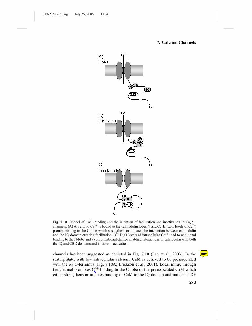
SVNY290-Chung July 25, 2006 11:34
7. Calcium Channels
Fig. 7.10 Model of Ca2+ binding and the initiation of facilitation and inactivation in Cav2.1channels. (A) At rest, no Ca2+ is bound to the calmodulin lobes N and C. (B) Low levels of Ca2+
prompt binding to the C-lobe which strengthens or initiates the interaction between calmodulinand the IQ domain creating facilitation. (C) High levels of intracellular Ca2+ lead to additionalbinding to the N-lobe and a conformational change enabling interactions of calmodulin with boththe IQ and CBD domains and initiates inactivation.
channels has been suggested as depicted in Fig. 7.10 (Lee et al., 2003). In theresting state, with low intracellular calcium, CaM is believed to be preassociatedwith the �1 C-terminus (Fig. 7.10A; Erickson et al., 2001). Local influx throughthe channel promotes C2+ binding to the C-lobe of the preassociated CaM whicheither strengthens or initiates binding of CaM to the IQ domain and initiates CDF
273

SVNY290-Chung July 25, 2006 11:34
Ben Corry and Livia Hool
(Fig. 7.10B). A global increase in Ca2+ encourages Ca2+ binding to the CaM N-lobe. The fully loaded CaM then binds to the CBD domain (while possibly remainingbound to the IQ domain) creating a conformational change that promotes inactivation(Fig. 7.10C).
A recent structure of Ca2+/CaM bound to the calcium channel IQ domainindicates the details of CaM–�1 interactions (Van Petegem et al., 2005). CaM isseen to bind to the IQ domain primarily through anchoring of a set of aromaticresidues in the C-terminus into hydrophobic pockets of CaM. As predicted, C-lobeCaM interactions appeared to be of higher affinity than N-lobe interactions. It issuggested that the difference in CDI and CDF between channel subtypes may be theresult of differing CaM anchor positions and the interplay between CDI and CDFappears to arise from competition for binding between Ca2+-loaded N and C-lobes.
One of the big remaining questions in this area is how the binding or unbindingof calmodulin is linked to changes in the channel pore. What are the conformationalchanges induced by partially or fully loaded CaM? Does CDI utilize the same inac-tivation gate as voltage-dependent inactivation? If so, what is the link between theC-terminus and the I–II linker of the �1 subunit? What are the differences betweenL-type and non-L-type channels that are responsible for the opposite effects of Ca2+
binding to the CaM C-lobes? How is the calcium influx enhanced by CDF? It shouldalso be noted that in addition to the calmodulin-dependent inactivation mechanisms,it has been suggested that calcium inhibits the channel via activation of proteinphosphatase 2B and/or by inhibition of adenylate cyclase (Schuhmann et al., 1997a;You et al., 1997).
7.8.3 Gating of LVA Channels
LVA channels have different activation, deactivation, and inactivation kinetics thanHVA channels. As described earlier, activation arises at more negative potentials.Inactivation is very rapid in these channels (� ∼ 10 to 50 ms) and also arises atmore negative potentials (Huguenard, 1996). In contrast, LVA channels are slowerto activate than HVA channels and are also 10-fold slower to deactivate upon hyper-polarization (Armstrong and Matteson, 1985).
7.9 Regulation of Channel Function
Many factors influence the function of voltage-gated calcium channels so that bio-chemical pathways can influence intracellular calcium levels. The electrochemicalgradient associated with varying concentrations of ions across the plasma membrane,notably calcium ions and H+ ions, influences channel gating, activation, and inacti-vation as described previously. It is well recognized that voltage-gated Ca2+ channelfunction is also regulated by many serine–threonine and tyrosine kinases as a resultof direct phosphorylation of the channel protein. However, more recently there is
274

SVNY290-Chung July 25, 2006 11:34
7. Calcium Channels
good evidence that the cell’s reduction–oxidation state can also influence channelfunction. In this section the factors that influence channel function will be discussedwith particular emphasis on the effects of oxygen and oxygen metabolites on theredox state of the channel protein and its function.
Acidosis has been reported to inhibit L-type Ca2+ channels by reducing chan-nel availability whereas alkalosis increases channel availability (Klockner and Isen-berg, 1994). The � subunit of the channel complex may be required for this effect(Schuhmann et al., 1997b), as if chemical modifications in the � subunit may affectits interaction with �1.
The �1 subunit is phosphorylated by cAMP-dependent protein kinase A atSer-1928 near the carboxy terminus and this results in an increase in the macroscopicpeak inward current while slowing the rate of inactivation (McDonald et al., 1994).It is now recognized that this response requires a direct association of protein kinaseA with the channel via a leucine zipper interaction with a kinase anchoring protein(Hulme et al., 2003). Protein kinase G inhibits both basal channel activity and theisoproterenol stimulated current in a voltage-dependent manner that may involvecompetition with cAMP (Wahler et al., 1990). Tyrosine kinase activation can increasechannel activity in smooth muscle (Wijetunge and Hughes, 1995) and mediates �-adrenergic receptor inhibition of �-adrenergically stimulated current at the level ofthe �-adrenergic receptor in cardiac myocytes probably by phosphorylation of the�-adrenergic receptor (Hool et al., 1998; Belevych et al., 2001).
Activators of protein kinase C (PKC) have been reported to have no effect(Lacerda et al., 1988; Mamas and Terrar, 2001), inhibit (Tseng and Boyden, 1991;Zhang et al., 1997; McHugh et al., 2000), and stimulate (Dosemeci et al., 1988; Heet al., 2000; Aiello and Cingolani, 2001; Alden et al., 2002; Blumenstein et al., 2002)basal channel activity while decreasing �-adrenergic receptor sensitivity of the chan-nel (Schwartz and Naff, 1997; Belevych et al., 2004). The variable responses withpharmacological activators of PKC (such as phorbol esters) may be due to nondis-crimatory activation of all PKC isoforms. PKC is a large family of lipid-derivedserine/threonine kinases that are classified according to their mode of activation andhomology in the regulatory domain. The classical isoforms (cPKC) are activated byCa2+ and diacylglycerol (DAG). This group are the most abundant and comprise the�, �I, �II, and � isozymes. �I and �II are C-terminal splice variants of the same genethat differ by 50 amino acids. The novel PKCs (nPKC) comprise ε, , �, and PKCand are activated by DAG but not Ca2+. The atypical PKCs (aPKC) are dependent onlipids but do not require DAG or Ca2+for activation. Isoforms appear to be restrictedto particular cell sites before stimulation and stimulation results in the localization ofisoforms to specific subcellular sites. For example, stimulation of cardiac myocyteswith an �-adrenergic receptor agonist causes the translocation of �I PKC from thecytosol to the nucleus and �II PKC from fibrillar structures outside the nucleus to themembrane (Mochly-Rosen et al., 1990; Disatnik et al., 1994; Mochly-Rosen, 1995).The translocation is then assumed to be associated with specific function at the site.The localization of activated isoforms requires protein–protein interactions betweenthe isoforms and anchoring proteins. These anchoring proteins bind unique sites
275

SVNY290-Chung July 25, 2006 11:34
Ben Corry and Livia Hool
on each isoforms and thereby regulate the activity of individual isoforms. It wouldappear that different isoforms of PKC produce variable responses depending on thelevel of expression and activity in tissue. The use of specific peptide inhibitors ofisoforms has helped to elucidate the role of PKC in physiological and pathologicalprocesses. Zhang et al. (1997) exploited the fact that peptides can be delivered intomyocytes via the patch-pipette, and recorded the effect of phorbol esters and peptideinhibitors of cPKC isozymes on basal and isoproterenol-stimulated L-type Ca2+
channel currents. The authors found that phorbol esters inhibit basal L-type Ca2+
current and dialysis of the cell with a peptide inhibitor of cPKC isozymes (�C2-2)partially attenuated the basal inhibition by PMA. In addition, PMA inhibited theisoproterenol-stimulated L-type Ca2+ current. Addition of �C2-2 and �C2-4 pep-tides to the patch pipette resulted in 89% attenuation of the effect of PMA. Theseresults suggest that C2 containing PKC isozymes are involved in the regulation ofbasal L-type Ca2+ channel current and isoproterenol-stimulated L-type Ca2+ cur-rents.
Many hormones and neurotransmitters regulate channel function by bindingto G proteins that themselves regulate channel function via activation of kinasesand second messengers. However, it is well recognized that P/Q-type and N-typeCa2+ channels are directly coupled to and regulated by G proteins. This occursas a result of direct interaction between the G protein �� complex and the �1
subunit at the �1 interaction domain AID that may disturb the interaction with the� subunit (Herlitze et al., 1996; Ikeda, 1996; De Waard et al., 1997). The small Gprotein kir/Gem inhibits calcium channels by interacting directly with the � subunitand decreasing �1-subunit expression (Beguin et al., 2001). This occurs with thebinding of Ca2+/CaM. The direct coupling of L-type Ca2+ channels to Gs proteinshas been reported (Yatani et al., 1987). Of the T-type Ca2+ channels, only �1H
but not �1G can be inhibited by activation of G protein �2�2 subunits that bind tothe intracellular loop connecting domains II and III suggesting unique control ofchannel function by G protein �� subunits in this class of channel (Wolfe et al.,2003).
More recently, it has been demonstrated that the channel can respond to changesin oxygen (O2) tension raising the possibility that the channel itself is an O2 sensor.It is assumed that acute changes in O2 tension will result in adaptive responsesthat are designed to restore O2 to tissue and maintain normal cellular function inmammals. This is true of cardiorespiratory reflexes in the brain that respond rapidlyto discharge levels from afferent chemosensory fibers in the carotid body as a resultof alterations in blood levels of O2, CO2, or pH. However, in the heart, an acutechange in cellular oxygen tension represents a trigger for cardiac arrhythmia wherean appropriate substrate such as myocardial infarction or a defect in a gene encodingan ion channel exists (Keating and Sanguinetti, 2001; Sanguinetti, 2002). Therefore,it would appear at least for cardiac myocytes that the cellular response to changesin oxygen tension is not always adaptive. Understanding how the myocyte senseschanges in oxygen tension is important in determining treatment strategies to preventlife-threatening arrhythmias.
276

SVNY290-Chung July 25, 2006 11:34
7. Calcium Channels
Ion channels have been considered O2 sensors because their modulation byhypoxia is rapid and occurs in excised membrane patches where cytosolic vari-ables such as second messengers, ATP, and Ca2+ are absent. A number of studieshave reported modulations in ion channel activity during hypoxia, the first being thedemonstration of O2-sensitive K+ channels in glomus or type I cells of the rabbitcarotid body (Lopez-Barneo et al., 1988). The closure of K+ channels by hypoxiaresults in membrane depolarization and Ca2+ influx, transmitter release to the inner-vated organ and activation of afferent sensory fibers. This is not the case howeverin some pulmonary resistance vessel myocytes where hypoxia increases Ca2+ chan-nel conductance (Franco-Obregon and Lopez-Barneo, 1996a). In fact, ion channelresponses to hypoxia appear to vary depending on the cell type and the functionalresponse. In arterial smooth muscle cells, hypoxia appears to inhibit L-type Ca2+
channels resulting in relaxation of vessels (Franco-Obregon et al., 1995; Franco-Obregon and Lopez-Barneo, 1996b).
In cardiac myocytes, there is good evidence that hypoxia inhibits the L-typeCa2+ channel. This has been demonstrated in the �1C subunit of the human cardiacL-type Ca2+ channel recombinant in HEK 293 cells (Fearon et al., 1997) and in nativechannels in guinea-pig cardiac myocytes (Fig. 7.11; Hool, 2000, 2001) suggestingthat there is a requirement for the �1C subunit of the channel in the hypoxia response.In search of the identity of the O2-sensing site, Fearon et al. (2000) produced splicevariants of the �1C subunit of the human cardiac L-type Ca2+ channel and examinedthe O2 sensitivity of the variants. They were able to isolate a 39 amino acid segmentof the C-terminal domain of the subunit as the O2-sensitive component. However, adirect effect of changes in O2 tension on the channel protein has not been demon-strated. In addition, the varied responses by ion channels to changes in O2 tensionhas made it difficult to assign a universal O2-sensing component to the channel.
A clue to the mechanism of O2 sensing arises from the fact that the function ofthe �1C subunit can be modulated by reduction or oxidation of critical thiol groups(Chiamvimonvat et al., 1995; Hu et al., 1997; Fearon et al., 1999). This has alsobeen demonstrated in native channels (Campbell et al., 1996; Hool, 2000, 2001;Hool and Arthur, 2002). In addition, the reactive oxygen species superoxide andH2O2 can modulate ion channel function (Anzai et al., 2000; Hool and Arthur,2002; Liu and Gutterman, 2002; Lebuffe et al., 2003). Since the channel does notpossess an O2 binding domain it is more likely that the O2-sensitive region on theC-terminal domain of the �1C subunit contains a number of critical thiol groups thatcan be modified in response to alterations in cellular redox state occurring duringhypoxia. Consistent with this, we have shown that exposure of myocytes to thiol-specific reducing agent dithiothreitol mimics the effect of hypoxia on the channelwhile the oxidizing agent 5,5’-dithio-bis[2-nitrobenzoic acid](DTNB) attenuates theeffect of hypoxia. In addition to decreasing basal channel activity, hypoxia increasesthe sensitivity of the channel to �-adrenergic receptor stimulation (Hool, 2000, 2001;Hool and Arthur, 2002). In the presence of hypoxia, the K0.5 for activation of thechannel by the �-adrenergic receptor agonist isoproterenol is decreased from 5.1 nMto 1.6 nM. We used the response of the channel to the �-adrenergic receptor agonist
277

SVNY290-Chung July 25, 2006 11:34
Ben Corry and Livia Hool
Fig. 7.11 Hypoxia inhibits the basal L-type Ca2+channel in guinea-pig ventricular myocytes.(A) Time course of changes in membrane current recorded during a 75 ms test pulse to 0 mVapplied once every 10 s. Basal current recorded during exposure to hypoxic Tyrode’s solution thatis reversed upon switching the superfusate to Tyrode’s solution containing room oxygen tension.(B) Membrane currents recorded at time points in protocol illustrated in (A). (C) Mean ± SEcurrent–voltage (I –V ) relationship for five cells exposed to hypoxia and hypoxia + nisoldipine(nisol). Reproduced with permission from Hool (2000).
278

SVNY290-Chung July 25, 2006 11:34
7. Calcium Channels
isoproterenol as a functional reporter of changes in cellular redox state. In cardiacmyocytes, hypoxia is associated with a decrease in cellular superoxide (derived fromthe mitochondria) that then signals a change in the redox state of the channel resultingin altered function. Perfusing myocytes intracellularly with catalase (that specificallyconverts H2O2 to H2Oand O2) mimics the effect of hypoxia on channel function.In addition, preexposing myocytes to H2O2 attenuates the effect of catalase andthe effect of hypoxia (Fig. 7.12). Given that the channel responds to alterations incellular redox state, it would appear that O2-sensing is not intrinsic to ion channels.The challenge now is to determine how a change in channel thiol group redox statealters the conformational state of the channel and ion conduction.
7.10 Conclusions and Outlook
Our knowledge of calcium channels has come a long way since the first calciumcurrents were recorded in the 1950s. At that time the importance and complexityof the calcium signaling network could not have been appreciated. We now have amuch better idea of the role of calcium in translating electrical stimuli into responsesand the many functions of Ca2+ within the cell such as stimulating neurotransmittersecretion and muscle contraction. Because Ca2+ is used to initiate a vast range ofresponses, a huge variety of biochemical pathways exist to control its movement.Although some of the pathways by which Ca2+ can interact with components of thecell, and by which the cell can influence the passage of Ca2+ through ion channelsare being clarified, many still remain to be elucidated.
Given that there is still much to be learnt about the biochemical propertiesof Ca2+ channels, it is probably not surprising that we know comparatively littleabout the physical mechanisms underlying their function. Indeed there are manyaspects of calcium channel functions that remain to be elucidated. Using channelinactivation as just one example, although the domain I–II linker is implicated asbeing directly involved in channel inactivation, we still do not have a clear answer asto how this inactivation takes place at a structural level. Does this region physicallyblock the pore along the lines of the ball and chain model? What are the relationshipsbetween the mechanisms of calcium- and voltage-dependent inactivation? How dointeractions with the � subunits or the calmodulin bound C-terminus affect theprocess of inactivation, and how does the binding of other regulatory factors to the� subunit get passed on to alter the current passing through the channel pore? Otherquestions also remain to be answered. How do the structures of calcium channelsrespond to phosphorylation, binding to G proteins, pharmaceutical agents, or changesin oxidation state? How accurate are our models of ion permeation and selectivity?
Much progress has been made on understanding the mechanisms of voltagesensing and gating in K+ channels, however, there is still much that is unknown.Indeed one important question that must be asked, given that so much of out knowl-edge of Ca2+ channel structure and function is derived from the study of bacterialK+ channels, is whether they really make good models of Ca2+ channel pores. It
279

SVNY290-Chung July 25, 2006 11:34
Ben Corry and Livia Hool
Fig. 7.12 Preexposure of guinea-pig ventricular myocytes to H2O2 attenuates the effect of hypoxiaand the effect of catalase on the sensitivity of the L-type Ca2+ channel to beta-adrenergic receptorstimulation. During hypoxia (or when the cell is perfused intracellularly with catalase) the currentproduced in response to 3 nM isoproterenol would typically represent approximately 80% of theresponse elicited by a maximally stimulating concentration of the agonist (1 �M) within the samecell. (A) Time course of changes in membrane current recorded in a cell during exposure tohypoxia and isoproterenol. The cell was preexposed to 8.8 �mol L−1 H2O2 for at least 5 min priorto superfusion with isoproterenol in the absence of H2O2. Membrane currents recorded at the timepoints indicated are shown inset at left. I –V relationship in the same cell shown inset at right. (B)Time course of changes in membrane current recorded in a cell during exposure to isoproterenolwhile being dialyzed with catalase. The cell was also preexposed to 8.8 �mol L−1 H2O2 for atleast 5 min prior to superfusion with isoproterenol in the absence of H2O2. Membrane currentsrecorded at the time points indicated are shown inset at left. I –V relationship in the same cellshown inset at right. Adapted from Hool and Arthur (2002).
280

SVNY290-Chung July 25, 2006 11:34
7. Calcium Channels
is known that the selectivity filter of K+ channels are lined by the backbone car-bonyl oxygen atoms, but it is believed that glutamate side chains perform that role incalcium channels. Whether there are significant differences elsewhere in the struc-ture can only really be determined from an atomic resolution structure of a calciumchannel.
Obtaining a detailed structure of a bacterial channel was a major step in K+
channel studies, and has advanced the field significantly. The study of calcium chan-nels would also benefit significantly from such a structure. This would, for example,enable the details of the selectivity filter to be determined, as well as the likely chan-nel gates. Furthermore, one hopes that the interactions between the pore and theinactivation gate on the �1 I–II linker and calcium sensors on the �1 C-terminuscould be deduced from such information. Although crystal structures of the � sub-unit and of CaM bound to the C-terminus of the �1 subunit have been obtainedrecently, a complete structure of the entire calcium channel appears a long way off.
Surprisingly, one of the most promising leads in detailed structural studies ofcalcium channels has come from the successful expression of a bacterial sodiumchannel NaChBac (Ren et al., 2001). Being more closely related to the family ofcalcium channels than K+ channels, deductions on the basic structure of NaChBacwill be particularly pertinent to Ca2+ channel studies. In particular, NaChBac is foundto contain an “EEEE” locus, and using appropriate mutations the NaChBac channelcan be made selective for Ca2+ (Yue et al., 2002). Although many difficulties anduncertainties will no doubt have to be overcome, if a crystal structure of NaChBac isdetermined it will undoubtedly provide important information about the selectivityand ion conduction properties of the family of Ca2+ channels.
References
Ahlijanian, M.K., R.E. Westenbroek, and W.A. Catterall. 1990. Subunit structureand localization of dihydropyridine-sensitive calcium channels in mammalianbrain, spinal cord, and retina. Neuron 4:819–832.
Aiello, E.A., and H.E. Cingolani. 2001. Angiotensin II stimulates cardiac L-typeCa(2+) current by a Ca(2+)- and protein kinase C-dependent mechanism. Am.J. Physiol. Heart Circ. Physiol. 280:H1528–H1536.
Alden, K.J., P.H. Goldspink, S.W. Ruch, P.M. Buttrick, and J. Garcia. 2002. Enhance-ment of L-type Ca(2+) current from neonatal mouse ventricular myocytes byconstitutively active PKC-betaII. Am. J. Physiol. Cell Physiol. 282:C768–C774.
Almers, W., E.W. McCleskey, and P.T. Palade. 1984. A non-selective cation conduc-tance in frog muscle membrane blocked by micromolar external calcium ions.J. Physiol. 353:565–583.
Almers, W., and E.W. McCleskey. 1984. Non-selective conductance in calcium chan-nels of frog muscle: Calcium selectivity in a single file pore. J. Physiol. 353:585–608.
281

SVNY290-Chung July 25, 2006 11:34
Ben Corry and Livia Hool
Anzai, K., K. Ogawa, T. Ozawa, and H. Yamamoto. 2000. Oxidative modificationof ion channel activity of ryanodine receptor [Review] [28 refs]. AntioxidantsRedox Signaling 2:35–40.
Armstrong, C.M., and J. Neyton. 1991. Ion permeation through calcium channels—aone site model. Ann. N.Y. Acad. Sci. 635:18–25.
Armstrong, C.M., and D.R. Matteson. 1985. Two distinct populations of calciumchannels in a clonal line of pituitary cells. Science 227:65–67.
Artalejo, C.R., M.A. Ariano, R.L. Perlman, and A.P. Fox. 1990. Activation of facili-tation calcium channels in chromaffin cells by D1 dopamine receptors throughcAMP/protein kinase A-dependent mechanism. Nature 348:239–242.
Artalejo, C.R., R.L. Perlman, and A.P. Fox. 1992. �-conotoxin GVIA blocks a Ca2+
current in bovine chromaffin cells that is not the “classic” N-type. Neuron 8:85–95.
Bahinski, A., A. Yatani, G. Mikala, S. Tang, S. Yamamoto, and A. Schwartz. 1997.Charged amino acids near the pore entrance influence ion-conduction of ahuman L-type cardiac calcium channel. Mol. Cell. Biochem. 166:125–134.
Barreiro, G., C.R. Guimaraes, and R.B. de Alencastro. 2002. A molecular dynamicsstudy of an L-type calcium channel model. Protein Eng. 15:109–122.
Barreiro, G., C.R. Guimaraes, and R.B. de Alencastro. 2003. Potential of mean forcecalculations on an L-type calcium channel model. Protein Eng. 16:209–215.
Bean, B.P., M.C. Nowycky, and R.W. Tsien. 1984. Beta-adrenergetic modulation ofcalcium channels in frog ventricular heart-cells. Nature 307:371–375.
Beguin, P., K. Nagashima, T. Gonoi, T. Shibasaki, K. Takahashi, Y. Kashima, N.Ozaki, K. Geering, T. Iwanaga, and S. Seino. 2001. Regulation of Ca2+ channelexpression at the cell surface by the small G-protein kir/Gem. Nature 411:701–706.
Belevych, A.E., I. Juranek, and R.D. Harvey. 2004. Protein kinase C regulates func-tional coupling of beta1adrenergic receptors to Gi/o-mediated responses incardiac myocytes. FASEB J. 18:367–369.
Belevych, A.E., A. Nulton-Persson, C. Sims, and R.D. Harvey. 2001. Role of ty-rosine kinase activity in alpha-adrenergic inhibition of the beta-adrenergicallyregulated L-type Ca(2+) current in guinea-pig ventricular myocytes. J Physiol.537:779–792.
Berrou, L., G. Bernatchez, and L. Parent. 2001. Molecular determinants of inacti-vation within the I–II linker of alpha1E (CaV2.3) calcium channels. Biophys.J. 80:215–228.
Bezanilla, F., and C.M. Armstrong. 1972. Negative conductance caused by entry ofsodium and cesium ions into the potassium channels of squid giant axons. J.Gen. Physiol. 60:588–608.
Bezprozvanny, I., J. Watras, and B.E. Ehrlich. 1991. Bell-shaped calcium-responsecurves of Ins(1,4,5)P3- and calcium-gated channels from endoplasmic reticu-lum of cerebellum. Nature 351:751–754.
Bichet, D., V. Cornet, S. Geib, E. Carlier, S. Volsen, T. Hoshi, Y. Mori, and M.De Waard. 2000. The I–II loop of the Ca2+ channel alpha1 subunit contains an
282

SVNY290-Chung July 25, 2006 11:34
7. Calcium Channels
endoplasmic reticulum retention signal antagonized by the beta subunit. Neuron25:177–190.
Birnbaumer, L., K.P. Campbell, W.A. Catterall, M.M. Harpold, F. Hofmann, W.A.Horne, Y. Mori, A. Schwartz, T.P. Snutch, T. Tanabe, et al. 1994. The namingof voltage-gated calcium channels. Neuron 13:505–506.
Birnbaumer, L., N. Qin, R. Olcese, E. Tareilus, D. Platano, J. Costantin, and E.Stefani. 1998. Structures and functions of calcium channel beta subunits. J.Bioenerg. Biomembr. 30:357–375.
Blumenstein, Y., N. Kanevsky, G. Sahar, R. Barzilai, T. Ivanina, and N. Dascal. 2002.A novel long N-terminal isoform of human L-type Ca2+ channel is up-regulatedby protein kinase C. J. Biol. Chem. 277:3419–3423.
Boda, D., D. Busath, D. Henderson, and S. Sokolowski. 2000. Monte Carlo sim-ulations of the mechanism for channel selectivity: The competition betweenvolume exclusion and charge neutrality. J. Phys. Chem. 104:8903–8910.
Bosanac, I., J.R. Alattia, T.K. Mal, J. Chan, S. Talarico, F.K. Tong, K.I. Tong, F.Yoshikawa, T. Furuichi, M. Iwai, T. Michikawa, K. Mikoshiba, and M. Ikura.2002. Structure of the inositol 1,4,5-trisphosphate receptor binding core incomplex with its ligand. Nature 420:696–700.
Budde, T., S. Meuth, and H.C. Pape. 2002. Calcium-dependent inactivation of neu-ronal calcium channels. Nat. Rev. Neurosci. 3:873–883.
Bultynck, G., E. Vermassen, K. Szlufcik, P. De Smet, R.A. Fissore, G. Callewaert,L. Missiaen, H. De Smedt, and J.B. Parys. 2003. Calcineurin and intracellu-lar Ca2+-release channels: Regulation or association? Biochem. Biophys. Res.Commun. 311:1181–1193.
Campbell, D.L., J.S. Stamler, and H.C. Strauss. 1996. Redox modulation of L-typecalcium channels in ferret ventricular myocytes. Dual mechanism regulationby nitric oxide and S-nitrosothiols. J. Gen. Physiol. 108:277–293.
Catterall, W.A. 2000. From ionic currents to molecular mechanisms: The structureand function of voltage-gated sodium channels. Neuron 26:13–25.
Catterall, W.A., J. Striessnig, T.P. Snutch, and E. Perez-Reyes. 2003. InternationalUnion of Pharmacology. XL. Compendium of voltage-gated ion channels: Cal-cium channels. Pharmacol. Rev. 55:579–581.
Catterall, W.A., E. Perez-Reyes, T.P. Snutch, and J. Striessnig. 2005. InternationalUnion of Pharmacology. XLVIII. Nomenclature and structure–function rela-tionships of voltage-gated calcium channels. Pharmacol. Rev. 57:411–425.
Cens, T., S. Restituito, S. Galas, and P. Charnet. 1999. Voltage and calcium use thesame molecular determinants to inactivate calcium channels. J. Biol. Chem.274:5483–5490.
Chandra, R., V.S. Chauhan, C.F. Starmer, and A.O. Grant. 1999. beta-Adrenergicaction on wild-type and KPQ mutant human cardiac Na+ channels: Shift ingating but no change in Ca2+:Na+ selectivity. Cardiovasc. Res. 42:490–502.
Chang, F.C., and M.M. Hosey. 1988. Dihydropyridine and phenylalkylamine recep-tors associated with cardiac and skeletal muscle calcium channels are struc-turally different. J. Biol. Chem. 263:18929–18937.
283

SVNY290-Chung July 25, 2006 11:34
Ben Corry and Livia Hool
Chaudhuri, D., B.A. Alseikhan, S.Y. Chang, T.W. Soong, and D.T. Yue. 2005. Devel-opmental activation of calmodulin dependent facilitation of cerebellar P-typeCa2+ current. J. Neurosci. 25:8282–8294.
Chen, X.H., I. Bezprozvanny, and R.W. Tsien. 1996. Molecular basis of proton blockof L-type Ca2+ channels. J. Gen. Physiol. 108:363–374.
Chen, X.H., and R.W. Tsien. 1997. Aspartate substitutions establish the concertedaction of p-region glutamates in repeats I and III in forming the protonationsite of L-type Ca2+ channels. J. Biol. Chem. 272:30002–30008.
Chen, Y.H., M.H. Li, Y. Zhang, L.L. He, Y. Yamada, A. Fitzmaurice, Y. Shen,H. Zhang, L. Tong, and J. Yang. 2004. Structural basis of the �1-� subunitinteraction of voltage gated Ca2+ channels. Nature 429:675–680.
Chiamvimonvat, N., B. O’Rourke, T.J. Kamp, R.G. Kallen, F. Hofmann, V. Flockerzi,and E. Marban. 1995. Functional consequences of sulfhydryl modification inthe pore-forming subunits of cardiovascular Ca2+ and Na+ channels. Circ. Res.76:325–334.
Chien, A.J., X. Zhao, R.E. Shirokov, T.S. Puri, C.F. Chang, D. Sun, E. Rios, and M.M.Hosey. 1995. Roles of a membrane-localized beta subunit in the formation andtargeting of functional L-type Ca2+ channels. J. Biol. Chem. 270:30036–30044.
Chow, R.H. 1991. Cadmium block of squid calcium currents. Macroscopic data anda kinetic model. J. Gen. Physiol. 98:751–770.
Cibulsky, S.M., and W.A. Sather. 2000. The EEEE locus is the sole high-affinity Ca2+
binding structure in the pore of a voltage-gated Ca2+ channel–Block by Ca2+
entering from the intracellular pore entrance. J. Gen. Physiol. 116:569–585.Cloues, R.K., S.J. Tavalin, and N.V. Marrion. 1997. Beta-adrenergic stimulation se-
lectively inhibits long-lasting L-type calcium channel facilitation in hippocam-pal pyramidal neurons. J. Neurosci. 17:6493–6503.
Colecraft, H.M., B. Alseikhan, S.X. Takahashi, D. Chaudhuri, S. Mittman, V. Yeg-nasubramanian, R.S. Alvania, D.C. Johns, E. Marban, and D.T. Yue. 2002.Novel functional properties of Ca(2+) channel beta subunits revealed by theirexpression in adult rat heart cells. J. Physiol. 541:435–452.
Corry, B., S. Kuyucak, and S.H. Chung. 2000a. Invalidity of continuum theories ofelectrolytes in nanopores. Chem. Phys. Lett. 320:35–41.
Corry, B., S. Kuyucak, and S.H. Chung. 2000b. Tests of continuum theories as modelsof ion channels: II. Poisson–Nernst–Planck theory versus brownian dynamics.Biophys. J. 78:2364–2381.
Corry, B., T.W. Allen, S. Kuyucak, and S.H. Chung. 2000c. A model of calciumchannels. Biochim. Biophys. Acta 1509:1–6.
Corry, B., T.W. Allen, S. Kuyucak, and S.H. Chung. 2001. Mechanisms of permeationand selectivity in calcium channels. Biophys. J. 80:195–214.
Corry, B., T. Vora, and S.H. Chung. 2005. Electrostatic basis of valence selectivityin cationic channels. Biochim. Biophys. Acta 1711:72–86.
Coulter, D.A., J.R. Huguenard, and D.A. Prince. 1989. Calcium currents in rat tha-lamocortical relay neurons: Kinetic properties of the transient, low thresholdcurrent. J. Physiol. 414:587–604.
284

SVNY290-Chung July 25, 2006 11:34
7. Calcium Channels
Curtis, B.M., and W.A. Catterall. 1984. Purification of the calcium antagonist re-ceptor of the voltage-sensitive calcium channel from skeletal muscle transversetubules. Biochemistry 23:2133–2138.
Dang, T.X., and E.W. McCleskey. 1998. Ion channel selectivity through stepwisechanges in binding affinity. J. Gen. Physiol. 111:185–193.
DeMaria, S.D., T.W. Soong, B.A. Alseikhan, R.S. Alvania, and D.T. Yue. 2001.Calmodulin bifurcates the local Ca2+ signal that modulates P/Q-type Ca2+
channels. Nature 411:484–489.De Waard, M., H. Liu, D. Walker, V.E. Scott, C.A. Gurnett, and K.P. Campbell. 1997.
Direct binding of G-protein betagamma complex to voltage-dependent calciumchannels. Nature 385:446–450.
DelPrincipe, F., M. Egger, and E. Niggli. 2000. L-type Ca2+ current as the predom-inant pathway of Ca2+ entry during I(Na) activation in beta-stimulated cardiacmyocytes. J. Physiol. 527(Pt 3):455–466.
Dipolo, R., and L. Beauge. 2006. Sodium/calcium exchanger: Influence of metabolicregulation on ion carrier interactions. Physiol. Rev. 86:155–203.
Disatnik, M.H., G. Buraggi, and D. Mochly-Rosen. 1994. Localization of proteinkinase C isozymes in cardiac myocytes. Exp. Cell Res. 210:287–297.
Dolphin, A.C. 1996. Facilitation of Ca2+ current in excitable cells. Trends Neurosci.19:35–43.
Dolphin, A.C. 1998. Mechanisms of modulation of voltage-dependent calcium chan-nels by G proteins. J. Physiol. 506(Pt 1):3–11.
Dolphin, A.C. 2003. Beta subunits of voltage-gated calcium channels. J. Bioenerg.Biomembr. 35:599–620.
Dosemeci, A., R.S. Dhallan, N.M. Cohen, W.J. Lederer, and T.B. Rogers. 1988.Phorbol ester increases calcium current and simulates the effects of an-giotensin II on cultured neonatal rat heart myocytes. Circ. Res. 62:347–357.
Doughty, S.W., F.E. Blaney, and W.G. Richards. 1995. Models of ion pores in N-typevoltage gated calcium channels. J. Mol. Graph. 13:342–348.
Douglas, W.W. 1968. Stimulus-secretion coupling: The concept and clues from chro-maffin and other cells. Br. J. Pharmacol. 34:453–474.
Dunlap, K., and G.D. Fischbach. 1978. Neurotransmitters decrease the calcium com-ponent of sensory neuron action potentials. Nature 276:837–839.
Doyle, D.A., J.M. Cabral, R.A. Pfuetzner, A. Kuo, J.M. Gulbis, S.L. Cohen, B.T.Chait, and R. MacKinnon. 1998. The structure of the potassium of K+ conduc-tion and channel: Molecular basis selectivity. Science 280:69–77.
Eisenman, G. 1962. Cation selective glass electrodes and their mode of operation.Biophys. J. 2(Pt 2):259–323.
Ellinor, P.T., J. Yang, W.A. Sather, J.F. Zhang, and R.W. Tsien. 1995. Ca2+ channelselectivity at a single locus for high-affinity Ca2+ interactions. Neuron 15:1121–1132.
Ellis, S.B., M.E. Williams, N.R. Ways, R. Brenner, A.H. Sharp, A.T. Leung, K.P.Campbell, E. McKenna, W.J. Koch, A. Hui, et al. 1988. Sequence and expression
285

SVNY290-Chung July 25, 2006 11:34
Ben Corry and Livia Hool
of mRNAs encoding the alpha 1 and alpha 2 subunits of a DHP-sensitive calciumchannel. Science 241:1661–1664.
Erickson, M.G., B.A. Alselkhan, B.Z. Peterson, and D.T. Yue. 2001. Preassociationof calmodulin with voltage-gated Ca2+ channels revealed by FRET in singleliving cells. Neuron 31:973–985.
Ertel, E.A., K.P. Campbell, M.M. Harpold, F. Hofmann, Y. Mori, E. Perez-Reyes,A. Schwartz, T.P. Snutch, T. Tanabe, L. Birnbaumer, R.W. Tsien, and W.A.Catterall. 2000. Nomenclature of voltage-gated calcium channels. Neuron25:533–535.
Fatt, P., and B. Katz. 1953. The electrical properties of crustacean muscle fibres.J. Physiol. 120:374–389.
Fatt, P., and B.L. Ginsborg. 1958. The ionic requirements for the production of actionpotentials in crustacean muscle fibres. J. Physiol. 142:516–543.
Fearon, I.M., A.C. Palmer, A.J. Balmforth, S.G. Ball, G. Mikala, A. Schwartz, and C.Peers. 1997. Hypoxia inhibits the recombinant alpha 1C subunit of the humancardiac L-type Ca2+ channel. J. Physiol. 500:551–556.
Fearon, I.M., A.C. Palmer, A.J. Balmforth, S.G. Ball, G. Varadi, and C. Peers. 1999.Modulation of recombinant human cardiac L-type Ca2+ channel alpha1C sub-units by redox agents and hypoxia. J. Physiol. 514:629–637.
Fearon, I.M., G. Varadi, S. Koch, I. Isaacsohn, S.G. Ball, and C. Peers. 2000. Splicevariants reveal the region involved in oxygen sensing by recombinant humanL-type Ca2+ channels. Circ. Res. 87:537–539.
Felix, R., C.A. Gurnett, M. De Waard, and K.P. Campbell. 1997. Dissection offunctional domains of the voltage-dependent Ca2+ channel alpha2delta subunit.J. Neurosci. 17:6884–6891.
Ferris, C.D., A.M. Cameron, D.S. Bredt, R.L. Huganir, and S.H. Snyder. 1991a. Inos-itol 1,4,5-trisphosphate receptor is phosphorylated by cyclic AMP-dependentprotein kinase at serines 1755 and 1589. Biochem. Biophys. Res. Commun.175:192–198.
Fenwick, E.M., A. Marty, and E. Neher. 1982. Sodium and calcium channels inbovine chromaffin cells. J. Physiol. 331:599–635.
Ferris, C.D., R.L. Huganir, D.S. Bredt, A.M. Cameron, and S.H. Snyder. 1991b. In-ositol trisphosphate receptor: Phosphorylation by protein kinase C and calciumcalmodulin-dependent protein kinases in reconstituted lipid vesicles. Proc. Natl.Acad. Sci. USA 88:2232–2235.
Fill, M., and J.A. Copello. 2002. Ryanodine receptor calcium release channels.Physiol. Rev. 82:893–922.
Franco-Obregon, A., and J. Lopez-Barneo. 1996a. Differential oxygen sensitivityof calcium channels in rabbit smooth muscle cells of conduit and resistancepulmonary arteries [published erratum appears in J Physiol (Lond) 1996 Jun15;493(Pt 3):923]. J. Physiol. 491:511–518.
Franco-Obregon, A., and J. Lopez-Barneo. 1996b. Low PO2 inhibits calcium channelactivity in arterial smooth muscle cells. Am. J. Physiol. 271:H2290–H2299.
286

SVNY290-Chung July 25, 2006 11:34
7. Calcium Channels
Franco-Obregon, A., J. Urena, and J. Lopez-Barneo. 1995. Oxygen-sensitive calciumchannels in vascular smooth muscle and their possible role in hypoxic arterialrelaxation. Proc. Natl. Acad. Sci. USA 92:4715–4719.
Franzini-Armstrong, C., and F. Protasi. 1997. Ryanodine receptors of striatedmuscles: A complex channel capable of multiple interactions. Physiol. Rev.77:699–729.
French, R.J., J.F. Worley III, W.F. Wonderlin, A.S. Kularatna, and B.K. Krueger 1994.Ion permeation, divalent ion block, and chemical modification of single sodiumchannels. J. Gen. Physiol. 103:447–470.
French, R.J., and G.W. Zamponi. 2005. Voltage-gated sodium and calcium channelsin nerve, muscle and heart. IEEE Trans. Nanobiosci. 4:58–69.
Fukuda, N., O.U. Jin, D. Sasaki, H. Kajiwara, S. Ishiwata, and S. Kurihara. 2001a.Acidosis or inorganic phosphate enhances the length dependence of tension inrat skinned cardiac muscle. J. Physiol. 536:153–160.
Fukuda, N., D. Sasaki, S. Ishiwata, and S. Kurihara. 2001b. Length dependence oftension generation in rat skinned cardiac muscle: Role of titin in the Frank-Starling mechanism of the heart. Circulation 104:1639–1645.
Fukushima, A., and S. Hagiwara. 1985. Currents carried by monovalent cationsthrough calcium channels in mouse neoplastic B lymphocytes. J. Physiol.358:255–284.
George, C.H., C.C. Yin, and F.A. Lai. 2005. Toward a molecular understand-ing of the structure-function of ryanodine receptor Ca2+ release channels–perspectives from recombinant expression systems. Cell Biochem. Biophys. 42:197–222.
Gerasimenko, O.V., J.V. Gerasimenko, A.V. Tepikin, and O.H. Petersen. 1996. Cal-cium transport pathways in the nucleus. Pflugers Arch. 432:1–6.
Giannattasio, B., S.W. Jones, and A. Scarpa. 1991. Calcium currents in the A7r5smooth muscle-derived cell line. Calcium-dependent and voltage-dependentinactivation. J. Gen .Physiol. 98:987–1003.
Groth, R.D., and P.G. Mermelstein. 2003. Brain-derived neurotrophic factor activa-tion of NFAT (nuclear factor of activated T-cells)-dependent transcription: Arole for the transcription factor NFATc4 in neurotrophin-mediated gene expres-sion. J. Neurosci. 23:8125–8134.
Gunter, T.E., D.I. Yule, K.K. Gunter, R.A. Eliseev, and J.D. Salter. 2004. Calciumand mitochondria. FEBS Lett. 567:96–102.
Guse, A.H., C.P. da Silva, I. Berg, A.L. Skapenko, K. Weber, P. Heyer, M. Ho-henegger, G.A. Ashamu, H. Schulze-Koops, B.V. Potter, and G.W. Mayr. 1999.Regulation of calcium signalling in T lymphocytes by the second messengercyclic ADP-ribose. Nature 398:70–73.
Guy, H.R., and S.R. Durrel. 1995. Structural models of Na+, Ca2+, and K+ chan-nels.Soc. Gen. Physiol. Ser. 50:1–16.
Gyorke, S., and M. Fill. 1993. Ryanodine receptor adaptation: Control mechanismof Ca(2+)-induced Ca2+ release in heart. Science 260:807–809.
287

SVNY290-Chung July 25, 2006 11:34
Ben Corry and Livia Hool
Hagar, R.E., A.D. Burgstahler, M.H. Nathanson, and B.E. Ehrlich. 1998. Type IIIInsP3 receptor channel stays open in the presence of increased calcium. Nature396:81–84.
Hagiwara, A., and K.I. Naka. 1964. The initiation of spike potential in barnaclemuscle fibres under low intracellular Ca++. J. Gen. Physiol. 48:141–161.
Hagiwara, S., J. Fukuda, and D.C. Eaton. 1974. Membrane currents carried by Ca,Sr, and Ba in barnacle muscle fiber during voltage clamp. J. Gen. Physiol.63:565–578.
Hagiwara, S., S. Miyazaki, S. Krasne, and S. Ciani. 1977. Anomalous permeabilitiesof the egg cell membrane of star fish in K+–Ti+ mixtures. J. Gen. Physiol.70:269–281.
Halestrap, A.P., E. Doran, J.P. Gillespie, and A. O’Toole. 2000. Mitochondria andcell death. Biochem. Soc. Trans. 28:170–177.
Halestrap, A.P., P.M. Kerr, S. Javadov, and K.Y. Woodfield. 1998. Elucidating themolecular mechanism of the permeability transition pore and its role in reper-fusion injury of the heart. Biochim. Biophys. Acta. 1366:79–94.
He, J.Q., Y. Pi, J.W. Walker, and T.J. Kamp. 2000. Endothelin-1 and photoreleaseddiacylglycerol increase L-type Ca2+ current by activation of protein kinase Cin rat ventricular myocytes. J. Physiol. 524:807–820.
Heinemann, S.H., H. Terlan, W. Stuhmer, K. Imoto, and S. Numa. 1992. Calciumchannel characteristics conferred on the sodium channel by single mutations.Nature 356:441–443.
Herlitze, S., D.E. Garcia, K. Mackie, B. Hille, T. Scheuer, and W.A. Catterall.1996. Modulation of Ca2+ channels by G-protein beta gamma subunits. Nature380:258–262.
Herlitze, S., G.H. Hockerman, T. Scheuer, and W.A. Catterall. 1997. Moleculardeterminants of inactivation and G protein modulation in the intracellular loopconnecting domains I and II of the calcium channel alpha1A subunit. Proc.Natl. Acad. Sci. USA 94:1512–1516.
Hess, P., and R.W. Tsien. 1984. Mechanism of ion permeation through calciumchannels. Nature 309:453–456.
Hess, P., J.B. Lansman, and R.W. Tsien. 1986. Calcium channel selectivity for diva-lent and monovalent cations: Voltage and concentration dependence of singlechannel current in ventricular heart cells. J. Gen. Physiol. 88:293–319.
Heidelberger, R., and G. Matthews. 1992. Calcium influx and calcium current in sin-gle synaptic terminals of goldfish retinal bipolar neurons. J. Physiol. 447:235–256.
Hille, B. 2001. Ion Channels of Excitable Membranes, 3rd Ed. Sinauer Associates,Sunderland, MA.
Hodgkin, A.L., and R.D. Keynes. 1957. Movements of labelled calcium in squidgiant axons. J. Physiol. 138:253–281.
Hofmann, F., L. Lacinova, and N. Klugbauer. 1999. Voltage-dependent calcium chan-nels: From structure to function. Rev. Physiol. Biochem. Pharmacol. 139:33–87.
288

SVNY290-Chung July 25, 2006 11:34
7. Calcium Channels
Hool, L.C. 2000. Hypoxia increases the sensitivity of the L-type Ca(2+) current tobeta-adrenergic receptor stimulation via a C2 region-containing protein kinaseC isoform. Circ. Res. 87:1164–1171.
Hool, L.C. 2001. Hypoxia alters the sensitivity of the L-type Ca(2+) channel toalpha-adrenergic receptor stimulation in the presence of beta-adrenergic recep-tor stimulation. Circ. Res. 88:1036–1043.
Hool, L.C., and P.G. Arthur. 2002. Decreasing cellular hydrogen peroxide with cata-lase mimics the effects of hypoxia on the sensitivity of the L-type Ca2+ channelto beta-adrenergic receptor stimulation in cardiac myocytes. Circ. Res. 91:601–609.
Hool, L.C., L.M. Middleton, and R.D. Harvey. 1998. Genistein increases the sen-sitivity of cardiac ion channels to beta-adrenergic receptor stimulation. Circ.Res. 83:33–42.
Hosey, M.M., J. Barhanin, A. Schmid, S. Vandaele, J. Ptasienski, C. Ocallahan,C. Cooper, and M. Lazdunski. 1987. Photoaffinity labelling and phospho-rylation of a 165 kilo-dalton peptide associated with dihydropyridine andphenylalkylamine-sensitive calcium channels. Biochem. Biophys. Res. Com-mun. 147:1137–1145.
Huguenard, J.R. 1996. Low-threshold calcium currents in central nervous systemneurons. Annu. Rev. Physiol. 58:329–348.
Hu, H., N. Chiamvimonvat, T. Yamagishi, and E. Marban. 1997. Direct inhibition ofexpressed cardiac L-type Ca2+ channels by S-nitrosothiol nitric oxide donors.Circ. Res. 81:742–752.
Hulme, J.T., T.W. Lin, R.E. Westenbroek, T. Scheuer, and W.A. Catterall. 2003.Beta-adrenergic regulation requires direct anchoring of PKA to cardiac CaV1.2channels via a leucine zipper interaction with A kinase-anchoring protein 15.Proc. Natl. Acad. Sci. USA 100:13093–13098.
Ikeda, S.R. 1996. Voltage-dependent modulation of N-type calcium channels byG-protein beta gamma subunits. Nature 380:255–258.
Jay, S.D., S.B. Ellis, A.F. McCue, M.E. Williams, T.S. Vedvick, M.M. Harpold, andK.P. Campbell. 1990. Primary structure of the gamma subunit of the DHP-sensitive calcium channel from skeletal muscle. Science 248:490–492.
Jiang, Y.X., A. Lee, J.Y. Chen, V. Ruta, M. Cadene, B.T. Chait, and R. MacKinnon.2003. X-ray structure of a voltage-dependent K+ channel. Nature 423:33–41.
Johnson, J.D., C. Snyder, M. Walsh, and M. Flynn. 1996. Effects of myosin lightchain kinase and peptides on Ca2+ exchange with the N- and C-terminal Ca2+
binding sites of calmodulin. J. Biol. Chem. 271:761–767.Jones, S.W., and T.N. Marks. 1989. Calcium currents in bullfrog sympathetic neu-
rons. II. Inactivation. J. Gen. Physiol. 94:169–182.Jones, SW. 1998. Overview of voltage-dependent calcium channels. J. Bioenerg.
Biomembr. 30:299–312.Jones, SW. 2003. Calcium channels: Unanswered questions. J. Bioenerg. Biomembr.
35:461–475.
289
SVNY290-Chung July 25, 2006 11:34
Ben Corry and Livia Hool
Jouaville, L.S., F. Ichas, E.L. Holmuhamedov, P. Camacho, and J.D. Lechleiter. 1995.Synchronization of calcium waves by mitochondrial substrates in Xenopuslaevis oocytes. Nature 377:438–441.
Ju, Y.K., and D.G. Allen. 1998. Intracellular calcium and Na+–Ca2+ exchange cur-rent in isolated toad pacemaker cells. J. Physiol. 508:153–166.
Kass, R.S., and M.C. Sanguinetti. 1984. Inactivation of calcium channel current inthe calf cardiac Purkinje fibre. Evidence for voltage- and calcium-mediatedmechanisms. J. Gen. Physiol. 84:705–726.
Keating, M.T., and M.C. Sanguinetti. 2001. Molecular and cellular mechanisms ofcardiac arrhythmias. Cell 104:569–580.
Khan, A.A., J.P. Steiner, M.G. Klein, M.F. Schneider, and S.H. Snyder. 1992. IP3receptor: Localization to plasma membrane of T cells and cocapping with theT cell receptor. Science 257:815–818.
Kim, M.S., T. Morii, L.X. Sun, K. Imoto, and Y. Mori. 1993. Structural determinantsof ion selectivity in brain calcium channel. FEBS Lett. 318:145–148.
Kirichok, Y., G. Krapivinsky, and D.E. Clapham. 2004. The mitochondrial calciumuniporter is a highly selective ion channel. Nature 427:360–364.
Klemic, K.G., C.C. Shieh, G.E. Kirsch, and S.W. Jones. 1998. Inactivation of Kv2.1potassium channels. Biophys. J. 74:1779–1789.
Klockner, U., and G. Isenberg. 1994. Intracellular pH modulates the availability ofvascular L-type Ca2+ channels. J. Gen. Physiol. 103:647–663.
Klockner, U., G. Mikala, A. Schwartz, and G. Varadi. 1996. Molecular studies of theasymmetric pore structure of the human cardiac voltage-dependent Ca2+ chan-nel. Conserved residue, Glu-1086, regulates proton-dependent ion permeation.J. Biol. Chem. 271:22293–22296.
Klugbauer, N., L. Lacinova, E. Marais, M. Hobom, and F. Hofmann. 1999. Moleculardiversity of the calcium channel alpha2delta subunit. J. Neurosci. 19:684–691.
Kobrinsky, E., S. Tiwari, V.A. Maltsev, J.B. Harry, E. Lakatta, D.R. Abernethy, andN.M. Soldatov. 2005. Differential role of the alpha1C subunit tails in regulationof the Cav1.2 channel by membrane potential, beta subunits, and Ca2+ ions.J. Biol. Chem. 280:12474–12485.
Koch, S.E., I. Bodi, A. Schwartz, and G. Varadi. 2000. Architecture of Ca2+ channelpore lining segments revealed by covalent modification of substituted cysteines.J. Biol. Chem. 275:34493–34500.
Kohr G., and I. Mody. 1991. Endogenous intracellular calcium buffering and theactivation/inactivation of HVA calcium currents in rat dentate gyrus granulecells. J. Gen. Physiol. 98:941–967.
Komalavilas, P., and T.M. Lincoln. 1994. Phosphorylation of the inositol 1,4,5-trisphosphate receptor by cyclic GMP-dependent protein kinase. J. Biol. Chem.269:8701–8707.
Kornhauser, J.M., C.W. Cowan, A.J. Shaywitz, R.E. Dolmetsch, E.C. Griffith, L.S.Hu, C. Haddad, Z. Xia, and M.E. Greenberg. 2002. CREB transcriptional ac-tivity in neurons is regulated by multiple, calcium-specific phosphorylationevents. Neuron 34:221–233.
290

SVNY290-Chung July 25, 2006 11:34
7. Calcium Channels
Kostyuk, P.G., S.L. Mironov, and Y.M. Shuba. 1983. Two ion-selecting filters in thecalcium channel of the somatic membrane of mollusc neurons. J. Membr. Biol.76:83–93.
Kostyuk, P.G., Y.M. Shuba, A.N. Savchenko, and V.I. Teslenko. 1988. Kinetic charac-teristics of different calcium channels in the nueronal membrane. In: Bayer Cen-tenary Symposium, the Calcium Channel: Structure, Function and Implications.M. Morad, W. Nayler, S. Kazda, and M. Schramm, editors. Springer-Verlag,Berlin, pp. 442–464.
Kraus, R.L., M.J. Sinnegger, A. Koschak, H. Glossmann, S. Stenirri, P. Carrera,and J. Striessnig. 2000. Three new familial hemiplegic migraine mutants affectP/Q-type Ca(2+) channel kinetics. J. Biol. Chem. 275:9239–9243.
Kuo, C.C., and P. Hess. 1993a. Ion permeation through the L-type Ca2+ channel in ratphaeochromocytoma cells: Two sets of ion binding sites in the pore. J. Physiol.466:629–655.
Kuo, C.C., and P. Hess. 1993b. Characterization of the high-affinity Ca2+ bind-ing sites in the L-type Ca2+ channel pore in rat phaeochromocytoma cells.J. Physiol. 466:657–682.
Lacerda, A.E., D. Rampe, and A.M. Brown. 1988. Effects of protein kinase C acti-vators on cardiac Ca2+ channels. Nature. 335:249–251.
Lansman, J.B., P. Hess, and R.W. Tsien. 1986. Blockade of current through sin-gle calcium channels by Cd2+, Mg2+, and Ca2+: Voltage and concentrationdependence of calcium entry into the pore. J. Gen. Physiol. 88:321–347.
Lebuffe, G., P.T. Schumacker, Z.H. Shao, T. Anderson, H. Iwase, and T.L. vandenHoek. 2003. ROS and NO trigger early preconditioning: Relationship to mito-chondrial KATP channel. Am. J. Physiol. Heart Circ. Physiol. 284:H299–H308.
Lee, K.S., and R.W. Tsien. 1984. High selectivity of calcium channels in singledialysed heart cells of guinea-pig. J. Physiol. 354:253–272.
Lee, A., S.T. Wong, D. Gallagher, B. Li, D.R. Storm, T. Scheur, and W.A. Catterall.1999. Ca2+/calmodulin binds to and modulates P/Q-type calcium channels.Nature 399:155–159.
Lee, A., H. Zhou, T. Scheuer, and W.A. Catterall. 2003. Molecular determinantsof Ca2+/calmodulin-dependent inactivation of L-type calcium channels. Proc.Natl. Acad. Sci. USA 100:16059–16064.
Leite, M.F., E.C. Thrower, W. Echevarria, P. Koulen, K. Hirata, A.M. Bennett, B.E.Ehrlich, and M.H. Nathanson. 2003. Nuclear and cytosolic calcium are regu-lated independently. Proc. Natl. Acad. Sci. USA 100:2975–2980.
Letts, V.A., R. Felix, G.H. Biddlecome, J. Arikkath, C.L. Mahaffey, A. Valenzuela,F.S. Bartlett II, Y. Mori, K.P. Campbell, and W.N. Frankel. 1998. The mousestargazer gene encodes a neuronal Ca2+-channel � subunit. Nat. Genet. 19:340–347.
Leung, A.T., T. Imagawa, and K.P. Campbell. 1987. Structural characterization ofthe 1,4-dihydropyridine receptor of the voltage dependent Ca2+ channel fromrabbit skeletal muscle. Evidence for two distinct molecular weight subunits. J.Biol. Chem. 262:7943–7946.
291

SVNY290-Chung July 25, 2006 11:34
Ben Corry and Livia Hool
Lipkind, G.M., and H.A. Fozzard. 2001. Modeling of the outer vestibule and selec-tivity filter of the L-type Ca2+ channel. Biochemistry 40:6786–6794.
Liang, H., C.D. DeMaria, M.G. Erickson, M.X. Mori, B.A. Alseikhan, and D.T.Yue. 2003. Unified mechanism of Ca(2+) regulation across the Ca(2+) channelfamily. Neuron 39:951–960.
Liu, H., M. De Waard, V.E.S. Scott, C.A. Gurnett, V.A. Lennon, and K.P. Campbell.1996. Identification of three subunits of the high affinity �-conotoxin MVIIC-sensitive Ca2+ channel. J. Biol. Chem. 271:13804–13810.
Liu, Y., and D.D. Gutterman. 2002. Oxidative stress and potassium channel function.Clin. Exp. Pharmacol. Physiol. 29:305–311.
Llinas, R., M. Sugimori, J.W. Lin, and B. Cherksey. 1989. Blocking and isolationof a calcium channel from neurons in mammals and cephalopods utilizing atoxin fraction (FTX) from funnel-web spider poison. Proc. Natl. Acad. Sci. USA86:1689–1693.
Long, S.B., E.B. Campbell, and R. MacKinnon. 2005. Crystal structure of amammalian voltage-dependent shaker family K+ channel. Science 309:897–903.
Lopez-Barneo, J., J.R. Lopez-Lopez, J. Urena, and C. Gonzalez. 1988. Chemotrans-duction in the carotid body: K+ current modulated by PO2 in type I chemore-ceptor cells. Science 241:580–582.
Lui, P.P., F.L. Chan, Y.K. Suen, T.T. Kwok, and S.K. Kong. 2003. The nucleus ofHeLa cells contains tubular structures for Ca2+ signaling with the involvementof mitochondria. Biochem. Biophys. Res. Commun. 308:826–833.
Lux, H. D., E. Carbone, and H. Zucker. 1990. Na+ currents through low-voltage-activated Ca2+ channels of chick sensory neurons: Block by external Ca2+ andMg2+. J. Physiol. 430:159–188.
Lytton, J., M. Westlin, and M.R. Hanley. 1991. Thapsigargin inhibits the sarcoplasmicor endoplasmic reticulum Ca-ATPase family of calcium pumps. J. Biol. Chem.266:17067–17071.
Mak, D.O., S. McBride, and J.K. Foskett. 1999. ATP regulation of type 1 inosi-tol 1,4,5-trisphosphate receptor channel gating by allosteric tuning of Ca(2+)activation. J. Biol. Chem. 274:22231–22237.
Mamas, M.A., and D.A. Terrar. 2001. Inotropic actions of protein kinase C activationby phorbol dibutyrate in guinea-pig isolated ventricular myocytes. Exp. Physiol.86:561–570.
Martin-Moutot, N., C. Leveque, K. Sato, R. Kato, M. Takahashi, and M. Seagar.1995. Properties of omega conotoxin MVIIC receptors associated with �1A
calcium channel subunits in rat brain. FEBS Lett. 366:21–25.Martin-Moutot, N., N. Charvin, C. Leveque, K. Sato, T. Nishi, S. Kozaki, M. Taka-
hashi, and M. Seagar. 1996. Interaction of SNARE complexes with P/Q-typecalcium channels in rat cerebellar synaptosomes. J. Biol. Chem. 271:6567–6570.
Matteson, D.R., and C.M. Armstrong. 1986. Properties of two types of channels inclonal pituitary cells. J. Gen. Physiol. 87:161–182.
292

SVNY290-Chung July 25, 2006 11:34
7. Calcium Channels
Mayer, M.L. 1985. A calcium-activated chloride current generates the after-depolarization of rat sensory neurons in culture. J. Physiol. 364:217–239.
McCleskey, E.W., and W. Almers. 1985. The Ca channel in skeletal muscle is a largepore. Proc. Natl. Acad. Sci. USA 82:7149–7153.
McCormack, J.G., A.P. Halestrap, and R.M. Denton. 1990. Role of calcium ionsin regulation of mammalian intramitochondrial metabolism. Physiol. Rev.70:391–425.
McDonald, T.F., S. Pelzer, W. Trautwein, and D.J. Pelzer. 1994. Regulation andmodulation of calcium channels in cardiac, skeletal, and smooth muscle cells.[Review] [1868 refs]. Physiol. Rev. 74:365–507.
McEnery, M.W., A.M. Snowman, A.H. Sharp, M.E. Adams, and S.H. Snyder. 1991.Purified �-conotoxin GVIA receptor of rat brain resembles a dihydropyridine-sensitive L-type calcium channel. Proc. Natl. Acad. Sci. USA 88:11095–11099.
McFarlane, M.B. 1997. Depolarization-induced slowing of Ca2+ channel deactiva-tion in squid neurons. Biophys. J. 72:1607–1621.
McGeown, J.G. 2004. Interactions between inositol 1,4,5-trisphosphate receptorsand ryanodine receptors in smooth muscle: One store or two? Cell Calcium35:613–619.
McHugh, D., E.M. Sharp, T. Scheuer, and W.A. Catterall. 2000. Inhibition of cardiacL-type calcium channels by protein kinase C phosphorylation of two sites inthe N-terminal domain. Proc. Natl. Acad. Sci. USA 97:12334–12338.
Mikami, A., K. Imoto, T. Tanabe, T. Niidome, Y. Mori, H. Takeshima, S. Narumiya,and S. Numa. 1989. Primary structure and functional expression of the cardiacdihydropyridine-sensitive calcium channel. Nature 340:230–233.
Mochly-Rosen, D. 1995. Localization of protein kinases by anchoring proteins: Atheme in signal transduction. Science 268:247–251.
Mochly-Rosen, D., C.J. Henrich, L. Cheever, H. Khaner, and P.C. Simpson. 1990. Aprotein kinase C isozyme is translocated to cytoskeletal elements on activation.Cell Reg. 1:693–706.
Moy, G., B. Corry, S. Kuyucak, and S.H. Chung. 2000. Tests of continuum theo-ries as models of ion channels: I. Poisson–Boltzmann theory versus Browniandynamics. Biophys. J. 78:2349–2363.
Nakagawa, T., H. Okano, T. Furuichi, J. Aruga, and K. Mikoshiba. 1991. The sub-types of the mouse inositol 1,4,5-trisphosphate receptor are expressed in atissue-specific and developmentally specific manner. Proc. Natl. Acad. Sci.USA 88:6244–6248.
Neher, E. 1975. Ionic specificity of the gramicidin channel and the thallous ion.Biochim. Biophys. Acta 401:540–544.
Nonner, W., and B. Eisenberg. 1998. Ion permeation and glutamate residues linkedby Poisson–Nernst–Planck theory in L-type calcium channels. Biophys. J.75:1287–1305.
Nonner, W., L. Catacuzzeno, and B. Eisenberg. 2000. Binding and selectivity inL-type Ca channels: A mean spherical approximation. Biophys. J. 79:1976–1992.
293

SVNY290-Chung July 25, 2006 11:34
Ben Corry and Livia Hool
Nowycky, M.C., A.P. Fox, and R.W. Tsien. 1985a. Long opening mode of gating ofneuronal calcium channels and its promotion by the dihydropyridine calciumagonist Bay K8644. Proc. Natl. Acad. Sci. USA 82:2178–2182.
Nowycky, M.C., A.P. Fox, and R.W. Tsien. 1985b. Three types of neuronal calciumchannel with different calcium agonist sensitivity. Nature 316:440–443.
Parent, L., and M. Gopalakrishnan. 1995. Glutamate substitution in repeat IV altersdivalent and monovalent cation permeation in the heart Ca2+ channel. Biophys.J. 69:1801–1813.
Parekh, A.B., and J.W. Putney. 2005. Store-operated calcium channels. Physiol. Rev.85:757–810.
Patil, P.G., D.L. Brody, and D.T. Yue. 1998. Preferential closed-state inactivation ofnueronal calcium channels. Neuron 20:1027–1038.
Peterson, B.Z., C.D. DeMaria, J.P. Adelman, and D.T. Yue. 1999. Calmodulin isthe Ca2+ sensor for Ca2+-dependent inactivation of L-type calcium channels.Neuron 22:549–558.
Perez-Reyes, E., H.S. Kim, A.E. Lacerda, W. Horne, X.Y. Wei, D. Rampe, K.P.Campbell, A.M. Brown, and L. Birnbaumer. 1989. Induction of calcium cur-rents by the expression of the alpha 1-subunit of the dihydropyridine receptorfrom skeletal muscle. Nature 340:233–236.
Pietrobon, D., and P. Hess. 1990. Novel mechanism of voltage-dependent gating inL-type calcium channels. Nature 346:651–655.
Plummer, M.R., and P. Hess. 1991. Reversible uncoupling of inactivation in N-typecalcium channels. Nature 351:657–659.
Polo-Parada, L., and S.J. Korn. 1997. Block of N-type calcium channels in checksensory neurons by external sodium. J. Gen. Physiol. 109:693–702.
Pragnell, M., M. De Waard, Y. Mori, T. Tanabe, T.P. Snutch, and K.P. Campbell.1994. Calcium channel beta-subunit binds to a conserved motif in the I–IIcytoplasmic linker of the alpha 1-subunit. Nature 368:67–70.
Priori, S.G., and C. Napolitano. 2005. Cardiac and skeletal muscle disorders causedby mutations in the intracellular Ca2+ channels. J. Clin. Invest. 115:2033–2038.
Qin, N., R. Olcese, M. Bransby, T. Lin, and L. Birnbaumer. 1999. Ca2+-inducedinhibition of the cardiac Ca2+ channel depends on calmodulin. Proc. Natl.Acad. Sci. USA 96:2435–2438.
Qin, N., S. Yagel, M.L. Momplaisir, E.E. Codd, and M.R. D’Andrea. 2002. Molec-ular cloning and characterization of the human voltage-gated calcium channelalpha(2)delta-4 subunit. Mol. Pharmacol. 62:485–496.
Ramakrishnan, V., D. Hendersen, and D.D. Busath. 2004. Applied field non-equilibrium molecular dynamics simulations of ion exit from a �-barrel modelof the L-type calcium channel. Biochim. Biophys. Acta 1664:1–8.
Randall, A.D., and R.W. Tsien. 1995. Pharmacological disection of multiple types ofCa2+ channel currents in rat cerebellar granule neurons. Neuroscience 15:2995–3012.
294

SVNY290-Chung July 25, 2006 11:34
7. Calcium Channels
Randall, A.D., and R.W. Tsien. 1997. Contrasting biophysical and pharmacolog-ical properties of T-type and R-type calcium channels. Neuropharmacology36:879–893.
Ren, D., B. Navarro, H. Xu, L. Yue, Q. Shi, and D.E. Clapham. 2001. A prokaryoticvoltage-gated sodium channel. Science 294:2372–2375.
Reuter, H., and H. Scholz. 1977. A study of the ion selectivity and the kineticproperties of the calcium dependent slow inward current in mammalian cardiacmuscle. J. Physiol. 264:17–47.
Root, M.J., and R. MacKinnon. 1994. Two identical noninteracting sites for an ionchannel revealed by proton transfer. Science 265:1852–1856.
Sah, P. 1996. Ca(2+)-activated K+ currents in neurons: Types, physiological rolesand modulation. Trends Neurosci. 19:150–154.
Sanguinetti, M.C. 2002. When the KChIPs are down. Nature Med. 8:18–19.Santana, L.F., A.M. Gomez, and W.J. Lederer. 1998. Ca2+ flux through promis-
cuous cardiac Na+ channels: Slip-mode conductance. Science 279:1027–1033.
Sather, W.A., and E.W. McCleskey. 2003. Permeation and selectivity in calciumchannels. Annu. Rev. Physiol. 65:133–159.
Schiefer, A., G. Meissner, and G. Isenberg. 1995. Ca2+ activation and Ca2+ inacti-vation of canine reconstituted cardiac sarcoplasmic reticulum Ca(2+)-releasechannels. J. Physiol. 489( Pt 2):337–348.
Schneider, T., and F. Hofmann. 1988. The bovine cardiac receptor for calcium channelblockers is a 195-kDa protein. Eur. J. Biochem. 174:369–375.
Schuhmann, K., C. Romanin, W. Baumgartner, and K. Groschner. 1997a. Intracellu-lar Ca2+ inhibits smooth muscle L-type Ca2+ channels by activation of proteinphosphatase type 2B and by direct interaction with the channel. J. Gen. Physiol.110:503–513.
Schuhmann, K., C. Voelker, G.F. Hofer, H. Pflugelmeier, N. Klugbauer, F. Hofmann,C. Romanin, and K. Groschner. 1997b. Essential role of the beta subunit inmodulation of C-class L-type Ca2+ channels by intracellular pH. FEBS Lett.408:75–80.
Sculptoreanu, A., T. Scheuer, and W.A. Catterall. 1993. Voltage-dependent poten-tiation of L-type Ca2+ channels due to phosphorylation by cAMP-dependentprotein kinase. Nature 364:240–243.
Schwartz, D.D., and B.P. Naff. 1997. Activation of protein kinase C by angiotensinII decreases beta 1-adrenergic receptor responsiveness in the rat heart. J. Car-diovasc. Pharmacol. 29:257–264.
Scriven, D.R., P. Dan, and E.D. Moore. 2000. Distribution of proteins implicatedin excitation-contraction coupling in rat ventricular myocytes. Biophys. J.79:2682–2691.
Sham, J.S., L.S. Song, Y. Chen, L.H. Deng, M.D. Stern, E.G. Lakatta, and H. Cheng.1998. Termination of Ca2+ release by a local inactivation of ryanodine receptorsin cardiac myocytes. Proc. Natl. Acad. Sci. USA 95:15096–15101.
295

SVNY290-Chung July 25, 2006 11:34
Ben Corry and Livia Hool
Singer, D., M. Biel, I. Lotan, V. Flockerzi, F. Hofmann, and N. Dascal. 1991. The rolesof the subunits in the function of the calcium channel. Science 253:1553–1557.
Sipido, K.R., E. Carmeliet, and F. Van de Werf. 1998. T-type Ca2+ current as a triggerfor Ca2+ release from the sarcoplasmic reticulum in guinea-pig ventricularmyocytes. J. Physiol. 508(Pt 2):439–451.
Sipido, K.R., M. Maes, and F. Van de Werf. 1997. Low efficiency of Ca2+ entrythrough the Na(+)–Ca2+ exchanger as trigger for Ca2+ release from the sar-coplasmic reticulum. A comparison between L-type Ca2+ current and reverse-mode Na(+)–Ca2+ exchange. Circ. Res. 81:1034–1044.
Snutch, T.P., J.P. Leonard, M.M. Gilbert, H.A. Lester, and N. Davidson. 1990. Ratbrain expresses a heterogeneous family of calcium channels. Proc. Natl. Acad.Sci. USA 87:3391–3395.
Storm, J.F. 1990. Potassium currents in hippocampal pyramidal cells. Prog. BrainRes. 83:161–187.
Stotz, S.C., J. Hamid, R.L. Spaetgens, S.E. Jarvis, and G.W. Zamponi. 2000. Fastinactivation of voltage-dependent calcium channels. A hinged-lid mechanism?J. Biol. Chem. 275:24575–24582.
Stotz, S.C., and G.W. Zamponi. 2001. Structural determinants of fast inactivation ofhigh voltage-activated Ca2+ channels. Trends Neurosci. 24:176–182.
Stotz, S.C., S.E. Jarvis, and G.W. Zamponi. 2004. Functional roles of cytoplasmicloops and pore lining transmembrane helices in the voltage-dependent inacti-vation of HVA calcium channels. J. Physiol. 554(Pt 2):263–273.
Striessnig, J., H.G. Knaus, M. Grabner, K. Moosburger, W. Steitz, H. Lietz, and H.Glossmann. 1987. Photoaffinity labelling of the phenylalkylamine receptor ofthe skeletal muscle transverse-tubule calcium channel. FEBS Lett. 212:247–253.
Sutko, J.L., and J.A. Airey. 1996. Ryanodine receptor Ca2+ release channels: Doesdiversity in form equal diversity in function. Physiol. Rev. 76:1027–1071.
Swandulla, D., and C.M. Armstrong. 1998. Fast deactivating calcium channels inchick sensory neurons. J. Gen. Physiol. 92:97–218.
Swandulla, D., and C.M. Armstrong. 1999. Calcium channel block by cadmium inchick sensory neurons. Proc. Natl. Acad. Sci. USA 86:1736–1740.
Takahashi, M., M.J. Seagar, J.F. Jones, B.F. Reber, and W.A. Catterall. 1987. Subunitstructure of dihydropyridine-sensitive calcium channels from skeletal muscle.Proc. Natl. Acad. Sci. USA 84:5478–5482.
Tanabe, T., H. Takeshima, A. Mikami, V. Flockerzi, H. Takahashi, K. Kangawa, M.Kojima, H. Matsuo, T. Hirose, and S. Numa. 1987. Primary structure of thereceptor for calcium channel blockers from skeletal muscle. Nature 328:313–318.
Taylor, R.E., C.M. Armstrong, and F. Bezanilla. 1976. Block of sodium channels byexternal calcium ions. Biophys. J. 16:27.
Taylor, W.R. 1988. Permeation of barium and cadmium through slowly inactivatingcalcium channels in cat sensory neurons. J. Physiol. 407:433–452.
296

SVNY290-Chung July 25, 2006 11:34
7. Calcium Channels
Thevenod, F., and S.W. Jones. 1992. Cadmium block of calcium current in frogsympathetic neurons. Biophys. J. 63:162–168.
Tseng, G.N., and P.A. Boyden. 1991. Different effects of intracellular Ca and pro-tein kinase C on cardiac T and L Ca currents. Am. J. Physiol. 261:H364–H379.
Tsien, R.W., P. Hess, E.W. McCleskey, and R.L. Rosenberg. 1987. Calcium channels:Mechanisms of selectivity, permeation and block. Ann. Rev. Biophys. Chem.16:265–290.
Tsien, R.W., D. Lipscombe, D.V. Madison, K.R. Bley, and A.P. Fox. 1988. Multi-ple types of neuronal calcium channels and their selective modulation. TrendsNeurosci. 11:431–438.
Unni, V.K., S.S. Zakharenko, L. Zablow, A.J. DeCostanzo, and S.A. Siegelbaum.2004. Calcium release from presynaptic ryanodine-sensitive stores is re-quired for long-term depression at hippocampal CA3–CA3 pyramidal neuronsynapses. J. Neurosci. 24:9612–9622.
Vanoevelen, J., L. Raeymaekers, J.B. Parys, H. De Smedt, K. Van Baelen, G. Calle-waert, F. Wuytack, and L. Missiaen. 2004. Inositol trisphosphate producingagonists do not mobilize the thapsigargin-insensitive part of the endoplasmic-reticulum and Golgi Ca2+ store. Cell Calcium 35:115–121.
Van Petegem, F., F.C. Chatelain, and D.L. Minor Jr. 2005. Insights into the voltage-gated calcium channel regulation from the structure of the CaV1.2 IQ domain-Ca2+/calmodulin complex. Nat. Struct. Mol. Biol. 12:1108–1115.
Van Petegem, F., K.A. Clark, F.C. Chatelain, and D.L. Minor Jr. 2004. Structure of acomplex between a voltage-gated calcium channel �-subunit and an �-subunitdomain. Nature 429:671–675.
Vereecke, J., and E. Carmeliet. 1971. Sr action potentials in cardiac Purkinje fibres. IIDependence of the Sr conductance on the external Sr concentration and Sr–Caantagonism. Pfluegers Arch. 322:565–578.
Vergara, C., R. Latorre, N.V. Marrion, and J.P. Adelman. 1998. Calcium-activatedpotassium channels. Curr. Opin. Neurobiol. 8:321–329.
Vora, T., B. Corry, and S.H. Chung. 2004. A model of sodium channels. Biochim.Biophys. Acta 1668:106–116.
Wang, Cl. 1985. A note on Ca2+ binding to calmodulin. Biochem. Biophys. Res.Commun. 130:426–430.
Wahler, G.M., N.J. Rusch, and N. Sperelakis. 1990. 8-Bromo-cyclic GMP inhibitsthe calcium channel current in embryonic chick ventricular myocytes. Can. J.Physiol. Pharmacol. 68:531–534.
Wehrens, X.H.T., S.E. Lenhart, and A.R. Marks. 2005. Intracellular calcium releaseand cardiac disease. Annu. Rev. Physiol. 67:69–98.
Weick, J.P., R.D. Groth, A.L. Isaksen, and P.G. Mermelstein. 2003. Interactionswith PDZ proteins are required for L-type calcium channels to activate cAMPresponse element-binding protein-dependent gene expression. J. Neurosci.23:3446–3456.
297

SVNY290-Chung July 25, 2006 11:34
Ben Corry and Livia Hool
Werz, M.A., K.S. Elmslie, and S.W. Jones. 1993. Phosphorylation enhances inac-tivation of N-type calcium current in bullfrog sympathetic neurons. Plugers.Arch. 424:538–545.
Wijetunge, S., and A.D. Hughes. 1995. pp60c-src increases voltage-operated cal-cium channel currents in vascular smooth muscle cells. Biochem. Biophys. Res.Commun. 217:1039–1044.
Witcher, D.R., M. De Waard, J. Sakamoto, C. Franzini-Armstrong, M. Pragnell, S.D.Kahl, and K.P. Campbell. 1993. Subunit identification and reconstitution of theN-type Ca2+ channel complex purified from brain. Science 261:486–489.
Wolfe, J.T., H. Wang, J. Howard, J.C. Garrison, and P.Q. Barrett. 2003. T-type calciumchannel regulation by specific G-protein betagamma subunits. Nature 424:209–213.
Wootton, L.L., C.C. Argent, M. Wheatley, and F. Michelangeli. 2004. The expression,activity and localisation of the secretory pathway Ca2+-ATPase (SPCA1) indifferent mammalian tissues. Biochim. Biophys. Acta. 1664:189–197.
Wu, X.S., H.D. Edwards, and W.A. Sather. 2000. Side chain orientation of the selec-tivity filter of a voltage-gated Ca2+ channel. J. Biol. Chem. 275:31778–31785.
Yamasaki, M., R. Masgrau, A.J. Morgan, G.C. Churchill, S. Patel, S.J. Ashcroft, andA. Galione. 2004. Organelle selection determines agonist-specific Ca2+ signalsin pancreatic acinar and beta cells. J. Biol. Chem. 279:7234–7240.
Yang, J., P.T. Ellinor, W.A. Sather, J.F. Zhang, and R.W. Tsien. 1993. Moleculardeterminants of Ca2+ selectivity and ion permeation in L-type Ca2+ channels.Nature 366:158–161.
Yang, H.T., D. Tweedie, S. Wang, A. Guia, T. Vinogradova, K. Bogdanov, P.D. Allen,M.D. Stern, E.G. Lakatta, and K.R. Boheler. 2002. The ryanodine receptormodulates the spontaneous beating rate of cardiomyocytes during development.Proc. Natl. Acad. Sci. USA 99:9225–9230.
Yang, Y., D. Hendersen, and D. Busath. 2003. Applied field molecular dynamicsstudy of a model calcium channel selectivity filter. J. Chem. Phys. 118:4213–4220.
Yatani, A., J. Codina, Y. Imoto, J.P. Reeves, L. Birnbaumer, and A.M. Brown. 1987.A G protein directly regulates mammalian cardiac calcium channels. Science238:1288–1292.
You, Y., D.J. Pelzer, and S. Pelzer. 1997. Modulation of L-type Ca2+ current by fastand slow Ca2+ buffering in guinea pig ventricular cardiomyocytes. Biophys. J.72:175–187.
Yue, D.T., P.H. Backx, and J.P. Imredy. 1990. Calcium-sensitive inactivation in thegating of single calcium channels. Science 250:1735–1738.
Yue, L., J.B. Peng, M.A. Hediger, and D.E. Clapham. 2001. CaT1 manifests the poreproperties of the calcium-release-activated calcium channel. Nature 410:705–709.
Yue, L.X., B. Navarro, D.J. Ren, A. Ramos, and D.E. Clapham. 2002. The cationselectivity filter of the bacterial sodium channel, NaChBac. J. Gen. Physiol.120:845–853.
298

SVNY290-Chung July 25, 2006 11:34
7. Calcium Channels
Zeng, W., D.O. Mak, Q. Li, D.M. Shin, J.K. Foskett, and S. Muallem. 2003. A newmode of Ca2+ signaling by G protein-coupled receptors: Gating of IP3 receptorCa2+ release channels by Gbetagamma. Curr. Biol. 13:872–876.
Zhang, J.F., A.D. Randall, P.T. Ellinor, W.A. Horne, W.A. Sather, T. Tanabe, T.L.Scwarz, and R.W. Tsien. 1993. Distinctive pharmacology and kinetics of clonedneuronal Ca2+ channels and their possible counterparts in mammalian CNSneurons. Nueropharmacology 32:1075–1088.
Zhang, S.L., Y. Yu, J. Roos, J.A. Kozak, T.J. Deerinck, M.H. Ellisman, K.A. Staud-erman, and M.D. Cahalan. 2005. STIM1 is a Ca2+ sensor that activates CRACchannels and migrates from the Ca2+ store to the plasma membrane. Nature437:902–905.
Zhang, Z.H., J.A. Johnson, L. Chen, N. El-Sherif, D. Mochly-Rosen, and M. Boutjdir.1997. C2 region-derived peptides of beta-protein kinase C regulate cardiac Ca2+
channels. Circ. Res. 80:720–729.Zhou, Z., and C.T. January. 1998. Both T- and L-type Ca2+ channels can contribute to
excitation–contraction coupling in cardiac Purkinje cells. Biophys. J. 74:1830–1839.
Zuhlke, R.D., G.S. Pitt, K. Deisseroth, R.W. Tsien, and H. Reuter. 1999. Calmodulinsupports both inactivation and facilitation of L-type calcium channels. Nature399:159–162.
Zuhlke, R.D., G.S. Pitt, R.W. Tsien, and H. Reuter. 2000. Ca2+-sensitive inactivationand facilitation of L-type Ca2+ channels both depend on specific amino acidresidues in a consensus calmodulin-binding motif in the �1C subunit. J. Biol.Chem. 275:21121–21129.
Zweifach, A., and R.S. Lewis. 1993. Mitogen-regulated Ca2+ current of T lympho-cytes is activated by depletion of intracellular Ca2+ stores. Proc. Natl. Acad.Sci. USA 90:6295–6299.
299

SVNY290-Chung July 25, 2006 11:34
300