60 y.o. nodule in the finger case 28. case history oct 2004 excision with split-skin grafting...
TRANSCRIPT

60 y.o. Nodule in the finger CASE 28


Case History
• Oct 2004 Excision with split-skin grafting
Histology moderately differentiated squamous cell carcinoma with large areas of necrosis and brisk mitotic activity.
Second episode of red cell aplasia- CLL-immuosupressed
Blood transfusion
Rituximab

October 2004October 2004

October 2005October 2005

Three months later

Histology

PAS StainPAS Stain

Revised DiagnosisEccrine porocarcinoma

Progress
• Dec 2005 Amputation declined
• Interferon alpha 5 million units three-times weekly
• April 06 New inguinal lymphadenopathy
CT appearance in keeping with metastatic disease
Commenced weekly Paclitaxel 70mg/m2

January 2006January 2006

April 2006April 2006

August 2006August 2006

December 2006December 2006

Management Summary
• Interferon-alpha Dec 2005-April 2006– 5MU sc 3 times a week
• Paclitaxel chemotherapy April 2006– weekly for 12/52
• Capecitabine August 2006– 2g bd for 14/7 2 cycles over 6/52; PO – (Fluoropyrimidine Tegafur)
• Thalidimide

Eccrine porocarcinoma (EP)
• Rare tumour derived from the acrosyringium of the eccrine gland
• First described in 1963 by Mehregan and Pinkus
• 200 cases in the literature
• Most prevalent malignant eccrine tumour


Eccrine Porocarcinoma– F>M
– Age 73 years (29-91years)
– Site• Lower Extremity (44%)• Trunk (24%)• Head (18%)
– Clinical Appearance • Variable
– Histologic Pattern• Wide variation →Diagnostic Error
– Prognosis• Mitosis (14mitosis/high power field)• Lymphovascular invasion• Tumour Depth (>7mm)

Eccrine Porocarcinoma

Clinical presentation
• 6th to 8th decade• Equal sex preponderance• Lower limb>>trunk>head&neck>upper limb• Soliatry enlarging nodule• Variable appearance• Diagnosis rarely suspected clinically• 18%-30% arise within benign eccrine poroma

ECCRINE POROCARCINOMA:HISTOLOGY
ECCRINE POROCARCINOMA:HISTOLOGY
• Cords and lobules of polygonal cell tumor in the dermis, some of which have squamoid features and central necrosis
• Overt nuclear atypia with nucleoli
• Permeative peripheral growth
• Intraepidermal tumour cells in "lakes," often centered on acrosyringial pores

Histology
• Poromatous basaloid epithelial cells• Ductal differentiation• Cytological atypia
Variety of patterns:• Squamous differentiation• Clear cell differentiation• Mucus metaplasia• Spindle cell differentiation
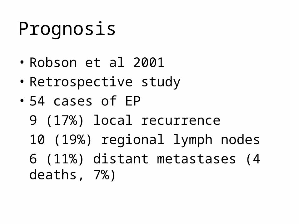
Prognosis
• Robson et al 2001
• Retrospective study
• 54 cases of EP
9 (17%) local recurrence
10 (19%) regional lymph nodes
6 (11%) distant metastases (4 deaths, 7%)

Histological parameters associated with aggressive disease
• >14 Mitoses per high power field
• Tumour depth >7mm
• Lymphovascular invasion
• Presence of an advancing infiltrative border

Treatment-metastatic disease
• Radiotherapy not effective
• ChemotherapyTamoxifen PaclitaxelIsotretinoin 5-FU/Cisplatin/RadiotherapyIFN-alpha Isotretinoin/IFN-alphaDocetaxel Docetaxel +topical 5-FU5-FU IFN-alpha +IL-2

Previous reports of patients with metastatic EP (mEP) treated with taxanes
Author Clinical presentation
Rx Outcome
Plunkett et al, 2001
45 y.o. Renal transplant, history of Hodgkins lymphoma, mEP to lung
Failed epirubicin
Docetaxel 100mg/m2
Disease stability 3/12 post treatment,required 2nd course
Gutermuth et al 2004
67 y.o. mEP to regional lymph nodes
IFN-alpha 9Miu 3x-weekly, 5# weekly Paclitaxel 100mg/m2
No evidence of disease progression after 7 months
De Bree et al 2005
69 y.o,mEP to ribs
Failed IFN-alpha and isotretinoin
Daily top 5-FU and 3# intra-arterial docetaxel 75mg/m2, 2# systemic docetaxel 80mg/m2
Disease stability after 25 months with complete histological response of skin lesions (anaphylaxis to systemic docetaxel after 3rd# so treatment stopped)

Pathogenesis
• Poorly understood-polyoma virus co-carcinogen?• Role of immunosuppression?• C Harwood et al, 2003
Immunosuppressed renal transplant population at greatly increased risk of appendageal tumour compared with immunocompetent population
Greater proportion of these were malignant
DERMATOPATHOLGY-CPC

WELCOME!
GRAZIE!2012