6. sources of population exposure to … radiation 233 6. sources of population exposure to ionizing...
TRANSCRIPT

IONIZING RADIATION 231
������������������������������������������������������������������������������������������������������������������������������������������������������������������������������������������������������������������������������������������������������
��������������������������������������������������������������������������������������������������������������������������������������������������������������������������������������������������������������������������������������������������������������������������������������������� �
���
�������������������������������������������������������������������������������������������������������������������������������������������������������������������������������������������������������������������������������������������������������������
���
�������������������������������������������������������������������������������������������������������������������������������������������������
������������������������������������������������������������������������������������������������������������������
����������������������������������������������������������������������������������������������������������������������������������������������������������������������������������������������������������������������������������������������������������������������������������������������������������������������������������������
���������������������������������������
�����������������������������������������������������������������������������������������������������������������������������������������������������������������������������������������������������������������������������������������������������������������������������������������
���������������������������������
������������������������������������������������������������������������������������������������������������������������������������������������������������������������������������������������������
������������������������������������������������������������������������������������������������������������������������������������������������������������������������������������������������������������������������������������������������������������������������������������������������������������������������������������������������������������������������������������������������������������������������������������������������������������������������������������������������������������������������������������������������������������������������������������������������������������������������������������������������������������������������������������������������������������������������������������������������������������������������������������
������������������������������������������������������������������������������������������������������������������������������������������������������������������������������������������������������������������������������������������������������������������������������������������������������������������������������������������������������������������������������������������������������������������������������������������������������������������������������������������������������������������������������������������������������������������������������������������������������������������������������������������������������������������������������������������������������������������������������������������������������������������������������������������������������������������������������������������������������������������������������������������������������������������������������������������������������������������������������������������������������������������������������������������������
Nuclear Medicine4%
Radon55%
X Rays11%
Consumer Products 3%
Other<1%
Terrestrial8%
Cosmic8%
Natural Internal11%
Figure 6-1. Radiation Exposure to the Average U.S. Citizenadapted from (NCRP1987a)
6. SOURCES OF POPULATION EXPOSURE TO IONIZING RADIATION
6.1 OVERVIEW
All organisms (e.g., bacteria, plants, or
animals, including humans) are exposed
everyday to varying amounts of ionizing
radiation. Figure 6-1 shows average
contributions from various sources of
radiation to which the average U.S.
citizen is exposed during his or her
lifetime. Approximately 82% of the
radiation dose is from natural sources:
55% from radon (see Figure 1-3), 8%
from cosmic radiation (from the sun and
stars), another 8% from terrestrial
sources (radioactive material in rocks and soil), and 11% from internal sources (radioactive materials,
primarily potassium-40, from food and water consumed in the daily diet).
The remaining 18% of the dose comes from anthropogenic (man-made) sources such as medical x ray
exposure (11%), nuclear medicine procedures exposure (4%), consumer products (3%), and other sources
(<1%). These other sources include occupational exposure, nuclear fallout, and the nuclear fuel cycle.
The total average annual effective dose equivalent for the population of the United States, natural and
anthropogenic, is approximately 360 mrem (3.6 mSv) and is described further in Chapter 1 of this profile
(BEIR V 1990).
The majority of exposure to radiation comes from natural sources. With the exception of indoor radon
exposure (and to some extent exposure from terrestrial sources), exposure to natural radiation is only
moderately controllable. Controllability in relation to radon refers to mitigation of radon concentrations
in buildings and homes. The average annual effective dose equivalent from all natural sources combined
is approximately 3 mSv (300 mrem). Of this amount, approximately 98 mrem (98 mSv) is due to

IONIZING RADIATION 232
6. SOURCES OF POPULATION EXPOSURE TO IONIZING RADIATION
background radiation; this includes cosmic rays, 29 mrem (0.29 mSv); terrestrial gamma rays, 29 mrem
(0.29 mSv); and naturally existing radionuclides within the body, 40 mrem (0.40 mSv). Individual doses
from natural sources may be much greater. The magnitude of natural exposures depends upon numerous
factors such as geographic location, height above sea level, and the construction and ventilation of
buildings. For instance, the average annual radiation dose received by a person living in Boston,
Massachusetts, is approximately 300 mrem (3 mSv), while people living in Denver, Colorado, and
Kerala, India, receive average annual doses of approximately 600 mrem and 1500 mrem, respectively.
The difference in these doses is due mainly to greater concentrations of radioactive materials found in the
soils of the Colorado and Kerala areas and to a smaller extent the increase in cosmic radiation at higher
altitudes in these areas (BEIR V 1990; Eisenbud 1987; Harvard Medical School 1996; UNSCEAR 1993).
Commonly used terms and scientific unit symbols and abbreviations used in this chapter are defined in
Table 6-1 and Table 6-2, respectively. These and other terms may also be found in the glossary in
Chapter 9 or in the index at the end of this toxicologic profile. The following sections discuss natural
external exposures (Sections 6.2 and 6.3), natural internal exposures (Section 6.4), and internal and
external man-made/industrial exposures (Sections 6.5, 6.6, and 6.7).
6.2 COSMIC RADIATION EXPOSURE
Cosmic radiation contributes an estimated 8% to the average population radiation dose. It is primarily
composed of galactic radiation originating outside the solar system in addition to a varying degree of
solar radiation. The primary cosmic rays that arrive in the upper atmosphere are high-energy subatomic
particles—primarily protons, but also nuclei and electrons— moving almost at the speed of light; these
primary rays create secondary rays that bathe the atmosphere in radiation. Austrian physicist Victor Hess
discovered cosmic rays in 1912 when he and two assistants flew a balloon to an altitude of 16,000 ft
(4,877 meters). Hess proved that the source of a mysterious radiation previously detected over the ocean,
where terrestrial radiation levels were expected to be very low, was actually coming from outside the
atmosphere; he also found that the rate of decline in radiation as the balloon ascended over land was
slower than would be expected if the radiation emanated from the earth. The difference is caused by the
cosmic rays. Only a small fraction of cosmic radiation originates from the sun; however, the proportion
of cosmic radiation contributed by the sun increases during periods of increased sunspot and solar flare
activity, which run in

IONIZING RADIATION 233
6. SOURCES OF POPULATION EXPOSURE TO IONIZING RADIATION
Table 6-1. Common Terms and Abbreviations
becquerel (Bq) SI unit for quantity of radioactive material; 1 Bq equals that quantity ofradioactive material in which there is 1 transformation or disintegration persecond (dps).
curie (Ci) Conventional unit for quantity of radioactive material. One Ci is the quantityof any radionuclide in which there are 37 billion transformations ordisintegrations in 1 second. This is the activity of 1 gram of 226Ra.
rad The unit of absorbed dose equal to 0.01 Joule/kg in any medium.
gray (Gy) SI unit of absorbed dose.
rem Conventional unit for dose equivalent. The dose equivalent in rem isnumerically equal to the absorbed dose in rad multiplied by the qualityfactor.
roentgen (R) A unit of x ray and gamma ray exposure. It is measured by the amount ofionization in air produced by x ray and gamma radiation. One R equals2.58x10-4 coulomb per kg of air.
sievert (Sv) The SI unit of dose equivalent. It is equal to the dose in grays times aquality factor; 1 Sv equals 100 rem.
quality factor (Q) The linear-energy-transfer-dependent factor by which absorbed doses aremultiplied to obtain (for radiation protection purposes) a quantity thatexpresses the effectiveness of the absorbed dose on a common scale for allionizing radiation.
Table 6-2. Scientific Units
Prefix (symbol) Power of 10 Decimal Equivalent
atto 10-18 0.000000000000000001
femto (f) 10-15 0.000000000000001
pico (p) 10-12 0.000000000001
nano (n) 10-9 0.000000001
micro (µ) 10-6 0.000001
milli (m) 10-3 0.001
centi (c) 10-2 0.01
deci (d) 10-1 0.1
kilo (k) 103 1,000
mega (M) 106 1,000,000
giga (G) 109 1,000,000,000
tera (T) 1012 1,000,000,000,000
peta (P) 1015 1,000,000,000,000,000
exa (E) 1018 1,000,000,000,000,000,000
IONIZING RADIATION 234
6. SOURCES OF POPULATION EXPOSURE TO IONIZING RADIATION
11-year cycles. Cosmic rays bombard the periphery of the earth’s atmosphere at a rate of 2x10¹⁸ particles
per second, at a density of about 4 rays/cm2-sec, and at an energy flux of 2,000 MeV/cm2•sec. These
rays, referred to as “primary cosmic rays,” are deflected and slowed by particles in the earth’s
atmosphere, creating “secondary cosmic rays” that often reach and even penetrate the earth’s surface.
The interaction of cosmic rays with the atmosphere leads to the production of several cosmogenic
radionuclides, notably carbon-14 (14C), tritium (3H) and beryllium-7 (7Be). Because of the shielding
effect of the atmosphere and the earth’s geomagnetic fields, which tend to deflect charged cosmic ray
particles towards the magnetic poles, the cosmic ray dose rate increases with altitude and latitude. The
average annual dose from cosmic radiation in the United States is 29 mrem (0.29 mSv), but this value
doubles for every 6,000-foot (1,828 meters) increase in altitude. Thus, the dose from cosmic rays
received in Denver, Colorado, and Leadville, Colorado (altitudes of 1,600 m and 3,200 m, respectively),
is approximately two and four times that received at sea level, respectively (Eisenbud 1987; Korff 1964;
NASA 1995; Shapiro 1990; UNSCEAR 1993). At altitudes of 30,000 to 40,000 feet (9144 to 12192
meters), where most jet aircraft fly, the cosmic ray dose rate is about 1 mrem per hour (0.01 mSv/hr).
6.3 TERRESTRIAL RADIATION EXPOSURE
Cosmic radiation contributes approximately the same amount of background radiation as terrestrial
radiation (8%), which is emitted by naturally occurring radioactive materials found in the earth’s crust,
such as 40K, uranium and its progeny, and thorium and its progeny (see Figure 6-1). Uranium, for
example, is found in all types of soil and rock at concentrations ranging from 0.003 ppm in meteorites to
120 ppm in phosphate rock from Florida. Exposure to radioactive materials in the soil and earthen
products occurs continuously since we are surrounded by these sources. The radiation dose varies
tremendously and is affected by such factors as geographic location, concentration of natural radioactive
materials in the soil and building materials, and the types of materials used in building structures.
Some communities situated on soil with high concentrations of granite or mineral sand receive doses
many times the average. Examples include coastal areas in Espiritos Santos and Rio de Janeiro in Brazil;
Kerala, on the southwest coast of India; and the Guangdong province in China. In Brazil, the black sand
beaches are composed of monazite, a rare earth mineral containing 9% radioactive thorium. External
radiation dose rates from these sands may be as high as 5 mrem/hr (0.05 mSv/hr); permanent residents
experience an average annual dose equivalent of approximately 500 mrem (5 mSv). In Kerala, on the

IONIZING RADIATION 235
6. SOURCES OF POPULATION EXPOSURE TO IONIZING RADIATION
west coast of India, residents receive 1,300–1,500 mrem (13–15 mSv) annually, due to the presence of
monazite sand. Some dose rates are as high as 3,000 mrem/yr (Eisenbud and Gesell 1997).
Apart from radiation exposures due to living in close proximity to the earth’s crust, people are also
exposed to additional radiation when earth crust products (oil, coal, coal ash, minerals) are extracted,
refined, and used. The naturally occurring radioactive materials in these products are concentrated into
what is called technologically enhanced naturally-occurring radioactive materials (TENORM). In
general, the hazards of exposure to TENORM during the extraction and processing of earth materials are
relatively small compared to the hazards of exposure to other chemicals. As a result, radiation exposure
from these sources, with the exception of uranium mining, milling, and processing, is not routinely
monitored (Eisenbud 1987; UNSCEAR 1993). The radiation hazards associated with the mining of coal,
oil, natural gas, phosphate rock products, and sand are discussed below.
Radon exposure makes up the largest fraction of total radiation dose and contributes to both internal and
external radiation exposures. The following subsections discuss various radioactive materials primarily
associated with external radiation exposures from various terrestrial activities. Radon is inherent in these
terrestrial sources, as well, and is discussed because of its association, but with the understanding that it is
a major internal radiation exposure source.
6.3.1 Coal Production
Exposure to radionuclides occurs during the mining and use of coal and coal ash. The methods of coal
usage vary considerably among countries; on average worldwide, about 40% of coal is burned in electric
power stations, 10% in dwellings, and 50% in other industries. Based on samples from 15 countries, the
average concentrations of 40K, 238U, and 232Th in coal are 50, 20, and 20 Bq/kg (1.35, 0.54, and
0.54 nCi/kg), respectively. These concentrations may vary considerably, depending upon the mine
location. For example, concentrations of these radionuclides in China are 104, 36, and 30 Bq/kg (2.81,
0.97, and 0.81 nCi/kg), respectively. Coal mine exhaust typically contains radon; the estimated annual
per person dose from radon in coal mine dust is 0.1–2 nSv. The average annual per person doses of
radiation from coal-fired power plants and from domestic cooking with coal are about 0.2 mrem (2 µSv)
and 0.04–0.8 mrem (0.4–8 µSv), respectively.

IONIZING RADIATION 236
6. SOURCES OF POPULATION EXPOSURE TO IONIZING RADIATION
About 280 million tons of coal ash are produced by power plants each year. Potential uses for the ash
include fertilizers and building materials for roads and dwellings. Most U.S. power plants recover fly ash
exhaust using scrubbers, electrostatic precipitators, or bag houses. The radioactive content of coal tends
to concentrate in the ash, resulting in 5- to 10-fold increases in the concentration of lead-210 (210Pb) and
polonium-210 (210Po) as compared to unburned coal. When fly ash is used in building materials, the
degree of external exposure to radiation and the inhalation of radon gas increases directly with the amount
of ash incorporated into these materials, and, for radon, the porosity of the materials. The average annual
exposure associated with living in concrete and wooden houses is 7 mrem (70 µSv) and 3 mrem (30 µSv),
respectively. An EPA report published in 1979 estimated that the exposure to radioactive materials
emitted from all 250 coal-fired power plants resulted in an additional 1.5 cancers per year
(Eisenbud 1987; EPA 1979 as cited by Eisenbud; UNSCEAR 1982, 1988).
6.3.2 Crude Oil and Natural Gas Production
About 3x10¹² kg of crude oil and 1012 m3 of natural gas are produced worldwide annually. Oil-fired
power plants use about 15% of all oil. Gas-fired power plants are estimated to use about 15% of all gas.
Radon is present in natural gas; concentrations of radon in gas at well heads average approximately
40 pCi/L (1.5 Bq/L). The processing and blending of liquefied petroleum gas (LPG) tends to enhance
radon concentrations, and the long-lived radon daughters (210Pb and 210Po) tend to accumulate on LPG
processing machinery, resulting in low level exposure to maintenance workers. The annual per person
doses from crude oil and gas are estimated to be 0.001 mrem (10 nSv) and 0.0001 mrem (1 nSv),
respectively. The estimated doses are small and result from inhalation of radioactive particles and radon
gas (Eisenbud 1987; UNSCEAR 1993).
6.3.3 Phosphate Rock Products
Phosphate rock, the precursor of all phosphorous products including fertilizer, is mined at a rate of
130 million tons per year worldwide. The worldwide use of fertilizer, estimated to be 30 million tons,
constitutes the greatest source of 40K and 226Ra mobility. In the United States, the application rate for
fertilizers ranges from 30 kg of phosphate per hectare (barley) to 150 kg/hectare (potatoes and tobacco)
for commercial agricultural application, and possibly less for residential applications. Concentrations of40K and 232Th in phosphate rock are similar to those in soil (a few grams per hundred grams of soil and a
few grams per million grams of soil, respectively). Levels of 238U and its transformation products are

IONIZING RADIATION 237
6. SOURCES OF POPULATION EXPOSURE TO IONIZING RADIATION
much higher in phosphate rock than in soil. Concentrations of 238U in phosphate deposits are typically
about 1,500 Bq/kg (40.5 nCi/kg). The practice of using phosphate fertilizers has resulted in uranium
concentrations in food at levels up to 8 ng/g (0.005 pCi/g). Exposure to the general public occurs near
areas of mining and processing through waste effluent. Several end-products of phosphate processing,
phosphogypsum and calcium silicate, are used for fertilizer, for back-fill and road-base material, in
additives to concrete, in mine reclamation, and in the recovery of sulphur. Phosphogypsum is also used
as a substitute for natural gypsum in the manufacture of cement, wallboard and plaster. The primary
radioactive material in phosphogypsum is 226Ra, which is found at concentrations of 900 Bq/kg
(24.3 nCi/kg).
Exposure to phosphate-borne radioactivity also occurs as a result of discharges into surface waters. The
primary pathway of exposure to radioactivity in humans is through the consumption of fish and shellfish.
Elevated concentrations of radon have been detected in structures built over reclaimed phosphate mines,
and over unmined mineral deposits. Maximum annual individual doses near phosphate facilities range
from 4 mrem (40 µSv) in the Netherlands to 600 mrem (6 mSv) in the United States. The average annual
per person dose of 40K derived from fertilizers is approximately 0.2 mrem (2 µSv); while the average per
person dose from potassium in the body is about 20 mrem (0.2 mSv) per year, maximum annual
individual doses, from consumption of seafood, have been estimated at 15 mrem (150 µSv), with 210Po as
the main contributor. The annual per person radiation dose from 226Ra-laden phosphogypsum in building
materials is estimated to be about 1 mrem (0.01 mSv) (Eisenbud 1987; Shapiro 1990; UNSCEAR 1982,
1988, 1993). The use and disposal of phosphogypsum is regulated by the EPA, and these regulations are
intended to ensure that the public is not exposed to unsafe levels of radionuclides from this material.
6.3.4 Sand
Mineral sands, defined as sands with a specific gravity of more than 2.9, originate from eroded rock.
These sands are mined in Australia, Bangladesh, Indonesia, Malaysia, Thailand, and Vietnam. The heavy
mineral is extracted from the sand and is processed into, among other items, paint pigment, titanium
metals, catalysts, and structural materials. The sand itself is used as abrasive material for sandblasting.
The principal radioactive components are 232Th and 238U. Exposure is mainly external through minerals
spilled at the processing plants. Although information on exposures is scant, annual levels are estimated
to be in the low µSv range (UNSCEAR 1993).

IONIZING RADIATION 238
6. SOURCES OF POPULATION EXPOSURE TO IONIZING RADIATION
6.3.5 Hot Springs and Caves
Geothermal energy, produced in Iceland, Italy, Japan, New Zealand, Russia, and the United States, is
produced from steam or water from high-temperature areas within the earth’s crust. Mineral springs and
spas, which are found in South America, Europe, Japan, and the United States, are also clustered around
these high-temperature areas of the earth’s crust. The primary radionuclides in this source are those of the
uranium transformation chain. Of these, 226Ra and 222Rn are considered to be the most important to public
health.
The diffusion of radon from ordinary rock and soils and from radon-rich water can cause notably elevated
radon concentrations in tunnels, caves, and spas. In Bad Gastein, Austria, approximately 5 million
gallons of mineral water are distributed to hotels and spas daily, allowing the release of about 58 Ci (2
Mbq) of radon per year into the environment. In comparison with levels in outdoor air, the concentrations
of radon and its transformation products in confined air spaces such as mines and caves are elevated. The
average annual per person radiation dose from this source is estimated to be 0.0001 mrem (1 nSv);
however, the doses received by those intentionally spending time in these environs (e.g., tourists,
workers, miners) are much greater than this amount (Eisenbud 1987; IARC 1988; UNSCEAR 1993).
6.4 NATURAL INTERNAL EXPOSURE
Natural internal radionuclides contribute an estimated 11% to the average population radiation dose.
Radioactive materials enter the body by inhalation, ingestion, or dermal absorption. Although radioactive
materials may also enter the body through punctures (either wounds or injections), this route of exposure
will not be addressed in this toxicological profile.
The effects induced by internally deposited nuclides or external radiation are classified as either "acute"
(early-occurring effects of radiation, which appear within days or weeks after exposure) or "latent"
(chronic or late-occurring effects of radiation, which appear months or years after exposure). Acute
effects are not expected for natural sources of radiation because they are not capable of producing high
dose rates in a short period of time. However, they may cause latent effects. The most common latent
radiation effect is an increased probability of certain types of cancer. More information on the health
effects of ionizing radiation can be found in Chapter 3 of this toxicological profile.

IONIZING RADIATION 239
6. SOURCES OF POPULATION EXPOSURE TO IONIZING RADIATION
6.4.1 Inhalation
The sources of inhaled radioactive materials include debris from atmospheric nuclear testing; nuclear
reactor and medical gaseous waste; radioactive materials manufacturing; diagnostic medical radionuclide
use; coal- and gas-burning power plants; airborne soil; and naturally emanating gases. The radionuclides
(and their average concentrations) commonly found in the atmosphere include: 222Rn and 220Rn
(270 pCi/m3 [10 MBq/m3] each); 210Pb (0.01 pCi/m3); 210Po (0.001 pCi/m3); 238U (12x10-5 pCi/m3); 232Th
(3x10⁻⁵ pCi/m3); 230Th; (4.5x10⁻⁵ pCi/m3); and 228Th (3x10⁻⁵ pCi/m3), 14C and 3H. In addition, smokers are
exposed to radiation from the radionuclide 210Po, which is found in tobacco; the resulting dose to the
bronchial epithelium can be as high as 20 mrem (0.2 mSv) per year (NCRP 1984; Shapiro 1990;
UNSCEAR 1993).
The largest dose of radiation from natural sources comes from the inhalation of 222Rn and 220Rn (thoron)
gases. These colorless and odorless gases, which are in the uranium and thorium transformation chains,
respectively, are continuously released from the soil. Worldwide, the total emanation rate of radon is
estimated to be 50 Ci/sec (2 TBq/sec); the total atmospheric content is estimated to be 25 MCi (1 EBq).
The main factors controlling the rate of radon release and subsequent exposure are: ground porosity,
ground cover, temperature, meteorological conditions, and the type of construction and ventilation
properties of dwellings. The rate of radon emanation from soil is thought to increase with diminished
atmospheric pressure and to decrease during periods of, or in areas of, elevated moisture, while the
atmospheric concentration of radon tends to increase during temperature inversions and as the humidity
decreases.
The health hazards of radon exposure were first recognized in the 1930s when radium miners in Scheen-
burg, Germany, and Joachimstal, Czechoslovakia, were found to have a high incidence of lung cancer.
Over half of all miner deaths were attributed to lung cancer, and most of the miners were less than 50
years of age when they died. In the general U.S. population, the EPA estimated that radon exposure
accounts for approximately 10% (17,000) of all lung cancers, while smoking accounts for approximately
85% (144,500) of all lung cancers. The average annual effective dose equivalent from radon is about
200 mrem (2 mSv), but individual doses may be much higher. It is estimated that 1–3% of all homes
have radon levels in excess of 8 pCi/L, which is twice the EPA recommended residential limit of 4 pCi/L.
Approximately 50,000 to 100,000 homes in the U.S. have radon concentrations exceeding 20 pCi/L,
which results in exposures equal to or exceeding the limit for occupational exposure. The 220Rn doses are
considerably lower than those of 222Rn, due to its short half-life (55 sec for 220Rn versus 3.8 days for
IONIZING RADIATION 240
6. SOURCES OF POPULATION EXPOSURE TO IONIZING RADIATION
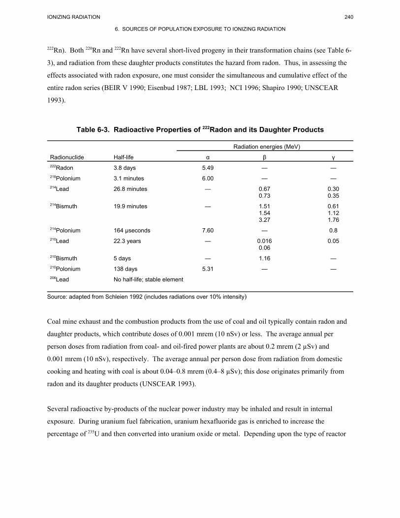
222Rn). Both 220Rn and 222Rn have several short-lived progeny in their transformation chains (see Table 6-
3), and radiation from these daughter products constitutes the hazard from radon. Thus, in assessing the
effects associated with radon exposure, one must consider the simultaneous and cumulative effect of the
entire radon series (BEIR V 1990; Eisenbud 1987; LBL 1993; NCI 1996; Shapiro 1990; UNSCEAR
1993).
Table 6-3. Radioactive Properties of 222Radon and its Daughter Products
Radiation energies (MeV)
Radionuclide Half-life α β γ222Radon 3.8 days 5.49 — —218Polonium 3.1 minutes 6.00 — —214Lead 26.8 minutes — 0.67
0.730.300.35
214Bismuth 19.9 minutes — 1.511.543.27
0.611.121.76
214Polonium 164 µseconds 7.60 — 0.8210Lead 22.3 years — 0.016
0.060.05
210Bismuth 5 days — 1.16 —210Polonium 138 days 5.31 — —206Lead No half-life; stable element
Source: adapted from Schleien 1992 (includes radiations over 10% intensity)
Coal mine exhaust and the combustion products from the use of coal and oil typically contain radon and
daughter products, which contribute doses of 0.001 mrem (10 nSv) or less. The average annual per
person doses from radiation from coal- and oil-fired power plants are about 0.2 mrem (2 µSv) and
0.001 mrem (10 nSv), respectively. The average annual per person dose from radiation from domestic
cooking and heating with coal is about 0.04–0.8 mrem (0.4–8 µSv); this dose originates primarily from
radon and its daughter products (UNSCEAR 1993).
Several radioactive by-products of the nuclear power industry may be inhaled and result in internal
exposure. During uranium fuel fabrication, uranium hexafluoride gas is enriched to increase the
percentage of 235U and then converted into uranium oxide or metal. Depending upon the type of reactor

IONIZING RADIATION 241
6. SOURCES OF POPULATION EXPOSURE TO IONIZING RADIATION
fuel or nuclear weapons material being produced, uranium must be enriched to a minimum of 3% 235U for
fuel and 93.5% for nuclear weapons. Emissions from fabrication facilities usually consist of the long-
lived isotopes 234U, 235U, and 238U, and the short-lived isotopes 234Th, and protactinium-234m (234mPa).
The major route of exposure from this source is inhalation. More information about uranium is available
in the ATSDR Toxicological Profile for Uranium (ATSDR 1999).
6.4.2 Ingestion
The sources of radionuclides that contribute to radiation exposure by ingestion include nuclear weapons
testing, the accidental or intentional release of radioactivity from nuclear reactors, the release of medical
or experimental radionuclides into sanitary sewers, and naturally occurring radionuclides (which normally
represent the source of highest oral dose). For most radionuclides present at waste sites containing low
levels of radioactive nuclides, oral exposure is not a major route of exposure. There is a small probability
of radionuclide ingestion because of the potential for surface water and groundwater contamination and
uptake by plants and animals following erosion of ground cover from a contaminated site.
Among the naturally occurring radionuclides, uranium, 40K, and 226Ra are found in soils and fertilizers; as
a result, they are incorporated into foods consumed by animals and humans. The practice of using
phosphate fertilizers has resulted in uranium concentrations in food at levels up to 8 ng/g, resulting in an
estimated average annual intake of uranium from dietary sources of 10 Bq; as a result, the average
skeletal content of uranium is estimated to be 25 µg, which is equivalent to approximately 17 pCi
(Eisenbud 1987; UNSCEAR 1993).
The most important radionuclides that are ingested are 40K, 226Ra, and the transformation products of226Ra. However, wherever it is found, all potassium is radioactive because its natural isotope, 40K, is
radioactive. The body content of potassium is under strict homeostatic control and is maintained at a
relatively constant level of about 140 g/70 kg. This amount of potassium contains approximately 0.1 µCi
(4,000 Bq) of 40K. Because the body controls the potassium balance, environmental variations have little
effect on the 40K content in the body (Eisenbud 1987; Shapiro 1990). This natural 40K delivers a dose of
20 mrem/year (0.2 mSv/year) to the gonads and other soft tissues and 15 mrem/year (0.15 mSv/year) to
bone.
FDA has developed guidelines for radionuclide levels in food for individuals from 3 months to adult that
are summarized in Chapter 7 (Regulations), Table 7-4, FDA Derived Intervention Levels (FDA 1998).

IONIZING RADIATION 242
6. SOURCES OF POPULATION EXPOSURE TO IONIZING RADIATION
6.4.3 Dermal
For the purposes of this profile, dermal exposure to radionuclides refers to exposures from a radionuclide
placed in direct contact with skin surface. Dermal exposure is typically a minor route of internal and
external exposure. In general, depending on the specific physical properties of the radionuclide that may
reside on the skin, the percutaneous absorption of radionuclides from particles is negligible, especially if
the skin is thoroughly washed immediately after exposure. The long-term biological effects of dermally
absorbed radionuclides are limited to the level of the epidermis and dermis (and its vasculature). More
soluble forms of the radionuclides may result in a small percentage of the nuclide being absorbed if it is
not removed from the skin's surface, for example tritium, as tritiated water or vapor, is readily absorbed
into the body through the skin. Generally, the skin is an effective barrier against absorption of
radionuclides (except for tritiated water) into the body. The dermal exposure pathway is, therefore, a
minor route of exposure at low-level radioactive waste sites.
6.5 X RAY AND NUCLEAR MEDICINE EXPOSURES
Radioactive materials and other sources of ionizing radiation are widely used in the diagnosis and
treatment of some diseases in human and veterinary medicine (NCRP 1989a; 1996). They represent 15%
of the average population dose, 11% for x rays and 4% for nuclear medicine. In 1989, the estimated
number of x ray machines used in the US was 109,000 for medical diagnosis, 143,000 for dental
diagnosis, and 1,300 for therapy: 3 million diagnostic examinations were made which produced a
collective US dose of 92,000 man•Sv (9,200,000 man-rem). Typical effective dose equivalents for
various procedures are 0.14 mSv (14 mrem) for a chest x ray, 1.0 mSv (100 mrem) for mammography,
and 7.2 mSv (720 mrem) for an upper GI tract evaluation. Due to the usefulness of nuclear medicine,
radioactive drugs and diagnostic compounds have become significant contributors to internal radiation
dose from man-made sources today. The average U.S. nuclear medicine examination gives an effective
dose equivalent of 5 mSv (500 mrem) with individual prodedured delivering 2.5 mSv (250 mrem) for a
thyroid uptake, 6.3 mSv (630 mrem) for a bone scan, and 14 mSv (1,400 mrem) for a cardiovascular
screen. Each year in the US, the collective effective dose from nuclear medicine procedures is 32,000
man•Sv (3,200,000 man-rem). Therapeutic doses are much larger to the individual but many fewer
individuals are exposed. After many millions of diagnostic radionuclide procedures, we have found no
increase in cancers from these procedures.
IONIZING RADIATION 243
6. SOURCES OF POPULATION EXPOSURE TO IONIZING RADIATION
The common sources of radiation exposure associated with radiotherapy and diagnosis include x rays,
thallium-201 (201Tl), technetium-99m (99mTc), 125I, and 131I. More exposures are related to diagnosis than
to therapy, and the average number of treatments per person increases as the level of health care
improves. Also, the average dose per individual treatment tends to decrease as techniques and equipment
improve. Overall, x ray treatments deliver a higher average per person dose in industrialized nations
(average of 0.3–2.2 mSv) than in countries with less developed health care (average exposure
0.02–0.2 mSv). On an individual basis, the average dose increases with age, from 52 mrem/year in
adolescents to 151 mrem/year in persons over 65 years of age. Exposures are usually lower for
examinations of the extremities and skull and higher for examination of the gastrointestinal (GI) tract. In
the United States, the average annual dose to the bone marrow from this source increased from 83 mrem
in 1964 to 103 mrem in 1970. A person receiving a full set of dental x rays would add approximately
40 mrem to his or her annual dose. On the other hand, the average annual dose per patient from the
diagnostic use of radionuclides is lower in industrialized nations, largely because of greater use of 99mTc.
This radionuclide is preferred over 131I because its shorter half-life (6 hours versus 8 days) gives a much
lower patient dose. There is currently no radioiodine in the atmosphere due to atmospheric testing
because of the 8-day half-life of 131I. The shorter half-life and higher cost of 99mTc make it more available
in developed than in developing nations, where 131I has frequent use. While the average dose (per
individual) in patients undergoing radiotherapy is much greater than in patients undergoing diagnosis, the
exposure group is much smaller, resulting in a lower overall population-at-risk. Unfortunately, serious
exposures resulting from failures of equipment, procedures, or personnel errors (usually a result of not
following procedures) sometimes occur, with several hundred failures out of several hundred million
procedures per year worldwide.
There are several emerging trends in diagnostic nuclear medicine. Some of these trends include: the
introduction of radiolabeled monoclonal antibodies for imaging and treatment; the emergence of new
compounds used in positron emission tomography (PET) and single-photon emission computed
tomography (SPECT) studies; and the use of computed x ray tomography (CAT scans). Radiolabeled
monoclonal antibodies have proven useful in the localization of tumors and metastases. Common
radionuclides associated with these antibodies include indium-111 (111In), 131I, and 99mTc. SPECT is used
for tumor localization, brain and cardiac studies, and bone or abdominal imaging. PET, which uses
nuclides such as 11C and 18F, can gather anatomical and physiological information that would otherwise be
difficult to collect. Whole-body imaging using radiolabeled compounds (e.g., anticancer drugs) is
becoming a common PET application (DOE 1996; Eisenbud 1987; Shapiro 1990; UNSCEAR 1993).

IONIZING RADIATION 244
6. SOURCES OF POPULATION EXPOSURE TO IONIZING RADIATION
The use of radiopharmaceuticals has stabilized in industrialized countries but is increasing in developing
countries. Long-lived radionuclides are used more frequently in developing countries, while
industrialized countries tend to use short-lived radionuclides. This results in increased exposures per
examination among developing country patients compared to those of industrialized nations. For
example, a typical thyroid scintigraphy with 99mTc can give an effective dose of less than 0.1 rem (1 mSv),
while the same procedure using 131I gives 10 rem (100 mSv); however, 99mTc is less readily available in
developing countries. Although the average per patient dose equivalent is lower in developed (2–5 mSv
[200-500 mrem]) than in less developed countries (20 mSv [2,000 mrem]), an apparently larger fraction
of individuals in developed nations receive nuclear medicine treatment, so the average per capita annual
dose from radiopharmaceuticals in developed countries (0.07 mSv [7 mrem]) is an order of magnitude
more than that of developing nations (0.004 mSv [4mrem]) (UNSCEAR 1993).
Radionuclides are frequently produced and used in industry, medicine, and research. The number of
users and frequency of radionuclide use are both steadily increasing. The number of establishments in
Japan that generate and/or use radionuclides has increased from 100 in 1960 to 5,000 in 1990. The public
may be exposed to radionuclides from these sources as a result of routine use or from being near someone
who has recently received a nuclear medicine procedure, as well as improper handling, use, or disposal.
In Japan, the usage of 14C, 125I, 3H, and 131I has been estimated to be 5.2, 6.1, 14, and 34 GBq (0.14, 0.16,
0.38, and 0.92 Ci) per million persons, respectively. In contrast, the production of 14C in the United
States and Britain has been estimated to be 30 and 55 GBq (0.81 and 1.49 Ci) per million, respectively.
The annual global production and usage of 14C has been estimated to be 30 GBq (810 mCi) per million
persons, or a total of 0.05 PBq (1.5 kCi). The total amount of 131I produced in Sweden for medical
purposes was estimated to be 0.9 TBq (110 GBq [2.97 Ci] per million) in 1986, while the amount of 131I
discharged from Australian hospitals in 1988 and 1989 was estimated to be 2.9 TBq
(190 GBq [5.13 Ci] per million) (UNSCEAR 1993). Information about some radionuclides used for
medical applications is shown in Table 6-4.
3H and noble gases are released to the air, while 14C release is through airborne and fluid effluents. The
isotopes 131I and 125I are primarily released through liquid effluent. The annual collective dose from
medical and radiopharmaceutical wastes to local populations is thought to be in the range of
10,000 man•rem (100 man•Sv). This level of exposure is relatively unimportant compared to that from
other sources (Eisenbud 1987; UNSCEAR 1993).
IONIZING RADIATION 245
6. SOURCES OF POPULATION EXPOSURE TO IONIZING RADIATION
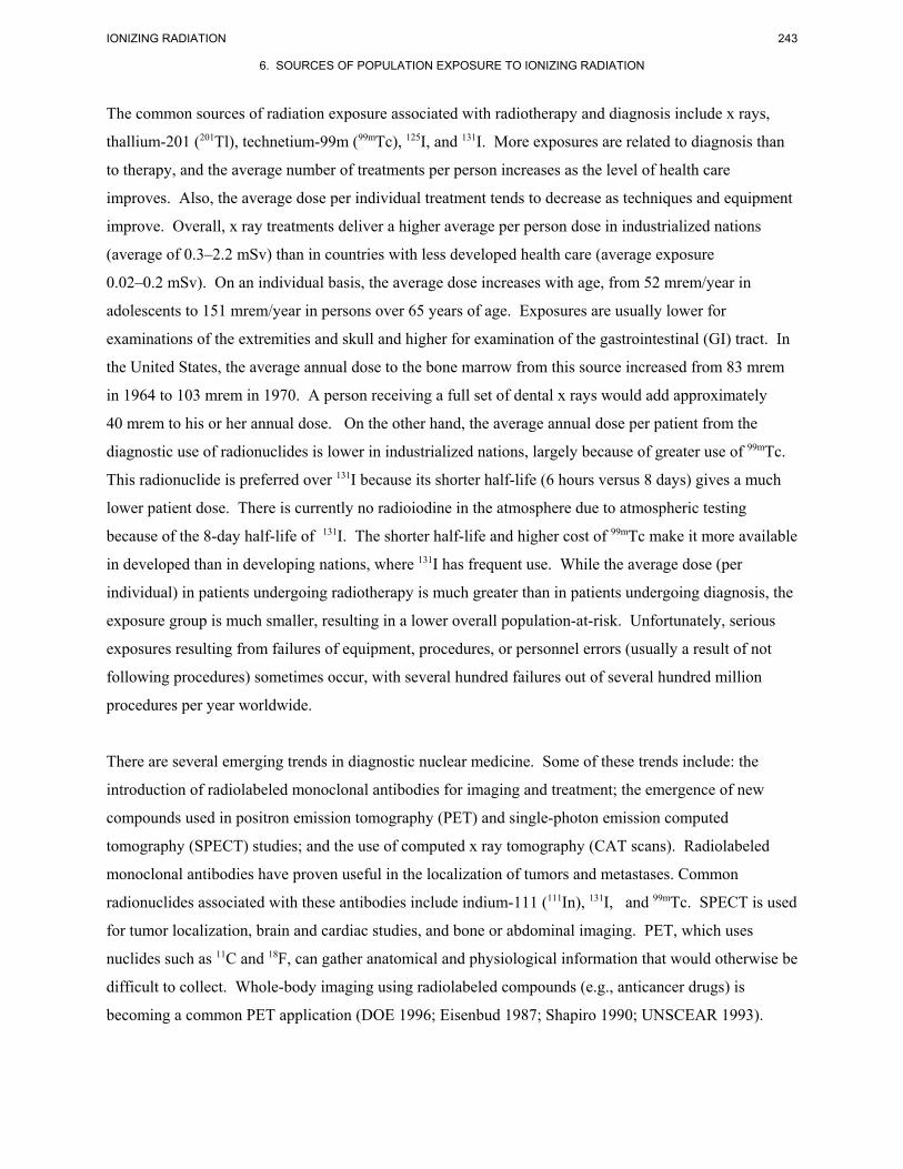
Table 6-4. Some Radiopharmaceuticals Used in Medicine
Radionuclide Preparation Use Properties99mTc Albumin Reduce pertechnetate 99mTc
in the presence of humanalbumin, ascorbic acid,FeCl2, and SnCl2.
Primarily used for lung imaging. Also used for imaging of coronary,urogenital, liver, gastrointestinal,lymphatic, and peripheralcirculation.
The biological clearance half-life from the lungs of 14 to15 hours.
111In Albumin Incubate 111In with humanalbumin in phosphate atpH 3; adjust pH to 11 andheat.
Primarily used for lung imaging. Also used for imaging of coronary,urogenital, liver, gastrointestinal,lymphatic, and peripheralcirculation.
The biological clearance half-life from the lungs of 14 to15 hours.
113mIn Albumin Incubate 113mIn with humanalbumin in phosphate atpH 3; adjust pH to 11 andheat.
Primarily used for lung imaging. Also used for imaging of coronary,urogenital, liver, gastrointestinal,lymphatic, and peripheralcirculation.
The biological clearance half-life from the lungs of 14 to15 hours.
203Pb Albumin Incubate ionic 203Pb withhuman albumin at pH 10with heat.
Primarily used for lung imaging. Also used for imaging of coronary,urogenital, liver, gastrointestinal,lymphatic, and peripheralcirculation.
The biological clearance half-life from the lungs of 14 to15 hours.
51Cr Albumin Incubate 51CrCl3 with humanalbumin
Detection and quantitation ofgastrointestinal protein loss andplacental localization
Cr (III) has strong affinity forplasma proteins withoutaffecting (binding to) redblood cells.
125I Albumin Mild iodination of humanalbumin at 10 EC in slightlyalkaline medium
Diagnostic aid in determining totalblood and plasma volumes
Longer shelf life than 131I;emits no beta radiation (allgamma emissions); lowerdoses needed to obtaingreater resolution comparedto 131I
131I Albumin Mild iodination of humanalbumin at 10 EC in slightlyalkaline medium
Diagnostic aid in determining totalblood and plasma volumes,circulation times, or cardiac output.
May cause sensitization.
131I Albumin,aggregated
Mild iodination of humanalbumin at 10 EC in slightlyalkaline medium
Diagnostic study of the lungs,especially the diagnosis ofpulmonary embolisms.
Aggregates block a smallpercentage (<0.5%) of thefine capillaries. Disintegratingaggregates are cleared byphagocytic Kupffer cells in theliver. Thyroid uptake may beblocked by prioradministration of Lugol’ssolution.
197HgChlormerodrin
Reflux allylurea with 197Hg mercuric acetate inmethanol; add aqueoussodium chloride
Diagnostic aid in scanning the brainfor lesions. Also used for scanningkidneys for anatomical andfunctional abnormalities.
Rapidly cleared by thekidneys. Provides smallerradiation dose compared to131I albumin. A hightumor:background ratio isobtained within 4 hours,allowing quicker scans withgreater resolution.
IONIZING RADIATION 246
6. SOURCES OF POPULATION EXPOSURE TO IONIZING RADIATION
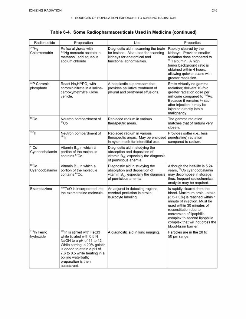
Table 6-4. Some Radiopharmaceuticals Used in Medicine (continued)
Radionuclide Preparation Use Properties203HgChlormerodrin
Reflux allylurea with203Hg mercuric acetate inmethanol; add aqueoussodium chloride
Diagnostic aid in scanning the brainfor lesions. Also used for scanningkidneys for anatomical andfunctional abnormalities.
Rapidly cleared by thekidneys. Provides smallerradiation dose compared to131I albumin. A hightumor:background ratio isobtained within 4 hours,allowing quicker scans withgreater resolution.
32P Chromicphosphate
React Na2H32PO4 withchromic nitrate in a saline-carboxymethylcellulosevehicle.
A neoplastic suppressant thatprovides palliative treatment ofpleural and peritoneal effusions.
Emits virtually no gammaradiation; delivers 10-foldgreater radiation dose permillicurie compared to 198Au. Because it remains in situafter injection, it may beinjected directly into amalignancy.
60Co Neutron bombardment of59Co
Replaced radium in varioustherapeutic areas.
The gamma radiationmatches that of radium veryclosely.
192Ir Neutron bombardment of191Ir
Replaced radium in varioustherapeutic areas. May be enclosedin nylon mesh for interstitial use.
Provides softer (i.e., lesspenetrating) radiationcompared to radium.
57CoCyanocobalamin
Vitamin B12 in which aportion of the moleculecontains 57Co.
Diagnostic aid in studying theabsorption and deposition ofvitamin B12, especially the diagnosisof pernicious anemia.
60CoCyanocobalamin
Vitamin B12 in which aportion of the moleculecontains 60Co.
Diagnostic aid in studying theabsorption and deposition ofvitamin B12, especially the diagnosisof pernicious anemia.
Although the half-life is 5.24years, 60Co cyanocobalaminmay decompose in storage;thus, frequent radiochemicalanalysis may be required.
Exametazime 99mTcO is incorporated intothe exametazine molecule.
An adjunct in detecting regionalcerebral perfusion in stroke;leukocyte labeling.
Is rapidly cleared from theblood. Maximum brain uptake(3.5-7.0%) is reached within 1minute of injection. Must beused within 30 minutes ofreconstitution due toconversion of lipophiliccomplex to second lipophiliccomplex that will not cross theblood-brain barrier.
113In Ferrichydroxide
113In is stirred with FeCl3while titrated with 0.5 NNaOH to a pH of 11 to 12. While stirring, a 20% gelatinis added to attain a pH of7.6 to 8.5 while heating in aboiling waterbath;preparation is thenautoclaved.
A diagnostic aid in lung imaging. Particles are in the 20 to50 µm range.

IONIZING RADIATION 247
6. SOURCES OF POPULATION EXPOSURE TO IONIZING RADIATION
Table 6-4. Some Radiopharmaceuticals Used in Medicine (continued)
Radionuclide Preparation Use Properties59Fe FerrousCitrate
59Fe complexed with citrate;neutron bombardment of58Fe.
A diagnostic aid in studying thekinetics of iron metabolism.
It may be administereddirectly into the bloodstreamwhere it reacts with the metal-binding globulin.
99mTc Ferroushydroxide
Add 99mTc to a vialcontaining ferrous sulfate;the hydroxide is precipitatedwith 0.1N NaOH at a pH of7.5 to 10.7. Gelatin isadded to stabilize theparticles; final pH should be7.1 to 8.3.
A diagnostic aid in pulmonaryscintigraphy.
Most of the particles are in the11 to 13 µm range; virtually allparticles fall in the 3 to 50 µmrange;.
125I Fibrinogen 125I in the form of I2, ICl or I- is combined with fibrinogenand is oxidized bychloramine-T, electrolyticallyor enzymatically. Unreactediodine is removed by theaddition of sodiumthiosulfate.
A diagnostic aid in the localization ofdeep vein thrombosis. Otherapplications include detection ofrenal transplant rejection, tumors,and the study of fibrinogen turnover.
Accumulates in clots, theradiation is easily detected atthe external surface of theaffected limb.
67Ga Galliumcitrate
67Ga is produced by protonirradiation of 67Zn-enrichedZnO2
Used in the diagnosis of lesions ofthe lung, breast, maxillary sinusesand liver. A positive 67Ga uptake ispotentially indicative ofmalignancies such as lymphomas,bronchogenic carcinoma, andHodgkin’s disease. Also useful forplacental localization and diagnosisof pancreatitis and disk disc-spaceinfection.
Concentrates in tumors of softtissues and bone. The half-life of the isotope is 78 hours;the biological half-life of thecitrate compound is 53 days.
111In Indiumchlorides
A cadmium target is bom-barded with deuterons. The111In is then etched from thetarget with HCl, carrier Fe3+
is added, and the mixture isprecipitated with NH4OH. The precipitate is dissolvedin HCl and the ferric iron isremoved by extraction withisopropyl ether.
111In has been used as a tag for avariety of compounds such astransferrin, EDTA and DTPA (usedin cisternography), bleoycin (usedfor tumor localization), platelets(detection of coronary thrombi),lymphocytes (monitoring cardiacantirejection therapy), andleukocytes (diagnosis of upper-abdominal infections).
Indium normally exists inaqueous solution as atrivalent cation. In aqueoussolution InCl exists as amixture of hydrated chlorides.
111In Oxyquinoline Used to label various bloodcomponents such as neutrophils,platelets and lymphocytes; cardiacimaging (labeled platelets);localization of infectious andinflammatory processes (labeledleukocytes).
IONIZING RADIATION 248
6. SOURCES OF POPULATION EXPOSURE TO IONIZING RADIATION
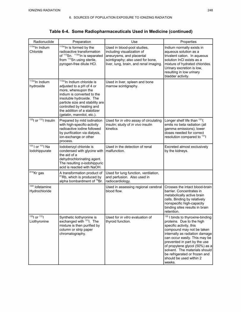
Table 6-4. Some Radiopharmaceuticals Used in Medicine (continued)
Radionuclide Preparation Use Properties113mIn IndiumChloride
113mIn is formed by theradioactive transformationof 113Sn. 113mIn is separatedfrom 113Sn using sterile,pyrogen-free dilute HCl.
Used in blood-pool studies,including visualization ofaneurysms, and placentalscintigraphy; also used for bone,liver, lung, brain, and renal imaging.
Indium normally exists inaqueous solution as atrivalent cation. In aqueoussolution InCl exists as amixture of hydrated chlorides. Urinary excretion is low,resulting in low urinarybladder activity.
113mIn Indiumhydroxide
113mIn Indium chloride isadjusted to a pH of 4 ormore, whereupon theindium is converted to theinsoluble hydroxide. Theparticle size and stability arecontrolled by heating andthe addition of a stabilizer(gelatin, mannitol, etc.).
Used in liver, spleen and bonemarrow scintigraphy.
125I or 131I Insulin Prepared by mild iodinationwith high-specific-activityradioactive iodine followedby purification via dialysis,ion-exchange or otherprocess.
Used for in vitro assay of circulatinginsulin; study of in vivo insulinkinetics
Longer shelf life than 131I;emits no beta radiation (allgamma emissions); lowerdoses needed for correctresolution compared to 131I
123 I or 131I NaIodohippurate
Iodobenzyl chloride iscondensed with glycine withthe aid of adehydrochlorinating agent. The resulting o-iodohippuricacid is reacted with NaOH.
Used in the detection of renalmalfunction.
Excreted almost exclusivelyby the kidneys.
81mKr gas A transformation product of81Rb, which is produced byalpha bombardment of 79Br.
Used for lung function, ventilation,and perfusion. Also used inradiocardiology.
123I IofetamineHydrochloride
Used in assessing regional cerebralblood flow.
Crosses the intact blood-brainbarrier. Concentrates inmetabolically active braincells. Binding by relativelynonspecific high-capacitybinding sites results in brainretention.
125I or 131ILiothyronine
Synthetic liothyronine isexchanged with 131I. Themixture is then purified bycolumn or strip paperchromatography.
Used for in vitro evaluation ofthyroid function.
125 I binds to thyroxine-bindingproteins. Due to the highspecific activity, thiscompound may not be takeninternally as radiation damagecan occur easily. This may beprevented in part by the useof propylene glycol (50%) as asolvent. The materials shouldbe refrigerated or frozen andshould be used within 2weeks.
IONIZING RADIATION 249
6. SOURCES OF POPULATION EXPOSURE TO IONIZING RADIATION
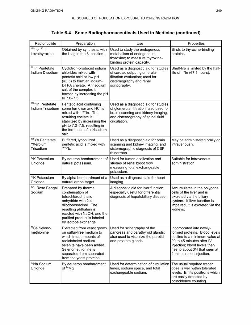
Table 6-4. Some Radiopharmaceuticals Used in Medicine (continued)
Radionuclide Preparation Use Properties125I or 131ILevothyroxine
Obtained by synthesis, withthe I-tag in the 3'-position.
Used to study the endogenousmetabolism of endogenousthyroxine; to measure thyroxine-binding protein capacity.
Binds to thyroxine-bindingproteins.
111In PentetateIndium Disodium
Cyclotron-produced indiumchlorides mixed withpentetic acid at low pH(#3.5) to form an indium-DTPA chelate. A trisodiumsalt of the complex isformed by increasing the pHto 7.0–7.5.
Used as a diagnostic aid for studiesof cardiac output, glomerularfiltration evaluation; used forcisternography and renalscintigraphy.
Shelf-life is limited by the half-life of 111In (67.5 hours).
113mIn PentetateIndium Trisodium
Pentetic acid containingsome ferric ion and HCl ismixed with 113mIn. Theresulting chelate isstabilized by increasing thepH to 7.0–7.5, resulting inthe formation of a trisodiumsalt.
Used as a diagnostic aid for studiesof glomerular filtration; also used forbrain scanning and kidney imaging,and cisternography of spinal fluidcirculation.
169Yb PentetateYtterbiumTrisodium
Buffered, lyophilizedpentetic acid is mixed with169Yb.
Used as a diagnostic aid for brainscanning and kidney imaging, andcisternographic diagnosis of CSFrhinorrhea.
May be administered orally orintravenously.
42K PotassiumChloride
By neutron bombardment ofnatural potassium.
Used for tumor localization andstudies of renal blood flowmeasuring total exchangeablepotassium.
Suitable for intravenousadministration.
43K PotassiumChloride
By alpha bombardment of anatural argon target.
Used as a diagnostic aid for heartimaging.
131I Rose BengalSodium
Prepared by thermalcondensation oftetrachlorophthalicanhydride with 2,4-diiodoresorcinol. Theresulting phthalein isreacted with NaOH, and thepurified product is labeledby isotope exchange
A diagnostic aid for liver function;especially useful for differentialdiagnosis of hepatobiliary disease.
Accumulates in the polygonalcells of the liver and isexcreted via the biliarysystem. If liver function isimpaired, it is excreted via thekidneys.
75Se Seleno-methionine
Extracted from yeast grownon sulfur-free medium towhich trace amounts ofradiolabeled sodiumselenite have been added. Selenomethionine isseparated from separatedfrom the yeast proteins.
Used for scintigraphy of thepancreas and parathyroid glands;also used to visualize the parotidand prostate glands.
Incorporated into newly-formed proteins. Blood levelsdecline to a minimum value at20 to 45 minutes after IVinjection; blood levels thenrise to about 3/4 that seen at2 minutes postinjection.
22Na SodiumChloride
By deuteron bombardmentof 24Mg
Used for determination of circulationtimes, sodium space, and totalexchangeable sodium.
The usual required tracerdose is well within toleratedlevels. Emits positrons whichare easily detected bycoincidence counting.

IONIZING RADIATION 250
6. SOURCES OF POPULATION EXPOSURE TO IONIZING RADIATION
Table 6-4. Some Radiopharmaceuticals Used in Medicine (continued)
Radionuclide Preparation Use Properties51Cr SodiumChromate
By neutron bombardment ofenriched 50Cr.
Used as a biological tracer tomeasure red-cell volume, red-cellsurvival time, and whole-bloodvolume. Also used to detect bloodcell loss due to hemolytic anemia orGI bleeding.
Requires 15–60 minutes todiffuse into red cells; binds toglobin molecules. Has nodeleterious effect onerythrocytes.
18F SodiumFluoride
By neutron bombardment ofenriched 6Li in the form oflithium carbonate. Contamination with 3H mustbe removed prior to use.
Useful for bone imaging, especiallyareas of altered osteogenic activity.
123I Sodium Iodide By proton bombardment ofenriched 124Te or bydeuteron bombardment ofenriched 122Te or bytransformation of 123Xe.
For diagnostic procedures in thyroidfunction studies; for organ imaging including the thyroid, liver, lung, andbrain.
Short half-life (13.2 hours)and radiation characteristicsresult in a smaller radiationdose compared to otheriodine isotopes.
125I Sodium Iodide By neutron bombardment ofxenon gas.
For diagnostic procedures in thyroidfunction studies; for organ imaging including the thyroid, liver, andbrain; treatment of deep-seatednon-resectable tumors.
For organ imaging, dose topatient may be decreasedwith better delineation oforgan and clearer resolutionthan 131I.
131I Sodium Iodide By neutron bombardment ofenriched 131Te or as abyproduct of uraniumfission.
For diagnostic procedures in thyroidfunction studies; a neoplasticsuppressant.
99mTc SodiumPertechnetate
Produced by the elution ofsodium pertechnetatethrough a generatorcontaining 99Mo whichdecays to 99mTc.
Used in the detection andlocalization of cranial lesions,thyroid and salivary gland imaging,placenta localization, and blood-pool imaging.
99mTc has an ideal half-lifewhich is long enough fordiagnostic procedures but isshort enough to minimizeradiation doses to the patient.Also, it lacks a beta radiationcomponent. Pertechnetate isreadily absorbed by thethyroid; this can be reducedby preinfusion of potassiumperchlorate.
32P SodiumPhosphate
By neutron bombardment ofelemental sulfur in anatomic reactor. 32P is thenseparated by leaching withNaOH.
A neoplastic and polycythemicsuppressant; a diagnostic aid for thelocalization of certain ocular tumors.
85Sr Strontium By neutron bombardment ofa strontium salt enriched in85Sr.
A diagnostic aid for scanning bonesand bony structures to detect anddefine lesions and to study bonegrowth and abnormal formations.
Has a long half-life (64 days),resulting in high bone doses.
99mTc Albumin Albumin is tagged with areduced form of thepertechnetate. Thepertechnetate may bereduced by one of severalmethods.
Diagnostic aid in determining totalblood and plasma volumes,circulation times, or cardiac output.
See earlier comment on99mTc.

IONIZING RADIATION 251
6. SOURCES OF POPULATION EXPOSURE TO IONIZING RADIATION
Table 6-4. Some Radiopharmaceuticals Used in Medicine (continued)
Radionuclide Preparation Use Properties99mTc Albumin,Aggregated
Denatured human albuminis tagged with a reducedform of the pertechnetate. The pertechnetate may bereduced by one of severalmethods.
Diagnostic aid in study of the lungs.Primary use is for diagnosingpulmonary embolism. Also usefulfor static blood-pool imaging,angiography, dynamic function testsand visualization of placentaltissues.
See earlier comment on99mTc. 99mTc is preferred over131I as the radioactive tagbecause of the smallerdelivered dose.
99mTc Etidronateor 99mTcOxidronate
Acetic acid is treated withPCl3; the disodium salt isformed when a solution ofetidronic acid is adjusted toa pH of 8.5. Stannouschloride and sometimes astabilizer such as sodiumascorbate are added. Freshly eluted 99mTc isadded.
Useful for bone imaging. See earlier comment on99mTc. This compound issuperior to 18F bone scansand to roentgen studies and isfrequently more sensitive indetecting metastases to thebone.
99mTcIminodiaceticAcid (IDA)
Usually provided in kit form,the compound isreconstituted and tagged byadding sterile 99mTc sodiumpertechnetate.
Useful for hepatobiliary imaging. See earlier comment on99mTc.
99mTc Ferpentate Usually in kit form, thecompound is made byadding a solution of99mTc sodium pertechnetate;the pH is adjusted withsodium hydroxide and asolution of pentetic acid isadded. The chelate isformed by gentle mixing.
Useful for kidney imaging. See earlier comment on99mTc.
99mTc Pentetate Prepared by adding sterile99mTc pertechnetate salinesolution to an aliquot ofbuffered stock solution ofDTPA containing stannouschloride as a reducingagent. Instant DTPA 99mTckits are available.
Useful for brain and kidneyvisualization, for vascular dynamicstudies for measurement ofglomerular filtration and for lungventilation studies.
See earlier comment on99mTc. Does not concentratein any organ. DTPA isuniformly distributedthroughout the extracellularspace and is rapidly clearedby the kidneys withoutretention.
99mTcPyrophosphate
Sodium pyrophosphate,mixed with stannous tin, arecombined with a solution of99mTc sodium pertechnetate.Kits are availablecommercially.
Used as a skeletal imaging agent;used to demonstrate areas ofaltered osteogenesis; also used asa cardiac imaging agent, as anadjunct in the diagnosis ofmyocardial infarction.
See earlier comment on99mTc. The pyrophosphatecompound has been found toconcentrate in muscle tissue,especially contused muscletissue. myocardium.
99mTc Sestamibi Usually in kit form, thecompound is reconstitutedand tagged with sterile 99mTc sodium pertechnetate.
Used as a myocardial perfusionagent in the evaluation of ischemicheart disease and distinguishingand locating abnormal myocardium.
Accumulates in viablemyocardial tissue.
IONIZING RADIATION 252
6. SOURCES OF POPULATION EXPOSURE TO IONIZING RADIATION
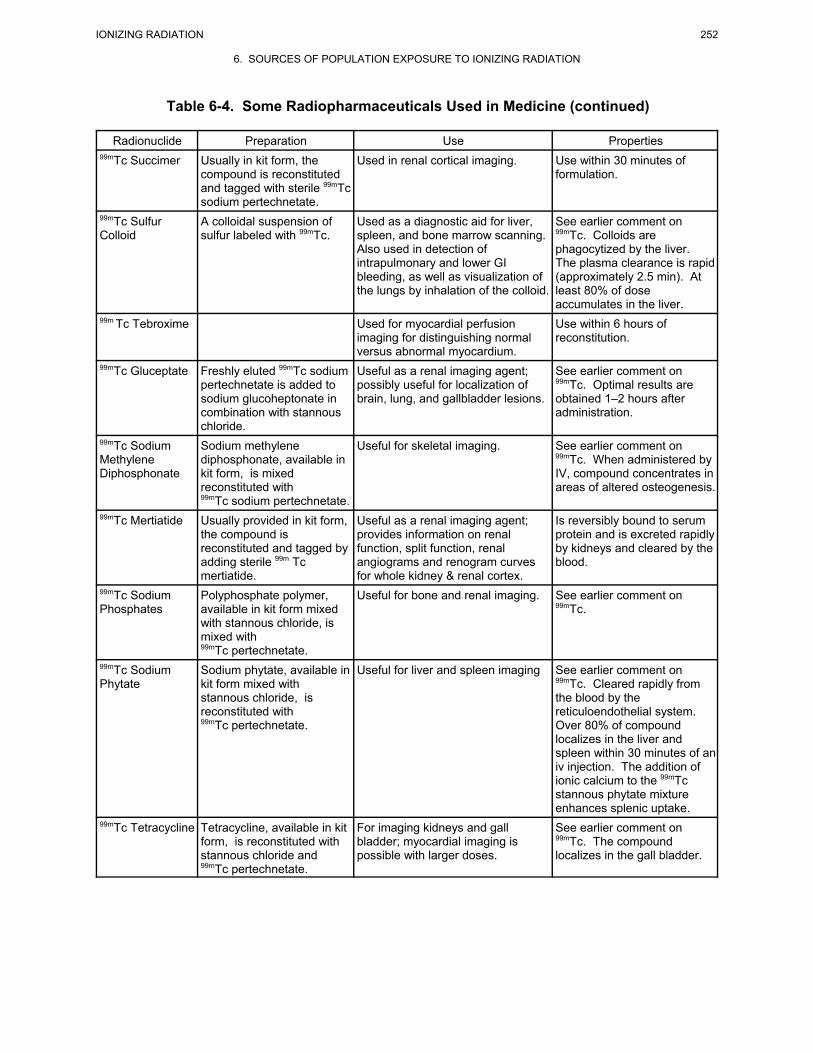
Table 6-4. Some Radiopharmaceuticals Used in Medicine (continued)
Radionuclide Preparation Use Properties99mTc Succimer Usually in kit form, the
compound is reconstitutedand tagged with sterile 99mTcsodium pertechnetate.
Used in renal cortical imaging. Use within 30 minutes offormulation.
99mTc SulfurColloid
A colloidal suspension ofsulfur labeled with 99mTc.
Used as a diagnostic aid for liver,spleen, and bone marrow scanning. Also used in detection ofintrapulmonary and lower GIbleeding, as well as visualization ofthe lungs by inhalation of the colloid.
See earlier comment on99mTc. Colloids arephagocytized by the liver. The plasma clearance is rapid(approximately 2.5 min). Atleast 80% of doseaccumulates in the liver.
99m Tc Tebroxime Used for myocardial perfusionimaging for distinguishing normalversus abnormal myocardium.
Use within 6 hours ofreconstitution.
99mTc Gluceptate Freshly eluted 99mTc sodiumpertechnetate is added tosodium glucoheptonate incombination with stannouschloride.
Useful as a renal imaging agent;possibly useful for localization ofbrain, lung, and gallbladder lesions.
See earlier comment on99mTc. Optimal results areobtained 1–2 hours afteradministration.
99mTc SodiumMethyleneDiphosphonate
Sodium methylenediphosphonate, available inkit form, is mixedreconstituted with99mTc sodium pertechnetate.
Useful for skeletal imaging. See earlier comment on99mTc. When administered byIV, compound concentrates inareas of altered osteogenesis.
99mTc Mertiatide Usually provided in kit form,the compound isreconstituted and tagged byadding sterile 99m Tcmertiatide.
Useful as a renal imaging agent;provides information on renalfunction, split function, renalangiograms and renogram curvesfor whole kidney & renal cortex.
Is reversibly bound to serumprotein and is excreted rapidlyby kidneys and cleared by theblood.
99mTc SodiumPhosphates
Polyphosphate polymer,available in kit form mixedwith stannous chloride, ismixed with99mTc pertechnetate.
Useful for bone and renal imaging. See earlier comment on99mTc.
99mTc SodiumPhytate
Sodium phytate, available inkit form mixed withstannous chloride, isreconstituted with99mTc pertechnetate.
Useful for liver and spleen imaging See earlier comment on99mTc. Cleared rapidly fromthe blood by thereticuloendothelial system. Over 80% of compoundlocalizes in the liver andspleen within 30 minutes of aniv injection. The addition ofionic calcium to the 99mTcstannous phytate mixtureenhances splenic uptake.
99mTc Tetracycline Tetracycline, available in kitform, is reconstituted withstannous chloride and99mTc pertechnetate.
For imaging kidneys and gallbladder; myocardial imaging ispossible with larger doses.
See earlier comment on99mTc. The compoundlocalizes in the gall bladder.

IONIZING RADIATION 253
6. SOURCES OF POPULATION EXPOSURE TO IONIZING RADIATION
Table 6-4. Some Radiopharmaceuticals Used in Medicine (continued)
Radionuclide Preparation Use Properties201Tl ThalliumChloride
Thallium target material isbombarded with protons toproduce 201Pb. The unusedthallium material is removedby ion exchange, and theremaining 201Pbsubsequently decays to201Tl.
Used for myocardial perfusionimaging for the localization ofmyocardial ischemia and infarction;used as an adjunct to angiography. Also useful for thyroid imaging,particularly the detection of goiterand thyroid carcinoma.
Thallium mimics potassiumions and is taken up by thecells of the heart; decreasedcell vitality is indicated bydecreased thallium uptake.Rapidly disappears from theblood.
15O Water Prepared by labeling with acyclotron-generatedradionuclide.
Used in blood-flow imaging usingpositron emission tomographyscanning.
127Xe Xenon gas Produced by protonbombardment of cesium-133 with 127Xe.
As a gas, used for lung imaging todetect alveolar block-age; also usedfor mapping cerebral blood flow.Dissolved in saline used as a tracerfor measurement of regional bloodflow.
The biological half-life of thegas is approximately15 minutes.
133Xe Xenon A product of nuclear fission;also formed by neutronactivation of 132Xe.
As a gas, used for lung imaging todetect alveolar blockage; also usedfor mapping cerebral blood flow.Dissolved in saline used as a tracerfor measurement of regional bloodflow.
The biological half-life of thegas is approximately15 minutes.
Source: Remington 1985; Remington and Gennaro 1995
6.6 EXPOSURE FROM CONSUMER PRODUCTS
Consumer products contribute an estimated 3% to the average population radiation dose. Several
consumer products, used both within the home and in many public areas, emit minuscule amounts of
radiation. Among these are ionization-type smoke detectors, television sets, and liquid propane gas
(LPG) appliances. The first smoke detectors contained radium (approximately 20 µCi [0.7 MBq]), but
now contain americium-241 (241Am), which is more economical and produces much less radiation dose.
While present-day detectors contain 0.5–1.0 µCi (0.02-0.04 MBq) of 241Am, the original units contained
approximately 80 µCi (3 MBq). In the 1980s, annual sales of smoke detectors approached 12 million,
representing approximately 8.5 Ci (300 GBq) of 241Am. Smoke detectors contain a small ionization
chamber in which the air between two electrodes is ionized by the source radionuclide. This ionization
allows the flow of current across the gap between the electrodes. When the flow is stopped by smoke
particles, the interruption in current flow is interpreted by the detector to indicate the presence of smoke.
Television sets accelerate electrons that bombard the screen; in the process, low-energy x rays are
emitted. The total annual dose associated with watching a color television has been estimated to be
2–3 mrad per year (0.02-0.03 mGy/yr). Radon is found in LPG, which may be used in water heaters,
IONIZING RADIATION 254
6. SOURCES OF POPULATION EXPOSURE TO IONIZING RADIATION
stoves, and fireplaces; it has been estimated that exposure to radon in homes using natural gas results in
an average annual dose of approximately 5 mrem (0.05 mSv) in the United States.
Among consumer products of the past are items that contained radium, such as medicines, tonics,
luminous paints, and ceramic glazes. After its discovery in the early part of the 20th century, radium was
used for many years in the treatment of rheumatism (arthritis) and mental disorders; oral solutions
contained 226Ra and 228Ra at concentrations up to 2 µCi/60 mL, while ampules for intravenous
administration contained 5–100 µg 226Ra and 228Ra. Radium was also used to produce luminescent paints
that were applied to wristwatches, clocks, static eliminators, fire alarms, electron tubes, and military and
educational products. During the peak years of production, approximately 3 million radium-laden
timepieces were sold annually in the United States. The radium content of a man’s wristwatch ranged
from 0.01 to 0.36 µCi (370-13,000 Bq), resulting in potential gonadal doses of 0.5–6 mrem/year
(0.005—0.06 mSv/yr). Radium has been replaced with 3H and prometium-147 (147Pm), and watch cases
are sufficiently thick to absorb the beta emissions from these radionuclides. Uranium has been used as a
coloring agent for ceramic glazes, resulting in doses to the hands of up to 20 mrad/hour (0.02 mGy/hr).
The dose from ceramics produced since 1944 is thought to be five-fold less than that from earlier pieces.
For more than 40 years, 224Ra has been used in Europe to treat the symptoms of tuberculosis and
ankylosing spondylitis. Although its use in children was curtailed in the 1950s, 224Ra has been used for
treating the pain associated with ankylosing spondylitis. In two studies of patients treated with 224Ra,
average calculated skeletal doses ranged from 0.65–4.2 Gy (65–420 rad) (Eisenbud 1987; Harley 1996;
Harvard Medical School 1996; NCRP 1993).
6.7 EXPOSURE FROM OTHER SOURCES
Other sources contribute less than 1% to the average population radiation dose. This radiation exposure
may result from several anthropogenic sources, including the radioactive debris still remaining from
atmospheric and underground detonation of nuclear weapons, electrical energy production,
radiopharmaceuticals, and radionuclide production and use (Shapiro 1990; UNSCEAR 1993).

IONIZING RADIATION 255
6. SOURCES OF POPULATION EXPOSURE TO IONIZING RADIATION
Figure 6-2. Schematic of the Nuclear Fuel Cycle
6.7.1 Exposure from the Nuclear Fuel Cycle
The nuclear fuel cycle contributes around 0.1% to the
average population radiation dose and refers to the mining,
milling and enrichment of uranium; fabrication of fuel
elements; the production of electricity; and the recycling,
transportation, and waste storage/disposal of radioactive
m a t e r i a l s u s e d i n n u c l e a r w e a p o n s o r
reactor-grade nuclear fuel. The steps involved in the
uranium fuel cycle are depicted in Figure 6-2 and described
in the Toxicological Profile for Uranium (ATSDR 1999b).
The primary radionuclide components of nuclear weapons
and reactors include 239Pu, 235U, 238U, and 3H.
Radionuclides associated with uranium mining and milling
include 235U, 238U, and their natural decay chain radionuclides, such as 226Ra, 234Th, 234mPa, 230Th, and 222Rn,
while those associated with power production and subsequent waste disposal include (but are not limited to)60Co, 3H, 14C, 129I, 131I, 134Cs, and 137Cs. Noble gas radionuclides of Kr, Xe, and Zr are only associated with
operational reactor releases. The various steps within this cycle provide multiple opportunities for the
exposure of humans to these materials.
Mining and milling. Uranium ore typically contains uranium at concentrations ranging from a tenth of a
percent to a few percent; thus, millions of tons of ore are mined and processed annually to meet the needs of
nuclear power plants for uranium fuel. Radon is the predominant radionuclide released from uranium mines.
Radon containing air is discharged from mines at a rate of approximately 0.5–20 µCi/min/1,000 ft3; these are
point releases whose concentrations dilute quickly with distance from the release shaft and, thus, pose no
additional health risk to the general public. Incomplete extraction of uranium during milling results in
uranium concentrations in mill tailings of 0.001–0.01%. The presence of radon precursors (226Ra and 230Th) in
mill tailings presents a potential long-term source for atmospheric radon. The rate of radon emanation varies
with meteorological factors such as barometric pressure and humidity. The rate of soil and mill tailings
migration depends primarily on wind and water erosion of the site (Eisenbud 1987; UNSCEAR 1993).

IONIZING RADIATION 256
6. SOURCES OF POPULATION EXPOSURE TO IONIZING RADIATION
Enrichment and fuel fabrication. In both nuclear weapons and nuclear fuel production, after being
mined and milled, uranium must be converted to uranium hexafluoride gas, which is then enriched and
converted to uranium oxide or metal. If enrichment is carried to about 90%, the uranium may be used to
make nuclear weapons or to fuel naval warships; alternatively, the uranium may be enriched by only a small
percentage for use in civilian nuclear energy facilities. Metallic uranium is capable of reacting with both air
and water exothermically; because of this reactivity, the more stable uranium oxide is the most commonly
used fuel in reactors. While this form is more stable, it has poor thermal conductivity, necessitating the use
of small-diameter fuel rods. The fuel is in the form of high melting point ceramic pellets, about 0.5 inches in
diameter and 1 inch long, in which UO2-enriched to 3–4% 235U is dispersed. These pellets are stacked end to
end in zirconium alloy or stainless steel tubes about 12 feet long (called cladding) and then sealed to retain
the fission products that are produced during operation. These fuel filled tubes are then assembled in groups
of 8 x 8 to 17 x 17 arrays into fuel rod assemblies. About 500 of these assemblies make up the core of a
nuclear power reactor. For a frame of reference, a single pellet contains the energy equivalent of about one
ton of coal or 3 barrels of oil. Emissions from fabrication facilities usually consist of the long-lived isotopes234U, 235U, and 238U, and the short-lived nuclides 234Th and 234mPa; however, the relative value of the refined
and enriched uranium and the high level of accountability for uranium stock preclude any long-term or
widespread loss of material. The major route of exposure from this source is inhalation (Eisenbud 1987;
UNSCEAR 1993). More information about the toxicological properties of uranium can be found in the
Toxicological Profile for Uranium (ATSDR 1999b).
Power generation. Power production from nuclear plants has increased steadily since the industry’s
birth in the 1950s. During the years between 1970 and 1989, the number of nuclear reactors worldwide
increased from 77 to 426, and total nuclear power generation increased from 9 to 212 gigawatts per year.
In 1996, nuclear power plants produced 17% and 19.4% of the world’s and U. S. electrical energy,
respectively (DOE 1997c). The annual worldwide production of uranium from 1979 to 1989 ranged from
19,000 to 44,000 tons; from 1985 to 1990, the annual production was approximately 50,000 tons.
USNRC regulations require that all the component parts of the nuclear fuel cycle be designed and
operated to limit the annual dose to a member of the public from the total nuclear fuel cycle to a
maximum of 25 mrem (0.25 mSv). Various regulations identified in Chapter 7 of this profile are
designed to limit human exposure to radiation and radioactive materials. Guidance documents, such as
the ANSI air sampling standard (ANSI 1999), are available to aid in establishing monitoring programs for
assessing discharges from nuclear facilities. During the energy production phase, radioactive
contamination of the coolant occurs through small defects in the protective cladding surrounding the fuel

IONIZING RADIATION 257
6. SOURCES OF POPULATION EXPOSURE TO IONIZING RADIATION
pellets, through fission of “tramp” uranium contamination on the outside surface of the fuel rods and
through neutron activation of contaminants in the cooling medium.
In general, the levels of radionuclide emissions from reactors are not typically detectable, except at points
close to effluent discharges; because of this, estimates of radionuclide discharge levels must be modeled.
Based on such models, the total collective dose due to reactor discharges through 1989 was estimated to
be 370,000 man•rem (3,700 man•Sv) over a 45-year period. This may be compared to the collective dose
to the U.S. public from the natural radioactive potassium within everyone’s body, which is about
5,000,000 man•rem (50,000 man•Sv) each year . A 1981 U.S. Nuclear Regulatory Commission
(USNRC) study of the doses received by 98 million people living within 80 km of 48 nuclear facilities
concluded, on the basis of a zero threshold model, that 0.02 excess fatal cancers per year, or 1 every 50
years, could be attributed to exposures from nuclear facilities (USNRC 1981). A study of the cancer rates
in populations surrounding 62 U.S. nuclear facilities, performed by the National Cancer Institute (NCI) in
1990, found no evidence of a relationship between proximity to a nuclear facility and the occurrence of
cancer (Eisenbud 1987; NCI 1990; UNSCEAR 1993).
Weapons production. There is little public information regarding the amount of radioactive materials
produced for use as weapons. The atmospheric content of krypton-85 (85Kr), a by-product of plutonium
extraction, has been used to estimate the plutonium stockpiles in both the United States and Russia. After
adjusting for production and release of 85Kr from nuclear reactors, it is estimated from the atmospheric content
of 85Kr that plutonium stockpiles in both the United States and Russia is about 100 tons each. United Nations
estimates from 1981 and 1990 state that nuclear arsenals are comprised of 40,000 weapons with a combined
explosive power of 13,000 Mt. Tritium, which has a half-life of 12.32 years, must be continually produced
to replace aging stockpiles. It is estimated that an annual production of 3 kg is sufficient to replace that lost
by transformation in the United States. By inference, this would indicate a total U.S. stockpile of 55 kg and
a world stockpile of about 110 kg (UNSCEAR 1993).
The dose from nuclear weapons research, development, and production is less than 1% of the dose from
atmospheric testing. Variations in local exposure from former weapons plants have been reported. In the
United States, the Hanford nuclear weapons facility has released a significant amount of radioactive
material into the atmosphere and the Columbia River from its plutonium production and reprocessing
plants. The majority of the radioactive material (131I) was released between 1944 and 1946 (18 PBq;
486 kCi), although additional releases are known to have occurred from 1947 to 1956 (2 PBq; 54kCi). A

IONIZING RADIATION 258
6. SOURCES OF POPULATION EXPOSURE TO IONIZING RADIATION
recent study reconstructed the radiation doses to individuals who were exposed to the 131I released from
the Hanford site, medically determined their thyroid health, and found that the Hanford releases had not
caused thyroid problems. The thyroid radiation doses to the 3,193 participants averaged 0.186 Gy
(18.6 rad) and ranged from 0–2.842 Gy (0–284.2 rad). Health impacts to the thyroid which have been
related to radiation exposure in other studies include thyroid cancer, benign thyroid nodules,
hypothyroidism, and autoimmune thyroiditis. The study assessed these conditions and concluded that the
occurrence of these diseases in the population were not related to the Hanford releases’ radiation dose to
the thyroid (CDC 1999).
The Chelyabinsk-40 center, located near Kyshtym in the Soviet Union, was the first nuclear weapons
processing facility in Russia. A uranium-graphite-moderated reactor and a fuel reprocessing plant were
opened in 1948. Due to poor waste handling and the storage of radioactive wastes in the open, significant
liquid releases (100 PBq; 2.7MCi) to the Techa River occurred from 1949 to 1956, with the majority of
the releases (95%) occurring from March 1950 to November 1951. The main nuclides released included89Sr, 137Cs, 95Zr, 95Nb, and ruthenium and rare-earth nuclides. The population along the Techa River was
exposed to both external and internal doses of radiation; a total of 20 settlements (7,500 people) were
eventually evacuated. The average doses to persons living in the village of Metlino, 7 km downstream
from the plant, were estimated to be as much as 140 rad (1.4 Gy) (UNSCEAR 1993).
Fuel reprocessing. Fuel reprocessing allows the recovery of uranium and plutonium from the
irradiated fuel pellets. Less than 10% of the nuclear fuel is consumed in a spent fuel rod. The
radionuclides most commonly associated with reprocessing waste are: 3H, 14C, 85Kr, 129I, 131I, 134Cs, 137Cs
and transuranium nuclides. At present, reprocessing is carried out in only a few countries, and only a
small portion of the total fuel inventory is being reprocessed (4% from 1985 to 1990). The remainder is
retrievably stored (UNSCEAR 1993).
The reprocessing of nuclear fuel has been performed almost exclusively at government-owned facilities
designed to meet military needs. Only 1 operable reprocessing facility exists in the U.S. As with nuclear
reactors, the facility and the equipment used have been designed with numerous safeguards to prevent
criticality and to ensure containment of radioactive material in the event of a non-nuclear explosion or
system failure. The estimated collective dose due to reprocessing to date is estimated to be 460,000
man•rem (4,600 man•Sv); the main radionuclide constituents (>90%) of these releases have been 137Cs
and 106Ru. Releases of gaseous 85Kr and 3H from reprocessing facilities have also been reported. The
main pathways of exposure are consumption of locally caught fish and shellfish, external (whole-body)
irradiation from intertidal areas, and external (dermal) irradiation of fishermen handling pots and nets.
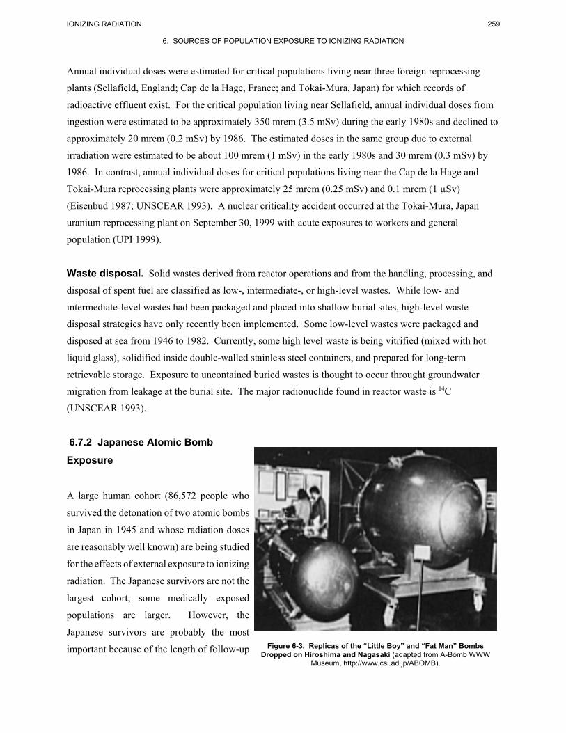
IONIZING RADIATION 259
6. SOURCES OF POPULATION EXPOSURE TO IONIZING RADIATION
Figure 6-3. Replicas of the “Little Boy” and “Fat Man” BombsDropped on Hiroshima and Nagasaki (adapted from A-Bomb WWW
Museum, http://www.csi.ad.jp/ABOMB).
Annual individual doses were estimated for critical populations living near three foreign reprocessing
plants (Sellafield, England; Cap de la Hage, France; and Tokai-Mura, Japan) for which records of
radioactive effluent exist. For the critical population living near Sellafield, annual individual doses from
ingestion were estimated to be approximately 350 mrem (3.5 mSv) during the early 1980s and declined to
approximately 20 mrem (0.2 mSv) by 1986. The estimated doses in the same group due to external
irradiation were estimated to be about 100 mrem (1 mSv) in the early 1980s and 30 mrem (0.3 mSv) by
1986. In contrast, annual individual doses for critical populations living near the Cap de la Hage and
Tokai-Mura reprocessing plants were approximately 25 mrem (0.25 mSv) and 0.1 mrem (1 µSv)
(Eisenbud 1987; UNSCEAR 1993). A nuclear criticality accident occurred at the Tokai-Mura, Japan
uranium reprocessing plant on September 30, 1999 with acute exposures to workers and general
population (UPI 1999).
Waste disposal. Solid wastes derived from reactor operations and from the handling, processing, and
disposal of spent fuel are classified as low-, intermediate-, or high-level wastes. While low- and
intermediate-level wastes had been packaged and placed into shallow burial sites, high-level waste
disposal strategies have only recently been implemented. Some low-level wastes were packaged and
disposed at sea from 1946 to 1982. Currently, some high level waste is being vitrified (mixed with hot
liquid glass), solidified inside double-walled stainless steel containers, and prepared for long-term
retrievable storage. Exposure to uncontained buried wastes is thought to occur throught groundwater
migration from leakage at the burial site. The major radionuclide found in reactor waste is 14C
(UNSCEAR 1993).
6.7.2 Japanese Atomic BombExposure
A large human cohort (86,572 people who
survived the detonation of two atomic bombs
in Japan in 1945 and whose radiation doses
are reasonably well known) are being studied
for the effects of external exposure to ionizing
radiation. The Japanese survivors are not the
largest cohort; some medically exposed
populations are larger. However, the
Japanese survivors are probably the most
important because of the length of follow-up

IONIZING RADIATION 260
6. SOURCES OF POPULATION EXPOSURE TO IONIZING RADIATION
and the wide range of doses received. The first atomic bomb was detonated on July 16, 1945, in Alamogordo,
New Mexico. The U.S. military, in an effort to bring a swifter end to World War II and to avoid a costly
ground invasion of Japan, which could claim more U.S. and Japanese lives, detonated a 235U atomic bomb,
nicknamed “Little Boy,” (see Figure 6-3) over the city of Hiroshima, Japan, on August 6, 1945. Three days
later, an atomic bomb using 239Pu, nicknamed “Fat Man,” was detonated over the city of Nagasaki, Japan.
The uranium used in the “Little Boy” bomb was enriched to >80% 235U. (Natural uranium contains 0.7%235U, and reactor fuel is enriched to 3–4% 235U). The uranium bomb design of Little Boy used a standard
explosion trigger, called the "gun" method, because it was originally made using a gun barrel. In this
configuration, a sub-critical uranium mass, referred to as the “bullet,” was propelled inside the gun barrel
toward a second sub-critical portion of the uranium mass (called the “target”), which was located at the
end of the gun barrel. The target contained slightly less than the amount of uranium needed to achieve
critical mass (the amount necessary to create a chain reaction). The instant the two subcritical pieces of
uranium came together, super-criticality was attained, and an explosion with a force equivalent to 15,000
tons (15 kt) of trinitrotoluene (TNT) occurred (Roesch 1987). In the case of Little Boy, the bullet was a
cylindrical stack of nine 235U wafers about 10 cm wide and 16 cm long, containing 40% of the bomb’s
total 235U mass (25.6 kg). The target was a hollow cylinder 16 cm long and wide; it weighed 38.4 kg and
was composed of two separate rings that were inserted into the bomb separately to prevent reaching
critical mass during assembly. The complete Little Boy weapon was 10.5 feet long, 28–29 inches in
diameter, and reportedly weighed between 8,900 and 9,700 pounds. The firing mechanism was so simple
and was considered so failproof that it was not tested prior to its use over Hiroshima. The gun-type firing
mechanism was, however, an unsafe weapon design, in that once the firing mechanism was loaded with
high explosive, anything that ignited it would cause a nuclear explosion. Also, a crash or even an
accidental drop of the bomb could have driven the bullet into the target, potentially resulting in a nuclear
explosion. No other weapon of this design was ever tested, and although several Little Boy units were
built, none ever entered the U.S. nuclear arsenal.
The plutonium bomb, Fat Man, was dramatically different from the Little Boy design. The gun-type
firing mechanism could not be used to unite two pieces of plutonium fast enough to achieve a nuclear
blast; impurities in the plutonium would have caused premature detonation. Fat Man contained a ball of
subcritical plutonium (plutonium core), which was surrounded with high explosives. The high explosives
were cast into spheres, called lenses, and were wired so they would all fire at the same instant. The
instantaneous pressure from all sides compressed the plutonium core in on itself, causing it to reach
critical mass and density, and create a nuclear blast. The combat configuration for the Fat Man bomb

IONIZING RADIATION 261
6. SOURCES OF POPULATION EXPOSURE TO IONIZING RADIATION
consisted of the implosion device encapsulated in a steel armor egg. Fat Man was 5 feet in diameter,
12 feet in length, and weighed 10,300 pounds (American Airpower Heritage Museum 1996; Sublette
1996). The explosion of Fat Man at Nagasaki was equivalent to 21,000 tons (21 kt) of TNT (Roesch
1987).
In Hiroshima and Nagasaki, a total of 64,000 people within 1 km of the hypocenter (the point on the
ground directly below where the bomb exploded in the air) died on the first day. The total numbers of
acute deaths from the explosions were 90,000–140,000 in Hiroshima and 60,000–80,000 in Nagasaki. For
bombs the size (approximate yield of 15 kilotons) and type of Little Boy, the energy released within the
first minute after detonation was in the forms of thermal radiation (35%), blast wave (50%), and ionizing
radiation (15%); most casualties (including fatalities) resulted from the heat and blast. Two-thirds of
those who died during the first day were burned. People close enough to suffer from radiation illness
were also well within the lethal zones from blast and heat; thus, the proportion of survivors experiencing
radiation illness (30%) was much smaller than the expected proportion based solely on exposure to
radiation. People within 1–2 km of the hypocenter who initially survived the blast and received several
hundred rad (several grays) of radiation, suffered the ill effects of acute radiation syndrome. It is
estimated that all persons whose bodies received a dose of 600 rad (6 Gy) and half of those whose
radiation doses were 450 rad (4.5 Gy) died shortly thereafter, as a direct result of radiation exposure. Of
those who survived the immediate radiation illness effects, a portion would suffer from the latent effects
of radiation-induced cancer (the excess cancer death rate among the exposed population is on the order of
5% greater than that of an unexposed population). It is estimated that approximately 500 survivors have
died of radiation induced cancers since 1945. Given the estimated altitudes at which Little Boy and Fat
Man detonated (1,900±50 ft. and 1,650±33 ft., respectively), little or no soil was carried up into the
fireball, so the fallout produced came mainly from the detonation itself. Of that, very little radioactive
material was deposited on the ground in the vicinity of ground zero; the majority was carried high into the
atmosphere by heat convection. A small amount of fallout did occur in areas close to the cities due to
rainfall that occurred shortly after the explosions; the affected areas were to the west and northwest of
Hiroshima and a few miles east of Nagasaki. Fatality rates in the Hiroshima and Nagasaki attacks were
1–2 orders of magnitude greater than rates from conventional bombings because of the nearly
instantaneous destruction of buildings that occurred without warning, and because survivors were so
incapacitated that they could not escape the rapidly ensuing fire storms. Approximately one-third of all
Japanese bombing fatalities occurred in these two cities (Masse 1996; Sublette 1995, 1996; Uranium
Information Center 1995; Zajtchak 1989).
IONIZING RADIATION 262
6. SOURCES OF POPULATION EXPOSURE TO IONIZING RADIATION
6.7.3 Exposure from Nuclear Weapons Testing
Nuclear weapons testing is conducted to assess design efficiency and magnitude of resulting damage.
Nuclear explosions world wide were carried out above ground from 1945 to 1980, with the periods of
greatest activity occurring from 1952 to 1958, and from 1961 to 1962. A total of 520 tests with an
estimated total equivalent energy of 545 megatons (Mt) of TNT was performed, resulting in the release of
220 PBq (6 MCi) of radioactive material. The first U.S. testing of nuclear weapons after World War II
was performed in the Marshall Islands in the Pacific Ocean from 1946 to 1948. The Soviet Union
conducted its first weapons test in 1948. In the 1950s, as the frequency of weapons testing escalated, so
did public concerns over radioactive fallout. In the fall of 1958, the United States, Britain, and Russia,
declared a moratorium on weapons testing; however, Russia broke the agreement in 1961, and another
rapid escalation in testing ensued. In 1963, the United States, Britain, and Russia, signed the Limited
Test Ban Treaty, which prohibited atmospheric testing. Although these three countries have remained
faithful to the treaty, other countries such as France, China, India, and Pakistan have since conducted
weapons testing (Eisenbud 1987; PBS 1999; UNSCEAR 1993).
The energy from nuclear weapons devices is generated by one or both of the following reactions: (1) the
fission of 235U or 239Pu in a chain reaction and (2) the fusion of the hydrogen isotopes deuterium and
tritium. Fission-type weapons accounted for 217 Mt of the total test yield, while fusion-type weapons
accounted for 328 Mt. Many radionuclides are produced by U and Pu fission. Fusion reactions produce
helium and result in the neutron activation of the surrounding substance. The most notable neutron
activation product is 14C, which is formed from the neutron bombardment of atmospheric nitrogen.
Unused weapons material is also liberated after detonation due to premature loss of critical mass (Radnet
1996; UNSCEAR 1993).
The most important radionuclides associated with nuclear weapons testing exposures are 14C; 137Cs;
zirconium-95 (95Zr); niobium-95 (95Nb); 90Sr; ruthenium-106 (106Ru); manganese-54 (54Mn); 144Ce; 131I;
and 3H. This is in addition to the large amounts of 14C and 3H in the biosphere’s carbon and hydrogen
inventories which are produced by the interaction of cosmic rays with atmospheric gases.
The disposition of ingested strontium-90 (90Sr) has been studied extensively due to its abundance
(15 MCi of that introduced into the atmosphere as a result of nuclear weapons testing fell to the earth by
January 1970), its long half-life (28 years), and its tendency to localize in bones. Metabolically, Sr
follows the pathways of calcium (Ca); however, the body discriminates against Sr in favor of Ca.

IONIZING RADIATION 263
6. SOURCES OF POPULATION EXPOSURE TO IONIZING RADIATION
Because of this parallel mechanism, Sr concentrations are often expressed as activity of Sr per gram of
Ca. In 1965, bone levels of 90Sr in 1- to 4-year-old Norwegian children averaged 11.8 pCi/g Ca. In New
York City, bone levels of 90Sr in 1- to 2-year-olds varied from 7 pCi/g Ca in 1965 to 1.6 pCi/g Ca in
1975. In 5- to 19-year-olds, bone levels varied from 3 pCi/g Ca during 1956–1968 to 1.4 pCi/g Ca in
1975 (Shapiro 1990). In an effort to quantify exposure resulting from nuclear testing, 90Sr deposition has
been monitored worldwide at 50 to 200 stations in cooperation with the Environmental Measurements
Laboratory (EML), and by a network of 26 stations organized by the United Kingdom Atomic Energy
Authority. These data are compiled into a report called Environmental Radiation Data (ERD), which is
distributed quarterly by the Office of Radiation and Indoor Air’s National Air and Radiation
Environmental Laboratory (NAREL). The report contains data from the Environmental Radiation
Ambient Monitoring System (ERAMS). ERAMS was established in 1973 by the EPA to provide air,
surface and drinking water, and milk samples from which environmental radiation levels are derived.
These samples are collected from locations that provide adequate population coverage and function to
monitor fallout from nuclear devices and other radioactive contamination from the environment. Samples
are subjected to analysis for gross alpha and beta emissions; gamma analyses for fission products; and
more specific analysis for uranium plutonium, strontium, iodine, radium, and tritium (EPA 1997).
Zirconium-95 (95Zr) deposition has been monitored as an indicator of exposure to short-lived radio-
nuclides. Monitoring levels of 3H and 14C is more difficult due to the rapid recycling of these elements in
a biosphere that contains large cosmogenically produced amounts of these same radionuclides.
Interhemispheric transfer is limited due to prevailing trade winds and the scavenging effect of
precipitation in the tropics. The best estimate of the average total per person dose, for persons in the
northern and southern hemispheres, for all 22 major radionuclides resulting from nuclear testing is 440
and 310 mrem (4.4 and 3.1 mSv), respectively. Worldwide, the average total cumulative dose is
370 mrem (3.7 mSv). However, as noted below, extreme variations in local exposures due to testing have
been noted (UNSCEAR 1993).
Approximately 23 billion Ci of 131I have been introduced into the atmosphere as a result of nuclear
weapons testing. In October 1961, the air concentration of 131I in the United States averaged 3.8 pCi/m3.
This was estimated to result in an annual dose of 24 mrad to a 1-year-old child. In 1962, the
concentration of 131I in milk in the United States averaged 32 pCi/L. Although about two-thirds of orally
administered 131I is excreted in the urine within the first 24 hours, the remainder concentrates in the
thyroid. It has been estimated that an infant receiving milk from cows that grazed on forage contaminated
with 1 µCi 131I/m2 could receive a dose to the thyroid of 30 rad (Shapiro 1990; UNSCEAR 1993). In
addition, studies of pregnant women who died suddenly of non-radiation-induced causes (e.g., car

IONIZING RADIATION 264
6. SOURCES OF POPULATION EXPOSURE TO IONIZING RADIATION
accidents) found that fetal tissue concentrations of 131I were 30% greater than those in maternal tissues
(Shapiro 1990). Due to the short 8-day half-life of 131I, there is no 131I remaining in the atmosphere or in
food from nuclear weapons testing.
Plutonium (Pu) has been introduced into the atmosphere from sources such as weapons testing (>5,000 kg
[>320 kCi]) and the vaporization of energy power packs from a Russian satellite (20 kg [0.27 TBq] of239Pu) and U.S. satellites (1 kg [17 kCi] of 238Pu ) that burned up upon re-entry. Air activity of Pu,
monitored in New York, peaked in 1963 at a concentration of 1.7 fCi/m3. The cumulative inhalation
intake from 1954 to 1975 averaged 43 pCi per person. For comparison, the EPA’s annual limit on intake
(ALI) for 239Pu for occupational exposure is 2,000 pCi. Cumulative individual tissue doses (through the
year 2000) due to inhalation are predicted to be: lungs, 1.6 mrad; liver, 1.7 mrad; and bone lining cells,
1.5 mrad (Shapiro 1990).
Radioactive debris from nuclear explosions falls into three categories: large particles, which fall out close
to the explosion site within hours of the explosion; smaller particles, which penetrate the troposphere,
behave like aerosols, and may not fall out for days; and the smallest particles, which penetrate the
stratosphere, distribute worldwide, and fall out over many months or years. The greatest portion of
fallout from nuclear weapons testing was injected into the stratosphere (78%), while 10% and 12% were
injected into the troposphere and in the locality of the test, respectively. As of 1993, the total cumulative
worldwide collective dose due to fallout was estimated to be 7x10⁶ man•Sv. The collective dose will
continue to climb, mainly due to long-lived 14C (Eisenbud 1987; UNSCEAR 1993).
Airborne radioactive materials, both naturally occurring and fallout-derived, usually attach to dust
particles; the potential for inhalation of radionuclides bound to particles, and the respective threat this
poses to animal and human health, varies considerably. In regions downwind of nuclear weapons test
sites or nuclear weapons production facilities, or areas with abnormally high concentrations of naturally
occurring radionuclides (e.g., New York state or Denver, Colorado), the potential for inhalation of
particulate-bound radionuclides increases. Although most inhaled radioactive particles are eliminated
from the lungs by normal clearing mechanisms, some of the particles remain in the lungs for extended
periods. Others are carried to lymph nodes by scavenger cells (Eisenbud 1987; Shapiro 1990).
Radiation dose from atmospheric testing is attributed to external exposure to radionuclides on the earth's
surface, internal exposure from inhalation of gases or particulate matter, and ingestion of contaminated
foods and water. Approximately 80% of the radiation dose from nuclear testing is estimated to be
IONIZING RADIATION 265
6. SOURCES OF POPULATION EXPOSURE TO IONIZING RADIATION
delivered through ingestion, with 16% and 4% of the dose delivered through external exposure and
inhalation, respectively. Radioactive particles resulting from fallout can contaminate food supplies
directly by foliar deposition, or indirectly by entry into the soil and subsequent incorporation into plants,
although this delayed incorporation through root uptake accounts for a small portion of that ingested.
Surface waters can be contaminated through soil runoff or direct contamination from the atmosphere.
The degree of radionuclide incorporation into plants through root uptake varies considerably among
radionuclides. For example, 137Cs and radium bind tightly to clay or organic minerals upon entering the
soil and are not amenable to root uptake; thus, foliar deposition is the primary route for oral exposure to137Cs. Likewise, 131I poses little threat through root uptake due to its short half-life (8 days). The
concentrations of radionuclides in food vary considerably across food types. The concentration of 226Ra
ranges from 0.15 pCi/kg in cow’s milk to 2,000 pCi/kg in Brazil nuts. Concentrations of 137Cs range from
20 pCi/kg in cow’s milk to 5,000 pCi/kg in beef. More important than concentrations in foods is the rate
of intake and absorption of a radionuclide. Individual intake varies considerably; for a given radionuclide
and locality, intake may vary as much as 500-fold. Likewise, the extent of absorption varies among the
various nuclides, from almost completely in the case of 40K, 137Cs, and 131I, to very poorly (0.003%) in the
case of Pu radionuclides. Many alpha emitters, such as 226Ra, are taken up and retained in the bone,
resulting in sustained alpha irradiation of the bone-forming cells and bone surface lining cells
(Eisenbud 1987; McClellan 1982; Shapiro 1990).
The primary radionuclides of concern are 90Sr, 137Cs, and 131I. Within the first year, 45% of 137Cs is
transferred to the food chain (through milk, grain and meat). 90Sr enters the food chain primarily through
milk and grain products. Exposure to 14C and 3H is through ingestion and inhalation; however, the
contribution of 14C and 3H by ingestion is trivial. The dose rate from naturally occurring 14C is about 1
mrem per year. At its peak effect, the dose rate from 14C due to weapons testing also was about 1 mrem
per year, and is now decreasing. The dose from the tritium due to weapons is considered to be even less.
At the peak, the additional 3H contributed less than 0.1 mrem per year (UNSCEAR 1993). The radiation
dose from 131I occurs largely during the first 2 months following detonation, due to its short 8-day half-
life, and the primary exposure route is via the pasture-cow’s milk ingestion pathway.
In addition to exposures from inhalation and ingestion, radiation exposure also occurs externally from
particles deposited on the ground. Since the debris spends more time on the ground than in the air, the
radiation dose from earthbound particles ranges from 100 to 1,000,000 times that from airborne particles
(UNSCEAR 1993).

IONIZING RADIATION 266
6. SOURCES OF POPULATION EXPOSURE TO IONIZING RADIATION
6.7.3.1 Atmospheric Testing
Atmospheric testing refers to the detonation of nuclear bombs above the earth’s surface.
Nevada Test Site Fallout. A total of 100 surface or near-surface tests with a total explosive yield of
about 1 Mt were performed at the Nevada test site between 1951 and 1962. The population around the
site at this time was approximately 180,000 persons. Within this population, thyroid doses in children
may have been as high as 100 rad (1 Gy). The collective dose received by this population was
approximately 50,000 man•rem (500 man•Sv); 90% of this dose was delivered between 1953 and 1957.
The dust from these tests also drifted over the United States, producing bands of exposure to radioactive
material. Deposition of fallout varied considerably because of meteorological conditions. For example,
the greatest (non-local) fallout levels from one of the Nevada test explosions occurred in New York State,
some 2,000 miles away, due to rainfall. The cumulative dose from gamma radiation in New York,
approximately 100 mrad (1 mGy), exceeded the doses received by any remote U.S. location for all of
1953 (Eisenbud 1987; UNSCEAR 1993).
Bikini Atoll Fallout. Operation Crossroads was a series of nuclear weapons tests that began in the
Marshall Islands, a group of atolls in the Pacific, on July 1, 1946. Prior to testing, the inhabitants of the
Bikini Island Atoll were evacuated. The second test in this series, designated “Baker,” was a 21-kiloton
bomb that was detonated underwater. This resulted in contamination of ships staged nearby and the atoll
itself. Local soil contamination prevented the return of the Bikini native population until 1969. Although
the island was still contaminated in 1969, it was thought that dietary restrictions and the importation of
foods would allow safe habitation. However, body burdens of plutonium began to increase in the natives,
resulting in their re-evacuation in 1978.
During Operation Castle, another series of nuclear tests, the second test, “Bravo,” resulted in significant
fallout and contamination of humans. Abrupt changes in wind direction after the 15-Mt detonation on
March 1, 1954, resulted in the inadvertent exposure of residents of the Rongelap and Utirik islands, which
lie 210 and 570 km to the east of Bikini, as well as exposure of a group of 23 Japanese fishermen whose
boat was caught in the fallout approximately 80 miles downwind. Since the device was mounted on a
barge situated in shallow water, a considerable amount of coral was incorporated into the fireball. The
fishermen reported that the fallout particles resembled snow and that deposits of fallout on the boat were
of sufficient depth to allow one to see footprints. Because they were unaware of the circumstances, the
fishermen took no precautionary measures to minimize exposure; they remained on the contaminated boat

IONIZING RADIATION 267
6. SOURCES OF POPULATION EXPOSURE TO IONIZING RADIATION
until returning to port some 13 days later. Within 1–2 days after exposure to the fallout, the fishermen
began to experience itching and burning sensations on exposed skin. By the third day, skin lesions and
epilation began to develop; the skin lesions became ulcerous in about 70% of the fishermen. Lesions
were less severe in those who had worn protective clothing such as hats (ACHRE 1995; Eisenbud 1987).
A more detailed description is available (Simon and Vetter 1997).
Within 78 hours of the explosion, 82 and 159 persons were evacuated from Rongelap and Utirik,
respectively. However, as with the fishermen, the island inhabitants took no precautionary measures to
minimize exposure to radioactive fallout. Within 1–2 days after exposure to fallout, itching and burning
sensations on exposed skin were experienced by the natives of Rongelap, but not those of Utirik. Skin
lesions and epilation occurred within 21 days of exposure, becoming ulcerous in about 25% of the
Rongelaps; lesions were less severe in those who had worn protective clothing or bathed during the
period prior to evacuation. The island of Utirik was not heavily contaminated, and its residents were
allowed to return within a few months; however, the Rongelap residents were not allowed to return to
their island until 1957, and they were monitored annually by U.S. medical teams thereafter. Despite the
monitoring, fears among the island residents that exposure-related health problems were occurring
prompted a second evacuation, initiated by the residents, in 1985. External doses, ranging from 10 to 190
rad (0.1 to 1.9 Sv), were mostly from short-lived radionuclides. Mean thyroid doses to adults, 9-year-
olds, and 1-year-olds, were 1,300, 2,200, and 5,200 rad (13, 22, and 52 Gy), respectively. Maximum
thyroid doses to these groups were 4,200, 8,200, and 20,000 rad (42, 82, and 200 Gy), respectively
(ACHRE 1995; Eisenbud 1987; NAS 1994; UNSCEAR 1993).
Average gamma dose rates 3 feet above ground level on Rongelap island, estimated from a survey
performed in July 1956, were 0.2–0.5 mR/hr (mean of 0.4 mR/hr) (DOE 1994b). Environmental samples
collected in 1964 showed 239Pu concentrations of 11 pCi/g (0.4 Bq/g) in a soil sample collected at a depth
of 0.5–1.0 inch. A more extensive survey that included 14 of the atoll islands was performed in April and
May of 1967. External radiation, as well as the radioactive content of food, vegetation, and soil, was
measured. On the islands closest to the detonations, the major contributor to the external gamma
radiation field was 60Co, which was associated with neutron activation of scrap metal; the major
contributor to the external gamma radiation field on distant islands was 137Cs. Additional samples were
collected during the U.S. cleanup operations in 1969. 239Pu concentrations on Bikini Island ranged from
1.3 to 190 pCi/g (0.1–7 Bq/g). 239Pu concentrations on Eneu Island ranged from 0.5 to <3 pCi/g
(0.02–<0.1 Bq/g) (DOE 1970). Measurements of gamma radiation exposure performed in June 1975
showed highly variable exposure rates on Bikini island (10–20 µR/hr at the shore versus 30–100 µR/hr in
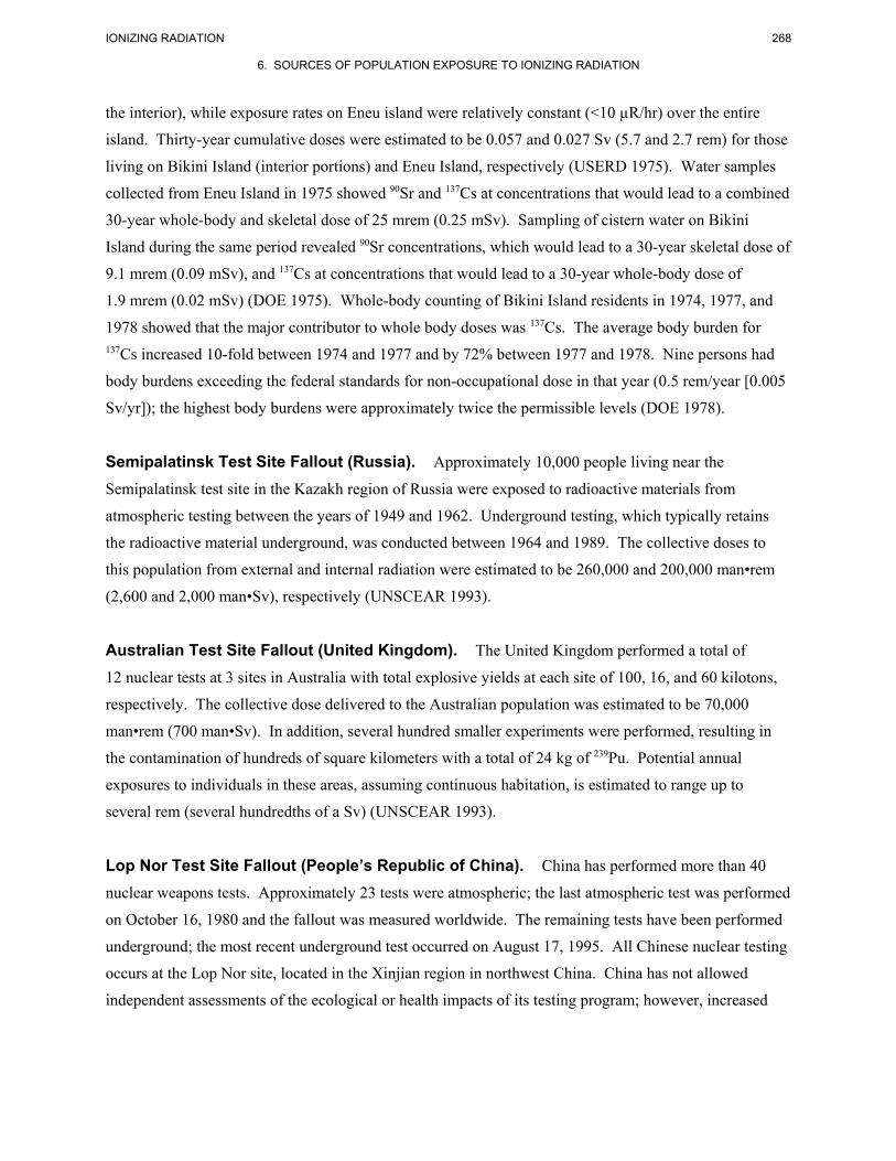
IONIZING RADIATION 268
6. SOURCES OF POPULATION EXPOSURE TO IONIZING RADIATION
the interior), while exposure rates on Eneu island were relatively constant (<10 µR/hr) over the entire
island. Thirty-year cumulative doses were estimated to be 0.057 and 0.027 Sv (5.7 and 2.7 rem) for those
living on Bikini Island (interior portions) and Eneu Island, respectively (USERD 1975). Water samples
collected from Eneu Island in 1975 showed 90Sr and 137Cs at concentrations that would lead to a combined
30-year whole-body and skeletal dose of 25 mrem (0.25 mSv). Sampling of cistern water on Bikini
Island during the same period revealed 90Sr concentrations, which would lead to a 30-year skeletal dose of
9.1 mrem (0.09 mSv), and 137Cs at concentrations that would lead to a 30-year whole-body dose of
1.9 mrem (0.02 mSv) (DOE 1975). Whole-body counting of Bikini Island residents in 1974, 1977, and
1978 showed that the major contributor to whole body doses was 137Cs. The average body burden for137Cs increased 10-fold between 1974 and 1977 and by 72% between 1977 and 1978. Nine persons had
body burdens exceeding the federal standards for non-occupational dose in that year (0.5 rem/year [0.005
Sv/yr]); the highest body burdens were approximately twice the permissible levels (DOE 1978).
Semipalatinsk Test Site Fallout (Russia). Approximately 10,000 people living near the
Semipalatinsk test site in the Kazakh region of Russia were exposed to radioactive materials from
atmospheric testing between the years of 1949 and 1962. Underground testing, which typically retains
the radioactive material underground, was conducted between 1964 and 1989. The collective doses to
this population from external and internal radiation were estimated to be 260,000 and 200,000 man•rem
(2,600 and 2,000 man•Sv), respectively (UNSCEAR 1993).
Australian Test Site Fallout (United Kingdom). The United Kingdom performed a total of
12 nuclear tests at 3 sites in Australia with total explosive yields at each site of 100, 16, and 60 kilotons,
respectively. The collective dose delivered to the Australian population was estimated to be 70,000
man•rem (700 man•Sv). In addition, several hundred smaller experiments were performed, resulting in
the contamination of hundreds of square kilometers with a total of 24 kg of 239Pu. Potential annual
exposures to individuals in these areas, assuming continuous habitation, is estimated to range up to
several rem (several hundredths of a Sv) (UNSCEAR 1993).
Lop Nor Test Site Fallout (People’s Republic of China). China has performed more than 40
nuclear weapons tests. Approximately 23 tests were atmospheric; the last atmospheric test was performed
on October 16, 1980 and the fallout was measured worldwide. The remaining tests have been performed
underground; the most recent underground test occurred on August 17, 1995. All Chinese nuclear testing
occurs at the Lop Nor site, located in the Xinjian region in northwest China. China has not allowed
independent assessments of the ecological or health impacts of its testing program; however, increased

IONIZING RADIATION 269
6. SOURCES OF POPULATION EXPOSURE TO IONIZING RADIATION
mortality rates due to fallout of radioactive materials have been reported by a political advocacy group in
neighboring eastern Turkestan (Eastern Turkestan Information Bulletin 1996). Both the data and the
claims are unsubstantiated.
6.7.3.2 Underground Testing
Underground testing refers to the detonation of nuclear bombs below the earth’s surface. About 1,400
underground nuclear tests have been performed worldwide, with a total explosive yield of 90 Mt. The
frequency of underground testing increased dramatically after the 1963 signing of the Limited Test Ban
Treaty, which banned atmospheric testing. The nature of underground tests typically causes all the
radioactive material produced to be retained underground. However, some radioactive material can be
released if the blast penetrates the surface or if inadvertent leaks occur due to ground structure damage or
the gradual diffusion of gases. Of the 500 underground tests performed at the Nevada test site, only 32
led to off-site contamination. The total activity of 131I inadvertently released was about 5 PBq (135 kCi),
which is about five orders of magnitude lower than that released during atmospheric testing. Based on
calculations of theoretical yields, it is estimated that the total release of noble gases from underground
testing resulted in a population dose of 500 man•rem (5 man•Sv). Of the noble gases, 133Xe is the
predominant radionuclide. It is estimated that the total dose from 3H resulting from underground testing
is 0.1 man•rem (0.001 man•Sv) (UNSCEAR 1993).
In addition to military-sponsored nuclear explosions, a series of about 100 test detonations was carried
out during the 1960s for the purpose of developing peaceful applications for nuclear explosives, such as
building flood prevention reservoirs and interoceanic canals similar to the Panama canal (designated as
Project Plowshare). As the benefits were far outweighed by the issues of contamination, the project was
subsequently terminated. Of these tests, six were performed at the Nevada test site. The estimated
collective dose delivered to the surrounding population (180,000 persons) from one of these tests (Sedan;
104 kt explosion) was estimated to be 300 man•rem (3 man•Sv). As a result of the Schooner cratering
experiment carried out in the United States in 1968, tungsten-181 (181Tu) generated from the neutron
shield was detected as far away as Europe. The estimated collective dose from this explosion to the
population living in the 40º–50º latitude band of the northern hemisphere was estimated to be 2,000
man•rem (20 man•Sv) (UNSCEAR 1993).
IONIZING RADIATION 270
6. SOURCES OF POPULATION EXPOSURE TO IONIZING RADIATION
6.7.4 Occupational Exposure
Occupational exposure to radiation occurs when workers handle radioactive materials or are exposed to
radiation sources (e.g., x rays and radioactive sources). The history of occupational exposure to
radioactivity is as old as its use. In the period between the discovery of x rays and the early 1930s, more
than 100 radiologists died of skin cancer, anemia, and leukemia, largely because they knew very little
about the hazards of radiation and how to protect themselves adequately from it. The frequencies of
anemia and noncancerous skin damage were also elevated. The concept of a “tolerance dose” was
developed early, based initially upon the levels of exposure that resulted in erythema. Originally, limits
of 5 R/month or 0.2 R/day were established, and these limits were successively lowered as the body of
knowledge concerning radiation health effects grew. The current limits are 5 rem/year (0.05 Sv/year)
total effective dose equivalent; 50 rem/year (0.5 Sv/year) for the sum of deep-dose equivalent (from
external radiation) and committed dose equivalent (from internal radionuclides) to any individual organ or
tissue other than the lens of the eye; 15 rem/year (0.15 Sv/year) shallow dose equivalent to the skin or to
any extremity. While most exposure is external, internal exposure occurrs in several occupations, such
as radium dial painters, powerplant workers, uranium miners, radiopharmaceutical manufacturers, and
nuclear medicine support staff.
In the early 1900s, it was discovered that radium, when mixed with zinc sulfide causes the zinc sulfide to
glow. This discovery spurred the development of radioluminescent paints, which consists of a mixture of
finely powdered radium salt and zinc sulfide crystals in an appropriate volatile vehicle. This paint was
used in the manufacture of dial faces, wristwatches, static eliminators, emergency exit signs, electron
tubes, and educational products. In 1924, bone damage that looked like phosphorus poisoning was
observed in radium dial painters employed at a northern New Jersey plant, and later it was determined to
be bone cancer caused by radium. It was determined that the young women were inadvertently ingesting
radium due to the practice of lip-pointing the brush tips when painting fine numerals. A group of 24 dial
painters ingested approximately 900–1,300 µCi (33–48 MBq) radium during the course of their careers,
which resulted in the formation of bone cancers (see Chapter 3 of this toxicological profile) (Eisenbud
1987; Shapiro 1990; UNSCEAR 1993).
Exposure to airborne uranium ore dust occurs in uranium miners and millers, while exposure to airborne
elemental uranium or uranium salts occurs in uranium processors. Uranium ore contains other radio-
nuclides including 226Ra, 222Rn, 220Rn, 218Po, 214Po, and 210Po. Radon diffuses from the rock into the mine
air, where the radon progeny become attached to particles of dust or moisture and are inhaled into the

IONIZING RADIATION 271
6. SOURCES OF POPULATION EXPOSURE TO IONIZING RADIATION
lungs. In the 1800s, silver and uranium miners in Europe were dying of a mysterious malady; the illness
was diagnosed as intrathoracic malignancy (lung cancer) in 1879. At that time, it was estimated that the
life expectancy of these miners was 20 years after entering the occupation. Death rates from lung cancer
in these miners were much higher than expected; as early as 1942, the deaths were attributed to radon
exposure. It has been estimated that as much as 40% of all lung cancers in miners may be due to
exposure to radon and its progeny (Archer et al. 1973a; Gottlieb and Husen 1982; Lubin et al. 1969,
1995; Samet et al. 1984, 1986). Although radon and its transformation products have been implicated as
causative agents in miners with lung cancer, it is difficult to isolate the cancer risk that may be specific to
the miners’ exposure because they were concurrently exposed to other suspected or known carcinogens
such as tobacco smoke, silica and other dusts, and diesel engine exhaust fumes (ACHRE 1995; Auerbach
et al. 1978; Band et al. 1980; Lundin et al. 1969; Saccomanno et al. 1971, 1976, 1986; Whittemore and
McMillan 1983).
A study of 16 male Navajo uranium miners who developed lung cancer between February 1965 and May
1979 found that the mean cumulative radon exposure was 1,140 working level months (WLM) (Gottlieb
and Husen 1982). The working level is a measure of airborne concentration of radon progeny. The
WLM is a measure of total exposure. It is the product of the concentration, in WL’s and the exposure
time, in months (1 working month = 170 hrs). One WLM corresponds to an alpha dose to the
tracheobronchial epithelium of approximately 1 rad (0.01 Gy). An excess of lung cancer deaths was also
found in uranium miners who had worked underground for at least 1 year in the Grants mineral belt area
of New Mexico. Mean exposures in these studies ranged from 2.6 to 42 WLM from 1954 to 1966 and
from 0.3 to 21.8 WLM from 1967 to 1982 (Acquavella et al. 1985; Samet et al. 1986). A National
Institutes of Health study (NIH 1994) summarized cumulative WLM for several mining cohorts and
Colorado miners had the highest average WLM of 807 (follow-up period 1950–1987). Exposed New
Mexico miners had an average WLM estimate of 110 and non- U.S. miners ranged from an average WLM
of 7 to 370. It should be noted that, in several of these studies, exposure to dust and cigarette smoke was
also found to be related in varying degrees to the incidence of cancer.
With the discovery of fission and the development of particle accelerators (Cockcroft-Walton, Van de
Graaff generator, cyclotron), numerous new radionuclides and new elements could be readily produced.
The number of users and frequency of radionuclide use are both steadily increasing.
Other professions in which workers receive elevated radiation doses include: commercial airline
personnel (pilots and flight attendants), military pilots, astronauts, industrial and nuclear power plant
workers, radiographers, and dental and medical personnel. A person flying cross-country receives about

IONIZING RADIATION 272
6. SOURCES OF POPULATION EXPOSURE TO IONIZING RADIATION
5 mrem (0.05 mSv) per flight due to the increased levels of cosmic radiation associated with the increase
in elevation; it has been estimated that pilots and flight attendants receive an annual dose that is
approximately 160 mrem (1.6 mSv) higher than that of the average population. Astronauts are exposed to
intense radiation emanating from solar flares, the earth’s radiation belts, and ambient cosmic radiation. The
average radiation doses for crews of the Apollo missions (5–12 days/mission) were 0.16–1.14 rad
(0.0016–0.0114 Gy); the average doses for the Skylab missions, which lasted 20–90 days, were
1.6–7.7 rad (0.016–0.77 Gy). These high doses of radiation require attention if people begin living in the
environments of outer space (space stations, interplanetary travel, etc.). The processing and blending of
LPG tends to enhance radon concentrations, and the long-lived radon daughters (210Pb and 210Po) tend to
accumulate inside LPG processing machinery, resulting in a possible risk of exposure to maintenance
workers. A nuclear power plant worker averages 300 mrem (3 mSv) additional dose per year, resulting in
doses about 80% higher than the average population (DOE 1996; Eisenbud 1987).
6.8 ADEQUACY OF THE DATABASE
The database is considered to be adequate for use as the basis for radiation safety standards.
6.9 CONCLUSIONS
The issue of radiation exposure is a matter of interest to the general public; however, radiation exposure is
inevitable as it is a natural part of the environment. Indeed, radioactive materials have always existed
around and even within us. While the risk of exposure to radiation from man-made sources exists, with
the exception of locally high exposures, the average individual dose received from man-made radiation is
small compared to that received from natural sources. When assessing the risks associated with a
radiation exposure, one must weigh the potential benefits (e.g., gain in quality of life related to medical
diagnoses and treatments) against the potential detriments (acute radiation sickness, cancer risk )
associated with the exposure. Conversely, in situations presenting minimal risks of exposure to radiation
and radioactive materials, one may also compare the potential risks associated with the use of alternatives.
For example, in the case of nuclear power versus power from fossil fuels, one may want to weigh the risk
of exposure to coal dust, radioactive materials, combustion products , and waste materials associated with
coal power versus the risk of radiation exposure from nuclear power production and waste disposal. The
regulations concerning radiation exposure limitations are based upon the studies and recommendations of
numerous scientific organizations to ensure the health of occupational workers and the public.