6 ommunica c tion, integration, and...
TRANSCRIPT

Microarray
Future progress in medicine will require a quantitative understanding of the many interconnected networks of molecules that comprise our cells and tissues, their interactions, and their regulation.
— Overview of the NIH Roadmap, 2003
Background Basics
Homeostasis Nucleotides
Cell junctions
Extracellular matrix
Endocrine glands
Membrane structure
Membrane proteins
Diff usion
Exocytosis
Cell-to-Cell Communication Gap Junctions Create Cytoplasmic Bridges
Contact-Dependent Signals Require Cell-to-Cell Contact
Paracrine and Autocrine Signals Carry Out Local Communication
Long-Distance Communication May Be Electrical or Chemical
Cytokines May Act as Both Local and Long-Distance Signals
Signal Pathways Receptor Proteins Are Located Inside the Cell or on the Cell Membrane
Membrane Proteins Facilitate Signal Transduction
Receptor-Enzymes Have Protein Kinase or Guanylyl Cyclase Activity
Most Signal Transduction Uses G Proteins
Many Lipophobic Hormones Use GPCR-cAMP Pathways
G Protein–Coupled Receptors Also Use Lipid-Derived Second Messengers
Integrin Receptors Transfer Information from the Extracellular Matrix
The Most Rapid Signal Pathways Change Ion Flow Through Channels
Novel Signal Molecules Calcium Is an Important Intracellular Signal
Gases Are Ephemeral Signal Molecules
Some Lipids Are Important Paracrine Signals
Modulation of Signal Pathways One Ligand May Have Multiple Receptors
Receptors Exhibit Saturation, Specifi city, and Competition
Up- and Down-Regulation Enable Cells to Modulate Responses
Cells Must Be Able to Terminate Signal Pathways
Many Diseases and Drugs Target the Proteins of Signal Transduction
Homeostatic Refl ex Pathways Cannon’s Postulates Describe Regulated Variables and Control Systems
Long-Distance Pathways Maintain Homeostasis
Control Systems Vary in Their Speed and Specifi city
Complex Refl ex Control Pathways Have Several Integrating Centers
Communication, Integration, and Homeostasis 6
184

Communication, Integration, and Homeostasis
6
extracellular fluid. Chemical signals are responsible for most communication within the body. Th e cells that respond to electri-cal or chemical signals are called target cells , or targets for short.
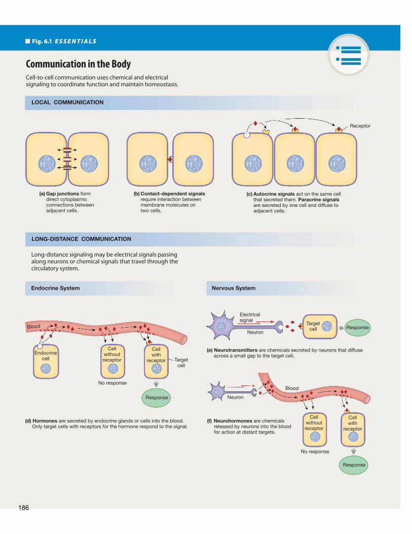
Our bodies use four basic methods of cell-to-cell commu-nication ( Fig. 6.1 ). Local communication includes (1) gap junctions, which allow direct cytoplasmic transfer of electrical and chemical signals between adjacent cells; (2) contact- dependent signals, which occur when surface molecules on one cell membrane bind to surface molecules on another cell’s mem-brane; and (3) chemicals that diff use through the extracellular fluid to act on cells close by. Long-distance communication (4) uses a combination of chemical and electrical signals carried by nerve cells and chemical signals transported in the blood. A given molecule can function as a signal by more than one method. For example, a molecule can act close to the cell that released it (local communication) as well as in distant parts of the body (long-distance communication).
Gap Junctions Create Cytoplasmic Bridges
Th e simplest form of cell-to-cell communication is the direct transfer of electrical and chemical signals through gap junctions, protein channels that create cytoplasmic bridges between adja-cent cells ( Fig. 6.1 a). A gap junction forms from the union of membrane-spanning proteins, called connexins, on two adjacent cells. The united connexins create a protein channel, or con-nexon , that can open and close. When the channel is open, the connected cells function like a single cell that contains multiple nuclei (a syncytium ).
When gap junctions are open, ions and small molecules such as amino acids, ATP, and cyclic AMP diffuse directly from the cytoplasm of one cell to the cytoplasm of the next. Larger molecules cannot pass through gap junctions. In addition, gap junctions are the only means by which electrical signals can pass directly from cell to cell. Movement of molecules and electrical signals through gap junctions can be modulated or shut off completely.
Gap junctions are not all alike. Scientists have discovered more than 20 diff erent isoforms of connexins that may mix or match to form gap junctions. Th e variety of connexin isoforms allows gap junction selectivity to vary from tissue to tissue. In mammals, gap junctions are found in almost every cell type, including heart muscle, some types of smooth muscle, lung, liver, and neurons of the brain.
Contact-Dependent Signals Require Cell-to-Cell Contact
Some cell-to-cell communication requires that surface mol-ecules on one cell membrane bind to a membrane protein of an-other cell ( Fig. 6.1 b). Such contact-dependent signaling occurs in the immune system and during growth and development, such as when nerve cells send out long extensions that must grow from the central axis of the body to the distal (distant) ends of
In 2003 the United States National Institutes of Health em-barked on an ambitious project to promote translation of basic research into new medical treatments and strategies
for disease prevention. Contributors to the NIH Common Fund Programs ( http://commonfund.nih.gov ) are compiling infor-mation on biological pathways in an eff ort to understand how cells communicate with one another and maintain the body in a healthy state. In this chapter, we examine the basic patterns of cell-to-cell communication and see how the coordination of function resides in chemical and electrical signals. Each cell in the body can communicate with most other cells. To maintain homeostasis, the body uses a combination of simple diff usion across small distances; widespread distribution of molecules through the circulatory system; and rapid, specifi c delivery of messages by the nervous system.
Cell-to-Cell Communication In recent years the amount of information available about cell-to-cell communication has mushroomed as a result of advances in research technology. Signal pathways that once seemed fairly simple and direct are now known to be incredibly complex net-works and webs of information transfer. In the sections that fol-low, we distill what is known about cell-to-cell communication into some basic patterns that you can recognize when you en-counter them again in your study of physiology.
By most estimates the human body is composed of about 75 trillion cells. Th ose cells face a daunting task—to communi-cate with one another in a manner that is rapid and yet conveys a tremendous amount of information. Surprisingly, there are only two basic types of physiological signals: electrical and chemical. Electrical signals are changes in a cell’s membrane potential. Chemical signals are molecules secreted by cells into the
R U N N I N G P R O B L E M
Diabetes Mellitus: A Growing Epidemic
It is 8:00 A.M. and Marvin Garcia, age 20, is hungry. He came to his family physician’s offi ce before breakfast to have a fasting blood glucose test as part of a routine physical examination. In this test, blood is drawn after an overnight fast, and the glucose concentration in the blood is measured. Because he knows he is in good condition, Marvin isn’t worried about the results. He is surprised, then, when the nurse practitioner in the doctor’s offi ce calls two days later. “Your fasting blood sugar is a bit elevated, Marvin. It is 150 milligrams per deciliter, and normal is 110 or less. Does anyone in your family have diabetes?” “Well, yeah—my dad has it. What exactly is diabetes?”
185
Blood
Fig. 6.1 E S S E N T I A L S
Cell-to-cell communication uses chemical and electricalsignaling to coordinate function and maintain homeostasis.
LONG-DISTANCE COMMUNICATION
LOCAL COMMUNICATION
Communication in the Body
(a) Gap junctions form direct cytoplasmic connections between adjacent cells.
(b) Contact-dependent signals require interaction between membrane molecules on two cells.
(c) Autocrine signals act on the same cell that secreted them. Paracrine signals are secreted by one cell and diffuse to adjacent cells.
Receptor
Long-distance signaling may be electrical signals passing along neurons or chemical signals that travel through the circulatory system.
(d) Hormones are secreted by endocrine glands or cells into the blood. Only target cells with receptors for the hormone respond to the signal.
(e) Neurotransmitters are chemicals secreted by neurons that diffuse across a small gap to the target cell.
Neuron
Electricalsignal
Endocrinecell
Cellwithoutreceptor
Cellwith
receptor
Cellwithoutreceptor
Cellwith
receptor
No response
No response
(f) Neurohormones are chemicals released by neurons into the blood for action at distant targets.
Neuron
Targetcell
Targetcell
Response
Response
Response
Blood
Endocrine System Nervous System
186
Communication, Integration, and Homeostasis
6
it is called a neurotransmitter ( Fig. 6.1 f). If a neurocrine acts more slowly as an autocrine or paracrine signal, it is called a neuromodulator . If a neurocrine released by a neuron diff uses into the blood for distribution, it is called a neurohormone( Fig. 6.1 e). Th e similarities between neurohormones and clas-sic hormones secreted by the endocrine system blur the distinc-tion between the nervous and endocrine systems, making them a functional continuum rather than two distinct systems.
Cytokines May Act as Both Local and Long-Distance Signals
Cytokines are among the most recently identifi ed communica-tion molecules. Initially the term cytokine referred only to pro-teins that modulate immune responses, but in recent years the defi nition has been broadened to include a variety of regulatory peptides. All nucleated cells synthesize and secrete cytokines in response to stimuli. Cytokines control cell development, cell diff erentiation, and the immune response. In development and diff erentiation, cytokines usually function as autocrine or para-crine signals. In stress and infl ammation, some cytokines may act on relatively distant targets and may be transported through the circulation just as hormones are.
How do cytokines differ from hormones? In general, cytokines act on a broader spectrum of target cells. In addi-tion, cytokines are not produced by specialized cells the way hormones are, and they are made on demand. In contrast, most protein or peptide hormones are made in advance and stored in the endocrine cell until needed. Also, the signal pathways for cytokines are usually different from those for hormones. However, the distinction between cytokines and hormones is sometimes blurry. For example, erythropoietin, the molecule that controls synthesis of red blood cells, is by tradition consid-ered a hormone but functionally fi ts the defi nition of a cytokine.
the developing limbs. Cell adhesion molecules (CAMs) first known for their role in cell-to-cell adhesion, have now been shown to act as receptors in cell-to-cell signaling. CAMs are linked to the cytoskeleton and to intracellular enzymes. Th rough these linkages, CAMs transfer signals in both directions across cell membranes.
Paracrine and Autocrine Signals Carry Out Local Communication
Local communication takes place through paracrine and auto-crine signaling. A paracrine signal { para −, beside + krinen, to secrete} is a chemical that acts on cells in the immediate vicinity of the cell that secreted the signal. A chemical signal that acts on the cell that secreted it is called an autocrine signal { auto −, self}. In some cases a molecule may act as both an autocrine sig-nal and a paracrine signal.
Paracrine and autocrine signal molecules reach their target cells by diff using through the interstitial fl uid ( Fig. 6.1 c). Because distance is a limiting factor for diffusion, the effective range of paracrine signals is restricted to adjacent cells. A good example of a paracrine molecule is histamine, a chemical released from dam-aged cells. When you scratch yourself with a pin, the red, raised wheal that results is due in part to the local release of histamine from the injured tissue. Th e histamine acts as a paracrine signal, diffusing to capillaries in the immediate area of the injury and making them more permeable to white blood cells and antibodies in the plasma. Fluid also leaves the blood vessels and collects in the interstitial space, causing swelling around the area of injury.
Several important classes of molecules act as local signals. Cytokines are regulatory peptides, and eicosanoids are lipid-de-rived paracrine and autocrine signal molecules. We discuss cy-tokines and eicosanoids in more detail below.
Long-Distance Communication May Be Electrical or Chemical
All cells in the body can release paracrine signals, but most long-distance communication between cells takes place through the nervous and endocrine systems. Th e endocrine system commu-nicates by using hormones { hormon, to excite}, chemical signals that are secreted into the blood and distributed all over the body by the circulation. Hormones come in contact with most cells of the body, but only those cells with receptors for the hormone are target cells ( Fig. 6.1 d).
Th e nervous system uses a combination of chemical signals and electrical signals to communicate over long distances. An electrical signal travels along a nerve cell ( neuron ) until it reaches the very end of the cell, where it is translated into a chemical signal secreted by the neuron. Such a chemical signal is called a neurocrine.
If a neurocrine molecule diff uses from the neuron across a narrow extracellular space to a target cell and has a rapid eff ect,
Concept Check Answers: End of Chapter
1. Match the communication method on the left with its property on the
right.
(a) autocrine
(b) cytokine
(c) gap junction
(d) hormone
(e) neurohormone
(f) neurotransmitter
(g) paracrine
Communication occurs by:
1. electrical signals
2. chemical signals
3. both electrical and chemical signals
2. Which signal molecules listed in the previous question are transported
through the circulatory system? Which are released by neurons?
3. A cat sees a mouse and pounces on it. Do you think the internal signal
to pounce could have been transmitted by a paracrine signal? Give two
reasons to explain why or why not.
187

Communication, Integration, and Homeostasis
patterns that you will encounter over and over as you study the systems of the body. Most physiological processes, from the beating of your heart to learning and memory, use some varia-tion of these pathways. One of the wonders of physiology is the fundamental importance of these signal pathways and the way they have been conserved in animals ranging from worms to humans.
Receptor Proteins Are Located Inside the Cell or on the Cell Membrane
Protein receptors for signal molecules play an important role in physiology and medicine. About half of all drugs currently in use act on receptor proteins. Target-cell receptor proteins may be found in the nucleus, in the cytosol, or on the cell membrane as integral proteins. Where a chemical signal binds to its recep-tor largely depends on whether that signal molecule is lipophilic or lipophobic ( Fig. 6.3 ).
Lipophilic signal molecules can diff use through the phos-pholipid bilayer of the cell membrane and bind to cytosolic re-ceptors or nuclear receptors ( Fig. 6.3 a). In these cases, receptor activation oft en turns on a gene and directs the nucleus to make new mRNA (transcription). Th e mRNA then provides a tem-plate for synthesis of new proteins (translation). Th is process is relatively slow and the cell’s response may not be noticeable for an hour or longer. In some instances the activated receptor can also turn off , or repress, gene activity. Many lipophilic signal molecules that follow this pattern are hormones.
Signal Pathways Chemical signals in the form of paracrine and autocrine mol-ecules and hormones are released from cells into the extracellu-lar compartment. Th is is not a very specifi c way for these signals to find their targets because substances that travel through the blood reach nearly every cell in the body. Yet cells do not respond to every signal that reaches them.
Why do some cells respond to a chemical signal while other cells ignore it? Th e answer lies in the target-cell receptor proteins to which chemical signals bind. A cell can respond to a chemical signal only if the cell has the appropriate receptor pro-teins for that signal ( Fig. 6.1 ).
If a target cell has a receptor for a signal molecule, binding of the signal to the receptor protein initiates a response. All sig-nal pathways share the following features ( Fig. 6.2 ):
1 The signal molecule is a ligand that binds to a protein receptor. The ligand is also known as a first messenger because it brings information to the target cell.
2 Ligand-receptor binding activates the receptor. 3 The receptor in turn activates one or more intracellular
signal molecules. 4 Th e last signal molecule in the pathway initiates synthesis
of target proteins or modifies existing target proteins to create a response.
In the following sections, we describe some basic signal pathways. They may seem complex at first, but they follow
Fig. 6.2
Signal Pathways
Membranereceptor protein
Intracellularsignal molecules
Signalmolecule
Target proteins
Response
binds to
activates
alter
create
Most signal pathways consist of the 5 steps shown. Use the shapes and colors of the steps shown here to identify the pattern in later illustrations.
R U N N I N G P R O B L E M
Later that day in the physician’s offi ce, the nurse practitioner explains diabetes to Marvin. Diabetes mellitus is a family of metabolic disorders caused by defects in the homeostatic pathways that regulate glucose metabolism. Several forms of diabetes exist, and some can be inherited. One form, called type 1 diabetes mellitus , occurs when endocrine cells of the pancreas are destroyed and stop making insulin, a protein hormone involved in blood glucose homeostasis. In another form, type 2 diabetes mellitus, insulin may be present in normal or above-normal levels, but the insulin-sensitive cells of the body do not respond normally to the hormone.
Q1: In which type of diabetes is the target cell signal pathway for insulin more likely to be defective?
Q2: Insulin is a protein hormone. Would you expect to fi nd its receptor on the cell surface or in the cytoplasm of the target cells?
188

Communication, Integration, and Homeostasis
6
Binding to cytosolic or nuclear receptors triggers
Slower responsesrelated to changes
in gene activity
Receptor in cytosol
Receptor in nucleus
Lipophilic signal molecules diffusethrough the cellmembrane.
Rapid cellularresponses
Extracellular signalmolecule binds to a cell membrane receptor.
Cellmembrane
ECF
ICFG protein
ReceptorChannel
Extracellularsignal
molecules
IntegrinReceptor
Enzyme
Anchorprotein
Cytoskeleton
Ligand binding opens or closes the channel.
Ligand binding tointegrin receptorsalters the cytoskeleton.
Ligand binding to areceptor-enzyme activates an intracellular enzyme.
Ligand binding to a G protein– coupled receptor opens an ion channel or alters enzyme activity.
Receptor-channel
Receptor-enzyme Integrin receptorG protein–coupled receptor
(a) Intracellular Signal Receptors (b) Cell Membrane Receptors
Binding triggers
(c) Four Categories of Membrane Receptors
Fig. 6.3 Target cell receptors may be located on the cell surface or inside the cell
Lipophobic signal molecules are unable to diff use through the phospholipid bilayer of the cell membrane. Instead, these signal molecules remain in the extracellular fl uid and bind to receptor proteins on the cell membrane ( Fig. 6.3 b). (Some lipophilic signal molecules also bind to cell membrane recep-tors in addition to their intracellular receptors.) In general, the response time for pathways linked to membrane receptor pro-teins is very rapid: responses can be seen within milliseconds to minutes.
We can group membrane receptors into four major catego-ries, illustrated in Figure 6.3 c. Th e simplest receptors are chemi-cally gated ( ligand-gated ) ion channels called receptor-channels . Ligand binding opens or closes the channel and alters ion fl ow across the membrane.
Three other receptor types are shown in Figure 6.3 c: receptor-enzymes, G protein–coupled receptors, and integrin receptors . For all three, information from the signal molecule must be passed across the membrane to initiate an intracellular
189

Communication, Integration, and Homeostasis
In biological systems, membrane proteins act as transducers. Th ey convert the message of extracellular signals into intracel-lular messenger molecules that trigger a response.
Th e basic pattern of a biological signal transduction path-way is shown in Figure 6.5 a and can be broken down into the following events.
1 An extracellular signal molecule (the fi rst messenger ) binds to and activates a membrane receptor.
2 Th e activated membrane receptor turns on its associated proteins and starts an intracellular cascade of second messengers.
3 Th e last second messenger in the cascade acts on intracel-lular targets to create a response.
A more detailed version of the basic signal transduction pathway is shown in Figure 6.5 b.
1 Membrane receptors and their associated proteins usually either (a) activate protein kinases, which are enzymes that
transfer a phosphate group from ATP to a protein. Phosphorylation is an important biochemical method of regulating cellular processes.
(b) activate amplifier enzymes that create intracellular second messengers .
2 Second messenger molecules in turn (a) alter the gating of ion channels. Opening or closing
ion channels creates electrical signals by altering the cell’s membrane potential.
(b) increase intracellular calcium. Calcium binding to proteins changes their function, creating a cellular response.
(c) change enzyme activity, especially of protein kinases or protein phosphatases, enzymes that remove a phosphate group. The phosphorylation or dephos-phorylation of a protein can change its confi guration and create a response. Examples of changes that occur with phosphorylation include increased or decreased enzyme activity and opening or closing of gated ion channels.
3 Th e proteins modifi ed by calcium binding and phosphory-lation control one or more of the following: (a) metabolic enzymes (b) motor proteins for muscle contraction and cytoskeletal
movement (c) proteins that regulate gene activity and protein synthesis (d) membrane transport and receptor proteins
If you think this list includes almost everything a cell does, you’re right!
Figure 6.6 a shows how the steps of a signal transduc-tion pathway form a cascade. A signaling cascade starts when
response. This transmission of information from one side of a membrane to the other using membrane proteins is known as sig-nal transduction . We will take a closer look at signal transduction before returning to the four receptor types that participate in it.
Concept Check Answers: End of Chapter
4. List four components of signal pathways.
5. Name three cellular locations of receptors.
Membrane Proteins Facilitate Signal Transduction
Signal transduction is the process by which an extracellu-lar signal molecule activates a membrane receptor that in turn alters intracellular molecules to create a response. The extra-cellular signal molecule is the fi rst messenger, and the intracel-lular molecules form a second messenger system . Th e term signal transduction comes from the verb to transduce, meaning “to lead across” { trans, across + ducere, to lead}.
A transducer is a device that converts a signal from one form into a different form. For example, the transducer in a radio converts radio waves into sound waves ( Fig. 6.4 ).
Radio
Radio waves
Receptor
Transducer
Amplifier
Response
External signal
Sound waves
Signal transduction converts one form of signal into a different form.
A radio contains an antenna to receive signals, a transducer that converts radio waves into sound waves, and an amplifier to increase the strength of the signal.
Fig. 6.4 Signal transduction
190

Communication, Integration, and Homeostasis
6
a stimulus (the signal molecule) converts inactive molecule A (the receptor) to an active form. Active A then converts inac-tive molecule B into active B, active molecule B in turn converts inactive molecule C into active C, and so on, until at the fi nal step a substrate is converted into a product. Many intracellular signal pathways are cascades. Blood clotting is an important example of an extracellular cascade.
In signal transduction pathways, the original signal is not only transformed but also amplifi ed { amplifi care, to make larger}. In a radio, the radio wave signal is also amplified. In cells, signal amplifi cation turns one signal molecule into multi-ple second messenger molecules ( Fig. 6.6 b). Th e process begins when the fi rst messenger ligand combines with its receptor. Th e receptor-ligand complex turns on an amplifier enzyme. The amplifier enzyme activates several molecules, which in turn each activate several more molecules as the cascade proceeds. By the end of the process, the effects of the ligand have been amplifi ed much more than if there were a 1:1 ratio between each step. Amplifi cation gives the body “more bang for the buck” by enabling a small amount of ligand to create a large eff ect. Th e most common amplifi er enzymes and second messengers are listed in the table in Figure 6.6 c.
Concept Check Answers: End of Chapter
6. What are the four steps of signal transduction?
7. What happens during amplifi cation?
8. Why do steroid hormones not require signal transduction and second
messengers to exert their action? ( Hint: Are steroids lipophobic or
lipophilic?)
Fig. 6.5 Biological signal transduction
Membranereceptor protein
Intracellularsignal molecules
Signalmolecule
Target proteins
Response
binds to
activates
alter
create
Transducer
Targets
Secondmessengersystem
Response
Firstmessenger
alter
Signalmolecule
Membrane receptor
Signal transduction by proteins
Amplifier enzymes
Second messengermolecules
Protein kinasesIncrease
intracellular Ca2+
Phosphorylatedproteins
Calcium-bindingproteins
Extracellularfluid
Intracellularfluid
Cell response
initiates
binds to
Ionchannel
(a) Basic Signal Transduction (b) Transduction Pathways
In the sections that follow, we will examine in more de-tail the four major types of membrane receptors (see Fig. 6.3 c). Keep in mind that these receptors may be responding to any of the diff erent kinds of signal molecules—hormones, neurohor-mones, neurotransmitters, cytokines, paracrines, or autocrines.
Receptor-Enzymes Have Protein Kinase or Guanylyl Cyclase Activity
Receptor-enzymes have two regions: a receptor region on the extracellular side of the cell membrane, and an enzyme region on the cytoplasmic side (see Fig. 6.3 c). In some instances,
191

Fig. 6.6 E S S E N T I A L S
Signal Transduction
ExtracellularFluid
IntracellularFluid
Cellmembrane
One ligand is amplified into manyintracellular molecules.
Receptor-ligand complex activates an
amplifier enzyme (AE).
L
R
AE
(a) Signal transduction pathways form a cascade.
(c) Second messenger pathways
(b) Signal amplification allows a small amount of signal to have a large effect.
Inactive A
Signal
Inactive B
Inactive C
Substrate
Active A
Active B
Active C
ProductConversion of substrate
to product is the finalstep of the cascade.
Nucleotides
Lipid-derived*
ATP
GTP
Adenylyl cyclase (membrane)
Guanylyl cyclase (membrane)
GPCR*
*GPCR = G protein–coupled receptor. IP3 = Inositol trisphosphate. DAG = diacylglycerol
Receptor-enzyme
Activates proteinkinases, especially PKA.Binds to ion channels.
Activates proteinkinases, especially PKG.
Phosphorylates proteins. Alterschannel opening.
Phosphorylatesproteins.
Guanylyl cyclase (cytosol)
Nitric oxide (NO) Binds to ion channels. Alters channel opening.
Membranephospholipids
Phospholipase C(membrane) GPCR
Releases Ca2+ fromintracellular stores.
See Ca2+ effectsbelow.
Activates proteinkinase C.
Phosphorylatesproteins.
cAMP
IP3
Ions
Binds to calmodulin.Binds to other proteins.
Alters enzyme activity.Exocytosis, musclecontraction, cytoskeleton movement,channel opening.
Ca2+
DAG
cGMP
MADE FROMSECOND MESSENGER AMPLIFIER ENZYME LINKED TO ACTION EFFECTS
192
Communication, Integration, and Homeostasis
6
to guanosine diphosphate (GDP). Exchanging the GDP for guanosine triphosphate (GTP) activates the G protein. When G proteins are activated, they either (1) open an ion channel in the membrane or (2) alter enzyme activity on the cytoplasmic side of the membrane.
G proteins linked to amplifi er enzymes make up the bulk of all known signal transduction mechanisms. The two most common amplifi er enzymes for G protein–coupled receptors are adenylyl cyclase and phospholipase C. Th e pathways for these amplifi er enzymes are described next.
Many Lipophobic Hormones Use GPCR-cAMP Pathways
Th e G protein – coupled adenylyl cyclase-cAMP system was the fi rst identifi ed signal transduction pathway ( Fig. 6.8 a). It was discovered in the 1950s by Earl Sutherland when he was study-ing the eff ects of hormones on carbohydrate metabolism. Th is discovery proved so signifi cant to our understanding of signal transduction that in 1971 Sutherland was awarded a Nobel prize for his work.
The G protein–coupled adenylyl cyclase-cAMP system is the signal transduction system for many protein hormones. In this system, adenylyl cyclase is the amplifier enzyme that converts ATP to the second messenger molecule cyclic AMP (cAMP). Cyclic AMP then activates protein kinase A (PKA), which in turn phosphorylates other intracellular proteins as part of the signal cascade.
G Protein–Coupled Receptors Also Use Lipid-Derived Second Messengers
Some G protein–coupled receptors are linked to a different amplifier enzyme: phospholipase C ( Fig. 6.8 b). When a signal molecule activates this G protein–coupled pathway, phospholipase C (PLC) converts a membrane phospholipid ( phosphatidylinositol bisphosphate ) into two lipid-derived second messenger molecules: diacylglycerol and inositol trisphosphate.
Diacylglycerol (DAG) is a nonpolar diglyceride that remains in the lipid portion of the membrane and interacts with protein kinase C (PKC), a Ca 2+ -activated enzyme asso-ciated with the cytoplasmic face of the cell membrane. PKC phosphorylates cytosolic proteins that continue the signal cascade.
Inositol trisphosphate (IP 3 ) is a water-soluble messenger molecule that leaves the membrane and enters the cytoplasm. Th ere it binds to a calcium channel on the endoplasmic reticu-lum (ER). IP 3 binding opens the Ca 2+ channel, allowing Ca 2+ to diff use out of the ER and into the cytosol. Calcium is itself an important signal molecule, as discussed below.
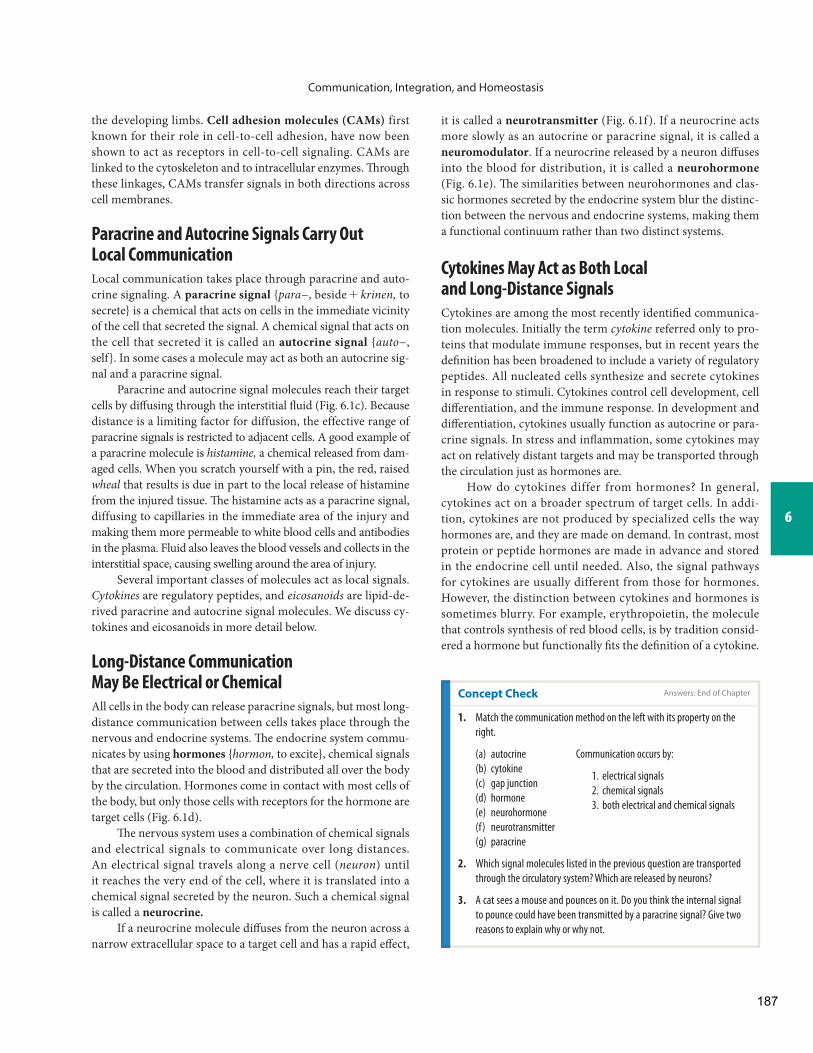
the receptor region and enzyme region are parts of the same protein molecule. In other cases, the enzyme region is a sep-arate protein. Ligand binding to the receptor activates the enzyme. The enzymes of receptor-enzymes are either pro-tein kinases, such as tyrosine kinase ( Fig. 6.7 ), or gua nylyl cyclase, the amplifier enzyme that converts GTP to cyclic GMP (cGMP) .
Ligands for receptor-enzymes include the hormone insu-lin as well as many cytokines and growth factors. Th e insulin receptor protein has its own intrinsic tyrosine kinase activity. In contrast, most cytokine receptor proteins do not have intrinsic enzyme activity. Instead, cytokine binding activates a cytosolic enzyme called Janus family tyrosine kinase, usually abbreviated as JAK kinase .
Most Signal Transduction Uses G Proteins
The G protein – coupled receptors (GPCR) are a large and complex family of membrane-spanning proteins that cross the phospholipid bilayer seven times (see Fig. 6.3 c). The cytoplasmic tail of the receptor protein is linked to a three-part membrane transducer molecule known as a G protein. Hundreds of G protein–coupled receptors have been iden-tified, and the list continues to grow. The types of ligands that bind to G protein–coupled receptors include hormones, growth factors, olfactory molecules, visual pigments, and neurotransmitters. In 1994, Alfred G. Gilman and Martin Rodbell received a Nobel prize for the discovery of G pro-teins and their role in cell signaling (see http://nobelprize.org/nobel_prizes/medicine ).
G proteins get their name from the fact that they bind guanosine nucleotides. Inactive G proteins are bound
R
TK
L
+ Protein
Active binding site
P
+ ADP
ECF
ICF
Signal molecule binds to surface receptor
Phosphorylatedprotein
Tyrosine kinase oncytoplasmic side
Cellmembrane
activates
ProteinATP
Tyrosine Kinase Receptor
Tyrosine kinase (TK) transfers a phosphate group from ATP to a tyrosine (an amino acid) of a protein.
Fig. 6.7
193

Communication, Integration, and Homeostasis
One signalmolecule
Adenylylcyclase
ATP
cAMP
G protein
GPCR
Proteinkinase A
Phosphorylatedprotein
Cellresponse
Signal molecule binds to G protein–coupled receptor (GPCR), which activates the G protein.
Protein kinase A phosphorylatesother proteins, leading ultimatelyto a cellular response.
55
G protein turns on adenylyl cyclase, an amplifier enzyme.
2
Adenylyl cyclase converts ATP to cyclic AMP.
3
cAMP activates protein kinase A.4
32
4
1
1
Ca2+stores
Membrane phospholipid
(a) GPCR-adenylyl Cyclase Signal Transduction and Amplification
(b) GPCR-phospholipase C Signal Transduction
PLC
IP3
PKC
Protein + Pi
Cellmembrane
Extracellularfluid
Intracellularfluid
DAG
Phosphorylatedprotein
PLCDAGPKCIP3
ER
=====
phospholipase Cdiacylglycerolprotein kinase Cinositol trisphosphateendoplasmic reticulum
ER
ReceptorG protein
Cellularresponse
Signal molecule
Signal moleculeactivates receptorand associatedG protein.
G protein activatesphospholipase C (PLC), an amplifier enzyme.
PLC converts membrane phospho-lipids into diacylglycerol (DAG), whichremains in the membrane, and IP3, which diffuses into the cytoplasm.
DAG activates proteinkinase C (PKC), whichphosphorylates proteins.
IP3 causes releaseof Ca2+ fromorganelles, creatinga Ca2+ signal.
1 2
2
3 4
3
5
5
4
1
Ca2+KEY
G Protein–Coupled Signal Transduction
FIGURE QUESTION
Using the pattern shown in Fig. 6.6a, create a cascade that includes ATP, cAMP, adenylyl cyclase, a phosphorylated protein, and protein kinase A.
Fig. 6.8
194

Communication, Integration, and Homeostasis
6
The Most Rapid Signal Pathways Change Ion Flow Through Channels
Th e simplest receptors are ligand-gated ion channels, and most of them are neurotransmitter receptors found in nerve and muscle. Th e activation of receptor-channels initiates the most rapid intracellular responses of all receptors. When an extracel-lular ligand binds to the receptor-channel protein, a channel gate opens or closes, altering the cell’s permeability to an ion. Increasing or decreasing ion permeability rapidly changes the cell’s membrane potential, creating an electrical signal that alters voltage-sensitive proteins ( Fig. 6.9 ).
One example of a receptor-channel is the acetylcholine-gated monovalent (“one-charge”) cation channel of skel-etal muscle. Th e neurotransmitter acetylcholine released from an adjacent neuron binds to the acetylcholine receptor and opens the channel. Both Na + and K + flow through the open channel, K + leaving the cell and Na + entering the cell along
Integrin Receptors Transfer Information from the Extracellular Matrix
The membrane-spanning proteins called integrins mediate blood clotting, wound repair, cell adhesion and recognition in the immune response, and cell movement during development. On the extracellular side of the membrane, integrin receptors bind either to proteins of the extracellular matrix or to ligands such as antibodies and molecules involved in blood clotting. In-side the cell, integrins attach to the cytoskeleton via anchor pro-teins ( Fig. 6.3 c). Ligand binding to the receptor causes integrins to activate intracellular enzymes or alter the organization of the cytoskeleton.
The importance of integrin receptors is illustrated by inherited conditions in which the receptor is absent. In one condition, platelets—cell fragments that play a key role in blood clotting—lack an integrin receptor. As a result, blood clotting is defective in these individuals.
Extracellularsignal
molecules
Ions
Ions
Ions
Ions
Receptor-channel
Ionchannel
G protein
Change in membranepermeability to
Na+, K+, Cl–
Creates electricalsignal
Voltage-sensitiveprotein
Cellularresponse
G protein–coupledreceptor
Intracellularsignal molecules
Receptor-channels open orclose in response to signalmolecule binding.
Some channels are directlylinked to G proteins.
Other channels respond to intracellular second messengers.
1
2
3
1 2
3
Signal Transduction Using Ion Channels
Fig. 6.9 Signal transduction using ion channels
195

their electrochemical gradients. Th e sodium gradient is stron-ger, however, so net entry of positively charged Na + depolarizes the cell. In skeletal muscle, this cascade of intracellular events results in muscle contraction.
Not all ion channel signal transduction is mediated by receptor-channels. Some ligand-gated ion channels are con-trolled by intracellular second messengers, such as cAMP or ATP. Th e ATP-gated K + channels of the pancreatic beta cell are
an example. Other ion channels open or close in response to extracellular signals, but the signal ligand does not bind to the channel protein. Instead it binds to a G protein–coupled recep-tor that is linked to the ion channel.
Figure 6.10 is a summary map of basic signal trans-duction, showing the general relationships among first mes-sengers, membrane receptors, second messengers, and cell responses.
Ions
Fig. 6.10 E S S E N T I A L S
Summary Map of Signal Transduction
Gated ion channel
alters
alter
creates
produces
activate
phosphorylate
will be a change in
phosphorylates
ExtracellularFluid
IntracellularFluid
Cell membrane
Protein kinases
Altered proteins
Change in ionconcentration
Motorproteins
Enzymeactivity
Membrane receptorsand transporters
Gene activity andprotein synthesis
Activates orinhibits
amplifier enzyme
Second messengermolecules
Triggersrelease ofCa2+ fromorganelles
Signalmolecule
ActivatesG protein
Cellular responses
bindto
Membranereceptor
Electrical signal
Ions moveinto or out
of cell
Activatestyrosinekinase
Alterscytoskeleton
196

Communication, Integration, and Homeostasis
6
Novel Signal Molecules The following sections introduce you to some unusual signal molecules that are important in physiology and medicine. Th ey include an ion (Ca 2+ ), three gases, and a family of lipid-derived messengers. Th e processes controlled by these signal molecules have been known for years, but the control signals themselves were discovered only relatively recently.
Calcium Is an Important Intracellular Signal
Calcium ions are the most versatile ionic messengers ( Fig. 6.11 ). Calcium enters the cell either through voltage-gated Ca 2+ chan-nels or through ligand-gated or mechanically gated channels. Calcium can also be released from intracellular compartments by second messengers, such as IP 3 . Most intracellular Ca 2+ is stored in the endoplasmic reticulum, where it is concentrated by active transport.
Release of Ca 2+ into the cytosol (from any of the sources just mentioned) creates a Ca 2+ signal, or Ca 2+ “spark,” that can be recorded using special Ca 2+ -imaging techniques (see Biotech-nology box on calcium signals). Th e calcium ions combine with cytoplasmic calcium-binding proteins to exert various eff ects. Several types of calcium-dependent events occur in the cell:
1 Ca 2+ binds to the protein calmodulin, found in all cells. Calcium binding alters enzyme or transporter activity or the gating of ion channels.
2 Calcium binds to other regulatory proteins and alters movement of contractile or cytoskeletal proteins such as microtubules. For example, Ca 2+ binding to the regulatory protein troponin initiates muscle contraction in a skeletal muscle cell.
Concept Check Answers: End of Chapter
9. Name the four categories of membrane receptors.
10. What is the difference between a first messenger and a second
messenger?
11. Place the following terms in the correct order for a signal transduction
pathway:
(a) cell response, receptor, second messenger, ligand
(b) amplifi er enzyme, cell response, phosphorylated protein, protein
kinase, second messenger
12. In each of the following situations, will a cell depolarize or hyperpolarize?
(a) Cl − channel opens
(b) K + channel opens
(c) Na + channel opens
Extracellularfluid
Intracellularfluid
Electricalsignal
released fromintracellularCa2+ stores.
Ca2+
Ca2+
Ca2+ binds toproteins.
Ca2+
Calmodulin
Calcium As an Intracellular Messenger
Other Ca2+-binding proteins
Alters proteinactivity
Exocytosis Movement
Chemicalsignal
Ca2+ in cytosolincreases.
Voltage-gated Ca2+ channel
opens.
Fig. 6.11
3 Ca 2+ binds to regulatory proteins to trigger exocytosis of secretory vesicles. For example, the release of insulin from pancreatic beta cells occurs in response to a calcium signal.
4 Ca 2+ binds directly to ion channels to alter their gating state. An example of this target is a Ca 2+ -activated K +channel found in nerve cells.
5 Ca 2+ entry into a fertilized egg initiates development of the embryo.
Concept Check
13. The extracellular fl uid Ca 2+ concentration averages 2.5 mmol/L. Free
cytosolic Ca 2+ concentration is about 0.001 mmol/L. If a cell is going to
move calcium ions from its cytosol to the extracellular fl uid, will it use
passive or active transport? Explain.
Gases Are Ephemeral Signal Molecules
Soluble gases are short-acting paracrine/autocrine signal mole-cules that act close to where they are produced. Th e best-known gaseous signal molecule is nitric oxide (NO), but carbon
Answers: End of Chapter
197
Communication, Integration, and Homeostasis
explain studies suggesting that eating garlic has protective eff ects on the heart.
Some Lipids Are Important Paracrine Signals
One of the interesting developments from sequencing the human genome and using genes to fi nd proteins has been the identifi ca-tion of orphan receptors, receptors that have no known ligand.
monoxide and hydrogen sulfide, two gases better known for their noxious eff ects, can also act as local signals.
For years researchers knew of a short-lived signal molecule produced by the endothelial cells lining blood vessels. Th ey initially named it endothelial-derived relaxing factor (EDRF). Th is molecule diff uses from the endothelium into adjacent smooth muscle cells, causing the muscle to relax and dilate the blood vessel. Scientists took years to identify EDRF as nitric oxide because it is rapidly broken down, with a half-life of only 2 to 30 seconds. ( Half-lifeis the time required for the signal to lose half of its activity.) As a result of this diffi cult work on NO in the cardiovascular system, Robert Furchgott, Louis Ignarro, and Ferid Murad received the 1998 Nobel prize for physiology and medicine.
In tissues, NO is synthesized by the action of the enzyme nitric oxide synthase (NOS) on the amino acid arginine:
Arginine + O2 nitric oxide synthase
NO + citrulline (an amino acid)
Th e NO produced in this reaction diff uses into target cells, where it binds to a receptor that activates the cytosolic form of guanylyl cyclase and causes formation of the second messenger cGMP. In addition to relaxing blood vessels, NO in the brain acts as a neurotransmitter and a neuromodulator.
Carbon monoxide (CO) , a gas known mostly for its toxic eff ects, is also a signal molecule produced in minute amounts by certain cells. Like NO, CO activates guanylyl cyclase and cGMP, but it may also work independently to exert its eff ects. Carbon monoxide targets smooth muscle and neural tissue.
The newest gaseous signal molecule to be described is hydrogen sulfide (H 2 S). Hydrogen sulfide also acts in the cardiovascular system to relax blood vessels. Garlic is a major dietary source of the sulfur-containing precursors, which may
From Dynamite to Medicine
Who would have thought that a component of smog and a derivative of dynamite would turn out to be a biologi-cal messenger? Certainly not the peer reviewers who initially rejected Louis Ignarro’s attempts to publish his research fi ndings on the elusive gas. However, the ability of nitrate-containing compounds to relax blood vessels has been known for more than 100 years, ever since workers in Alfred Nobel’s dynamite factory complained of headaches caused by nitrate-induced vasodilation. Since the 1860s, physicians have used nitroglycerin to relieve angina, heart pain that results from constricted blood vessels. Even today heart patients carry little nitroglycerin tablets to slide under their tongues when angina strikes. Still, it took years of work to isolate nitric oxide (NO), the short-lived gas that is the bio-logically active molecule derived from nitroglycerin. Despite all our twenty-fi rst-century technology, direct research on NO is still diffi cult. Many studies look at its infl uence indi-rectly by studying the location and activity of nitric oxide synthase (NOS), the enzyme that produces NO.
C L I N I C A L F O C U S
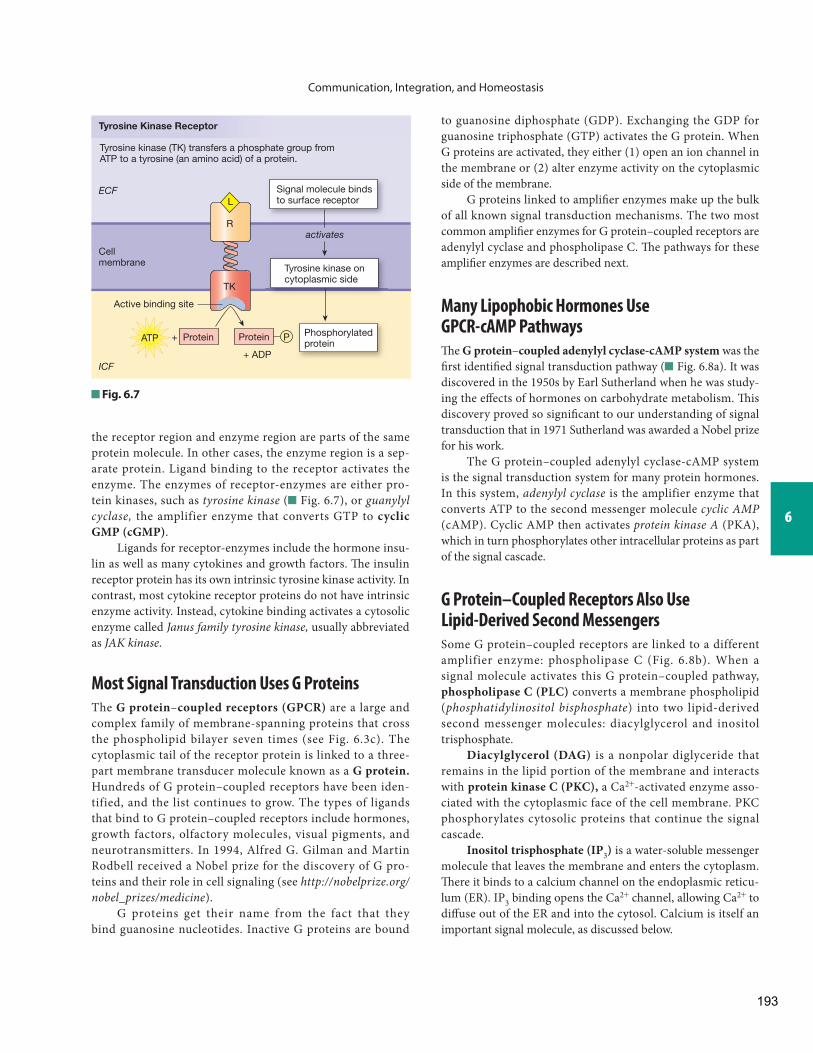
Calcium Signals Glow in the Dark
If you’ve ever run your hand through a tropical ocean at night and seen the glow of bioluminescent jellyfi sh, you’ve seen a calcium signal. Aequorin, a protein complex isolated from jellyfi sh, is one of the molecules that scientists use to monitor the presence of calcium ions during a cellular response. When aequorin combines with calcium, it releases light that can be measured by electronic detection systems. Since the fi rst use of aequorin in 1967, researchers have been designing increasingly sophisticated indicators that allow them to follow calcium signals in cells. With the help of molecules called fura, Oregon green, BAPTA, and chameleons, we can now watch calcium ions diff use through gap junctions and fl ow out of intracellular organelles.
B I O T E C H N O L O G Y
The sea nettle Chrysaora fuscescens.
198
Communication, Integration, and Homeostasis
6
pharmaceutical companies have been actively developing drugs to block leukotriene synthesis or action.
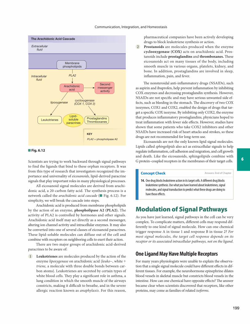
2 Prostanoids are molecules produced when the enzyme cyclooxygenase (COX) acts on arachidonic acid. Pros-tanoids include prostaglandins and thromboxanes. Th ese eicosanoids act on many tissues of the body, including smooth muscle in various organs, platelets, kidney, and bone. In addition, prostaglandins are involved in sleep, infl ammation, pain, and fever.
Th e nonsteroidal anti-infl ammatory drugs (NSAIDs), such as aspirin and ibuprofen, help prevent infl ammation by inhibiting COX enzymes and decreasing prostaglandin synthesis. However, NSAIDs are not specifi c and may have serious unwanted side ef-fects, such as bleeding in the stomach. Th e discovery of two COX isozymes, COX1 and COX2, enabled the design of drugs that tar-get a specifi c COX isozyme. By inhibiting only COX2, the enzyme that produces infl ammatory prostaglandins, physicians hoped to treat infl ammation with fewer side eff ects. However, studies have shown that some patients who take COX2 inhibitors and other NSAIDs have increased risk of heart attacks and strokes, so these drugs are not recommended for long-term use.
Eicosanoids are not the only known lipid signal molecules. Lipids called sphingolipids also act as extracellular signals to help regulate infl ammation, cell adhesion and migration, and cell growth and death. Like the eicosanoids, sphingolipids combine with G protein–coupled receptors in the membranes of their target cells. Scientists are trying to work backward through signal pathways
to find the ligands that bind to these orphan receptors. It was from this type of research that investigators recognized the im-portance and universality of eicosanoids, lipid-derived paracrine signals that play important roles in many physiological processes.
All eicosanoid signal molecules are derived from arachi-donic acid, a 20-carbon fatty acid. The synthesis process is a network called the arachidonic acid cascade ( Fig. 6.12 ). For simplicity, we will break the cascade into steps.
Arachidonic acid is produced from membrane phospholipids by the action of an enzyme, phospholipase A2 (PLA2). The activity of PLA2 is controlled by hormones and other signals. Arachidonic acid itself may act directly as a second messenger, altering ion channel activity and intracellular enzymes. It may also be converted into one of several classes of eicosanoid paracrines. These lipid-soluble molecules can diffuse out of the cell and combine with receptors on neighboring cells to exert their action.
There are two major groups of arachidonic acid-derived paracrines to be aware of:
1 Leukotrienes are molecules produced by the action of the enzyme lipoxygenase on arachidonic acid { leuko −, white + triene, a molecule with three double bonds between car-bon atoms}. Leukotrienes are secreted by certain types of white blood cells. Th ey play a signifi cant role in asthma, a lung condition in which the smooth muscle of the airways constricts, making it diffi cult to breathe, and in the severe allergic reaction known as anaphylaxis . For this reason,
Concept Check
14. One drug blocks leukotriene action in its target cells. A diff erent drug blocks
leukotriene synthesis. Use what you have learned about leukotrienes, signal
molecules, and signal transduction to predict what these drugs are doing to
have those eff ects.
Fig. 6.12
Extracellularfluid
Intracellularfluid
Membranephospholipids
Arachidonicacid
Secondmessenger
activity
Leukotrienes
The Arachidonic Acid Cascade
PLA2 = phospholipase A2
ProstaglandinsThromboxanes
PLA2
lipoxygenasecycloxygenase(COX 1, COX 2)
Lipid-soluble
paracrines
KEY
Modulation of Signal Pathways As you have just learned, signal pathways in the cell can be very complex. To complicate matters, diff erent cells may respond dif-ferently to one kind of signal molecule. How can one chemical trigger response A in tissue 1 and response B in tissue 2? For most signal molecules, the target cell response depends on its receptor or its associated intracellular pathways, not on the ligand .
One Ligand May Have Multiple Receptors
For many years physiologists were unable to explain the observa-tion that a single signal molecule could have diff erent eff ects in dif-ferent tissues. For example, the neurohormone epinephrine dilates blood vessels in skeletal muscle but constricts blood vessels in the intestine. How can one chemical have opposite eff ects? Th e answer became clear when scientists discovered that receptors, like other proteins, may come as families of related isoforms .
Answers: End of Chapter
199

Communication, Integration, and Homeostasis
norepinephrine and its cousin the neurohormone epinephrine (also called adrenaline ). Both molecules bind to a class of receptors called adrenergic receptors . ( Adrenergic is the adjective relating to adrenaline.) The ability of adrenergic receptors to bind these neurocrines, but not others, demonstrates the specificity of the receptors.
Epinephrine and norepinephrine also compete for a sin-gle receptor type. Both neurocrines bind to subtypes of adren-ergic receptors designated alpha (a) and beta (b). However, a- receptors have a higher binding affi nity for norepinephrine, and the b 2 -receptor subtype has a higher affi nity for epinephrine.
Agonists and Antagonists When a ligand combines with a receptor, one of two events follows. Either the ligand activates the receptor and elicits a response, or the ligand occupies the binding site and prevents the receptor from responding ( Fig. 6.14 ). Ligands that turn receptors on are known as agonists. Ligands that block receptor activity are called antagonists.
Pharmacologists use the principle of competing agonists to design drugs that are longer-acting and more resistant to enzy-matic degradation than the endogenous ligand produced by the body { endo-, within + - genous, developing}. One example is the family of modified estrogens (female sex hormones) in birth control pills. Th ese drugs are agonists of naturally occurring es-trogens but have chemical groups added to protect them from breakdown and extend their active life.
The cellular response that follows binding of a signal molecule to a receptor depends on which isoform of the receptor is involved. For example, the a- and b 2 -adrenergic receptors for epinephrine are isoforms of each other. When epinephrine binds to a-receptors on smooth muscle in intestinal blood vessels, signal pathways begin that cause the vessels to constrict ( Fig. 6.13 ). When epinephrine binds to b 2 -receptors on certain skeletal muscle blood vessels, the associated signal transduction pathways cause the vessels to dilate. In other words, the response of the blood vessel to epinephrine depends on the receptor isoform and its signal transduction pathway, not on the ligand that activates the receptor. Many drugs now are designed to be specifi c for only one receptor isoform.
Receptors Exhibit Saturation, Specifi city, and Competition
Because receptors are proteins, receptor-ligand binding exhibits the general protein-binding characteristics of specifi city, com-petition, and saturation. Similar protein-binding reactions oc-cur in enzymes and transporters.
Specificity and Competition: Multiple Ligands for One Receptor Receptors have binding sites for their ligands, just as enzymes and transporters do. As a result, diff erent ligand molecules with similar structures may be able to bind to the same receptor. A classic example of this principle involves two neurocrines responsible for the fight-or-flight response: the neurotransmitter
Epinephrine can bind to different isoforms of the adrenergic receptor.
Intestinalblood vessel Skeletal muscle
blood vessel
α-Receptor Response β2-Receptor Response
Epinephrine + α-Receptor
β2-Receptorα-Receptor
Epinephrine + β2-Receptor
Vessel constricts.
Target response depends on the target receptor.
In this example, blood vessels dilate or constrict depending on their receptor type.
Vessel dilates.
Fig. 6.13
Fig. 6.14
The primaryligand activates
a receptor.
An agonistalso activatesthe receptor.
An antagonistblocks receptor
activity.
ResponseNo
response
Concept Check Answers: End of Chapter
15. What do receptors, enzymes, and transporters have in common that
explains why they all exhibit saturation, specifi city, and competition?
16. Insulin increases the number of glucose transporters on a skeletal
muscle cell but not on the membrane of a liver cell. List two possible
mechanisms that could explain how this one hormone can have these
two diff erent eff ects.
200

Communication, Integration, and Homeostasis
6
Cells Must Be Able to Terminate Signal Pathways
In the body, signals turn on and off, so cells must be able to tell when a signal is over. Th is requires that signaling processes have built-in termination mechanisms. For example, to stop the response to a calcium signal, a cell removes Ca 2+ from the cyto-sol by pumping it either back into the endoplasmic reticulum or out into the extracellular fl uid.
Receptor activity can be stopped in a variety of ways. Th e extracellular ligand can be degraded by enzymes in the extra-cellular space. An example is the breakdown of the neurotrans-mitter acetylcholine. Other chemical messengers, particularly neurotransmitters, can be removed from the extracellular fl uid through transport into neighboring cells. A widely used class of antidepressant drugs called selective serotonin reuptake in-hibitors, or SSRIs, extends the active life of the neurotransmitter serotonin by slowing its removal from the extracellular fl uid.
Once a ligand is bound to its receptor, activity can also be terminated by endocytosis of the receptor-ligand complex. Aft er the vesicle is in the cell, the ligand is removed, and the receptors can be returned to the membrane by exocytosis.
Many Diseases and Drugs Target the Proteins of Signal Transduction
As researchers learn more about cell signaling, they are realizing how many diseases are linked to problems with signal pathways. Diseases can be caused by alterations in receptors or by problems with G proteins or second messenger pathways
Up- and Down-Regulation Enable Cells to Modulate Responses
Saturation of proteins refers to the fact that protein activity reaches a maximum rate because cells contain limited numbers of protein molecules. Saturation can be observed with enzymes, transporters, and receptors. A cell’s ability to respond to a chem-ical signal therefore can be limited by the number of receptors for that signal.
A single cell contains between 500 and 100,000 receptors on the surface of its cell membrane, with additional receptors in the cytosol and nucleus. In any given cell, the number of receptors changes over time. Old receptors are withdrawn from the mem-brane by endocytosis and are broken down in lysosomes. New receptors are inserted into the membrane by exocytosis. Intracel-lular receptors are also made and broken down. Th is fl exibility permits a cell to vary its responses to chemical signals depending on the extracellular conditions and the internal needs of the cell.
What happens when a signal molecule is present in the body in abnormally high concentrations for a sustained period of time? Initially the increased signal level creates an enhanced response. As this enhanced response continues, the target cells may attempt to bring their response back to normal by either down-regulation or desensitization of the receptors for the signal.
Down-regulation is a decrease in receptor number. The cell can physically remove receptors from the mem-brane through endocytosis. A quicker and more easily revers-ible way to decrease cell response is desensitization, which can be achieved by binding a chemical modulator to the recep-tor protein. For example, the b-adrenergic receptors described in the previous section can be desensitized by phosphoryla-tion of the receptor. The result of decreased receptor num-ber or desensitization is a diminished response of the target cell even though the concentration of the signal molecule re-mains high. Down-regulation and desensitization are one
R U N N I N G P R O B L E M
“My dad takes insulin shots for his diabetes,” Marvin says. “What does insulin do?” The nurse practitioner replies that normally insulin helps most cells take up and use glucose. In both types of diabetes, however, fasting blood glucose concentrations are elevated because the cells are not taking up and using glucose normally. If people with type 1 diabetes are given shots of insulin, their blood glucose levels decline. If people with type 2 diabetes are given insulin, blood glucose levels may change very little.
Q3: In which form of diabetes are the insulin receptors more likely to be up-regulated?
Concept Check
17. To decrease a receptor’s binding affi nity, a cell might (select all that apply):
(a) synthesize a new isoform of the receptor
(b) withdraw receptors from the membrane
(c) insert new receptors into the membrane
(d) use a covalent modulator
explanation for the development of drug tolerance, a condition in which the response to a given dose decreases despite con-tinuous exposure to the drug.
In the opposite situation, when the concentration of a ligand decreases, the target cell may use up-regulation in an attempt to keep its response at a normal level. In up-regulation, the target cell inserts more receptors into its membrane. For example, if a neuron is damaged and unable to release normal amounts of neu-rotransmitter, the target cell may up-regulate its receptors. More receptors make the target cell more responsive to whatever neu-rotransmitters are present. Up-regulation is also programmed during development as a mechanism that allows cells to vary their responsiveness to growth factors and other signal molecules.
Answers: End of Chapter
201

Communication, Integration, and Homeostasis
change occurs in a cell or tissue, and the chemical paracrine or autocrine signals released there are the entire pathway. But in more complicated reflex control pathways , information must be transmitted throughout the body using chemical signals or a combination of electrical and chemical signaling. In the last section of this chapter we look at some of the patterns of refl ex control pathways you will encounter as you study the various organ systems of the body.
Cannon’s Postulates Describe Regulated Variables and Control Systems
Walter Cannon, the father of American physiology, described a number of properties of homeostatic control systems in the 1920s based on his observations of the body in health and dis-ease states. *
Cannon’s four postulates are:
1 Th e nervous system has a role in preserving the “fi tness” of the internal environment. Fitness in this instance means conditions that are compatible with normal func-tion. Th e nervous system coordinates and integrates blood volume, blood osmolarity, blood pressure, and body tem-perature, among other regulated variables. (In physiology, a regulated variable is also known as a parameter { para -, beside + meter, measure}).
2 Some systems of the body are under tonic control {tonos, tone}. To quote Cannon, “An agent may exist which has a moderate activity which can be varied up and down.” Tonic control is like the volume control on a radio. The radio is always on, but by turning a single knob, you can make the sound level louder or soft er.
A physiological example of a tonically controlled sys-tem is the neural regulation of diameter in certain blood vessels, in which increased input from the nervous system decreases diameter, and decreased input from the nervous system increases diameter ( Fig. 6.15 a ). Tonic control is one of the more diffi cult concepts in physiology because we have a tendency to think of responses stopping and starting when a controller turns off or on rather than as a response that is always on but can increase or decrease.
3 Some systems of the body are under antagonistic control. Cannon wrote, “When a factor is known which can shift a homeostatic state in one direction, it is reasonable to look for a factor or factors having an opposing eff ect.” Systems that are not under tonic control are usually under antagonistic control, either by hormones or the nervous system.
In pathways controlled by the nervous system, the sympathetic and parasympathetic divisions often have opposing effects. For example, chemical signals from a
* W. B. Cannon. Organization for physiological homeostasis. Physiological Reviews 9: 399–443, 1929.
(see Tbl. 6.1 for some examples). A single change in the amino acid sequence of a receptor protein can alter the shape of the receptor’s binding site, thereby either destroying or modifying its activity.
Pharmacologists are using information about signal-ing mechanisms to design drugs to treat disease. Some of the alphabet soup of drugs in widespread use are ARBs (angioten-sin receptor blockers), “beta blockers” (b-adrenergic receptor blockers), and calcium-channel blockers for treating high blood pressure; SERMs (selective estrogen receptor modulators) for treating estrogen-dependent cancers; and H 2 (histamine type 2) receptor antagonists for decreasing acid secretion in the stom-ach. You may encounter many of these drugs again if you study the systems in which they are eff ective.
Homeostatic Refl ex Pathways Th e cellular signal mechanisms just described are oft en just one small component of the body’s signaling systems that maintain homeostasis. For local control mechanisms, a relatively isolated
Some Diseases or Conditions Linked to Abnormal Signaling Mechanisms
Genetically Inherited Abnormal Receptors
Receptor Physiological Alteration
Disease or Condition That Results
Vasopressin receptor (X-linked defect)
Shortens half-life of the receptor
Congenital diabetes insipidus
Calcium sensor in parathyroid gland
Fails to respond to increase in plasma Ca 2+
Familial hypercalcemia
Rhodopsin receptor in retina of eye
Improper protein folding
Retinitis pigmentosa
Toxins Aff ecting Signal Pathways
Toxin Physiological Eff ect
Condition That Results
Bordetella pertussis toxin
Blocks inhibition of adenylyl cyclase (i.e., keeps it active)
Whooping cough
Cholera toxin Blocks enzyme activity of G proteins; cell keeps making cAMP
Ions secreted into lumen of intestine, causing massive diarrhea
Table6.1
202

Communication, Integration, and Homeostasis
6
Fig. 6.15 Tonic and antagonist control of regulated variables
(b) Antagonistic control uses different signals to send a parameter in opposite directions. Inthis example, antagonistic neurons control heart rate: some speed it up, while others slow it down.
0 1 2 3Time (sec)
0 1 2 3Time (sec)
Stimulation by parasympathetic nerves decreases heart rate.
Stimulation by sympathetic nerves increases heart rate.
ANTAGONISTIC CONTROL
TONIC CONTROL
Heartbeats
Heartbeats
Sympatheticneuron
Parasympatheticneuron
Time
Moderate signal rate results in a blood vessel of intermediate diameter.
Electricalsignals
fromneuron
Change in
signal rateTime
If the signal rate decreases,the blood vessel dilates.
Time
If the signal rate increases,the blood vessel constricts.
(a) Tonic control regulates physiological parameters in an up-down fashion. The signal is always present but changes in intensity.
Increased signal rate vessel constricts
Decreased signal rate vessel dilates
What heart rates (in beats/min)are shown on the two ECGtracings?
FIGURE QUESTION
203

Communication, Integration, and Homeostasis
Long-Distance Pathways Maintain Homeostasis
Long-distance refl ex pathways are traditionally considered to involve two control systems: the nervous system and the endo-crine system. However, cytokines are now known to be involved in some long-distance pathways. During stress and systemic infl ammatory responses, cytokines work together with the ner-vous and endocrine systems to integrate information from all over the body into coordinated responses.
All refl ex pathway response loops have three primary com-ponents: an input signal, integration of the signal, and an output signal . These three components can be broken down into the following sequence of seven steps to form a pattern that is found with slight variations in all refl ex control pathways ( Fig. 6.16 ):
Stimulus b sensor or receptor b input signal b integrating center b
output signal b target b response
sympathetic neuron increase heart rate, but chemical signals from a parasympathetic neuron decrease it ( Fig. 6.15 b).
When chemical signals have opposing effects, they are said to be antagonistic to each other. For example, insulin and glucagon are antagonistic hormones. Insu-lin decreases the glucose concentration in the blood and glucagon increases it.
4 One chemical signal can have diff erent eff ects in diff erent tissues. Cannon observed correctly that “homeostatic agents antagonistic in one region of the body may be cooperative in another region.” However, it was not until scientists learned about cell receptors that the basis for the seemingly contradictory actions of some hormones or nerves became clear. As you learned in the fi rst part of this chapter, a single chemical signal can have diff erent eff ects depending on the receptor and signal pathway of the target cell. For example, epinephrine constricts or dilates blood vessels, depending on whether the vessel has a- or b 2 - adrenergic receptors ( Fig. 6.13 ).
Th e remarkable accuracy of Cannon’s postulates, now con-fi rmed with cellular and molecular data, is a tribute to the obser-vational skills of scientists in the nineteenth and early twentieth centuries.
Concept Check Answers: End of Chapter
18. What is the diff erence between tonic control and antagonistic control?
19. How can one chemical signal have opposite effects in two different
tissues?
R U N N I N G P R O B L E M
“Why is an elevated blood glucose concentration bad?” Marvin asks. “The elevated blood glucose itself is not bad,” says the nurse practitioner, “but when it is high after an overnight fast, it suggests that there is something wrong with the way your body is regulating its glucose metabolism.” When a normal person absorbs a meal containing carbohydrates, blood glucose levels increase and stimulate insulin release. When cells have taken up the glucose from the meal and blood glucose levels fall, secretion of another pancreatic hormone, glucagon, increases. Glucagon raises blood glucose and helps keep the level within the homeostatic range.
Q4: The homeostatic regulation of blood glucose levels by the hormones insulin and glucagon is an example of which of Cannon’s postulates?
Fig. 6.16 Steps of a refl ex pathway
STIMULUS
SENSOR
INPUTSIGNAL
INTEGRATINGCENTER
OUTPUTSIGNAL
TARGET
RESPONSE
Reflex Steps
Feedback loop
204

Communication, Integration, and Homeostasis
6
Chemoreceptor(pH, gases,chemicals)
Eyes (vision)
Nose (smell)
Tongue (taste)
Ears (hearing,
equilibrium)
Osmoreceptor(osmolarity)
Thermoreceptor(temperature)
Baroreceptor(pressure)
Proprioceptor(body position)
Othermechanoreceptors
(pain, vibration,touch)
RECEPTORS
Sensors: Specialized Cells or Structures That Convert Various Stimuli into Electrical Signals
Cell Membrane orIntracellular Receptor
Proteins
Peripheral receptorslie outside the brain.
Centralchemoreceptors,
osmoreceptors, andthermoreceptors
Central receptorsare in or closeto the brain.
can be
Fig. 6.17 Multiple meanings of the word receptor. The word receptor may mean a pro-tein that binds to a ligand. Receptor can also mean a specialized cell or structure for trans-duction of stimuli into electrical signals (a sensory receptor or sensor ). Sensory receptors are classifi ed as central or peripheral, depending on whether they are found in the brain or out-side the brain.
A stimulus is the disturbance or change that sets the path-way in motion. The stimulus may be a change in tem-perature, oxygen content, blood pressure, or any one of a myriad of other regulated variables.
A sensor or sensory receptor continuously monitors its environment for a particular variable.
When activated by a change, the sensor sends an input ( aff erent ) signal to the integrating center for the refl ex.
Th e integrating center compares the input signal with the setpoint, or desired value of the variable.
If the variable has moved out of the acceptable range, the integrating center initiates an output signal.
Th e output ( eff erent ) signal is an electrical and/or chemi-cal signal that travels to the target.
The target, or effector { effectus, the carrying out of a task} is the cell or tissue that carries out the appropriate response to bring the variable back within normal limits.
Let’s look in more detail at these components of a refl ex.
Sensors In the fi rst step in a physiological response loop, a stim-ulus activates a sensor or receptor. Notice that this is a new and
diff erent application of the word receptor. Like many other terms in physiology, receptor can have diff erent meanings ( Fig. 6.17 ). Th e sensory receptors of a neural refl ex are not protein receptors that bind to signal molecules, like those involved in signal trans-duction. Rather, neural receptors are specialized cells, parts of cells, or complex multicellular receptors (such as the eye) that respond to changes in their environment.
Th ere are many sensory receptors in the body, each located in the best position to monitor the variable it detects. Th e eyes, ears, and nose are receptors that sense light, sound and motion, and odors, respectively. Your skin is covered with less complex recep-tors that sense touch, temperature, vibration, and pain. Other sen-sors are internal: receptors in the joints of the skeleton that send information to the brain about body position, or blood pressure and oxygen receptors in blood vessels that monitor conditions in the circulatory system. Th e sensory receptors involved in neural refl exes are divided into central receptors and peripheral recep-tors. Central receptors are located in the brain or are closely linked to the brain. An example is the brain’s chemoreceptor for carbon dioxide. Peripheral receptors reside elsewhere in the body and include the skin receptors and internal receptors just described.
205

Communication, Integration, and Homeostasis
neural signals to the diaphragm. Output pathways in the ner-vous system are given the anatomical name of the nerve that carries the signal. For example, we speak of the vagal control of heart rate ( vagal is the adjective for vagus ).
In the endocrine system, the anatomical routing of the out-put signal is always the same—all hormones travel in the blood to their target. Hormonal output pathways are distinguished by the chemical nature of the signal and are therefore named for the hormone that carries the message. For example, the output signal for a reflex integrated through the endocrine pancreas will be either the hormone insulin or the hormone glucagon, depending on the stimulus and the appropriate response.
Targets Th e targets of refl ex control pathways are the cells or tissues that carry out the response. Th e targets of neural path-ways may be any type of muscle, endocrine or exocrine glands, or adipose tissue. Targets of an endocrine pathway are the cells that have the proper receptor for the hormone.
Responses There are two levels of response for any reflex control pathway. One is the very specifi c cellular response that takes place in the target cell. Th e more general systemic response describes what those specifi c cellular events mean to the organ-ism as a whole. For example, when the hormone epinephrine combines with b 2 -adrenergic receptors on the walls of certain blood vessels, the cellular response is relaxation of the smooth muscle. Th e systemic response to relaxation of the blood vessel wall is increased blood fl ow through the vessel.
All sensors have a threshold, the minimum stimulus needed to set the refl ex response in motion. If a stimulus is be-low the threshold, no response loop is initiated.
You can demonstrate threshold in a sensory receptor easily by touching the back of your hand with a sharp, pointed object, such as a pin. If you touch the point to your skin lightly enough, you can see the contact between the point and your skin even though you do not feel anything. In this case, the stimulus (pressure from the point of the pin) is below threshold, and the pressure receptors of the skin are not responding. As you press harder, the stimulus reaches threshold, and the receptors respond by sending a signal to the brain, causing you to feel the pin.
Endocrine refl exes that are not associated with the nervous system do not use sensory receptors to initiate their pathways. Instead, endocrine cells act both as sensor and integrating cen-ter for the refl ex. For example, a pancreatic beta cell sensing and responding directly to changes in blood glucose concentrations is an endocrine cell that is both sensor and integrating center.
Input Signal Th e input signal in a refl ex varies depending on the type of reflex. In a neural pathway, such as the pin touch above, the input signal is electrical and chemical information transmitted by a sensory neuron. In an endocrine refl ex, there is no input pathway because the stimulus acts directly on the en-docrine cell, which serves as both sensor and integrating center.
Integrating Center Th e integrating center in a refl ex pathway is the cell that receives information about the regulated variable and can initiate an appropriate response. In endocrine refl exes, the integrating center is the endocrine cell. In neural refl exes, the integrating center usually lies within the central nervous sys-tem (CNS), which is composed of the brain and the spinal cord.
If information is coming from a single stimulus, it is a rela-tively simple task for an integrating center to compare that infor-mation with the setpoint and initiate a response if appropriate. Integrating centers really “earn their pay,” however, when two or more confl icting signals come in from diff erent sources. Th e center must evaluate each signal on the basis of its strength and impor-tance and must come up with an appropriate response that inte-grates information from all contributing receptors. Th is is similar to the kind of decision-making you must do when on one evening your parents want to take you to dinner, your friends are having a party, there is a television program you want to watch, and you have a major physiology test in three days. It is up to you to rank those items in order of importance and decide what you will do.
Output Signals Output signal pathways are relatively simple. In the nervous system, the output signal is always the electrical and chemical signals transmitted by an eff erent neuron. Because all electrical signals traveling through the nervous system are identical, the distinguishing characteristic of the signal is the anatomical pathway of the neuron—the route through which the neuron delivers its signal. For example, the vagus nerve car-ries neural signals to the heart, and the phrenic nerve carries
Concept Check Answers: End of Chapter
20. What is the diff erence between local control and refl ex control?
21. Name the seven steps in a refl ex control pathway in their correct order.
R U N N I N G P R O B L E M
Marvin is fascinated by the ability of the body to keep track of glucose. “How does the pancreas know which hormone to secrete?” he wonders. Special cells in the pancreas called beta cells sense an increase in blood glucose concentrations after a meal, and they release insulin in response. Insulin then acts on many tissues of the body so that they take up and use glucose.
Q5: In the insulin refl ex pathway that regulates blood glucose levels, what are the stimulus, the sensor, the integrating center, the output signal, the target(s), and the response(s)?
Now that you have been introduced to the basic parts of a re-fl ex control pathway, we can turn to an analysis of the two primary control systems, the nervous system and the endocrine system.
206
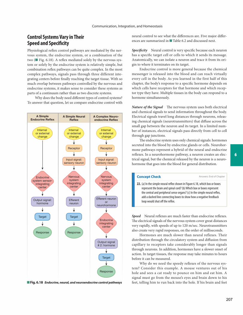
Communication, Integration, and Homeostasis
6
neural control to see what the diff erences are. Five major diff er-ences are summarized in Table 6.2 and discussed next.
Specifi city Neural control is very specifi c because each neuron has a specifi c target cell or cells to which it sends its message. Anatomically, we can isolate a neuron and trace it from its ori-gin to where it terminates on its target.
Endocrine control is more general because the chemical messenger is released into the blood and can reach virtually every cell in the body. As you learned in the first half of this chapter, the body’s response to a specifi c hormone depends on which cells have receptors for that hormone and which recep-tor type they have. Multiple tissues in the body can respond to a hormone simultaneously.
Nature of the Signal Th e nervous system uses both electrical and chemical signals to send information throughout the body. Electrical signals travel long distances through neurons, releas-ing chemical signals (neurotransmitters) that diff use across the small gap between the neuron and its target. In a limited num-ber of instances, electrical signals pass directly from cell to cell through gap junctions.
Th e endocrine system uses only chemical signals: hormones secreted into the blood by endocrine glands or cells. Neurohor-mone pathways represent a hybrid of the neural and endocrine refl exes. In a neurohormone pathway, a neuron creates an elec-trical signal, but the chemical released by the neuron is a neuro-hormone that goes into the blood for general distribution.
Control Systems Vary in Their Speed and Specifi city
Physiological refl ex control pathways are mediated by the ner-vous system, the endocrine system, or a combination of the two ( Fig. 6.18 ). A refl ex mediated solely by the nervous sys-tem or solely by the endocrine system is relatively simple, but combination refl ex pathways can be quite complex. In the most complex pathways, signals pass through three different inte-grating centers before fi nally reaching the target tissue. With so much overlap between pathways controlled by the nervous and endocrine systems, it makes sense to consider these systems as parts of a continuum rather than as two discrete systems.
Why does the body need diff erent types of control systems? To answer that question, let us compare endocrine control with
Concept Check
22. (a) In the simple neural refl ex shown in Figure 6.18 , which box or boxes
represent the brain and spinal cord? (b) Which box or boxes represent
the central and peripheral sense organs? (c) In the simple neural refl ex,
add a dashed line connecting boxes to show how a negative feedback
loop would shut off the refl ex.
Fig. 6.18 Endocrine, neural, and neuroendocrine control pathways
A SimpleEndocrine Reflex
A Simple NeuralReflex
A Complex Neuro-endocrine Reflex
Internal or external
change
Internal or external
change
Internal or external
change
Receptor Receptor
Input signal:sensory neuron
Input signal:sensory neuron
Endocrinesystem sensor-
integratingcenter
Nervoussystem
integratingcenter
Nervoussystem
integratingcenter
Endocrineintegrating
center
Output signal:hormone
Output signal# 2: hormone
Efferent neuronor
neurohormone
Efferentneuron
Target Target
Target
Response Response
Response
Speed Neural refl exes are much faster than endocrine refl exes. Th e electrical signals of the nervous system cover great distances very rapidly, with speeds of up to 120 m/sec. Neurotransmitters also create very rapid responses, on the order of milliseconds.
Hormones are much slower than neural reflexes. Their distribution through the circulatory system and diff usion from capillary to receptors take considerably longer than signals through neurons. In addition, hormones have a slower onset of action. In target tissues, the response may take minutes to hours before it can be measured.
Why do we need the speedy refl exes of the nervous sys-tem? Consider this example. A mouse ventures out of his hole and sees a cat ready to pounce on him and eat him. A signal must go from the mouse’s eyes and brain down to his feet, telling him to run back into the hole. If his brain and feet
Answers: End of Chapter
207

Communication, Integration, and Homeostasis
were only 5 micrometers (5 μm = 1/200 millimeter) apart, it would take a chemical signal 20 milliseconds (msec) to diff use across the space and the mouse could escape. If the brain and feet were 50 μm (1/20 millimeter) apart, diff usion would take 2 seconds and the mouse might get caught. But because the head and tail of a mouse are centimeters apart, it would take a chemical signal three weeks to diff use from the mouse’s head to his feet. Poor mouse!
Even if the distribution of the chemical signal were accel-erated by help from the circulatory system, the chemical mes-sage would still take 10 seconds to get to the feet, and the mouse would become cat food. Th e moral of this tale is that refl exes
R U N N I N G P R O B L E M
“OK, just one more question,” says Marvin. “You said that people with diabetes have high blood glucose levels. If glucose is so high, why can’t it just leak into the cells?”
Q6: Why can’t glucose always diff use into cells when the blood glucose concentration is higher than the intracellular glucose concentration?
Q7: What do you think happens to the rate of insulin secretion when blood glucose levels fall? What kind of feedback loop is operating?
Comparison of Neural and Endocrine Control
Property Neural Refl ex Endocrine Refl ex
Specificity Each neuron terminates on a single target cell or on a limited number of adjacent target cells.
Most cells of the body are exposed to a hormone. The response depends on which cells have receptors for the hormone.
Nature of the signal Electrical signal that passes through neuron, then chemical neurotransmitters that carry the signal from cell to cell. In a few cases, signals pass from cell to cell through gap junctions.
Chemical signals secreted in the blood for distribution throughout the body.
Speed Very rapid. Distribution of the signal and onset of action are much slower than in neural responses.
Duration of action Usually very short. Responses of longer duration are mediated by neuromodulators.
Responses usually last longer than neural responses.
Coding for stimulus intensity Each signal is identical in strength. Stimulus intensity is correlated with increased frequency of signaling.
Stimulus intensity is correlated with amount of hormone secreted.
requiring a speedy response are mediated by the nervous system because they are so much more rapid.
Duration of Action Neural control is of shorter duration than endocrine control. Th e neurotransmitter released by a neuron combines with a receptor on the target cell and initiates a re-sponse. Th e response is usually very brief, however, because the neurotransmitter is rapidly removed from the vicinity of the receptor by various mechanisms. To get a sustained response, multiple repeating signals must be sent through the neuron.
Endocrine reflexes are slower to start, but they last lon-ger. Most of the ongoing, long-term functions of the body, such as metabolism and reproduction, fall under the control of the endocrine system.
Coding for Stimulus Intensity As a stimulus increases in in-tensity, control systems must have a mechanism for conveying this information to the integrating center. Th e signal strength from any one neuron is constant in magnitude and therefore cannot reflect stimulus intensity. Instead, the frequency of signaling through the aff erent neuron increases. In the endo-crine system, stimulus intensity is refl ected by the amount of hormone released: the stronger the stimulus, the more hor-mone is released.
Complex Refl ex Control Pathways Have Several Integrating Centers
Figure 6.19 summarizes variations in the neural, neuroendo-crine, and endocrine refl ex control pathways.
Table6.2
208

R
T
T
R
R
RR
E
E
T
T
T
T
T
R
E2
E
S
Simple NeuralReflex
NeurohormoneReflex Complex Neuroendocrine Reflexes
Simple EndocrineReflex
Sensor
Sensory neuron
CNS CNS CNS CNS CNS
Efferentneuron
Neuro-transmitter
Targetcell
Endocrinecells
Bloodvessel
Example:The knee jerk reflex
Example:Release of breast milk in response to suckling
Example:Insulin secretion in response to a signal from the brain
Example:Secretion ofgrowth hormone
Example:Insulin releasewhen bloodglucose increases
Example:This pattern occurswith hormonesreleased by theanterior pituitary.
Response
Response
Response
Response
Response
Response
Hormone
Neurohormone
Neurotransmitter
Hormone #2
1 2 3 4 5 6
R
Stimulus Stimulus StimulusStimulus Stimulus Stimulus
Stimulus
Neurotransmitter
Neurohormone
Classic hormone
Endocrine integrating center
Sensor Efferent neuron
Sensory neuron(input pathway)
CNS integrating center
Target cell (effector)
Output Pathways
E E1
Fig. 6.19 E S S E N T I A L S
Simple and Complex Reflex Pathways
KEY
209
Communication, Integration, and Homeostasis
In complex pathways, there may be more than one in-tegrating center. Figure 6.19 shows three examples of com-plex neuroendocrine pathways. The simplest of these, Figure 6.19 3 , combines a neural refl ex with a classic endocrine refl ex. An example of this pattern can be found in the control of insulin release. Th e pancreatic beta cells monitor blood glu-cose concentrations directly ( Fig. 6.19 6 ), but they are also controlled by the nervous system. During a meal, the presence of food in the stomach stretches the wall of the digestive tract and sends input signals to the brain. Th e brain in turn sends ex-citatory output signals to the beta cells, telling them to release insulin. Th ese signals take place even before the food has been absorbed and blood glucose levels have gone up (a feedforward refl ex ). Th is pathway therefore has two integrating centers (the brain and the beta cells).
In a simple endocrine reflex pathway ( Fig. 6.19 6 ), the endocrine cell monitors the regulated variable and acts as both sensor and integrating center; there is no input pathway. The output pathway is the hormone, and the target is any cell having the appropriate receptor protein.
An example of a simple endocrine refl ex is secretion of the hormone insulin in response to changes in blood glucose level. Th e endocrine cells that secrete insulin monitor blood glucose concentrations by using ATP production in the cell as an indi-cator. When blood glucose increases, intracellular ATP produc-tion exceeds the threshold level, and the endocrine cells respond by secreting insulin into the blood. Any target cell in the body that has insulin receptors responds to the hormone and initiates processes that take glucose out of the blood. Th e removal of the stimulus acts in a negative feedback manner, and the response loop shuts off when blood glucose levels fall below a certain concentration.
In a simple neural refl ex, all the steps of the pathway are present, from receptor to target ( Fig. 6.19 1 ). Th e neural refl ex is represented in its simplest form by the knee jerk (or patel-lar tendon) refl ex. A blow to the knee (the stimulus) activates a stretch receptor. An electrical and chemical signal travels through an aff erent neuron to the spinal cord (the integrating center). If the blow is strong enough (exceeds threshold), a sig-nal travels from the spinal cord through an eff erent neuron to the muscles of the thigh (the target or effector). In response, the muscles contract, causing the lower leg to kick outward (the knee jerk).
Concept Check
23. Match the following terms for parts of the knee jerk refl ex to the parts
of the simple neural refl ex shown in Figure 6.19 1 : blow to knee, leg
muscles, neuron to leg muscles, sensory neuron, brain and spinal cord,
stretch receptor, muscle contraction.
Concept Check
24. Match the following terms with the appropriate parts of the simple
neuroendocrine refl ex in Fig. 6.19 3 (terms may be used more
than once): food in stomach following a meal, brain and spinal cord,
endocrine cells of pancreas, stretch receptors, eff erent neuron to
pancreas, insulin, adipose cell, blood, sensory neuron.
The neurohormone reflex, shown in Figure 6.19 2 , is identical to the neural reflex except that the neurohormone released by the neuron travels in the blood to its target, just like a hormone. A simple neurohormone refl ex is the release of breast milk in response to a baby’s suckling. Th e baby’s mouth on the nipple stimulates sensory signals that travel through sensory neurons to the brain (integrating center). An elec-trical signal in the eff erent neuron triggers the release of the neurohormone oxytocin from the brain into the circulation. Oxytocin is carried to the breast, where it causes contraction of smooth muscles in the breast (the target), resulting in the ejection of milk.
Another complex reflex ( Fig. 6.19 4 ) uses a neurohor-mone to control the release of a classic hormone. Th e secretion of growth hormone is an example of this pathway. The most complex neuroendocrine pathways, shown as Figure 6.19 5, include a neurohormone and two classic hormones. Th is pat-tern is typical of some hormones released by the anterior pitu-itary, an endocrine gland located just below the brain.
In describing complex neuroendocrine reflex pathways, we identify only one receptor and input pathway, as indicated in Figure 6.19 . In the three complex pathways shown, the brain is the fi rst integrating center and the neurohormone is the fi rst output pathway. In Figure 6.19 5 the endocrine target (E 1 ) of the neurohormone is the second integrating center, and its hormone is the second output pathway. Th e second endocrine gland in the pathway (E 2 ) is the third integrating center, and its hormone is the third output pathway. Th e target of the last sig-nal in the sequence is the eff ector.
Table 6.3 compares the various steps in neural, neuro-endocrine, and endocrine refl exes. R U N N I N G P R O B L E M C O N C L U S I O N
Answers: End of Chapter
Answers: End of Chapter
210

Communication, Integration, and Homeostasis
6
Comparison of Neural, Neuroendocrine, and Endocrine Refl exes
Neural Neuroendocrine Endocrine
Sensor Special and somatic sensory receptors
Special and somatic sensory receptors
Endocrine cell
Input signal Sensory neuron Sensory neuron None
Integrating center Brain or spinal cord Brain or spinal cord Endocrine cell
Output signal Efferent neuron (electrical signal and neurotransmitter)
Efferent neuron (electrical signal and neurohormone)
Hormone
Target(s) Muscles and glands, some adipose tissue
Most cells of the body Most cells of the body
Response Contraction and secretion primarily; may have some metabolic effects
Change in enzymatic reactions, membrane transport, or cell proteins
Change in enzymatic reactions, membrane transport, or cell proteins
Diabetes Mellitus
Marvin underwent further tests and was diagnosed with early type 2 diabetes. With careful attention to his diet and with a regular exercise program, he has managed to keep his blood glucose levels under control. Diabetes is a growing epidemic in the United States, with more than 25 million diabetics in the United States in 2011 (about 8% of the population). Even scarier is the estimate that another 79 million people are considered “prediabetic”—at signifi cant risk of becoming diabetic. You will learn more about diabetes as you work through the chapters
in this course. To learn more about diabetes now, see the American Diabetes Association web site ( www.diabetes.org ) or the Centers for Disease Control and Prevention ( www.cdc.gov/diabetes ).
In this running problem, you learned about glucose homeostasis and how it is maintained by insulin and glucagon. The disease diabetes mellitus is an indication that glucose homeostasis has been disrupted. Check your understanding of this running problem by comparing your answers to the information in the summary table.
R U N N I N G P R O B L E M C O N C L U S I O N
Question Facts Integration and Analysis
1. In which type of diabetes is the signal pathway for insulin more likely to be defective?
Insulin is a peptide hormone that uses membrane receptors linked to second messengers to transmit its signal to cells. People with type 1 diabetes lack insulin; people with type 2 diabetes have normal-to-elevated insulin levels.
Normal or high insulin levels suggest that the problem is not with amount of insulin but with the action of the insulin at the cell. The problem in type 2 diabetes could be a defective signal transduction mechanism.
2. Insulin is a protein hormone. Would you expect to fi nd its receptor on the cell surface or in the cytoplasm of the target cells?
Lipophilic signal molecules have intracellular receptors. Lipophobic molecules have cell membrane receptors.
Proteins are lipophobic so protein hormones have cell surface receptors.
Table6.3
211

Communication, Integration, and Homeostasis
R U N N I N G P R O B L E M C O N C L U S I O N ( c o n t i n u e d )
Question Facts Integration and Analysis
3. In which form of diabetes are the insulin receptors more likely to be up-regulated?
Up-regulation of receptors usually occurs if a signal molecule is present in unusually low concentrations. In type 1 diabetes, insulin is not secreted by the pancreas.
In type 1 diabetes, insulin levels are low. Therefore, type 1 is more likely to cause up-regulation of the insulin receptors.
4. The homeostatic regulation of blood glucose levels by the hormones insulin and glucagon is an example of which of Cannon’s postulates?
Cannon’s postulates describe the role of the nervous system in maintaining homeostasis, and the concepts of tonic activity, antagonistic control, and diff erent eff ects of signals in diff erent tissues.
Insulin decreases blood glucose levels, and glucagon increases them. Therefore, the two hormones are an example of an antagonistic control.
5. In the insulin pathway that regulates blood glucose levels, what are the stimulus, the sensor, the integrating center, the output signal, the target(s), and the response(s)?
See the steps of refl ex pathways. Stimulus: increase in blood glucose levels; sensor: beta cells of the pancreas that sense the change; integrating center: beta cells; output signal : insulin; targets: any tissues of the body that respond to insulin; responses: cellular uptake and use of glucose.
6. Why can’t glucose always diff use into cells when the blood glucose concentration is higher than the intracellular glucose concentration?
Glucose is lipophobic. Simple diff usion goes across the phospholipid bilayer. Facilitated diff usion uses protein carriers.
Because glucose is lipophobic, it cannot cross the membrane by simple diff usion. It must go by facilitated diff usion. If a cell lacks the necessary carriers, facilitated diff usion cannot take place.
7. What do you think happens to the rate of insulin secretion when blood glucose levels fall? What kind of feedback loop is operating?
The stimulus for insulin release is an increase in blood glucose levels. In negative feedback, the response off sets the stimulus. In positive feedback, the response enhances the stimulus.
An increase in blood glucose concentration stimulates insulin release; therefore, a decrease in blood glucose should decrease insulin release. In this example, the response (lower blood glucose) off sets the stimulus (increased blood glucose), so a negative feedback loop is operating.
Two of the major themes in physiology stand out in this chapter: the control of homeostasis and communication . Th e sensors, integrating cent-ers, and targets of physiological control systems are described in the context of refl ex control pathways, which vary from simple to complex. Functional control systems require effi cient communication that uses
Chapter Summary
various combinations of chemical and electrical signals. Th ose signals that cannot cross cell membranes must use membrane receptor proteins and signal transduction to transfer their information into the cell. Th e interaction of signal molecules with protein receptors illustrates another fundamental theme of physiology, molecular interactions .
• PhysioExTM Lab Simulations • Interactive Physiology
Animations
Test your understanding with:
www.masteringaandp.com
• Practice Tests • Running Problem Quizzes • A&PFlixTM Animations
212

Communication, Integration, and Homeostasis
6
Cell-to-Cell Communication 1. There are two basic types of physiological signals: chemical and
electrical. Chemical signals are the basis for most communication within the body.
2. There are four methods of cell-to-cell communication: (1) direct cytoplasmic transfer through gap junctions, (2) contact-dependent signaling, (3) local chemical communication, and (4) long-distance communication. ( Fig. 6.1 )
3. Gap junctions are protein channels that connect two adjacent cells. When they are open, chemical and electrical signals pass directly from one cell to the next.
4. Contact-dependent signals require direct contact between surface molecules of two cells.
5. Local communication uses paracrine signals , chemicals that act on cells close to the cell that secreted the paracrine. A chemical that acts on the cell that secreted it is called an autocrine signal . Th e activity of paracrine and autocrine signal molecules is limited by diff usion distance.
6. Long-distance communication uses neurocrine molecules and electrical signals in the nervous system, and hormones in the endocrine system. Only cells that possess receptors for a hormone will be target cells.
7. Cytokines are regulatory peptides that control cell development, diff erentiation, and the immune response. Th ey serve as both local and long-distance signals.
Signal Pathways 8. Chemical signals bind to receptors and change intracellular signal
molecules that direct the response. 9. Lipophilic signal molecules enter the cell and combine with cytoplasmic
or nuclear receptors. Lipophobic signal molecules and some lipophilic molecules combine with membrane receptors. ( Fig. 6.3 )
10. Signal transduction pathways use membrane receptor proteins and intracellular second messenger molecules to translate signal infor-mation into an intracellular response. ( Fig. 6.4 )
11. Some signal transduction pathways activate protein kinases . Oth-ers activate amplifi er enzymes that create second messenger mol-ecules. ( Fig. 6.5 )
12. Signal pathways create intracellular cascades that amplify the origi-nal signal. ( Fig. 6.6 )
13. Receptor-enzymes activate protein kinases, such as tyrosine kinase ( Fig. 6.7 ), or the amplifi er enzyme guanylyl cyclase, which produces the second messenger cGMP .
14. G proteins linked to amplifi er enzymes are the most prevalent sig-nal transduction system. G protein – coupled receptors also alter ion channels. ( Fig. 6.8 )
15. Th e G protein – coupled adenylyl cyclase-cAMP-protein kinase A pathway is the most common pathway for protein and peptide hor-mones. ( Fig. 6.8 a)
16. In the G protein – coupled phospholipase C pathway, the amplifi er enzyme phospholipase C creates two second messengers: IP 3 and diacylglycerol (DAG). IP 3 causes Ca 2+ release from intracellular stores. Diacylglycerol activates protein kinase C . ( Fig. 6.8 b)
17. Integrin receptors link the extracellular matrix to the cytoskeleton. ( Fig. 6.3 c)
18. Ligand-gated ion channels open or close to create electrical signals. ( Fig. 6.9 )
Novel Signal Molecules 19. Calcium is an important signal molecule that binds to calmodulin
to alter enzyme activity. It also binds to other cell proteins to alter movement and initiate exocytosis. ( Fig. 6.11 )
20. Nitric oxide (NO), carbon monoxide (CO), and hydrogen sulfi de (H 2 S) are short-lived gaseous signal molecules. NO activates guany-lyl cyclase directly.
21. The arachidonic acid cascade creates lipid signal molecules, such as leukotrienes , prostaglandins , and thromboxanes .( Fig. 6.12 )
Modulation of Signal Pathways 22. Th e response of a cell to a signal molecule is determined by the cell’s
receptor for the signal. 23. Receptors come in related forms called isoforms . One ligand
may have different effects when binding to different isoforms.( ; Fig. 6.13 )
24. A receptor may have multiple ligands. Agonists mimic the action of a signal molecule. Antagonists block the signal pathway. ( Fig. 6.14 )
25. Receptor proteins exhibit specifi city, competition, and saturation. 26. Cells exposed to abnormally high concentrations of a signal for a
sustained period of time attempt to bring their response back to normal through down-regulation or by desensitization . In down-regulation , the cell decreases the number of receptors. In desen-sitization , the cell decreases the binding affinity of the receptors. Up-regulation is the opposite of down-regulation and involves in-creasing the number of receptors for a signal.
27. Cells have mechanisms for terminating signal pathways, such as re-moving the signal molecule or breaking down the receptor-ligand complex.
28. Many diseases have been linked to defects in various aspects of signal pathways, such as missing or defective receptors. (Tbl. 6.1)
Homeostatic Refl ex Pathways 29. Walter Cannon first stated four basic postulates of homeostasis:
(1) Th e nervous system plays an important role in maintaining ho-meostasis. (2) Some parameters are under tonic control , which al-lows the parameter to be increased or decreased by a single signal ( Fig. 6.14 a). (3) Other parameters are under antagonistic control , in which one hormone or neuron increases the parameter while an-other decreases it ( Fig. 6.14 b). (4) Chemical signals can have diff er-ent eff ects in diff erent tissues of the body, depending on the type of receptor present at the target cell. ( Fig. 6.15 )
30. In refl ex control pathways, the decision to respond to a change is made by an integrating center. A chemical or electrical signal to the target cell or tissue then initiates the response. Long-distance refl ex pathways involve the nervous and endocrine systems and cytokines.
31. Neural control is faster and more specifi c than endocrine control but is usually of shorter duration. Endocrine control is less specifi c and slower to start but is longer lasting and is usually amplifi ed. (Tbl. 6.2)
32. Many refl ex pathways are complex combinations of neural and en-docrine control mechanisms. ( Fig. 6.19 )
213

Communication, Integration, and Homeostasis
Questions
Level One Reviewing Facts and Terms 1. What are the two routes for long-distance signal delivery in the
body? 2. Which two body systems maintain homeostasis by monitoring and
responding to changes in the environment? 3. What two types of physiological signals does the body use to send
messages? Of these two types, which is available to all cells? 4. In a signal pathway, the signal ligand, also called the first messen-
ger, binds to a(n) , which activates and changes intracellular .
5. Th e three main amplifi er enzymes are (a) , which forms cAMP; (b) , which forms cGMP; and (c) , which converts a phospholipid from the cell’s membrane into two diff erent second messenger molecules.
6. An enzyme known as protein kinase adds the functional group to its substrate, by transferring it from a(n) molecule.
7. Distinguish between central and peripheral receptors. 8. Receptors for signal pathways may be found in the ,
, or of the cell. 9. Down-regulation results in a(n) (increased or
decreased?) number of receptors in response to a prolonged signal. 10. List two ways a cell may decrease its response to a signal. 11. In a negative feedback loop, the response moves the system in the
(same/opposite) direction as the stimulus moves it.
Level Two Reviewing Concepts 12. Explain the relationships of the terms in each of the following sets.
Give a physiological example or location if applicable. (a) gap junctions, connexins, connexon (b) autocrine, paracrine, cytokine, neurocrine, hormone (c) agonist, antagonist (d) transduction, amplifi cation, cascade
13. List and compare the four classes of membrane receptors for signal pathways. Give an example of each.
14. Who was Walter Cannon? Restate his four postulates in your own words.
15. Briefl y defi ne the following terms and give an anatomical example when applicable: input signal, integrating center, output signal, response, sensor, stimulus, target.
16. Compare and contrast the advantages and disadvantages of neural versus endocrine control mechanisms.
17. Would the following refl exes have positive or negative feedback? (a) glucagon secretion in response to declining blood glucose (b) increasing milk release and secretion in response to baby’s
suckling (c) urgency in emptying one’s urinary bladder (d) sweating in response to rising body temperature
18. Identify the target tissue or organ for each example in question 17. 19. Now identify the integrating center for examples (a), (c), and (d) in
question 17.
Level Three Problem Solving 20. In each of the following situations, identify the components of the
refl ex. (a) You are sitting quietly at your desk, studying, when you become
aware of the bitterly cold winds blowing outside at 30 mph, and you begin to feel a little chilly. You start to turn up the thermo-stat, remember last month’s heating bill, and reach for an afghan to pull around you instead. Pretty soon you are toasty warm again.
(b) While you are strolling through the shopping district, the aroma of cinnamon sticky buns reaches you. You inhale appreciatively, but remind yourself that you’re not hungry, because you had lunch just an hour ago. You go about your business, but 20 min-utes later you’re back at the bakery, sticky bun in hand, raven-ously devouring its sweetness, saliva moistening your mouth.
21. A researcher is studying the smooth muscle of the respiratory sys-tem airways. When she exposes the airways to the neurotransmitter acetylcholine, the smooth muscle contracts. When she exposes the airways to the neurohormone epinephrine, the airways relax. (a) Th e phenomenon just described is an example of
control. (b) What distinguishes a neurotransmitter from a neurohormone? (c) Which chemical messenger is secreted in higher concentrations:
acetylcholine or epinephrine? Defend your answer.
Level Four Quantitative Problems 22. In a signal cascade for rhodopsin, a photoreceptor molecule, one
rhodopsin activates 1000 molecules of transducin, the next mol-ecule in the signal cascade. Each transducin activates one phos-phodiesterase, and each phosphodiesterase converts 4000 cGMP to GMP. (a) What is the name of the phenomenon described in this paragraph? (b) Activation of one rhodopsin will result in the production of
how many GMP molecules?
214

Communication, Integration, and Homeostasis
6
17. Choices (a) and (d) could decrease binding affi nity. Changing re-ceptor number would not aff ect binding affi nity.
18. Tonic control usually involves one control system, but antagonistic control uses two.
19. A signal can have opposite eff ects by using diff erent receptors or diff erent signal pathways.
20. In local control, the stimulus, integration of the signal, and re-sponse all take place in or very close to the target cell. With refl ex control, integration of the input signal and initiation of a response may take place far from the location where the change occurred. In addition, the refl ex response is oft en systemic and not localized.
21. Stimulus, sensor or sensory receptor, input signal (aff erent path-way), integrating center, output signal (eff erent pathway), target or eff ector, response (tissue and systemic)
22. (a) Th e “neural system integrating center” is the brain and spinal cord. (b) “Receptor” represents the sense organs. (c) Th e dashed line indicating negative feedback runs from “Response” back to “Internal or external change.”
23. blow to knee = internal or external change; leg muscles = targets; neuron to leg muscles = eff erent neuron; sensory neuron = input signal; brain and spinal cord = CNS integrating center; stretch re-ceptor = sensor or receptor; muscle contraction = response.
24. food in stomach = stimulus; brain and spinal cord = CNS integrat-ing center; endocrine cells of pancreas = E (integrating center); stretch receptors = receptor; eff erent neuron to pancreas = eff er-ent neuron; insulin = classic hormone; adipose cell = target cell; sensory neuron = aff erent neuron. Blood is the anatomical route that hormones use to reach their target but is not part of the refl ex pathway.
Answers to Figure Questions
Figure 6.8 : A (inactive and active) = adenylyl cyclase; inactive B =ATP; active B = cAMP; C (inactive and active) = protein kinase A; product = phosphorylated protein.
Figure 6.15 : 180 beats/min for the top ECG and 60 beats/min for the bottom ECG.
1. All methods listed are chemical signals except for (c) gap junctions, which transfer both chemical and electrical signals. Neurohor-mones (e) and neurotransmitters (f) are associated with electrical signaling in neurons but are themselves chemicals.
2. Cytokines, hormones, and neurohormones travel through the blood. Cytokines, neurohormones, and neurotransmitters are released by neurons.
3. Th e signal to pounce could not have been a paracrine signal because the eyes are too far away from the legs and because the response was too rapid for it to have taken place by diff usion.
4. Th e components of signal pathways are signal molecule, receptor, intracellular signal molecule(s), and target proteins.
5. Th e cellular locations of receptors are cell membrane, cytosol, and nucleus.
6. Th e steps of signal transduction are (1) signal molecule binds to receptor that (2) activates a protein that (3) creates second messen-gers that (4) create a response.
7. Amplification turns one signal molecule (first messenger) into multiple second messenger molecules.
8. Steroids are lipophilic, so they can enter cells and bind to intracel-lular receptors.
9. Receptors are either ligand-gated ion channels, receptor-enzymes, G protein–coupled receptors, or integrins.
10. First messengers are extracellular; second messengers are intracellular.
11. (a) ligand, receptor, second messenger, cell response; (b) amplifi er enzyme, second messenger, protein kinase, phosphorylated pro-tein, cell response
12. (a) Cl − channel opens: cell hyperpolarizes; (b) K + channel opens: cell hyperpolarizes; (c) Na + channel opens: cell depolarizes.
13. Th e cell must use active transport to move Ca 2+ against its concen-tration gradient.
14. A drug that blocks leukotriene action could act at the target cell
receptor or at any step downstream. A drug that blocks leukotriene synthesis might inhibit lipoxygenase.
15. Receptors, enzymes, and transporters are all proteins. 16. Insulin could be using one receptor isoform with diff erent second
messenger systems in diff erent cells or could be binding to diff erent receptor isoforms.
Answers to Concept Check Questions
Answers
Answers to Figure Questions
215
Communication, Integration, and Homeostasis
Answers to Review Questions
Level One Reviewing Facts and Terms
1. Neurons and blood 2. Nervous and endocrine systems 3. Chemical (available to all cells) and electrical 4. receptor, targets (effectors) or proteins 5. (a) adenylyl cyclase, (b) guanylyl cyclase, (c) phospholipase C 6. phosphate, ATP 7. Central: located within the central nervous system. Peripheral: found outside
the CNS 8. nucleus, cytosol , cell membrane 9. decreased 10. It may down-regulate receptor number or decrease receptor affinity for the
substrate. 11. opposite
Level Two Reviewing Concepts
12. (a) Gap junctions connect two cells using protein channels called connexons , made from connexin subunits. (b) All are chemicals secreted into the ECF. Paracrines act on nearby cells; autocrines act on the cell that secretes them. Cytokines are peptide autocrine and paracrine signals or hormones . Neuro-crines are chemicals secreted by neurons. (c) Agonists have the same action as another molecule; antagonists act to oppose the action of another molecule. (d) Transduction: a signal molecule transfers information from ECF to the cytoplasm. Cascade: a series of steps. Amplification: one signal molecule cre-ates a larger signal.
13. Ligand-gated channels (ATP-gated channel); integrin receptors (platelet receptors); receptor enzymes (tyrosine kinase receptor); G-protein–coupled receptors (adenylyl cyclase>cAMP-linked receptors).
14. The father of American physiology. (1) The nervous system keeps body functions within normal limits. (2) Some functions have tonic control rather than on-off control. (3) Some signals act in opposition to each other. (4) Cell response depends on the cell’s receptor for a signal.
15. Input signal: information transmission from stimulus to integrating center (sensory nerve). Integrating center: cell or cells that receive information, decide whether and how it should be acted upon, and send a signal to initiate a response (the brain). Output signal: electrical or chemical signals that take information from integrating center to target (nerve or hormone). Response: what target cell does to react to the stimulus (pull hand away from hot stove). Sensor: cell that perceives the stimulus (temperature receptor). Stimulus: change that begins a response (touching a hot stove). Target: the cell or tissue that carries out the response (muscle).
16. Neural control is faster than endocrine and better for short-acting responses. Endocrine can affect widely separated tissues with a single signal and better for long-acting responses.
17. (a) negative, (b) positive, (c) negative, (d) negative 18. (a) tissues that respond to glucagon, such as liver, (b) breast, (c) bladder,
(d) sweat glands 19. (a) pancreatic endocrine cells that secrete glucagon, (b)–(d) nervous system
Level Three Problem Solving
20. (a) stimulus = decrease in body temperature to decrease; sensor = temperature receptors; input = sensory neurons; integrating center = CNS; output = efferent neurons; targets = muscles used to pull up afghan; response = afghan conserves heat (b) stimulus = smell of sticky buns; sensor = odor receptors in the nose; input = sensory neurons; integrating center = CNS; output = efferent neurons; target = skeletal muscles; response = walk to bakery, buy buns, and eat
21. (a) antagonistic (b) Neurotransmitters act on nearby cells (paracrine ac-tion). Neurohormones act on distant targets. (c) Epinephrine is secreted in larger amounts because it will be diluted by the blood volume before reaching its target.
Level Four Quantitative Problems
22. (a) amplification and a cascade (b) (1000 � 4000) or 4,000,000 GMP
Photo Credits
CO: Alila Sao Mai /Shutterstock.com Biotechnology: Russ Bishop/Alamy.
216
Introduction to the Endocrine System
From Chapter 7 of Human Physiology: An Integrated Approach, Sixth Edition. Dee Unglaub Silverthorn. Copyright © 2013 by Pearson Education, Inc. All rights reserved.
217