5dgldwlrqsurwhfwlrq - european commission · european commission 5dgldwlrqsurwhfwlrq...
TRANSCRIPT

5DGLDWLRQ�SURWHFWLRQ����
,PSOHPHQWDWLRQ�RI�WKH³0HGLFDO�([SRVXUH�'LUHFWLYH´
�������(XUDWRP�3URFHHGLQJV�RI�WKH�LQWHUQDWLRQDO
ZRUNVKRS�KHOG�LQ
0DGULG��RQ����$SULO�����
(XURSHDQ�&RPPLVVLRQ

European Commission
5DGLDWLRQ�SURWHFWLRQ����
,PSOHPHQWDWLRQ�RI�WKH³0HGLFDO�([SRVXUH�'LUHFWLYH´
�������(XUDWRP�3URFHHGLQJV�RI�WKH�LQWHUQDWLRQDO�ZRUNVKRS�KHOG�LQ
0DGULG��RQ����$SULO�����
1998
Directorate-GeneralEnvironment, Nuclear Safety
and Civil Protection
3
)RUHZRUG
The Council of the European Union issued the Medical Exposure Directive(MED) 97/43/Euratom on 30 June 1997. Member States must implement theDirective in national legislation no later than 13 May 2000.
To assist Member States with transposing the Directive the EuropeanCommission developed a number of technical guidelines directly related toexplain further parts of the Directive.
This conference was designed to promote knowledge on the MED and therelated guidelines on the one hand and to bring about a discussion onpractical ways of transposing the Directive on the other.

4
3URJUDPPH�&RPPLWWHH
L. ARRANZ Spain
M. DE ROO E.U.M.S. Section Nuclear Medicine (Belgium)
K. FAULKNER United Kingdom
A. FINCH I.S.R.R.T. (United Kingdom)
G.D. HURLEY Ireland
A. NOEL E.F.O.M.P. (France)
H. RINGERTZ President European Association of Radiology(Sweden)
P. SMEESTERS Belgium
B. WALL United Kingdom
C. ZUUR The Netherlands
G. MENZEL EC / DG XII (Research & TechnologicalDevelopment – Energy)
D. TEUNEN EC/DG XI.C.1 – Radiation Protection(scientific secretariat)

5
&RQWHQWV
)25(:25'�������������������������������������������������������������������������������������������������������������������������������������������������������������������������� �
23(1,1*�$''5(66 ����������������������������������������������������������������������������������������������������������������������������������������������������������� �
BY MR. JOSÉ-MANUEL ROMAY-BECCARIA
23(1,1*�$''5(66 ����������������������������������������������������������������������������������������������������������������������������������������������������������� �
BY MRS. SUZANNE FRIGREN
23(1,1*�$''5(66 ����������������������������������������������������������������������������������������������������������������������������������������������������������� �
BY MR. JUAN MANUEL KINDELAN
0$-25�&+$1*(6�,1�7+(�0(',&$/�(;32685(�',5(&7,9( ��������������������������������������������������������������������������� ��
DIEDERIK TEUNEN
5$',$7,21�3527(&7,21�)2//2:,1*�,2',1(�����7+(5$3< ����������������������������������������������������������������������� ��
CISKA ZUUR
5$',$7,21�3527(&7,21�2)�7+(�81%251�&+,/'��������������������������������������������������������������������������������������������� ��
WOLFRAM LEITZ
$&&(37$%,/,7<�&5,7(5,$�)25�5$',2/2*,&$/�,167$//$7,216�$1'�48$/,7<�$6685$1&(352*5$00(6������������������������������������������������������������������������������������������������������������������������������������������������������������������ ��
HANS ZOETELIEF
5$',$7,21�3527(&7,21�,1�0(',&$/�$1'�%,20(',&$/�5(6($5&+������������������������������������������������������� ��
FRANCIS P. CRAWLEY
327(17,$/�(;32685(6�$1'�$&&,'(17�35(9(17,21�,1�0(',&$/�$33/,&$7,21������������������������������ ��
P. ORTIZ
',$*1267,&�5()(5(1&(�/(9(/6��,1&/8',1*�3$7,(17�'26,0(75<������������������������������������������������������ ��
BARRY F. WALL
-867,),&$7,21�2)�0(',&$/�(;32685(6�$1'�0(',&2�/(*$/�(;32685(6������������������������������������������ ��
WILLIAM BINCHY
75$,1,1*�)25�0(',&$/�$33/,&$7,21�2)�,21,=,1*�5$',$7,21�������������������������������������������������������������� ��
ELISEO VAÑO - L. GONZÁLEZ
63(&,$/�(;32685(6��3$(',$75,&6���������������������������������������������������������������������������������������������������������������������� ���
KARL SCHNEIDER
*(1(5$/�',6&866,21������������������������������������������������������������������������������������������������������������������������������������������������� ���
&21&/86,216 ���������������������������������������������������������������������������������������������������������������������������������������������������������������� ���
DR. PATRICK SMEESTERSMR. STEPHEN KAISER

6
2SHQLQJ�DGGUHVV
E\�0U��-RVp�0DQXHO�520$<�%(&&$5,$6SDQLVK�0LQLVWHU�RI�+HDOWK
Mr Kindelán (President of the Nuclear Safety Council), Mrs Frigren (Director of the Nuclear SafetyDirectorate, DG XI), Mr Juan José Francisco Polledo (Director-General for Public Health), Mr Kaiser(Head of the Radiation Protection Unit at the European Commission), Ladies and Gentlemen:
It is a great pleasure for me to take part in this opening session of the Workshop on the implementationof the Directive on health protection against the dangers of ionising radiation in relation to medicalexposures. I wish you all a warm welcome to our hospitable city and would like to thank you forselecting the Ministry of Health and Consumer Affairs as the venue for this important meeting. Iwould also like to congratulate the European Commission’s Directorate-General for the Environment,Nuclear Safety and Civil Protection on its initiative in calling this meeting. Finally, I would like tothank the Spanish Radiation Protection Society and, of course, all the other scientific bodies, industryrepresentatives, public authorities and experts involved in the radiation protection of patients for theirparticipation and collaboration.
During the Workshop we will have an opportunity to exchange views and experiences and thusbecome more familiar with the principles underlying Directive 97/43/Euratom. I am sure that the finalresults of this meeting will make it easier for the Member States to transpose the new directive intonational legislation.
The Directive is basically intended to bring about improvements in patient safety and protection byextending the application of the principles of justification and optimisation of medical exposuresalready set out in earlier directives. This will bring undoubted health benefits and, above all, willimprove the protection afforded to patients by reducing exposures.
Of special interest are the newly-introduced requirements concerning paediatric exposures, healthscreening programmes, practices involving high doses, preventing the exposure of pregnant andbreastfeeding women, and the introduction of quality assurance programmes in radiologicalinstallations.
The radiation protection of patients and health professionals is one of the priority aims of the Ministryof Health and Consumer Affairs and is organised jointly with the Nuclear Safety Council and theAutonomous Communities. To do this, and ensure that radiation protection programmes are effectivelyimplemented, a coordinating committee has been set up within the Interregional Health Council.Through this committee’s efforts, there has been a national census of diagnostic radiology, nuclearmedicine and radiotherapy installations, criteria have been drawn up to avoid the unnecessaryproliferation of installations, and the inspection and monitoring programmes for these installationshave been harmonised.
I would like to stress that the setting up of this committee within the Interregional Council, which isthe highest coordinating body in the national health service, enables us to ensure in the most effectiveway possible that European and national legislation on protection against ionising radiation is applieduniformly throughout Spain.
I would also like to point out that many of the requirements in the new Directive have been transposedinto Spanish legislation by Royal Decree 1841/1997, which lays down quality criteria in the nuclearmedicine field. In addition, the Government will shortly be approving another royal decree on quality

7
criteria for radiotherapy. Finally, there is a royal decree on quality criteria for diagnostic radiology,which will be submitted to the European Commission within the next few weeks.
This legislation clearly demonstrates that the Ministry of Health and Consumer Affairs is determinedto make the necessary arrangements by the time the new Directive enters into force in May 2000.
In this connection, I would like to express my thanks for the excellent advice given by officials fromDG XI’s Radiation Protection Unit, who collaborated with officials from the Ministry of Health’sDirectorate-General for Public Health.
To end, it is my view that this workshop, in which I hope you will all play an active part, will enableus to exchange ideas and information so as to arrive at a thorough understanding of the contents andpurpose of Directive 97/43/Euratom. I have no doubt that this will make it easier to transpose thedirective into national legislation throughout the European Union.
Thank you for your attention. I hope you have a happy stay in Madrid and trust that you will find timeto enjoy the attractions of our city.

8
2SHQLQJ�DGGUHVV
E\�0UV��6X]DQQH�)5,*5(1(XURSHDQ�&RPPLVVLRQ
Ladies and Gentlemen,
It is my pleasure to welcome you in name of the European Commission to this workshop on theimplementation of the Medical Exposure Directive which was approved by the Council of Ministers on30 June 1997.
First of all, I would like to thank the Spanish co-organisers of this workshop, the Ministry of Healthand Consumers affairs and the Spanish Association for Radiation Protection, for their invaluablecontribution to ensure the success of the workshop.
The European Commission considers the Medical Exposure Directive a very important tool toconsolidate the good level of radiation protection achieved under the 1984 Directive and, at the sametime, to optimise protection where possible taking into account, among other factors, scientific andtechnical progress.
In its effort to create the best possible conditions for implementation, the European Commission hasorganised today’s workshop as a first attempt to establish a positive environment for a moreharmonised approach to implementation. An intensive exchange of views between administrators,professionals daily involved in the medical use of ionising radiation and the manufacturers ofradiological equipment coming from all Member States as well as from outside the Union, shouldallow competent authorities to learn about practical aspects of implementing the Directive.
This workshop focuses on a number of key-issues, which may require special attention because theyare new compared to the 1984 Directive or simply because experience has shown that problems canarise. Nine experts will give you a brief introduction to each issue highlighting essential questions.
The final success of this workshop, however, largely depends on your input to the discussion bearingin mind that bringing up proposals for solutions today may prevent difficulties in a later stage of theprocedure.
For the European Commission this workshop is not the end but rather the beginning of a continuouseffort of assisting the Member States and it is my conviction that similar multilateral and bilateralconsultations will take place during the next two years.
Finally, I would like to thank you all for attending in such large number and I wish you a verysuccessful meeting.

9
2SHQLQJ�DGGUHVV
E\�0U��-XDQ�0DQXHO�.,1'(/$1&RQVHMR�GH�6HJXULGDG�1XFOHDU
Minister, Mrs Frigren, Ladies and Gentlemen:
I would like to start by thanking the organisers of this Workshop – the European Commission, theMinistry of Health and the Spanish Radiation Protection Society – for their invitation to take part inthis opening ceremony, which gives me an opportunity to share some thoughts on certain aspectsassociated with the implementation of Directive 97/43/Euratom.
Throughout this century, and particularly in the last 50 years, the use of ionising radiation haddeveloped enormously in both the diagnostic and therapeutic fields. Medical imaging protocols,procedures for isotope-based function tests and the use of radiotherapy facilities embrace a range oftechniques in which ionising radiation makes a fundamental contribution to the detection and treatmentof particular diseases.
However, it is important to remember that all applications of ionising radiation must be carried outwith a wide safety margin, by controlling the risks to individuals and preventing environmentalcontamination. Achieving this aim has been and will continue to be one of the main priorities of thehealth authorities and the regulatory organisations in every country, not to mention the professionalsworking in this field. As an illustration of this, an international conference was held in Seville lastNovember, sponsored by the International Atomic Energy Agency (IAEA) and the World HealthOrganisation (WHO) and coordinated by the Nuclear Safety Council. It was attended by over 500experts from 65 countries, who examined the biological, epidemiological and regulatory aspects of lowradiation doses and reviewed the latest scientific knowledge on this subject.
As for Spain, my own organisation (the Nuclear Safety Council) and the Ministry of Health have setup a working group within the Interregional Health Council, which is the body that coordinates healthpolicy in Spain and includes representatives from the Autonomous Communities and centralgovernment. The remit of this group is to look at all aspects of radiation protection in hospitals andclinics throughout the country.
The European Commission has always been extremely vigilant and active in this area, as shown by theapproval in 1984 of an initial directive laying down basic measures for the radiation protection ofpersons undergoing medical examination or treatment. This has been replaced by Directive 97/43,which we are here to discuss and which regulates in a much more precise and exhaustive way thevarious aspects of protection for patients and medical staff against the risks associated with medicaluses of ionising radiation.
The transposition of the Directive into the national legislation of the Member States will signal theincorporation into daily medical practice within the EU of the three principles recommended by theInternational Commission on Radiological Protection, namely justification, optimisation and limitationof individual doses. This will benefit the ever-increasing number of patients who are exposed toionising radiation.
Today’s workshop provides a valuable and timely opportunity to foster debate and encourage thecompetent authorities of the Member States, those responsible for health issues at the EuropeanCommission, general practitioners, prescribing physicians, radiation protection experts, professionals

10
involved in medical uses of radiation and industry representatives to exchange views on the best wayof implementing the Directive in the area that concerns us.
As President of the Nuclear Safety Council, I would like to reiterate our commitment to collaboratingwith the Ministry of Health, as the law provides, in transposing the Directive into our domesticlegislation and medical practice in the most effective way possible.
To end, I would like to congratulate you on taking this wonderful initiative. I am sure the workshopwill be a great success.
Thank you very much.

11
0DMRU�FKDQJHV�LQ�WKH�0HGLFDO�([SRVXUH�'LUHFWLYH
'LHGHULN�7(81(1(XURSHDQ�&RPPLVVLRQ
European legislation on radiation protection is governed by the EURATOM Treaty and theregulations, directives and other legislation developed in implementation of it. Directives are legalinstruments that are binding on Member States as to their objectives but leave the freedom to theMember States to choose how to implement the directive into national law. The framework directiveis the Basic Safety Standards directive (BSS) on the protection of the public and exposed workersagainst the dangers of ionising radiation. It covers practices, interventions and work activities. Thedirective actually in force dates back to 1980 and 1984 (80/836/Euratom and 84/467/Euratom). It wasrevised in 1996 (96/29/Euratom) to take into account the recommendations of the InternationalCommission on Radiological Protection in ICRP 60. The scope of this directive covers all practices,including medical practices, but no specific requirements are present to protect the individualundergoing medical exposure. In fact, article 6 of the BSS confirms that a medical exposure is subjectto the principles of justification and optimisation but excludes individuals who receive such exposurefrom the dose limitation principle. BSS are to be implemented in national law by Member States notlater than 13 May 2000.
On 3 September 1984 the Council of Ministers issued a directive laying down basic measures for theradiation protection of persons undergoing medical examination or treatment (84/466/Euratom). Thisdirective complemented the BSS as regards the protection of individuals undergoing medicalexposures. The so-called “Patient directive” was the first specific attempt of the European Commissionto define radiation protection concepts in medical application of ionising radiation.
The merit of the Patient directive is not only that it entailed legal initiatives to regulate medicalradiation protection in all Member States but it also created the necessary platform for the furtherdevelopment of a “radiation protection culture” in this field. The directive was concise and had only 5operational articles together with an annex with practical recommendations that were not binding onMember States.
The transposition of the directive in national law, which took place the following decade, showed thatnot all of the requirements present were clearly formulated and that in some cases improvement of thewording was required.
In 1994 the European Commission started the procedure for revising the Patient directive. The majorobjectives of this revision were to harmonise the directive with the new BSS, taking into accountexperience gained with the implementation of the Patient directive and taking account of scientific andtechnical evolution in medical practice.
The revised Medical Exposures Directive (MED) (97/43/Euratom), approved by the Council ofMinisters on 30 June 1997, reaffirms the major objectives of the Patient directive i.e. to aim atoptimum diagnostic efficacy at reasonable dose to the patient and to reduce the number of inadequateexposures. These objectives are pursued by 4 types of requirement:
– provisions relating to duties, responsibilities and qualifications of the staff of medicalfacilities
– provisions related to equipment
– provisions related to procedural requirements

12
– provisions related to ‘special practices’
The MED can be summarised as follows:
$UWLFOH�� entitled “Purpose and scope” is new. It links the MED directly to the BSS. According to thisarticle not only ‘patients’ are covered by its requirements but other individuals, directly or indirectlyexposed to a medical exposure, are covered as well. It gives a list of exposures such as in biomedicalresearch, occupational health surveillance and medico-legal procedures.
$UWLFOH�� entitled “Definitions” is also new. It gives a list of definitions of terms used in the directive.The purpose of this article is to clarify further some requirements of the directive and to minimisepossible misunderstandings. It also gives the directive a more updated look according to modern legalpractice in the Union.
$UWLFOH�� “Justification” takes over the principles already present in the Patient directive and in the1996 BSS. It makes the distinction between justification of practices which usually is a genericjustification and justification of individual exposures. It also identifies those exposures where thejustification requires special procedures or particular attention, such as exposure for medical orbiomedical research, medico-legal exposure and the exposure of an individual assisting a patient.
$UWLFOH�� “Optimisation” also refers to a basic principle already present in the Patient directive butnow makes a distinction between radiodiagnostic and radiotherapeutic procedures. It introduces theconcept and use of diagnostic reference levels as a tool for optimisation and specifies particularrequirements for exposures such as healthy individuals exposed during medical or biomedical researchand individuals willingly and knowingly helping patients. These categories of people are not coveredby the BSS as far as dose limitation is concerned. Therefore dose constraints to control exposures arerequired.
$UWLFOH�� “Responsibilities” is a new article stipulating the role of the practitioner, prescriber and therest of the staff involved in a radiological procedure. It states clearly that the medical practitioner hasthe clinical responsibility for the exposure, but he may delegate parts of his responsibility to other,qualified and recognised, individuals. This article also requires Member States to lay down proceduresto be observed in the case of medico-legal examinations. This means that the Member State mustcreate a legal framework within which those exposures can take place, on condition that this type ofexposure is considered justified by the Member State in the first place.
$UWLFOH�� “Procedures” identifies a number of requirements, some of which were already present in thePatient directive; others are new. It introduces three concepts of importance: the obligation to lay downa protocol per piece of equipment, the availability of referral criteria for prescribers and theintroduction of clinical audit as part of the quality assurance programme. It also specifies that if thediagnostic reference levels as mentioned in article 4 are consistently exceeded, the practitioner shouldreview his procedures and possibly take corrective action. Finally, it describes in greater detail theinvolvement of the expert in medical physics in radiological procedures. For this purpose differentlevels of involvement are given referring to the different types of exposures i.e. radiotherapy, diagnosisand nuclear medicine.
$UWLFOH�� “Training” refers back to article 2 of the Patient directive. It adds, however, the requirementto establish the necessary curricula and the recognition of corresponding diplomas, certificates orqualifications. It also insists on continuing professional education being available and Member Statesmust encourage the introduction of radiation protection in the basic curriculum of medical and dentalschools. It creates the possibility for those individuals still in training to participate in radiologicalprocedures.

13
$UWLFOH�� “Equipment” groups requirements related to the duties of the holder of the installation and tothe duties of competent authorities regarding equipment. Many of these requirements were present inthe Patient directive such as the avoidance of unnecessary proliferation of equipment, strictsurveillance, the availability of an inventory, steps to be taken in case of inadequate or defectiveequipment and the drawing up of criteria of acceptability (minimum criteria) for equipment. Some ofthe requirements of 1984, however, were strengthened, for example the prohibition of fluoroscopicexaminations without image intensification. It also introduces the concept of quality assuranceprogrammes and acceptance testing and performance testing of the equipment. For new diagnosticequipment a device informing the practitioner on the quantity of radiation produced during theexposure should be present with the aim to enhance his awareness as far as administered dose isconcerned.
$UWLFOH�� “Special practices” identifies three categories of exposure that merit special attention fromthe radiation protection’ point of view. The exposure of children, because of their greater sensitivity toradiation, health screening programmes because healthy individuals are exposed and those proceduresinvolving exposure to high doses, particularly if deterministic effects may occur. The MED stresses theneed to pay special attention in this case to quality assurance and quality control measures forequipment and to ensure adequate training of the staff.
$UWLFOH��� “Special protection during pregnancy and breastfeeding” lays down the framework to berespected in the case of radiological examinations of women of childbearing age or breastfeeding. Inparticular the justification (urgency) and the optimisation of the procedure are addressed.
$UWLFOH� �� “Potential exposures” makes the link to the BSS’s equivalent articles in specifying theparticularities of accident prevention in medical applications, especially in radiotherapy. It also givesthe tools to be used for this purpose i.e. application of quality assurance programmes and the correctuse of the criteria of acceptability as mentioned in article 8.
$UWLFOH��� “Estimates of population doses” links up with article 14 of the BSS. It says that individualdose estimates from medical exposures should be made. There is also an indirect link with theestablishment of diagnostic reference levels as mentioned in article 4.
$UWLFOH��� “Inspection” makes clear that Member States have a duty to control the provisions of thedirective. This requirement was implicitly present in the Patient directive, but now distinction is madebetween quality control at the level of the holder (and user) of the installation and an external controlperformed by the Member State’s competent authority or an equivalent recognised body.
$UWLFOH��� “Transposition into Member State law” gives, consistent with the BSS, 13 May 2000 as thelatest date for complying with the MED. The Patient directive will then be repealed.
The European Commission, with the help of the scientific experts established according to article 31 ofthe EURATOM Treaty, is developing several technical guidelines to assist Member States with theimplementation of the MED in national law. These documents are not binding on Member States butmust be considered as practical examples. Guidelines on the development of criteria of acceptability ofradiological and nuclear medicine installations were recently published in collaboration with MemberStates’ competent authorities. Other topics covered are protection of family and friends of patientstreated with I-131, protection of the unborn child, protection during medical and biomedical researchand the establishment and use of diagnostic reference levels.
&RQFOXVLRQ
The Medical Exposures Directive (97/43/Euratom) strengthens the provisions of the old Patientdirective (84/466/Euratom). It also expands the scope of application to individuals other than patients.

14
It introduces several new concepts such as quality assurance, clinical audit and acceptance andperformance testing of equipment etc. This MED is to be implemented in national law before 13 May2000 at which date the Patient directive will be repealed.

15
5DGLDWLRQ�SURWHFWLRQ�IROORZLQJ�,RGLQH�����WKHUDS\
&LVND�=8850LQLVWU\�RI�+RXVLQJ��3ODQQLQJ�DQG�WKH�(QYLURQPHQW
7KH�+DJXH�±�7KH�1HWKHUODQGV
This paper addresses the most important aspects of the publication " Radiation Protection followingI-131 Therapy" from the regulatory point of view. The guidance given in this publication also appliesto I-131 diagnosis in those (rare) cases where relatively high levels of I-131 (up to 400 MBq) are used,but does not apply to I-131-MIBG therapy or therapies with other radionuclides because the situationsdiffer widely and require different guidance.
In his paper, Mr Teunen mentioned medical exposures as being radiological exposures relating to:
• patients
• occupational health surveillance
• health screening
• biomedical and medical research
• medico-legal procedures
For the present paper, it is important to note that the Medical Exposures Directive (MED) also appliesto ‘H[SRVXUH�RI�LQGLYLGXDOV�NQRZLQJO\�DQG�ZLOOLQJO\�KHOSLQJ��RWKHU�WKDQ�DV�SDUW�RI�WKHLU�RFFXSDWLRQ��LQWKH�VXSSRUW�DQG�FRPIRUW�RI�LQGLYLGXDOV�XQGHUJRLQJ�PHGLFDO�H[SRVXUH¶��$UWLFOH�������
This paper deals with patient discharge levels, the justification and optimisation of these levels, theinstructions to be given before discharging and the responsibility for discharging.
$LP�RI�WKH�JXLGDQFH
For convenience the term ‘discharge levels’ is often used, even though out-patients cannot, strictlyspeaking, be discharged. The term covers the levels of discharge of in-patients as well as the level upto which out-patient treatment is allowed.
A working party of the Article 31 group recently tried to come up with harmonised discharge levelsfollowing iodine treatments. But it soon became apparent that this was for the time being not possible.The differences between Member States were too great.
One reason for this is the different outcome of the justification process as applied to discharge levels inthe Member States: in some Member States there is a lack of specially protected rooms to hold patientsfor one or more days while in other countries the costs for an in-patient are considered too high for thisparticular purpose. Therefore, higher levels than elsewhere are justified in such cases. On the otherhand, there are countries where doses received by other people should be very low before the patientcan go home, resulting in discharge levels which are quite low.
As a result of the social and economic factors taken into account when justifying them, dischargelevels in the Member States differ widely, ranging from 95 to 800 [sometimes even 1100] MBq I-131.
In the Netherlands the discharge level up to about 4 years ago was 200 MBq, but it is now 400 MBq asa result of a review of the justification. In Germany it is still 95, but will probably be raised to250 MBq.

16
These different approaches made it impossible to agree harmonised discharge levels, but the article 31group did succeed in recommending harmonised dose constraints and harmonised instructions forbehaviour after leaving the hospital.
-XVWLILFDWLRQ�DQG�RSWLPLVDWLRQ
Under Article 6(4) of the Basic Safety Standards (BSS), dose limits do not apply to the type ofexposures being discussed. However, if there are no dose limits, the principles of justification andoptimisation are even more important than for other (non-medical) sources. That was also made clearin the last presentation: in relative terms, much more attention is given to these principles in the MEDthan in the BSS.
The benefits of these exposures are different since they accrue to the same persons as the onesreceiving the doses, resulting in the MXVWLILFDWLRQ of doses higher than the dose limits. Moreover, atherapeutic or diagnostic procedure should not be interrupted or stopped half-way through becausesome dose level is exceeded.
The second important principle is RSWLPLVDWLRQ. Within the optimisation process dose constraints arerequired. This is firstly strongly advised (µGRVH�FRQVWUDLQWV�VKRXOG�EH�XVHG¶) in Article 7 of the BSS,but subsequently Article 4(4)(a) of the MED (0HPEHU�6WDWHV�VKDOO�HQVXUH�WKDW���GRVH�FRQVWUDLQWV�DUHHVWDEOLVKHG�) specifically requires that, for the purpose of optimisation, dose constraints be set fordoses received by others owing to treatment or diagnosis undergone by patients.
In addition, guidance should be given on dose optimisation. The published guidelines are designed toanswer this need.
'RVH�FRQVWUDLQWV
According to a joint NEA/CEC report, dose constraints are:
• Tools for optimisation
• Ceiling values, not supposed to be exceeded
• Prospectively set
• Based on well-managed practices or expert judgement
• NOT dose limits!
The most important points are that dose constraints are prospectively set and they are not dose limits.Hence, exceeding a dose constraint may lead to a review or an investigation but does not represent aviolation of a regulation.
The guidance also indicates the rationale behind the recommended dose constraints.
Firstly, the constraints are based on risk factors: average risk is not an appropriate measure because therisk does not continue over a life time, given that this type of exposure happens perhaps once or twicein a person’s life. Therefore, 5 mSv can be seen as a useful reference value (according to the BSS, inspecial circumstances people are allowed to receive more than 1 mSv in a single year as long as theaverage over 5 years does not exceed 1 mSv). Moreover, the actual risk at the age of exposure and,hence, age-dependant risk factors should be applied, and not the average over a lifetime.
According to ICRP 60, these age-dependant risk factors are as follows: for adults the average, forunborn children and children up to 10 years old a 3 times higher risk than the average, and for elderlypeople a risk which is from 3 or 5 up to 10 times lower.

17
Secondly, dose limits are considered not as values to be adopted but as providing a frame of referencefor the acceptability of certain exposures.
([SRVHG�JURXSV�WR�EH�FRQVLGHUHG
Different exposed groups can be considered.
Let us begin with the group closest to the patient. This group is called ‘family and close friends’.
The exposure of this group can be justified because these people also derive some benefit from the factthat the patient is at home and no longer in the hospital. They have their father, mother or partner athome and don’t have to visit the patient in the hospital. (Such patients really need some visits becausethey normally stay in an isolated room and often have psychological problems owing to the nature oftheir illness.)
The second group is formed by so-called ‘third persons’. These are people who derive no benefit at allfrom the fact that the patient is not staying in the hospital, except perhaps when the patient is acolleague and they have to take over his/her work.
In general, third persons do not help knowingly and willingly. Accordingly, the MED does not applyfor them but they are covered by dose limits.
In some countries, parents are not allowed to take decisions on behalf of children who are still tooyoung to make an informed decision. These children cannot help knowingly and willingly, and shouldtherefore be regarded as ‘third persons’.
If a family refuses to have a patient at home as long as he/she is a radioactive source, such a familyalso qualifies as ‘third persons’.
5HFRPPHQGHG�GRVH�FRQVWUDLQWV
Based on the above considerations, the recommended dose constraints for the different groups are:
For family and close friends: 3 mSv
(For adults, if no other sources of exposure at all were present, 5 mSv could be the referencevalue for acceptability. However, other sources are always present, so 3 mSv in a year is anappropriate value.)
For unborn children and children under 10 years old : 1 mSv
(The risk factor is about 3 times higher, so the recommended level for adults 3 is divided by 3 =1 mSv)
For adults aged 60 or over: 15 mSv
(The risk factor is about 5 times lower, so 15 mSv seems an acceptable value.)
As mentioned before, for third persons dose constraints do not apply but the dose limit does. As thereare also other sources exposing third persons, the level should be well below the cumulative dose limitof 1 mSv in a year. That is why a fractional value of 0.3 mSv has been chosen.

18
,QVWUXFWLRQV�RQ�EHKDYLRXU
The instructions on behaviour are of course based on the ways in which other people may be irradiatedor contaminated, so exposure pathways are important.
There are various conceivable pathways but some of them have little or no relevance.
The important pathways are the external irradiation of people in the immediate vicinity of the patient,including those present at autopsies, laying out, vigils or attendance and funeral services. In addition,internal contamination by inhalation of I-131 aerosols exhaled by the patient could cause a significantdose.
The last pathway is often not recognised and some people even doubt whether it exists. However, ifyou work in a laboratory with iodine, you know how quickly iodine aerosols can be formed.Furthermore, significant traces of iodine are found in the urine of small babies after treatment of themother.
A simple general rule can be derived from this: NHHS�\RXU�GLVWDQFH!
As for external irradiation, when a patient dies shortly after administration of I-131, it is the best toseek advice from a qualified expert and to consult the family etc. about their intentions.
In some countries such as the Netherlands, this consultation is compulsory but no further restrictionsare applied to burial or cremation because dose calculations have shown that the doses are not relevant.However, if the family wants to take the ashes home, this is only allowed after some time has elapsed.
The instructions concerning FKLOGUHQ are the most restrictive.
There are two types of ‘sources’, namely those who work with children, and parents, grandparents etc.
It is important to keep children at a distance or, if this is not possible (e.g. when small children arearound), let them stay somewhere else with friends or grandparents, for example, or stay away yourself(e.g. if working with small children).
There is, however, no harm in visiting the parent, but the patient should not change nappies or hug ayoung child too long or too frequently.
It should not be too difficult to explain to the parents that this is in the best interests of the child. It isastonishing, though, that the same people who happily give their babies to a neighbour or othermembers of their family when they want to go on holiday sometimes object to this.
If relevant, breastfeeding should be stopped totally. It is also recommended that pregnancy should beavoided for 4 months, in order to stay under the 1mSv limit for the unborn child.
An important additional recommendation is that the period for following instructions should beextended by one week when young children are involved, because the risk for this group is higher.
In the case of SDUWQHUV etc., it is worth noting that elderly partners need not follow strict instructionsbecause of the high dose constraint of 15 mSv that applies to their age group.
As regards the WRLOHW� FXWOHU\�and� FURFNHU\, the aim is to avoid direct contamination, but otherwisethere is no need for special instructions.

19
The instructions concerning WKLUG�SHUVRQV are more restrictive than for the family and close friendsbecause the dose constraint is only 0.3 mSv. In general they are intended to minimise close contactover longer periods of time.
A question frequently asked is: Why are instructions needed given that the dose limits do not apply?(The fact that dose limits do not apply also gives the impression that doses are not important).
Indeed, dose limits do not apply but the ALARA principle still does, because doses (e.g. in children)may in certain cases be as high as 20 mSv (sleeping in the same bed as the patient). This dose cannotbe called really ‘dangerous’ but it is unacceptably high.
An average dose of 2 mSv is already high compared to other sources, especially when the maximumdose for all sources together is only 1 mSv. Therefore the instructions based on the ALARA principleare not superfluous; on the contrary, they should be carefully followed.
5HVSRQVLELOLWLHV��$UWLFOH������F��
The responsibilities are clearly stated in the Medical Exposures Directive:
"7KH�PHGLFDO�SUDFWLWLRQHU�RU� WKH�KROGHU�RI� WKH�UDGLRORJLFDO� LQVWDOODWLRQ�SURYLGHV� WKH�SDWLHQW�RU� OHJDOJXDUGLDQ�ZLWK�ZULWWHQ�LQVWUXFWLRQV��ZLWK�D�YLHZ�WR�WKH�UHVWULFWLRQ�RI�GRVHV�WR�SHUVRQV�LQ�FRQWDFW�ZLWK�WKHSDWLHQW�DV�IDU�DV�UHDVRQDEO\�DFKLHYDEOH�DQG�WR�SURYLGH�LQIRUPDWLRQ�RQ�WKH�ULVNV�RI�LRQLVLQJ�UDGLDWLRQ�
7KHVH�LQVWUXFWLRQV�VKDOO�EH�KDQGHG�RXW�EHIRUH�OHDYLQJ�WKH�KRVSLWDO�RU�FOLQLF�RU�D�VLPLODU�LQVWLWXWLRQ��
The Guidance specifies and expands on this requirement as follows:
The instructions should not only be given in written form. It is not enough to hand out a piece of paperjust before the patient leaves the hospital and to say goodbye. The instructions should be explainedorally to the patient (and his family, if present), who should also be given the opportunity to ask forfurther explanations. To be reasonably sure the instructions are given properly, these procedures mustto be noted down.
The medical practitioner is also responsible for deciding, before the patient leaves the hospital,whether the condition of the patient is good enough to follow the instructions.
It is also important for the medical practitioner to make certain that the situation at home (or possiblyelsewhere) is such that the instructions can be complied with. He should therefore ensure that thepatient is asked about these conditions.
It is important to note that the medical practitioner takes the decision to discharge the patient or to treatthe patient as an out-patient on the basis of the information given but that he cannot be heldresponsible if incorrect information is supplied.
It is the patient himself who is responsible for his own behaviour and for keeping the doses to otherpersons as low as reasonably achievable by applying the instructions, in the same way as it is theresponsibility of a pregnant woman not to smoke or drink.

20
4XHVWLRQV��4����$QVZHUV��$����5HPDUNV��5�UHOHYDQW�WR�WKH�VXEMHFW�SUHVHQWHG�E\�0UV��=885
4� There is a clear trend towards prescribing fractionated cancer treatment of I-131 to patients inorder to avoid hospitalisation. Is this justified?
$� The guidance is very clear about that: it is not justified because you give the patient a highertotal dose and, indirectly, people at home receive higher doses as well.
4� How can you calculate doses to other persons?
$� 400 MBq activity in the patient gives a calculated maximum dose of 5 mSv to an individualwho is continuously within 1 meter of the patient. This is the reasoning behind the instructionin the guide to keep 2 meters away at all times. However, measurements made in Sweden andthe United Kingdom found values lower than 1 mSv.
4� These days people are often treated abroad and this could present a problem when a patient diessuddenly shortly after receiving a therapeutic dose. Would it not be useful to draw upinternational regulations on the transport of corpses containing radionuclides ?
$� Yes, that would of course be useful, but unfortunately such regulations do not exist yet.
4� How can you check that a treated patient does comply with the instructions? For example,when the patient goes to work, who should inform the employer?
$� That is the responsibility of the patient. I really think it is up to him to inform his colleaguesand the employer of his condition. You cannot expect the practitioner to inform everyindividual who might be in contact with the patient.
4� In a hospital you may have the situation where several treated patients are in the same roomand they have additional doses due to each other. Is this a medical exposure and do we need toapply the ALARA-principal in this case or maybe set a dose constraint?
$� When we calculate the additional dose, it is low compared to the original dose due to thetreatment. If you have 2 or 3 patients in a room it is not a big problem for the patients but itmight create an occupational risk for the staff.
21
5DGLDWLRQ�SURWHFWLRQ�RI�WKH�XQERUQ�FKLOG
:ROIUDP�/(,7=6ZHGLVK�5DGLDWLRQ�3URWHFWLRQ�,QVWLWXWH
6WRFNKROP���6ZHGHQ
,QWURGXFWLRQ
Frequently this topic is referred to – incorrectly - as 3URWHFWLRQ�RI�SUHJQDQW�ZRPHQ. The concern is theprotection of the unborn child and not possible health effects for the mother-to-be herself. There aremany good reasons to address this topic in the context of the Medical exposure directive (MED97).First of all the MED requires that special attention shall be given to this issue. For other but medicalexposures the basic safety standards (BSS96) are providing dose limits, both when the mother-to-be isa worker and when she can be regarded as a member of the public. For medical exposures the situationneeds clarification. The unborn child is not a patient and will not direct benefit from the medicalprocedures and strict dose limits are not applicable. In addition, the unborn child and the infant aremore sensitive to radiation and hence radiation protection should be given more attention than toadults.
A working group set up by the EURATOM Article 31 group is preparing a document giving guidancefor this task. In this document information can be found on matters that had to be omitted in thepresent overview. Note that the term XQERUQ� FKLOG is used as to cover the entire time period ofdevelopment, from conception to delivery.
0HGLFDO�H[SRVXUH�GLUHFWLYH��0('�
Article 10 in the MED states:
��� �D� ,Q�WKH�FDVH�RI�D�IHPDOH�RI�FKLOGEHDULQJ�DJH��WKH�SUHVFULEHU�DQG�WKH�SUDFWLWLRQHU�VKDOO�LQTXLUHDV�VSHFLILHG�E\�WKH�0HPEHU�6WDWHV�ZKHWKHU�VKH�LV�SUHJQDQW��RU�EUHDVWIHHGLQJ��LI�UHOHYDQW��DQG
�E� LI�SUHJQDQF\�FDQQRW�EH�H[FOXGHG��GHSHQGLQJ�RQ�WKH�W\SH�RI�PHGLFDO�H[SRVXUH��LQ�SDUWLFXODU�LIDEGRPLQDO� DQG� SHOYLF� UHJLRQV� DUH� LQYROYHG�� VSHFLDO� DWWHQWLRQ� VKDOO� EH� JLYHQ� WR� WKHMXVWLILFDWLRQ��SDUWLFXODUO\�WKH�XUJHQF\��DQG�WR�WKH�RSWLPL]DWLRQ�RI�WKH�PHGLFDO�H[SRVXUH�WDNLQJLQWR�DFFRXQW�WKH�H[SRVXUH�ERWK�WR�WKH�H[SHFWDQW�PRWKHU�DQG�WKH�XQERUQ�FKLOG�
��� ,Q� WKH� FDVH� RI� EUHDVWIHHGLQJ� IHPDOHV�� LQ� QXFOHDU� PHGLFLQH� GHSHQGLQJ� RQ� WKH� W\SH� RI� PHGLFDOH[DPLQDWLRQ� RU� WUHDWPHQW�� VSHFLDO� DWWHQWLRQ� VKDOO� EH� JLYHQ� WR� WKH� MXVWLILFDWLRQ�� SDUWLFXODUO\� WKHXUJHQF\��DQG�WR�WKH�RSWLPL]DWLRQ�RI�WKH�PHGLFDO�H[SRVXUH��WDNLQJ�LQWR�DFFRXQW�WKH�H[SRVXUH�ERWK�IRUWKH�PRWKHU�DQG�WKH�FKLOG�
��� :LWKRXW�SUHMXGLFH�WR�$UWLFOH��������DQG������DQ\�PHDVXUH�FRQWULEXWLQJ�WR�LQFUHDVLQJ�WKH�DZDUHQHVVRI�ZRPHQ�VXEMHFW�WR�WKLV�$UWLFOH��VXFK�DV�SXEOLF�QRWLFHV�LQ�DSSURSULDWH�SODFHV��FRXOG�EH�KHOSIXO�
The strategies for dealing with the radiation risk of unborn children and infants from parental medicalexposures in an effective way can be divided into four main categories:
• To avoid unintentional exposures.
• To minimise the exposure to the unborn child or infant when parental exposure is necessary
• To assess the radiation risk for the unborn child after the exposure.

22
• To provide the mother-to-be with balanced information about the impact of the irradiation.
What is said about the unborn child is also valid, where applicable, for breast-fed children inconnection with nuclear medicine examination or treatment of the mother.
In this paper possible measures will be given that may be taken by the member states, by thecompetent authorities, by the professional organisations and by the individuals involved in the medicalcare of patients. As will be shown, it is crucial for measures to be effective that the latter have thenecessary knowledge and commitment.
5DGLDWLRQ�ULVN
Different types of risk are associated with irradiation of the unborn child (see also Table 1). They aregenerally dependent on the time during the pregnancy where the exposure occurs. Carcinogenic effectsare believed to be present during the entire period of pregnancy, with a risk factor as for children, 15 %per sievert for fatal cancer.
In the early phase, lasting from fertilisation to the settling of the embryo into the uterine mucosa, thenumber of cells is small. Failure of implantation or death of the conceptus would be the most likelyradiation effects, even if carcinogenic effects cannot be dismissed.
During the period of organogenisis there is the potential of malformation of organs. However, there areindications that a threshold in the order of 100 mSv exists and hence malformations of organs are veryunlikely to be caused by diagnostic exposures.
Exposures during the period of major formation of the central nerve system (8th through 15th week)have lead to a downward shift of the distribution of IQ with increasing dose. A figure of about 30 IQpoints per sievert is reported. On this basis, a dose of 100 mSv would lead to a reduction of 3 IQ pointsfor an individual, a figure not clinically observable.
+RZ�WR�DYRLG�XQLQWHQWLRQDO�LUUDGLDWLRQ�RI�WKH�XQERUQ�FKLOG"
The following procedures are applicable to radiation treatment or examinations that might cause aconsiderable dose to the unborn child. They are not applicable if these doses would be rather low, saybelow 1 mSv, e.g. for x-ray examinations that don’t involve the uterus in the primary beam.
Whenever a female patient of childbearing age is subject for a medical examination or treatmentinvolving ionising radiation the presence of pregnancy should be evaluated. The patient should beexplicitly asked, orally or in writing, whether she might be pregnant or may have missed a period.According to national rules the prescriber and the practitioner should asked this at referral and at thetime of the examination or treatment. The outcome of the questioning should be recorded. A noticerequesting the patient to inform the staff about pregnancy should be displayed prominently.
If the patient is found not to be pregnant, without any doubt, the considered medical examination ortreatment can be performed as planned. If there are doubts, e.g. because of a missed period or becausethe period is known to be irregular, pregnancy should be considered and the planned examination ortreatment postponed until after the next period, if this is acceptable from a medical point of view.
If the planned examination or treatment would involve a high dose to the uterus, say more than10 mSv, the evaluation of the presence of pregnancy is very crucial. This is a situation where the so-called ten-day-rule (exposure only during the first ten days after the beginning of the period wherepregnancy is unlikely), which has been abandoned as a general routine application many years ago,still may be considered. Alternatively a pregnancy test may be performed.

23
+RZ�WR�SURFHHG�ZKHQ�SUHJQDQF\�LV�DVFHUWDLQHG"
When pregnancy is verified, or cannot be ruled out, the radiation risk for the unborn child must betaken into account in the decision about radiological procedures of the mother-to-be. One of thefollowing alternatives may be appropriate:
• The examination is postponed until after delivery, if this is acceptable from a clinical point ofview, balancing the risk and benefit for the mother and the child.
• Other methods of diagnosis are chosen, leading to a lower dose or no dose at all to the unbornchild, taking into account their potential drawbacks.
• If delay is not considered medically acceptable, the examination/treatment is performed withspecial concern about the dose to the unborn child. Due attention is given to the possibleconsequences for the mother such as reduced efficacy of the examination/ treatment. The doseto the unborn child is estimated before the radiological procedure is carried out andreassessed, if relevant, afterwards.
Possible alternative procedures for x-ray or nuclear medicine examinations could be magneticresonance imaging or ultrasound. When an x-ray examination is performed, a reduction of the dose tothe unborn child could be achieved by reducing the number of images, limiting the fluoroscopy time toa minimum, careful selection of the projections and collimation of the radiation beam. A protocolshould be available for various x-ray examinations of the abdomen to ensure that the radiation dose tothe unborn child is as low as reasonably achievable, whilst taking due attention to the outcome for thepatient herself.
All decisions concerning the selection of alternatives, especially if they may influence the medical careof the mother-to-be or involve high doses to the unborn child, must be done in consensus with thepatient after she has been informed about the consequences of the various options.
:KDW�LV�WR�EH�GRQH�DIWHU�WKH�H[DPLQDWLRQ�WUHDWPHQW"
When pregnant women have undergone medical procedures involving ionising radiation, the dose tothe unborn child should always be assessed and the risk associated discussed with the mother-to-be. Itis extremely important that the facts are presented in a balanced way, not depreciating and, generallyeven more important, not exaggerating the risks. Normally the risk will be small compared with the"naturally" occurring risks, and reassurance of the mother-to-be not worry for the child because of hermedical exposure may be the ultimate task of the physician responsible for the patient.
It must be emphasised that abortion is a very drastic decision that should not be taken without veryserious reasons. Below 100 mSv abortion on the ground of radiation alone should not be considered.Above 100 mSv individual circumstances should be taken into account. However, even a dose to theunborn child as high as several hundreds of millisievert may not in all circumstances lead to advice forabortion.
5DGLDWLRQ�SURWHFWLRQ�RI�WKH�EUHDVW�IHG�FKLOG
If the planned procedure for a female of fertile age is a nuclear medicine examination or treatment withradionuclides, the patient should be asked, orally or in writing, whether she is breast-feeding a child. Anotice requesting the patient to inform the staff about breast-feeding should also be prominentlydisplayed in the waiting area. If the answer is yes, advice about restriction of breast-feeding should begiven to the patient. These recommendations are ranging, dependent on the diagnostic or therapeuticprocedure, from continuing without interruption to a complete stop of breast-feeding.

24
3UHFDXWLRQV�DIWHU�QXFOHDU�PHGLFLQH�SURFHGXUHV
Unlike after x-ray examinations the exposure continues after administration of radionuclides. In orderto avoid undue radiation doses to the child to be born, advice should be given to the patient on notbecoming pregnant after the nuclear medicine procedure within a specified time. This time varies,depending on the kind of procedure, between zero and 24 months.
&RQFOXVLRQV
One of the major strategies for the protection of the unborn child and of infants from parental medicalexposure is the introduction of administrative procedures with the aim of avoiding unintentionalexposures. A condition for this strategy to be successful is that all personnel involved is well informedand committed. Another important issue is the communication with the mother or mother-to-beconcerning the risk to the offspring from the parental medical exposure. It is a difficult task to give abalanced view that is understood by the patient. Nevertheless, much effort should be undertaken inorder to prevent unnecessary irradiation of unborn children and infants and to avoid psychicaldisturbances for the mother or mother-to-be.

25
5HIHUHQFHV
BSS96 &RXQFLO� GLUHFWLYH�������(85$720�RI����0D\������ OD\LQJ� GRZQ�EDVLF� VDIHW\� VWDQGDUGVIRU� WKH� SURWHFWLRQ� RI� WKH� KHDOWK� RI� ZRUNHUV� DQG� WKH� JHQHUDO� SXEOLF� DJDLQVW� WKH� GDQJHUVDULVLQJ�IURP�LRQL]LQJ�UDGLDWLRQ� Official Journal of the European Communities L 159, 1-28, 1996.
MED97� &RXQFLO� GLUHFWLYH� ������(85$720�RI� ��� -XQH� ����� RQ� KHDOWK� SURWHFWLRQ� RI� LQGLYLGXDOVDJDLQVW� WKH�GDQJHUV�RI� LRQL]LQJ� UDGLDWLRQ� LQ� UHODWLRQ� WR�PHGLFDO� H[SRVXUH�� DQG� UHSHDOLQJ'LUHFWLYH��������(85$720� Official Journal of the European Communities L 180, 22-27,1997.
7DEOH�� Radiation risks associated with exposures of the unborn child
3HULRG�RI�SUHJQDQF\ 7\SH�RI�GHWULPHQW 5LVN�ILJXUH
Pre-implantation phase Failure to implant/ deathof conceptus
?, relatively low risk
Organogenesis,3rd - 8th week
Malformation of organs Threshold 100 mSv?
Formation of CNS, 8th -15th (25th ) week
Mental retardation, loss IQ40%/Sv severe mental
retardation loss of 3 IQ-points/100mSv
Whole pregnancy Cancer 15%/Sv

26
4XHVWLRQV��4����$QVZHUV��$����5HPDUNV��5�UHOHYDQW�WR�WKH�VXEMHFW�SUHVHQWHG�E\�0U��/(,7=
5� On the subject of making patients aware of the risks involved, and not only on this particularsubject, it is very important that radiographers are well trained because very often they are thepersons who are asked initially and they can overly worry patients or overly reassure them.
4� In the medical exposure directive the term ‘unborn child’ is not defined and therefore it is notclear when we are talking about a foetus or a conceptus or an unborn child.
$� The Article 31 experts deliberately chose the term “unborn child” so as to include all stagesfrom conception until delivery. In the guide, “unborn child” is defined in this way.
4� At what dose level can we recommend an abortion to the pregnant women?
$� This is a difficult question to answer because it is not only a question of dose. Many otherfactors can be of importance: for example, if the pregnant woman has had difficulty inconceiving in the past, finally gets pregnant and absolutely wants to keep the baby. However,when the dose is sufficiently high, above deterministic threshold levels, then abortion could berecommended. But there are also cases of exposure of equivalent doses of several 100’s of mSvwhere no abortion was performed and the children were perfectly normal.
4� What do you recommend regarding the planning of a pregnancy just after a nuclear-medicineexamination?
$� This is clearly explained in the guide. It contains a table where, depending on the type ofexamination or treatment, women are advised not to become pregnant for a given time period.For example, following treatment with I-131 for carcinoma of the thyroid we recommendavoiding pregnancy for 4 months.
4� In the past, we used the 10-day rule to determine whether or not to perform an examination.Then we were told that in the first weeks of development it was an all or nothing event meaningthat either the foetus is OK or it dies following exposure. Now I have the impression we aregoing back to the old system where each time we want to perform an X-ray or nuclear-medicine examination we should make a detailed evaluation. Is this correct?
$� It depends on the type of examination you perform. If there is direct exposure of the foetusbecause it is in the beam, or if the expected equivalent dose of a nuclear-medicine examinationis above 10 mSv, the woman should always be asked if she is pregnant or has missed a period.If the foetus is not in the beam or the expected dose is low, for example 1mSv or less, I do notthink this whole procedure is necessary.
4� What do we do about the first two weeks of the menstruation? No woman can tell you whetheror not she is pregnant during this period.
$� Again, it depends of the type of examination that you intend to perform but if there is doubt, forwhatever reason, the woman must be assumed to be pregnant.

27
$FFHSWDELOLW\�FULWHULD�IRU�UDGLRORJLFDO�LQVWDOODWLRQV�DQGTXDOLW\�DVVXUDQFH�SURJUDPPHV
+DQV�=2(7(/,()712�&HQWUH�IRU�5DGLRORJLFDO�3URWHFWLRQ�DQG�'RVLPHWU\
5LMVZLMN���7KH�1HWKHUODQGV
,1752'8&7,21
The Medical Exposure Directive (MED) (97/43/Euratom)(1) states that Member States shall ensure thatappropriate quality assurance programmes including quality control measures and patient dose oradministered activity assessments are implemented in practice by the holder of the radiological instal-lation. Acceptance testing shall be carried out before the first use of the equipment for clinical purposesand thereafter performance testing on a regular basis, and after any major maintenance procedure.Furthermore, competent authorities shall adopt specific criteria of acceptability for equipment in order toindicate when remedial action is necessary.
Differing interpretations of quality assurance (QA) are possible. According to the InternationalOrganization for Standardization (ISO)(2), QA is defined as all those planned and systematic actionsnecessary to provide adequate confidence that a structure, system or component will performsatisfactorily in service. Quality control is defined by the ISO(3) as the set of operations (programming,co-ordinating, carrying out) intended to maintain or improve quality. The World Health Organization(WHO)(4) definition of QA in diagnostic radiology implies the optimum quality of the entire diagnosticprocess, i.e., the consistent production of adequate diagnostic information with minimum exposure ofpatients and personnel. According to WHO(4) Quality Control (QC) as applied to a diagnostic procedurecovers monitoring, evaluation and maintenance at optimum levels of all characteristics of performancethat can be defined, measured, and controlled. The WHO(5) provides the following definition for QA inradiotherapy: all those procedures that ensure consistency of the medical prescription and the safefulfilment of that prescription as regards dose to the target volume, together with minimal dose to normaltissue, minimal exposure of personnel, and adequate patient monitoring aimed at determining the endresult of treatment. The definition of QC according to the WHO(5) is the measures taken to restore,maintain and/or improve the quality of treatment. The MED(1) defines QA and QC as a combination ofthat of ISO(2) and WHO(4).
The European Commission (EC) criteria for acceptability of radiological (including radiotherapy) andnuclear medicine installations(6) specify minimum standards of performance. The criteria are applicableto facilities in use for diagnostic radiology, radiotherapy and nuclear medicine.
In the present contribution various approaches of QA and QC are presented for medical applications ofionizing radiation, with emphasis on diagnostic radiology.
&5,7(5,$�2)�$&&(37$%,/,7<�)25�5$',2/2*,&$/� �,1&/8',1*�5$',27+(5$3<�$1'�18&/($5�0(',&,1(�,167$//$7,216
To assist the competent authorities of the European Union (EU) Member States in their task to establishor to review minimum criteria, the EC has prepared a document on criteria of acceptability for radiolo-gical and nuclear medicine installations(6). The purpose of the document was to specify minimumstandards of performance. The criteria are to be considered as “remedial levels”, i.e., levels ofperformance at which remedial action need to be initiated. It is stressed that the proposed criteria are notto be used as recommended values for QC purposes.

28
A summary of the types of equipment dealt with in the document is presented in Table 1. Criteria fordigital radiography, except computed tomography (CT), were not included due to a lack of experience.For paediatric radiology only some general statements are made, mainly in relation to the small size ofthe patient.
As an example, the criteria for film processing are presented in Table 2. This example shows that criteriaare either given in terms of absolute values (base and fog) or with reference to baseline values (speedindex, contrast index). It can also be learned from the table that, e.g., test methods and frequencies arenot included in the document.
48$/,7<�&21752/�2)�(48,30(17�86('�,1�',$*1267,&�5$',2/2*<
In general, the situation in Europe after the publication of the EC document on criteria of acceptability ofequipment resembles that in The Netherlands after the amendment(7), dated May 25, 1997 to the DutchDecree on Radiation Protection of September 10, 1986(8). In an annex to this amendment technicalcriteria are included for equipment used in diagnostic radiology (radiotherapy and nuclear medicine).The criteria were to be considered as a first attempt to implement quality control. In general, nomeasurement methods are indicated or referred to. Furthermore, the limiting values presented can befulfilled easily in practice. Finally, the number of parameters included was rather limited. Therefore, theDutch Ministry of Health, Welfare and Sports (VWS) invited (professional) societies to participate in aworking group on “Quality criteria for equipment used in diagnostic radiology” with the aim toformulate guidelines for QC. Each guideline should include criteria for technical parameters andaccompanying measurement methods.
Four Netherlands Societies, i.e., for Radiology, of Radiographers and Radiological Technologists, forRadiological Protection and for Clinical Physics participated in the working group. Scientific andsecretarial support to the working group was provided by the TNO Centre for Radiological Protectionand Dosimetry. The working group restricted its activities to formulating guidelines including limitingvalues and measurement methods for conventional installations employed in diagnostic radiology. Basedon information in the literature and the experience of the members of the working group, eleventechnical parameters or parts of equipment having a major impact on image quality and patient dosewere selected (Table 3). The structure of the guidelines was kept identical for each technical parameter orpart of equipment. The limiting values included in the guidelines are based on values recommended ininternationally accepted standards, for instance those from the International ElectrotechnicalCommission (IEC). The measurement methods, as described in international standards, were not alwaysconsidered to be easily applicable to a clinical situation. Therefore, on various occasions alternativemeasurement methods were drafted by the working group with emphasis on simplicity of measurementsand cost effectiveness. Drafts of the guidelines were tested in 20 departments of radiology at universityand peripheral hospitals. The tests were performed by radiographers, instrumentation engineers andmedical physicists. The results of these tests were implemented in the guidelines where appropriate.Further details on limiting values and principles of the measurement methods can be found elsewhere(9).About 400 copies of the final version of the guidelines have been distributed among the boards ofdirectors of health institutes having a diagnostic X-ray installation and among all diagnostic radiologydepartments in The Netherlands.
The professional societies and the Dutch Minister of Health consider these guidelines as a valuableinstrument for QC of equipment used in diagnostic radiology. The Minister of Health recommends thatholders of radiological installations gain experience with these guidelines to be able to meet futurerequirements(1). Guidelines are lacking for computed tomography, fluoroscopy and digital imagingsystems.
At present, Report 77 of the Institute of Physics and Engineering in Medicine (IPEM)(10) is the mostcomprehensive document within Europe for QC of diagnostic X-ray imaging systems. It covers

29
radiographic equipment; films, intensifying screens, processors and the darkroom; mammographicequipment, dental equipment; image intensifiers; digital fluorography systems and CT. The documentincludes or refers to test methods, contains test frequencies, level of expertise required, priority of tests,and remedial and suspension levels.
48$/,7<�&21752/�2)�(48,30(17�86('�,1�5$',27+(5$3<
The WHO(5) identifies the following areas for QC in radiotherapy: mechanical and geometrical aspectsof external therapy machines and simulators; dosimetry; treatment planning system; brachytherapy;safety. It is further stated by the WHO(5) that the QA programme of the technical and physical aspectsshould include the specifications drawn up when the equipment is ordered, acceptance tests after thepurchase of the equipment and the determination of a baseline standard, the initial calibration, periodicconstancy checks and special tests after major repairs. The responsibility for the QC of equipment restswith the (clinical) physicist. Table 4 shows the recommendations for some tests on beam performanceand light-field accuracy selected from WHO(5). Mentioned are tolerance levels, test frequencies and onsome occasions an indication of the test method.
Various publications(11-21) exist which provide more detailed information on QC of equipment used forradiotherapy. For example, The Netherlands Commission on Radiation Dosimetry (NCS) recentlypublished a comprehensive report on methods for QC of medical linear accelerators(19). NCS report 8(19)
covers a large number of technical parameters, including an extensive description of test methods, testfrequencies and tolerance levels and is meant to serve as a model for good clinical practice. The checksdescribed in the report are not meant to be mandatory. A more differentiated set of regulations had to beset up for the following reasons. Firstly, NCS report 8 is rather extensive, i.e., describing checks for alarge variety of circumstances. Secondly, the test frequencies and tolerance levels are to be considered asa suggestion and can therefore be adapted to the local situation by a responsible physicist. Thirdly, inorder to test a certain parameter, more than one method can be suitable, making it difficult to impose onetest method for all radiotherapy institutions. It was, therefore, considered to be desirable to draftguidelines that should be used in any institution in The Netherlands.
In NCS Report 9(20) such a minimum set of parameters to be checked regularly for medical linearaccelerators has been formulated together with minimum test frequencies and action levels suitable forall radiotherapy institutions in the Netherlands. In formulating these, use was made of QC protocols andother reports on quality assurance for radiotherapy(5,11,12,14,15,16,17,18).
A comparison of the recommendations of the various QC protocols was made in NCS Report 9. Inaddition, the test frequencies among all (21) radiotherapy centres in The Netherlands were collectedthrough a questionnaire. As an example, the frequency distribution of the field flatness check for photonbeams is shown in Figure 1 and the recommended test frequencies and tolerance levels are presented inTable 5. It can be concluded that the tolerance levels are not too much different. The test frequenciesshow large variations.
It should be borne in mind that, contrary to the concept of (minimum) test frequency, differentinterpretations of tolerance levels exist. The stated tolerance level sometimes represents just a guidelinefor acceptable deviations. In other cases, a tolerance level has a stricter character in the sense that actionsare (immediately) required if a tolerance level has been exceeded. The values of the tolerance levels inNCS Report 8(19) should be considered as desirable during normal clinical use of a medical linearaccelerator. In the report of Brahme et al.(12) and Johansson et al.(14) the concept of tolerance level has adifferent meaning. According to their definitions, the equipment is suitable for high quality radiationtherapy, if a parameter remains within the tolerance level. In these cases no actions are required unless aseries of measured values stays close to one tolerance level. Beside this tolerance level, an action level isdefined in such a way that whenever this is reached, it is essential that appropriate actions are taken.From this point of view, tolerance levels are appropriate limits for performance specification and for

30
acceptance testing procedures, while action levels might be regarded as more relevant values for use inongoing quality control activities. As a consequence, “tolerance levels”, e.g., those presented in Table 5,can have different interpretations, depending on the definitions used. The limits presented in NCS Report9(20) should be regarded as action levels as defined by Brahme et al. (see also Figure 1).
However, some parameters are not easily and quickly corrected or repaired; it may sometimes bejustified to use the radiation equipment clinically, even if an action level has been exceeded. Such adelicate decision can only be taken after careful consideration of the responsible clinical physicist, withthe knowledge of the clinicians and radiographers. For example, curative treatments demand a highstability of the treatment table height, especially during lateral irradiation. If due to mechanicaltolerances, the table height cannot be adjusted within 1 cm, it still may be justified to perform palliativeposterior-anterior or anterior-posterior treatments if no alternatives are present at all. The decision to usea treatment unit clinically, in spite of the fact that an action level has been exceeded, has to be discussedthoroughly and documented for every treatment method. Under these special circumstances the actionlevel can no longer be considered as restrictive; i.e., since the clinical relevance of a parameter can differconsiderably from one treatment to another, it is impossible to implement an action level as a mandatoryminimum demand.
Recommendations on QC of simulators and CT are made, e.g., by Brahme et al.(12), Kutcher et al.(11) andthe NCS(21). The situation is, in principle, similar to that for medical linear accelerators. For QC oftreatment planning systems recommendations are less advanced.
The importance of QC of dosimetry has been recognized already much earlier than that of other parts orperformances of equipment. In general, national protocols and/or codes of practice exist for dosimetry inradiotherapy. The NCS, e.g., has published protocols/codes of practice for the dosimetry of high-energyphoton beams, dosimetry and quality control of radioactive sources used in brachy-therapy, dosimetry ofhigh-energy electron beams, calibration of Iridium-192 high dose rate sources and dosimetry of low andmedium energy X rays.
48$/,7<�&21752/�2)�(48,30(17�86('�,1�18&/($5�0(',&,1(
In principle, QC of equipment in nuclear medicine is not different from that in diagnostic radiology orradiotherapy. The types of equipment, however, generally differ. The Dutch recommendations(22)
concern the types of equipment listed in Table 6. The recommendations include measurement methods,test frequencies, criteria, equipment required, archiving and references. Similar protocols for QC ofequipment used in nuclear medicine are produced in various countries, e.g., in the UK by the IPEM, inthe USA by the American Association of Physicists in Medicine (AAPM) and the National ElectricalManufacturers Association (NEMA) and by international bodies such as the International Atomic EnergyAgency (IAEA) and IEC.
$/7(51$7,9(� $3352$&+� 72� 48$/,7<� &21752/� 2)� (48,30(17� 86('� ,1',$*1267,&�5$',2/2*<
QC in diagnostic radiology based on proper functioning of equipment has as a disadvantage that thenumber of parameters which has to be tested is quite large and still increasing. For instance in mammo-graphy, initially by TNO eight technical parameters were tested, whereas the European Guidelines onQuality Control in Mammography Screening(23) contain about 40 technical parameters, to be tested atvarious time intervals. Consequently, QC can become time consuming and thus expensive. Anotherapproach, which might be less time consuming, is proposed by the EC(24,25). This approach consists ofthree major elements, i.e., diagnostic requirements, criteria for radiation dose to the patient and examplesof good radiographic techniques. Reference levels for entrance surface dose were proposed forexamination of chest, lungs and heart (PA and lateral projection), skull (PA and lateral), lumbar spine(PA/AP, lateral and lateral of lumbo-sacral joint), pelvis (AP), urinary tract (AP before and after

31
administration of contrast medium) and breast (medio-lateral oblique and craniocaudal). The ICRP(26)
adopted this approach and introduced the term diagnostic reference level, which applies to an easilymeasurable quantity, usually the absorbed dose in air or muscle tissue at the surface of a simple standardphantom or a representative patient.
The selection of the quantity entrance surface dose (in air or muscle tissue) for specification of diagnosticreference levels by the ICRP(26) has a number of disadvantages. First, entrance surface dose is not themost appropriate dosimetric quantity for risk assessment. Second, other basic dosimetric quantities suchas dose area product (DAP) and computed tomography dose index (CTDI) which are often used for doseassessment are excluded. Third, both air and an unspecified tissue equivalent material are allowed fordose specification. Fourth, measurements are allowed on patients and on phantoms.
In The Netherlands, patient doses are usually specified in terms of effective dose, whereas DAP (orCTDI) is commonly the parameter measured. The possible restriction of the use of DAP in relation torisk assessment is illustrated by the following example. A study on dose in paediatric patients undergoingPA chest examinations, showed an approximately linear increase in measured DAP values with age fromabout 0.005 Gy cm2 (at about 1 year of age) to over 0.020 Gy cm2 (at about 11 years of age). Althoughan increase by a factor of approximately four was observed in DAP the effective dose remainedapproximately constant at 0.005 mSv. The use of entrance surface dose would have resulted in a similardependence on age as DAP, although probably less pronounced.
Various methods are available for assessment of image quality including quality criteria for diagnosticradiographic images, use of specific test phantoms, use of contrast-detail (C-D) phantoms, morefundamental methods based on determination of modulation transfer functions, noise spectra and contrasttransfer and application of so-called receiver operating characteristic (ROC) curves. Among thesemethods some are close to daily practice in a department of radiology, whereas others are most likely notreadily interpreted by radiologists. In The Netherlands, the use of C-D phantoms and the image qualityfigure (IQF)(27) is the most widespread method for assessment of image quality. IQF has as adisadvantage that it is not the most sensitive parameter (especially in the case of mammography) whichcan be derived from images of a C-D phantom. Another disadvantage of the use of C-D phantoms forimage quality assessment is the dependence of the results on the human observer. This disadvantagemight be resolved by computer assisted scoring of digital images. It will be of great interest to comparethe results from all mentioned methods for assessment of image quality for different types andtechniques of examination employed in diagnostic radiology.
48$/,7<�$6685$1&(�,1�0$002*5$3+<
As stated in the introduction QA can be interpreted in different ways. Among the types of examination indiagnostic radiology, mammography deserved special attention with respect to QA and QC. This wasmost likely due to the fact that this type of examination involves large groups of healthy women whomay not get a benefit, but will be at (some) risk due to exposure to ionizing radiation. Furthermore, itwas recognized that the benefits of mammography screening can only be achieved when optimal imagequality is attained.
In the first edition, the European Guidelines(29) were predominantly devoted to QC of technical aspects ofmammographic equipment. It was, however, already indicated that organizational aspects includingobjectives, age group, population to be screened and call/recall systems and medical aspects, includingrecognition of pathology, epidemiology and treatment are also of great importance.
In the second edition of the European Guidelines(23) the number (and details) of QA aspects covered wasconsiderably increased. In the general section, key organizational aspects (conditions and objectives), theQA programme (identification of target population, data-administrative system, screening age groupsand intervals, the screening examination, professional communication, radiation protection, cost-

32
effectiveness and training), quality maintenance and assessment process are addressed. In addition, theroles of the radiographer and the radiologist are described.
Detailed protocols and guidelines are presented on QA in epidemiology, cytopathology, pathology andon QC of the physical and technical aspects of breast cancer screening. In addition, reference is made tothe European protocol on Dosimetry in Mammography(30).
Although the European Guidelines were extended considerably there are still various aspects that are notcovered (in detail), e.g., the role of the physicist, cost-effectiveness calculations and guidelines fortreatment.
',6&866,21�$1'�&21&/86,216
The interpretation of QA for medical application of ionizing radiation can be quite different. Usually,implementation of QA programmes starts with QC of equipment, except in radiotherapy where accuratedosimetry deserved primary attention. Concerning QC of equipment, the first step is often theformulation of criteria of acceptability such as those published by the EC(6) for a restricted number ofparameters. A next step is to add measurement (test) methods, test frequencies, registration ofmeasurements, evaluation and interpretation (including criteria) and reporting, see, e.g., Dutch guidelinesfor QC of equipment used in diagnostic radiology(9). A further step is to cover (almost) the whole field ofe.g., diagnostic X-ray imaging, and add levels of expertise needed, priority of tests and to refine thecriteria in terms of, e.g., remedial and suspension levels (IPEM)(10).
Although the types of equipment in radiotherapy and nuclear medicine are generally different from thatused in diagnostic radiology, the general trends in QC are similar. Points of concern for QC of equipmentused for medical application of ionizing radiation are the large diversity of protocols for the samepurpose and the still increasing number of tests.
An example of the confusion that can be caused due to the large number of protocols is the term used forconstraint, e.g., limiting value, tolerance, remedial level, action level, suspension level (see also Figure2). Usually, the recommendations in different protocols are not too much different (see, e.g., Table 5)although the frequencies of tests might be quite different. A situation as described above also existed fordosimetry in mammography, where national protocols in Europe were available in Germany, France, theUK, The Netherlands, the Nordic countries, Spain and Italy. The national protocols dealt in varyingextent with the evaluation of absorbed dose as a part of QA. For countries where protocols wereavailable the European protocol aimed to serve comparability of the reported dose values. For othercountries, the European protocol provides consistent methods of dosimetry. A similar approach mightalso be followed for QC of equipment used in diagnostic radiology (except mammography) withinEurope with the aim to increase harmonization. For mammography European protocols exist.
QC of equipment can become too time consuming due to the increasing number of technical parametersto be tested. In diagnostic radiology, an alternative approach can be to evaluate the integral imagingsystem through assessment of patient dose and image quality. However, this approach requires still anappreciable research effort especially with respect to assessment of image quality. A similar approachhas been used for WHO interlaboratory comparison studies in nuclear medicine imaging using totalperformance phantoms (see, e.g., Busemann Sokole(31)). For radiotherapy, the use of electronic portalimaging devices (EPIDs) allows the treatment beam alignment to be visualized with respect to patientanatomy. The use of EPIDs might reduce the number of tests on mechanical parameters.
The European guidelines for quality assurance in mammography contain detailed protocols/ guidelines.In addition, organizational aspects, QA programme, quality maintenance and assessment process areaddressed. Still, extensions are possible, e.g., guidelines for treatment.

33
With regard to radiotherapy, e.g., comprehensive QA has been formulated in a report of the AAPMradiation therapy committee task group 40(11). This report includes the comprehensive QA programme,which details the quality control tests and procedures, the action criteria, the records required and thepersonnel to perform them; QA of radiotherapy equipment (radiation sources, simulators, treatmentplanning) including test frequency and guidelines for tolerance values; brachytherapy; QA of clinicalaspects; and the roles and responsibilities of the radiation oncologist, oncology physicist, medicalradiation dosimetrist and radiation therapist (radiologic technologist).
For nuclear medicine, e.g., the Dutch guidelines(22) contain recommendations on diagnostic methods forvarious types of examination, including principles, reasons for examination, data available beforeexamination, radiopharmaceutical substance (type and activity), patient preparation, ancillary equipment,performance of the examination, equipment used, record-keeping, interpretation of results and pitfalls,alternative diagnostic methods and references. Similarly, recommendations are given for therapeuticapplications of nuclear medicine. A separate section is devoted to radiopharmaceuticals includingpreparation, QC, interactions, unwanted side effects and references. Recommendations for QC ofequipment are already mentioned before. Appendices deal with radiation dosimetry, paediatric patients,and measures in case of radioactive contamination.
It can be concluded that QA in radiotherapy and nuclear medicine is, in general, more advanced than indiagnostic radiology, except mammography. Thus, a larger effort seems to be necessary for diagnosticradiology than for nuclear medicine and radiotherapy, although QC of treatment planning systems is stillan area of research. In view of the complexity of QA in diagnostic radiology, radiotherapy and nuclearmedicine it might be worthwhile to restrict the activities to the aspects most closely correlated withradiation protection.

34
5()(5(1&(6
1. European Commission. &RXQFLO�'LUHFWLYH�RI����-XQH�������������(XUDWRP��RQ�+HDOWK�3URWHFWLRQRI�,QGLYLGXDOV�$JDLQVW�WKH�'DQJHUV�RI�,RQL]LQJ�5DGLDWLRQ�LQ�5HODWLRQ�WR�0HGLFDO�([SRVXUH� OfficialJ. Eur. Communities. No L180/22(1997).
2. International Organization for Standardization. 1XFOHDU� 3RZHU� 3ODQWV�4XDOLW\� $VVXUDQFH�,QWHUQDWLRQDO�6WDQGDUG�,62���������� (Geneva: ISO) (1980).
3. International Organization for Standardization (ISO). 6WDWLVWLFV�YRFDEXODU\� DQG� V\PEROV�,QWHUQDWLRQDO�6WDQGDUG�,62���������� (Geneva: ISO) (1977).
4. World Health Organization (WHO). 4XDOLW\�$VVXUDQFH�LQ�5DGLRORJ\ (Geneva: WHO) (1982).
5. World Health Organization (WHO).�4XDOLW\�$VVXUDQFH�LQ�5DGLRWKHUDS\ (Geneva: WHO) (1988).
6. European Commission.�&ULWHULD� IRU�$FFHSWDELOLW\� RI� 5DGLRORJLFDO� �,QFOXGLQJ�5DGLRWKHUDS\�� DQG1XFOHDU�0HGLFLQH� ,QVWDOODWLRQV� (Luxembourg: Office for Official Publications of the EuropeanCommunities) (1997).
7. Dutch Decree of May 25, 1993. $PHQGPHQW�RI�WKH�'HFUHH�RQ�5DGLDWLRQ�3URWHFWLRQ��Staatsblad 317(1993). (In Dutch).
8. Dutch Decree of 10 September 1986. (QIRUFHPHQW�RI�WKH�$UWLFOHV����8SWR�DQG�,QFOXGLQJ����DQGWKH�$SSOLFDWLRQ�RI�$UWLFOH����RI� WKH�$WRPLF�(QHUJ\�$FW (Decree on Radiation Protection AtomicEnergy Act). Staatsblad 465 (1986) (In Dutch).
9. Berg, L. van den, Aarts, J.C.N.M., Beentjes, L.B., Dalen, A. van, Elsakkers, P., Julius, H.W.,Kicken, P.J.H., Meer, F. van der, Teeuwisse, W., Thijssen, M.A.O., and Zoetelief, J. *XLGHOLQHV�IRU4XDOLW\� &RQWURO� RI� (TXLSPHQW� XVHG� LQ� 'LDJQRVWLF� 5DGLRORJ\� LQ� WKH� 1HWKHUODQGV� Submitted toRadiat. Prot. Dosim.
10. The Institute of Physics and Engineering in Medicine, IPEM. 5HFRPPHQGHG� 6WDQGDUGV� IRU� WKH5RXWLQH�3HUIRUPDQFH�7HVWLQJ�RI�'LDJQRVWLF�;�UD\� ,PDJLQJ�6\VWHPV. IPEM Report no 77 (York:IPEM) (1997).
11. Kutcher G.J., Coia, L., Gillin, M., Hanson, W.F., Leibel, S., Morton, R.J., Palta, J.R., Purdy, J.A.,Reinstein, L.E., Svensson, G.K., Weller, M. and Wingfield, L. &RPSUHKHQVLYH�4$� IRU� UDGLDWLRQRQFRORJ\��5HSRUW�RI�$$30�5DGLDWLRQ�7KHUDS\�&RPPLWWHH�7DVN�*URXS���� Med.Phys. 21: 581-618(1994).
12. Brahme, A., Chavaudra, J., Landberg, T., McCullough, E., Nüsslin, F., Rawlinson, A., Svensson,G. and Svensson, H. $FFXUDF\�UHTXLUHPHQWV�DQG�TXDOLW\�DVVXUDQFH�RI�H[WHUQDO�EHDP�WKHUDS\�ZLWKSKRWRQV�DQG�HOHFWURQV� Suplementum 1 to Acta Oncologica, Stockholm, Sweden (1998).
13. DIN-Standard 6847, part 5.�0HGL]LQLVFKH�(OHNWURQHQEHVFKOHXQLJHU�$QODJHQ��.RQVWDQ]�SU�IXQJHQDSSDUDWLYHU�4XDOLWlWVPHUNPDOH� Beuth-Verlag, Berlin, 1986.
14. Johansson, K.-A., Sernbo, G., Van Dam, J. 4XDOLW\� FRQWURO� RI� PHJD�YROWDJH� WKHUDS\� XQLWV�5DGLRWKHUDS\�SK\VLFV�LQ�SUDFWLFH. Oxford, United Kingdom (1993).
15. International Electrotechnical Commission (IEC). Technical Report 976. Medical electricalequipment. Medical accelerators - Functional performance characteristics (Geneva: IEC) (1989).
16. International Electrotechnical Commission (IEC). Technical Report 977. Medical electricalequipment. Medical electron accelerators in the range 1 MeV to 50 MeV - Guidelines forfunctional performance characteristics (Geneva: IEC) (1989).
17. Institute of Physical Sciences in Medicine (IPSM). Report no. 54. Commissioning and QualityAssurance of Linear Accelerators. (York, United Kingdom: IPSM) (1988).
35
18. Société Française des Physiciens d’Hôpital (SFPH). Publication 4. Quality control of electronaccelerators for medical use. (Paris, France: Institut Curie) (1989).
19. Netherlands Commission on Radiation Dosimetry (NCS), Report 8. Quality Control of MedicalLinear Accelerators: methods for quality control, desirable tolerances and frequencies (Delft: NCS)(1995) (In Dutch).
20. Netherlands Commission on Radiation Dosimetry (NCS). Report 9. Quality Control of MedicalLinear Accelerators: Current practice and minimum requirements (Delft: NCS) (1996).
21. Netherlands Commission on Radiation Dosimetry (NCS). Report 11. Quality Control (QC) ofSimulators and CT scanners and some basic QC methods for Treatment Planning Systems (Delft:NCS) (1997).
22. Netherlands Society for Nuclear Medicine. Recommendations for Nuclear Medicine (Delft:Eburon) (1996) (In Dutch).
23. European Commission (EC). (XURSHDQ� *XLGHOLQHV� IRU� 4XDOLW\� $VVXUDQFH� LQ� 0DPPRJUDSK\6FUHHQLQJ. Second Edition. (Luxembourg: EC) (1996).
24. European Commission (EC)�4XDOLW\�&UWLHULD� IRU�'LDJQRVWLF�5DGLRJUDSKLF� ,PDJHV (2nd Edition,working Document XII/173/90) (Luxembourg: EC) (1990).
25. European Commission (EC)� (XURSHDQ� *XLGHOLQHV� RQ� 4XDOLW\� &ULWHULD� IRU� 'LDJQRVWLF5DGLRJUDSKLF�,PDJHV� EUR 16260. (Luxembourg: EC) (1996).
26. International Commission on Radiological Protection (ICRP). 5DGLRORJLFDO�3URWHFWLRQ�DQG�6DIHW\LQ�0HGLFLQH� ICRP Publication 73 (Oxford: Pergamon Press) (1996).
27. Thijssen, M.A.O., Thijssen, H.O.M., Merx, J.L., Lindeijer, J.M., and Bijkerk, K.R. $�'HILQLWLRQ�RI,PDJH� 4XDOLW\�� 7KH� ,PDJH� 4XDOLW\� )LJXUH. In: Optimization of Image Quality and PatientExposure in Diagnostic Radiology. Eds.: B.M. Moores, B.F. Wall, H. Eriskat and H. Schibilla.BIR Report 20, pp. 29-34 (London: British Institute of Radiology) (1989).
28. Zoetelief, J., Jansen, J.T.M., and Wit, N.J.P. de. 'HWHUPLQDWLRQ� RI� ,PDJH�4XDOLW\� LQ�5HODWLRQ� WR$EVRUEHG�'RVH�LQ�0DPPRJUDSK\. Radiat. Prot. Dosim. 48, 157-161 (1993).
29. European Commission (EC). (XURSHDQ� *XLGHOLQHV� IRU� 4XDOLW\� $VVXUDQFH� LQ� 0DPPRJUDSK\6FUHHQLQJ (Luxembourg: EC) (1993).
30. European Commission (EC). (XURSHDQ� 3URWRFRO� RQ�'RVLPHWU\� LQ�0DPPRJUDSK\� EUR 16263.(Luxembourg: EC) (1996).
31. Busemann Sokole, E. 4XDOLW\� DVVXUDQFH� LQ� QXFOHDU� PHGLFLQH� LPDJLQJ�� KDUGZDUH� DQG� VRIWZDUHDVSHFWV� Thesis. University of Amsterdam (1990).

36
7DEOH�� Types of equipment included in the EC Acceptability Criteria Document(6)
• Diagnostic radiographic installations in general
• Film processors, image receptors and viewing boxes
• Fluoroscopic equipment
• Conventional and computed tomography units
• Equipment for dental radiography
• Mammographic installations
• Radiotherapy equipment
• Nuclear medicine installations
7DEOH�� Criteria for film processing for diagnostic radiology applications in the EC AcceptabilityCriteria Document(6)
• Base and fogBase and fog should be less than 0.30 OD
• Speed indexDeviation of the speed index from the baseline value should be less than 0.20 OD
• Contrast indexDeviation of the contrast index from the baseline value should be less than 0.20 OD
7DEOH�� Technical parameters included in the Dutch guidelines for QC of equipment used indiagnostic radiology(9)
• Tube voltage
• Automatic exposure control
• Film processing
• Film screen combination
• Light tightness and illumination of the darkroom
• Half value layer and filtration
• Light beam alignment
• Grid
• Focal spot size
• Viewing boxes
• Geometrical indicators of the X-ray unit
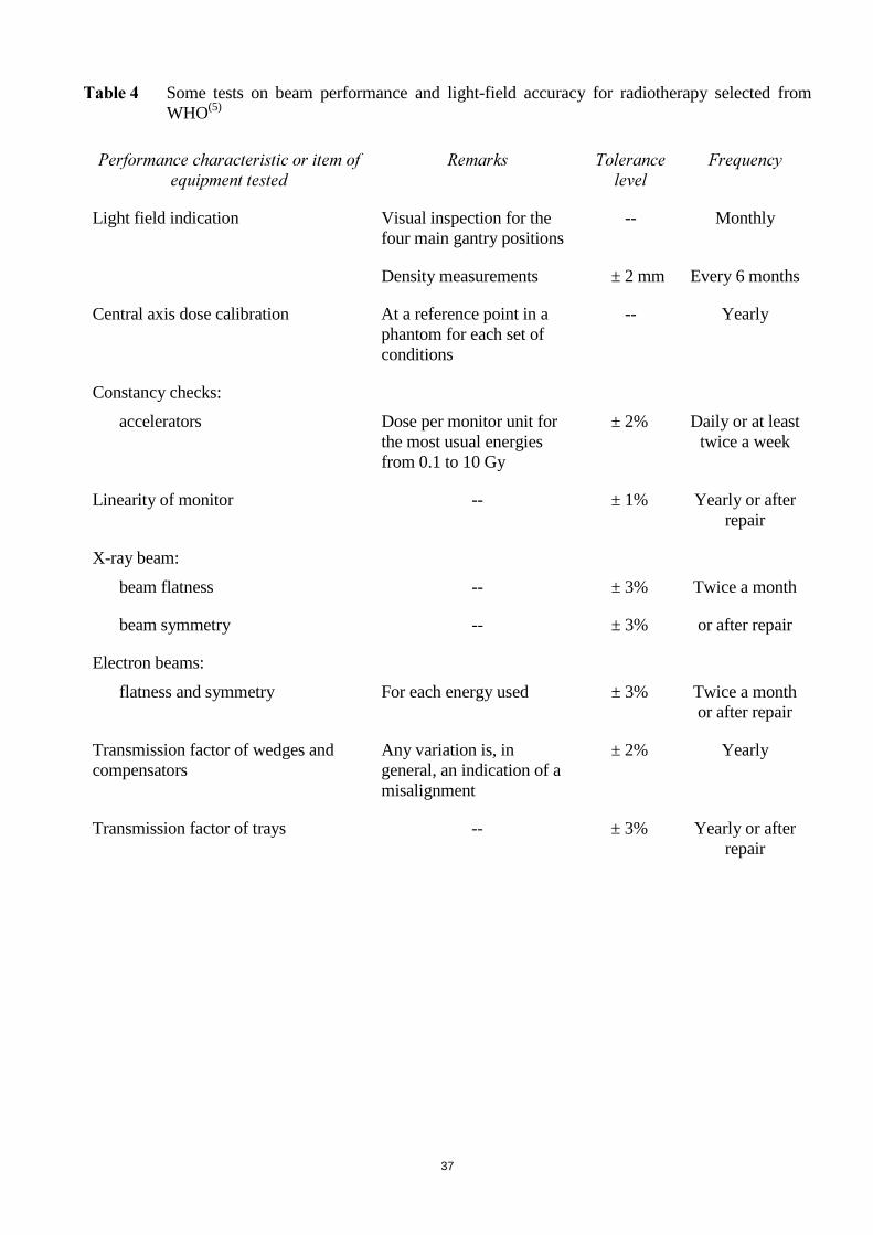
37
7DEOH�� Some tests on beam performance and light-field accuracy for radiotherapy selected fromWHO(5)
3HUIRUPDQFH�FKDUDFWHULVWLF�RU�LWHP�RIHTXLSPHQW�WHVWHG
5HPDUNV 7ROHUDQFHOHYHO
)UHTXHQF\
Light field indication Visual inspection for thefour main gantry positions
-- Monthly
Density measurements ± 2 mm Every 6 months
Central axis dose calibration At a reference point in aphantom for each set ofconditions
-- Yearly
Constancy checks:
accelerators Dose per monitor unit forthe most usual energiesfrom 0.1 to 10 Gy
± 2% Daily or at leasttwice a week
Linearity of monitor -- ± 1% Yearly or afterrepair
X-ray beam:
beam flatness -- ± 3% Twice a month
beam symmetry -- ± 3% or after repair
Electron beams:
flatness and symmetry For each energy used ± 3% Twice a monthor after repair
Transmission factor of wedges andcompensators
Any variation is, ingeneral, an indication of amisalignment
± 2% Yearly
Transmission factor of trays -- ± 3% Yearly or afterrepair

38
7DEOH�� Intercomparison of recommended test frequencies and tolerance levels for the photon fieldflatness and symmetry of linear accelerators(20)
5HSRUW )UHTXHQF\� 7ROHUDQFH�OHYHO
IODWQHVV V\PPHWU\
Ref. 12 M ±2% ±3%
Ref. 13 M ±4% (±2.5)% --
W -- ±3% (±1.5%)
Ref. 14 A1 -- --
Ref. 17 W2 ±3% (≤30 cmx30cm) ±3%
±5% (>30cmx30cm)
Ref.18 W ±1.5% ±3%
Ref. 20 W/M ±3% (±2%) --
Ref. 21 A ±3% --
M -- ±3%
Ref. 19 M ±3% ±3%1 Eight measurements of the “5-point” test and four measurements of the scan test2 One energy each week with alternating four gantry angles with four collimator angles3 W = weekly; M = monthly; A = annually
7DEOH�� Equipment subjected to QC in nuclear medicine in the Netherlands(22)
• Gamma camera planar
• Gamma camera whole body scanning
• Gamma camera SPECT
• Imaging equipment
• Scintillation counter (thyroid probe)
• Surgical detector/probe
• Dose calibrator
• Semi-conductor detector
• Flood source

39

40

41
4XHVWLRQV��4����$QVZHUV��$����5HPDUNV��5�UHOHYDQW�WR�WKH�VXEMHFW�SUHVHQWHG�E\�0U��=2(7(/,()
4� Would it be useful to harmonise quality control programs in Europe?
$� Yes, I am totally in favour of harmonization of quality control programs in Europe, especiallyif no such programs are available at national level.
4� No European guide on quality control for radiotherapy equipment exists for the moment.Would it not be a good idea to use the AAPM Report 46 entitled ‘Comprehensive qualityassurance for radiation oncology’?
$� The AAPM report is a very good document and can be used as reference document. However,in some Member States protocols on radiotherapy exist already and it also seems useful todevelop European guidelines in addition to that.
4� Referring to Article 8.6 of the MED, what is exactly the device informing the practitioner of thequantity of radiation produced? Could an mAs-meter be used for this purpose?
$� What a practitioner ideally should be informed of is the cumulated effective dose or, in case ofinterventional radiology, also the maximum dose to the patient’s body. An mAs-meter does notindicate these quantities but, by calculating the conversion between the two measurements, itmight in practice be possible to use an mAs-meter.
4� What is the suggested policy for the future use of single-phase fully-rectified generators?
$� I do not think it is to be expected that this type of generators will fulfil the nationalrequirements, if available, or the European criteria for acceptability of equipment.
4� Does all medical equipment in use have to comply with the Medical Devices Directive andshould it bear the CE mark?
$� The Medical Devices Directive refers to new equipment whilst the Medical Exposure Directiverefers to equipment in use. The answer to the question is no.
4� Don’t you think that too stringent quality control programs might impair the access to certainexaminations or treatments in radiology or nuclear medicine which could provoke a setback forpatient’s health?
$� That could theoretically be possible. Therefore, as I said before, we should be careful with ourdefinitions of suspension level or of an acceptability criterion. For example, some radiotherapyinstallations, no longer accepted for curative use, may be used under specified conditions togive palliative treatment.
4� Large quality control programs can put a heavy workload on the staff of a department. Do youthink this can be a restriction on introducing such programs?
$� Hospitals often are subject to budget restrictions. A large radiology department doing about100,000 examinations per year needs 1 full-time person to perform control checks. There issome reluctance to spend this money on someone who only checks the performance ofequipment. Moreover, many radiologists do not like to have somebody in the department whois ‘always looking over their shoulder when they are doing their job’. If there is no legalobligation, quality control is clearly not a priority.

42
4� You said image quality and dose are the two relevant parameters to be taken into account. Doyou agree that phantoms can be sufficiently relevant to evaluate image quality in diagnosticradiology?
$� Useful phantoms exist but what still needs to be achieved is to relate image quality asdetermined by radiologists with the physical methods behind this image to convince theradiologist that a better physical ‘performance’ also results in better diagnostic images.

43
5DGLDWLRQ�3URWHFWLRQ�LQ�PHGLFDO�DQG�ELRPHGLFDO�UHVHDUFK
)UDQFLV�3��&5$:/(<(WKLFV�:RUNLQJ�3DUW\
(XURSHDQ�)RUXP�IRU�*RRG�&OLQLFDO�3UDFWLFH
8QLYHUVLW\�RI�%UXVVHOV.HVVHO���%HOJLXP
7KH�SULQFLSOH�RI�PHGLFDO�DQG�VXUJLFDO�PRUDOLW\�FRQVLVWV�LQ�QHYHUSHUIRUPLQJ�RQ�PDQ�DQ�H[SHULPHQW�ZKLFK�PLJKW�EH�KDUPIXO�WR�KLP�WR�DQ\H[WHQW��HYHQ�WKRXJK�WKH�UHVXOW�PLJKW�EH�KLJKO\�DGYDQWDJHRXV�WR�VFLHQFH�
L�H���WR�WKH�KHDOWK�RI�RWKHUV�&ODXGH�%HUQDUG������
.H\ZRUGV
Radiation medicine, biomedical ethics, biomedical research, health policy
6XPPDU\
This paper situates the ‘Medical Exposures’ Directive within the framework of biomedical ethics. Itstresses the importance of ethical conduct in biomedical research in order to respect and maintainindividual and public trust in the use of radiation in biomedical research. After locating the researchreferred to in the Directive within the broader field of biomedical research in general, the paperexamines the importance of the principles of respect, beneficence, and justice for biomedical researchinvolving exposure to radiation. Following a consideration of the ethical importance of risk in theresearch setting involving radiation exposure, there is a discussion of the importance of informedconsent and ethical review in protecting research subjects. The importance of the ‘Medical Exposures’Directive is situated within the framework of an evolving European Community public health policy isdiscussed in the conclusion. Finally, a recommendation is made for the establishment of an expertgroup to draft guidelines on informed consent and ethical review for research projects involving theexposure of persons to ionising radiation.
,QWURGXFWLRQ
Medical and biomedical research on human subjects involving ionising radiation poses specific ethicalchallenges requiring scientific, procedural, and legal approaches to their resolution. Council Directive97/43/EURATOM of 30 June 1997 (the ‘Medical Exposures’ Directive)1, 2 imposes for the first time inEuropean Union law specific protection for healthy individuals and patients participating in medicaland biomedical research involving radiation. While the scope of the Directive is much broader, givingparticular attention to increased protection to patients, health workers, and other individuals3 exposedto radiation in standard medical and medico-legal procedures, the specific concern with medical andbiomedical research has special significance. 1 The author wishes to thank Diederik Teunen of the European Commission, DG XI.C.1 Radiation Protection for his
guidance and support in the preparation of this paper. Additionally, Professor Joseph J. Hoet of the WHO CollaboratingCentre, Faculty of Medicine, Université Catholique de Louvain, provided valuable advice regarding issues of scienceand ethics discussed below. Any remaining errors or shortcomings of the paper are attributable solely to the author.
2 Council Directive 97/43/EURATOM of 30 June 1997on health protection of individuals against the dangers of ionizingradiation in relation to medical exposure, and repealing Directive 84/466/Euratom. (OJ L-180 of 9 July 1997).
3 The more direct coverage of health workers and the general public is covered in Council Directive 96/29/EURATOM of13 May 1996 laying down basic safety standards for the health protection of the general public and workers against thedangers of ionizing radiation. (OJ L-159 of 05/10/84 page 1).

44
The exposure of individuals to radiation in research settings requires a legal framework that promoteshigh scientific standards and establishes rigorous ethical guidelines. The Directive provides afundamental basis on which to build and maintain public trust in medical experimental procedureswhere participants are exposed to radiation. The implementation of this Directive requires reflection onthe ethical conduct of medical and biomedical research, while at the same time it engages a furtherdevelopment of high ethical and scientific standards in the interest of public health.
7UXVW�DQG�+XPDQ�5DGLDWLRQ�([SHULPHQWV
The primary purpose of the Directive is stated in Article 1.1. as follows:
This Directive supplements Directive 96/29/Euratom and lays down the general principles of radiation protectionof individuals in relation to . . . exposure.
The development and implementation of well considered legal principles for the protection ofEuropean citizens in biomedical research is important in order to foster trust in research. At the heartof biomedical research, involving radiation or any other form of medical intervention, is a relationshipof confidence between a patient and her physician (or other health care professional). This is arelationship in which an individual entrusts not just bodily health to a caring professional, but indeedone’s general sense of well-being. The patient trusts that, in this very private and confidentialrelationship, her well-being will be the predominant, if not sole, concern of the health care provider.Biomedical research introduces a peculiar challenge to this relationship because it requires that thephysician’s interest also be directed, in part, towards the general concerns of science. By bringingexplicitly under its scope the protection of individuals ‘in medical or biomedical, diagnostic ortherapeutic, research programmes’, the Directive reinforces the primacy of the physician-patientrelationship over the researcher-subject relationship.
One of the most decisive factors for patients entering biomedical research is the fact that they haveconfidence in the advice of their physicians. In the article ‘Trust: The Fragile Foundation ofContemporary Biomedical Research’ the authors document the force of trust in the physician-patientrelationship with anecdotes of the following sort:
‘My doctor told me if I do not take the drug, in a couple of months I [will] die. So, I had no choice. Who wantsto die? Nobody.’‘Well, he [the oncologist] said he’d already ‘been through everything he knew what to do. He would try to keepme as comfortable as he could. That’s when he told me about this new treatment. I told him we would try it.’‘There’s not a lot that you can control when you’re sick, so you have to trust your doctors . . . if he suggests thatyou should go into a research project, I think you should really take his advice or her advice . . . because if youtake the time to get yourself a good doctor and they’re involved in research, they would never steer you wrong.’‘[T]o me, they are the doctors, and once I trusted them . . . it was pretty much up to them.’4
Patients are in an essentially vulnerable, dependent relationship with their physician. Their readiness totrust is understandable, given the weakness of their condition and the power of the physician to treatthat condition. It is essential that in the process of discussion and decision-making with the patient thatthe physician respond with integrity regarding a proposed treatment’s potential benefits and potentialrisks. In the case of research, the physician-researcher may encounter extraordinary moral challengesin maintaining the integrity of their interactions with the patient (potential subject). This challenge mayarise because of the physician-investigator’s interest in science and the possibilities for benefitingfuture patients. However, it may also arise out of the physician-investigator’s interest in the patient andthe potentially bleak outlook on a specific health problem. Respecting the trust of the relationshipdepends on maintaining an integrity that is responsive to the patient’s trust.
4 Nancy E. Kass, Jeremy Sugarman, Ruth Faden, and Monica Schoch-Spana, “Trust, The Fragile Foundation of
Contemporary Biomedical Research,” +DVWLQJV�&HQWHU�5HSRUW 26.5: September-October 1996. Pp. 5-10

45
Additionally, trust is at the heart of the public’s confidence in government health policy and themedical profession in general. The effective implementation and development of (public) healthpolicies and medical practices requires an open dialogue with all the different sectors of societyinvolved in health care, including the patients. Helen Carter of the Research Trust for MetabolicDiseases in Children, United Kingdom, recently expressed this concern at a meeting in the EuropeanParliament as follows:
We live in exciting times. We have unprecedented opportunities for industry, patient groups and governments towork together to develop our economies, strengthen industry and promote and enable our health and well being.5
The dialogue between the different sectors of society in health care should be based on a concern topromote medical and biomedical practices that are safe and efficacious, permitting only a minimallevel of risk. As is indicated in the draft document entitled ‘Guidance on Medical and BiomedicalResearch for the Working Party on Medical Exposures’6, radiation experiments are of specific concernin the area of public trust in health policy:
A comprehensive culture of radiation protection and safety in medicine has been progressively developingthroughout the European Union with regard to the medical use of ionising radiation and has been integrated intothe various branches of diagnosis and treatment.
The European Commission has contributed to this evolution with the establishment of legal requirements to beimplemented by Member States for the radiation protection of persons undergoing medical examinations ortreatment. (p. 1)
The ‘Medical Exposures’ Directive makes a particularly important contribution to this protection bothincluding medical and biomedical research within its scope and by providing guidance for radiationresearch similar to the guidance legislated at the European level for clinical trials involvingpharmaceuticals and for research involving medical devices. A ‘comprehensive culture of radiationprotection and safety’ needs to be situated within, and co-ordinated with, the wider culture of medicaland biomedical research concerned with public health.
There is a recent and disturbing historical memory of experimentation on human beings involvingradiation that has been extensively documented in the United States by the Advisory Committee onHuman Radiation Experiments7. The Committee’s )LQDO�5HSRUW shows in exhausting detail repeatedabuses of human subjects in radiation experiments, largely by the government, military, academic, andhospital communities between 1944 and 1974. The experiments were carried out during the period ofthe Cold War usually under a veil of secrecy (often understood as ‘national security’) with therationale that the national interest and the general public welfare were at stake. Testifying before theCommittee on Governmental Affairs of the US Senate in the Hearing on Radiation and OtherExperimentation on Human Beings in 1994, an expert methodologist in the design and conduct ofclinical trials, Curtis Meinert, stated the following:
The recent flurry of events, and accompanying publicity, regarding radiation studies done in the past has createda crisis of confidence that extends far beyond the specifics of radiation research. It is a crisis that concerns us allwhatever our perspective – that of a research subject, a researcher, citizen, or human being. Hence, it isappropriate to have a public airing of the specifics, and of the general issues involved, to restore the trust of thepublic in the collective research enterprise where indicated, and to correct deficiencies where noted. We will nothelp anyone, starting with those we treat, if this debate leads the public to believe that they are likely to beunwitting guinea pigs every time they enter a health care facility. We have a crisis of confidence that, unlessdealt with forthrightly and resolutely, will affect us all, to the extent that we depend on mutual trust and respect
5 Helen Carter, “Rare Diseases: The Availability of Cures and the 5th Framework Programme,” %LRPHGLFDO�5HVHDUFK�DQG
2USKDQ�0HGLFLQDO�3URGXFWV, Hessel Dijkstra, Erica Poot, and Ilse Wilczek, eds. (Baarn, The Netherlands: EuropeanPlatform for Patients’ Organisations, Science and Industry, 1998).
6 Working Group – Article 31, ‘Guidance on Medical and Biomedical Research for the Working Party on MedicalExposures,’ Draft document, DGXI-1, 10 April 1998.
7 Advisory Committee on Human Radiation Experiments, )LQDO�5HSRUW (US Government Printing Office, October 1995).

46
in carrying out any research on human beings. The ability to do such work is, itself, a form of public trust andviolations of that trust have the potential of pushing us back to the era [of] nonexperimental empiricism inchoosing the treatments in use.8
The ‘crisis of confidence’ Meinert refers to is perhaps considerably less in Europe than in the UnitedStates.9 However, the potential for abuses to occur in medical research – while actual occurrencestoday are perhaps few in number – demands a high level of public responsibility in reviewing,authorising, and monitoring biomedical research.
'HILQLQJ�%LRPHGLFDO�5HVHDUFK
Certainly many of the standard practices today involving radiation medicine can be improved upon,and it is likely that as our understanding of human biology and nuclear medicine increases newpractices will be proposed using radiation in diagnostic and therapeutic health interventions. Therevision of standards or the introduction of new practices in biomedicine requires research on personsfor their justification.
Biomedical research may be broadly defined as any systematic investigation in the field of healthintended to develop or contribute to medical understanding and practices. This includes all testing,evaluation, and development of medical products (pharmaceuticals), medical devices, surgicaltechniques, and radiation exposure. Falling clearly within the scope of this definition are allexperimental interventions involving persons.
This definition includes all research done on or relating to people or their body fluids or tissues, as well as thatdone on records or data on human beings, even if the specific project in question does not involve any contactwith persons to whom the records or data pertain.10
Thus, biomedical research involving persons has a broad scope, and we need to be aware of this whenwe consider the application of ethical principles. Neither the World Medical Associations’ (WMA)'HFODUDWLRQ�RI�+HOVLQNL��, the Council for International Organizations of Medical Sciences’ (CIOMS),QWHUQDWLRQDO� (WKLFDO� *XLGHOLQHV� IRU� %LRPHGLFDO� 5HVHDUFK� ,QYROYLQJ� +XPDQ� 6XEMHFWV��, nor theEuropean &RQYHQWLRQ� RQ� +XPDQ� 5LJKWV� DQG� %LRPHGLFLQH�� provides a definition of biomedical (ormedical) research, although all of these internationally accepted ethical guidelines are to be applied tobiomedical research.
However, within the definition of biomedical research lies a complex dilemma: the inherent freedomof the inquiring mind versus the privilege of carrying out research on persons. Most often we see oneside or another of this dilemma put forward, which then bears directly on the scope of what biomedical
8 Reprinted in Advisory Committee on Human Radiation Experiments, )LQDO�5HSRUW, Supplementary Volume 1, p. 822.9 Yet there is good reason to believe that human radiation experiments were also widely undertaken in European
countries. For one example, see Campaign for Nuclear Disarmament, “British Nuclear Guinea-Pigs: Human RadiationExperiments in Britain from 1957 to the Current Day” (London: CND Information Office, November 1996). Alsoavailable on the World Wide Web at http://www.mcb.net/cnd/radexpts/report.htm.
10 Curtis L. Meinert, “Hearing on Radiation and Other Experimentation on Human Being[s],” 20 January 1994,Testimony, reprinted in Advisory Committee on Human Radiation Experiments, )LQDO�5HSRUW, Supplementary Volume1, p. 824. See also United States Department of Health and Human Services, 45 CRF §46.102(d); 18 June 1991.
11 World Medical Association,� :RUOG� 0HGLFDO� $VVRFLDWLRQ� 'HFODUDWLRQ� RI� +HOVLQNL�� 5HFRPPHQGDWLRQV� *XLGLQJ3K\VLFLDQV�LQ�%LRPHGLFDO�5HVHDUFK�,QYROYLQJ�+XPDQ�6XEMHFWV. Adopted by the 18th World Medical Assembly Helsinki,Finland, June 1964. Amended by the 29th World Medical Assembly, Tokyo, Japan, October 1975; 35th World MedicalAssembly, Venice, Italy, October 1983; 41st World Medical Assembly, Hong Kong; and the 48th General Assembly,September 1989. Somerset West, Republic of South Africa, October 1996.
12 Council for International Organizations of Medical Sciences (CIOMS), in collaboration with the World HealthOrganization (WHO). ,QWHUQDWLRQDO� (WKLFDO�*XLGHOLQHV� IRU� %LRPHGLFDO� 5HVHDUFK� ,QYROYLQJ�+XPDQ� 6XEMHFWV. Geneva1993.
13 Council of Europe (Directorate of Legal Affairs). &RQYHQWLRQ�IRU�WKH�3URWHFWLRQ�RI�+XPDQ�5LJKWV�DQG�'LJQLW\�RI�WKH+XPDQ�%HLQJ�ZLWK�5HJDUG�WR�WKH�$SSOLFDWLRQ�RI�%LRORJ\�DQG�0HGLFLQH��&RQYHQWLRQ�RQ�+XPDQ�5LJKWV�DQG�%LRPHGLFLQH.European Treaty Series – No. 164. Oviedo, 4 November 1997.

47
research may be. The European &RQYHQWLRQ�RQ�+XPDQ�5LJKWV�DQG�%LRPHGLFLQH comes squarely downon the side of freedom, never referring to the privilege of research. Article 15 reads as follows:
Scientific research should be carried out freely, subject to the provisions of this Convention and the other legalprovisions ensuring the protection of the human being.
Although the freedom of research is not absolute, its limitations are strictly legal in nature. Thejustification for this ‘General Rule’ is provided by the supporting ([SODQDWRU\�5HSRUW:
Freedom of scientific research in the field of biology and medicine is justified not only by humanity’s right toknowledge, but also by the considerable progress its results may bring in terms of the health and well-being ofpatients.14
The ‘right to knowledge’ guarantees the researcher broad freedom in the design and execution ofbiomedical research projects. Further, this right needs to be upheld in order to prophylactic, diagnostic,and therapeutic treatment at the disposal of standard medicine for addressing the health needs of thepopulation.
On the other hand, it is becoming increasingly common today for ethicists and experts in biomedicalresearch to emphasise the privilege of biomedical research on human subjects. Curtis Meinert writesthe following:
I argue that being able to carry out research on human beings is a privilege, not a right . . . .15
Those emphasising the privilege of biomedical research stress the importance of the consideration ofthe safety and well-being of the research subject over the interests of science. At times linked to thisclaim is the suggestion that biomedical researchers be certified or that their institutions be accredited.16
However, the dilemma of the freedom versus the privilege of biomedical research involving humansubjects need not lead to an impasse. One of the great challenges to biomedical ethics today is toensure that the freedom of research and the privilege of research compliment one another in practice.
The ‘Medical Exposures’ Directive regulates biomedical research in the European Union insofar as itinvolves the exposure of persons to ionising radiation. It does not attempt to define biomedicalresearch, nor does it address the freedom or the privilege of biomedical research. However, theDirective’s concern is clearly with a limited scope of biomedical research that can be termed ‘clinicalresearch’: biomedical research on persons within the setting of a medical clinic, or co-ordinated ordirected by a medical clinic, and aimed at testing the safety and/or efficacy of an intervention. The
14 Directorate of Legal Affairs, Council of Europe, ([SODQDWRU\�5HSRUW� WR� WKH�&RQYHQWLRQ� IRU� WKH�3URWHFWLRQ�RI�+XPDQ
5LJKWV�DQG�'LJQLW\�RI�WKH�+XPDQ�%HLQJ�ZLWK�5HJDUG�WR�WKH�$SSOLFDWLRQ�RI�%LRORJ\�DQG�0HGLFLQH��&RQYHQWLRQ�RQ�+XPDQ5LJKWV�DQG�%LRPHGLFLQH� DIR/JUR 5. Strasbourg, May 1997, p. 23.
15 Curtis L. Meinert, Letter to Ruth R. Faden, Chair of the United States Advisory Committee on Human RadiationExperiments, 24 February 1995, published in Advisory Committee on Human Radiation Experiments, )LQDO�5HSRUW,Supplementary Volume 1, p. 820. This argument finds support in the 3ULQFLSOHV� IRU� 7KRVH� LQ� 5HVHDUFK� DQG([SHULPHQWDWLRQ�of the World Medical Association (adopted by the Eighth General Assembly of the World MedicalAssociation, 1954) which states in its first principle:1. Scientific and Moral Aspects of Experimentation
The word experimentation applies not only to experimentation itself but also to the experimenter. An individualcannot and should not attempt any kind of experimentation. Scientific qualities are indisputable and must alwaysbe respected. Likewise, there must be strict adherence to the general rules of respect of the individual.
Here the very definition of a valid (bio)medical experiment infers the privilege of the undertaking.16 On the same subject, Ernest Prentice points to the following situation in the United States:
It is ironic to think that the American Association for the Accreditation of Laboratory Animal Care (AAALAC)has accredited 550+ institutions and this accreditation requires submission of a detailed self-study and AAALACsite visit [sic] every 3 years. We do not, however, have a comparable system for accreditation of institutionsinvolved in human subject research.
See Ernest D. Prentice, Letter to Ruth R. Faden, Chair of the United States Advisory Committee on Human RadiationExperiments, 24 February 1995, published in Advisory Committee on Human Radiation Experiments, )LQDO�5HSRUW,Supplementary Volume 1, p. 832.

48
Good Clinical Practice Guideline of the International Conference on Harmonization (ICH) defines aclinical trial/study as follows:
Any investigation in human subjects intended to discover or verify the clinical, pharmacological and/or otherpharmacodynamic effects of an investigational product(s) and/or to identify any adverse reactions to aninvestigational product(s), and/or to study absorption, distribution, metabolism, and excretion of aninvestigational product(s) with the object of ascertaining its safety and/or efficacy. The terms clinical trial andclinical study are synonymous.17
While this definition was written in the context of biomedical research involving pharmaceuticals, itseems that it would be largely applicable to biomedical research involving exposure to radiation.
The ‘Medical Exposures’ Directive applies to all biomedical research in which persons are exposed toionising radiation. Article 1.2 establishes this in broad lines:
This Directive shall apply to the following medical exposure:. . . .
(d) the exposure of healthy individuals or patients voluntarily participating in medical or biomedical, diagnosticor therapeutic, research programmes.
The requirements of the Directive thus apply to all instances of research where there is exposure toionising radiation, whether or not the safety or efficacy of the exposure is to be a measured result ofthe research. These would also apply to research involving dosage studies, whether studyingexperimental dose quantities or comparing standard dose quantities. They apply equally to biomedicalresearch involving either external radiation or radiopharmaceuticals. The Directive also applies toresearch involving the testing of medical or biomedical equipment where there is exposure to (or thepotential for exposure to) radiation, e.g., radiographic equipment, mammographic equipment, imageintensifiers, digital fluoroscopy systems, or computed tomography installations. In addition, theDirective applies to a wide range of studies where evaluating the efficacy of a pharmaceutical, device,or surgical intervention requires radiation exposure.
7KH�3XUSRVH�RI�%LRPHGLFDO�5HVHDUFK�,QYROYLQJ�([SRVXUH�WR�5DGLDWLRQ
Biomedical research is generally understood to have as its purpose the improvement of knowledgeregarding the health of individuals or groups in society. The 'HFODUDWLRQ� RI� +HOVLQNL provides thefollowing guidance:
The purpose of biomedical research involving human subjects must be to improve diagnostic, therapeutic andprophylactic procedures and the understanding of the aetiology and pathogenesis of disease.
The 'HFODUDWLRQ insists that biomedical research on persons may only be carried out with the aim ofdeveloping or contributing to current biomedical knowledge. This imposes a strong responsibility onthe researcher to be informed of the current state of understanding in her field, and to ensure thatrepetitive, redundant, or in some other way ill-informed or ill-advised research proposals regarding thescientific community’s current state of knowledge are not undertaken. Biomedical research on humansubjects that is not expected to advance current scientific knowledge is considered to be unethical, nomatter how benign. Poorly designed (or poorly conducted) research is without scientific or ethicalmerit. Even where minimum risk is present it should not be permitted.
The ‘Medical Exposures’ Directive provides for clear guidance regarding the situations in whichbiomedical research exposing persons to radiation is acceptable. Article 3.1 (a) states the following:
all new types of practices involving medical exposure shall be justified in advance before being generallyadopted,
17 International Conference on Harmonisation of Technical Requirements for the Registration of Pharmaceuticals for
Human Use (ICH). *XLGHOLQH�IRU�*XLGDQFH�RQ�*RRG�&OLQLFDO�3UDFWLFH (CPMP/ICH/135/95) 1 May 1996.

49
existing types of practices involving medical exposure may be reviewed whenever new, importantevidence about their efficacy or consequences is acquired.
Research is required in order to justify either new types of interventions, or to review or modifyexisting practices. The Directive places a strong emphasis on the need to justify new or modifiedpractices, and this seems to require research. It continues in paragraph 3. of the same article byinsisting
If an exposure can not be justified, it should be prohibited.
The justification for the introduction of new interventions or modification of existing ones wouldrequire biomedical research, which, in all cases, would require exposure to persons. However, as isimplied by the same article’s insistence on ethical review, the research that would justify standardpractices needs to be justified itself in advance of its being performed.
The 'HFODUDWLRQ�RI�+HOVLQNL provides a clear statement on this:
The design and performance of each experimental procedure involving human subjects should be clearlyformulated in an experimental protocol which should be transmitted for consideration, comment and guidance toa specially appointed committee independent of the investigator and the sponsor provide that this independentcommittee is in conformity with the laws and regulations of the country in which the research experiment isperformed.
The essential justification for a biomedical research project needs to be clearly defined and motivatedin a protocol. The protocol should be designed according to current scientific standards and it shoulddemonstrate
1. a thorough understanding of the current level of scientific knowledge in the field,2. a need for the research (including a need to carry out the research on persons),3. the identity and qualifications of the researchers,4. a statement of the objective of the research,5. a statement of the hypothesis to be examined,6. a well defined methodology for examining the hypothesis,7. a well defined method for radiation dose calculation (for radiation research),8. a statement of the potential risks,9. a well considered ethical appreciation of the risks and inconveniences for the participants,
a means for evaluating the data,10. the inclusion/exclusion criteria for potential research subjects,11. procedures for the ongoing monitoring of the research,12. criteria for the discontinuation of individual subject participation,13. criteria for the discontinuation of the study as a whole,14. a plan for the communication of the results of the research.
Responsible biomedical research is never a freewheeling, unstructured activity. No matter howinnovative or seemingly promising, the departure from standard or accepted practice by a medicalpractitioner does not by itself constitute biomedical research.18 Research requires a critical evaluation
18 The ‘Medical Exposures’ Directive leaves open to practitioners the possibility to deviate from standard or accepted
practices in individual cases. Article 3.1 (b), in the second paragraph, reads as follows:If a type of practice involving a medical exposure is not justified in general, a specific individual exposure of thistype could be justified in specific circumstances, to be evaluated on a case-by-case basis.
Certainly no single case of a deviation from standard medical practice could be considered research. The example of aspecific case could not by itself contribute to general knowledge. More importantly, while such a deviation might beenvisaged (for example, in an effort to provide the best care to a terminally ill patient) it could not be justified in anyscientific sense beyond the judgement, however professional or expert, of those taking the decision. How the proposedevaluation is to be made in such specific cases, the Directive does not address. Even if this sort of deviation from

50
of the problem to be addressed and a well prepared plan for assessing a proposed intervention. Alreadyin 1979 the %HOPRQW�5HSRUW stated in summary fashion:
[T]he term research designates an activity designed to test an hypothesis, permit conclusions to be drawn, andthereby to develop or contribute to generalizable knowledge (expressed, for example, in theories, principles, andstatements of relationships). Research is usually described in a formal protocol that sets forth an objective and aset of procedures designed to reach that objective.19
New practices or modifications of standard practices in radiation medicine require justification throughresearch, i.e., clear demonstrations that the practices to be introduced are safe and efficacious. At thesame time, the research itself needs to be justified in advance in order to show that it is ethicallyresponsible and scientifically sound.
$SSO\LQJ�(WKLFDO�3ULQFLSOHV�WR�%LRPHGLFDO�5HVHDUFK�,QYROYLQJ�5DGLDWLRQ
Today it is becoming increasingly clear that good science is always essentially ethical. Scientificresearch that is sound and well considered takes into account, not only the interests of building uponthe general body of knowledge shared by the international community of scientists, but also theinterests and welfare of those involved in the research and those for whom the research will have adirect benefit. High scientific thought and conduct is not distinguishable from high ethical thought andconduct. In recent years ethical principles have been introduced as part of accepted biomedicalresearch. These principles are firmly rooted in Western moral philosophy, and they are considered tobe universal in their application:
• The principle of respect for persons• The principle of beneficence• The principle of justice
These principles were first described in relation to the ethics of research in the %HOPRQW�5HSRUW andthey were later articulated in much the same fashion in the CIOMS’ ,QWHUQDWLRQDO�(WKLFDO�*XLGHOLQHVIRU� %LRPHGLFDO� 5HVHDUFK� ,QYROYLQJ� +XPDQ� 6XEMHFWV. While the European &RQYHQWLRQ� RQ� +XPDQ5LJKWV�DQG�%LRPHGLFLQH does not enunciate these principles, they clearly form the underlying ethicalstructure of the document.
In the context of biomedical research, these principles function as a guide to the conscience of theresearcher in the preparation of a research protocol and in the activity of carrying out the research. Inaddition, through the demonstration of their application, they serve as a justification for the ethicalintegrity of a biomedical research project. The principles can be explained in their relationship tobiomedical research as follows:
RESPECT FOR PERSONS
The principle of respect for persons derives from the fundamental recognition of the dignity of eachand every person. Each individual deserves to be treated with respect because of the dignity of theirperson. Perhaps best formulated and described in the philosophy of Immanual Kant, the dignity of aperson refers to the capacity for independent and autonomous decision making. The respect due everyperson, in biomedical research as well as in all other activities of human life, is a fundamentalrecognition of their freedom in thought and self-determination, whether or not they enjoy the
standard practice would be highly exceptional, which one would expect, there remains the real problem of establishingin advance an appropriate mechanism for evaluating such types of practice.
19 Department of Health, Education, and Welfare, Office of the Secretary, Protection of Human Subjects. %HOPRQW�5HSRUW�(WKLFDO�3ULQFLSOHV�DQG�*XLGHOLQHV�IRU�WKH�3URWHFWLRQ�RI�+XPDQ�6XEMHFWV�RI�5HVHDUFK��5HSRUW�RI�WKH�1DWLRQDO�&RPPLWWHHIRU�WKH�3URWHFWLRQ�RI�+XPDQ�6XEMHFWV�RI�%LRPHGLFDO�DQG�%HKDYLRXUDO�5HVHDUFK. DHEW Publication No. (OS) 78-0013and No. (OS) 78-0014. 18 April 1979. p. 3.

51
capacities or the circumstances to exercise that freedom. Respect for the dignity of the human person isthe unquestionable foundation for ethical conduct in biomedical research.
In the European &RQYHQWLRQ�RQ�+XPDQ�5LJKWV�DQG�%LRPHGLFLQH, this principle underlies Article 1:
Parties to this Convention shall protect the dignity and identity of all human beings and guarantee everyone,without discrimination, respect for their integrity and other rights and fundamental freedoms with regard to theapplication of biology and medicine.
The principle of the respect for the dignity of the human person has led directly in the last fifty years tothe central role of the informed consent and ethical review in biomedical research. These importantstructures for the protection of the (potential) research participant are discussed later in this paper. Theprinciple of respect for the dignity of each and every person applies to all activities of biomedicalresearch. Its application supersedes all other considerations. There is no goal or achievement, inscience, politics, business, or any other activity, whose value or esteem exceeds the recognition of thefundamental dignity of the individual person. Even the lofty and praiseworthy pursuit of knowledge inthe service of humanity loses its force and is blasphemous to science when respect for the dignity of anindividual is shunned. The principle of respect gives force and honour to the activities of biomedicalresearch. It also underlies its two companion principles.
BENEFICENCE
The principle of beneficence derives from the obligation that persons be addressed in the interest oftheir well-being in each and every circumstance. This principle asserts that the aim of every medical orbiomedical intervention, whether in standard practice or in research, be aimed at promoting the health(the good) of the patient or recipient of the intervention. The first sentences of the 'HFODUDWLRQ� RI+HOVLQNL reinforce the importance of this principle in biomedical research:
It is the mission of the physician to safeguard the health of the people. His or her knowledge and conscience arededicated to the fulfillment of this mission.
Essentially, this principle asserts the obligation to maximise benefits and minimise risk. TheHippocratic maxim ‘do no harm’ applied to the situation of biomedical research requires the researcherto ensure that the risks of the research are reasonable in respect of the expected benefits.
However, it is the very nature of research that there is an area of the intervention where no currentknowledge exists regarding potential risks and benefits. In all medical and biomedical research thereare inherent risks, most of which – and the most threatening of which – will hopefully be seen inadvance of the undertaking of the research. Still the risks (foreseen and hidden) can only be properlyevaluated by well designed and executed research projects.
Learning what will in fact benefit may require exposing persons to risk. The problem posed by these imperativesis to decide when it is justifiable to seek certain benefits despite the risks involved, and when the benefits shouldbe foregone because of the risks.20
Biomedical research is continually challenged by the principle of beneficence. Concern with the healthand well-being of the individual research subject may appear at odds with the interest of science andthe need to advance biomedical understanding. Evaluating and balancing potential risks with potentialbenefits forms an essential task in the design and approval of biomedical research proposals.
In many biomedical situations, and certainly in the field of nuclear medicine, research and treatmentcan be very well joined. This is very often the case in attempts to find an improved diagnosis or 20 Department of Health, Education, and Welfare, Office of the Secretary, Protection of Human Subjects. %HOPRQW�5HSRUW�
(WKLFDO�3ULQFLSOHV�DQG�*XLGHOLQHV�IRU�WKH�3URWHFWLRQ�RI�+XPDQ�6XEMHFWV�RI�5HVHDUFK��5HSRUW�RI�WKH�1DWLRQDO�&RPPLWWHHIRU�WKH�3URWHFWLRQ�RI�+XPDQ�6XEMHFWV�RI�%LRPHGLFDO�DQG�%HKDYLRXUDO�5HVHDUFK. DHEW Publication No. (OS) 78-0013and No. (OS) 78-0014. 18 April 1979. p. 4.

52
treatment for a patient’s condition or for attempting to diagnosis or treat cases for which there is nostandard of care. In such cases the potential benefits justify the potential risks. However, in a great dealof biomedical research (for example, phase I radiopharmaceutical trials) healthy volunteers are thesubject of investigation, and this principle appears difficult, if not impossible, to apply. There are onlypotential risks, no potential health benefits for the research subject. Jay Katz posed the dilemma in thefollowing manner:
When, if ever, should conflicts between advancing medical knowledge for our benefit and protecting theinviolability of citizen-subjects of research be resolved in favour of the former?21
The 'HFODUDWLRQ�RI�+HOVLQNL tries to ensure that the interests of the physician-research, qua physician,will always take precedence over the interests of the physician-research, qua researcher. However,Katz indicates rightly that the ethical challenge is not so easily removed. Instead, he concludes with thefollowing response to the dilemma:
1. that any exception to the principle of individual autonomy, since it tampers with fundamental democraticfreedoms, must be rigorously justified by clear and sufficient reasons; and
2. that such exceptions cannot be made by investigators or IRBs [ethics committees] alone, but only by anauthoritative and highly visible body.22
Katz’s proposal that the dilemma can be addressed by justifying exceptions to the ethical principle inan open and publicly acceptable manner reinforces the importance of trust in biomedical research.However, it does not resolve the dilemma. It remains difficult, if not impossible, to find ‘clear andsufficient reasons’ for violating ethical principles in biomedical research. With respect to the principleof beneficence, where this violation appears most threatening in research involving radiation exposure,perhaps the best researchers and society can do at the present time is to remain aware of the dilemmaand vigilant in ensuring that it does not become an instrument for abuse.
JUSTICE
The principle of justice requires that biomedical researcher projects engage potential and actualresearch subjects equally and fairly according to the health needs of the subjects, not according to theireconomic means, race, creed, class, or some other form of societal entitlement. The 'HFODUDWLRQ� RI/LVERQ� RQ� WKH� 5LJKWV� RI� WKH� 3DWLHQW�� demonstrates a strong concern that the principle of justice isapplied, not only in standard medical care, but also in biomedical research. In the Preamble it makesthe following statement:
In the context of biomedical research involving human subjects – including non therapeutic biomedical research– the subject is entitled to the same rights and consideration as any patient in a normal therapeutic situation.24
21 Jay Katz, “Statement by Committee Member Jay Katz,” in Advisory Committee on Human Radiation Experiments,
)LQDO�5HSRUW (US Government Printing Office, October 1995). p. 849. Katz seems to limit the dilemma only to caseswhere full and satisfying informed consent has not been obtained from the citizen-subject. However, it is our contentionhere that the dilemma is broader, that it appears in many standard practices where informed consent is duly obtained.Having obtained informed consent (and/or favourable ethical review by an appropriate committee) does not release aninvestigator from any of the obligations of the ethical principles of biomedical research.
22 Jay Katz, “Statement by Committee Member Jay Katz,” in Advisory Committee on Human Radiation Experiments,)LQDO�5HSRUW (US Government Printing Office, October 1995). p. 853.
23 World Medical Association,�:RUOG�0HGLFDO�$VVRFLDWLRQ�'HFODUDWLRQ�RI /LVERQ�RQ�WKH�5LJKWV�RI�WKH�3DWLHQW. Adopted bythe 34th World Medical Assembly, Lisbon, Portugal, September/October 1981 and amended by the 47th GeneralAssembly, Bali, Indonesia, September 1995.
24 This is an important statement in the consideration of research subjects. Indeed the idea may be better stated moreforcefully: All potential subjects in biomedical research, including healthy volunteers, are to be considered as patientsonce enrolled in biomedical research, entitled to the same rights and considerations as any other patient in a research orstandard care situation. It would be irresponsible for a physician or other health care professional to expose individualsto risks for the sake of research without at the same time admitting them into their care.

53
The 'HFODUDWLRQ�RI�/LVERQ continues with the following principles:
1. Right to medical care of good qualitya. Every person is entitled without discrimination to appropriate medical care.c. The patient shall always be treated in accordance with his/her best interests. The treatment applied
shall be in accordance with generally approved medical principles.e. In circumstances where a choice must be made between potential patients for a particular treatment
which is in limited supply, all such patients are entitled to a fair selection procedure for that treatment.That choice must be based on medical criteria and made without discrimination.
One of the concerns for the principle of justice in biomedical research has to do with the fair selectionof research subjects. On the one hand, this concerns the equal possibility for persons to be included inbiomedical research projects, particularly with respect to their health conditions. In recent yearspatients have become better informed regarding the potential for participating in clinical research andthey have become increasingly more proactive in asserting their ‘right’ to participate. This has beenmost forcefully illustrated among communities of persons living with HIV/AIDS around the world.Biomedical research has also been faulted for its demographic make-up. Women and minority cultureshave felt themselves discriminated against in the designs of protocols and the recruitment of subjects.The principle of justice, thus, concerns the fair distribution of the opportunity to participate in researchamong individuals and across society.
On the other hand, the principle of justice concerns the maintenance of a vigilant guard against theinclusion of persons because of a vulnerable situation in which they find themselves. Vulnerabilityhere refers to situations of an institutionalised hierarchy (e.g., persons in prisons, the military, hospitalor academic settings), of a compromised position (e.g., persons who are poor, who belong to minoritycultures, or who are institutionalised), or of a physical, intellectual, or emotional impairment (e.g.,persons who are unconscious, who are mentally or emotionally handicapped, or children andadolescents). With respect to vulnerability we need also to take into account persons who are in asituation that is easily manipulated or persons who are simply available. In these last two groups, weshould not forget, we find patients:
Patients have always been the most vulnerable group for purposes of research.25
Patients are usually the most readily available and most easily manipulated group for the recruitingbiomedical researcher. Patients are notably not only interested in participating in research because ofthe hope for the amelioration of their own conditions, but they are very often anxious to contribute topreventing future patients/persons from suffering the same illnesses and pains they themselvesexperienced.
The principle of justice is also concerned with the distribution of biomedical research within societiesand across societies. There is a growing awareness that biomedical research is often motivated byeconomic or political interests. This leads to the situation where, on the one hand, adverse healthconditions that are widely prevalent or affect a well represented group within a society receive anundue proportion of biomedical research and are even subject to redundant research. On the otherhand, while diseases affecting a small part of a population or weaker groups within society are largelyignored in the distribution of research. This has led to the situation in Europe (as in other developedparts of the world) where there is an increasing polarisation between ‘mainstream medicine’ and‘orphan medicine’. At a meeting held at the European Parliament in September 1997, P. Peters of theEuropean Commission expressed the problem as follows:
Public health is by definition addressed to the health needs of the entire population. This means that availablehealth services and resources . . . should be used for the benefit of as many people as possible. This principleapplies just as much to those affected by rare diseases as it does to those affected by the “major health scourges”.
25 Jay Katz, “Statement by Committee Member Jay Katz,” in Advisory Committee on Human Radiation Experiments,
)LQDO�5HSRUW (US Government Printing Office, October 1995). p. 851.

54
However, the very fact of the rareness of the low-prevalence diseases and conditions and the consequent lack ofinformation about them can lead to many affected by these conditions not receiving the health resources andservices they need.26
It can be well understood that the biomedical research community focuses its efforts on diseases thataffect the larger part of the population. After all, research in these areas is usually best financed andadvances in these areas lead to the largest recognition. However, a patient that suffers wants in the firstplace to have his or her suffering ended, regardless of the condition that causes the suffering. Theprinciple of justice imposes upon the biomedical research community and society as a whole that abalance be developed in research investments across the entire range of health afflictions.27
In addition, a growing ethical concern in the biomedical research is focused on research in developingcountries, as well as research in Central and Eastern Europe. There is a growing tendency, very oftenwell founded, to export biomedical research on persons either for reasons of economics, of requiredstudy populations, or of regulatory restrictions. Three general ethical concerns arise here. First, theethical and/or scientific standards in the community hosting the research may be unacceptably lowfrom a European point of view. Second, the communities from which the research subjects are drawnmay not benefit from the outcome of the research once the investigations are completed. Third, theinfrastructure of the host community may not be able to support the research in an acceptablemanner.28 The principle of justice requires researchers to consider the situation in which the research isto be conducted and the community from which the research subjects are to be drawn. Further, theEuropean Community has a responsibility to ensure that biomedical research sponsored by instanceswithin the Community and carried out in other regions of the world meet the same ethical andscientific standards that are imposed on research conducted within the Community.29
The principle of justice also applies to considerations of compensation for subjects who are injured dueto their participation in a biomedical research. The Guideline 13 of the CIOMS ,QWHUQDWLRQDO�(WKLFDO*XLGHOLQHV reads as follows:
Research subjects who suffer physical injury as a result of their participation are entitled to such financial orother assistance as would compensate them equitably for any temporary or permanent impairment or disability.In the case of death, their dependants are entitled to material compensation. The right to compensation may notbe waived.
26 R. Peters, ‘The European Commission’s Views on Rare Diseases,’ %LRPHGLFDO� 5HVHDUFK� DQG� 2USKDQ� 0HGLFLQDO
3URGXFWV, proceedings of A Roundtable Conference Organized by European Platform for Patients’ Organisations,Science and Industry, Brussels, 23 September 1997. Baarn, The Netherlands: European Platform for Patients’Organisations, Science and Industry, 1998.
27 Not discussed here is the increasingly dramatic global distinction between the investment in biomedical research fordiseases that are prevalent in the ‘developed world’ and the investment in biomedical research in the ‘undevelopedworld’. While the economic and political reasons are clear for this distinction, the principle of justice leaves us feelingmore than a little ill at ease with the current investment and distribution of biomedical research in different regions ofthe world.
28 For a summary of the current discussion, see Vassilike Leontis, “Ethical Challenges Posed by Trials of BiomedicalIntervention on Human Subjects Conducted in Developing Countries,” [manuscript] Information paper for thepreparation of the European Conference of National Ethics Committees in Porto on 9-10 November 1998. The paperwas prepared under the auspices of the Bioethics Section, Directorate of Legal Affairs, Council of Europe. It waspresented to the Working Party on Biomedical Research in the Spring of 1998. See also Dale Guenter, Rapporteur,“Final Report: UNAIDS-Sponsored Regional Workshops to Discuss Ethical Issues in Preventive HIV Vaccine Trials(Geneva, UNAIDS, 3 June 1998 [draft]).It should be pointed out that currently an important amount of biomedical research on persons is being ‘exported’ out ofthe European Union because of improved efficiency and quality in the research process in other countries. This isespecially the case with many clinical trials in Central and Eastern Europe. See Suzanne Pozsonyi, “Update on GCP inCentral and Eastern Europe,” $SSOLHG�&OLQLFDO�7ULDOV�7.8 (1998): 32-34; and Danielle M. Jacobs, “GCP Compliance inCentral and Eastern Europe,” $SSOLHG�&OLQLFDO�7ULDOV 7.8 (1998): 36-37.
29 It is expected that the Protocol on Biomedical Research currently being drafted by the Council of Europe as anaddendum to the European &RQYHQWLRQ�RQ�+XPDQ�5LJKWV�DQG�%LRPHGLFLQH.

55
There is general agreement among Member States in Europe today that the right to compensation forresearch subjects in the case of injury needs to be taken into account in all biomedical research30. Theobligation to provide compensation remains whether the injury is due to malpractice (negligence) orproduct liability. Although there is no European legislation addressing this issue31, in all MemberStates researchers are now required by law to provide for insurance coverage where there is risk ofinjury to the subject.32
Finally, the principle of justice plays an important role in considerations of the copyrighting andpatenting of products of biomedical research. This includes considerations of intellectual propertyregarding scientific results and publications, as well as the manufacturing and marketing of products.
The application of the principle of justice to biomedical research requires the careful design ofresearch protocols with regard to the recruitment, inclusion, and compensation of subjects. It alsorequires the development of a public health policy that address the distribution of biomedical researchand the licensing of the products of the research.. Such a policy must take into account both the rightsof patient (citizen) to participate in research and the need for society to protect the vulnerability ofpersons participating in research.
SUMMARY OF THE PRINCIPLES OF RESPECT, BENEFICENCE, AND JUSTICE
These principles provide guidance to biomedical research. They all derive from a fundamentalreverence for the dignity of each and every person in all situations. A mere reference to, or enunciationof, these principles is never a guarantee that they have been applied and adhered to in preparing andcarrying out biomedical research. Rather, they serve as valuable touchstones, both for the researcherengaged in clinical trials and for those outside who may demand justification of the research. Theseprinciples apply to both individual researchers and their institutions in carrying out particular researchprojects, and to society at large in its concern with biomedical progress and the care for public health.
5LVN
The central ethical concern in biomedical research on human subjects is with the kinds and extents ofthe risks to which subjects will be exposed. In biomedical research risk refers to all potential adverseevents arising from a subject’s participation in a research project. Any potential untoward healthcondition that may be causally linked to a subject’s participation in an investigation must beconsidered a risk. This is the case whether the undesired condition is a result of the agent or instrumentbeing tested, or whether it is a result of an intervention in support of the testing of the agent orinstrument. For example, a patient enrolled in a cancer trial involving the testing of a registeredpharmaceutical for a new indication may be exposed to additional risks other than those that belong tothe drug, e.g., the risks of radiation exposure used to measure the progression of a tumour during thecourse of the trial. Indeed, in some instances, the risks associated with the procedures may be greaterthan the risks associated with the agent or instrument being tested.
30 See Francis P. Crawley, “The Role of Insurance Coverage in the Ethical Review System,” Special Edition: The Present
Position and Outlook for Clinical Trials of Medicines in Europe: The Legal Stature in the European Union and theProblems of International Harmonisation. Peter Bennett, ed. (XURSHDQ�3KDUPDFHXWLFDOV�/DZ�1RWHERRN. forthcoming in1998. See also Y. Lambert-Faivre, “La responsibilité médicale confrontée á l’évolution du droit et de la science,”-RXUQDO� GH� 0pGHFLQH� /pJDOH� 'URLW� 0pGLFDO 39.2 (1996): 83-86; Robert Saury, “Le contrat médical: Les aspectsjuridiques du ‘colloque singulier’ entre le médecin et le malade,” 5HYXH�IUDQoDLVH��GRPPDJH�FRUS. 2 (1996): 117-132;P.A.W. Edgar, “Insurance against Injury in Clinical Trials,” %0- 308 (1994): 1638-1639; and Denis Lacombe,“Insuring International Clinical Trials: An Academic Viewpoint,” $SSOLHG�&OLQLFDO�7ULDOV 7.3 (1998): 24-29
31 However, the Directive on product liability would be applicable to many products used in biomedical research,including some studies involving radiation exposure. See ‘The Council of the European Commission, EC ProductLiability Directive, Council Directive of 25 July 1985, no. 85/374/EEC.
32 The one exception to this is the United Kingdom which, while requiring insurance for biomedical research privatelysponsored, does not require insurance for research sponsored by the government.

56
The design of each research project should take into account an assessment of the predictable risks tosubjects. The risks then need to be weighed against any foreseeable or expected benefits. The fifthbasic principle of the 'HFODUDWLRQ�RI�+HOVLQNL is specifically concerned with the evaluation of risk:
Every biomedical research project involving human subjects should be preceded by careful assessment ofpredictable risks in comparison with foreseeable benefits to the subject or to others. Concern for the interests ofthe subject must always prevail over the interests of science and society.
While the 'HFODUDWLRQ reinforces the dignity of the individual over the over the interests of science andsociety, it does not insist that persons may be subjected to risk in biomedical research only when theirown prospects for benefits outweigh the risks. Article 16 of the European &RQYHQWLRQ� RQ� +XPDQ5LJKWV�DQG�%LRPHGLFLQH makes a similar statement:
Research on a person may only be undertaken if all the following conditions are met:. . . .ii the risks which may be incurred by that person [the research subject] are not disproportionate to the
potential benefits of the research.
It is generally agreed that for patients (with some exceptions) the benefits must outweigh the risks.However, in cases where biomedical research needs to be performed in healthy volunteers, it isgenerally accepted that, if the risks to the individual are kept to a minimum and there is a real prospectthat the research will advance knowledge and benefit future persons, then the research may bejustified.
The ‘Medical Exposures’ Directive follows these guidelines in its concern with risk in biomedicalresearch involving the exposure to radiation. Article 4.2 (b) states the following:
[Member States shall] ensure that for each biomedical and medical research project . . .. . . . a does constraint is established for individuals for whom no direct medical benefit is expected from this
exposure, in the case of patients, who voluntarily accept to undergo an experimental diagnostic or therapeutic practice
and who are expected to receive a diagnostic or therapeutic benefit from this practice, the target levels ofdoses shall be planned on an individual basis by the practitioner and/or prescriber.
Risk in procedures involving ionising radiation is evaluated in terms of the expected effects of specificradiation doses on the health of subjects. The extent of the risk depends on the amount of radiationabsorbed by a subject, the dose rate of exposure, the length of the exposure, and the particular organ(s)exposed. The extent of risk is usually evaluated according to the concept of ‘whole body effective doseequivalent’. Because radiation affects cellular structure, the predominant risks in radiation exposureare (1) the risk of cancer and (2) the risk of genetic mutations.
In 1977 the International Commission on Radialogical Protection published recommendations inwhich it identified two types of radiation effects for which protection is required:
“Stochastic” [random] effects are those for which the probability of an effect occurring, rather than its severity,is regarded as a function of dose without threshold, whereas “non-stochastic” effects are those for which theseverity varies with the dose and for which a threshold may occur.33
The potential for stochastic and non-stochastic effects of the projected radiation exposure need to beevaluated in terms of risk in the course of design a research project. Within the framework of theDirective, it is important to bear in mind that radiation risk in terms of biomedical research may needto be assessed in other ways than when considering other circumstances of exposure. M. C. Thorneexpresses this as follows:
33 MC Thorne, “Principles of the International Commission on Radiological Protection System of Dose Limitation,” %U�-
5DGLRO 60.709 (1987): p. 32.

57
In the case of patients deliberately exposed to ionising radiations, the objectives of radiation protection differsomewhat from those applying to radiation workers and members of the public. For patients, risks and benefitsrelate to the same person and upper limits on acceptable risks may differ grossly from those appropriate tonormal individuals.34
Thus, for the cases of patients and healthy volunteers in biomedical research involving exposure toresearch, the risk levels need to take into consideration the objectives of the intervention and thepotential diagnostic or therapeutic benefit for the research subject. At the same time, it is essential thatthe exposure be limited to the smallest dose required to effectively carry out the investigation.
One of the most difficult challenges in biomedical research involving exposure to radiation lies incommunicating the risks to the potential subjects. Robert Levine makes the following point:
There is one important continuing problem in the field of “radiation research.” That is, it is difficult to conveymeaningful information to prospective research subjects about the risks of radiation exposure.35
While the biomedical community’s knowledge and appreciation of the use of ionising radiation hasincreased in the last fifty years leading to refined and considerably safe practices, the general publiccontinues to be thwarted in its attempts to come to terms with the use of radiation in medicine.
[T]he lack of public understanding of radiation and the misleading treatment of issues of radiation safety in thepopular press can confound the best efforts of physicians and researchers to explain these matters.36
The communication of risk to potential research subjects with terms and references that are readilyunderstandable is one of the most important challenges to biomedical research involving radiationexposure. A carefully assessed risk-benefit ratio needs to be included in the informed consent process.
,QIRUPHG�&RQVHQW
The most important single safeguard to the subject in biomedical research is informed consent. Theessential focus of informed consent is the subject’s informed and considered decision to eitherparticipate or not to participate in a proposed research project. Informed consent is a process in whicha physician (or other health care professional) proposes to a subject that she participate in a researchprocess and explains the aim(s) of the project, the nature of the intervention, the procedures, theforeseeable risks and benefits, and other information relevant to the subject’s decision to participate ornot. The informed consent process is primarily concerned with extending the trust of the physician-patient relationship into the context of the investigator-subject relationship. Although the informedconsent process, and especially the written informed consent procedures, has come to have legalimplications, informed consent is essentially an ethical engagement.37 The motivation behind theinformed consent procedure is the ethical principle of autonomy: an individual can only freely decide
34 MC Thorne, “Principles of the International Commission on Radiological Protection System of Dose Limitation,” %U�-
5DGLRO 60.709 (1987): p. 32.35 Robert J. Levine, Letter to Ruth R. Faden, Chair of the United States Advisory Committee on Human Radiation
Experiments, 24 February 1995, published in Advisory Committee on Human Radiation Experiments, )LQDO�5HSRUW,Supplementary Volume 1, p. 816.
36 James W. Ryan, Letter to Ruth R. Faden, Chair of the United States Advisory Committee on Human RadiationExperiments, 24 February 1995, published in Advisory Committee on Human Radiation Experiments, )LQDO�5HSRUW,Supplementary Volume 1, p. 816.
37 See D.D. Kerrigan, et al., ‘Who’s afraid of informed consent? %0- 306(1993): 298-300; J.P. Demarez,‘Consentements’, /D�/HWWUH�GX�3KDUPDFRORJXH, 6.9 (1992): 215-218; J.P. Gérard, et al. ‘Evaluation des conséquencesde la signature d’un consentement écrit sur la relation médecin-malade. A propos de la loi Huriet’, %XOO� &DQFHU 80(1993): 903-904; Y. Lambert-Faivre, “La responsibilité médicale confrontée á l’évolution du droit et de la science,”-RXUQDO�GH�0pGHFLQH�/pJDOH�'URLW�0pGLFDO 39.2 (1996): 83-86; See also Robert Saury, “Le contrat médical: Les aspectsjuridiques du ‘colloque singulier’ entre le médecin et le malade,” 5HYXH�IUDQoDLVH��GRPPDJH�FRUS. 2 (1996): 117-132.

58
to engage in an activity if she is informed by an understanding of the potential consequences of thatactivity.38
Informed consent became the core pillar of (bio)medical in 1946 as a result of an American Tribunaldecision during the trials held in Nuernberg for German’s accuse of war crimes.39 The NurembergMilitary Tribunal's decision in the case of the United States v. Karl Brandt, et al. the NurembergMilitary Tribunal issued a ten-point statement on human experimentation as part of its decision. Thefirst point of that statement reads as follows:
1. The voluntary consent of the human subject is absolutely essential.
This means that the person involved should have legal capacity to give consent; should be so situated as tobe able to exercise free power of choice, without the intervention of any element of force, fraud, deceit,duress, over-reaching, or other ulterior form of constraint or coercion; and should have sufficientknowledge and comprehension of the elements of the subject matter involved as to enable him to make anunderstanding and enlightened decision. This latter element requires that before the acceptance of anaffirmative decision by the experimental subject there should be made known to him the nature, duration,and purpose of the experiment; the method and means by which it is to be conducted; all inconveniencesand hazards reasonably to be expected; and the effects upon his health or person which may possibly comefrom his participation in the experiment.
The duty and responsibility for ascertaining the quality of the consent rests upon each individual whoinitiates, directs or engages in the experiment. It is a personal duty and responsibility which may not bedelegated to another with impunity.40
While the 1XUHPEHUJ� &RGH has a strong foundation for the requirement of informed consent inbiomedical research, it also left a trace on the ethics of human experimentation that has been difficultto overcome. On the one hand, the emphasis on the ‘legal capacity’ of the subject to consent hastended toward an implementation of the informed consent process in a purely procedural manner.Indeed, even today very often the focus is on ‘securing the signature’ of the prospective subject,perhaps at the expense of ensuring the autonomy of the decision. On the other hand, the historicalbackground to the &RGH, the fact that it arose in the confrontation of war criminals with heinous crimesthat were clearly beyond the pale of science, led to a complacent attitude among researchers: ‘Oh, ourexperiments are of an entirely different nature. They are sound and intended to benefit individuals orsociety.’ In a certain sense, many researchers rejected the import of the &RGH on their work, quitelogically, because they also sought to reject any importance to the ‘science’ carried out by Nazidoctors in the shadow of the war.41
The 'HFODUDWLRQ�RI�+HOVLQNL reinforced the requirement of informed consent in 1964. It brought theconcept more securely into the realm of professional responsibilities in biomedical research. BasicPrinciple 9 of the 'HFODUDWLRQ states the following:
In any research on human beings, each potential subject must be adequately informed of the aims, methods,anticipated benefits and potential hazards of the study and that he or she is at liberty to abstain from participation
38 See Francis P. Crawley. “The Role of Insurance Coverage in the Ethical Review System.” Special Edition: The Present
Position and Outlook for Clinical Trials of Medicines in Europe: The Legal Stature in the European Union and theProblems of International Harmonisation. Peter Bennett, ed. (XURSHDQ�3KDUPDFHXWLFDOV�/DZ�1RWHERRN. forthcoming in1998.
39 For a history of informed consent see Tom L. Beauchamp and Ruth L. Faden, “History of Informed Consent,”(QF\FORSHGLD�RI�%LRHWKLFV, rev. ed., Warren T. Reich, ed. (New York: Macmillan, 1995).
40 Nuremberg Code. "Permissible Medical Experiments." Trials of War Criminals Before the Nuernberg MilitaryTribunals Under Control Council Law No. 10: Nuernberg, October 1946-April 1949. Washington, D.C.: U.S.Government Printing Office (n.d.), vol. 2, pp. 181-182.
41 See Evelyne Shuster, ‘Fifty Years Later: The Significance of the Nuremberg Code,” 1HZ�(QJODQG�-RXUQDO�RI�0HGLFLQH338 (1997): 1436-40. See also Jay Katz, “The Consent Principle of the Nuremberg Code: Its Significance Then andNow,” in G.J. Annas and M.A. Grodin, eds. 7KH� 1D]L� 'RFWRUV� DQG� WKH� 1XUHPEHUJ� &RGH�+XPDQ�5LJKWV� LQ� +XPDQ([SHULPHQWDWLRQ (New York: Oxford University Press, 1992).

59
in the study and that he or she is free to withdraw his or her consent to participation at any time. The physicianshould then obtain the subject’s freely-given informed consent, preferably in writing.
While informed consent is an integral part of biomedical research, informing the prospective subject ofnature of her participation and allowing her to freely decide whether or not to participate, it needs to befirmly situated within the more global relationship of that between a patient and her physician. Indeed,only when viewed within this wider context is it possible to appreciate the importance of informedconsent in biomedical research. The 'HFODUDWLRQ� RI� /LVERQ� RQ� WKH� 5LJKWV� RI� WKH� 3DWLHQW insistsimportance of the patient’s right to free decision in all therapeutic (or diagnostic) situations. Principle3.a. states the following:
The patient has the right to self-determination, to make free decisions regarding himself/herself. The physicianwill inform the patient of the consequences of his/her decisions.
Similarly the European &RQYHQWLRQ�RQ�+XPDQ�5LJKWV�DQG�%LRPHGLFLQH establishes informed consent asthe General Rule for interventions in the field of health. Article 5 sets forth the following requirement:
An intervention in the health field may only be carried out after the person concerned has given free andinformed consent to it.This person shall beforehand be given appropriate information as to the purpose and nature of the intervention aswell as on its consequences and risks.The person concerned may freely withdraw consent at any time.
The patient’s ‘right’ to informed consent goes beyond the research situation. It is the very basis of thephysician’s expression of respect for her patient. In paragraph 37 of the ([SODQDWRU\� 5HSRUWaccompanying the European &RQYHQWLRQ, the different forms of consent are set forth, generally dividedinto express or implied, with express consent being either verbal or written. Express consent may notbe required in many standard medical interventions, but this only so long as the patient is ‘sufficientlyinformed’.
At the same time, research introduces a specific kind of situation outside of the standard physician-patient relationship. This calls for more attention to the informed consent process, ensuring that thepatient (prospective subject) is aware that the intervention lies outside that of standard treatment andmay involve more risk. One of the more important instruments used to address this exceptionalsituation is to request the individual’s informed consent in writing. Neither the 'HFODUDWLRQ�RI�+HOVLQNL,the ,QWHUQDWLRQDO� (WKLFDO� *XLGHOLQHV� IRU� %LRPHGLFDO� 5HVHDUFK� ,QYROYLQJ� +XPDQ� 6XEMHFWV, nor theEuropean &RQYHQWLRQ� RQ� +XPDQ� 5LJKWV� DQG� %LRPHGLFLQH require that informed consent in abiomedical research setting at all times be provided in writing. The lack of emphasis of, and evenreference to in the latter two documents, written informed consent can be understood as a move towarda more ethical, rather than legalistic interpretation of the informed consent procedure and its value.More recently, the ICH GCP Guideline has insisted, without exception, that biomedical researchinvolving pharmaceuticals does require the potential subject’s signature on an informed consent form.
The ‘Medical Exposures’ Directive does not invoke the term ‘informed consent’. However, withrespect to biomedical research involving exposure to radiation, the Directive is clear that potentialsubjects need to be informed and participate voluntarily in the research project. Article 4.2 (a) requiresthat Member States
ensure for each biomedical and medical research project . . . the individuals concerned shall participate voluntarily, these individuals shall be informed about the risks of this exposure,. . . .
These two points taken together would appear to require that informed consent be a part of theprocedures for medical and biomedical research projects involving exposure to radiation. TheDirective’s requirement that individuals ‘participate voluntarily’ would seem to require their consent.

60
Similarly, the requirement that they be ‘informed about the risks of this exposure’ could be understoodas an additional requirement for the informed consent process with research involving radiationexposure.
However, at the same time, the Directive would have been stronger on this subject if some referencehad been given to the complete process of informed consent. This could have been done, for example,by reiterating (in legislation) the requirement for physicians (and other health care professionals) toabide by the 'HFODUDWLRQ�RI�+HOVLQNL. For the purposes of the ethics of biomedical research, there isperhaps a too strong concentration in the Directive on the specific science being addressed, the use ofionising radiation in medicine. Thus, the ethics of the Directive is largely limited to concerns regardingthe safety of the use of radiation, and tends to ignore the wider concerns regarding the respect of thedignity of the individual in the biomedical research setting.
(WKLFDO�5HYLHZ
While the requirement for informed consent in biomedical research involving human subjectsaddresses the core of the ethical concern with regard to respecting the dignity of the individual, thecomplexity of decision-making in biomedical research and the stringent need to safeguard the interestof the patient have led to the development of ethics committees. Ethics Committees are nowestablished in all European countries. They provide a valuable resource for the investigator in herdecision to engage a research project. From the point of view of the research subject, the ethicscommittee provides additional assurance that the engagement of her physician (health careprofessional) is sound and in keeping with the scientific and ethical requirements of high qualitybiomedical research.42 The primary role of an ethics committee in biomedical research is to serve as anindependent and competent body for examining the possible effects of proposed research on thedignity and rights of the patient-subject.43 In this sense, an ethics committee provides an open andreliable field for the extension of the investigator’s conscience.
Neither did the 1XUHPEHUJ�&RGH or the first version of the 'HFODUDWLRQ�RI�+HOVLQNL in 1964 make anyreference to ethical review as a requirement for biomedical research.44 It was not until the first revisionof the 'HFODUDWLRQ� RI�+HOVLQNL in 1975 that the first international guideline for biomedical researchincluded ethical review as a requirement. This was subsequently refined further in the 1989 revision.45
The current statement on ethical review in the 'HFODUDWLRQ�RI�+HOVLQNL reads as follows:
The design and performance of each experimental procedure involving human subjects should be clearlyformulated in an experimental protocol which should be transmitted for consideration, comment and guidance toa specially appointed committee independent of the investigator and the sponsor provided that this independentcommittee is in conformity with the laws and regulations of the country in which the research experiment isperformed.
As this statement indicates, the role of an ethics committee is not simply to approve or to disapprove aproposed research project. Rather, as an independent body, the ethics committee should reflect on theproject proposed, provide comments where appropriate, and assist the investigator through guidance. Itis the fact of the independence (and competence) of such a committee that it can assist an investigatorin the appreciation of the bearing the proposed intervention may have on perspective subjects. 42 See Francis P. Crawley, Guest Editor. Special Edition: Ethics and Quality Assurance in Clinical Trials. 7KH�4XDOLW\
$VVXUDQFH�-RXUQDO. Forthcoming.43 See Francis P. Crawley, “Ethics Committees and Informed Consent: Locating Responsibility in Clinical Trials.”
+XPDQ�5LJKWV�DQG�'LJQLW\� LQ� WKH�3UDFWLFH�RI�0HGLFLQH. Japan: Tokai University Press, 1997: 19-30. Republished in7RNDL�-�([S�&OLQ�0HG� 22.6 (1997): 259-265.
44 For a history of the ethical review process, see Robert J. Levine, “Research Ethics Committees,” (QF\FORSHGLD� RI%LRHWKLFV, rev. ed., Warren T. Reich, ed. (New York: Macmillan, 1995); and Robert J. Levine, (WKLFV�DQG�5HJXODWLRQ�RI&OLQLFDO�5HVHDUFK, 2d ed. (Baltimore: Urban and Schwarzenberg, 1986).
45 See World Medical Association, “Summary History of The World Medical Association Declaration of Helsinki:Recommendations Guiding Physicians in Biomedical Research Involving Human Subjects,” Manuscript, n.d.

61
The ‘Medical Exposures’ Directive has recognised the importance of the role of the ethics committeewith respect to biomedical research involving exposure to radiation. Article 3.1 (c) states thefollowing:
[M]edical exposure for biomedical and medical research shall be examined by an ethics committee, set up inaccordance with national procedures and/or by the competent authorities.
The advantage legislated by this Directive is, firstly, to reinforce the requirement of ethical review inthe biomedical research setting where there is exposure to radiation and, secondly, to require that theexposure itself makes up part of the evaluation by the ethics committee. Thus, the implementation ofthis Directive should strengthen both the legal and the ethical protections for European citizensparticipating in biomedical research where there is exposure to radiation. The specific risks associatedwith exposure to radiation require that the investigator seek the advise of a multi-disciplinary andpluralistic body whose reflection can provide assurance that the foreseeable risks of the exposure havebeen clearly defined and that they are justifiable.
The Directive also recognises the great diversity in the ethical review process between differentMember States. Largely due to historical circumstances, as well as the difference of culture in medicalpractice, each Member State has developed a system of ethics committees that addresses the need forethical review in a significantly different manner.46 There is no European system for ethical review andneither is there a recognised authoritative body for harmonising the varying procedures.47 In order toensure the legitimacy and quality in ethical review, the ‘Medical Exposures’ Directive requires that theethics committee reviewing the exposure is in conformity with the laws or procedures already inpractice in a particular country, or that the ethics committee has been established by ‘the competentauthorities’, usually referring to a department of a member states’ government having responsibilityfor biomedical research. In order to guarantee the required needs to protecting the safeguards to theresearch subject, an ethics committee must be duly established and follow well defined and sufficientlycomprehensive review procedures.48
Ethics committees have the potential to play an important role in assisting the implementation of the‘Medical Exposures’ Directive with regard to protecting the individual in the biomedical researchsetting involving exposure to radiation49. Ethics committees are not purely perfunctory instruments foranalysing proposed research protocols. Today they play a tremendously important role in Europeansociety by providing a forum for education regarding both ethics and science. Established outside of
46 See Francis P. Crawley and Joseph J. Hoet, eds., $Q� ,QWHUQDWLRQDO� DQG� &RPSDUDWLYH� 6WXG\� RI� (WKLFDO� 5HYLHZ
0HFKDQLVPV�IRU�&OLQLFDO�7ULDOV�DQG�%LRPHGLFDO�5HVHDUFK�LQ�*HQHUDO. London: Chapman & Hall, forthcoming. See alsoEigill F. Hvidberg, ‘Continuous Improvement of Ethics Committees’, 'UXJ�,QIRUPDWLRQ�-RXUQDO�28 (1994): 1125-1128;M.E. Redshaw, and others, “Research Ethics Committee Audit: Differences between Committees,” -RXUQDO�RI�0HGLFDO(WKLFV 22 (1996): 78-82; and C. Legrand, and others, “Clinical Trial Initiation Procedures in Europe: The LegalFramework and Practical Aspects,” 'UXJ�,QIRUPDWLRQ�-RXUQDO 29 (1995): 201-259.
47 See Francis P. Crawley and Robert N. Smith. ‘Facilitating transnational clinical and epidemiological research.’ Editorialin the Special Edition: Co-ordinating and Harmonising Clinical Research in Europe. ,QWHUQDWLRQDO� -RXUQDO� RI3KDUPDFHXWLFDO�0HGLFLQH. 12.3 (1998): 125-26.
48 See European Forum for Good Clinical Practice, *XLGHOLQHV�DQG�5HFRPPHQGDWLRQV� IRU�(XURSHDQ�(WKLFV�&RPPLWWHHV,Revised ed. (Brussels: EFGCP, 1997 [1st ed. 1995]).
49 It should be pointed out in this context that the majority of ethics committees in Europe today provide only an initialreview of proposed biomedical research projects. However, there is a need for ongoing monitoring of research in orderto ensure ‘a sustained state of equipoise’. This requires regular intervals of data analysis and project evaluation.
‘[O]ne is obliged to stop enrolling or treating and to modify the design or stop the trial when the data from the trialor elsewhere indicate that the state [of equipoise] no longer prevails.’Curtis L. Meinert, Letter to Ruth R. Faden, Chair of the United States Advisory Committee on Human RadiationExperiments, 24 February 1995, published in Advisory Committee on Human Radiation Experiments, )LQDO5HSRUW, Supplementary Volume 1, p. 820.
While the concern with ‘a sustained state of equipoise’ would seem to be of essential concern in biomedical researchinvolving exposure to ionising radiation, the ‘Medical Exposures’ Directive makes no reference to the need for ongoingmonitoring of biomedical research involving exposure.

62
other traditional institutional structures, such as the government or university, they provide a place fora multidisciplinary, pluralistic education.50 Recently, the European &RQYHQWLRQ�RQ�+XPDQ�5LJKWV�DQG%LRPHGLFLQH stressed the importance of open discussion regarding the biology and technology of newadvancements in biomedical science. Article 28 is entitled ‘Public Debate’:
Parties to this Convention shall see to it that the fundamental questions raised by the developments of biologyand medicine are the subject of appropriate public discussion in the light, in particular, of relevant medical,economic, ethical and legal implications, and that their possible application is made the subject of appropriateconsultation.
While certainly the ‘public debate’ envisioned here includes such forums as the media, parliamentarydebate, and traditional institutions of education, ethics committees need to play a key role in providingsignificant focus and continuity to the debates. Marie-Hélène Parizeau described this special role asfollows:
[T]he ethics committee engenders a place for discussion where the fundamental problems touching the patientcan finally be approached outside the usual inter-professional rivalry and the role of professional authority. Theethics committee is seen as the means by which the attitudes and norms can be put into question, can be modifiedin the long term by an ethical sensitisation of the hospital atmosphere centred on the patient and not on themedical technique.51
With the growing complexity of medical and biomedical science, and the increasing specialisation ofbiomedical research, it is essential that European society promote forums for mutual educationbetween scientists, as well as education between the scientific community and the general public.
So much depends today on an analysis of, not only procedures and methodologies in biomedicalresearch, but also perspectives and values. The fact that ethics committees are composed, not only ofscientists and medical professional, but also individuals drawn from other walks of live, normallyhaving other interests in their daily concerns, ensures a richness of exchange between biomedicalscience and society in general. The beneficiary of this pluralistic, multidisciplinary dialogue iscertainly the individual patient/research subject. At the same time, the biomedical community gains theadvantage of a critical appreciation of its projects as well as perhaps the opening up of new dimensionsfor future investigation.
&RQFOXVLRQ��5DGLDWLRQ�5HVHDUFK�DQG�+HDOWK�3ROLF\
As the European Commission proceeds further in supporting and advising on the implementation ofthe Directive, it is of essential importance that further mechanisms are devised in order to ensure thatthe dignity and interests of the research participant will always be advanced ahead of the interests ofscience and society. While in the comfort of an armchair discussion or meeting room this principleappears obvious, it may not always take precedence in actual research situations where so often manyother forces try to gain the upper hand over our moral commitments. Informed consent and ethicalreview today provide the very best safeguards for ensuring that the dignity of the person supersedes allother interests.
With respect to the ethics of biomedical research in the European Union, the implementation of the‘Medical Exposures’ Directive is in some important ways redundant. First, there is a general
50 See Francis P. Crawley, “Culture and Community in Bioethics: The Case for an International Education Programme.”
%LRHWKLFV�LQ�$VLD. Norio Fujiki and Darryl R.J. Macer, eds. Christchurch, NZ: Eubios Ethics Institute, 1998.51 ‘[L]e comité d’éthique apporte un lieu de discussion où des problèmes fondamentaux touchant le patient peuvent enfin
être abordé hors des rivalités interprofessionelles usuelles et des rôles d’autorité professionnelle. Le comité d’éthiqueest alors perçue comme le moyen par lequel l’éthique peut remettre en question des attitudes et des normes, voire lesmodifier à long terme par une sensibilisation éthique du milieu hospitalier centrée sur le patient et non sur la techniquemédicale’. Marie-Hélène Parizeau, ‘Comité d’éthique’, /HV� PRWV� GH� OD� ELRpWKLTXH�� 8Q� YRFDEXODLUH� HQF\FORSpGLTXH(Brussels: De Boeck Université, 1993): 75.

63
confidence in Europe today regarding the quality of biomedical research, including research involvingexposure to radiation. Second, the 'HFODUDWLRQ�RI�+HOVLQNL is a binding document for all physicians andmost health care workers in all Member States, and at the Community level adherence to the'HFODUDWLRQ is require for all EU funded research on persons. Third, the European &RQYHQWLRQ� RQ+XPDQ�5LJKWV�DQG�%LRHWKLFV, although still in the process of ratification, is applicable to all biomedicalresearch on persons, including research involving exposure to radiation. Expected shortly from theCouncil of Europe is also an additional Protocol on Biomedical Research to be appended to the&RQYHQWLRQ. Finally, there is a large battery of legislation at the Member State and Community levelsguiding biomedical research in other fields, which would provide sufficient guidance for carrying outbiomedical research involving radiation exposure.
And yet, however redundant in a general sense, the ethical requirements of the ‘Medical Exposures’Directive do have the advantage of reinforcing the protection of the individual citizen-subject inbiomedical research involving. The Directive helps to tie together the general ethical requirements ofbiomedical research with the specific risks of exposure to radiation. Additionally, as a Directive, asopposed to a Regulation, it requires the legislative bodies of the Member States to reflect on thecurrent status of their laws governing biomedical research and radiation exposure. The law educates,Plato insisted, not only legislatures, but also practitioners and the general public.
We cannot substitute law for ethics. The most important guarantee we have for the ethical conduct ofbiomedical research is conscientious reflection by researchers, patients, institutions, and citizens. The'HFODUDWLRQ� RI�+HOVLQNL¶V first principle clearly puts forward conscious as the fundamental guide toethical conduct. However, the 'HFODUDWLRQ�RI�+HOVLQNL cannot stand alone. States and institutions havea responsibility to ensure that when conscience fails, or is arrested, instruments are in place tosafeguard the health and integrity of persons. The ‘Medical Exposures’ Directive, as legal instrument,contributes to the protection of persons in biomedical research involving exposure to radiation.52
As the European Union moves toward the development of a European health policy, under themandate of the 0DDVWULFKW� 7UHDW\ and the proposed 7UHDW\� RI� $PVWHUGDP, it is necessary that weconsider the role of medical and biomedical research in the health and well-being of the Europeancitizen. A high standard for every type of clinical research – be it research involving pharmaceuticals,medical devices, surgical techniques, or radiation – needs to be included in a comprehensive policy forthe conduct of human experimentation. The recent Commission communication ‘On the Developmentof Public Health Policy in the European Community’53 points to three strands of action for a possiblenew Community public health policy:
• Improving information for the development of public health,• Reacting rapidly to threats to public health,• Tackling health determinants through health promotion and disease prevention.
The intention of this communication is to foster discussion on how a Community policy can bedesigned and implemented that provides for a ‘health watch’ across Member States. Although theCommunication includes no discussion of the role of medical and biomedical research in health policy,certainly a key area will have to be the development of an overall approach to the conduct of humanexperimentation. The implementation of the ‘Medical Exposures’ Directive will be of significant valuein generating a co-ordinated and harmonised policy across the Member States with respect to humanradiation experiments.
52 See Francis P. Crawley and Joseph J. Hoet. “Ethics and Law: The 'HFODUDWLRQ�RI�+HOVLQNL under Discussion.” Guest
Editorial. $SSOLHG�&OLQLFDO�7ULDOV 7.4 (1998): 36-40.53 Commission of the European Communities, “Communication from the Commission to the Council, the European
Parliament, the Economic and Social Committee and the Committee of the Regions on the Development of PublicHealth Policy in the European Community,” Brussels, April 15, 1998, COM (1998) 230 final.

64
At the same time, in the field of research on human subjects involving radiation exposure, as in otherfields of medical and biomedical experiments on human subjects, there is a need to appreciate thelimits of governmental laws and regulations when considering the ethics of the research. If we are tomove forward in the protection of individuals (citizens) in medical experimentation, then it is essentialthat an ongoing and open discussion be pursued within Europe concerning ethical conduct in research.Ruth Faden, Chair of the US Advisory Committee on Human Radiation Experiments, has emphasisedthis point, adding:
Our report also called for the establishment of a mechanism to provide for the continuing interpretation andapplication of ethics rules and principles for the conduct of human subject research in an open and public forum,an essential process if research involving human subjects is to have an ethical framework responsive to changingscientific and social times.54
Ultimately, good ethical practice, like Good Clinical Practice, cannot be enforced from outside theresearch community. While vigilance and circumspect is always required, our best guarantees forethical conduct in medical and biomedical research lie in the development of high moral values withinour research institutions and among the researchers themselves. These values, focused on theunimpeachable dignity of the human person, need to be continually responsive to the changing cultureof research. The patient’s trust in her physician and the public’s trust in its government and healthinstitutions need to be supported in medical and biomedical research involving radiation by law, publichealth policy, and the integrity of researchers and their institutions.
Already today much of what we accept as standard medical interventions depends on justified andcontrolled exposure to ionising radiation. As our understanding of the genetic structure of humanbiology increases alongside our growing understanding of nuclear physics we can well expect that theexposure to radiation will become an increasingly important instrument in biomedicine. Presently,there are many afflictions to the health of persons in our societies for which the advancement of theknowledge and use of radiation medicine may provide healing and/or comforting solutions. In theinterests of the well-being of the individual citizen and public health, sound ethical and scientificresearch in radiation medicine need to be pursued by responsible researchers and institutions. Thisrequires the development of appropriate legislation that ensures the protection of the citizen-subjectand supporting guidance for the researcher. The ‘Medical Exposures’ Directive makes an importantcontribution in ensuring that the trust European citizens are largely apt to freely confide in theirresearch institutions is neither misappropriated nor abused.
5HFRPPHQGDWLRQ
While the ethics of biomedical research remains an area of broad discussion today, the ethicalrequirements for biomedical research are generally well defined and readily applicable to all research.However, legislation on biomedical research at the Member State and EU levels is less co-ordinated.No connection is made between biomedical research involving exposure to ionising radiation and otherforms of research, e.g., research involving pharmaceuticals, medical devices, or surgical techniques.Certainly while each of these forms of research have their own specific considerations, the larger partof the ethical and legal concerns are shared. A framework needs to be developed in Europeanlegislation that takes into account all forms of biomedical research. Patients require this. EthicsCommittees require this. The research community and the general population would also benefit. Sucha framework should be clearly and securely tied to general health policy. This is lacking at the presentmoment at the level of the European Union. One hopes that recent developments will move us closerto this goal.
54 Ruth Faden, “The Advisory Committee on Human Radiation Experiments: Reflections on a Presidential Commission,”
+DVWLQJV�&HQWHU�5HSRUW 26.5: September-October 1996.

65
Currently, an ethics committee or regulatory office reviewing a protocol for testing aradiopharmaceutical would need to consider legislation in the areas of pharmaceuticals, medicaldevices, and radiation exposure. For the experts working in a particular field there is normally alimitation to the number of legislative acts with which they need comply. However, ethics committeesand patients, as well as the general public, should not be expected to weigh through volumes oflegislation in order to ensure that a single protocol is, not only in conformity with the present law, butalso adheres to generally accepted ethical practice in biomedical research. There is a need, perhapsurgent, to harmonise this legislation.55
A step in improving the situation could be taken alongside the implementation of the ‘MedicalExposures’ Directive. It would be useful to consider establishing a European expert group that woulddraft guidelines for informed consent and ethical review in medical and biomedical research onpersons involving ionising radiation. These guidelines could be tied to other existing guidelines onethics and good research practice, while providing specific guidance for research where there isexposure to radiation.
55 See Francis P. Crawley and Joseph J. Hoet. “Clinical research ethics and the proposed directive on the implementation
of GCP III/5778/96, final. Cuadernos de Derecho Europeo Farmacéutico 3.7 (1997): 177-185.

66
4XHVWLRQV��4����$QVZHUV��$����5HPDUNV��5�UHOHYDQW�WR�WKH�VXEMHFW�SUHVHQWHG�E\�0U��&5$:/(<
4� In your opinion, do we need special rules for research on women in childbearing age, i.e.different from those for men, or are ethics committees sufficient also in this case?
$� In the case of pregnancy there must be particular justification. Pregnant women should only beinvolved in research if the subject is relevant to pregnancy. The normal considerations forwomen that might be pregnant by chance still apply.
4� How should one treat exposed workers who want to participate as healthy volunteers forresearch involving ionising radiation?
$� This category of people is particularly vulnerable and therefore it seems advisable to makespecial provision for them within a trial protocol.

67
3RWHQWLDO�H[SRVXUHV�DQG�DFFLGHQW�SUHYHQWLRQ�LQ�PHGLFDODSSOLFDWLRQ
3��257,=,QWHUQDWLRQDO�$WRPLF�(QHUJ\�$JHQF\
9LHQQD���$XVWULD
��� ,1752'8&7,21
Radiation exposure is divided into two broad categories: normal exposure and potential exposure(ICRP, 1991, 1993, 1997). “Normal exposure will occur with certainty while potential exposure is notcertain to occur. Normal exposure includes both exposure from operations conducted as planned andexposure from events which are unintended but have a KLJK� SUREDELOLW\� DQG� ORZ� FRQVHTXHQFHV´.Normal exposure is thus certain (or almost certain with ORZ� FRQVHTXHQFHV) as opposed to potentialexposure, which is not certain and PD\�KDYH� VLJQLILFDQW� FRQVHTXHQFHV. Once the potential exposureoccurs it becomes real, i.e. an accident.
The term accident is defined in (IAEA, 1996) as: “any unintended event, including operating errors,equipment failures or other mishaps, the consequences or potential consequences of which are notnegligible from the point of view of protection or safety”. Also in this definition, the FRQVHTXHQFHV�RUSRWHQWLDO�FRQVHTXHQFHV�is the feature, which determines if an event is considered an accident.
Medical exposure is administered in three major disciplines: diagnostic radiology, nuclear medicineand radiotherapy. In diagnostic radiology, the most relevant situations from the point of view ofpotential exposure are interventional procedures. Nevertheless, diagnostic radiology is discussed underanother topic of this workshop. In nuclear medicine, the most important consequences arise fromtherapeutic procedures with unsealed sources, in which the wrong patient is treated, or the wrongradiopharmaceuticals or a nursing mother is administered a radiopharmaceuticals without measures toavoid an accidental exposure to the child (IAEA, 1998). Measures to prevent those accidents aresimilar to those applicable in radiotherapy. Therefore this presentation will be focused on accidents inradiotherapy.
Modern radiotherapy has three major concerns: efficacy, quality of life, and safety (WHO, 1995).From the point of view of safety, radiotherapy is a very special application of radiation: humans aredirectly placed in a very intense radiation beam during 25 or 30 sessions, or sources are placed indirect contact with tissue; very high doses are intentionally delivered. No physical barriers can beplaced between the radiation beam and the patient. Moreover, a radiotherapy treatment involves manyprofessionals participating in a large number of steps between treatment prescription and treatmentdelivery, and a large number of treatment sessions where many parameters have to be adjusted. Notonly overdosage but also underdosage may have severe consequences in radiotherapy, and constitutesan accidental exposure. The potential for an accident in radiotherapy is therefore very significant anddeserves special measures for prevention.
Accidents are triggered by an initiating event that, if it passes undetected, it can progress until itbecomes an accidental exposure. Accidents can be then prevented by placing safety barriers (bothphysical and procedural) to intercept the progression from an initiating event towards an accidentalexposure. For the radiation safety specialist the method of placing layers of safety is called defence-in-depth. For the radiation oncologist and the medical physicist, accident prevention is an integral part ofthe overall quality assurance programme that is tailored “to obtain the best possible results fromradiotherapy” and that “aims to ensure consistency of the medical prescription, as regards dose to the

68
target volume, together with minimal dose to normal tissue and minimal exposure of personnel”(WHO, 1988).
The combination of both approaches, i.e., use defence-in-depth methodology as a test for a qualityassurance programme, can bring a substantial benefit to radiotherapy. In order to identify layers ofsafety needed, it is necessary to first identify accident scenarios. For this purpose prospective andretrospective methods can be used. Retrospective methods (the use of experience to learn aboutaccident scenarios) has led to improvements of equipment and procedures in industrial applicationsand reduced the probability of fatal accidents by as much as three orders of magnitude (ICRP, 1997).However, retrospective methods may overlook potential accidents that never occurred and therefore noinformation of experience can be used. This remaining potential for accidents, not detected byretrospective methods needs to be identified by prospective methods (the most important approachesare event and fault tree analysis).
In an IAEA document on “Lessons learned from accidents in radiotherapy” (IAEA, 1998),retrospective methods, i.e., lessons from experience have been used to identify accident scenarios andto provide a comprehensive list of initiating events and contributing factors. The followingpresentation highlights the main lessons from (IAEA, 1998) and proposes measures for defence-in-depth. For a more comprehensive review the reader is referred to the original document that will bepublished this year. Details on prospective methods can be found in (ICRP, 1997).
��� $&&,'(176�,1�(;7(51$/�%($0
����� $FFLGHQWV� UHODWHG� WR� GHVLJQ�� PDQXIDFWXUH�� WHVWLQJ� DQG� PDLQWHQDQFH� RI� UDGLRWKHUDS\HTXLSPHQW�
a) 'HVLJQ��PDQXIDFWXUH�DQG�WHVWLQJ
An operator of an accelerator changed too quickly from x-ray mode to electron mode before themachine was able to complete the previous demand to operate in x-ray mode and it operated withhybrid instructions. The accident repeated in six different hospitals before the problem was identifiedand two patients died from overdosage in the order of 160-180 Gy.
The following contributing factors were identified:
• The computer controlled accelerator did not seem to have been tested under extremeconditions that occurred afterwards in real practice in six different hospitals
• The problem was handled inefficiently and before the cause was identified, the same accidenthad occurred in six different hospitals and caused the death of two patients
Once the initiating event and the contributing factors are identified the following measures forprevention can be drawn: an authorization by the Regulatory Authority should be mandatory, not onlyfor users of radiotherapy equipment, but also for manufacturers and suppliers of radiotherapyequipment. As part of the authorization process, the Regulatory Authority needs to require a formalsafety assessment, which should include prospective (event and fault tree) analysis for acomprehensive identification of accident scenarios, as well as testing of the radiotherapy equipment inclinical conditions, i.e., challenging the equipment to all possible operating conditions by clinicalpersonnel.

69
In addition, as precondition for authorization, the Regulatory Authority should require manufacturersand suppliers to:
1. establish a maintenance network and methodology for central compilation, analysis and closefollow up of unusual equipment behaviour that may affect safety;
2. train and certify maintenance personnel, including identification of safety critical behaviourand accurately report to the manufacturer event circumstances through formally establishedprocedures;
3. promptly disseminate information on any detected safety problem, together with preventiveand corrective measures and a report to Regulatory Authorities;
��� incorporate knowledge on unusual events into the training of users and maintenancepersonnel.
E�� 0DLQWHQDQFH
There were two major accidents related to maintenance problems: in one of them, the initiating eventwas the misadjustment of energy in an accelerator (27 patients involved). The other accident was dueto intermittent failures, followed by frequent interruption of the treatment and several unsuccessfulattempts to repair the machine which finally led to the decision to disable interlocks, thus to treatpatients in “physical mode”, thus disabling interlocks. Both accidents had lethal consequences.
The following factors contributed to the accidents:
• insufficient understanding on the machine circuitry and of the consequences of the mis-adjustment of physical beam parameters,
• informal transfer of the accelerator for maintenance and return for clinical use withoutnotification to the medical physicist, which resulted in resumed treatment without dosimetriccheck of the beam
• it was possible for the accelerator to operate with the energy selector disabled (which isequivalent to non-clinical mode): the selected energy was not the actual energy
• conflicting display and signals (one from a key indicating the selected electron energy andanother from an instrument indicating the actual energy) were not correctly interpreted by thestaff, who decided to accept the wrong one. Remark: staff, which has not been trained to dealwith unusual situations, tends to accept the signal which allows resuming the work, thusbecoming a contributing factor to accidents.
• tools for quick constancy checks were not available at that time; therefore, frequent (daily)quick checks of the beam were not possible
• in the second accident, repeated, intermittent, unresolved equipment faults caused interruptionof treatments several times; this led the radiation oncologist to decide to operate in physicalmode thus eliminating defence-in-depth (safety interlocks).
From these contributing factors it follows that the requirements by the Regulatory Authority on themanufacturers and suppliers, as described above, should be complemented with additional defence-in-depth measures, such as:
1. training of maintenance personnel on the consequences of mis-adjustment of beamparameters and on dealing with and accurate reporting of intermittent equipment faults;
2. formal procedures for transferring the equipment for repair and return through the medicalphysicist;

70
3. the formal procedures should include the decision to check the beam before resumingtreatment depending on the maintenance done;
4. training of the radiotherapy staff on identifying and dealing with unusual and conflictingsignals;
5. availability of quality control instrument for quick (daily) check of accelerator beams
����� $FFLGHQWV�UHODWHG�WR�FDOLEUDWLRQ�RI�EHDPV
The most important events were those resulting in an error in the dose rate, and therefore in wrongirradiation times for all patients treated in these conditions. In one case (115 patients involved) an errorin the beam calibration exposure time was made, that led an underestimation of the dose rate and thusto overdosage of patients. In the second case (207 patients), the dose at 5 cm depth was confused withthe dose at the depth of maximum (25% overdosage). In the third case (426 patients) the decay curvesfor a new Co-60 source were wrongly drawn, which led to an overdosage that increased with timebecause the wrong curve was departing from the correct decay; the problem remained undetected for22 months in which the physicist was devoting his time to a new accelerator.
In addition, there were also other cases of misinterpretations of calibration certificates and errors incorrection factors for atmospheric pressure, as data reported by the weather station, which werecorrected to sea level, were taken as the pressure at the weather station level. In one reported case, aCo-60 beam was used without calibration (the exposure value in Roentgen from the radiation sourcecertificate was simply taken as cGy). In another case, a plane-parallel chamber was used incorrectly(upside down), as the label was misplaced and a new physicist was not acquainted with the chamber.
The following contributing factors can be identified from this group of accidents:
• staff had insufficient overall training on beam calibration, which resulted in misunderstandingof calibration certificates (absorbed dose to water was taken for dose to air) of the use ofdosimetry equipment (the orientation of the plane-parallel ionization chamber was reversed);
• there was no redundant and independent absorbed dose determination (a simple calculationmistake after a source change was not detected before treating patients);
• in one of the cases, there was a lack of periodic measurements of absorbed dose during 22months (dedication of the physicist to a new accelerator resulted in neglecting the Co-60unit);
• there were no clear procedures and protocols and of overall supervision of compliance withprocedures;
• there was a change of physicist with poor communication and transfer of information
From the above information the following general measures can be inferred: a) formal education,training and certification of medical physicists; it should include accident case histories to increaseawareness of potential mistakes in the calibration of beams; b) formal commissioning of equipmentand new beams (including source change) following approved protocols and c) a comprehensivequality assurance programme, including external audits. In addition, the following, more specificdefence-in-depth measures and cross-checks can be implemented:
1. comparison of the obtained dose rate of the beam with the manufacturer’s source certificate(diversification of methods)
2. redundant calibration by an independent person with an independent instrument beforeinitiating treatments (redundancy)

71
3. participation in postal dose check services (redundancy and diversification)
4. periodic re-measurement of the beam as part of the quality assurance programme (monthlyfor Co-60 beam)
5. “in vivo” dosimetry of the first fraction of a patient (redundancy and diversification)
It seems that any radiotherapy department can easily afford at least three of these layers that couldhave prevented all of the reported accidents.
����� $FFLGHQWV�UHODWHG�WR�WUHDWPHQW�SODQQLQJ
The most important accident occurred when a manual distance correction was made, not knowing thatthe computer algorithm of the treatment planning system (TPS) already included the correction, thusthe correction was applied twice. As a result, an underdosage of more than 1,000 patients, by as muchas 30%, depending of the target volume depth. This led to a much lower tumour control probabilitythan expected from the prescription.
In addition to this accident, there were several cases with entering wrong tables with basic data to beused by the computerized TPS. In another case, wedge factors were introduced twice and finally, amanufacturer provided inaccurate data that were used without acceptance test.
The following contributing factors were identified:
• an insufficient understanding of the TPS lead to a mistaken use of the correction factor;
• the absence of a formal commissioning of the TPS, following accepted protocols, made itpossible for the mistake to be used in actual planning
• the lack of an independent check of the planning (either by manual calculations to selectedpoints or by measurement on a phantom, or by “in vivo” dosimetry) resulted in wrong dosesto patients and that remained undetected for several years.
The following measures would have prevented the accidents under this group:
1. training of staff on the specific type of TPS (more generally, training on any new equipment)
2. formal procedures for commissioning of TPS using approved testing protocols;
3. redundant test of a the TPS by a second independent person;
4. manual dose calculation of doses to reference points;
In addition, the following provisions would avoid accidents with individual patients:
1. in vivo” dosimetry for the first fraction of individual patients
2. formally approved protocols to transfer the prescription into the treatment planning to avoidmiscommunication
����� $FFLGHQWV�UHODWHG�WR�WUHDWPHQW�VLPXODWLRQ
In one reported case, the wrong side of the patient was treated (laterality accident) due to incorrectlabelling of the simulator film. Factors contributing to this accident were that the simulation was madein an unusual position and that there was no check of the anatomical site relative to the film.

72
����� $FFLGHQW�UHODWHG�WR�WKH�WUHDWPHQW�VHW�XS�DQG�GHOLYHU\
In one case, a patient responded when another patient was called by his name and a fraction of 2.5Gywas given to the wrong site; in several cases the use of the wrong patient’s chart led to treating thewrong site (in one of them the accident was prevented by the presence of a second technologist whodetected the wrong site and terminated the session); in another case, a tattoo from a previous treatmentwas confused with the correct one; in another case, the oncologist relied just on asking the patient toidentify the site and the patient received a treatment with a strontium plaque to the eye instead of theprescribed 10 Gy external beam treatment; a technologist continued six more sessions than prescribed.
The factors contributed to the accidents:
• the procedure for identification of patients against the chart was not followed,
• the procedure for identification of the treatment site against anatomical marks on the patientwere not followed or were ambiguous and patient objections about being treated on the wrongsite were not thoroughly verified (looking at both patient and chart), or
• in the opposite case, the oncologist relied only on the answer of the patients with regard to thetreatment site
• irradiation was initiated with the settings for rotational therapy from a previous patient
Provisions for accident prevention based on defence-in-depth philosophy are:
1. a picture of the patient should be on the chart (picture)
2. additionally, the patient should be requested to identify him/herself and verified against thechart, rather than the staff saying the patient’s name and relying only on the answer “yes” or“I am”, which is more likely to fail
3. anatomical marks should be unambiguously recognizable following accepted anddocumented procedures
4. involvement of two technologists in identifying the patient and site as well as in the wholeset-up and delivery
��� $&&,'(176�,1�%5$&+<7+(5$3<
����� (TXLSPHQW�GHVLJQ��PDQXIDFWXUH��WHVWLQJ�DQG�PDLQWHQDQFH
The most important case, related to equipment design and manufacture was an accident with fatalconsequences in which sources were dislodged from the drive mechanism of an HDR remoteafterloading machine; the warning signal from an area radiation monitor was ignored and the patientand clothes were not monitored before leaving the room. In another case, a treatment to the wrong sitewas given because a kink in the catheter prevented the sources from reaching the treatment site. Thefollowing were thought to contribute to the accidents:
• equipment seemed not sufficiently tested for extreme conditions such as stuck source train inthe catheter or kink catheter
• conflicting signals were misinterpreted (the equipment signal was showing “source shielded”while an area monitor was indicating that there was radiation) and the wrong one wasaccepted
• there was a history of malfunctions of the radiation monitor that favored the misinterpretation,since the staff did not trust the monitor and believed in a false alarm

73
• the patient, clothes and room were not checked with a portable radiation monitor for thepresence of a radiation source.
The same measures proposed in 2.1 for external beam are applicable to brachytherapy equipment.Additional special measures are required for HDR brachytherapy: since the contingency plan has to beapplied within a few seconds and there is no time to wait for the person in charge of safely returningthe sources, his/her presence is permanently needed (during all treatments).
����� $FFLGHQWV�UHODWHG�WR�VRXUFH�RUGHU�DQG�GHOLYHU\��VRXUFH�FDOLEUDWLRQ�DQG�DFFHSWDQFH
Different units were used by the hospital (mCi) and the manufacturer (mg-Ra-equivalent); the use ofdifferent units led to an overdosage of 74% of one patient. In three cases there was an underdosage dueto the use of the sources without checking the source activity. In one of them the sources were usedover years thus involving many patients, and the deviation in dose ranged from -5 to -29%. The factorsthat contributed to these accidents were:
• in one case the supplier delivered “wrong” sources;
• the delivery documents and source certificate were not sufficiently checked against thepurchase order (only the number was checked but not the unit) and
• sources were used without acceptance test and without source calibration;
• sources were used as interchangeable without check for consistency;
• in one case, a wrong conversion factor was used when the hospital switched from Ra-226 toCs-137;
• sources no longer in use were not withdrawn and physically separated from the others.
This group of accidents shows that accidents generally occur when more than one contributing factorare present, that is to say when more than one protective “barriers” fail (in these cases, a mistake bythe manufacturer and lack of check by the user). Defence-in-depth measures would need a regulatoryrequirement to use the same units in all documents and a quality assurance programme in both themanufacturer’s and user’s procedures to cover the contributing factors listed above.
����� $FFLGHQWV�UHODWHG�WR�WUHDWPHQW�SODQQLQJ
The errors under this group resulted in a wrong time calculation, the deviation from the prescribeddose ranging from -59% to +49%. Contributing factors were:
• copies of an obsolete form were still available for clinical use;
• miscommunication between radiation oncologist, physicist and dosimetrist (a treatment planwas modified but the unmodified protocol was used for treatment);
• lack of independent time calculations;
• in one case the records did not include the treating distance for the time calculation of thebrachytherapy treatment.
The same defence-in-depth measures listed under 2.3 for external beam are applicable tobrachytherapy.
����� 6RXUFH�SUHSDUDWLRQ�IRU�WKH�WUHDWPHQW
Wrong sources were used in some of the cases (-50% of the prescribed dose in one case, and doseslower than intended during three months in another case), in one case the manufacturer had delivered a

74
source with essentially no activity; two Ir-192 sources were lost (they were separated from the ribbonand left unsecured); a leaking I-125 source was re-used; sources withdrawn from clinical use wereused with incompatible applicator. Contributing factors were:
• personnel handling sources and applicators lacked proper training;
• sources withdrawn from clinical use were not removed and were re-used by mistake;
• source activity was not verified;
• leakage of a source was not detected during preparation, and a similar incident that hadoccurred in another hospital had not triggered a check action;
• a source ribbon end was not correctly identified;
• survey of all radiation sources before implanting was not performed.
����� $FFLGHQWV�UHODWHG�WR�WKH�WUHDWPHQW�GHOLYHU\
Events of this group were: a resident physician did not implant all prescribed sources; the wrongpatient was treated; a source ribbon was dislodged from the catheter and was taped by a nurse on theface of the patient; sources became displaced; sources were removed by the patient; and a source thatdid not match the applicator was loose and fell out of the applicator.
The following factors contributed to these events:
• an untrained physician worked without supervision; general nurses were caring forbrachytherapy patients with no specific training, no procedures and poor communication ofinstructions, that were not understood;
• a prescription was misunderstood;
• procedures for accountability of the sources were not in place;
• the wrong chart was left on the console of the remote afterloading machine and there was alack of verification;
• errors were made with unusual treatments.
Defence-in-depth for prevention this group of accidents should include the measures to identify thepatient, site and type of treatment similar to those listed under 2.5 for external beam. In addition, sincethe lack of specific training of nurses was a contributing factor to some of the accidents, safetyprovisions should include:
1. regulatory requirement of training for nurses of brachytherapy on caring for patients withsources inside;
2. formal procedures for caring for brachytherapy patients;
3. formal procedures to inform new staff including change of duty of nurses for brachytherapypatients);
4. rehearsal of brachytherapy procedures.
����� $FFLGHQW�UHODWHG�WR�VRXUFH�UHPRYDO
In addition to the case described under equipment design (HDR sources left inside the patient), therewere several cases in which sources were lost due to lack of check after removal.
75
Contributing factors were:
• sources were not accounted for after removal;
• the patient, clothes and room and/or waste from the treatment room were not monitored;
• in one case, the sources were checked after removal against the total number of sourcesimplanted, but not against the total number of sources that were sent to the room (moresources were sent than needed);
The preventive measures to remove the above contributing factors are obvious.
��� 0,7,*$7,21�2)�$&&,'(176
The above overview of accidents shows that, often, staff was unable to recognize an accidentalsituation (although there was some evidence through signals and displays) and made the wrongdecision or chose to ignore a warning signal. In most cases people engaged in such behaviour becauseof frequent prior false alarms, time or work pressure, or a feeling that the “rules don’t match the job”(ICRP, 1997). Training on recognizing unusual events, and procedures and rehearsal to deal with themare essential for early detection and mitigation. An important element is the frequent clinicalobservation of patients’ reaction and side effects: since a certain degree of side effects is expectedunder normal conditions, too little or too much side effects and complications may be an indicator ofan under or overdosage, i.e., a significant deviation from the prescribed treatment.
��� 6800$5<�2)�&21&/86,216�$1'�0($685(6�)25�$&&,'(17�35(9(17,21
A formal safety assessment should be mandatory, not only to the radiotherapy department but also tomanufacturers, suppliers and maintenance companies. The distribution of equipment in a countryshould only be authorized if there is a comprehensive strategy for maintenance.
Safety culture should permeate the use of radiotherapy and awareness should be promoted by trainingnot only radiation oncologists, medical physicists, and technologist but also brachytherapy nursesmaintenance engineers. The training should not only cover normal situation but should also be focusedon detecting and effectively dealing with unusual events. Hospital managers and health authoritiesshould undergo a training focused on raising awareness on the potential for accidents in medicalexposures, their consequences and measures for prevention.
A comprehensive quality assurance programme, incorporating defence-in-depth commensurate to thepotential for accidents and to the consequences, should be precondition to any authorization and bepresent in all steps of radiotherapy. Examples of defence-in-depth are given in the preceding sections.The programme should include external audits to detect incipient degradation of safety before it resultsin an accident.
Procedures for mitigation should be in place in order to effectively deal with detected unusual events.Close observation of patient’s side effects and complications (frequency and severity) may detectunder or overdosage.
��� 5()(5(1&(6
(IAEA 1996). International basic safety standards for protection against ionizing radiation and for thesafety of radiation sources, Safety Standards. 6DIHW\� 6HULHV 115. International AtomicEnergy Agency, Vienna, Austria.

76
(IAEA 1998). Lessons learned from accidents in radiotherapy. Safety Report. To be published.International Atomic Energy Agency, Vienna, Austria.
(ICRP 1991). 1990 recommendations of the International Commission on Radiological Protection.ICRP Publication 60. $QQDOV�RI�WKH�,&53����(1-3), Pergamon Press, Oxford, UK.
(ICRP, 1993). Protection from potential exposure: a conceptual framework. ICRP Publication 64.$QQDOV�RI�WKH�,&53����(1), Pergamon Press, Oxford, UK.
(ICRP 1997). Protection from potential exposures: application to selected radiation sources. ICRPPublication 76. $QQDOV�RI�WKH�,&53�����(2), Pergamon Press, Oxford, UK.
(WHO 1988). Quality assurance in radiotherapy. World Health Organization, Geneva, Switzerland.
(WHO, 1995). World Health. The Magazine of World Health Organisation, No. 3. May-June, 1995.100 years of radiological sciences. X-rays, 1895-1995. Radioactivity 1896-1996. Geneva,Switzerland.

77
4XHVWLRQV��4����$QVZHUV��$����5HPDUNV��5�UHOHYDQW�WR�WKH�VXEMHFW�SUHVHQWHG�E\�0U��257,=
4� What are the most common accidents in nuclear medicine?
$� In diagnostic applications, most of the ‘accidents’ are due to human failure, for exampleconfusing examinations, isotopes or patients. Fortunately the consequences are not dramatic inmost cases.
4� Don’t you think it would be useful if information on incidents and accidents in medicalapplications could be published more widely so that we all could learn from them?
$� Yes, I agree. In some countries it is obligatory to inform the authorities of accidents.Sometimes such events are also published in journals, for example journals of radiationprotection associations. The Medical Device Directive contains a requirement that users ofequipment should inform producers if the equipment is found to have failed.

78
'LDJQRVWLF�5HIHUHQFH�OHYHOV��LQFOXGLQJ�SDWLHQW�GRVLPHWU\�
%DUU\�)��:$//1DWLRQDO�5DGLRORJLFDO�3URWHFWLRQ�%RDUG
2[RQ�±�8QLWHG�.LQJGRP
��� ,1752'8&7,21
Article 4 of the new Medical Exposure Directive (MED97)(1) deals with the optimisation of medicalexposures. As an aid to keeping doses $V� /RZ� $V� 5HDVRQDEO\� $FKLHYDEOH (ALARA) it requiresMember States to promote the establishment and use of GLDJQRVWLF� UHIHUHQFH� OHYHOV� �'5/V�� and fornational regulations implementing this requirement to be in place by May 2000.
Article 2 of MED97 defines DRLs as:- “dose levels in medical radiodiagnostic practices or, in the caseof radiopharmaceuticals, levels of activity, for typical examinations for groups of standard-sizedpatients or standard phantoms for broadly defined types of equipment. These levels are expected not tobe exceeded for standard procedures when good and normal practice regarding diagnostic andtechnical performance is applied.” Article 6 furthermore requires “that appropriate local reviews areundertaken whenever GLDJQRVWLF�UHIHUHQFH�OHYHOV are consistently exceeded and that corrective actionsare taken where appropriate”.
The definition and promotion of DRLs in MED97 were preceded by remarkably similarrecommendations from ICRP in its publication 73 in 1996(2). In ICRP’s recommendations DRLs - “area form of investigation level and (for x-ray examinations) apply to an easily measured quantity, usuallythe absorbed dose in air, or in a tissue equivalent material at the surface of a simple standard phantomor representative patient. In nuclear medicine, the quantity will usually be the administered activity.DRLs should be related only to common types of diagnostic examination and to broadly defined typesof equipment. They will be intended for use as a simple test for identifying situations where the levelsof patient dose or administered activity are unusually high”.
It is clear from both MED97 and ICRP Publication 73 that with their primary function as the initialstandards in a local radiology (or nuclear medicine) audit process, GLDJQRVWLF�UHIHUHQFH�OHYHOV need tobe expressed in terms of quantities that are readily known or can be easily measured in all medicalimaging departments. Moreover, the values at which they are set by Member States should be at theborderline between good and bad current national practice rather than at some “optimum” level basedon the latest and best technology.
��� $335235,$7(�'26(�48$17,7,(6�$1'�0($685(0(17�7(&+1,48(6
����� ;�UD\�H[DPLQDWLRQV
To achieve widespread use, diagnostic reference levels need to be expressed in terms of dose quantitiesthat are clearly defined and that can be easily measured with readily available dosemeters of sufficientprecision and accuracy. They should provide a measurement of the typical dose received by patientsexamined in a particular facility from either a particular type of individual radiograph or a particulartype of complete x-ray examination.
European experience in national, regional and local surveys of patient doses has been extensive overthe past decade or so. Two dose quantities have become established for practical routine monitoring of

79
patient doses in conventional radiology (i.e. excluding CT). They are Entrance Surface Dose (ESD) forindividual radiographs and the Dose-Area Product (DAP) for complete examinations.
The ESD can be directly measured with small dosemeters (usually TLDs) attached to the skin wherethe centre of the x-ray beam enters the patient. It is defined to include the radiation backscattered fromthe patient. Alternatively, ESD can be estimated from x-ray tube output measurements made in free airwith an ionisation chamber dosemeter during routine quality assurance tests. In this case anappropriate backscatter factor should be applied and the measurement corrected for any differencebetween the measuring point and the position of the entrance surface of the patient. Backscatter factorsare significant for the x-ray spectra and beam sizes used in diagnostic radiology and are likely to rangebetween 1.2 and 1.4.
DAP is conveniently measured with a specially designed ionisation chamber DAP meter which can beattached to the x-ray tube diaphragm housing. The total DAP from a complete examination even whenit involves fluoroscopy as well as radiography can be accumulated by the DAP meter and comparedwith the appropriate reference level. This provides a measure of the degree of patient protectionafforded both by the imaging equipment and the examination procedures (e.g. collimation, number ofimages taken, duration of fluoroscopy, etc.) that are adopted in a particular facility.
DRLs are not intended to be used as investigation levels for individual patients but are to be comparedwith measured or assessed mean values for a representative sample of patients. Since doses arecritically dependent on patient size, it is recommended that measurements be made on a representativesample of about 10 adult patients with mean weight close to 70 kg. The average dose to such a samplefor each particular type of radiograph or examination should provide a good indication of typicalclinical practice in each room of an x-ray department. The average doses can then be compared withnational DRLs to assess local performance.
Different dose quantities are needed for CT where the exposure conditions are quite different fromthose in conventional radiology. Patient doses from CT examinations are relatively high, making theestablishment of diagnostic reference levels for CT particularly important. A recently published ECWorking Document on 4XDOLW\�&ULWHULD�IRU�&RPSXWHG�7RPRJUDSK\����provides guidelines on DRLs forCT and recommends two dose quantities somewhat analogous to ESD per radiograph and DAP perexamination for conventional x-rays. These are a weighted CT Dose Index (CTDIW) per CT slice and aDose-Length Product (DLP) per CT examination. Unlike the case for conventional x-rays, thesequantities can only be derived from measurements on phantoms. The phantoms however are widelyavailable and represent the head and trunk of average-sized adult patients.
����� 1XFOHDU�0HGLFLQH
Both ICRP Publication 73 and the EC Medical Exposure Directive recommend that diagnosticreference levels in nuclear medicine be expressed in terms of the quantity - administered activity. Theactivity administered to patients for diagnostic nuclear medicine procedures should be known andchecked before every procedure, so this is readily available information that requires no additionalmeasurements.
��� 3+,/2623+<�%(+,1'�7+(�6(/(&7,21�2)�5()(5(1&(�9$/8(6
����� ;�UD\�H[DPLQDWLRQV
The method for selecting DRL values depends critically on a clear understanding of their intendedpurpose. Both MED97 and the ICRP recommendations state that DRLs are intended to act asLQYHVWLJDWLRQ�OHYHOV triggering a local investigation if the typical dose for a specific type of diagnostic

80
procedure is found consistently to exceed the relevant reference level. Unless such relatively highdoses can be justified by sound clinical judgement, appropriate corrective action should be taken toimprove practice. Appropriate action will involve changes in procedures or equipment to reduce dosesto below the DRL without compromising the quality of the diagnostic information.
Essentially, diagnostic reference levels act as a simple test for identifying situations where patientdoses are EHFRPLQJ� XQXVXDOO\� KLJK� DQG� DFWLRQ� LV�PRVW� XUJHQWO\� UHTXLUHG. With this function inmind, they should not be set at an “optimum” or “minimum achievable” level but more at theborderline between acceptable and unacceptable practice.
A pragmatic way of setting this level, and one which has been adopted in earlier UK(4) and European(5)
protocols, is to use the third quartile values observed in wide scale surveys of typical doses forcommon procedures. It was argued that if 75% of x-ray departments can operate satisfactorily belowthis dose level, then the remaining 25% should be made aware of their considerably less than optimumperformance and should be encouraged to alter their imaging equipment or examination techniques tobring their doses in line with the majority. Indeed, ICRP Publication 73 recommends that - “...initialDRL values be chosen as a percentile point on the observed distribution of dose to patients.” A reporton 1RUGLF�*XLGDQFH�/HYHOV�IRU�3DWLHQW�'RVH�LQ�'LDJQRVWLF�5DGLRORJ\ published in 1996(6) uses valuescloser to the mean rather than the third quartile. However, the survey was spread over 10 years inNorway and most Nordic hospitals are now expected to be able to meet the DRLs derived from themean of measurements going back over such a long period.
Tables 1 and 2 show the DRLs currently quoted in the above UK, European and Nordic guidelines.The UK national reference dose values of ESD per radiograph and DAP per examination for standardadult patients are based on rounded values of the third quartile of the distributions of the mean dosesseen on representative samples of patients from each hospital in the NRPB national survey of 1984.The same ESD reference dose values appear in the (XURSHDQ� *XLGHOLQHV� RQ� 4XDOLW\� &ULWHULD� IRU'LDJQRVWLF� 5DGLRJUDSKLF� ,PDJHV(5) since a 1991 European trial(7) showed similar mean dosedistributions to the 1984 UK survey. More recent UK patient dose data (up to 1995)(8) show a cleartrend towards lower doses for common conventional x-ray examinations. However, although thedistributions of typical doses have shifted downwards, the variability between hospitals remains ashigh as before, indicating a continuing need for (perhaps lower) reference doses to help identify andbring more into line those hospitals at the top end of the dose range. Some UK x-ray departments arealready using the more recent survey data to set lower reference doses for local use. The national levelshave, so far, remained unchanged but are under review by NRPB and representatives of theprofessional bodies in radiology in the UK.
Table 3 shows the proposed DRLs for routine CT examinations as quoted in the EC WorkingDocument on 4XDOLW\�&ULWHULD�IRU�&RPSXWHG�7RPRJUDSK\(3). They are also based on the third quartilevalues of the distributions of mean doses observed for representative samples of patients at each CTcentre in a national survey. The most comprehensive national CT survey for which results wereavailable at the time the document was published was an UK survey performed in 1989.
Article 9 of MED97 requires that special attention be given to patient dose assessment for childrenundergoing medical exposures. If DRLs are to be established for paediatric radiology they willundoubtedly have to be different from those for adult patients due to the large differences in patientsize between children and adults (and indeed amongst children ranging from neonates to adolescents).So far within Europe only the most rudimentary DRLs have been developed for a few commonpaediatric radiology examinations and appear as dose criteria in the (XURSHDQ�*XLGHOLQHV�RQ�4XDOLW\&ULWHULD� IRU� 'LDJQRVWLF� 5DGLRJUDSKLF� ,PDJHV� LQ� 3DHGLDWULFV���. Further studies are continuingsupported by the EC Radiation Protection Research Programme.

81
����� 1XFOHDU�0HGLFLQH
The only published reference levels for common nuclear medicine procedures at the present timeappear in the IAEA et al Basic Safety Standards(10) as *XLGDQFH� /HYHOV� based largely on the“maximum usual activities” (MUAs) quoted by the UK Administration of Radioactive SubstancesAdvisory Committee (ARSAC)(11). However, the derivation and purpose of these MUAs has not beenformally established and they have not yet been officially recognised as diagnostic reference levels inthe UK.
��� :+2�,6�5(63216,%/(�)25�6(77,1*�'5/6"
ICRP Publication 73 recommends that - “the (DRL) values should be selected by professional medicalbodies and reviewed at intervals that represent a compromise between the necessary stability and thelong-term changes in the observed dose distributions.” In MED97, the appropriate regulatoryauthorities within each Member State are required to ensure that guidance on the establishment and useof DRLs is available, but there is no indication of where this guidance should come from apart from areference to - “having regard to European DRLs where available”.
Whereas it is known that many aspects of radiodiagnostic and nuclear medicine practice differ betweenMember States, the impact that these differences might have on patient dose distributions within eachcountry is not clear. It would seem to be most appropriate for individual Member States, in the firstinstance, to establish their own DRLs based on national dose distributions for a few commonprocedures. The regulatory authority should consult the professional bodies involved in medicalradiology and radiological protection to develop national guidance on these matters. However, theresources required to determine national dose distributions are considerable and if they are notavailable in a particular Member State in time for implementation of this MED97 requirement by May2000, then the levels published by the EC(3.5) could possibly be used.
82
5HIHUHQFHV
1. European Commission. &RXQFLO�'LUHFWLYH�������(85$720�RI����-XQH������RQ�KHDOWK�SURWHFWLRQRI� LQGLYLGXDOV� DJDLQVW� WKH� GDQJHUV� RI� LRQL]LQJ� UDGLDWLRQ� LQ� UHODWLRQ� WR� PHGLFDO� H[SRVXUH�� DQGUHSHDOLQJ�'LUHFWLYH��������(XUDWRP. Official Journal of the European Communities, No. L 180/22(1997)
2. ICRP.�5DGLRORJLFDO�3URWHFWLRQ�DQG�6DIHW\�LQ�0HGLFLQH. ICRP Publication 73, (Oxford, Pergamon)(1996)
3. EC Study Group. 4XDOLW\�&ULWHULD� IRU�&RPSXWHG�7RPRJUDSK\� Working Document, EUR 16262(Brussels, EC) (1997)
4. IPSM/NRPB/CoR. 1DWLRQDO� SURWRFRO� IRU� SDWLHQW� GRVH� PHDVXUHPHQWV� LQ� GLDJQRVWLF� UDGLRORJ\(Chilton, NRPB) (1992)
5. EC Study Group. (XURSHDQ�*XLGHOLQHV�RQ�4XDOLW\�&ULWHULD�IRU�'LDJQRVWLF�5DGLRJUDSKLF�,PDJHV.EUR 16260 (Luxembourg, EC) (1996)
6. Nordic Radiation Protection Authorities. 1RUGLF�*XLGDQFH�/HYHOV�IRU�3DWLHQW�'RVHV�LQ�'LDJQRVWLF5DGLRORJ\�� Report No. 5 on Nordic Radiation Protection Cooperation. (Oslo, NorwegianRadiation Protection Authority)
7. Maccia, C, Moores, B M and Wall, B F. 7KH������&(&�7ULDO�RQ�4XDOLW\�&ULWHULD�IRU�'LDJQRVWLF5DGLRJUDSKLF�,PDJHV��'HWDLOHG�5HVXOWV�DQG�)LQGLQJV. EUR 16635 (Luxembourg, EC) (1996)
8. Hart D, Hillier M C, Wall B F, Shrimpton P C and Bungay D.�'RVHV�WR�SDWLHQWV�IURP�PHGLFDO�[�UD\�H[DPLQDWLRQV�LQ�WKH�8.��������UHYLHZ. NRPB-R289 (London, HMSO) (1996)
9. EC Study Group. (XURSHDQ�*XLGHOLQHV�RQ�4XDOLW\�&ULWHULD�IRU�'LDJQRVWLF�5DGLRJUDSKLF�,PDJHVLQ�3DHGLDWULFV. EUR 16261 (Luxembourg, EC) (1996)
10. FAO/IAEA/ILO/NEA/PAHO/WHO. ,QWHUQDWLRQDO�%DVLF�6DIHW\�6WDQGDUGV�IRU�3URWHFWLRQ�$JDLQVW,RQL]LQJ�5DGLDWLRQ�DQG�IRU�WKH�6DIHW\�RI�5DGLDWLRQ�6RXUFHV��Safety Series No 115 (Vienna, IAEA)(1996)
11. ARSAC.� 1RWHV� IRU� *XLGDQFH� RQ� WKH� $GPLQLVWUDWLRQ� RI� 5DGLRDFWLYH� 6XEVWDQFHV� WR� 3HUVRQV� IRU3XUSRVHV�RI�'LDJQRVLV��7UHDWPHQW�RU�5HVHDUFK. (London, DH) (1993)
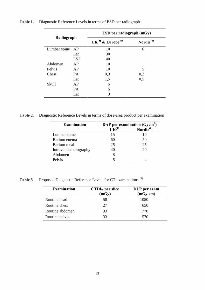
83
7DEOH��� Diagnostic Reference Levels in terms of ESD per radiograph
(6'�SHU�UDGLRJUDSK��P*\�5DGLRJUDSK
8.�����(XURSH��� 1RUGLF���
Lumbar spine AP 10 6Lat 30LSJ 40
Abdomen AP 10Pelvis AP 10 5Chest PA 0,3 0,2
Lat 1,5 0,5Skull AP 5
PA 5Lat 3
7DEOH��� Diagnostic Reference Levels in terms of dose-area product per examination
([DPLQDWLRQ '$3�SHU�H[DPLQDWLRQ��*\FP��8.��� 1RUGLF���
Lumbar spine 15 10Barium enema 60 50Barium meal 25 25Intravenous urography 40 20Abdomen 8Pelvis 5 4
7DEOH�� Proposed Diagnostic Reference Levels for CT examinations (3)
([DPLQDWLRQ &7',Z�SHU�VOLFH�P*\�
'/3�SHU�H[DP�P*\�FP�
Routine head 58 1050
Routine chest 27 650Routine abdomen 33 770Routine pelvis 33 570

84
4XHVWLRQV��4����$QVZHUV��$����5HPDUNV��5�UHOHYDQW�WR�WKH�VXEMHFW�SUHVHQWHG�E\�0U��:$//
4� Diagnostic reference levels are set at the 75 percentile, as you illustrated. Why are they set onlyon the upper side of the distribution whilst examinations with low exposures usually do notprovide the practitioner with useful diagnostic information?
$� Diagnostic reference levels form part of a quality assurance programme. The nature of such aprogramme is that it combines several criteria including image quality and diagnostic efficacy.So we should consider all aspects at the same time.
4: What is the exact meaning of a reference level? It is not a level which your are aiming at, is it?And if you apply the system of revising these levels towards the lower part of the dosedistribution on a continuous basis, where do you stop?
$� As I said before, reference levels are based on today’s practice. They are used as a first step inthe optimisation process to identify the ‘bad guys’ who are well away from the optimum.Corrective measures will have a maximum impact in this case. Reference levels are certainlynot target values. If you are below them, it does not mean that you should not continue theoptimisation effort. As far as the downward trend is concerned I do not see any problem insetting tighter goals when techniques improve and doses come down. Any way, we are still faraway from the lower limit therefore this question is not quite relevant for the moment.
85
-XVWLILFDWLRQ�RI�PHGLFDO�H[SRVXUHV�DQGPHGLFR�OHJDO�H[SRVXUHV
:LOOLDP�%,1&+<7ULQLW\�&ROOHJH'XEOLQ�±�,UHODQG
,QWURGXFWLRQ
I have the task of addressing the subject of justification of medical exposures, in particularmedico-legal exposures. The Medical Exposure Directive gives prominence to the argument ofjustification. In doing so it requires us to be conscious of the broader philosophical, ethical and socialcontexts in which the question of justification of medical exposures is to be resolved. No scientificcalibration, however sophisticated, can give us answers to this question. The matter is particularlydifficult when, as m the case of medico-legal exposures, there is no way of balancing a�direct healthbenefit to a particular person against the individual detriment that the exposure may cause him or her.Where there is such a direct health benefit, the calculus has the aura of the exercise of a conventionalmedical decision; without that benefit, the decision clearly has to be made by reference to otherimportant non-medical criteria.
-XVWLILFDWLRQ�IRU�PHGLFDO�H[SRVXUH�XQGHU�$UWLFOH��
Article 3(1) sets out the general principle for justification, requiring a sufficient net benefit, weighingthe total potential diagnostic or therapeutic benefits the medical exposure produces against theindividual detriment that it may cause. Article 3 (3) provides that, if an exposure can not be justified,"it should be prohibited." The concept of prohibition is worthy of reflection. It implies some edict orlaw which specifically provides, not merely the delimitation of what should be done, but also thespecific banning of any activity falling outside the scope of that determination. I am not sufficientlyfamiliar with the civil law systems to know whether the terminology of Article 3 (3) implies in thosesystems that there must be a legal sanction against exposures that can not be justified. Certainly, in thecommon law system of Britain and Ireland, Article 3 (3) would suggest that there has to be such asanction. Indeed one normally speaks of prohibitions in the context of criminal sanctions.
I will now discuss in detail some of the other aspects of Article 3. Clearly it is significant that Article 3(1) (a) requires that QHZ types of�practices must be justified in advance before being generally adoptedand that H[LVWLQJ types of practices PD\ (not PXVW) be reviewed whenever new important evidenceabout their efficacy and consequences is acquired. Article 3 (1) (b) requires all individual medicalexposures to be justified in advance. If a type of practice involving a medical exposure is not justifiedin general, a specific individual exposure of this type "could be justified in special circumstances, to beevaluated on a case-by-case basis." The tentative use of "could be" and the emphasis on specialcircumstances and case-by-case evaluation make it clear that this exception is not envisaged as aparticularly wide one. Article 3 (1) (c) requires that medical exposure for biomedical and medicalresearch be examined by an ethics committee. This is in line with the general developments in regardto such research in most member states. Article 3 (2) deals with "helpers." It requires that the exposureshow a sufficient net benefit, taking into account also the direct health benefits for the patient, thebenefit to the helper and the detriment that it might cause. Some complicated ethical issues arise in thiscontext, which I will merely adumbrate here, since they are reflected in the context of medico-legalexposures, which I discuss below. What is at stake is the practical conflict between the benefit to thepatient and the detriment to the helper. In contrast to the person subjected to medico-legal exposures,the helper is not merely suffering detriment. His or her altruism is part of the tapestry of human

86
solidarity on which society is founded. Those called on to make decisions relating to justification inthis context have the difficult task of determining the point at which paternalistic principles shouldoverride or limit the helper’s exercise of altruistic autonomy.
0HGLFR�OHJDO�SURFHGXUHV
I now turn to medico-legal procedures. It may be the useful, first, to mention the provisions of theDirective which refer explicitly to medico-legal procedures. Article 1(2)(e) makes it clear that theDirective applies to the exposure of individuals as part of medical procedures. Article 2 definesmedico-legal procedures as "procedures performed for insurance or legal purposes without a medicalindication". Article 3(1), which sets out the requirement that medical exposures referred to in Article1(2) must be justified, provides in paragraph (d) that:
"special attention shall be given to the justification of those medical exposures where thereis no direct health benefit for the person undergoing the exposure and especially for thoseexposures on medico-legal grounds".
Article 4(2)(c), expanding on the optimization principle, requires Member States to "ensure thatspecial attention be given, to keep the dose arising from the medico-legal exposure referred to inArticle 1(2) (e) as low as reasonably achievable". Finally, we may note Article 4(4), which requiresMember States to "ensure the laying down of procedures to be observed in case of medico-legalexaminations".
:KDW�DUH�PHGLFR�OHJDO�SURFHGXUHV"
As we have noted, Article 2 of the Directive defines medico-legal procedures as "proceduresperformed for insurance or legal purposes without a medical indication". This definition isunderstandably broad. It would have been unwise for the Directive to have attempted to provide anexhaustive list. Clearly, a procedure performed for insurance purposes falls within the scope of thedefinition. A person who wants life insurance cover, for example, may be required to submit to amedical examination, with the extra requirement of being subjected to a medical exposure. It might beconsidered that exposures of this kind are elective in the sense that the person need not apply for theinsurance cover if he or she objects to the fact that it involves a medical exposure. In the real world,however, the insurance cover may in practice be compulsory, since it may be tied to an importantaspect of the person’s life -such as employment or a financial commitment (a mortgage, for example)which cannot be shaken off.
Article 2 speaks of procedures "for.... legal purposes". These can range widely. The legal process mayrequire a person to subject himself or herself to a medical exposure. Legal systems throughout Europevary widely but there would appear nothing in principle which is contrary to the European Conventionin Human Rights for legislation or a court to require that a person can be subjected to a medicalexposure where due regard is taken of that person’s entitlements to life, health, bodily integrity, privacyand autonomy. I do not here wish to address in detail this aspect of the law other than to note that, incriminal law proceedings, concern for the presumption of innocence and principle against self-incrimination must be taken into account in determining the limits of the scope of the requirement tosubject oneself to any medical or other intervention which impacts on one’s bodily integrity andprivacy. It is interesting to note that in the recent Irish decision of J. v C. (otherwise C.T.), High Court,14 October 1996, Budd J. held that the Master of the High Court had competence to appoint apsychiatrist as medical inspector of both parties to a suit of nullity of marriage. He specifically rejectedthe argument that the appointment of such an inspector infringed the respondents’ rights to bodilyintegrity and privacy.

87
In civil litigation, it may be necessary, or at all events highly desirable, that a litigant be subjected to amedical exposure. In some instances, the authenticity of a plaintiffs complaint of a medical conditioncan be tested effectively only by means of such an exposure. In other cases, where it is necessary tocheck a plaintiff’s medical progress, a medical exposure may also be essential.
7KH�-XVWLILFDWLRQ�3ULQFLSOH
I now turn to consider exposures on medico-legal grounds in the context of Article 3, which sets outthe justification principle. Article 3(1) requires that medical exposure referred to in Article 1(2) mustshow "a sufficient net benefit", weighing the total potential diagnostic or therapeutic benefits itproduces, including the direct health benefits to an individual and the benefit to society, against theindividual detriment that the exposure might cause, taking into account the efficacy, benefits and risksof available alternative techniques having the same objective but involving no or less exposure toionizing radiation.
Thus, on the one side are all the total diagnostic or therapeutic benefits, including (but clearly notlimited to) the direct health benefits to an individual and the benefits to society. In the case ofmedico-legal exposures, as the Directive acknowledges in Article 2, there are no medical indications.One can envisage some relatively rare cases where beneficial medical results may follow from suchexposures but these are not the reason for their occurrence and cannot seriously be considered underthe justification principle.
If medico-legal exposures are to be justified as showing "a sufficient net benefit", therefore, thejustification must be based on some other ground. What precisely is this ground? Article 3 wouldappear to be limited to "the benefits to society". It is hard to identify any other "potential diagnostic ortherapeutic benefits". Undoubtedly society benefits from the phenomenon of medico-legal proceduresinvolving medical exposures. The administration of justice, the litigation process, the delivery ofinsurance services and other areas of life would all be compromised, to a greater or a lesser extent, ifthese procedures were not available. But the fact that there are social benefits associated with anyparticular practice does not mean that it is necessarily justified. One must weigh the benefits againstthe damage that can result. In the present context, human individuals will suffer the damage in theinterests of society.
The question of the extent to which an individual must risk injury or death for the benefit of society isof profound philosophical and ethical proportions. Much of the history of philosophical debate overthe past two and a half millennia has been concerned with this question.
Some philosophers have argued in favour of broad utilitarian principles which allow for a "felicificcalculus" in which individual welfare is subsumed into a social calculation and in which the languageof individual rights that are capable of withstanding social convenience is anathema. Philosophers whoadopt a materialist understanding of humanity do not necessarily give priority to social benefits(though many do), but they sometimes reject the concept of individual rights, either in its totality or inparticular contexts. Other philosophers give centrality to human dignity, capacity, freedom andautonomy. Some of them seek to integrate that understanding with the vision of human beings asessentially "social animals", owing duties to their society and being entitled to its protection. Othersadopt a more individualistic approach, some going so far as to assert that society has no legitimateclaim to intrude upon individual autonomy.
The Directive makes no attempt to adopt any particular philosophy in this context. What is noteworthyis that its terminology nowhere seeks to answer in any specific way that is capable of empiricaltranslation the question of what practical outcome should follow from the fact that a particular medicalexposure is one that is part of a medico-legal procedure.
88
$UWLFOH������G�V�DSSURDFK
In this context the language of Article 3(1)(d) is of interest. As has been mentioned, it requires that"special attention" be given to the justification for those medical exposures where there is no directhealth benefit for the person undergoing the exposure and "especially" for those exposures onmedico-legal grounds. A requirement to give special attention to the justification issue is not the sameas a requirement to desist from authorising an exposure in any particular instance. What Article 3(1)(d) does is to call the decision-maker to a state of particular solemnity when reaching a decision asto justification in this particular context. It does not seek to guide the decision-maker in any way as tothe particular outcome of that decision.
I point this out not in criticism of the Directive but merely to show what might not otherwise havebeen obvious, which is that the Directive, having highlighted the issue of exposures on medico-legalgrounds, is neutral on the question of which philosophical approach should be taken to resolve thejustification question. If I had a criticism of the language of the Directive, it would be that Article 3(1)in its references to exposures on medico-legal grounds, uses language which makes no overt referenceto the concepts of human dignity, autonomy and bodily integrity. I can fully understand why thereshould be some reticence about doing so, but it would seem possible to have given someacknowledgement of the true complexity of the problem and of the fact that there is a profounddifference between balancing benefits against detriment in respect of one particular person andbalancing social benefits against one particular person’s detriment. These two questions raise quitedifferent issues.
7KH�H[LVWLQJ�OHJDO�IUDPHZRUN
I cannot speak with any authority about the position in civil law systems. It may, however, be worthmentioning that courts in common law systems are well used to addressing in specific contextsprecisely the balancing process which Article 3 requires. This arises most frequently in negligencelitigation. Negligence is the all-pervasive tort (or delict) of the late twentieth century. Negligencelitigation is used, not merely to respond to such manifest wrongdoing as careless driving, sub-standardmedical treatment and ill-considered financial advice, but also to address and resolve in a judicial formcomplex issues of social policy.
Thus, for example, in the English decision of Knight v Home Office [1990] 3 All E.R. 237, the courthad to determine whether a prison hospital should be required to come up to the same standards inmonitoring the condition of a disturbed prisoner with suicidal tendencies as would be expected in aspecialist psychiatric hospital. In Watt v Hertfordshire County Council [1954] 1 W.L.R. 835 and inHeeney v Dublin County Council (Irish High Court 1988) the courts had to consider the extent towhich the emergency nature of a fire-fighter’s occupation might affect the scope of an employer’s dutyof care to the fire-fighter. The courts have accepted that they have to balance the risk of injury to theplaintiff against the social benefits that result from the risky activity and the costs of removing ormitigating the risk. In addressing this balancing process, the courts are not guided by any overtphilosophy since, as with Article 3, they operate on an analytic formula which allows them fill in theparticular philosophy to which they subscribe. That philosophy changes over time and differs fromcountry to country, yet the skeletal formula remains unchanged, it is capable of accommodating suchdiverse approaches in the "law and economics" school of Chicago with a more humane, person-centredapproach favoured in some other jurisdictions. As McHugh J.A observed in the Australian decision ofWestern Suburbs Hospital v Currie, 9 N.S.W. L.R. 511, at 523 (1987),
"negligence is not an economic cost/benefit equation. Immeasurable ’soft’ values such ascommunity concepts of justice, health, life and freedom of conduct have to be taken intoaccount".

89
)XWXUH�DSSURDFKHV
Perhaps the best way for the law at a Community level to deal further with the issue of medicalexposures in the context of medico-legal procedures is to address specific areas and deal with themdirectly in relation to those areas. It would have been folly for the Medical Exposure Directive to haveattempted this task. What is necessary is for these particular areas to be identified and for policiesregarding medical exposures to be developed in the light of the corpus of law associated with theseareas. Thus, for example, criminal law contains its own unique limitations which have no directparallel in other legal contexts. In criminal law, an issue may arise as to the circumstances (if any) inwhich it is legally permissible to obtain evidence by means of a medical exposure against the wishes ofthe accused person. Over and above any questions of bodily integrity, privacy and autonomy, thecriminal law has its own reasons for restraint, concentrating on the principle of the presumption ofinnocence and what in the common law system is identified as "the right to silence". In civil law, it isworth noting that circumstances can differ radically. It might possibly be thought fair to take the viewthat a plaintiff in personal injury litigation who refused to subject himself or herself to a necessarymedical exposure should suffer the consequences in having the action dismissed or at least havingserious inferences drawn against him or her. There are, however, other types of civil litigation wherethe party who is reluctant to subject himself or herself to a medical exposure is not such a free agent. Aperson who claims that he or she has been wrongfully committed to a mental hospital is placed in adilemma rather than having a free choice, since the only option here may be between undergoing theexposure or remaining in the hospital. Somewhat similar considerations could arise in relation torefugees and asylum-seekers, who may not be truly free to decline the option of subjecting themselvesto undesired exposures.
In this context, Article 4 rather than Article 3 may in practice offer the best protection. The obligationit sets down in paragraph (c) of Section (1) to ensure that special attention be given to keeping the doseas low as reasonably achievable requires that explicit consideration be given to the purpose for whichthe exposure is required.
&RQFOXGLQJ�2EVHUYDWLRQV
The framers of the Directive are to be commended for their inclusion of medico-legal exposures withinits terms. The principle of justification set out in Article 3 has the flexibility to render it capable ofeasy implementation throughout the Community.

90
4XHVWLRQV��4����$QVZHUV��$����5HPDUNV��5�UHOHYDQW�WR�WKH�VXEMHFW�SUHVHQWHG�E\�0U��%,1&+<
4� Would it be of interest to define a European list of prohibited radiological practices?
$� In the first place the MED does not require such a list. I do not think that it would be feasible todevelop such a list within the transposition period of only 2 years. It might be envisaged atnational level, but I do not think that MS are keen on this.
4� Does the EC plan a uniform approach towards medical-legal exposures, for example theradiological examination of drug smugglers?
$� I do not think so. There is not enough specificity and detail available to produce somethingrealistic within such a short period of time. Also at the national level I do not see it happening.The question of calculating ratios of individual detriment versus social benefits is a difficultone. As regard the particular case of drug smuggling, criminal law not only considers theviolation of the integrity of the individual but also the presumption of innocence and theentitlement of the suspect not to incriminate himself. Both principles have a bearing on acompulsory intervention such as an X-ray examination.
4� What is your attitude towards consenting children?
$� I cannot give you a bold and clear answer to this question. The traditional view in law practicewas to seek consent from the parents. Now there is a trend towards a greater autonomy for thechild and the age of consent is clearly coming down - 10 to 15 years old, for example. But itremains a grey area where no absolute prohibitions exist and where courts are reluctant to gobeyond general phrases such as: ‘…special attention shall be given … ‘ which you also find inthe MED.
4� In your opinion, does the MED contain requirements that could lead to court cases claimingmoney from practitioners for malpractice as we often see in the USA?
$� In Europe, no tradition of medical legal litigation exists. In the USA, the situation is differentbecause they have the jury system which is not applied to such cases in Europe. Here medicallegal negligence actions are not easily won because courts are reluctant to sanction�practitionersfor malpractice. Secondly, the level of damages awarded for malpractice in Europe is lowerthan in the USA. Therefore, I do not think the same situation will develop in Europe.

91
7UDLQLQJ�IRU�PHGLFDO�DSSOLFDWLRQ�RI�LRQL]LQJ�UDGLDWLRQ
(OLVHR�9$f2���/��*21=È/(=0HGLFLQH�6FKRRO�±�5DGLRORJ\�'HSDUWPHQW�±�&RPSOXWHQVH�8QLYHUVLW\
0DGULG�±�6SDLQ
,QWURGXFWLRQ
Article 7 of Council Directive 97/43/EURATOM of 30 June 1997 on health protection of individualsagainst the dangers of ionising radiation in relation to medical exposure (1), establishes requirementsconcerning education and training. Member States may find that certain aspects of this Article requiresome clarification and guidance and this paper could provide an initial platform for discussion of afuture EC Guideline containing some specific recommendations for the application of the Directive.
We assume that “adequate theoretical and practical training for the purpose of radiological practices”already exists in accordance with national requirements and that there are in existence nationalmechanisms to control this level of training. Nevertheless, some specific cases may need to bereviewed. One such instance concerns radiology technicians: there are very major differences betweenMember States in the content and length of their training (especially in the case of Spain).
For those aspects related to “relevant competence in radiation protection”, harmonization at Europeanlevel would be desirable. Great differences may exist between the Member States. Work along theselines was carried out in 1991-1993, as part of the European Commission (EC) VALUE programme forRadiologists and Radiographers. Draft documents concerning “Specific Educational Objectives inRadiological Protection and Quality Assurance for Diagnostic Radiology Installation Personnel” wereproduced (2). Within the framework of the ongoing European Concerted Action DIMOND (DigitalImaging: measures for optimisation of radiological information content and dose), a similar documentfor Interventional Radiology has also been drafted (3).
Some indication of the required level of training in radiation protection (RP) would be convenient toensure appropriate curricula in the different Member States, establishing differences between:
– radiologists, nuclear medicine and radiotherapy specialists– cardiologists– other medical doctors using x-ray systems (specially fluoroscopy systems), such as urologists,
vascular surgeons, traumatologists, etc.– dentists– orthopedists– radiographers, nuclear medicine and radiotherapy technicians (if they have different previous
training in a particular country)– medical physicists– maintenance engineers and maintenance technicians
Training for nurses, radiographers and other radiology technicians is required under Article 5.3 of theDirective. Medical physics experts are included in Article 6.3. Maintenance Engineers and otherauxiliaries involved in medical exposures are not specifically mentioned, but obviously they needtraining in RP.
The World Health Organisation (WHO) recommendations for Interventional Radiology, producedduring a Workshop held in Munich, in October 1996 (4), could be a good example of the training andcredentialing requirements for people involved in interventional radiology practice.
92
During the workshop held in Grado (Italy) in September 1993, sponsored by the EC, some relevantconclusions about training in radiation protection were stated (5). One of the conclusions was that “ageneral and strong impression which came up was the need and demand to improve training inradiation protection and in quality assurance. Common training programmes at European level must becontinued. A practical way to make progress in the harmonisation of training could be the elaborationof specific educational objectives, as made within the EC VALUE Programme for radiologists andradiographers”.
The introduction of topics on quality assurance (QA) as part of the training programmes in radiationprotection would contribute to the promotion of the safety culture. The routine application of QAprogrammes requires the participation of radiology technicians and a good training of this group ofprofessionals is essential.
ERPET (European Radiation Protection Education and Training) courses are also a source of goodtraining material. EC departments will publish the proceedings of the last course, held in Madrid inMay 1997, on radiation protection in interventional radiology (6).
The experience of the American College of Radiology (ACR) could be valuable in this field. The ACRhas established a Commission on Education, including specific Committees on Accreditation forContinuing Medical Education, Systematised Refresher Courses, Residency Training, ProfessionalSelf-evaluation Committee, etc.
Guidelines for the basic aspects of continuing education programmes are needed in Europe and ACRstandards can be a relevant source of information.
It seems that in some European countries, national regulations do not deal very effectively with aspectsconcerning training. PHS Smith has recently published a paper in The British Journal of Radiology (8)stating that “some aspects of the POPUMET regulations (Protection of Persons Undergoing MedicalExaminations or Treatment, UK regulation 1988), have caused considerable difficulties and have beenineffective, particularly with regard to training”.
Other British authors (AD Quinn et al.) stress that the UK regulations state that a person clinically orphysically directing a medical exposure shall have received adequate training to obtain a core ofknowledge. Clinical directing is defined as having clinical responsibility for the decision to effect amedical exposure. In some fluoroscopy examinations this responsibility may lie with non-radiologists.The “core of knowledge” is defined in the statutory instrument and includes:
– knowledge of the nature of ionising radiation;– risk of ionising radiation;– range of dose associated with a particular procedure;– principles of dose reduction.
These authors think that the introduction of a compulsory core of knowledge in medical school coursesmay be a more appropriate way to achieve the principles of POPUMET.
The International Commission on Radiological Protection also stresses the importance of training forgood protection in medical exposures in its recent recommendations about “Radiological Protectionand Safety in Medicine” (10).
*HQHUDO�UHFRPPHQGDWLRQV�IRU�7UDLQLQJ�3URJUDPPHV�LQ�5DGLDWLRQ�3URWHFWLRQ
A list of topics to be included in the training programmes in RP for the different groups ofprofessionals should be established. Good examples are the VALUE documents mentioned above (2),

93
agreed during a number of meetings attended by radiologists, radiographers and medical physicistsfrom various European Countries. The following training areas have been agreed as being relevant toradiation protection:
– Atomic structure and the interaction of radiation– Radiological quantities and units– Physical characteristics of x-ray machines– Fundamentals of radiation detection– Detectors used in diagnostic installations– Fundamentals of radiobiology: cell, systemic and whole body responses– Radiation protection. General criteria– Operational radiological protection– General RP aspects in diagnostic radiology– Particular aspects of patient and staff RP– Quality control and quality assurance– National and European regulations and standards– Practical training
The WHO recommendations for interventional radiology (IR) requires a specific second level oftraining in RP for these specialists (11), defined for the following areas:
– X-ray systems for IR.– Dosimetric quantities specific for IR.– Radiobiology: risks in IR.– Radiological protection of the staff in IR.– Radiological protection of the patients in IR.– Quality assurance in IR.– Local and international rules concerned with IR.– Procedures optimisation in IR.
There are also British recommendations for topics to be included in RP training for nuclear medicine(12):
– Nature of ionising radiation and its interaction with tissue.– Genetic and somatic effects and how to assess their risks.– Patient Doses– Quality assurance and quality control– Dose limitation– Pregnancy– Unsealed sources– Organisation of radiation protection– Statutory responsibilities
German regulations include special radiation-protection courses with the following content (13):
– Radiation physics, dose, and dosimetry– Radiobiology and radiation risk– Radiation protection of personnel and patients– Radiation protection law– And a second part about practical continuing training (physicians perform this training during
the residency period)

94
Additionally, it is obvious that the topics to be included in the training courses, and the level ofknowledge of the topics, should be different and adapted to the various specialities (diagnosticradiology, radiotherapy, cardiology, dentistry, etc.) and the different kind of work and responsibility(medical doctors, medical physicists, maintenance engineers, radiographers, etc.). During the WHOMunich meeting (11), different levels of training were proposed for interventional radiology. A list oftopics and a level of knowledge were drawn up where relevant, for medical doctors, radiographers,nurses and maintenance engineers (medical physicists should know all the listed topics).
Table 1 presents a proposal of training topics and level of knowledge for the different groups of healthprofessionals involved in medical exposures.
A formal recommendation about the number of hours for the training programmes (theory andpractical work) should be established. This training period will depend on the prior knowledge ofradiation physics, radiobiology, etc., possessed by the various groups of professionals in the differentcountries. A good tool for defining the number of hours need for training could be the use ofguidelines containing the specific educational objectives (2, 3), whereby the length of the trainingperiod would depend on the objective to be achieved.
It is easier to agree a list of topics to be taught and a catalogue of specific educational objectives, thana set number of hours of training. Training is not just a matter of courses. Self-training and goodtraining material should be promoted. A common level of knowledge in RP throughout Europe for thedifferent groups of health workers would be desirable.
The range of recommended training time varies very widely, from half a day in the UK for practisingnurses and doctors in Nuclear Medicine (12) to several weeks in Spain (14).
Practical exercises and practical sessions should be included in these programmes. As a minimum, apractical demonstration, lasting between one and two hours, in a clinical installation should beincluded even in the most simple training programmes. Practical sessions should make up 25-50% ofthe total time scheduled in more extensive courses.

95
TABLE 1
TRAINING TOPIC DRMD
RTMD
NMMD
CDMD
DT MD TE NU ME
Atomic structure and interaction of radiation m h M L l l l l m
Nuclear structure and radioactivity -- h H -- -- -- l l m
Radiological quantities and units m h M M l l m l m
Physical characteristics of x-ray or therapymachines
m h l M l m m l h
Fundamentals of radiation detection l m h L l l m l h
Fundamentals of radiobiology m h h M l m l l l
Radiation protection. General criteria h h h H m m m l m
Operational radiological protection h h h H m m m m m
Particular RP aspects of patient h h h H m h m m m
Particular RP aspects of staff h h h H m h m m m
Quality control and quality assurance m h m M l l m l m
National and European regulations and standards m m m M m m m l h
Recommended training hours20-30
40-60
30-50
20-30
10-15
15-20
15-50
10-15
40-60
DR/MD = Diagnostic Radiology Specialists (Medical Doctors)RT/MD = Radiotherapy Specialists (Medical Doctors)NM/MD = Nuclear Medicine Specialists (Medical Doctors)CD/MD = Cardiology Specialists (Medical Doctors)DT = Dentists MD = Other Medical Doctors using X-ray systemsTE = TechniciansNU = NursesME = Maintenance Engineers
Level of knowledge:
l = Low level of knowledgem = Medium level of knowledgeh = High level of knowledge
The lecturers for these kinds of training programmes should be carefully selected. They should haveprevious experience in radiation protection in medical installations and in practical work in a clinicalenvironment. Installations conducting practical training should be medical installations and not onlylaboratory or simulation exercises.
An important element in the outcome of these training activities is the availability of training material.Specific material in the form of books, slide collections, videos, interactive CD ROMs etc., is scarceand not always at the right level for the various courses. The EC should encourage the publication ofthe proceedings of the different ERPET courses and promote the drafting of specific training material

96
for a set of basic courses (e.g. basic training in radiation protection for cardiologists). Exploiting theexperience gained in the various European countries in this field could also prove very useful.
Some specialities such as interventional radiology require, according to WHO (11) recommendations,a second level of specific training in radiation protection, over and above the general level received bydiagnostic radiology specialists.
Special consideration should be given to the case of medical doctors (non radiologists) usingfluoroscopy x-ray systems on a regular basis (urologists, vascular surgeons, traumatologists, etc.). Inthe USA, these doctors require a specific credentialing process in radiation protection (15): in Europe asimilar system should be established.
Paediatric Radiology also requires some specific topics in RP for radiologists and radiographersinvolved in these examinations.
5HFRPPHQGDWLRQV�FRQFHUQLQJ�WKH�FUHGHQWLDOLQJ�SURFHVV�LQ�UDGLDWLRQ�SURWHFWLRQ
A system for credentialing the training programmes in RP should be established at national or regionallevel. This responsibility should be assumed by the regulatory authority or by academic institutions(such as universities). A register of accredited bodies should be established.
The minimum requirements for credentialing a training programme should take account of all theaspects involved: enough administrative support, guarantees for the archiving of files, diplomas, etc.,for a minimum number of years, enough didactic support (classroom, audiovisual support, etc.),teachers qualified in the topics to be taught and with experience in hospital medical physics work,instrumentation for practical exercises, availability of clinical installations for practical sessions, etc.
In the case of accreditation programmes for radiology technicians, it seems logical to require thattraining be conducted in hospital radiology departments with radiologists, medical physicists andsenior radiology technicians as teachers.
Various alternatives should be proposed for credentialing different professionals with different dutiesand responsibilities (examination at the end of the course, residency, continuous evaluation, etc.).
The basic information cited in the diplomas or certificates received by people attending a trainingprogramme in RP should include: the centre conducting the training, number of accredited traininghours, process of credentialing (examination or other form of assessment), date of the training, name ofthe tutor with responsibility for the training programme, etc.
The second level of training in RP for Interventional Radiology, recommended by the WHO (11)would also require a specific accreditation. The same applies in the special case of medical doctorsusing fluoroscopy on a regular basis.
5HFRPPHQGDWLRQV� FRQFHUQLQJ� UDGLDWLRQ� SURWHFWLRQ� RI� WKH� SDWLHQW� LQ� UHODWLRQ� WR� LQGLYLGXDOVXQGHUJRLQJ�WUDLQLQJ�SURJUDPPHV�LQ�KHDOWK�FHQWUHV
The RP of the patient during the training of residents and other health workers (radiographers,technicians, etc.) requires special consideration. The criteria of justification and optimisation should beapplied carefully and all the procedures should be performed under the responsibility of a seniorspecialist.
Some specific recommendations could also be given for planning practical RP training in medicalinstallations (e.g. X-ray systems must remain operational after the training sessions; if patients are

97
involved, simple procedures (low doses) must be selected; training must not involve any additionalirradiation of the patients, etc.).
In the case of high patient dose procedures (interventional and some vascular diagnostic procedures), astrict patient dose control should be performed to guarantee that no significant additional doses areimparted.
5HFRPPHQGDWLRQV� RQ� FRQWLQXLQJ� HGXFDWLRQ� DQG� WUDLQLQJ� DIWHU� TXDOLILFDWLRQ� DQG� ZKHQ� QHZWHFKQLTXHV�DUH�LPSOHPHQWHG
The ACR Continuing Medical Education (CME) Standard (for physicians and medical physicists)requires a minimum of 150 hours of approved education in category 1 and 2 every three years (thiswill be renewed in a three-year cycle) (7).
Category 1 (designated by ACR or other recognised organisations). The minimum number of hours is60. Accredited residencies and fellowships up to 50 hours per year can be included.
Category 2. The maximum number of hours allowed is 90. Activities accepted: medical meetings,lectures, course syllabi, study of authoritative medical literature, teaching radiology-related services tomedical students, preparation and publication of scientific papers, presentation of papers, courses, orscientific exhibits, clinical consultations, use of computer-assisted learning materials designed toenhance patient care, review of manuscripts for peer-reviewed journals and review of abstracts forscientific meetings.
If we consider this figure as a realistic recommendation, percentages between 2 and 6% (1 - 3hours/year) of training devoted to continuing education in RP could be considered, depending on thekind of work. A radiotherapist or a medical physicist could need more time than a dentist.
Such continuing education in RP should be promoted by the Regulatory and Health Authorities andsome basic courses, when necessary, should be organised by health centres, academic institutions orprofessional and scientific societies.
When a new radiation system is introduced in a Health Centre (e.g. a new CT or interventionalradiology X ray system), staff should receive specific training before any clinical use of the system andthe participation of the engineers of the firm supplying the system should be required. This trainingshould be part of the commissioning process of the new radiation system. It is important to considerthe responsibility of the supplier concerning the availability of full and understandable instructions inthe local language.
Also, when a new technique is implemented in a centre (e.g. intravascular brachytherapy or some newinterventional procedures) prior training for staff should be required. In this case the training should becarried out in another centre with previous experience in the technique to be implemented and theconsiderations stated in the previous section about “Recommendations concerning radiation protectionof the patient in relation with individuals undergoing training programmes in health centres”, shouldbe taken into account. A certain number of examinations and/or procedures, which should beperformed under the control of an experienced physician could be considered in some cases.
5HFRPPHQGDWLRQV� UHJDUGLQJ� WKH� FRXUVH� RQ� UDGLDWLRQ� SURWHFWLRQ� LQ� WKH� EDVLF� FXUULFXOXP� RIPHGLFDO�DQG�GHQWDO�VFKRROV
The EC Directive states that “Member States shall encourage the introduction of a course on radiationprotection in the basic curriculum of medical and dental schools”.

98
Assuming that a basic knowledge of radiation physics exists as part of the content of the disciplinestaught during the preclinical period (basic Medical Physics or equivalent), the recommended RPcourse could concentrate on topics which focus on patient protection. A possible outline could be thecontent of ICRP 73, Radiological Protection and Safety in Medicine (10).
1. Introduction2. The Quantification of Radiation Dose and Risks3. The Framework of Radiological Protection4. The Justification of a Practice5. The Optimisation of Protection6. Individual Dose Limits8. Practical Methods of Protection9. Operational Guides and Reference Levels10. Accidents and Emergencies11. Institutional Arrangements
Obviously, differences between Medical and Dental Faculties should be considered where appropriate.Also a RP course should be introduced in nurses and orthopedic schools.
The duration of these courses should be between 15 and 30 hours, assuming some prior knowledge ofradiation physics. Approximately 20-30% should consist of practical sessions analysing typical casespresented in clinical practice (pregnant patient who needs a radiological examination, paediatricexposures, dose and risk comparison between conventional and CT examinations, etc.).
This course should be mandatory and taught at the end of the preclinical or during the clinical period.
2XWOLQH�IRU�VSHFLILF�WUDLQLQJ�LQ�5DGLDWLRQ�3URWHFWLRQ�IRU�,QWHUYHQWLRQDO�5DGLRORJ\
As example of the usefulness of formulating specific educational objectives when preparing trainingactivities, we present some items defined by a group of DIMOND experts (3), in line with the topicsproposed during the WHO meeting in Munich:
��� ;�UD\�V\VWHPV�IRU�LQWHUYHQWLRQDO�UDGLRORJ\�
½ To explain the effect of high additional filtration (e.g., copper filters) on conventional x-raybeams.
½ To explain the operation of continuous and pulsed X-ray emission modes.
½ To explain the benefits of the grid controlled X-ray tube when using pulsed beams.
½ To explain road mapping.
½ To explain temporal integration and its benefits in terms of image quality.
½ To analyse the changes on the dose rate when varying the distance from intensifier to patient.
��� 'RVLPHWULF�TXDQWLWLHV�VSHFLILF�IRU�LQWHUYHQWLRQDO�UDGLRORJ\�
½ To define the dose-area product (DAP) and the units it is usually measured in.
½ To discuss the correlation between surface dose and DAP.
½ To discuss the relationship between DAP and effective dose.
½ To correlate the dose at the entry point in the patient with the dose at the exit surface and thedose at the intensifier input surface.

99
��� 5DGLRELRORJ\��ULVNV�LQ�LQWHUYHQWLRQDO�UDGLRORJ\�
½ To describe deterministic effects which may be observed in IR.
½ To analyse the risks of deterministic effect induction as a function of the surface dosesreceived by the patients.
½ To analyse the relationship between received doses and deterministic effects in the lens ofthe eye.
½ To be aware of the likely time intervals between irradiation and occurrence of the differentdeterministic effects, the required follow up and control of patients.
��� 5DGLRORJLFDO�SURWHFWLRQ�RI�WKH�VWDII�LQ�LQWHUYHQWLRQDO�UDGLRORJ\�
½ To comment on the most important factors which influence dose levels to staff in IRlaboratories.
½ To analyse the influence of X-ray C-arm positioning on occupational doses.
½ To analyse the effects of using different fluoroscopy modes on occupational doses.
½ To analyse the effects of using personal protection (e.g. leaded aprons, gloves, eyeglasses,thyroid protectors, etc.).
½ To analyse the benefits and drawbacks of using articulated screens suspended from theceiling.
½ To understand the importance of choosing a suitable location for personal dosimeters.
��� 5DGLRORJLFDO�SURWHFWLRQ�RI�WKH�SDWLHQWV�LQ�LQWHUYHQWLRQDO�UDGLRORJ\�
½ To analyse the correlation between image number taken in a procedure and dose received bypatients.
½ To discuss the effects of the focus to skin distance and patient image intensifier inputdistance.
½ To analyse the dose reductions attainable by modifying the image rate in cine or in digitalacquisition.
½ To give typical examples of patient entrance dose value per image in different procedures.
½ To analyse the effect on the patient dose of using different magnifications.
½ To discuss the parameters which should be recorded in the patient’s history related to (orwith reference to data concerning) the received doses.
��� 4XDOLW\�DVVXUDQFH�LQ�LQWHUYHQWLRQDO�UDGLRORJ\�
½ To discuss the difference between parameters that usually do not downgrade over time andthose which could do so and require regular checks.
½ To discuss the importance of establishing simple criteria to compare doses at the patient or atthe intensifier entrance, in different controls.
½ To note the importance in QA programs of regular checks of patient dose and its comparisonwith reference dose levels.

100
��� /RFDO�DQG�LQWHUQDWLRQDO�UXOHV�FRQFHUQHG�ZLWK�LQWHUYHQWLRQDO�UDGLRORJ\�
½ To discuss the different national regulations which apply in IR installations.
½ To describe the international recommendations on IR (WHO, IAEA, ICRP, EC, etc.).
½ To provide information about the international recommendations concerning to limitation ofhigh-dose modes.
��� 3URFHGXUH�RSWLPLVDWLRQ�LQ�LQWHUYHQWLRQDO�UDGLRORJ\�
½ To note the value of optimisation in IR radiation procedures.
½ To discuss the importance of reference levels related to the patient dose, at the local, nationaland international levels.
½ To analyse the importance of regular patient dose checks in each room.
½ To discuss the possibility of using different C-arm orientations addressing long procedures inwhich the threshold of deterministic effects may be attained.
½ To analyse the importance of recording the dose values imparted to every patient.
$FNQRZOHGJHPHQWV
The authors acknowledge the useful comments received from B. Bauer (Germany), R. Van Loon(Belgium), H. Bosmans (Belgium), K. Faulkner (UK), K. Harding (UK), D. Teunen (CE), M. Bezares(Spain), E. Guibelalde (Spain) and J.M. Fernández (Spain).
5HIHUHQFHV
1. European Commission. Council Directive 97/43/EURATOM of 30 June 1997 on healthprotection of individuals against the dangers of ionising radiation in relation to medicalexposure. Official Journal of the European Communities, L 180:22-27; 9.7.97.
2. Vañó E, González L, Maccia C, Padovani R. Specific Educational Objectives in RadiologicalProtection and Quality Assurance for Diagnostic Radiology Installation Personnel. Radiologistsand Radiographers. CE VALUE Programme. Edited by Cátedra de Física Médica. Facultad deMedicina. Universidad Complutense. 28040 Madrid. Spain. June 1993.
3. Vañó E, González L, Faulkner K, Padovani R, Malone JF. Specific training objectives inRadiation Protection for medical staff in Interventional Radiology installations Draft document.DIMOND European Concerted Action. January 1997.
4. Bäum A, Bauer B, Bernhardt JH, Stieve FE, Veit R, Zeitlberger I, editors. Efficacy andRadiation Safety in Interventional Radiology: BfS-ISH-178/97. Proceedings of the 1995 JointWHO/ISH Workshop on Efficacy and Radiation Safety in Interventional Radiology; October 9-13; Munich-Neuherberg. Salzgitter, Germany: Bundesamt für Strahlenschutz, 1997.
5. Data Analysis and Optimisation in Quality Control and Radiation Protection of the Patient inDiagnostic Radiology and Nuclear Medicine, Proceedings of the Workshop jointly organisedby the CEC, the Unitá Sanitaria Locale N' 7, Udine (I) and the World Health Organisation, heldin Grado (Italy), 29 September- 1 October, 1993. Edited by G. Contento, B. Wall, H. Schibillaand D. Teunen. Report EUR 15257, EN, Radiation Protection Dosimetry, Vol. 57 Ns. 1-4,1995.
6. Radiation protection in interventional radiology. ERPET Course. Madrid, 12-14 May 1997.European Commission. Complutense University (Radiology Department) and Ciemat.Proceedings to be published by the EC in 1998.

101
7. ACR Standard for continuing medical education. Revised 1996. ACR Standards, 1997.
8. Smith PHS. EC Directive: 97/43/Euratom. Br J Radiol 1998; 71:108-108.
9. Quinn AD, Taylor CG, Sabharwal T, Sikdar T. Radiation protection awareness in non-radiologists. B J Radiol. 1997; 70:102-106.
10. ICRP 73, Radiological Protection and Safety in Medicine. Annals of the ICRP, Vol. 26, Num.2, 1996. Pergamon. U.K.
11. Efficacy and Radiation Safety in Interventional Radiology; Proceedings of a joint WHO/ISHWorkshop held in Munich-Neuherberg, Germany; October 9-13, 1995. To be published byWHO, 1998.
12. Harding LK. Training in radiation protection for those physically directing medical exposures(nuclear medicine). Newsletter. Nucl Med Comm 1989; 10: 531-532.
13. Bauer B. BfS, Institut für Strahlenhygiene. Germany. Personal communication. February 1998.
14. Spanish Nuclear Safety Council. Guidelines for Training Courses on Radiation Protection inDiagnostic Radiology and Radioactive Installations (in Spanish). 1998.
15. Wagner LK, Archer BR. Minimizing Risks from Fluoroscopy X-rays. A CredentialingProgramm for Anesthesiologists, Cardiologists, Gastroenterologists, Interventionalists,Orthopedists, Pulmonologists, Radiologists, Surgeons and Urologists. 1996, Partners inRadiation Management, Houston, USA.

102
4XHVWLRQV��4����$QVZHUV��$����5HPDUNV��5�UHOHYDQW�WR�WKH�VXEMHFW�SUHVHQWHG�E\�0U��9$f2
4� In medical physics, a shortage of trained physicists in diagnostic radiology is imminent. Doesthe EC envisage doing something about this?
$� Training is in the first place a national matter. However, in the past EC training schemes incollaboration with EFOMP have tried to give physicists access to the latest knowledge in thevarious application fields. For example, last summer we had a summer-school in Nice onradiology physics which was quite successful. The EC is planning to continue this effort to setan example and to help Member States to achieve a good level of education.
4� Do predefined training programs for medical physicist in patient dosimetry, quality assurance,etc. exist?
$� For the moment I don’t think such training programs exist. It would be a good suggestion forthe EC to organise such courses in collaboration with EFOMP.
4� Do you think that minimum levels of education for radiographers and radiology technicians canbe set at European level together with an accreditation system.
$� The EC has no legal basis for doing this. It is, however, currently preparing a general guide,non-binding on Member States, where proposals for minimum knowledge for all the relevantprofessionals will be suggested. This guide could help to harmonise the approach of MemberStates in this respect.
4� Which countries in the Union have regulations on continuing training?
$� No data are yet available but the European guide in preparation will suggest ways of realisingcontinuous education together with a defined level of knowledge and an accreditation system.
5� The professional associations of radiologists and nuclear medicine specialists have developedcurricula for practitioners on a European level. They are available to the Member Stateauthorities who can use them as the basis for drafting legislation.
4� In the undergraduate curriculum of a medical school, students in some countries receivetraining courses of about 10 hours on radiation protection aspects. In which year of the 6 years’basic curriculum do you think this training should take place?
$� The MED recommends providing such training in medical and dental schools. There is norecommendation yet to what extent such training should be given but I think it is important thatthe radiation protection training should be given at a level where the students are aware of whatradiology means, what its purpose is, etc. Therefore I would say that the last two years, whenclinical training is given, seems the correct moment to do so.
103
6SHFLDO�H[SRVXUHV��3DHGLDWULFV
.DUO�6&+1(,'(5'U��YRQ�+DXQHUVFKHV�.LQGHUVSLWDO
0�QFKHQ�±�*HUPDQ\
,QWURGXFWLRQ
There are basic differences in anatomy, physiology, distribution of radiosensitive tissue and age relateddiseases of paediatric patients as compared to adults. The most important points are listed in 7DE��.These differences greatly affect radiographic and fluoroscopic imaging. Furthermore, the susceptibilityof paediatric patients to ionizing radiation is assumed to be higher than that of adult patients.
The MEDICAL EXPOSURE DIRECTIVE (Council Directive 97/43/Euratom) stresses many factors relevantfor the of use of ionizing radiation in relation to medical exposures. According to�$UWLFOH�� exposuresof paediatric patients have been defined as special exposures which have relevance in respect toeducation and training of radiographers/radiologists, appropriate equipment as well as effectiveoptimization procedures. Optimization of diagnostic imaging ($UWLFOH� �) depends on many factors:education and continual training and regular quality control measures organized by state authorities,medical physicists and the radiology departments itself.
63(&,),&�,0$*,1*�352%/(06�,1�3$(',$75,&�3$7,(176
+ROGLQJ�SHUVRQV
Ideally, no holding persons should be involved in radiography and fluoroscopy of patients. But inreality, severely ill patients require assisting persons to monitor the patient’s condition. Furthermore,partially cooperating paediatric patients, especially after trauma or with severe pain, need holdingpersons to stabilize the patient’s position for exact exposure.
If at all possible, the helping person should be a technician or a nurse. But, practically, often theparents present can and often will hold the child. If they hold the patient, they should be clearlyinformed as to the details of the radiologic examination so that they could properly fixate their childduring the whole examination. If female persons assist the radiographer or the radiologist, they shouldbe asked about a possible pregnancy.
According to a recent European survey on paediatric fluoroscopy in children’s hospitals holdingpersons are present in frequently performed examinations, e.g. micturition cysturethrography, bariummeal, in about 50% )LJ��.
;�UD\�HTXLSPHQW
X-ray tables and stands must be equipped with special paediatric holding systems and fixation devicesto allow safe and quick examinations of paediatric patients of all ages �$UWLFOHV� �� DQG���. Thesesupporting devices for uncooperative patients in different sizes must be available for radiography,fluoroscopy and computed tomography. Correct positioning of the infant and toddler is a key point toobtain high quality radiographs. This can only be reached with fixation techniques (taping), specificrestraining devices and tubes. As a consequence, radiology departments and private practices whichare not appropriately equipped should not be allowed to x-ray paediatric patients.

104
The use of automatic exposure control (AEC) for radiography in young paediatric patients is often oflimited value, even for such simple examinations as chest X-ray, pelvis etc. Currently, availablemeasuring systems are oversized, and even special designs of AEC do not function properly becauseyoung patients will frequently move of out of the measuring area. Very often, these systems arecombined with non-removable grids which are not needed in young infants and children. As the use ofgrids will often unnecessarily increase the dose to the patient these systems should be appropriatelymodified (removable grid) for use on paediatric patients )LJ��.
For this reason, most paediatric radiology departments use free hand settings based on age and weightof the patient for radiography. However, weight is not an ideal parameter to calculate the optimalexposure parameters of paediatric patients. In the future modern X-ray systems will use the diameter ofthe patient, e.g. by measuring the focus film distance and also the entrance site of the patient withultrasound. Thereby, the optimal mAs-product can be calculated.
Blurring of spot films caused by rapid, unforeseeable movements in uncooperative young childrenoccur frequently if the exposure time is too long. However, short exposure times are only possible withmodern converter generators with high power (50 - 80 kW). Even mobile generators should be able toswitch a minimum exposure time of at least 2 ms and very small mAs products. This problem isespecially important since the number of X-rays in intensive care units (ICUs) of ventilated patientshas enormously increased over the last 10 years.
It is well known that the kV pulse curve and the switching behaviour is highly dependent on the typeand the performance of the generator. Regular constancy tests in defined intervals should be done,especially in equipment which are used exclusively or mainly in paediatric patients �$UWLFOH���. Forthese tests special paediatric phantoms should be used. The test intervals should not be too long, e.g. 4weeks would be sufficient. It is important to include all equipment, especially mobile equipments andC-arm systems, e.g. used in orthopaedic surgery into the quality assurance programmes.
237,0,=$7,21�,1�5$',2*5$3+<
Recommendations for good radiographic technique and image quality criteria for the most frequent X-ray examination of paediatric patients were officially published in the (XURSHDQ�*XLGHOLQHV�RQ�4XDOLW\&ULWHULD� IRU� 'LDJQRVWLF� 5DGLRJUDSKLF� ,PDJHV� LQ� 3DHGLDWULFV EUR 16261 EN, Luxembourg. Thepracticability of the image criteria were evaluated in several Europe-wide dosimetric trials in fourdifferent age groups — newborns, infants, the 5 year old and the 10 year old patient. A relationshipbetween the reduction of the entrance surface dose (ESD) with increasing compliance to the guidelineswithout deteriorating effect on the image quality score was found in almost all examinations and agegroups tested by the European trials. )LJ�� shows this for the chest X-rays of the five year old patients.However, these graphs also show that only 30% of the radiology departments participating in thesesurveys had implemented the criteria for good radiographic technique.
The reasons for the lack of introduction of the radiographic criteria of the European guidelines were:technical factors, e.g. old-fashioned equipment, insufficient knowledge of the radiographers andradiologists on radiation physics, but also, adherance to traditional procedures and strict protocols, e.g.routine use of grids, use of only one low speed screen-film system.
Simple optimization procedures in radiography are the use of the recommended voltage, addit-ionalfiltration, reasonable use of grids and high speed screen-film-systems/ digital image receptor systems
Following these guidelines the dose to patient can be reduced by a factor of 10 to 20. The onlyobjective against this introduction is the adverse effect on the image contrast and the increase in noise.In most cases, this contrast degradation is acceptable for chest and abdominal films because mostquestions of the clinicians, e.g. catheter control, tube positioning, bowel obstruction etc., can still be

105
answered. Occasionally, bone radiographs for special questions, e.g. battered child syndrome,hyperparathyroidism etc., must be of high contrast and high resolution. In these cases, no additionalfiltration and a screen-film combination equivalent to a 200 system should be used. However, themaximal dose saving effect is only reached when all components — screen, film and cassette material(carbon fibre material, Kevlar®), and the film developing system— fit together.
Digital radiography, mostly storage phosphor plates, is now increasingly used as an image producingsystem especially in intensive care radiography. The resolution of newer systems is comparable to thestandard screen-film combinations with a speed of 200 to 400. The advantage over conventionalscreen-film combination is the wider range of exposure with no significant loss or even enhancementof information and the possibility of image storage, transfer and postprocessing. Because the dose indigital radiography can inadvertently increase )LJ��, periodical or continual dose measurements areindicated. The paradoxical increase in dose is caused by unnecessary high mAs-product values set bythe technician under the conditions in an ICU which are often highly variable and not beneficial fordose saving methods. Because the film blackening in digital computed radiography is to be keptconstant internally, there is no proof of the increase of dose if no such controls were performed.
63(&,$/�352%/(06�,1�3$(',$75,&�)/82526&23<
In fluoroscopy five basic factors determine the dose to the patient:
½ the specific design of the equipment,
½ special adaption to paediatric fluoroscopy technique,
½ irradiated area (volume) of the patient,
½ the screening time and the number of spot films taken by the radiologist.
½ The entrance dose to the patients is higher with the under couch systems compared to the overcouch equipment. On the other hand, the dose to the radiologist and other people close to thepatient (technician, nurse, parents) is higher with the over couch equipment. Training of allpeople working at the equipment is very important to use it safely and with the minimumoccupational dose �$UWLFOHV� ��� �� DQG� ���. In order to keep the X-ray field small a formatlimiting device (tubus) for under couch equipment and strict collimation for over couch systemsshould routinely be used. However, extreme coning using under couch systems canparadoxically increase the dose to patient. Therefore, practical teaching of young radiologist byan experienced teacher who knows the performance of the individual fluoroscopy unit is of greatimportance (see also training and education). The collimating system must regularly beinspected during quality control checks �$UWLFOH� ��. All edges of the X-ray-field should beconstantly visible on the viewing screen.The image intensifier size must not be larger than thetelevision image.
As in plain film radiography, additional filtration and the selective use of a grid which must be easilyremovable can considerably save fluoroscopy dose to patients. Of great importance is the generalrecording of the whole fluoroscopic procedure, e.g. video tape recording, storage of digital images,cine-loops.
Schumacher showed in a phantom study that with simple changes of the equipment (grid, additionalfiltration) and digital fluoroscopy the dose can be diminished to as low as 10% of the original value)LJ��. In addition, pulsed digital fluoroscopy (grid controlled or generator controlled) lowers theeffective screening time and therefore diminishes the dose to the paediatric patient enormously. Forexample, using the 3 pulse mode the screening dose can be decreased to 10% compared with

106
continuous fluoroscopy )LJ��. Digital fluoroscopy can be combined with pulsed fluoroscopy yieldingan optimal imaging process. and a dramatically reduction of dose to the patients.
With the introduction of digital technology the fluoroscopy equipment has also been considerablychanged, but also the behaviour of the radiologist. As the images can be stored and reevaluated duringthe examination, the “last image hold” function can be used for radiation-free collimation and theframe grabbing function can replace extra spot films. Thereby, the total fluoroscopy dose cantheoretically be significantly decreased. However, also the opposite can happen, if the screening timewill be increased because some investigators will produce a “perfect fluoroscopy study”. Therefore,guidelines for fluoroscopy in pediatric patients for the most frequent examination are urgently needed.They are now under progress.
Further developments of an optimized paediatric fluoro equipment are necessary. The design of themeasuring chambers is too large for most of the small paediatric patients. Smaller measuring devices,variable positioning and different shape could optimize the fluoroscopic investigation in very youngpatients. Furthermore, the dose rate controlling curves should be adapted to smaller diameters ofpaediatric patients and the fact, that very rarely a high contrast situation is needed. Extreme dosereduction is possible, e.g. if only tube positioning is done.
'26(�0($685,1*�6<67(06
6SHFLILF�SUREOHPV�LQ�GRVH�PHDVXUHPHQW
Dosimetric surveys in European children’s hospitals have shown a wide variation in radiographictechnique and entrance surface dose (TLD-measurements) )LJ��. TLDs can be used for large surveys,but they are impractical in busy radiology departments. In addition, TLD measurements do not provideimmediate feedback. In contrast, dose area product (DAP) measurement are more convenient andprovide immediate feedback. DAP measurements have advantages: No direct application of themeasuring device on the patient is needed, no shadow on the radiograph, and inclusion of the irradiatedarea. Ideally not only the DAP, but also the irradiated field and the dose should be measuredseparately. It is important to perform these measurements for all paediatric examinations, in order notto overlook deviations caused by the equipment or to detect systematic faults �$UWLFOHV��������DQG���.When DAP in fluoroscopy is measured separately for screening and spot films it can help to improvethe education of young technicians and radiologists, e.g. reduce unnecessary screening.
However, the sensitivity of the measuring device in small patients must be adapted to very small dosesand small fields. The minimum field in newborn babies can be as small es 6 x 6 cm. Additional displayof dose at the entrance side of the patient and the mean irradiated area would be very desirable. Allequipment, also mobile X-ray machines, should have DAP-meters. All measured doses, should bestored together with the individual exposure data (kV, mA, s, grid use; image receptor system and thegenerator).
5()(5(1&(�'26(6�25�'26(�&21675$,176
Diagnostic reference dose levels have been defined according to DUWLFOH�� for standardized patients orphantoms. This concept is impractical for paediatric patients because a “standard sized paediatricpatient” does not exist. The variation in size (height, length), weight and diameter of paediatric patientsin the same age group can be extreme. Dose constraints for 3 or 4 different age groups and for the mostfrequent examinations would be a more reasonable way to give guidance and information to theresponsible radiographers and radiologist on the dose to the patients (7DE���DQG�7DE��).

107
('8&$7,21�$1'�75$,1,1*�2)�5$',2*5$3+(56�$1'�5$',2/2*,676
The aim of quality assurance programmes is to optimize the imaging process in order to avoidunnecessary exposures or overexposures to the patients. If paediatric patients are x-rayed, particularage-specific facts must be considered, i.e. specific knowledge of the anatomy in all age groups, theawareness of rapid movements of uncooperative patients, tight coning down to the area of interest,selection of the appropriate radiographic technique and application of specific radiation protectionmeasures.
Optimal coning is extremely important in neonatal radiography and in most cases a matter ofeducation. But, some radiographers were not able to change their careless attitude and inaccuratetechnique )LJ��. Therefore, regular quality control measures and education/ training programmesshould be organized in the departments by the chief technicians and/or the responsible radiologists. Influoroscopy the most important factor to save or increase dose is the radiologist (in some membersstates physicians with no license are allowed to screen), who performs the examination. Before anfluoroscopy examination is started, all information on the patients must be available, e.g. patienthistory, protocols of previous operations, all previous radiographs should be of course available andread by the radiologist, finally a definitive clinical questions to the radiologist has to be clearlyformulated �$UWLFOHV���DQG���.
Specific training programmes in radiation protection in paediatric radiology for radiographers andradiologists must be introduced during their curriculum. In the future, national radiological societiesand the European scientific organizations must put all efforts to insure that radiography andfluoroscopic examinations in paediatric patients are only allowed to be performed either byradiographers or paediatric radiologists who have passed a special training, confirmed by a specialdiploma. Finally, postgraduate training and education in Europe should be accomplished by theintroduction of a credit point system established and controlled by the scientific societies and nationalas well as European authorities.

108
7DEOHV
$QDWRP\ body proportionssize and shape of organsskeletal developmentbone marrow distribution shift during growth
3DWKRORJ\ specific age dependant diseases(congenital heart disease, meconium ileus; battered child)
%LRFKHPLVWU\ total body water (newborns 90%, adult 60%)low fat massdifferent handling and metabolism of contrast media
3K\VLRORJ\ higher respiratory rate (newborns 80 – 120 breaths/ min.)higher heart rate (newborns 160 – 200 beats/ min.)fast body movements
7DE�� Basic differences between paediatric and adult patients with impact onradiographic imaging
(QWUDQFH�6XUIDFH�'RVH�YDOXHV�� *\�
XQ�RSWLPL]HG�WHFKQLTXH RSWLPL]HG�WHFKQLTXHn mini-
mum3rd
quartilemaxi-mum
n mini-mum
3rdquartile
maxi-mum
&KHVW�DS�SUHPDWXUH 122 9 99 386 41 11 37 83
&KHVW�DS����PRQWK 124 20 140 1373 32 21 67 115
&KHVW�DS����PRQWKZLWK�PRELOH�XQLWV
140 31 151 718 17 32 123 194
7DE�� Reference dose values (shaded) for various chest ap X-rays in infants
(QWUDQFH�6XUIDFH�'RVH�YDOXHV�� *\�
XQ�RSWLPL]HG�WHFKQLTXH RSWLPL]HG�WHFKQLTXHn mini-
mum3rd
quartilemaxi-mum
n mini-mum
3rdquartile
maxi-mum
&KHVW�SD�DS 74 26 115 1347 24 19 63 120
&KHVW�ODWHUDO 83 40 251 554 15 37 125 145
&KHVW�DSZLWK�PRELOH�XQLWV
83 29 126 333 15 30 68 94
7DE�� Reference dose (shaded) for various chest ap X-rays in the five year old child

109
),*85(6
)LJ�� Patient fixation in micturition cysturethrography and esophagram - results of a European widesurvey
)LJ�� Relationship between mean entrance surface dose and the use of a grid in chest ap/paradiography in paediatric patients of different age

110
)LJ�� Impact of radiographic technique on the entrance surface dose and image quality in chest ap/paradiography in 5 year old patients
)LJ�� Relationship between image receptor system and mean entrance surface for abdomen ap in the5 and 10 year old patient - results of European wide surveys

111
)LJ�� Dose reduction in fluoroscopy by simple changes of the equipment - phantom studies ref.Schuhmacher (1994)
)LJ�� Dose reduction with digital pulsed fluoroscopy ref. Hermann et al. (1994)

112
)LJ�� Variation of entrance surface dose for the abdomen ap in paediatric patients - results ofEuropean wide surveys
)LJ�� Variation of coning in neonatal chest radiography for 7 different radiographers from apaediatric radiology department

113
5HIHUHQFHV
Fauré C.The Present Status of Paediatric Radiology in Europe (Results of an ESPR-Inquiry) in: TheStatus of Paediatric Radiology in Europe. Editors: Fendel H, Sweet EM, Thomas PS, ScheringBerlin 1991, p. 49-63.
Schneider K., Fendel H., Bakowski C., Stein E., Kohn M.M., Kellner M, Schweighofer K., CartagenaG., Padovani R., Panzer W., Scheurer C., Wall B.F.Results of a Dosimetry Study in the European Community on Frequent X-ray Examinations inInfants.Radiat. Protect. Dosimetry 43:31-36, (1992)
Schneider K. , M.M. Kohn, Bakowski C., Stein E., Freidhof C., Horwitz A.E., Padovani R.,Wall B., Panzer W., Fendel H.Impact of radiographic imaging criteria on dose and image quality in infants in an EC-widesurvey.Radiat. Protect. Dosim. 49:73-76 (1993)
Fichtner C., Schneider K., Freidhof C., Endemann B., Horwitz A.E., Kohn M.M., Fendel H.Critical analysis of field size in chest x-rays of infants © a EC-wide survey in children'sclinics. ECR`93 in Wien.Eur. Radiol. Suppl. 3:389 (1993)
Zeiler M., Weisbach M., Weigel A., Kohn M.M., Schneider K., Fendel H..Patient exposure and radiographic technique in neonatal chest radiography - a survey inGermany. ECR`93 in Wien.Eur. Radiol. Suppl. 3:85 (1993)
Schneider K.Evolution of quality assurance in paediatric radiology. Radiation protection dosimetry. 57:119-123 (1995)
Krüger I., Schneider K.Analyse zur Feldgröße von Lungenübersichtsaufnahmen bei Neugeborenen - Wie oft wird dieobere Extremität unnötig mitexponiert?Radiologe 36:766 (1996)
Schneider K., Kohn M.M., Ernst G., Panzer W., Padovani R., Wall B.Effect of good radiographic technique on dose and image quality: Analysis of the results ineuropean children’s clinics.Pediatr. Radiol. 26: 591 (1996)
Kohn M.M., Moores B.M., Schibilla H., Schneider K., Stender H.St., Stieve F.E., Teunen D., Wall D.European guidelines on quality criteria for diagnostic radiographic images in paediatrics.European Commission, Luxembourg, EUR 16261EN, (1996)
Schneider K., Ernst G., Krüger I., Kohn M.M.Paediatric fluoroscopy — results of an European-wide surveyPediatr. Radiol. 27: 469 (1997)
Schneider K., Kohn M.M., Ernst G.The derivation of reference dose values for chest X-rays in paediatric radiography.Radiation Protection Dosimetry (under revision)

114
4XHVWLRQV��4����$QVZHUV��$����5HPDUNV��5�UHOHYDQW�WR�WKH�VXEMHFW�SUHVHQWHG�E\�0U��6&+1(,'(5
4� Dose reduction and improvement of image quality in paediatric radiology do not always gohand in hand. Sometimes, it is a long and slow learning process, don’t you agree?
$�� Yes, recent technological progress, and I am thinking in particular of digital radiography anddigital fluoroscopy, dramatically improves image quality but also entails higher doses.
4� How can one regulate the justification of paediatric exposures in a more restrictive way than foradults?
$� I do not think regulating this question will solve the problem. I think you must start in yourown department to investigate where improvement is possible and this is the only practical wayforward. I can recommend for this purpose WHO-report 757 entitled ‘Rational use of imagingin paediatric patients’.

115
*(1(5$/�',6&866,21The general discussion was grouped into two major themes, presented as question (Q), answer (A) orremark (R). The answers were given by an expert-panel of the invited speakers and CE officials.
��� 7UDQVSRVLWLRQ�RI�WKH�0HGLFDO�([SRVXUH�'LUHFWLYH��0('��DQG�LWV�KDUPRQLVDWLRQ
4� European Directives are general requirements, whereas national legislation is an interpretationof these general requirements. In view of the free market with free movement of persons andgoods, what does the European Commission (EC) envisage doing as regards harmonisation ofimplementation of the MED?
$� The MED is an umbrella document and is quite general. Member States (MS), however, arerequired under Chapter III of the Euratom Treaty to send proposals for transposition to the EC.The EC can try to harmonise legal provisions at that level. The EC also intends to continuediscussion with the MS through meetings like this one and bilateral meetings to shareexperiences. Finally, specific guidance on technical issues is being drawn up by the EC withthe help of the Article 31 experts group. The EC has also started an information campaigntargeted at the candidate MS and bilateral discussions on legislation will be initiated soon.
5� Nuclear medicine specialists consider the MED a good piece of legislation. It is now importantthat it is well transposed into national law. This transposition should not be too restrictivebecause, in that case, it could be counterproductive for the health benefit that the patient canobtain from ionising radiation in general and from some specific techniques in particular.
5� European radiologists accept the MED since it is in line with ICRP 73. However, they are quitedisappointed in the lack of transparency of the procedure used to draw up the MED.Radiologists were not consulted sufficiently and this is a negative point. Furthermore, Europeanlegislation should not try to dilute achievements in some countries by bringing the generalstandard down to the lowest common level. Radiologists also fear bureaucracy, for exampleregarding informed consent which, if too strictly applied, could hamper medical procedures.Finally, giving the prescriber who has 1 week of training in radiation protection just as muchresponsibility in the justification procedure to as the practitioner who has a much longertraining is an unbalanced approach and therefore not acceptable. For all these reasons,radiologists want to be closely involved in the transposition of the MED into nationallegislation in all MS. They could, for example, play a major role in developing referral criteriafor prescribers.
4� Clinical responsibility is a much broader concept as defined in the MED and goes beyondradiation protection aspects alone. In this context, what is meant by “informed consent”?
$� The concept of informed consent varies from MS to MS. Generally speaking, there are twoviews: the first is that the profession should set standards of disclosure, the second is a patient-centred test where the practitioner should look at the patient to determine which material riskshe prefers to be disclosed. In practice, the outcome is often similar and debate is still going onas to the degree of liberality and as to whether the patient or on the profession should be thefocus of the question of informed consent.

116
4�� Regarding informed consent, it is clear that the practitioner should never forget that the healthof the patient is his major objective and to preserve this health the application of ionisingradiation may be indicated. The patient, for his part, must have confidence in the knowledgeand professionalism of the practitioner, don’t you agree?
$�� The situation is not quite that clear cut. Not every medical exposure is for the benefit of thepatient. Some of them are not justified at all. Often a thorough clinical examination may revealthe necessary diagnostic information so that a radiological examination becomes irrelevant. Toask for a radiological examination in this situation, just to be sure, is not justified. It is also theduty of the prescriber to check whether the diagnostic information provided by previousradiological examinations can be used again. Once the request for a radiological examination ismade, it is the practitioner who should make an evaluation of this request to see whether it isthe best way to obtain the required information. Of course, this supposes that he has access toall relevant background information concerning the patient.
4� To avoid repeated examinations, particularly in interventional radiology, would it not be a goodidea to develop on a European level a document which informs the practitioner (and theprescriber) of previous examinations?
$� In some MS, the radiation-passport system was tested. It was not a success for several reasons,one of them being the bureaucratic burden. The EC therefore has no intention of initiating sucha measure and MS are not keen on it either. The MED clearly puts assigns to the practitionerthe responsibility for defining clinical responsibility. He must seek co-operation with otherspecialists, must actively look for previous information and must provide other colleagues withsuch information. And do not forget that the patient himself can also be an important source ofinformation if the relevant questions are asked.
5� The MED allows flexibility regarding the responsibilities attributed to the prescriber and thepractitioner. This is positive because it takes account of the different health care systems inplace in the MS. In the UK, the prescriber will never have the same level of training as thepractitioner and therefore it is the latter who will be fully responsible for the justification of anexposure. The role of the prescriber is to adequately inform the practitioner of what informationis required so that he can make his decision in an informed way. In other words, the prescriberprescribes an investigation but not a technique and the practitioner is responsible to make achoice on the adequate technique.
5� It is an illusion to think that the prescriber can take real responsibility as far as radiationprotection is concerned. Moreover, the direct risk due to a diagnostic examination is minimal.The risk of ionising radiation in medicine usually forms part of a general risk such as aninvasive procedure which is performed under radiological control or it presents an indirect risk,that is, if the diagnostic information is inadequate the patient will be badly treated. It is the roleof the practitioner to obtain the best benefit/risk ratio.
��� 3URWHFWLRQ�RI�WKH�XQERUQ�FKLOG
5� The Article 31 expert group’s point of view on the protection of the unborn child is that fordiagnostic examinations the 10 day rule is no longer applicable. The prescriber/practitionermust ask the woman if she is pregnant or has missed a period. If the answer is yes, specialjustification should be applied and in the case where the pelvic region might be exposed to highdoses the examination should be postponed, if possible. If the answer is not clear, the womanshould be treated as if she was pregnant. The philosophy behind this system is based on thepresumption of an all-or-nothing effect of ionising radiation on foetal cells in the early stages of

117
development, although some studies on animals show that a small risk for genetic effects mayexist.
4� Two problems may arise: the first is that adolescent females (10-16years) accompanied by aparent often do not admit that they might be pregnant. The radiographer, the person who is inclosest contact with the girl during the procedure, plays a very important role in finding out ifthere might be a possibility of pregnancy. The second problem is that if we apply the proposedsystem, the number of female patients that cannot exclude a possibility of pregnancy mightbecome quite large. Performing a pregnancy test on all of them might considerably disturb thegood functioning of a (large) radiological department.
$� Women usually know when a possibility of pregnancy is present. The important thing for thepractitioner is not to forget to ask the relevant question. Moreover, except for trauma situations,very few examinations with high risk to the fetus are performed on 10-16 year old females.Obviously, no such questioning is necessary for an examination of the wrist.
5� � Awareness on the part of the woman is an important issue. In Spain, the Association ofRadiation Protection, together with the Ministries concerned, developed an informative posterfor women which urges them to inform the practitioner if they might be pregnant. It isdisplayed in all radiological practices and hospitals.

118
&RQFOXVLRQV
'U��3DWULFN�60((67(560LQLVWU\�RI�3XEOLF�+HDOWK�DQG�(QYLURQPHQW
5DGLDWLRQ�3URWHFWLRQ�6HUYLFH%UXVVHOV�±�%HOJLXP
An international seminar on the transposition and implementation in the Member States of the newDirective on health protection in relation to medical exposure (Directive 97/43/Euratom), organised bythe European Commission in conjunction with the Spanish Ministry of Health and Consumer Affairsand the Spanish Radioprotection Society, was held in Madrid on 27 April 1998. Its aim was to promotediscussion and the exchange of views between all concerned, not only the competent authorities in theMember States but also, and perhaps above all, the representatives of the medical services, a group nottraditionally open to new legislation in its field. This aim can certainly be said to have been achieved.
The seminar opened with a general presentation by a Commission representative explaining thechanges which the new Directive has introduced, the main ones being as follows:
Firstly, it builds on the previous «patients» Directive:
– the VFRSH has been extended (all exposure relating to medical practice, rather than just patientexposure);
– more detailed information is provided on the application of the principles of justification -(henceforth the SUHVFULEHU is also involved) and optimisation (introduction of the concept ofTXDOLW\�DVVXUDQFH);
– more detailed provisions are laid down for certain problem types of exposure (KLJK�ULVN, KLJKGRVH or involving persons who are QRW�LOO��including embryos or foetuses);
– more stringent training requirements are introduced (it is now necessary to have undergonetraining and to have a UHFRJQLVHG diploma or qualifications);
– the UHVSRQVLELOLWLHV of the parties involved are more clearly defined.
The Commission also drew attention to the various new concepts introduced: diagnostic referencelevels, potential medical exposure, the requirement to carry out audits and the principle ofradioprotection training as part of basic medical training (again the responsibility of the prescriber).
Nine specific topics were then presented by different speakers before the floor was opened for generaldiscussion.
Of the subjects examined, several actually related to a set of draft 5HFRPPHQGDWLRQV drawn up by theCommission (or its experts), and were thus not always directly connected with the new Directive'sprovisions:
– The first was «5DGLRSURWHFWLRQ�GXULQJ�WUHDWPHQW�XVLQJ�LRGLQH����». This document was drawn upby the Article 31 experts' «Working Group on medical exposure». One important point whichemerged was that the European experts have abandoned the idea of achieving agreement onharmonising the criteria for discharging patients after treatment with radioiodine, which was clearlydisappointing for some of the participants. One debatable (and debated!) point was whether youngchildren should be regarded as part of the «helping family» (with flexible «dose constraint») ortreated as third parties (subject to the exposure limits).

119
– The document entitled «5HFRPPHQGDWLRQV�IRU�WKH�SURWHFWLRQ�RI�XQERUQ�DQG�EUHDVWIHHGLQJ�FKLOGUHQDJDLQVW�LUUDGLDWLRQ�DV�D�UHVXOW�RI�PHGLFDO�H[SRVXUH�RI�WKH�SDUHQWV» is in the advanced GUDIW stage. Ittoo was drawn up by the Article 31 experts' «Working Group on medical exposure».
The main problem with this document was that the basic scientific information it contained wassometimes incomplete or TXDOLILHG� Some of the participants called for full information (notconcealing the existence of XQFHUWDLQWLHV��such as the risk threshold values, but putting them intoperspective). Others felt it was more important to find SUDJPDWLF solutions and provide reassurancefor the physicians involved. The fundamental question, therefore, was whether it was right for ahigh-profile document of this kind to leave out or to present in a veiled manner certain observationsor analyses which might encourage greater caution, and what the ethical and legal implications ofsuch an approach would be. This led to a discussion on the question of maintaining the 10-day rule(possibly only in certain circumstances), because it is the very beginning of pregnancy which is themost uncertain, not only in terms of ascertaining that there is a pregnancy or establishing the dateof conception but also in terms of the nature of any risk or the threshold at which it occurs.
Another important point raised was the precise meaning of the term «unborn child». The authorsintended this phrase to cover all phases of pregnancy, from the egg to the baby immediately beforebirth. However, it appeared that in some quarters it had been interpreted as meaning the humanbeing at the advanced or final stage of the pregnancy, which might suggest, for example, that thedeath of a very young egg or embryo (a risk inevitably carried by irradiation during the first days ofpregnancy) was of no importance in terms of radioprotection.
– The «5HFRPPHQGDWLRQV�RQ�PHGLFDO�DQG�ELRPHGLFDO�UHVHDUFK», another draft document drawn upby the above Working Group. This issue seemed to create few difficulties in itself, but thediscussion showed the need to look more closely at a series of specific practical questions and to«create a culture» in this field.
– The «5HFRPPHQGDWLRQV�RQ�GLDJQRVWLF�UHIHUHQFH�OHYHOV�IRU�PHGLFDO�H[SRVXUH», again drawn up bythe above Group and at the advanced draft stage, are one of the main new concepts introduced bythe Directive. The main problem was of a semantic nature: many people did not really understandthe significance of these levels, so that discussion was fairly pointless. It should be rememberedthat, in the field of radiodiagnostics at least, this term has nothing to do with optimum levels orobjectives to be achieved in terms of patient dose. It actually refers to «examination» levels,indicating the upper level of what is acceptable when «good practice» is applied.
– The «CULWHULD� IRU� DFFHSWDELOLW\� RI� UDGLRORJ\� �LQFOXGLQJ� UDGLRWKHUDS\�� DQG� QXFOHDU� PHGLFLQHHTXLSPHQW», which has been the subject of lengthy discussion, were published in 1997 by theCommission (Radioprotection 91 collection) and have the status of a technical guide. There was nodiscussion on this document. A talk on quality assurance programmes, given by the same speaker,did give rise to discussion, the main conclusions of which were that recommendations should bedrawn up to specify – and harmonise – the VFDOH of the programmes and the PHWKRGV�WR�EH�XVHG andthat these programmes, which were costly in time and money, should not be overdeveloped.
The other topics discussed are not the subject of specific draft recommendations as yet. Theyconcerned:
– potential exposure in medical applications: the emphasis was on the need for widedissemination of information about accidents (databases) and the need for procedures tofacilitate or ensure their disclosure;
– the justification of exposure from the legal angle: the main feeling was that the application ofthe Directive should not create difficulties on this level;

120
– the harmonisation of training for medical and paramedical staff: this was a good idea, but noteasily achievable, judging from past experience in comparable fields; this point might beexamined by the Working Group mentioned above;
– radioprotection in paediatric medicine: this was a very technical talk given by a Germanpaediatric radiologist, which might provide a good source of inspiration for many.
The general discussion covered three «key issues» raised in oral and written questions: the conflictbetween harmonisation and subsidiarity, the responsibility of the prescriber in justification (asurprisingly sensitive issue) and the possible return to the «10-day rule» in radiology (examiningwomen during the first 10 days of the cycle).
One notable aspect of the workshop was that, after initial hostility on the part of the physicians, whofelt that they had not been consulted adequately or at all, they were ultimately positive about theDirective's provisions.
In conclusion, we could say that the power of discussion has once again been confirmed: it is essentialto specify «what we are talking about» (semantics and definitions), to «talk about it together»(consultation), to «talk about it in the right place» (training and information of all involved) and to«say little but to say it properly» (teaching and strategy of disseminating methods outside the expertcircles).
0U��6WHSKHQ�.$,6(5(XURSHDQ�&RPPLVVLRQ
/X[HPERXUJ
Ladies and Gentlemen,
On behalf of the European Commission I have the honour to close this workshop on theimplementation of the Medical Exposure Directive.
This workshop, designed to discuss important aspects of transposition of the Medical ExposureDirective into national legislation with Member States’ competent authorities and with all interestedprofessional societies, was extremely well attended and the debates were of great interest andconducted with great enthusiasm. It resulted in a number of practical recommendations andsuggestions to the Commission that I consider very useful and that certainly will help us in our futurework.
Today’s experience will also stimulate the European Commission to continue its efforts in supportingthe Member States in the transposition of the Directive. Therefore a second meeting in the spring ofnext year to evaluate the status of transposition seems to be the logical continuation.
Finally, let me congratulate the Spanish organizers for their firm commitment to bring this workshopto a successful conclusion and all of you for your invaluable contribution to the meeting.
Thank you very much and goodbye.

121
/LVW�RI�3DUWLFLSDQWV
3URI��+HOPXW�%(5*0$11Medical PhysicistDept. of Biomedical Engineering and PhysicsWaehringer Guertel 18-20A-1090 VIENNA
'U��(UQVW�+$9/,.MedizinphysikerInstitut für Biomedizin Technik & PhysikWähnüger Gürtel 18-20A-1090 WIEN
'U�$OIUHG�+()1(5Radiation Protection OfficerAbteilung StrahlenschutzForschungszentrum SeibersdorfA-2444 SEIBERSDORF
'U��(PLO�2*5,6Univ. Prof.Vorstand der Abteilung für NuklearmedizinDonauspital – SMZOLangobardenstraße 122A-1220 WIEN
'U�3��257,=International Atomic Energy AgencyP.O.Box 100A-1400 VIENNA
0U�)UDQFLV�&5$:/(<Chair (University of Brussels)EFGCP (European Forum for Good Clinical Practice)Ethics Working PartySchoolbergenstraat 47B-3010 KESSEL-LO
0U��0LFKHO�'(�522Dr. in Medicine (Nuclear Medicine)Herendreef 26B-3001 HEVERLEE
3URI��'U��/��'(�7+,%$8/7�'(�%2(6,1*+(Rijksuniversiteit GentArbeidsgeneeskundige dienst2KIII - U.Z.De Pintelaan 185B-9000 GENT
0U��(**(50217Head Radiation Protection OfficeVrije Universiteit Brussel (VUB)Dienst Fysische Controle - CyclotronHaarbeeklaan 103B-1090 BRUXELLES
0UV�6X]DQQH�)5,*5(1Director DG XI.C – Nuclear Safety and Nuclear ProtectionEuropean CommissionRue de la Loi, 200Office TRMF 03/24B-1049 BRUSSELS
0U�'��*2'(&+$/AIB VINCOTTE CONTROLATOMAvenue du Roi, 157B-1190 BRUXELLES
0U�+DQV�*HRUJ�0(1=(/European CommissionDG XII/F/6 - RadioprotectionOffice MO 75 04/21rue de la Loi 200B-1049 BRUXELLES
0U�+DUULH�02/Physicien médicalVUB dep. CyclotronLaarbeeklaan 103B-1090 BRUXELLES
0U�3LHWHU�3(5',(86Ingénieur, Contrôle de QualitéAGFA-GEVAERT N.V.Septestraat 27B-2640 MORTSEL
0U�3DWULFN�3,1(7Sales EngineerHOLOGIC Europe“Horizon Park”Leuvensesteenweg, 510Bus 31B-1930 ZAVENTEM
'U�3DWULFN�60((67(56Service de Protection contre les Radiations IonisantesMinistère de la Santé Publique et de l’EnvironnementRue Montagne de l’OratoireB-1010 BRUXELLES
0U�&KULVWLDQ�7+,(/(0$16Médecine du TravailPrésident ABRClos Sainte Anne, 27B-1332 GENVAL
0U��5RQDOG�9$1�/221PhysicienAgence Fédérale de Contrôle NucléaireRue de LombardieB-1060 BRUXELLES
0UV�5R]D�=/$7$129$Health PhysicistNational Centre of Radiobiology and Radiation Protection132 bd Kliment Ohridski1156 SOFIABulgaria
0UV�/XFLD�9LYLDQD�&$1(9$52Medical PhysicLaboratorio de Ciencias RadiológicasRua São Francisco Xavier, 524Pqv. Haroldo L. Da Cunha - Sala 13620550-013 RIO DE JANEIROBrasil
0V�$OHQD�+(5,%$129ÈRadiation InspectorState Office for Nuclear Safety (SONS)Senovázné namesti110 00 PRAGUE 1Czech Republic

122
0U�-DQ�6$/$9$Radiation InspectorState Office for Nuclear SafetySenovázné námestí 9110 0 PRAHA 1Czech Republic
0UV�/RLV�*58(1$8(5Ph.D., Staff ScientistPicker InternationalRobert-Bosch-Strasse 11D-65719 HOFHEIM-WALLAU
'U��*�QWHU�+(,1(0$11Arzt-InternistSecretary of the working Group “Radiation effects andradiation biology”Hochwaldweg 11D-70771 LEINFELDEN-ECHTERDINGEN
3�'��'U��PHG��.��+2)0$11�35(,�MedizinerJohannes-R-Becher-Str. 1D-07546 GERA
3URI��'U��)ULGWMRI�1h66/,1Medical Physicist, Head of Dept.President of EFOMPRadiologische Univ.-KlinikAbt. f. Medizinische PhysikHoppe-Seyler-Str. 3D-72076 TÜBINGEN
'U��1RUEHUW�3(,16,33Verwaltungsbeamber, MinisterialratBundesministerium für Umwelt, Naturschutz undReaktorsicherheitReferatsleiter RS II 1Postfach 12 06 2953048 BONN
'LSO��,QJ��*�QWHU�6&+0,'7Dipl. Ing. StrahlenschutzMedizin Einrichtungen der Universität BonnStabstelle StrahlenschutzSigmund Freud Str. 25D-53127 BONN
'U�.DUO�6&+1(,'(5RöntgenabteilungLMU-Ludwig Maximilians-UniversitätLindwurmstr. 4D-80337 MÜNCHEN
3URI��-�UJHQ�6&+h7=PhysicianZentrum StrahlenmedizinA.-Schweitzer-Str. 33D-48129 MÜNSTER
0U�5LFKDUG�9,(7RadiologeBundesamt für StrahlenschutzIngolstädter Landstr. 1D-85764 OBERSCHLEIßHEIM
0U�-HQV�-¡UJHQ�*5�1+$8*Radiograf/TechnologistRadiologisk AfdelingVejle SygehusDK-7100 VEJLE
0UV�(OOHQ�*5�1+$8*Radiograf/TechnologistRadiologisk AfdelingVejle SygehusDK-7100 VEJLE
0U�.DUO�$UQH�-(66(1Chief Physicist, Ph.D.Dept. of Medical PhysicsÅrhus KommunehospitalDK-8000 ÅRHUS C
0U�1LHOV�:,,1%(5*MDHillerod CentralsygehusDept. Clinical Physiology and Nuclear MedicinDK-3400 HILLEROD
0UV�0DUtD�-RVpTecnico Especialista RadiodiagnosticC/ Monasterio de Fitero N°18 (4° Izda)E-PAMPLONA
0U�)UDQFLVFR�$*8$'2Médico Jefe de ServicioAlcobendas 12 - ch 43La MoralejaE-28109 MADRID
0U�-XVWR�$/+$0%5$�6(55$12�&58=T.E.RadiodiagnósticoC/ Comendador, n°7 (2°)LA SOLANAE-13240 CIUDAD REAL
0U�0DQXHO�$/2162�',$=Radiofísico HospitalarioHospital U. “M. Valdecilla”Avda. Valdecilla s/nE-39008 SANTANDER-CANTABRIA
0UV�,VDEHO�$/2162�6$,=T.E.R.C/ La Torre N°2E-09557 PUENTEDEY (Burgos)
0U�-DYLHU�$/9$5(=�9$=48(=T.E.R.C/ Padre Don Rua N°1 (8°B)E-36203 PONTEVEDRA
0U�-XDQ�$0$'25Dr. QuímicoHospital Militar Gomez UllaServicio de Protección Radiologicac/ Glorieta del Ejercito s/nE-28047 MADRID
0U�-DYLHU�$0$'25�%/$1&2QuímicoCIS ESPAÑA S.A.C/ Prim, 5E-28004 MADRID
0UV�0DUtD�0HUFHGHV�$1'5(6�5(&/21'2FísicoHospital Miguel ServetServicio de FísicaIsabel la Católica N°1-3E-50009 ZARAGOZA

123
'U�*UHJRULR�$5$*Ï1�'(�/$�&58=PrésidentAssociation Espagnole de Radiothérapie Oncologique (AERO)C/ Amador de los Rios, 5E-28010 MADRID
MédicoClinica Puerta de HierroServicio de RadioterapiaC/ San Martín de Porres, 4E-28035 MADRID
0UV�3LODU�$5$*21�6$17$�0$5,$QuímicaInstituto de Salud Carlos IIIServicio RadioprotecciónCrtra. Majadabonda a Pozuelo Km.2E-28220 MAJADAHONDA (Madrid)
0UV�0DUtD�GHO�&DUPHQ�$5,$6�%/$1&2Médico Profesora UniversidadAvda de manolete No 22, 1°3E-14005 CORDOBA
0U�-RDTXtQ�$50,6(1QuímicoAGFAProvenza 392E-08025 BARCELONA
0U�)HUQDQGR�$51È,=�%8(12Médico NuclearPradillo, 8E-28002 MADRID
0U�/HRSROGR�$55$1=Físico MédicoHospital Ramón y CajalServicio de Protección RadiologicaE-28034 MADRID
0U�-RVp�0DQXHO�$57,*8(6�3('52/$Físico Responsible de P.R.Hospital de Sant Joan, S.A.M.C/ Sant Joan, s/nE-43201 REUS
'U�%DUWRORPp�%$//(67(5�02//PresidentSociété Espagnole de Physique Médical (SEFM)C/ Apolonio Morales, 27E-28036 MADRID
RadiofísicoHospital Universitario de San JuanServicio Protección RadiológicaE-03550 SAN JUAN DE ALICANTE
0U�-��/XLV�%$55(72MédicoClínica Los NaranjosAvda Enrique S. Otaño, s/nE-06004 BADAJOZ
0U�-RVp�,JQDFLR�%$55262�0$7(8ATS de EmpresaCentral Nuclear de Santa María de Garoña (Burgos)E-09200 BURGOS
0U�/HRQDUGR�%(1,7(=�)5$*8(/$QuímicoCUALICONTROL-ACI S.A.C/ Caleruega N° 67 - 1a PlantaE-28033 MADRID
0U�5REHUWR�%(1,72�*21=$/(=Director de NegocioMallinckrodt Iberica S.A.Avda. San Pablo, 28E-28820 COSLADA (Madrid)
0U�5LFDUGR�%(5(1*8(5�6$1726T.E.R.C/ Ermitagaña, 10, 6°AE-31008 PAMPLONA
0UV�0HUFHGHV�%(=$5(6Direccion General de Salud PublicaMinisterio de Sanidad y ConsumoP° Prado 14E-28071 MADRID
0U�-RVp�ÈQJHO�%/$1&2�58%,2Tec. Esp. en RadiologíaIsla de Tavira, 26 (5°C)E-28035 MADRID
0UV�$QD�%/$1(6MédicoConsejo de Seguridad Nuclearc/Justo Dorado 11E-28040 MADRID
0U�2FWDYLR�&$%$//(52�&$53(1$MédicoMedicina NuclearHospital San JuanCtra Nnal 332E-03550 SAN JUAN DE ALICANTE
0U�'DYLG�&$1&,2FarmaceuticoCIEMATAv. Complutense 22E-28040 MADRID
0U�0LJXHO�&$1(//$6�$12=FísicoHospital Clínico UniversitarioS° de Física y Protección Radiologicac/ San Juan Bosco 15E-50009 ZARAGOZA
0U�-RVp�/XLV�&$55$6&2�52'5Ë*8(=Radiofísico HospitalarioUnidad de Protección RadiológicaHospital Universitario “Virgen de la Victoria”Campus Universitario de TeatinosE-29071 MALAGA
0U�)UDQFLVFR�&$55(5$Radiofisico HospitalarioHospital “Juan Ramón Jimenez”Ronda Norte S/NE-21005 HUELVA
0U�(PLOLR�&$6$/FísicoCentro Nacional DosimetriaAvda Campanar 21E-46009 VALENCIA

124
0U�7RPiV�&$6$129$�%/$1&2FísicoJEFE Departamentoc/ Trespaderne 29E-28042 MADRID
0UV�-RVHID�&$67$f('$�$55217(Radiofisico HospitalarioHospital U. “M. Valdecilla”Avda. Valdecilla s/nE-39008 SANTANDER-CANTABRIA
0U�/XLV�0LJXHO�&$67(-Ï1�&$67È1FísicoJefe Servicio Protección RadiologicaC/ Pablo Casals N°12 - 9°BE-28011 MADRID
0UV�/RXUGHV�&$67,//(-26�52'5Ë*8(=MédicoHospital Universitario de GetafeServicio Medicina NuclearCrtra. de Toledo, Km. 12.500, GetafeE-28905 MADRID
0U�0DUWLQ�&$67,//2�+(5$6FísicoC/ Arturo Soria, 192E-28043 MADRID
0U�-HV~V�&$6752�&$7$/,1$FísicoInstituto de Salud Carlos IIIC.N. Sanidad AmbientalCrtra. Majadabonda a Pozuelo Km.2E-28220 MAJADAHONDA (Madrid)
0UV�0DUWD�&(%5,$1�(&8$55(FarmaceuticaCentro Investigaciones Biologicas (CSIC)C/ Velazquez 144E-28006 MADRID
0UV�&DUPHQ�&(59(/2Enfermera Medicina NuclearHospital Ramón y CajalServicio Medicina NuclearE-28034 MADRID
0UV�0DUtD�-HV~V�&(67(526�025$17(Radiofísico – Servicio de Oncología RadioterápicaHospital de LéonAltos de Nava s/nE-24008 LEON
0U�-RVp�&+2&$12�025(12T.E.RadiagnósticoURB. Las Orquideas n°7La PoblachuelaE-13197 CIUDAD REAL
0UV�$QD�,VDEpO�&2$5$6$T.E. RadiodiagnosticoC/ del Dibujo No 13GetafeE-28905 MADRID
0U�-RVp�&25'(52MédicoHospital Militar Gomez UllaServicio de Protección Radiologicac/ Glorieta del Ejercito s/nE-28047 MADRID
0UV�(YD�&255('2,5$�6,/9$RadiofísicoHospital Universitario “La Paz”Paseo de la Castellana 261E-28046 MADRID
0U�-XDQ�&DUORV�'(�$1'5(6Técnico RadiologiaHospital Ramón y CajalServicio de Protección RadiologicaE-28034 MADRID
0U�-HV~V�0DUtD�'(�)58726�%$5$-$RadiofísicoHospital Universitario de ValladolidAvda. de Ramón y Cajal, 3E-47011 VALLADOLID
0U�)��-DYLHU�'(�+$52�'(/�025$/Médico Medicina NuclearClinica Puerta de HierroServicio Medicina NuclearC/ San Martin de Porres 4E-28035 MADRID
0UV�5RVD�'(�9,'$1,$�08f2=Dr en BiologiaCIEMATAvda. Complutense, 22Edif. 3AE-28040 MADRID
0UV�&ULVWLQD�1��'(�9,//$9,&(1&,2FísicoServicio de Protección RadiologicaFundacion Jimenez. DíazAvda. de los Reyes CatólicosE-28040 MADRID
0UV�1LHYHV�',$=�&$1(-$�52'5,*8(=Profesora de Física MédicaFacultad de MedicinaUniversidad de CantabriaAvda. Cardenal Herrera Oria s/nE-39011 SANTANDER
0U�5LFDUGR�',(=�*21=$/(=Radiofísicoc/ Taquígrafo Marti, 14E-46005 VALENCIA
0U�/XLV�'20,1*8(=�*$'($Médico – Jefe de Sección del Servicio de Medicina NuclearC/ Marqués de Valdivia, 94, (2° Esc., 3C)E-28108 ALCOBENDAS (Madrid)
0U�3HGUR�'20,1*8(=�0217(52MédicoC/ Ferrol 30E-28029 MADRIDTel: +34-1-323.30.14
0UV�&DUPHQ�(6&$/$'$�3$6725Radiofísico de hospitalServicio de RadiofísicaHospital Universitario Clínica Puerta de HierroC/ San Martín de Porres, 4E-28035 MADRID
0UV�0DUtD�/XLVD�(63$f$�/Ï3(=RadiofísicoHospital de la Princesac/ Diego de León, 62E-28006 MADRID

125
0U�-DFLQWR�(67$55,$*$�$162Técnico en RadiologiaC/. Dr. J. Ma Reparaz N°4, 5° AE-31012 PAMPLONA
0UV�-XDQD�0DUtD�(67(12=�$/)$52MédicoHospital “12 de Octubre”Servicio NuclearCtra. Andalucía, Km. 4E-28041 MADRID
0U�)UDQFLVFR�)$<26�)(55(5Residente RadiofísicoC/ Luis Marin, 6 (3°B)E-28038 MADRID
0UV�%HOpQ�)(51$1'(=RadiofisicoServicio Física Médica y P.R.Hospital Central de AsturiasJulian Claveria s/nE-33006 OVIEDO
0U�0DQXHO�)(51$1'(=�%25'(6RadiofísicoHospital UniversitarioRadiofisicaPaseo San Vicente 58E-37007 SALAMANCA
'U�-RDTXtQ�)(51$1'(=�&58=PrésidentSociété Espagnole de Radiologie Médicale (SERAM)C/ Goya, 38E-28001 MADRID
RadiologoServicio de RadiodiagnósticoHospital Virgen del RocioC/ Manuel Siurot, s/nE-41013 SEVILLA
0U�-pVXV�)(51È1'(=�3e5(=MédicoJefe Servicio Oncologia RadioterápicaHospital “Virgen de la Arrixaca”E-MURCIA
0U�$GROIR�)(51$1'(=�35,(72Médico RadiologoHospital “Virgen de la Luz” del Insalud de CuencaServicio de RadiologiaE-CUENCA
0U�-RVp�0LJXHO�)(51$1'(=�6272Medical PhysicistServicio de Física MédicaHospital Clinico San CarlosE-28040 MADRID
0UV�1DWLYLGDG�)(55(5Físico MédicoHospital Ramón y CajalServicio de Protección RadiologicaE-28034 MADRID
'U�-RVp�0��)5(,5(Médico NuclearLangosta 12E-PUERTO SANTA MARIA-CADIZ
0UV�$UDFHOL�*$%$/'21FísicoEl Greco, 4E- MADRID
0UV�0DUtD�)p�*$%$/'Ï1�526,//ÏT.E.R.T.C/ Holanda, 2E-28916 LEGANÉS (MADRID)
3URI��&DUPHQ�*$/9$1Doctor en MedicinaServicio de RadioterapiaHospital Clínico San CarlosE-28040 MADRID
0U�0DQXHO�*$/9(=�'(/*$'2Profesor Universidad JEFE SPRAvda. Menendez Pidal s/nE-14004 CORDOBA
0U�-XDQ�-RVp�*$0%$57(�9$/(1&,$Técnico Especialista en RadiodiagnósticoTravesía Francisco Alesón n°2 (5-c)E-31008 PAMPLONA
0UV�3LODU�*$5&Ë$�$/2162MédicoHospital Universitario de GetafeServicio Medicina NuclearCrtra. de Toledo, Km. 12.500, GetafeE-28905 MADRID
0UV�7HUHVD�*$5&Ë$�&$55(5$FísicoHospital “Ramón y Cajal”Oficina Técnica-BioingenieríaCarretera de Colmenar Viejo Km.9100E-28034 MADRID
0U�$QWRQLR�*$5&,$�&85,(/Médico NuclearPaseo Marítimo n°1-(4°B)E-11010 CADIZ
0UV�6ROHGDG�*$5&,$�'(/�9,//$5FarmaceuticaHospital Militar CentralUniversitario Gomez UllaServicio de Medicina NuclearGlorieta del Ejercito s/nE-28047 MADRID
0U�$QJHO�*$5&,$�0,*8(/RadiofisicoHospital UniversitarioPaseo San VicenteE-37007 SALAMANCA
0UV�,VDEpO�*$5&,$�5(&8(52Médico ImmunologaLas Dalias, Casa 2Piso 7°DE-49008 SEVILLA
0UV�<RODQGD�*$55$/'$�3$6&8$/T.E.R.P/ de los Olmos N° 6 - 3°DUrbanizacion ZizurE-ZIZUR MAYOR (Navarra)

126
0UV�0DUtD�&ULVWLQD�*$55,'2�'(/*$'2Lic. Ciencias QuímicasAvda. Burgos 41 - 3°AE-28036 MADRID
0U�-XDQ�0DQXHO�*,/�*$+(7(Técnico del EstadoJusto Dorado N° 11E-28040 MADRID
0UV�0HUFH�*,1-$80(Dir. Tecnica Lab. DosimetriaINTE-UPCDiagonal 647E-08028 BARCELONA
0UV�6XVDQQD�*Ï0(=�&25(6RadiofisicoMGV Gregorio Marañónc/Doctor Esquerdo, 46E-28007 MADRID
0U�-RVp�5DPRQ�*20(=�)8(17(6Médico residenteHospital “12 de Octubre”Servicio de Medicina NuclearCarretera de Andalucía Km.5,400E-28041 MADRID
0UV�6\OYLD�*Ï0(=�7(-('25�$/2162Radiofísicoc/ Antonio Toledano, 24 (5°C)E-28028 MADRID
0UV�,QPDFXODGD�*21=$/(=Técnico Medicina NuclearMar Menor 34, 7°AE-28033 MADRID
0UV�&ULVWLQD�*21=È/(=�58,=RadiofisicoServicio Física Médica y P.R.Hospital Central de AsturiasJulian Claveria s/nE-33006 OVIEDO
0UV�&DUPHQ�*21=$/(=�6��6(*81'2Médico Onc. Radiot.C/ Cerrada N° 2 (5°J)E-47010 VALLADOLID
0U�9LFWRULDQR�*21=$/(=�9,/$RadiofísicoHospital U. “Virgen del Rocio”Avd. Manuel Siurot s/nE-41013 SEVILLA
0U�$QJHO�*5$&,$Medical PhysicC/ Donoso Cortés 80, 5°-8E-28040 MADRID
0U�(GXDUGR�*8,%(/$/'(Prof. of Medical PhysicsSchool of MedicineDept. RadiologyUniversity ComplutenseE-28040 MADRID
0UV�0DUtD�GHO�&DUPHQ�*8,1'(/�58,=Técnico en RadioterapiaC/ Rafael Fernández Hijicos N°47 (3°B)E-28038 MADRID
*8,6$62/$PhysicistInstituto OncológicoAldakonea, 44E-20012 SAN SEBASTIAN
0UV�,VDEpO�*87,(55(=FísicoFacultad de MedicinaUniversidad de CantabriaAvda. Cardenal Herrera Oria s/nE-39011 SANTANDER
0UV�(OID�+$52�6$/9$7,(55$Médico residenteHospital “12 de Octubre”Servicio de Medicina NuclearCarretera de Andalucía Km.5,400E-28041 MADRID
0U�9LGDO�+(51$1'(=�*$5&,$Médico Oncologia RadioterapicaC/ Severo Ochoa 3 (2°A)E-18001 GRANADA
0UV�$UDFHOL�+(51$1'(=�9,725,$FísicoHospital Clínico UniversitarioS° de Física y Protección Radiologicac/ San Juan Bosco 15E-50009 ZARAGOZA
0U�%DOWDVDU�+(51$1'2�0$&+,1IngenieroCUALICONTROL-ACI S.A.C/ Caleruega N° 67 - 1a PlantaE-28033 MADRID
0U�0DQXHO�+(55$1=RadiofisicoPlza. Virgen del Manzano, 2-7°DE-09004 BURGOS
0U�$QWRQLR�+(55(526�0$57,1(=Técnico en Protección RadiológicaC/ Rafael Batlle, 24 BajosE-08017 BARCELONA
0UV�0DUtD�GHO�3LODU�,*/(6,$6�*20(=T.E.R.Avda. Virgen de Argeme, 7 (3°D)E-10800 CÁCERES
0U�(PLOLR�,5$1=2Protección RadiológicaPaseo de la Castellana, 201E-28046 MADRID
0UV�<RODQGD�-($1�0$,5(7Técnico-P.R.C.S.N.c/Justo Dorado N° 11E-28040 MADRID
0U�-RVp�0LJXHO�-,0(1(=�*21=$/(=Radiofísico HospitalarioAvda. Cardenal Herrera Oria, n°54 (3° Dcha)E-39012 SANTANDER
0UV�6XVDQD�-,0(1(=�0$57,1(=Tecnico Especialista en RadiodiagnósticoC/ Delfin N°5 (9°B IZG)E-18015 GRANADA

127
0U�-XDQ�0DQXHO�.,1'(/$1President du Conseil de Sécurité NucléaireConsejo de Seguridad NuclearC/ Justo Dorado, 11E-28040 MADRID
0U�-RVp�3HGUR�/$�%$1'$�7(-('25MédicoServicio de Medicina NuclearHospital del AireC/ Arturo Soria 82E-28027 MADRID
0UV�0DUtD�7HUHVD�/(Ï1�*21=È/(=BiológaCIS ESPAÑA S.A.C/ Prim, 5E-28004 MADRID
0UV�1LHYHV�//25&$�'20$,&$FísicaCentro Nacional de Dosimetriaav/Campanar 21E-46009 VALENCIA
0U�5DPyQ�/2%$72�%8672RadiofísicoComplexo Hospitalario Universitario de Santiago deCompostelaC/ Galeras s/nE-15705 SANTIAGO
0U�3HGUR�/XLV�/23(=�%(558(=2T.E.R.P/ Navarra 6-7° C.P.E-31300 TAFALLA (Navarra)
0UV�0DUtD�3LODU�/Ï3(=�)5$1&2RadiofísicoHospital de la Princesac/ Diego de León, 62E-28006 MADRID
0UV�0DUtD�$QWRQLD�/Ï3(=�3217(Física - Dosimetría InternaCIEMATEdificio 34Avda. Complutense, 22E-28040 MADRID
0U�0LJXHO�/Ï3(=�725726$FísicoFísica MédicaServicio de Protección RadiológicaUniversitat Rovira i VirgiliFacultat de MedicinaC/ Sant Llorenç 21E-43201 REUS (Tarragona)
0UV�3LODU�/25(1=Técnico P.R.Consejo de Seguridad Nuclearc/Justo Dorado, 11E-28040 MADRID
0UV�0DUtD�7HUHVD�0$&Ë$6Responsable Protección RadiológicaInstituto de Investigaciones Biomédicas (CSIC)C/ Arturo Duperier 4E-28029 MADRID
0U�0RKDPHG�0$18=,Ingeniero IndustrialHospital “Ramón y Cajal”Oficina Técnica-BioingenieríaCarretera de Colmenar Viejo Km.9100E-28034 MADRID
0UV�0DUtD�-HV~V�0$1=$1$6Físicoc/Marques de Mondejar No 22 - 7°CE-28028 MADRID
0UV�3DORPD�0$5&+(1$BiologaUNESAC/ Francisco Gervas 3E-28020 MADRID
0UV�0DULVD�0$5&2Head of Training Dept. (RP)CIEMAT / IEEAvda. Complutense, 22E-28040 MADRID
0U�5DPyQ�&DUORV�0$548(=Técnico Especialista Médicina NuclearCIEMATAvenida Complutense, 22E-28040 MADRID
0U�/XLV�0LJXHO�0$57,1�&8572Médicoc/ Sangenjo 14E-28034 MADRID
0U�3HGUR�0$57,1�/(521(6Ldo. en Ciencias Físicasc/Las Eras N° 17, 6°AE-47009 VALLADOLID
0U�5REHUWR�0$57,1�2/,9$FísicoPza. San Roque n°5E-35450 SANTA MARIA DE GUIA (Las Palmas)
'U�-RVHS�0$57,1�&20,1PresidenteSociedad Española de Medicina Nuclear (SEMN)C/ Apolonio Morales, 27E-28036 MADRID
CSUB Hospital Princeps d’EspanyaS. Medicina NuclearC/ Feixa Llarga, s/nE-08907 HOSPITALET DE LLOBREGAT
0U�0LJXHO�$QJHO�0$57Ë1(=X-Ray SupervisorCtra. de Fuencarral Km,15.1E-28108 ALCOBENDAS (Madrid)
0UV�0DUtD�1LHYHV�0$57,1(=�/2,=$*$Médico especializado Med. Preventiva NuclearMEDYCSAPrincipe de Vergara N°134E-28002 MADRID
0U�)UDQFLVFR�5DPyQ�0$57,1(=�5$026T.E. Radiodiagnosticoc/ Aldeanueva de la Vera N° 21, 4°BE-28044 MADRID

128
0U�-RVp�)UDQFR�0$57,�9,'$/Radiofísicoc/ Alginet, 13 - 21°E-46989 TERRAMELARPaterna (Valencia)
0UV�0DUtD�-RVp�0$7(26�/23(=T.E.R.Avda. Madrid, 21 (1°B)E-18012 GRANADA
0U�5DIDHO�0$7(26�257,*26$Tec. Esp. RadiodiágnosticoC/ Jazmin N°9CAJARE-18198 GRANADA
0UV�/\GLD�0(,**6�&25%(//$Médicoc/ General Zabala No 13 - 5°FE-28002 MADRID
0UV�0DUJDULWD�0(/(1'52FarmaceuticaDu Pont PharmaC/ Albacete N°5E-28027 MADRID
0UV�0DUtD�0(1*8$/�*,/FísicoHospital Miguel ServetServicio de FísicaIsabel la Católica N°1-3E-50009 ZARAGOZA
0UV�9LFWRULD�0(675(�'(�-8$1FísicaCentro Nacional de Dosimetriaav/Campanar 21E-46009 VALENCIA
0UV�(VWKHU�0,//$1�&(%5,$1RadiophysicistServicio de Física y P.R.Hospital Clinico Universitario San JuanBosco, 15E-50009 ZARAGOZA
0UV�-RDTXLPD�0,5$/3(,;Licenciada en FísicaSant Salvador 131E-08024 BARCELONA
0UV�0DUWD�*XDGDOXSH�02/,1$�08f2=Licenciada en Derechoc/ Buenos Aires, 9 2°JE-18004 GRANADA
0U��-RVp�/XLV�02152<�$1721MédicoDon Ramón de la Cruz No 89E-28006 MADRID
0UV�*ORULD�0217(52RadiofarmaceuticaDu Pont PharmaC/ Albacete N°5E-28027 MADRID
0U�-XDQ�-RVp�025$17�(&+(9$51(FísicoServicio de Protección RadiológicaUniversitat Rovira i VirgiliFacultat de MedicinaC/ Sant Llorenç, 21E-43201 REUS (Tarragona)
0U�0LJXHO�$QJHO�08f2=�$=1$5(=T.E. RadiodiagnosticoC/ del Dibuto No 13GetafeE-28905 MADRID
0UV�0DUtD�6ROHGDG�1$-(5$�*$5&Ë$Podóloga/EnfermeraCollado de Marichiva n°8 (3B) - Escaleras DE-28035 MADRID
0UV�0DULD�3LODU�2/,9$5(6�08f2=FísicoHospital General Universitario “Gregorio Marañón”c/Dr. Esquerdo 46E-28007 MADRID
0U�0LJXHO�$QJHO�2/02�/Ï3(=T.E.RadiodiagnósticoC/ Alava N°7 (1°E)E-28017 MADRID
0UV�/XLVD�2/25,=�/$1'$Tecnico Especialista RadiodiagnosticoConcejo de Elcano N°11-Bajo BE-31016 PAMPLONA
0UV�9LFWRULD�3$/$&/26�3(5(=Tecnico Especialista en Radiodiagnostico (TER)C/ Artes Graficas, 34, 3°, 12°E-46010 VALENCIA
0UV�0DUtD�&UX]�3$5('(6�*$5&Ë$Radiofísico de hospitalServicio de RadiofísicaHospital Universitario Clínica Puerta de HierroC/ San Martín de Porres, 4E-28035 MADRID
0UV�&pOLD�3(&+$5520$1�6$&5,67$1Médico residenteHospital “12 de Octubre”Servicio de Medicina NuclearCarretera de Andalucía Km.5,400E-28041 MADRID
3URI��-��-RVp�3(f$Profesor Física MédicaCátedra Física MédicaFacultad de MedicinaUniversidad de ExtremaduraE-06070 BADAJOZ
0U�)UDQFLVFR�-DYLHU�3(1Ë1�*21=$/e=MédicoHospital Universitario de GetafeServicio Medicina NuclearCrtra. de Toledo, Km. 12.500 GetafeE-28905 MADRID
0UV�$PSDUR�3(5(=Enfermera Medicina NuclearHospital Ramón y CajalServicio Medicina NuclearE-28034 MADRID

129
0UV�3DORPD�3(5(=�02/,1$Director Técnico FarmacéuticoMallinckrodt Iberica S.A.Avda. San Pablo, 28E-28820 COSLADA (Madrid)
0U�&DUORV�3(<�,//(5$MédicoHospital Universitario de GetafeServicio Medicina NuclearCrtra. de Toledo, Km. 12.500, GetafeE-28905 MADRID
0U�;DYLHU��3,)$55e�0$57Ë1(=Radiofísico de hospitalServicio de RadiofísicaHospital Universitario Clínica Puerta de HierroC/ San Martín de Porres, 4E-28035 MADRID
0U�-XDQ�-RVp�)UDQFLVFR�32//('2Director General de Salud PublicaMinisterio de Sanidad y ConsumoP° Prado 14E-28071 MADRID
0U�$OHMDQGUR�35(16$Físico-Med. NuclearHospital Clínico San Carlosc/ Isaac Peral s/nE-28040 MADRID
0U�&DUORV�35,(72�0$57Ë1RadiofísicoHospital de la Princesac/ Diego de León, 62E-28006 MADRID
0U�-XDQ�,JQDFLR�5$%$Radiofísico HospitalarioAvda. Cardenal Herrera Oria, n°36 (5B)E-39011 SANTANDER
0UV�0DUtD�/XLVD�5$0,5(=Médico NuclearConsejo de Seguridad Nuclearc/Justo Dorado 11E-28040 MADRID
0U�)UDQFLVFR�5$0,5(=�)(51$1'(=Técnico Rayos XC/ Lima n°34 - (2°A)E-28945 FUENLABRADA (Madrid)
0U�-XDQ�,��5$<2�0$'5,'Medicina NuclearHospital Infanta CristinaCtra de Portugal, s/nE-06080 BADAJOZ
0UV�0DUtD�$QJHOHV�5,9$6�%$//$5Ë1Físico de HospitalAvda. Cesareo Alierta N°31, Esc. Dcha. 1°AE-50008 ZARAGOZA
0UV�/X]�0DUtD�52%5('2Dosimetría InternaCIEMATEdificio 7Avda Complutense, 22E-28040 MADRID
0U�3HGUR�52'5,*8(=Físico MédicoSMART SolutionsC/ Ortega y Gasset 20 (3°B)E-28006 MADRID
0UV�0yQLFD�52'5Ë*8(=FísicaIEE / CIEMATAvda. Complutense, 22E-28040 MADRID
0UV�/XFLD�52'5,*8(=�$6725*$Profesora Escuela de EnfermeriaC/ Severo Ochoa, 3 (2°A)E-18001 GRANADA
0U�0DQXHO�)UDQFLVFR�52'5Ë*8(=�&$67,//2FísicoHospital Universitario de ValmeServicio de RadiofísicaCtra. de Cadiz s/nE-41014 SEVILLA
0UV�)iWLPD�52-$6�&,0$'(9,/$PeriodistaConsejo Seguridad NuclearJusto Dorado, 11E-28040 MADRID
0U�-RVp�5DPyQ�520È1�&2//$'2FísicoHospital Universitario de ValmeServicio de RadiofísicaCtra. de Cadiz s/nE-41014 SEVILLA
0U�-RVp�0DQXHO�520$<�%(&&$5,$Minister of HealthMinisterio de Sanidad y ConsumoPaseo del Prado, 18E-28014 MADRID
0UV�0DUtD�/XLVD�526$/(6�&$/92BiologaConsejo de Seguridad NuclearJusto Dorado 11E-28040 MADRID
0U�)UDQFLVFR�-DYLHU�526$/(6�(63,=8$Técnico en Protección RadiológicaGobierno VascoDepartamento de Sanidadc/ María Díaz de Haro, 60E-48010 BILBAO
0UV�$OPXGHQD�58$12�*20(=T.E.R.C/ Crta. de Plasencia, 15E-10691 CÁCERES
0U�&DUORV�58,=�%/$1&2Tec. Esp. en RadiodiagnosticoResidencia Los Alfares P.I., I.C.E-16002 CUENCA
0U�5DIDHO�58,=�&58&(6Médico-RadiologoDpto. RadiologiaFacultad de Medicinac/Campus de Teatinos s/nE-29071 MALAGA

130
0UV�&DUPHQ�58,=�*,0(12FísicaInstituto de Salud Carlos IIIC.N. Sanidad AmbientalCrtra. Majadabonda a Pozuelo Km.2E-28220 MAJADAHONDA (Madrid)
0U�6HEDVWLDQ�58,=�62/,6Médico residenteHospital “12 de Octubre”Servicio de Medicina NuclearCarretera de Andalucía Km.5,400E-28041 MADRID
0UV�&DUPHQ�6$+848,//2�/23(=Tecnico Especialista en Radiodiagnostico (TER)C/ Ingeniero José Sirera N° 29 PTA 3E-46017 VALENCIA
0U�$EUDKDP�6$/&('2�3/$=$Técnico Especialista en Radiodiagnósticoc/ Bernardo Balbuena N°6E-CIUDAD REAL
0U�$QJHOHV�6$1&+(=�6$*5$'2T.E.RadiodiágnosticoAda Mediterraneo N° 52, Bajo FE-28007 MADRID
0UV�0DULQD�6$1&+(=�6$1&+(=MédicoConsejo de Seguridad Nuclearc/Justo Dorado 11E-28040 MADRID
0UV�&RQVXHOR�6È1&+(=�6(55$12T.E. RadiodiagnósticoPuerto el Esquinazo, 2 (1°)E-10300 NAVALMORAL DE LA MATA (Cáceres)
0UV�$QD�0DUtD�6$1&+2�3$6&8$/FarmaceuticaInstituto de Salud Carlos IIIServicio RadioprotecciónCrtra. Majadabonda a Pozuelo Km.2E-28220 MAJADAHONDA (Madrid)
0UV�0DUtD�3RORQLD�6$17,$*2�7(035$12Tecnico en RadiodiagnósticoC/ Pio XII, N°1 (7°A)E-15001 LA CORUÑA
0U�$QWyQ�6$1726�0,5$1'$Médico Onc. Radiot.Avda. de Burgos N° 16B, esc. 2, Bajo BE-28036 MADRID
0U�)HOL[�6$5$%,$�*$5&,$MédicoHospital “12 de Octubre”Avda. Andalucia Km 5.400E-28041 MADRID
0U�-�0��6$675(FísicoHospital Ramón y CajalServicio de Protección RadiologicaE-28034 MADRID
0U�,JQDFLR�6(&$'(6�$5,=Médico EspecialistaHospital Militar CentralUniversitario Gomez UllaServicio de Medicina NuclearGlorieta del Ejercito s/nE-28047 MADRID
0U�)UDQFLVFR�-��6(1,6(�%$55,2Médico NuclearC/ Doctor Eduardo Arroyo N°1, 4° DchaE-23003 JAÉN
0U��$QWRQLR�6(55$'$�+,(552RadiofísicoHospital Universitario “La Paz”Paseo de la Castellana 261E-28046 MADRID
0UV�&HOHVWLQD�6(55$12FísicoHospital Ramon y CajalSección de RadiofísicaServicio de RadioterapiaCarretera Colmenar km 9,100E-28034 MADRID
0U�'DUtR�6(59$12�$6(16,2MédicoHospital Militar Gomez UllaServicio de Protección Radiologicac/ Glorieta del Ejercito s/nE-28047 MADRID
0U�)UDQFLVFR�6,(55$�&$/92T.E.R.Prado Magdalena, 14 (5°A)E-47005 VALLADOLID
0U�)HUQDQGR�6,(55$�',$=Físico (Radiofisica Hospitalaria)Hospital Gregorio Marañón (S. Dosimetria y Radioprotección)c/Doctor Esquerdo 46E-28007 MADRID
0U�(GXDUGR�62//(7PresidenteSociedad Española de Protección Radiologica (SEPR)C/ Apolonio Morales, 27E-28036 MADRID
Físico NuclearIBERDROLAC/ Hermosilla 3E-28001 MADRID
0UV�6XVDQD�68È5(=�58'$Estudiante AprendizC/ Eva Levantes, 13 (6°B)E-41006 SEVILLA
0U�-RUJH�7(,-(,52�9,'$/Catedratico UniversidadC/ San Andres 114 (2°)E-15003 LA CORUÑA
0UV�0DULQD�7(//(=�'(�&(3('$�58,=RadiofísicoHospital Universitario “La Paz”Paseo de la Castellana, 261E-28046 MADRID

131
0U�-RVp�,JQDFLR�7(1�02521Medical PhysicistServicio de Física MédicaHospital Clinico San CarlosE-28040 MADRID
0UV�&RQVXHOR�7,(512�5(*,'25Periodista“Enfermería actualidad”C/ Fuente del Rey, n°2 (Esquina Ctra. de Castilla)E-28023 MADRID
0U�%RQLIDFLR�72%$55$�*21=$/(=Físico“Virgen de la Arrixaca”Servicio de Protección RadiologicaE-30120 EL PALMAR (Murcia)
0U�3HGUR�85,$57(MédicoHospital de LéonMédicina NuclearAltos de Nava, s/nE-24008 LEON
0UV�0DUtD�GHO�0DU�9$/%8(1$�0$5,1Tecnico Especialista en RadioterapiaC/ Luis Marín, 6 (3°B)E-28038 MADRID
3URI��(OLVHR�9$f2�&$558$1$Professor of Medical PhysicsRadiology DepartmentMedicine SchoolComplutense UniversityE-28040 MADRID
0U�(OLVHR�9$f2�*$/9È1Student in medicineC/ Gabriela Mistral, 19E-28035 MADRID
'U�)UDQFLVFR�9$5*$6Subdirector General de Sanidad AmbientalMinisterio de Sanidad y ConsumoP° Prado 14E-28071 MADRID
0UV�0DUtD�(OHQD�9(,*$�2&+2$FarmaceuticaInstituto de Salud Carlos IIIServicio RadioprotecciónCrtra. Majadabonda a Pozuelo Km.2E-28220 MAJADAHONDA (Madrid)
0U�6DQWLDJR�9(/È=48(=�0,5$1'$Radiofísicoc/ Luis Arenas Ladislao n° 7 (6°B)E-41005 SEVILLA
0UV�,QJULG�9(77(56Técnico de RadiologiaHospital Ramón y CajalServicio de Protección RadiologicaE-28034 MADRID
0UV�5RVD�0DUtD�9,&(17(�5$0,5(=Técnico en Imagen Médica (destino Protección Radiológica)Av. Retamas, 10 1°AE-28922 ALCORCÓN (MADRID)
0UV�6XVDQD�9,/&+(=�3(5$/(6Tec. Esp. RadiodiagnosticoC/ Cañada Bodega N°4E-18813 GRANADA
0UV�,ULQD�),/,3329$Head of Department of Supervision and InspectionEstonian Radiation Protection CentreKopli 76EST-EE0004 TALLINNEstonia
'U��(OOH�7$11(5Head of DepartmentEstonian Radiation Protection Centre76 Kopli Str.EE0004 TALLINNEstonia
0U�%HUQDUG�$8%(57Physicien d’HôpitalService de PhysiqueInstitut Gustave-RoussyF-94805 VILLEJUIF Cédex
3URI���%2.Professeur Médecine NucléaireHôpital Beaujon100 Bd Général LeclercF-92110 CLICHY
0U�+HQUL�&$66$*128O.P.R.I.B.P. 35F-78110 LE VESINET
0UV�6RSKLH�&+$,//(7JuristeMinistère de la SantéD.G.S. bureau Ethique et Droit8 av. de SégurF-75350 PARIS 07 SP
0V�<DQQD�&+(9$/0(PharmacienneAgence du MédicamentUnité Pharmaceutique-Chimie143/147 Bd Anatole FranceF-93200 SAINT-DENIS
0U�6HUJH�&2(48<7Médecin NucléaireService Central de Médecine Nucléaire1 rue O. LambretF-59037 LILLE CEDEX
0UV�/$92&$7�',56&+(5/PharmacienMallinckrodt Medical26 rue Gustave MadiotB.P. 3F-91923 BONDOUFLE CEDEX
0U�&KULVWLDQ�/()$85(Chef de ProjetCEPNCentre d’Etude sur l’Evaluation de la Protection dans leDomaine NucléaireB.P. 48F-92263 FONTENAY-AUX-ROSES

132
0U�&DUOR�0$&&,$PhysicistCAATS93 bd Maréchal JoffreF-92340 BOURG-LA-REINE
0U��3KLOLSSH�0$5(//(Médecin RadiologueFNMR60 bd Latour-MaubourgF-75007 PARIS
0UV�(OLVDEHWK�0$56+$//�'(3200,(5Comité Technique Interministériel pour l’Euratom31-33 rue de la FédérationF-75752 PARIS CEDEX 15
'U�$��12(/Centre Alexis VautrinUnité de RadiophysiqueRoute de BourgogneF-54511 NANCY
'U�'DQLHO�62Médecine NucléaireHôpital Bel Air – CHR Metz-ThionvilleService de Médecine NucléaireRue de FriscatyF-57100 THIONVILLE
0U�-�1��7$/%27MédecinHôpital Tenon4 rue de ChineF-75020 PARIS
0V�<OLWDUNDVWDMD�5LWYD�+$98.$,1(1Senior AdvisorSTUKRadiation and Nuclear Safety AuthorityPL 14FIN-00881 HELSINKI
0U�6DXOL�6$92/$,1(1Ph.D., Chief PhysicistHelsinki University Central HospitalDept. of RadilogyP.O.Box 380FIN-00029 HYKS
0U�0DWWL�6820(/$Research departmentSTUKP.O.Box 14FIN-00881 HELSINKI
0U�-HDQ�5�)��%((5RadiographerMedical Physics Dept.Mount Vernon HospitalRickmansworth RoadGB-HA6 2RN Northwood Middlesex
0U�&KULV�%5,**6Eastman Kodak CompanyHealth Imaging, EAMERThe AtriumP.O.Box 5911, Harefield RoadGB-UXBRIDGE UB8 1YD (Middlesex)
0U��5�+��&25%(77Diagnostic RadiologistDepartment of Diagnostic RadiologyHairmyres HospitalEast KilbrideGB- GLASGOW G75 8RG
0U�6WHYH�(%'21�-$&.621Department of HealthRoom 417Wellington House135-155 Waterloo RoadGB-LONDON SE1 8UG
0UV�0DU\�(0%/(721RadiographyThe College of Radiographers2 Carriage Row183 Eversholt StreetGB-LONDON NW1 1BU
'U�.��)$8/.1(5Regional Medical Physics Dept.Freeman HospitalFreeman RoadGB- NE7 7DN NEWCASTLE UPON TYNE
0UV�$GULHQQH�),1&+RadiographerISRRT52 Priory WayGB-N. HARROW HA2 6DH
'U��/��.HLWK�+$5',1*Medical DirectorCity Hospital NHS TrustDept. of Physics and Nuclear MedicineDudley RoadGB-BIRMINGHAM B18 7QH
0U��1LDOO�021$*+$1Radiation Protection Advisor8 second Cross RoadGB-TWICKENHAM TW2 5RF
'U�&5+�3(11OncologyDept o Clinical OncologyRoyal Devon & Exeter HospitalBarrack RoadGB-EXETER
0U�%��:$//National Radiological Protection BoardChilton, DidcotGB-OXON OX11 0RQ
'U��,DQ�:$77Consultant RadiologistDepartment of Clinical RadiologyBristol Royal InfirmaryGB-BRISTOL BS2 8HW
0U�1LQR�5��9(3.+9$'=(M.D., Ph.D, Radiation HygienistTbilisi State Medical UniversityDepartment of Preventive Medicine33, Vazha-Pshavela avenue380077 TBILISIGeorgia

133
0U�'HPHWULRV�&+5,672),'(6Civil Servant (Radiophysist)Environmental Health DivisionMinistry of Health and Welfare7, Aristotelous str.GR-ATHENS 10187
0U�3��',0,75,28Ass. Prof. in Medical PhysicsGreek Atomic Energy CommissionP.O.Box 60092GR-15310 Aghia Paraskevi
'U�(��<$.280$.,6SecretaryGreek Radiation Protection Associationc/o Dept. of Medical PhysicsMedical SchoolGR-115 27 ATHENS GOUDI
'U�6iQGRU�3(//(7Medical Deputy DirectorNational Research Institute for Radiobiology and RadiohygieneAnna u. 5H-1221 BUDAPEST
'U�3��=$5È1'Med. PhysicistUzsoki HospitalUzsoki N°29H-1145 BUDAPEST
3URI��3LHWUR�/XLJL�,1'29,1$Full Professor of PhysicsVia G. Donati, 32I-00159 ROMA
'U�)LORPHQD�0$==(,ResearcherLaboratorio di FisicaIstituto Superiore di SanitàViale Regina Elena 299I-00161 ROMA
0U�)UDQFR�0,/$12University of FlorenceViale Morganni 85I-50134 FIRENZE
0U�$��3$5,6,Radiation Protection ExpertMinistero della Sanità - ISPESLVia Urbana 167I-00184 ROMA
0U��$QWRQLR�686$11$DirigenteA.N.P.A.Via Brancati 48I-00144 ROMA
'U�*LRYDQQD�=$7(//,Member of the Executive Board of AIRPFisica SanitariaA.O. CareggiI-50139 FIRENZE
0UV�(OLVDEHWWD�=8&&+,Medical PhysicistServizio Física SanitariaAzienda Ospedaliera Ospedale San MartinoLargo Benzi 10I-GENOVA
3URI��:LOOLDP�%,1&+<Regius Chair of Laws and Head of the Law SchoolUniversity of DublinTrinity CollegeIRL-DUBLIN 2
'U�*HUDUG�+85/(<Chairman EAR/POCPresident UEMS Radiology Section & BoardDepartment of RadiologyMeath HospitalIRL-DUBLIN 8
'U�%UHQGDQ�0F&/($1Chief Medical PhysicistSt Luke’s HopitalHighfield RoadRathgarIRL-DUBLIN 6
0U�/XLV�(6&2%$5AdministratorEuropean CommissionRadiation Protection (XI.C.1)Centre Wagner C/340L-2920 LUXEMBOURG
0U�/DXUHQW�-20eAttaché d’AdministrationMinistère de la Santé57 Bd de la PétrusseL-2935 LUXEMBOURG
0U�6WHSKHQ�.$,6(5Head of UnitEuropean CommissionRadiation Protection (XI.C.1)Centre Wagner C/320L-2920 LUXEMBOURG
0UV�3DORPD�/23(=Biologiste11A rue de l’EgalitéL-3983 OLM
0U�'LHGHULN�7(81(1AdministratorEuropean CommissionRadiation Protection (DG XI.C.1)Centre Wagner C/325L-2920 LUXEMBOURG
0U�$OELQDV�0$67$8.$6DoctorRadioprotection CentreKalvariju 153VILNIUSLithuania
0U�$QWDQDV�9$,7.86Head of Radiation Protection ServiceKaunas Medical Academy HospitalEiveniu 23007 KAUNASLithuania
3URI��<XUL�'(.+7<$5PhysicistRiga Technical UniversityEEMT Institute1 Kalku Str.LV-1658 RIGALatvia

134
0U�(GPXQGV�3$.(56MD PhysicianP. Stradins Clinical Hospital13 PilsonuLV-1002 RIGALatvia
0UV�1��'(�+$$1Radiation Protection OfficerDVM - Vrije Universiteitvan der BoechorststraatNL-1081 BT AMSTERDAM
0U�)�-�'��)(/'(5+2)Training ManagerStichting BIGRAp/a Hogeschool HollandPostbus 261NL-1110 AG DIEMEN
�-��*(/,-16PhysicistDept. Clinical Oncology LUMCP.O.Box 9600NL-2300 RC LEIDEN
0UV��0U�-��5,-/$$56'$0Policy MakerMinistry of Health, Welfare and SportP.O.Box 5406NL-2280 HK RIJWIJK
0U�:��7(50256+8,=(1Health PhysicistAcademisch Ziekenhuis LeidenDienst Veiligheid, Stralenbescherming en MilieuInterne Postcode C0-QPostbus 9600NL-2300 RC LEIDEN
0U�3�-��9$1�'(5�-$*7DirectorRadionuclide CentreFree UniversityDe Boelelaan 1085cNL-1081 HV AMSTERDAM
'U��+�+��9$1�522,-Associate Director Regulatory affairsMallinckrodt Medical B.V.Westerduinweg 3NL-1755 LE PETTEN
0U�5LFKDUG�9$1�6216%((.Radiation Protection ExpertRöntgen Technische Dienst B.V.Radiation Protection ServicesPostbus 10065NL-3004 AB ROTTERDAM
'U�+DQV�=2(7(/,()PhysicistTNO Centre for Radiological Protection and DosimetryP.O.Box 5815NL-2280 HV RIJSWIJK
'U�&��=885Ministry of EnvironmentDGM/SVS/SNV/655P.O.Box 30945NL-2500 GX THE HAGUE
0UV�0DULD�'2�5$6$5,2�9,(,5$Medical DoctorAv. Acacias, 6R/C ESQ B°A, Monte EstorilP-2765 ESTORIL
0UV�'pOLD�(6&$-$�*$==2PhysicistDirecção-Geral da SaúdeAv. João Crisóstomo, N°9P-1093 LISBON
0U�-RmR�-RVp�48,17(/$�'(�%5,72PresidenteSociedade Portuguesa de Protecção Contra RadiaçõesRua 5 de Outubro Lote 33 1°EP-2685 S. JOÃO DA TALHA
0UV�$PiOLD�52'5,*8(6�1$*8(,5$Physicist Nuclear MedicineServiço de Medicina NuclearIstituto Portugues de Oncologia de GemfilP-1093 LISBOA Codex
0U�0DFLHM�6.$5=(:6.,Engineer-Radiation Safety OfficerNational Atomic Energy Agencyul.Krucza 36 – room N° 14300-921 WARSAWPoland
'U�&RQVWDQWLQ�0,/8Ph. D.President of the Romanian Society for Radiation ProtectionInstitute of Hygiene and Public Health, Radiation HygieneLaboratoryStr. Dr. Leonte N°1-376256 BUCHAREST 35Romania
0UV�.DULQ�(./81'RadiographerRadiologica DepartmentUniversity HospitalS-22185 LUND
0U�(ULN�-859,1Medical Physicist (Radiology)Central HospitalDept. of RadiologyS-37185 KARLSKRONA
0UV�7HUHVD�.83)(5Radiation Protection InspectorSwedish Radiation Protection InstituteS-17116 STOCKHOLM
'U�:ROIUDP�/(,7=Swedish Radiation Protection InstituteDiv. for Occupational & Medical ExposuresS-171 16 STOCKHOLM
0U�+DQV�*��5,1*(57=M.D., Ph.D., Prof. and ChairmanKarolinska HospitalS-17176 STOCKHOLM
0U�9ODGLPtU�-85,1$Head of Radiation ProtectionMinistry of Health of the Slovak RepublicLimbova 2830 07 BRATISLAVASlovak Republic

135
-XUH�)(77,&+Nuclear PhysicianNuclear Medicine Dept.Zaloska 71525 LJUBLJANASLOVENIA
0UV�0HWND�0$&$52/�0,7,M.D.Institute of Public HealthTrubaljeva 21000 LJUBLJANASlovenia
0U�8UEDQ�='(6$5PhysicistInstitute of Occupational SafetyBohoriceva 22A1000 LJUBLJANASlovenia
136
$%675$&7
The objective of the workshop was to promote discussion between competent authorities of theMember States, medical practitioners, pre-scribers, radiation protection experts and representatives ofthe industry on the transposition of the Medical Exposure Directive (MED) (97/43/Euratom). Ninerelevant topics, on many of which the EC is preparing technical guidance, were introduced by key-notespeakers, each followed by a discussion. In particular, the question of the usefulness of a return of the10-day rule for women in child bearing age, the significance of diagnostic dose reference levels, theharmonisation of method and scale of quality assurance programmes, the responsibility of the pre-scriber in the justification process and the possibilities of harmonising training were discussed indepth. It was agreed that a second meeting on the transposition of the MED early 1999 should takeplace to evaluate transposition in a further stage.