document
TRANSCRIPT

articles
Late endosomal membranes rich in lysobisphosphatidic acid regulate cholesterol transport
Toshihide Kobayashi*, Marie-Hélène Beuchat*, Margaret Lindsay†, Sonia Frias*, Richard D. Palmiter‡,Hitoshi Sakuraba§, Robert G. Parton† and Jean Gruenberg*¶
*Department of Biochemistry, Sciences II, University of Geneva, 30 quai E. Ansermet, 1211 Geneva 4, Switzerland†Center for Microscopy and Microanalysis, Center for Molecular and Cellular Biology and Department of Physiology and Pharmacolo gy,
University of Queensland, Queensland 4072, Australia‡Howard Hughes Medical Institute and Department of Biochemistry, Box 357370, University of Washington, Seattle, Washington 98195-7370, USA
§Department of Clinical Genetics, The Tokyo Metropolitan Institute of Medical Sciences, Bunkyo-ku, Tokyo 113, Japan¶e-mail: [email protected]
The fate of free cholesterol released after endocytosis of low-density lipoproteins remains obscure. Here we report that late endosomes have a pivotal role in intracellular cholesterol transport. We find that in the genetic disease Niemann–Pick type C (NPC), and in drug-treated cells that mimic NPC, cholesterol accumulates in late endosomes and sorting of the lysosomal enzyme receptor is impaired. Our results show that the characteristic network of lysobisphosphatidic acid-rich membranes contained within multivesicular late endosomes regulates cholesterol transport, presumably by acting as a collection and distribution device. The results also suggest that similar endosomal defects accompany the anti-phospholipid syndrome and NPC.
ost animal cells acquire cholesterol by receptor-mediatedendocytosis of low-density lipoproteins (LDL), which arethen transported first to early endosomes and then towards
late endosomes/lysosomes for degradation1. Although this pathwayis well established, the fate of free cholesterol remains obscure. Inanimal cells, late endosomes function not only as an obligatory sta-tion for LDL and other endocytosed ligands destined to bedegraded, but also as a major protein-sorting compartment2. Lyso-somes, in contrast, are generally considered as the end-point of theendocytic pathway, and delivery of endocytosed ligands to lyso-somes may be mediated by direct fusion of late endosomes and lys-osomes, leading to the formation of a hybrid organelle3. One of thecharacteristic features of late endosomes is a complex system ofinternal membranes within the lumen4. We previously showed thatthis membrane network contains high amounts of the unique,poorly degradable phospholipid lysobisphosphatidic acid (LBPA),and thus forms a specialized membrane domain withinendosomes5. Here, we report investigations into the role of LBPA-rich membrane domains in intracellular cholesterol transport.
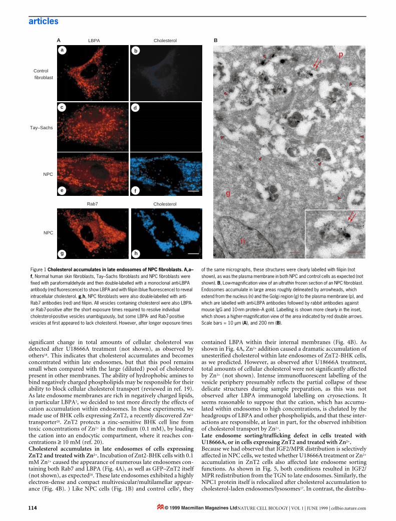
ResultsCholesterol accumulates in late endosomes in Niemann–Pick typeC fibroblasts. Electron-dense multivesicular and/or multilamellarstructures are often observed in cells affected by lysosomal storagediseases. In the human autosomal recessive Niemann–Pick type Cdisease (NPC), an abundance of these structures is accompanied byintracellular accumulation of unesterified cholesterol6. The precisefunction of the protein encoded by the NPC gene is not known,although it is presumably involved in the regulation of cholesteroltransport7–10. Accumulated cholesterol is conveniently detected bylight microscopy after cell fixation using filipin as a fluorescentmarker11 (Fig. 1A). In skin fibroblasts from NPC patients, accumu-lated cholesterol is present within vesicles containing both the smallGTPase Rab7, a late endosomal marker12, and LBPA (Fig. 1Ae–h).This is in contrast to control fibroblasts (Fig. 1Aa,b) or fibroblastsfrom patients with Tay–Sachs disease (Fig. 1Ac,d), an autosomalrecessive lipidosis resulting in ganglioside GM2 accumulation.Immunogold labelling of cryosections from NPC cells showed that
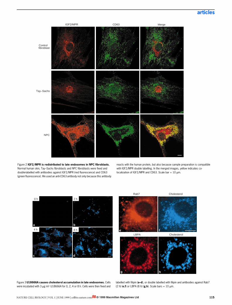
late endosomes in these cells (Fig. 1B) revealed the same character-istic distribution of LBPA within their internal membranes as didnormal BHK cells5. Although LBPA is increased in the liver andspleen of NPC patients13, we found similar amounts of LBPA inNPC and control fibroblasts (not shown). This agrees well withrecent observations14, and supports the view that lipid metabolismvaries between tissues in NPC patients13. Altogether, our observa-tions demonstrate that NPC is not only a lysosomal, but also anendosomal, storage disorder.NPC fibroblasts have a late endosome sorting/trafficking defect.This finding has important implications, as late endosomes, unlikelysosomes, are not the end-point of the endocytic pathway. One ofthe main functions of late endosomes is the sorting of the multi-functional receptor (IGF2/MPR) for ligands bearing mannose 6-phosphate, which include lysosomal enzymes and insulin-likegrowth factor 2 (ref. 15). IGF2/MPR delivers newly synthesized lys-osomal enzymes from the trans-Golgi network (TGN) to late endo-somes, and then recycles back to the TGN for reutilization. Wefound that, at steady state, IGF2/MPR localized predominantly tothe TGN in control fibroblasts or Tay–Sachs fibroblasts (Fig. 2). InNPC fibroblasts, however, the receptor was largely redistributed toabundant vesicles identified as late endosomes using CD63 as amarker16 (Fig. 2). Immunogold labelling of cryosections showedthat IGF2/MPR then co-localized with LBPA within internal mem-branes of large multivesicular endosomes (not shown). Some IGF2/MPR remained in the Golgi region of NPC cells (Fig. 2), perhapscorresponding, at least in part, to newly synthesized molecules.These experiments indicate that cholesterol accumulation in theNPC disease is accompanied by an endosomal sorting/traffickingdefect, in agreement with studies suggesting that NPC fibroblastsare deficient in vesicle-mediated clearance of endocytosed[14C]sucrose17.Cholesterol accumulates in late endosomes after U18666A treat-ment. The accumulation of LDL-derived cholesterol characteristicof NPC cells can be mimicked after treatment of healthy cells overa short time with hydrophobic amines such as U18666A (ref. 18).When BHK cells were treated with U18666A, cholesterol accumu-lated rapidly (within 2–8 h) in late endosomes that contained bothLBPA and Rab7 (Fig. 3), much as in NPC fibroblasts. However, no
M
NATURE CELL BIOLOGY | VOL 1 | JUNE 1999 | cellbio.nature.com 113© 1999 Macmillan Magazines Ltd
articles
significant change in total amounts of cellular cholesterol wasdetected after U18666A treatment (not shown), as observed byothers18. This indicates that cholesterol accumulates and becomesconcentrated within late endosomes, but that this pool remainssmall when compared with the large (diluted) pool of cholesterolpresent in other membranes. The ability of hydrophobic amines tobind negatively charged phospholipids may be responsible for theirability to block cellular cholesterol transport (reviewed in ref. 19).As late endosome membranes are rich in negatively charged lipids,in particular LBPA5, we decided to test more directly the effects ofcation accumulation within endosomes. In these experiments, wemade use of BHK cells expressing ZnT2, a recently discovered Zn2+
transporter20. ZnT2 protects a zinc-sensitive BHK cell line fromtoxic concentrations of Zn2+ in the medium (0.1 mM), by loadingthe cation into an endocytic compartment, where it reaches con-centrations ≥ 10 mM (ref. 20).Cholesterol accumulates in late endosomes of cells expressingZnT2 and treated with Zn2+. Incubation of Znt2-BHK cells with 0.1mM Zn2+ caused the appearance of numerous late endosomes con-taining both Rab7 and LBPA (Fig. 4A), as well as GFP–ZnT2 itself(not shown), as expected20. These late endosomes exhibited a highlyelectron-dense and compact multivesicular/multilamellar appear-ance (Fig. 4B). ) Like NPC cells (Fig. 1B) and control cells5, they
contained LBPA within their internal membranes (Fig. 4B). Asshown in Fig. 4A, Zn2+ addition caused a dramatic accumulation ofunesterified cholesterol within late endosomes of ZnT2-BHK cells,as we predicted. However, as observed after U18666A treatment,total amounts of cellular cholesterol were not significantly affectedby Zn2+ (not shown). Intense immunofluorescent labelling of thevesicle periphery presumably reflects the partial collapse of thesedelicate structures during sample preparation, as this was notobserved after LBPA immunogold labelling on cryosections. Itseems reasonable to suppose that the cation, which has accumu-lated within endosomes to high concentrations, is chelated by theheadgroups of LBPA and other phospholipids, and that these inter-actions are responsible, at least in part, for the observed inhibitionof cholesterol transport by Zn2+.Late endosome sorting/trafficking defect in cells treated withU18666A, or in cells expressing ZnT2 and treated with Zn2+.Because we had observed that IGF2/MPR distribution is selectivelyaffected in NPC cells, we tested whether U18666A treatment or Zn2+
accumulation in ZnT2 cells also affected late endosome sortingfunctions. As shown in Fig. 5, both conditions resulted in IGF2/MPR redistribution from the TGN to late endosomes. Similarly, theNPC1 protein itself is relocalized after cholesterol accumulation tocholesterol-laden endosomes/lysosomes17. In contrast, the distribu-
Figure 1 Cholesterol accumulates in late endosomes of NPC fibroblasts. A,a–f, Normal human skin fibroblasts, Tay–Sachs fibroblasts and NPC fibroblasts were fixed with paraformaldehyde and then double-labelled with a monoclonal anti-LBPA antibody (red fluorescence) to show LBPA and with filipin (blue fluorescence) to reveal intracellular cholesterol. g,h, NPC fibroblasts were also double-labelled with anti-Rab7 antibodies (red) and filipin. All vesicles containing cholesterol were also LBPA- or Rab7-positive after the short exposure times required to resolve individual cholesterol-positive vesicles unambiguously, but some LBPA- and Rab7-positive vesicles at first appeared to lack cholesterol. However, after longer exposure times
of the same micrographs, these structures were clearly labelled with filipin (not shown), as was the plasma membrane in both NPC and control cells as expected (not shown). B, Low-magnification view of an ultrathin frozen section of an NPC fibroblast. Endosomes accumulate in large areas roughly delineated by arrowheads, which extend from the nucleus (n) and the Golgi region (g) to the plasma membrane (p), and which are labelled with anti-LBPA antibodies followed by rabbit antibodies against mouse IgG and 10-nm protein–A gold. Labelling is shown more clearly in the inset, which shows a higher-magnification view of the area indicated by red double arrows. Scale bars = 10 µm (A), and 200 nm (B).
NPC
NPC
Rab7 Cholesterol
Tay Sachs
Cholesterol
Control
fibroblast
LBPA
_
A B
g h
e f
c d
a b p
g
n
114 NATURE CELL BIOLOGY | VOL 1 | JUNE 1999 | cellbio.nature.com© 1999 Macmillan Magazines Ltd
articles
NATURE CELL BIOLOGY | VOL 1 | JUNE 1999 | cellbio.nature.com 115
Figure 2 IGF2/MPR is redistributed to late endosomes in NPC fibroblasts. Normal human skin, Tay–Sachs fibroblasts and NPC fibroblasts were fixed and double-labelled with antibodies against IGF2/MPR (red fluorescence) and CD63 (green fluorescence). We used an anti-CD63 antibody not only because this antibody
reacts with the human protein, but also because sample preparation is compatible with IGF2/MPR double labelling. In the merged images, yellow indicates co-localization of IGF2/MPR and CD63. Scale bar = 10 µm.
IGF2/MPR CD63 Merge
Controlfibroblast
Tay Sachs
NPC
_
Figure 3 U18666A causes cholesterol accumulation in late endosomes. Cells were incubated with 3 µg ml-1 U18666A for 0, 2, 4 or 8 h. Cells were then fixed and
labelled with filipin (a–d), or double labelled with filipin and antibodies against Rab7 (2 h) (e,f) or LBPA (8 h) (g,h). Scale bars = 10 µm.
8 h
d
2 h
b
0 h
a4 h
c
Rab7 Cholesterol
CholesterolLBPA
e f
g h
© 1999 Macmillan Magazines Ltd

articles
tion of the protein TGN38, which also cycles between TGN andendosomes, was not affected by Zn2+ treatment (not shown), indi-cating that intralumenal accumulation of cations did not cause gen-eral perturbations of endosomal membranes, and suggesting thatTGN38 may recycle from early, and not late, endosomes.
After U18666A addition, cholesterol accumulation was rapid(8 h, see Fig. 3), as expected from the known action of the drug18.In contrast, IGF2/MPR redistribution occurred more slowly (24–48 h, see Fig. 5). Similarly, after Zn2+ addition cholesterol wasaffected before IGF2/MPR (Fig. 4,5), although Zn2+ accumulationwithin endosomes required longer incubation. These observationssuggest that the IGF2/MPR-sorting defect is a consequence, ratherthan a cause, of impaired cholesterol transport, and demonstratethat late endosomes have a critical role in intracellular cholesterolmovement.Cholesterol accumulates in late endosomes of cells treated with amonoclonal antibody against LBPA. In previous studies weshowed that a well-characterized monoclonal antibody againstLBPA, when internalized by fluid-phase endocytosis into livingcells, selectively accumulated in late endosomes upon binding to itsantigen5. This treatment caused late endosomes to become elec-tron-dense and compact, and IGF2/MPR to redistribute from TGNto late endosomes. These striking similarities prompted us to testthe effect of our antibody on cholesterol transport. As shown in Fig.6A, treatment with the anti-LBPA antibody resulted in massiveaccumulation of cholesterol, much as in NPC cells or cells treatedwith U18666A or Zn2+. However, as with our observations afterZn2+ or U18666A treatment, total amounts of cellular cholesterolwere not significantly affected by internalization of anti-LBPA anti-bodies (not shown). Cholesterol then co-localized with the endocy-tosed antibody within late endosomes. In contrast, the distributionof cholesterol (not shown) or IGF2/MPR5 was not affected after
Figure 4 Zn2+ causes cholesterol accumulation in late endosomes of cells expressing ZnT2. A, Cells expressing ZnT2 were incubated with 0.1 mM Zn2+ for 0, 1, 2 or 3 days, fixed, and then labelled with filipin (a–d), or double labelled with antibodies against LBPA and Rab7 at day 2 (e,f), and against LBPA and filipin at day 3 (g,h). Differences in the appearance of late endosomes containing LBPA, Rab7 and cholesterol between human NPC fibroblasts (Fig. 1A), BHK cells treated with U18666A (Fig. 3) or BHK cells expressing ZnT2 and treated with Zn2+ may reflect, at least in part, differences in the preservation of the fragile endosomal
ultrastructure after preparation of the samples for light microscopy. B, The upper panels show thin-section electron micrographs of control cells (left) and ZnT2-BHK cells treated for 3 days with Zn2+ (right), which causes a massive accumulation of huge electron-dense multivesicular structures throughout the cells. The bottom panel shows an ultrathin frozen section of these structures labelled with anti-LBPA antibodies (as in Fig. 1B). Labelling efficiency is lower than in NPC cells, perhaps because of reduced accessibility after Zn2+ accumulation; (m) mitochondrion. Scale bars = 10 µm (A) and 200 nm (B).
7
B
a b
c d
2 d 3 d
LBPA rab7
LBPA Cholesterol
e
g h
f
A 0 d 1 d
Figure 5 IGF2/MPR is redistributed to late endosomes after treatment with U18666A or Zn2+. BHK cells were incubated with 3 µg ml-1 U18666A for 0 (control, a,b) or 48 h (c,d), fixed, and double labelled with antibodies against IGF2/MPR and Lgp120/Lamp1 (refs 5,21). We used an anti-Lgp120/Lamp1 antibody, because this antibody reacts with the BHK protein and because sample preparation is compatible with IGF2/MPR double-labelling. BHK cells expressing ZnT2 were incubated with 0.1 mM Zn2+ for 3 days, fixed, and double labelled with filipin and antibodies against IGF2/MPR (e,f). Scale bars = 10 µm.
IGF2/MPR Lgp120/Lamp1
Control
U18666A
IGF2/MPR Cholesterol
ZnT2
a
c
e
b
d
f
116 NATURE CELL BIOLOGY | VOL 1 | JUNE 1999 | cellbio.nature.com© 1999 Macmillan Magazines Ltd

articles
endocytosis of control antibodies, including a monoclonal anti-body against Lgp120/Lamp1, which is a major constituent of thelimiting membranes of the same compartment5,21. Much as afterU18666A treatment, cholesterol accumulated rapidly after anti-body addition (∼ 4–8 h, see Fig. 6A), whereas ≥ 24-h incubationswere necessary to cause IGF2/MPR redistribution5, supporting thenotion that IGF2/MPR trafficking was affected by cholesterol accu-mulation. More important, these data show that selective perturba-tion of LBPA-rich membrane domains is involved in the regulationof intracellular cholesterol transport.Cholesterol accumulates in late endosomes of cells treated withsera from patients with anti-phospholipid syndrome. We had pre-viously reported that antibodies from patients suffering from theanti-phospholipid (aPL) syndrome22,23 specifically recognized LBPAin vivo and in vitro, and altered IGF2/MPR sorting when endocy-tosed by living cells, much like the monoclonal anti-LBPAantibody5. As shown in Fig. 6B, treatment with sera from patients,but not with sera from healthy individuals, caused significant accu-mulation of cholesterol within late endosomes containing LBPA,which was both reminiscent of Niemann–Pick type C, and similarto that observed with the monoclonal anti-LBPA antibody. We pre-viously speculated that antibodies associated with the aPL syn-drome affect IGF2/MPR sorting by altering LBPA-rich membranedynamics5. In the light of our new observations, it is attractive topropose that such alterations result from cholesterol accumulation.Our observations also suggest that endosome functions may beaffected in a similar manner in the genetic NPC disease and in theaPL syndrome, despite the fact that the onset of these diseases isentirely different.
DiscussionInternal, LBPA-rich membranes account for the vast majority oftotal late endosomal membranes, and form an extensive lumenalnetwork5. Thus, newly internalized cholesterol, which is first hydro-lysed to unesterified cholesterol, is very likely to partition preferen-
tially within these internal membranes when released into thelumen. Then, free cholesterol is rapidly redistributed intracellularly,as it does not accumulate within late endosomes under normalsteady-state conditions. However, intralumenal accumulation ofcations, which is predicted to affect the structures of internal mem-branes, causes cholesterol accumulation. More important, choles-terol accumulation in late endosomes is caused by highly specificperturbations of internal membranes with a monoclonal antibodyagainst LBPA, or with human antibodies which recognize LBPA.Thus, cholesterol transport depends not only on its intrinsic proper-ties (diffusion, flip-flop), but also on the physicochemical anddynamic properties of LBPA-rich internal membranes. Accumula-tion of cholesterol within LBPA-rich internal membranes is pre-dicted to alter membrane properties even further, and to contributeto the observed inhibition of IGF2/MPR-mediated sorting and traf-ficking. Consistent with this notion, IGF2/MPR accumulates withininternal membranes rich in LBPA in cells treated with anti-LBPAantibodies5. Cholesterol accumulation in late endosomes, and theresulting sorting/trafficking defect, may also alter transfer from lateendosomes to lysosomes, as recent studies suggest that this stepoccurs via the formation of a hybrid organelle3. Although the role ofthe NPC protein is not known, an attractive hypothesis is that itfunctions as a sensor of the endosomal cholesterol content7–10.
In conclusion, we propose that the network of LBPA-containinginternal membranes within late endosomes functions, in effect, as acollecting and distribution device, thereby controlling intracellularcholesterol transport. In addition, our observations support thenotion that LBPA-rich membrane domains are affected in bothNPC and the aPL syndrome, resulting in cholesterol accumulationwithin late endosomes and similar endosomal sorting/traffickingdefects. h
MethodsBaby hamster kidney (BHK-21) cells, BHK cells expressing ZnT2, and their parental zinc-sensitive line 3286-8-8 (ref. 20) were grown and maintained as described24. Cultured skin fibroblasts from patients with NPC25 or Tay–Sachs disease26 and from healthy subjects were established and maintained as
Figure 6 Antibodies against LBPA cause cholesterol accumulation in late endosomes. A, Cells were incubated with the monoclonal antibody against LBPA (50 µg ml-1) for 0, 4, 8 or 24 h, fixed, and labelled with filipin (a–d), or double labelled with anti-mouse antibodies (to visualize the endocytosed anti-LBPA antibody) and filipin (arrows point at examples of double-labelled structures) (e,f). B, Typical examples of cells incubated with sera from patients with the anti-phospholipid syndrome (a,b) or
with control sera (c,d), and then double labelled, after fixation, with antibody against LBPA and with filipin. Lower LBPA labelling intensity when compared with A is likely to reflect competition for LBPA molecules between the anti-LBPA monoclonal antibody used for detection and the internalized human antibodies, as both recognize LBPA5. Arrows point at double-labelled structures, and arrowheads at structures that do not seem to contain high levels of LBPA. Scale bars = 10 µm.
A B0 h 4 h
8 h 24 h
LBPA
LBPA
Cholesterol
Cholesterol
Patient
Control
a
a
c
c
e
d
d
f
b
b
NATURE CELL BIOLOGY | VOL 1 | JUNE 1999 | cellbio.nature.com 117© 1999 Macmillan Magazines Ltd

articles
described. Diagnosis was confirmed by the typical clinical manifestations and by lysosomal enzyme assays and filipin staining11. Briefly, double labelling with filipin and antibodies was performed on cells fixed with 3% paraformaldehyde11. Cells were incubated with the first antibodies in PBS containing 10% FCS and 50 µg ml-1 filipin, washed, and then incubated with the second antibodies in PBS-FCS only. Human antisera from patients with antiphospholipid syndrome were a gift from P. de Moerloose (University Hospital of Geneva). The monoclonal antibodies against LBPA5 and Lgp120/Lamp1 (ref. 21) have been described, as well as the antibody against Rab7 (ref. 5). Polyclonal antibodies against IGF2/MPR and TGN38 were gifts from B. Hoflack (Institut Pasteur, Lille) and G. Banting (University of Bristol), respectively. The monoclonal antibody against CD63 was from Chemicon. U18666A was from Biomol, and filipin from Sigma. Immunofluorescence microscopy5, and electron microscopy27 were performed as described.
RECEIVED 26 MARCH 1999; REVISED 26 APRIL 1999; ACCEPTED 4 MAY 1999; PUBLISHED 15 MAY 1999.
1. Goldstein, J. L., Brown, M. S., Anderson, R. G. W., Russell, D. W. & Schneider, W. J. Receptor-
mediated endocytosis. Annu. Rev. Cell Biol. 1, 1–40 (1985).
2. Gruenberg, J. & Maxfield, F. Membrane transport in the endocytic pathway. Curr. Opin. Cell Biol. 7,
552–563 (1995).
3. Mullock, B. M., Bright, N. A., Fearon, C. W., Gray, S. R. & Luzio, J. P. Fusion of lysosomes with late
endosomes produces a hybrid organelle of intermediate density and is NSF dependent. J. Cell Biol.
140, 591–601 (1998).
4. Kobayashi, T., Gu, F. & Gruenberg, J. Lipids and lipid domains in endocytic membrane traffic. Semin.
Cell Dev. Biol. 9, 517–526 (1998).
5. Kobayashi, T. et al. A lipid associated with the antiphospholipid syndrome regulates endosome
structure/function. Nature 392, 193–197 (1998).
6. Liscum, L. & Klansek, J. J. Niemann–Pick disease type C. Curr. Opin. Lipidol. 9, 131–5 (1998).
7. Loftus, S.K. et al. Murine model of Niemann–Pick C disease: mutation in a cholesterol homeostasis
gene. Science 277, 232–235 (1997).
8. Carstea, E. D. et al. Niemann–Pick C1 disease gene: homology to mediators of cholesterol
homeostasis. Science 277, 228–231 (1997).
9. Watari, H. et al. Niemann–Pick C1 protein: obligatory roles for N-terminal domains and lysosomal
targeting in cholesterol mobilization. Proc. Natl Acad. Sci. USA 96, 805–810 (1999).
10. Patel, S. C. et al. Localization of Niemann–Pick C1 protein in astrocytes: implications for neuronal
degeneration in Niemann–Pick type C disease. Proc. Natl Acad. Sci. USA 96, 1657–1662 (1999).
11. Sokol, J. et al. Type C Niemann–Pick disease. Lysosomal accumulation and defective intracellular
mobilization of low density lipoprotein cholesterol. J. Biol. Chem. 263, 3411–3417 (1988).
12. Chavrier, P., Parton, R. G., Hauri, H. P., Simons, K. & Zerial, M. Localisation of low molecular weight
GTP binding proteins to exocytic and endocytic compartments. Cell 62, 317–329 (1990).
13. Pentchev, P. G., Vanier, M. T., Suzuki, K. & Patterson, M. in The Metabolic and Molecular Bases of
Inherited Disease (eds Scriver, C. R., Beaudet, W. S. & Sly, D.) 2625–2639 (McGraw-Hill, New-York,
1995).
14. Koike, T. et al. Decreased membrane fluidity and unsaturated fatty acids in Niemann–Pick disease
type C fibroblasts. Biochim. Biophys. Acta 1406, 327–335 (1998).
15. Kornfeld, S. Structure and function of the mannose-6-phosphate/insulin-like growth factor II
receptors. Annu. Rev. Biochem. 61, 307–330 (1992).
16. Escola, J. M. et al. Selective enrichment of tetraspan proteins on the internal vesicles of multivesicular
endosomes and on exosomes secreted by human B lymphocytes. J. Biol. Chem. 273, 20121–20127
(1998).
17. Neufeld, E. B. et al. The Niemann–Pick C1 protein resides in a vesicular compartment linked to
retrograde transport of multiple lysosomal cargo. J. Biol. Chem. 274, 9627–9635 (1999).
18. Liscum, L. & Faust, J. R. The intracellular transport of low density lipoprotein-derived cholesterol is
inhibited in Chinese hamster ovary cells cultured with 3-beta-[2-(diethylamino)ethoxy]androst-5-
en-17-one. J. Biol. Chem. 264, 11796–11806 (1989).
19. Pentchev, P.G. et al. The Niemann–Pick C lesion and its relationship to the intracellular distribution
and utilization of LDL cholesterol. Biochim. Biophys. Acta 1225, 235–243 (1994).
20. Palmiter, R. D., Cole, T. B. & Findley, S. D. ZnT-2, a mammalian protein that confers resistance to
zinc by facilitating vesicular sequestration. EMBO J. 15, 1784–1791 (1996).
21. Aniento, F., Emans, N., Griffiths, G. & Gruenberg, J. Cytoplasmic dynein-dependent vesicular
transport from early to late endosomes. J. Cell Biol. 123, 1373–1388 (1993).
22. Asherson, R. A., Cervera, R., Piette, J.-C., Shoenfeld, Y. (Eds) The Antiphospholipid Syndrome (CRC
Press, Boca Raton, New York, London, Tokyo, 1996).
23. Alarcon-Segovia, D. & Cabral, A. R. The antiphospholipid/cofactor syndromes. J. Rheumatol. 23,
1319–1322 (1996).
24. Gruenberg, J., Griffiths, G. & Howell, K. E. Characterisation of the early endosome and putative
endocytic carrier vesicles in vivo and with an assay of vesicle fusion in vitro. J. Cell Biol. 108, 1301–
1316 (1989).
25. Omura, K. et al. Type C Niemann–Pick disease: clinical and biochemical studies on six cases. Brain
Dev. 11, 57–61 (1989).
26. Sakuraba, H. et al. GM2 gangliosidosis AB variant: clinical and biochemical studies of a Japanese
patient. Neurology 52, 372–377 (1999).
27. Griffiths, G., McDowell, A., Back, R. & Dubochet, J. On the preparation of cryosections for
immunocytochemistry. J. Ultrastruct. Res. 89, 65–78 (1984).
ACKNOWLEDGEMENTS
We thank R. Gagescu, G. van der Goot, F. Perez, and M. Rojo for critical reading of the manuscript. This
work was supported by the Swiss National Science Foundation (J.G.), the NHMRC of Australia (R.G.P.)
and by the International Human Frontier Science Program (J.G., R.G.P., T.K).
Correspondence and requests for materials should be addressed to J.G.
118 NATURE CELL BIOLOGY | VOL 1 | JUNE 1999 | cellbio.nature.com© 1999 Macmillan Magazines Ltd