55152
TRANSCRIPT

7/28/2019 55152
http://slidepdf.com/reader/full/55152 1/16
IP 914
NATIONAL INSTITUTE FOR HEALTH ANDCLINICAL EXCELLENCE
INTERVENTIONAL PROCEDURES PROGRAMME
Interventional procedure overview of balloondilatation of the Eustachian tube
Treating a blocked Eustachian tube using a small inflatable balloon
The Eustachian tube is a narrow tube that connects the middle ear with theback of the nose. If it is blocked, symptoms such as muffled hearing, pain, afeeling of fullness in the ear, tinnitus or dizziness may occur. In balloon
dilatation of the Eustachian tube, a flexible tube with a small balloon isinserted into the Eustachian tube through the nose. The balloon is filled withsaline (salt water) and left in place for a short time, with the aim of wideningthe Eustachian tube.
Introduction
The National Institute for Health and Clinical Excellence (NICE) has preparedthis overview to help members of the Interventional Procedures AdvisoryCommittee (IPAC) make recommendations about the safety and efficacy of aninterventional procedure. It is based on a rapid review of the medical literature
and specialist opinion. It should not be regarded as a definitive assessment of the procedure.
Date prepared
This overview was prepared in March 2011.
Procedure name
Balloon dilatation of the Eustachian tube
Specialty societies
ENT UK – The British Association of Otorhinolaryngologists, Head and
Neck Surgeons.

7/28/2019 55152
http://slidepdf.com/reader/full/55152 2/16
IP 914
Description
Ind icat ions and cu rrent treatment
Chronic Eustachian tube dysfunction
The Eustachian tube is a narrow tube that connects the middle ear with theback of the nose. If it is blocked, symptoms such as muffled hearing, pain, afeeling of fullness in the ear, tinnitus or dizziness may occur. The Eustachiantube typically becomes blocked following the onset of an upper respiratorytract infection or allergic rhinitis. It is usually a temporary problem thatresolves spontaneously, but sometimes symptoms persist and treatment isnecessary. Long-term Eustachian tube dysfunction is associated with damageto the eardrum and middle-ear transformer mechanism.
Medical treatments include oral and nasal steroids, decongestants andantihistamines. Autoinflation is a technique whereby the Eustachian tube isreopened by raising pressure in the nose. This can be achieved in a number of ways, including forced exhalation against a closed mouth and nose.
If Eustachian tube dysfunction persists, insertion of a tympanostomy tube(also known as a ventilation tube or grommet) through a small incision in theeardrum may be indicated. Repeated tube insertions may be required. Long-acting tubes are sometimes used but these are subject to crusting, infection,obstruction and permanent tympanic membrane perforation.
What the proc edure involves
Preoperative tubomanometry is initially used to assess the level of Eustachiantube dysfunction. Balloon dilatation of the Eustachian tube is usuallyperformed with the patient under general anaesthesia. A balloon catheter isintroduced into the Eustachian tube via the nose, under transnasalendoscopic vision. Once the balloon is correctly positioned in the cartilaginousand bony portion of the Eustachian tube, it is filled with saline up to a pressureof about 10 bars. Pressure is maintained for approximately 2 minutes. Theballoon is then emptied and removed.
The aim of the procedure is to widen the Eustachian tube and improve itsfunction.
Assessing Eustachian tube dysfunction
In tubomanometry, the opening of the Eustachian tube and the transportationof gas into the middle ear is registered by a pressure sensor in the occludedouter ear after applying the stimulus of a controlled gas bolus into thenasopharynx during swallowing. If tube opening is registered, the time of opening in relation to pressure applied can be measured (opening latencyindex or index R). An R value of < 1 indicates early opening of the Eustachiantube, which is considered optimal.

7/28/2019 55152
http://slidepdf.com/reader/full/55152 3/16
IP 914
The Eustachian Tube Score is a summation point score based on the patient’shistory and tubomanometry results. It ranges from 0 (worst value) to 10 (bestvalue):
2 points 1 point 0 points
Clicking noise duringswallowing
Always Infrequent Never
Clicking noise duringValsalva manoeuvre
Always Infrequent Never
Tubomanometry 30 mbar R < 1 R ≥ 1 No opening
Tubomanometry 40 mbar R < 1 R ≥ 1 No opening
Tubomanometry 50 mbar R < 1 R ≥ 1 No opening
Literature review
Rapid review o f l i terature
The medical literature was searched to identify studies and reviews relevant toballoon dilatation of the Eustachian tube. Searches were conducted of thefollowing databases, covering the period from their commencement to30 March 2011: MEDLINE, PREMEDLINE, EMBASE, Cochrane Library andother databases. Trial registries and the Internet were also searched. Nolanguage restriction was applied to the searches (see appendix C for detailsof search strategy). Relevant published studies identified during consultation
or resolution that are published after this date may also be considered for inclusion.
The following selection criteria (table 1) were applied to the abstractsidentified by the literature search. Where selection criteria could not bedetermined from the abstracts the full paper was retrieved.
Table 1 Inclusion criteria for identification of relevant studies
Characteristic Criteria
Publication type Clinical studies were included. Emphasis was placed onidentifying good quality studies.
Abstracts were excluded where no clinical outcomes werereported, or where the paper was a review, editorial, or alaboratory or animal study.
Patient Patients with Eustachian tube dysfunction.
Intervention/test Balloon dilatation of the Eustachian tube.
Outcome Articles were retrieved if the abstract contained informationrelevant to the safety and/or efficacy.
Language Non-English-language articles were excluded unless they werethought to add substantively to the English-language evidence
base.

7/28/2019 55152
http://slidepdf.com/reader/full/55152 4/16

7/28/2019 55152
http://slidepdf.com/reader/full/55152 5/16
IP 914
IP overview: Balloon dilatation of the Eustachian tube Page 5 of 16
Table 2 Summary of key efficacy and safety findings on balloon dilatation of the Eustachian tube
Abbreviations used: CT, computed tomography
Study details Key efficacy findings Key safety findings Comments
Ockermann T (2010)
Case series
Germany, UK
Recruitment period: not reported
Study population: adults with symptoms of chronic obstructiveEustachian tube dysfunction or adults who had undergone oneor more recent tympanoplasties for relapsing acute or chronicpersistent otitis media with effusion as a consequence of obstructive tube dysfunction.
n = 8 patients (13 Eustachian tubes)
Mean age: 44 years (range 21 –81)
Sex: 50% (4/8) female
Patient selection criteria: patients were over 18 years old andnot pregnant. Tubomanometry was used to quantify thedegree of Eustachian tube dysfunction. All patients underwenta transnasal endoscopic examination of their Eustachian tubesand microscopic examination of their ears. Preoperative CTdid not show any significant findings.
Technique: A modified endoscope with suction and a workingchannel was used. The tip of the endoscope was placedadjacent to the pharyngeal ostium of the Eustachian tube anda balloon catheter (Spiggle & Theiss, Germany) was pushedthrough the working channel of the endoscope, under vision,2 cm into the Eustachian tube. Pressure of 10 bars wasapplied for 2 minutes.
Follow-up: 2 months
Conflict of interest/source of funding: none
Number of patients analysed: 8 (13 Eustachiantubes)
Successful catheter placement = 100% (8/8)
The Eustachian tube score relied on thepatient’s history (clicking noise during
swallowing or Valsalva manoeuvre) andtubomanometry results. The score ranged from0 (worst value) to 10 (best value).
Mean Eustachian tube score:
Preoperative = 1.08 (± 0.61)
1 week postoperative = 4.15 (± 3.02)
2 weeks postoperative = 5.85 (± 2.61)
8 weeks postoperative = 7.54 (± 1.39)p < 0.05 between preoperative andpostoperative scores at all follow-up periods.
At 1-week follow-up, Eustachian tube openingwas found in all 3 tubomanometrymeasurements (30 mbar, 40 mbar and 50 mbar)in 7 cases.
All patients answered positively to the questionof whether they would undergo the procedure
again.
There were no intraoperative or postoperative complications.
Postoperative CT revealed noevidence of change in the bonylumen of the Eustachian tube.
Follow-up issues:
All patients underwerepeated computedtomography (CT) beand after the proced
Study design issues:
CT scans were anafor any abnormal finby 2 independent stradiologists.

7/28/2019 55152
http://slidepdf.com/reader/full/55152 6/16
IP 914
IP overview: Balloon dilatation of the Eustachian tube Page 6 of 16
Abbreviations used: CT, computed tomography
Study details Key efficacy findings Key safety findings Comments
Yu V (2010)2
Case series
Country: not reported
Recruitment period: 2009
Study population: patients with persistent middle-ear effusions,
aural fullness or other middle-ear disease.
n = 30 patients (53 Eustachian tubes)
Age: not reportedSex: not reported
Patient selection criteria: not reported
Technique: endoscopic placement of a 5 mm sinus ballooncatheter into the proximal Eustachian tube. 75% of procedureswere performed in the operating theatre at the same time asanother procedure, 25% were performed under localanaesthesia in the office.
Follow-up: not reported
Conflict of interest/source of funding: not reported
Number of patients analysed: 30(53 Eustachian tubes)
60% of patients reported that their aural fullnesshad resolved.
No patients reported any pain,hearing loss or infection.
Study design issues:
Conference abstracdetails of study desare given.

7/28/2019 55152
http://slidepdf.com/reader/full/55152 7/16
IP 914
IP overview: Balloon dilatation of the Eustachian tube Page 7 of 16
Abbreviations used: CT, computed tomography
Study details Key efficacy findings Key safety findings Comments
Poe D (2010)3
Case series
Country: not reported
Recruitment period: 2009
Study population: adults with longstanding otitis media with
effusion or tympanic membrane atelectasis.
n = 12 patients
Age: not reportedSex: not reported
Patient selection criteria: patients were unable to autoinsufflatetheir Eustachian tube by Valsalva, swallow or yawn.
Technique: balloon dilatation of the cartilaginous Eustachiantube was performed under general anaesthetic in a daysurgery setting. Inflation was to a maximum of 12 atm for 1minute.
Follow-up: not reported
Conflict of interest/source of funding: not reported
Number of patients analysed: 12
All Eustachian tubes were successfully dilated.
Mucoid otorrhoea persisted in 1/8 patients.
All patients could autoinsufflate after theprocedure.
All atelectases resolved.
The procedures were welltolerated, without pain or complications related to dilation.
Study design issues:
Conference abstracdetails of study desare given.
Study population issu
11 patients hadpreviously been treawith tympanoplastie
the time of the proc6 patients had a tubplace. 2 patients hatympanic membranperforation.

7/28/2019 55152
http://slidepdf.com/reader/full/55152 8/16
IP 914
IP overview: Balloon dilatation of the Eustachian tube Page 8 of 16
Eff icacy
Eustachian tube function.
A case series of 8 patients with 13 treated Eustachian tubes reported that themean Eustachian tube score (range 0 –10) improved from 1.08 at baseline to4.15, 5.85 and 7.54 at 1, 2 and 8 weeks postoperatively, respectively (p < 0.05) 1.
At 1-week follow-up, Eustachian tube opening was found in all 3 tubomanometrymeasurements (30 mbar, 40 mbar and 50 mbar) in 7 cases1.
Safety
A case series of 8 patients with 53 treated Eustachian tubes reported that therewere no intraoperative or postoperative complications1.
A case series of 30 patients described in a conference abstract reported that nopatients had any postoperative pain, hearing loss or infection2. Another caseseries of 12 patients described in a conference abstract reported that theprocedures were well tolerated, without pain or complications3.
Val idi ty and g eneralisabi l i ty of the stud ies
Only 1 small case series published as a full article was identified1. Two further
small case series were described in published conference abstracts; these
provided limited information on the study methodology2,3
.
There is a lack of long-term data.
Exist ing assessments of th is proc edure
There were no published assessments from other organisations identified at thetime of the literature search.
Related NICE gu idanc e
Below is a list of NICE guidance related to this procedure. Appendix B gives
details of the recommendations made in each piece of guidance listed.
Interventional procedures
Balloon catheter dilation of paranasal sinus ostia for chronic sinusitis. NICEinterventional procedures guidance 273 (2008). Available fromwww.nice.org.uk/guidance/IPG273

7/28/2019 55152
http://slidepdf.com/reader/full/55152 9/16
IP 914
IP overview: Balloon dilatation of the Eustachian tube Page 9 of 16
Clinical guidelines
Surgical management of children with otitis media with effusion in children.NICE clinical guideline 60 (2008) (Note: this only refers to children younger than 12 years). Available from www.nice.org.uk/guidance/CG60
Specialist Advisers’ opinions
Specialist advice was sought from consultants who have been nominated or ratified by their Specialist Society or Royal College. The advice received is their individual opinion and does not represent the view of the society.
Mr A Banerjee, Mr R Irving, Mr C Raine (ENT UK - British Association of Otorhinolaryngologists, Head and Neck Surgeons).
All 3 Specialist Advisers describe the procedure as definitely novel and of
uncertain safety and efficacy.
The comparator to the procedure would be insertion of ventilation tubes or
laser tuboplasty.
Theoretical adverse events include damage to the Eustachian tube(scarring,
stenosis), ear infection, pain, bleeding (carotid rupture), and permanent
conductive hearing loss.
Adverse events reported in the literature include mucosal tears in the
cartilaginous portion of the Eustachian tube.
The key efficacy outcomes are improvement of tubal function, resolution of
symptoms, and fluid drainage from the middle ears.
There are uncertainties about the use of the procedure in children.
There are no long-term follow-up data available.
Two Advisers consider the potential impact on the NHS to be minor and 1
Specialist Adviser thought that the potential impact was major.
Patient Commentators’ opinions
NICE’s Patient and Public Involvement Programme was unable to gather patient
commentary for this procedure.
7/28/2019 55152
http://slidepdf.com/reader/full/55152 10/16
IP 914
IP overview: Balloon dilatation of the Eustachian tube Page 10 of 16
Issues for consideration by IPAC
To date, the procedure has only been used to treat adults.

7/28/2019 55152
http://slidepdf.com/reader/full/55152 11/16
IP 914
IP overview: Balloon dilatation of the Eustachian tube Page 11 of 16
References
1. Ockermann T, Reineke U, Upile T et al. (2010) Balloon dilatationEustachian tuboplasty: a clinical study. Laryngoscope 120: 1411 –6.
2. Yu V, Jonnalagadda S, Catalano P. (2010) Balloon catheter dilation of Eustachian tube: pilot study. Otolaryngology –Head and Neck Surgery143: 86.
3. Poe D, Silvola J. (2010) Balloon dilation of the cartilaginous Eustachiantube. Otolaryngology –Head and Neck Surgery 143: 87.

7/28/2019 55152
http://slidepdf.com/reader/full/55152 12/16
IP 914
IP overview: Balloon dilatation of the Eustachian tube Page 12 of 16
Appendix A: Additional papers on balloon dilatation of the Eustachian tube
There were no additional papers identified.

7/28/2019 55152
http://slidepdf.com/reader/full/55152 13/16
IP 914
IP overview: Balloon dilatation of the Eustachian tube Page 13 of 16
Appendix B: Related NICE guidance for balloon
dilatation of the Eustachian tube
Guidance Recommendations
Interventional procedures Balloon catheter dilation of paranasal sinus ostia for chronic sinusitis. NICE interventional proceduresguidance 273 (2008).1.1 Current evidence on the short-term efficacy of ballooncatheter dilation of paranasal sinus ostia for chronicsinusitis is adequate and raises no major safety concerns.Therefore, this procedure can be used provided that normalarrangements are in place for clinical governance, consentand audit.1.2 This procedure should only be carried out by surgeonswith experience of complex sinus surgery, and specifictraining in both the procedure and the use of fluoroscopy.
1.3 Publication of long-term outcomes will be helpful inguiding the future use of this technique. NICE may reviewthe procedure upon publication of further evidence.
Clinical guidelines Surgical management of children with otitis media witheffusion (OME). NICE clinical guideline 60 (2008)1.4.1 Children with persistent bilateral OME documentedover a period of 3 months with a hearing level in the better ear of 25 –30 dBHL or worse averaged at 0.5, 1, 2 and 4kHz (or equivalent dBA where dBHL not available) shouldbe considered for surgical intervention.1.4.2 Exceptionally, healthcare professionals shouldconsider surgical intervention in children with persistentbilateral OME with a hearing loss less than 25 –30 dBHLwhere the impact of the hearing loss on a child’sdevelopmental, social or educational status is judged to besignificant.1.5 Surgical interventions
1.5.1 Once a decision has been taken to offer surgicalintervention for OME in children, the insertion of ventilationtubes is recommended. Adjuvant adenoidectomy is notrecommended in the absence of persistent and/or frequentupper respiratory tract symptoms.1.5.2 Children who have undergone insertion of ventilationtubes for OME should be followed up and their hearingshould be re-assessed.1.6 Non-surgical interventions
1.6.2 Autoinflation may be considered during the activeobservation period for children with OME who are likely tocooperate with the procedure.1.6.3 Hearing aids should be offered to children withpersistent bilateral OME and hearing loss as an alternative

7/28/2019 55152
http://slidepdf.com/reader/full/55152 14/16
IP 914
IP overview: Balloon dilatation of the Eustachian tube Page 14 of 16
to surgical intervention where surgery is contraindicated or not acceptable.1.7 Management of OME in children with Down’ssyndrome
1.7.1 The care of children with Down’s syndrome who aresuspected of having OME should be undertaken by a
multidisciplinary team with expertise in assessing andtreating these children.1.7.2 Hearing aids should normally be offered to childrenwith Down’s syndrome and OME with hearing loss. 1.7.3 Before ventilation tubes are offered as an alternativeto hearing aids for treating OME in children with Down’ssyndrome, the following factors should be considered:• the severity of hearing loss
• the age of the child
• the practicality of ventilation tube insertion
• the risks associated with ventilation tubes
• the likelihood of early extrusion of ventilation tubes.
1.8 Management o f OME in chi ldren w ith cleft palate
1.8.1 The care of children with cleft palate who aresuspected of having OME should be undertaken by thelocal otological and audiological services with expertise inassessing and treating these children in liaison with theregional multidisciplinary cleft lip and palate team.1.8.2 Insertion of ventilation tubes at primary closure of thecleft palate should be performed only after careful otologicaland audiological assessment.1.8.3 Insertion of ventilation tubes should be offered as an
alternative to hearing aids in children with cleft palate whohave OME and persistent hearing loss.1.9 Information fo r chi ldr en, parents and carers
1.9.1 Parents/carers and children should be giveninformation on the nature and effects of OME, including itsusual natural resolution.1.9.2 Parents/carers and children should be given theopportunity to discuss options for treatment of OME,including their benefits and risks.1.9.3 Verbal information about OME should besupplemented by written information appropriate to thestage of the child’s management.
7/28/2019 55152
http://slidepdf.com/reader/full/55152 15/16
IP 914
IP overview: Balloon dilatation of the Eustachian tube Page 15 of 16
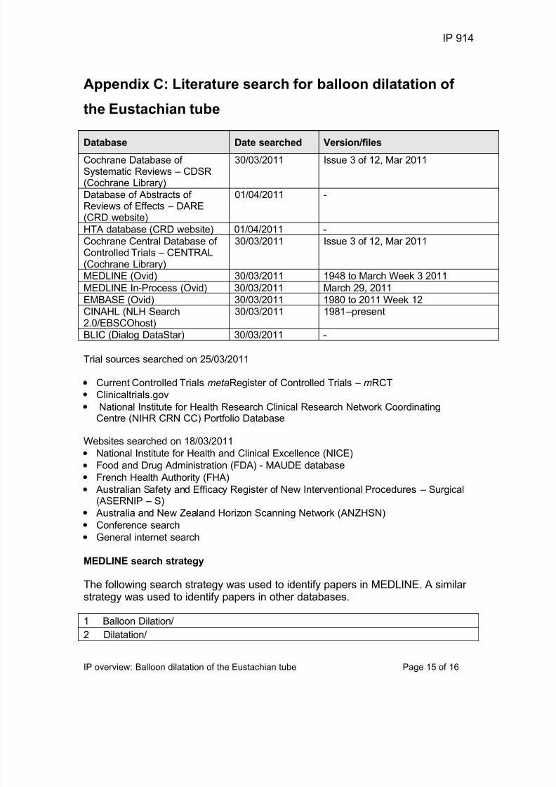
Appendix C: Literature search for balloon dilatation of
the Eustachian tube
Database Date searched Version/files
Cochrane Database of Systematic Reviews – CDSR(Cochrane Library)
30/03/2011 Issue 3 of 12, Mar 2011
Database of Abstracts of Reviews of Effects – DARE(CRD website)
01/04/2011 -
HTA database (CRD website) 01/04/2011 -
Cochrane Central Database of Controlled Trials – CENTRAL(Cochrane Library)
30/03/2011 Issue 3 of 12, Mar 2011
MEDLINE (Ovid) 30/03/2011 1948 to March Week 3 2011MEDLINE In-Process (Ovid) 30/03/2011 March 29, 2011
EMBASE (Ovid) 30/03/2011 1980 to 2011 Week 12CINAHL (NLH Search2.0/EBSCOhost)
30/03/2011 1981 –present
BLIC (Dialog DataStar) 30/03/2011 -
Trial sources searched on 25/03/2011
Current Controlled Trials metaRegister of Controlled Trials – mRCT
Clinicaltrials.gov
National Institute for Health Research Clinical Research Network CoordinatingCentre (NIHR CRN CC) Portfolio Database
Websites searched on 18/03/2011
National Institute for Health and Clinical Excellence (NICE)
Food and Drug Administration (FDA) - MAUDE database
French Health Authority (FHA)
Australian Safety and Efficacy Register of New Interventional Procedures – Surgical(ASERNIP – S)
Australia and New Zealand Horizon Scanning Network (ANZHSN)
Conference search
General internet search
MEDLINE search strategy
The following search strategy was used to identify papers in MEDLINE. A similar strategy was used to identify papers in other databases.
1 Balloon Dilation/
2 Dilatation/

7/28/2019 55152
http://slidepdf.com/reader/full/55152 16/16
IP 914
IP overview: Balloon dilatation of the Eustachian tube Page 16 of 16
3 Dilatation, Pathologic/
4 catheterization/ or catheterization, peripheral/
5 (Balloon* adj3 (dilat* or cathet*)).tw.
6 BET.tw.
7 (Cathet* adj3 peripher*).tw.
8 (Dilat* adj3 pathologic*).tw.
9 or/1-8
10 Eustachian tube/
11 exp Ear Diseases/
12 exp Otitis Media/
13 (Middle ear* adj3 (inflamm* or infect* or disease* or effus* or atelectas*)).tw.
14 ((Eustach* or auditory or pharyngotympanic* or obstructive or chronic) adj3 tubeadj3 (dysfunct* or obstruct* or block* or inflamm* or disorder*)).tw.
15 (otit* adj3 media*).tw.
16 or/10-15
17 9 and 1618 Animals/ not humans/
19 17 not 18