course1.winona.educourse1.winona.edu/twilson/documents/lab 5 respiratory... · web viewdetermine...
TRANSCRIPT

Biology 212: Anatomy and Physiology IILab #5: Respiratory System Anatomy and Measurement of Blood Pressure
There are two very important constants that exist in most tissues of the human body, #1: all tissues require oxygen (O2) in order to generate ATP; and #2: these tissues generate carbon dioxide (CO2) as a waste product during ATP generation. As a result, the body needs an efficient way to both supply O2
and eliminate CO2. Our circulatory system transports blood to the lungs where blood gases are exchanged. The alveolar surfaces of the lungs are where most gas exchange occurs with blood passing through the capillaries that lie immediately next to the alveoli. So, it is easy to see that the respiratory and circulatory systems are intimately linked to one another. Without the circulatory system, the respiratory system is useless. Without the respiratory system, the circulatory system is also useless (since we need O2 to generate ATP and stay alive!).
In order to exchange O2 and CO2 efficiently, several processes must occur 24 hours a day, 7 days a week. First, we must exchange the CO2-rich/ O2-depleted gases in our lungs with O2-rich/ CO2-depleted air from the external atmosphere (pulmonary ventilation). Second, there must be an exchange of gases between the gases in our lungs and those gases in the blood (alveolar gas exchange). Third, these gases must be delivered to the peripheral tissues (mostly the job of the circulatory system). Fourth, gases need to be exchanged between the blood and peripheral tissues.
Pulmonary ventilation and alveolar gas exchange occur exclusively within the respiratory system. The delivery of gases between the tissues and the lungs occurs partly within the respiratory system via the movement of blood through the pulmonary arteries and veins. In the well-ventilated lung hemoglobin found within the erythrocytes becomes saturated with oxygen before the blood is delivered back to the left atrium through the pulmonary vein.
Organs of the respiratory system:The respiratory system consists of two broad divisions based on their anatomical and physiological functions. The CONDUCTING portion of the respiratory system functions to condition and transport air between the external atmosphere and the lungs, and the RESPIRATORY portion of the respiratory system, is where gas exchange at the alveolar-capillary interface actually occurs.
EXERCISE #1: Conducting division anatomy Air travels from the external environment into either the nose (primary) or mouth (secondary). As air enters the nasal cavity large particles are filtered by the hairs within the nose and the air then encounters the nasal conchae which generate turbulence in the air flow and causes it to swirl around. As the air swirls around, some of the particulate matter (dirt, dust, allergens, pathogens) stick to the moist surface of the mucous membranes lining the nasal cavity, but just the simple contact with the membrane humidifies the air prior to entering the NASOPHARYNX. After leaving the nasopharynx, the air travels through the OROPHARYNX and then the LARYNGOPHARYNX before reaching the LARYNX (a.k.a., the voicebox). After passing through the larynx, the air enters the TRACHEA. The trachea is a large, tube-like structure that transmits air to the PRIMARY BRONCHI, which then branches to carry air to the left or right lung. Once inside the lung, the primary bronchi give off many branches which decrease in diameter with each division. Most of the larger and mid-sized bronchi are surrounded by cartilaginous supports, which limit changes in conduit diameter during inhalation and exhalation. The terminal bronchioles are the smallest of the conduction system airways.
Using Figures 22.1, 22.3, 22.4, and 22.7, the torso models (bullet points) and the larynx models (check marks) you should be able to identify the following and discuss how inhaled air moves from the largest to
Nasal cavity Naso Oropharynx
Laryngopharynx Epiglottis

Larynx Thyroid cartilage Cricoid cartilage Vestibular fold Vocal cord
Esophagus: non-respiratory
Trachea Tracheal cartilage Carina Primary bronchi Secondary bronchi Tertiary bronchi
Note: You should also be able to identify the above structures using the provided models (bronchial tree, larynx) and torso models.
EXERCISE #2: Respiratory division anatomyOnce air enters the more distal portion of the lungs via the conducting division, it is able to participate in the exchange of O2 and CO2 in the RESPIRATORY DIVISION. Distal to the terminal bronchioles (part of the conducting division), we encounter the respiratory bronchioles, alveolar ducts, and ultimately, the alveoli (pleural). While a minimal amount of actual gas exchange occurs in the respiratory bronchioles and alveolar ducts, the alveolus is the primary area for gas exchange. A key adaptation that allows the lung alveolus (singular) to exchange gas so efficiently is the extremely thin structure of the RESPIRATORY MEMBRANE. This is not a classic cell membrane, rather this membrane is composed of multiple layers including: TYPE 1 ALVEOLAR EPITHELIAL CELL, the acellular basement membrane, as well as the endothelium of the capillary bed that surrounds each alveolus.
Using Figures 22.11 and 22.12, be sure to be able to identify these structures and how they differ in terms of anatomy, the presence of cilia, and where gas exchange can occur:
Bronchiole Respiratory bronchiole Alveolus
Also, be sure to understand the organization of the respiratory membrane and the layers of tissue that gases need to cross in order to get from the bloodstream into the interior of the alveolus (and vice versa). For oxygen to pass from the alveolar sac to hemoglobin inside a erythrocyte, the O2
must pass into and out of the alveolar epithelial cell (two membrane bilayers), diffuse through the basement membrane and then into and out of the endothelial cell (simple squamous epithelial tissue) that lines the blood capillary (two membrane bilayers) before crossing the membrane of the erythrocyte.
EXERCISE #3: Gross anatomy of the lungThe lungs are situated in the lateral portions of the thoracic cavity and are surrounded by two layers of serous membrane. One membrane lies directly on top of the lung and is referred to as the VISCERAL PLEURA, while the other layer lines the thoracic cavity and is known as the PARIETAL PLEURA. Between these two layers is a potential space known as the PLEURAL CAVITY, which contains a thin, slippery fluid known as pleural fluid (about 15 ml/pleural cavity). One function of this fluid is to allow the lungs to move within the pleura sac in a relatively frictionless environment.
You may remember that the heart is not situated perfectly centered within the thorax. Rather, the heart tends to be slightly angled towards the left half of the body and as a result occupies more space on the left side of the thorax than the right. To accommodate this, the left lung is slightly smaller than the right lung in order to accommodate the positioning of the heart.
Identify the Lobes of the Lung: The left lung consists of two lobes (SUPERIOR and INFERIOR), while the right lung consists of three lobes (SUPERIOR, MIDDLE, and INFERIOR). The left lung is

separated by one fissure (OBLIQUE FISSURE), while the right lung is separated by two fissures: one HORIZONTAL FISSURE and one OBLIQUE FISSURE.
Identify the Surfaces of the Lung: The lung exhibits several surfaces which are named for the structures they face. There is a DIAPHRAGMATIC surface (inferior, faces the diaphragm), a COSTAL surface (lateral, anterior, and posterior, faces the rib cage), and a MEDIASTINAL surface (medial surface, basically facing the heart and trachea). On the mediastinal surface, you will find the HILUM. The hilum is the name given to the indented region of the lung where the primary bronchi and pulmonary arteries enter the lung, and the pulmonary veins exit the lung. In addition to the primary bronchi and pulmonary vessels, a number of lymphatic vessels and nerves also enter the lung here.
A. Using Thorax Model and Figures 22.9 and A.8 (atlas A on pg. 35 of Saladin), you should be able to identify (great test questions):
Trachea Primary bronchi Superior, middle, inferior lobes Horizontal and Oblique fissures Costal surface Mediastinal surface
Diaphragmatic surface Hilum Visceral pleura Parietal pleura Pleural cavity Cardiac impression
B. Gross Anatomy of Human Preserved Lungs and Fresh Pig Heart-Lung tissues: In addition, we also have preserved specimens of actual human lungs (GREAT practice but not on test). And we have fresh sheep or pig heart-lung combos. When handling these specimens, it is important to wear disposable gloves and only handle the lung specimens on the provided dissecting trays. You are encouraged to squeeze, inflate, pull, overinflate the bronchi you can find with the equipment provided. With instructor permission feel free to cut as well. Note how each bronchi ventilated only a very specific part of each lung or lung lobe. On these specimens, you are responsible for identifying:
Superior, middle, inferior lobes Horizontal and oblique fissures Costal surface Mediastinal surface Diaphragmatic surface Hilum Cardiac impression
C. Gross Anatomy: Examination of the Female Cadaver. (GREAT practice but not on test) Identify the larynx in the neck and palpate (feel) both its thyroid cartilage and cricoid
cartilage. Follow the trachea inferiorly from the neck into the thorax. Palpate the cartilage rings
which hold it open. Notice the trachealis muscle on the posterior surface. Note that the trachea passes posteriorly to the heart and great vessels. These structures
block your view of where the trachea divides into right and left primary bronchi, but from figures in your textbook you should understand where that division should be on the cadaver.
Note the position of the lungs within the thoracic cavity, surrounding the heart. Identify all of the lobes, surfaces and fissures of both lungs which you identified on the
preserved specimens above.
EXERCISE #4: Histology of the lung

Some portions of the respiratory system are also shared by the digestive system and will transmit both food/drink as well as air (mainly the oropharynx and laryngopharynx). These tissues need to withstand a certain amount of abrasion and are thus lined by a non-keratinized, stratified squamous epithelium. Most of the remainder of the conducting portion of the respiratory system is lined by pseudostratified columnar epithelium possessing many cilia extending from their surface. Scattered in between the epithelial cells are GOBLET cells which function to produce mucus. This mucus (produced by goblet cells, as well as actual mucus glands) lines the epithelia of the conducting airways and traps much of the dirt, debris, allergens, and pathogens before they can reach the alveoli. The cilia of the pseudostratified columnar epithelium then “sweep” the debris-laden mucus up towards the esophagus where it can be swallowed. The method of eliminating particulate matter is known as the MUCOCILIARY ELEVATOR.
As you travel more distally into the lung, the diameter of the bronchi decreases, the amount of hyaline cartilage surrounding the bronchi decreases, and the shape of the epithelial cells gradually transitions from pseudostratified columnar to cuboidal (respiratory bronchioles) to simple squamous (within the alveoli).
Within the alveolus, we find three main types of cells: the ALVEOLAR MACROPHAGES which clear debris and pathogens from the alveoli; the TYPE 2 ALVEOLAR CELLS which produce pulmonary surfactant; and finally the TYPE 1 ALVEOLAR EPITHELIAL CELLS which participate primarily in gas exchange. The simple squamous epithelium of the alveolus in conjunction with the basement membrane and endothelium of the lung capillary form the RESPIRATORY MEMBRANE, across which gas exchange occurs.
Using Figures 22.7, 22.11, and 22.12, you should be able to identify these structures and their physiological function: Respiratory membrane Type 1 alveolar epithelial cell Type 2 alveolar epithelial cell Alveolar macrophage
Hyaline cartilage ring (in trachea and bronchi)
Pseudostratified columnar epithelium
Histology: In addition to the above figures from your textbook, you should also examine the slides of lung and trachea that are found in your slide boxes (slides #2 and #7). On these slides you should be able to identify all of the previous structures except the respiratory membrane and alveolar macrophage, although you certainly may be able to see one! Go to this website for more:Lung Histology picture from: http://adademutia.blogspot.com/2011/02/organs-histology.htm
Trachea X40 Slide #7 Trachea lining X400 Slide#7 Lung X100 Slide #2

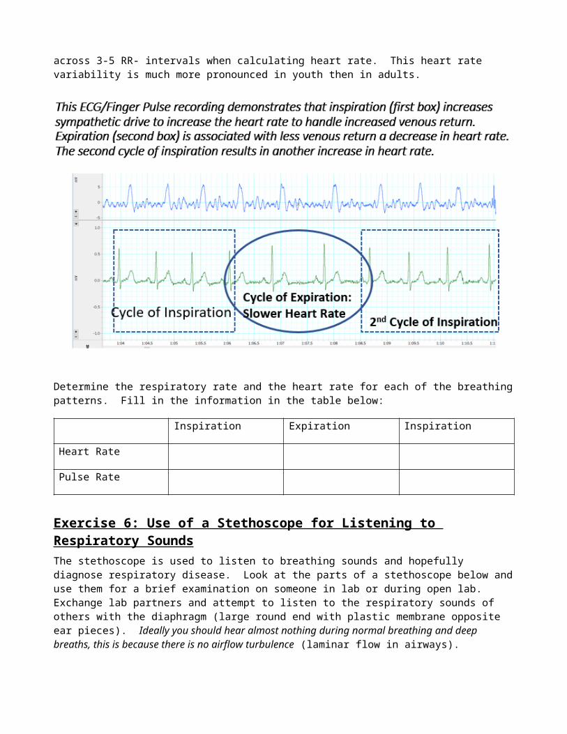
Exercise 5: Integration of ventilation with heart rate There is a relationship between the heart rate and ventilation patterns that is intended to optimize efficiency. When the thoracic cavity expands and a negative pressure is created, air enters the lungs. This negative pressure can also create pressure gradients that deliver more blood from the vena cava into the heart. During expiration the reverse occurs and the SA node receives less sympathetic stimulation, the rate of autorhythmicity decreases and the heart rate drops because venous return drops (your heart would have less blood to pump out). This causes entirely normal variations in the RR-interval, this also explains why is it important to average across 3-5 RR- intervals when calculating heart rate. This heart rate variability is much more pronounced in youth then in adults.
Determine the respiratory rate and the heart rate for each of the breathing patterns. Fill in the information in the table below:
Inspiration Expiration Inspiration
Heart Rate
Pulse Rate
Exercise 6: Use of a Stethoscope for Listening to Respiratory SoundsThe stethoscope is used to listen to breathing sounds and hopefully diagnose respiratory disease. Look at the parts of a stethoscope below and use them for a brief examination on someone in lab or during open lab. Exchange lab partners and attempt to listen to the respiratory sounds of others with the diaphragm (large round end with plastic membrane opposite ear pieces). Ideally you should hear almost nothing during normal breathing and deep breaths, this is because there is no airflow turbulence (laminar flow in airways).

The movement of air through the bronchi can become turbulent in places with restrictions to flow (i.e. asthma and bronchitis) these restrictions create turbulent air flow and sounds referred to as “Wheezes”. What would happen if an asthmatic had “wheezing” and then used their albuterol inhaler (a beta-2 agonist that promotes bronchodilation). If inflammation was the cause of wheezing and you administered a steroid like cortisone, what would happen to the sounds?
Another serious sound is called “Rales” or “Crackles”, which occurs when air in the alveoli and small bronchioles must “bubble up” through mucous of edema during exhalation, this sound is commonly associated with and accumulation of fluid in the small airways that is often associated with pneumonia or left side heart failure. Rales and crackles are often heard in the nursing home in a patient just before death resulting from a severe left side heart attack or left side heart failure. Ominously pulmonary edema can also occur even in normal healthy people upon ascent to high altitude (for example a family ski trip to a place like Winter Park Colorado at 13,000 feet). The respiratory events that cause a high pulmonary blood pressure and edema are poorly understood, but the observation of rales and crackles is a very dangerous and potentially fatal, just as it is in the elderly.
Examples of breathing sounds relevant for nursing practice are reviewed at this website: Give this a listen at home! https://www.youtube.com/watch?v=U8byn2NT_lo
Exercise 7: HEART SOUNDS, PULSE RATE AND SYSTEMIC BLOOD PRESSURE:Work in groups when you measure the Heart Sounds, Pulse Rate, and Systemic Blood Pressures. It will be easier for you to do these if you can coax a lab partner who has used these methods before to help you get started. Do this set of exercises in the last 30 minutes of lab or in the Open Labs.
A. Heart Sounds:The heart typically makes two sounds that correlate with the closure of the valves of the heart. Sounds can also be created whenever there is turbulence in blood (think of the sounds you hear when water rolls out of a dam into a turbulent pool). Take the stethoscope and listen to the sounds of the heart. The stethoscope has a pair of earpieces that set gently in your ears and a bell with a plastic diaphragm that sits on the surface where you want to listen. There should be two heart sounds in normal, healthy hearts. The first sound in each cardiac cycle is a “Lub” which is created by the closure of the right and left atrioventricular valves. The second sound is a sharp-ending “Dup” sound, which is created by the closure of the right and left semilunar valves. (Some diseases can result in valves closing at different times to create a third or fourth heart sound, such as when right ventricular pressure is increased in congestive heart failure and the pulmonary semilunar valve closes at a later time than the aortic semilunar valve.)
Try This In Lab: Listen to the heart sounds of a volunteer or yourself. Compare the heart sounds that you hear when the bell is placed on the left or right side of the heart. Compare sounds from the bell side of the stethoscope with sound from the diaphragm side. (The bell side favors low-frequency sounds and the diaphragm enhances higher-frequency sounds.) Do the heart sounds on the two sides of the heart sound alike or different? Why? How can the heart sounds be used to verify that the heart is positioned more to the left side of the thoracic cavity? Also Note: if your volunteer inhales and exhales deeply you may also hear some sounds that are created by respiration, especially if they have bronchitis. Also, compression of the heart with forced exhalation or extension of the heart with the vacuum of inhalation will change heart sounds.
You may also hear a heart murmur if the valves of the heart do not function correctly. If the AV valve did not close, the sound would appear right after the “lub” because turbulent blood is accidentally shooting back into the atrium. If you hear a heart murmur right after the second sound, it would likely be from a failure of the aortic semilunar valve to close properly, resulting in turbulent blood flow leaking from the aorta (high pressure) back into the relaxing left ventricle (low pressure).
B. Heart Rate: What are four clinical tools for measuring heart and pulse rate?Just because the heart depolarizes and contracts, does it HAVE to create a pulse? What if the stroke volume is very small or what if the semilunar valve never opened? For these reasons, it is dangerous to use an ECG as a measure of pulse rate. The two are different. The use of a radial pulse is a commonly used technique to determine the pulse rate.#1) Finger Pulse Oximeter (HR and %SpO 2): is a device (See below) that fits over the end of the index finger with the nail facing up (towards print) and finger pad facing down. An infrared or red light is directed through the tissue and the absorbance of the transmitted light is measured. These devices measure oxyhemoglobin and deoxyhemoglobin, but does not measure other hemoglobin species (like carbon monoxyhemoglobin). Place finger in groove, close oximeter and record the pulse rate and the percent oxygen saturation of the blood in the finger after 15 seconds. Typically the saturation will be around 94-98%. Note: If there is paint on the fingernail, light cannot pass through and the system will not work.
#2) Stethoscope: You listen for the number of beats (“lub”-AV valve closure + “dub”-semilunar valve closure = 1 cycle) heard in 15 seconds. Take this number and multiply it by 4 to get beats/minute based on heart sounds. For example: 22 sounds/15 seconds X 60 seconds/minute = 88 beats/minute.
#3) Radial Pulse Gently hold the wrist of your volunteer in the palm of your hand such that both palms face “up”. Place your index and middle fingers on the lateral surface of their wrist and feel the radius bone. Gently move the finger pressure point medially until you detect a pulse. Measure the pulses you feel in a 15 second period and multiply this number times four to get pulse rate

(pulse/minute). Ask around and find someone near you who has done this before if you need help. Remember that when you measure the pulse, a longer measurement period will give you a more accurate pulse rate measurement.
While a pulse rate is easy to determine by palpating the carotid arteries of the neck, this is dangerous for elderly patients! Sometimes a patient (especially the elderly) may have atherosclerotic plaque in this vessel and simply pressing on the surface of the vessel could cause a rupture in the plaque. If the plaque ruptures a piece can easily break off and travel downstream, causing a cerebral artery to become occluded and this could result in a severe stroke. You would not want to cause brain damage in a patient as a result of simply measuring their pulse rate, so please remember this.#4) Electrocardiogram (ECG): We looked at this last week, please review that lab. Please review the electrical conduction pathway, and how to measure heart rate, intervals and wave amplitudes.
C. Systemic Blood Pressure:Background: Since the left ventricle expels blood in a pulsatile fashion, both blood flow and blood pressure in the aorta and other large arteries is pulsatile. This means that a bolus of blood will be pushed through the system followed by another bolus. As a result our blood flow is not smooth or continuous. Systolic pressure is the highest pressure reached during ventricular systole. Diastolic pressure is the lowest pressure reached during diastole of the ventricle. Remember that it takes a bit of time for the wave of pressure (and blood flow) to move from the semilunar valve out to the location where the pulse was registered (felt).
The typical normal value for blood pressure in a normal person is about 120mmHg/80mmHg. It is important to know that blood pressure tends to increase as a result of aging. The reality is that if we live long enough, most of us will develop hypertension (high blood pressure). The increase with aging is more prominent in women after tehir 40s, after menopause estrogen levels drop and this somewhat delays the increase in arterial pressure. Soon after menopause there are few differences in blood pressure in men and women as they age. The figure below illustrates these changes (Burt VL, et al. Prevelance of hypertension in the U.S. adult population. Results from the Third National Health and Nutrition Examination Survey, 1988–1991. Hypertension 1995;25(3):305–13)

What are manual and automated tools for blood pressure measurement?An indirect method for measuring arterial blood pressure involves the use of a stethoscope and a sphygmomanometer. Atrial blood pressure is determined by placing an inflatable rubber cuff, attached to a pressure gauge, around the arm, inflating the cuff to collapse the underlying artery (blood flow stops) and listening over the vessel below the cuff with a stethoscope. When cuff pressure exceeds systolic arterial pressure, the artery collapses and blood flow ceases. As pressure in the cuff is reduced, blood flow through the artery begins again when cuff pressure falls just below systolic arterial pressure. This first blood squeezes through the tiny opening because the pressure in the blood vessel at systole is greater than the pressure in the cuff. Because the blood squeezes through if creates a special turbulence called a Korotkoff Sound. At this point a soft, but sharp tapping sound may be heard with the stethoscope over the artery and the associated cuff pressure is taken as an approximation of the systolic pressure. As cuff pressure is further reduced, the Korotkoff sounds increase in intensity and then suddenly become muffled when the cuff pressure matches the diastolic pressure and the artery remains open throughout the entire pulse cycle. By convention, blood pressure is expressed as a ratio: systolic pressure/diastolic pressure, where each pressure is expressed in mM of Hg. A typical value is approximately 120/80.The aneroid sphygmomanometer measures cuff pressure by means of a spring-operated gauge attached to the cuff. Manual units require a stethoscope and a good sense of hearing. Factors that contribute to possible inaccuracy when using this device includes friction and wear associated with internal moving parts, stretch of the spring with age and repeated use, and a relatively small indicator dial that makes reading more difficult. However, these sphygmomanometers are relatively small, lightweight, and portable.
Manual sphygmomanometer Automated sphygmomanometer

Try This in Lab: Have your volunteer sit up-right with their arm at their side. Open the silver pressure release valve and wrap the cuff around the upper arm of your volunteer, fasten it with the cuff’s Velcro strip. Place the bell of the stethoscope over the cubital fossa (notch between the biceps muscle and the brachioradialis muscle). The bell will now sit over the brachial artery and the bell will be at about the same level as the origin of the aorta. Close the cuff valve and pump the cuff pressure up to 180 mmHg. Placement of the cuff on the upper arm with the cuff level with where you think the heart is located in the thoracic cavity is VERY important. This is because gravity affects blood pressure very distinctly. Each inch above the heart results in a loss of 1.92 mmHg. Each inch below the heart results in an increase of 1.92 mmHg. This principle explains why your sprain ankle throbbed in pain when you were standing up, but the pain and throbbing were reduced when you laid on your back and elevated the ankle above the heart. Blood pressure in an ankle artery could be 200/100 when standing.
Now while listening carefully with the stethoscope, slowly release pressure from the cuff and watch the dial on the sphygmomanometer slowly move to lower and lower cuff pressures. Note the pressure indicated by the dial at the first instant that you hear a snapping turbulence as blood squeezes through the mostly occluded artery (this is the systolic blood pressure when you first hear two repetitive sounds). The faint tapping sound will grow louder as more blood is forced through, then it will grow fainter as you continue to release more cuff pressure and blood is impeded less. The instant the sound is no longer detectable, note the pressure indicated by the dial….this is the diastolic blood pressure.Automated unit are easier to operate. Simply put the arm in the cuff and press the button. The cuff will inflate and the systolic pressure, diastolic pressure and heart rate will appear when the reading is complete. The problem is that automated units while “easier” to use are not always as accurate. The gold standard is and for some time to come always be the manual cuff.
What other terms are used to discuss blood pressure in the clinic? The pulse pressure is the difference between the systolic and diastolic pressures. Mean Arterial Pressure (MAP) is often calculated in order to asses the average pressure present over time on a persons blood vessels. MAP= [Diastolic BP + (Pulse Pressure/3)]. This is important when a clinician wants to consider the severity of a person’s hypertension. Example: If your volunteer had a blood pressure of 190/100, their pulse pressure would be 190-100= 90 mmHg. Their MAP would be [100+ (90/3)]=130 mmHg…. YES this person would have severe hypertension.
For the lab test: How would you measure the blood pressure of the professor? If given a blood pressure, could you measure the pulse pressure and MAP?How does gravity affect blood pressure measurement? If your patient had no arm, or if their arms were in two casts, how could you measure blood pressure? Look at the ankle. As long as they lie flat so ankle is even with the heart, you are good to go for an estimate of blood pressure.

If you measured the blood pressure in the anterior tibial artery of your ankle while standing up (you can do this if your ankle is narrow) what would you expect the blood pressure to be if the pressure in the aortic arch was 120/80? [Assume the ankle was 50 inches below the level of the aortic arch.]
What would the blood pressure be at the top of your brain [assume 20 inches above aortic arch]? Try the math and verify it, and remember that each inch above the heart translates to 1.92 mmHg less arterial pressure, and that each inch below the heart adds an additional 1.92 mmHg (you can approximate this as 2 mmHg/inch).
What are the advantages and disadvantages of each of the above four systems for the assessment of heart rate and pulse rate?