44th annual spring meeting scientific program april …
TRANSCRIPT

Page 1
V/Q Lung Scintigraphy for PE
Mark Tulchinsky, MD, FACNM
Ventilation-Perfusion Scintigraphy: Trinary Versus Probabilistic Interpretation
and SPECT Versus Planar Imaging
Mark Tulchinsky, MD, FACNM�44th ANNUAL SPRING MEETING
SCIENTIFIC PROGRAM�April 11, 2014�
Disclosure: Consultant & Investigator, Bracco Diagnostics Inc.
Alex. Gottschalk, M.D., FACR, FCCP Professor of Radiology
Michigan State University Department of Radiology
2008 Courtesy of Alex Gottschalk, M.D., FACR, FCCP
Is CTA always the method of choice for diagnosing PE ?
Courtesy of Alex Gottschalk, M.D., FACR, FCCP
Introduction – CT and patient irradiation
Courtesy of Alex Gottschalk, M.D., FACR, FCCP
To Refresh Your Memories
Exposure is expressed in mGy [aka RADs]. 10 mGy = 1 RAD Absorbed dose is expressed in mSv [aka REMs] 10 mSv = 1 REM. At the energies of the gammas we are talking about there is no
difference, i.e 1 mGy = 1 mSv
Courtesy of Alex Gottschalk, M.D., FACR, FCCP
CT – An increasing source of radiation exposure*
� 60 million CT exams done in 2002 � BEIR VII says 10 mSv associated with a lifetime
risk of 1 in 1000 for developing a solid CA or leukemia.
� If CT gives a dose of 10 mSv or more, that is 60,000 new cancers
� They ESTIMATE this could be the cause of 1.5% to 2% of US cancer cases
*Brenner DJ, Hall EJ, NEJM 2007; 357: 2277-2284
Courtesy of Alex Gottschalk, M.D., FACR, FCCP

Page 2
V/Q Lung Scintigraphy for PE
Mark Tulchinsky, MD, FACNM
Soaring use of CT worries physicians
Courtesy of Alex Gottschalk, M.D., FACR, FCCP
Alternative strategies to CT*
� BEIR VII says low dose is less then 100 mSv � BEIR VII recent study of Japanese A- bomb
survivors in lowest dose group have increased incidence of cancer. The mean whole body exposure is 29 mSv equiv., the mean organ equiv dose is 34 mSv. There is no safe threshold.
� Leukemia, thyroid, and breast most common Ca. � Latency 10-20 years for breast. � And, 100 mSv causes Cancer in 1 in 100.
*Martin DR, Smelka RC Appl. Radiol. 2007; 36(6) 2007 discussed in Medscape 12-10-07
Courtesy of Alex Gottschalk, M.D., FACR, FCCP
Radiation Exposure of V/Q Scintigraphy vs. MDCTA
The bottom line is that the radiation exposure to the breasts of women in the child bearing age group from MDCTA is somewhere between 65-250 times greater than that from V/Q scintigraphy. Most quotes are in the 70-100 X range.
Additionally, the estimated radiation exposure from a standard 2 view mammogram is associated with 3-4.4 mSv which makes the MDCTA radiation dose approximately 10-20 times greater.
Courtesy of Leonard Freeman, MD
Why Diagnosis of PE is Critically Important? The Answer from the 1970s:
Total PE Incidence 630,000
Survive > 1 Hr 563,000 (89%)
Death in 1Hr 67,000 (11%)
Diagnosis missed 400,000 (71%)
Diagnosis made, treatment started
163,000 (29%)
Survival 280,000 (70%)
Death 120,000 (30%)
Survival 150,000 (92%)
Death 13,000 (8%)
Dalen JE, Alpert JS (1975) Natural history of pulmonary embolism. Prog Cardiovasc Dis 17:259–270
PE Epidemiology – Update, 21st Century • Mortality�
– 3rd cause of cardiovascular death (after myocardial infarction and stroke)�
– Untreated episode: mortality up to 30%�
– On treatment (anticoagulant therapy)�• In clinical trials: � �0.3% for DVT, 1.3% for PE�
• In ‘real-life’ cohorts: �0.6% for DVT, 3.0% for PE�
• Risk of bleeding on treatment �– Major bleed 3.0% / y, fatal bleed 0.5% / y �
Courtesy of Grégoire Le Gal, MD PhD

Page 3
V/Q Lung Scintigraphy for PE
Mark Tulchinsky, MD, FACNM
Diagnosis of PE • Challenge in daily clinical practice�
– Clinical signs and symptoms lack sensitivity and specificity�
– No simple, non-invasive, accurate, widely available, single diagnostic test�
• Important not to misdiagnose patients with suspected VTE�– False-negative: high mortality of untreated PE�
– False-positive: duration and risks of anticoagulant therapy�
• As a result�– Low threshold for clinical suspicion�
– Low proportion of patients with confirmed VTE�
Courtesy of Grégoire Le Gal, MD PhD
What’s the Main Diagnostic Challenge in the 21st Century
Western (US) Medical Practice?
Le Gal, J Thromb Haemost 2004
0% 10% 20% 30% 40% 50% 60%
1960s 1990s 2000s (Europe) 2000s
(North America) 2000s
Year
Prop
ortio
n of
con
firm
ed c
ases
am
ong
patie
nts
with
sus
pect
ed P
E
0
5
10 15
20
25
Num
ber o
f pat
ient
s ne
eded
to b
e in
vest
igat
ed to
find
one
PE
Decreasing Prevalence
Courtesy of Grégoire Le Gal, MD PhD
Is the Low Prevalence the Western Medicine’s Problem?
Prevalence of 59%
It sure looks like it!
What will be the main problem for us for PE diagnosis in the US as of April 2014?
Example from a representative University medical center (circa April 9, 2014)
Radiopharmaceuticals by Jubilant DraxImage�
April 1st, 2014 Price Change� % increase in price�Before� After�
��������� �� �� � ������������������ �� ��� ��������������� �� �� �������������������� ��� � � ����
Why?
Because they can � and we did nothing to object, prevent or reverse it.
If you like to object effectively, drop me an email at [email protected]
Wells’ score for PE History of DVT or PE
+ 1,5 Immobilization or surgery (< 4 weeks)
+ 1,5 Cancer + 1 An alternative diagnosis is less likely + 3 Hemoptysis + 1 Heart rate > 100/min + 1,5 Signs of DVT + 3
Low 0-1; intermediate 2-6; high: ≥ 7 Unlikely ≤ 4; likely > 4
Revised Geneva score Age > 65 years +1 History of DVT or PE +3 Surgery or fracture (<1 month) +2 Cancer +2 Unilateral leg pain +3 Hemoptysis +2 Heart rate
75 - 94 / minute +3 ≥ 95 / minute +5
Pain at calf palpation and swelling +4
Low 0-3 ; intermediate 4-10 ; high ≥ 11+
Wells, Thromb Haemost 2001 ; Le Gal, Ann Intern Med 2006
Clinical Probability Clinical Decision Rules

Page 4
V/Q Lung Scintigraphy for PE
Mark Tulchinsky, MD, FACNM
D-Dimer • D-Dimer�
– Fibrin degradation products�
– Measured through a simple, cheap, fast, blood test�
– Highly sensitive to the presence of a blood clot�• Positive in almost all patients with DVT or PE�
• Low likelihood of VTE if negative�
– Not specific�• Positive in many other conditions than VTE (infection,
surgery, cancer, pregnancy, elderly, etc.)�
• If positive, doesn’t mean that there it an active clot�
Diagnostic Tests for PE
• Gold-standard test for PE: pulmonary angiography (?)�– Invasive (mortality related to the test 1%)�– Expensive�
• Risk of thromboembolic event following a negative pulmonary angiography ≈ 2 %�– Used to validate diagnostic strategies�– A diagnostic strategy is considered to safely
exclude the diagnosis if the 3-month risk of recurrent VTE doesn’t exceed 3%�
van Beek, Clin Radiol 2001
V/Q (Ventilation/Quotient) Imaging
• Classic PE would have no perfusion�• Classic PE should have normal ventilation�• Classic PE mostly happens in textbooks�
• PE may occur in a previously diseased lung – CXR opacities - poorly ventilated and/or perfused – CTPA should be favored, but if no opacities – V/Q should be favored�
• PE may cause hypoxic bronchospasm, decreasing local ventilation�
• PE may cause hemorrhage, decreasing local ventilation (�Hampton�s hump�)�
Ventilation Agents
• 133Xenon gas�• 127Xenon gas�• 81mKrypton gas�• 99mTc DTPA aerosol�• 99mTc Technegas ( not available
in USA)�
Advantages of Xenon Gas
• Offers �Single breath� image, equilibrium image and washout phases of ventilation�
• Washout identifies areas of air trapping in obstructive airway conditions.�
• Washout is the most sensitive part of the test for airway disease�
• 3 minutes is the minimum of re-breathing at equilibrium to produce diagnostic washout study�

Page 5
V/Q Lung Scintigraphy for PE
Mark Tulchinsky, MD, FACNM
S.B. EQ.
Wash-out
POST
RPO LPO
RT LAT LT LAT LAO
ANT RAO
Tc on Tc Tc on Xe
S.B.
RT LAT
EQ.
�Re-Vent� with Xe-133
Xe on Xe
Xe-133 in COPD

Page 6
V/Q Lung Scintigraphy for PE
Mark Tulchinsky, MD, FACNM
Perfusion in COPD
Anterior
LPO Posterior
RAO
LAO Left Lateral
Right Lateral RPO
VENTILATION AGENTS Xenon-133
CHARACTERISTIC���Energy (keV)�Pulmonary Radiation Dose�Spatial Resolution�Sensitivity for COPD�Portability�Disposal�Cost�
81�High (ß)�Poor�Excellent�None�Trap + Neg Pressure�Low�
Disadvantages of Xenon Gas
• Poor energy for imaging, significant internal attenuation (Xe-133)�
• Requires significant patient cooperation�• Adds significant dead space and resistance to
breathing�• Requires a low pressure room and trapping
system�• Cannot be done on ventilator patients�• Long half life when room is contaminated.
Xenon sits in a layer on the floor�
Disadvantages of Xenon Gas (continued)
• Not always available on call�• Not portable�• Hypoxic patient may have to breathe
room air for several minutes�• Can only obtain single projection first
breath images, however, can be repeated in desired view (�re-vent�)�
VENTILATION AGENTS Tc-99m DTPA Aerosol
CHARACTERISTIC Energy (keV)�Pulmonary Radiation Dose�Spatial Resolution�Sensitivity for COPD�Portability�Disposal�Cost�
140�Low�Good�Good�Good�Easy�Low�
Advantages of Aerosols
• Patient is maintained on high flow oxygen throughout the study�
• No resistance to breathing�• Minimal dead space�• No patient cooperation required�
– Works with tidal breathing�– Easily inserted in line with a respirator. Allows
ventilation of a respirator dependent patient without contaminating the respirator�
• No gas trapping equipment required�• Does not require a negative pressure room�
Page 7
V/Q Lung Scintigraphy for PE
Mark Tulchinsky, MD, FACNM
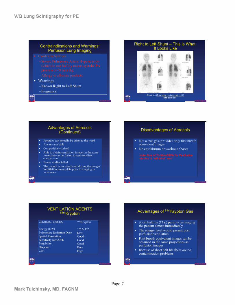
Contraindications and Warnings: Perfusion Lung Imaging
• Contraindication�– Severe Pulmonary Artery Hypertension
(which in our facility means systolic PA pressure > 69 mm Hg)�
– Allergy to albumin products�
• Warnings�– Known Right to Left Shunt�
– Pregnancy�
Right to Left Shunt – This is What It Looks Like
Shunt %= (Total body cts-lung cts) x100 Total body cts
Advantages of Aerosols (Continued)
• Portable, can actually be taken to the ward�• Always available�• Competitively priced�• Able to obtain ventilation images in the same
projections as perfusion images for direct comparison�
• Fewer studies failed�• The patient is not ventilated during the images.
Ventilation is complete prior to imaging in most cases.�
Disadvantages of Aerosols
• Not a true gas, provides only first breath equivalent images�
• No equilibrium or washout phases�
Note: Use of Tc-99m-DTPA for Ventilation studies is “off-label” use!
VENTILATION AGENTS 81mKrypton
CHARACTERISTIC��Energy (keV)�Pulmonary Radiation Dose�Spatial Resolution�Sensitivity for COPD�Portability�Disposal�Cost�
81mKrypton��176 & 192�Low�Good�Good�Good�Easy�High�
Advantages of 81mKrypton Gas
• Short half life (13 s.) permits re-imaging the patient almost immediately �
• The energy level would permit post perfusion ventilation�
• First breath equivalent images can be obtained in the same projections as perfusion images�
• Because of short half life there are no contamination problems�

Page 8
V/Q Lung Scintigraphy for PE
Mark Tulchinsky, MD, FACNM
Disadvantages of 81mKrypton Gas
• Relatively Expensive�• Short half life rubidium generator (4.5
hours)�• Images must be obtained while patient
is being ventilated�• Limited availability, patients must be
scheduled in advance. No availability for after hours studies.�
VENTILATION AGENTS 127Xenon
CHARACTERISTIC� � � 127Xenon��Energy (keV)� � � �203�Pulmonary Radiation Dose� �Moderate�Spatial Resolution � � �Fair�Sensitivity for COPD � �Excellent�Portability � � � �None�Disposal � � � �Trap, - Pressure�Cost � � � � �Moderate�
Ventilation Technique (PSU/HMC)
• 99mTc DTPA Aerosol�• 30-50 mCi in Nebulizer�• 0.8 to 1 mCi to patient�• LEAP or LEHR Collimator�• Patient supine with back to camera�• Aerosolize until 100 K/60 seconds�• Imaging Matrix 256 x 256 W�• Eight Views: Anterior, then every 45º�• If count rate below 50 K/min, re-
aerosolize�
Perfusion Technique (PSU/HMC)
• 99mTc MAA, 6 mCi �• Approximately 250-500 K particles�• >90%of particles10-90micron range
(Mean, 20-40 micron)�• Approximately 0.1% to 0.2% of
Pulmonary capillary bed is embolized�• Inject supine�• Do Not draw blood back into syringe�• Image for 1 minute (minimum) or
longer if 1000K counts are not achieved in 1 minute�
Causes of Perfusion Defects on Lung Scans
• Pulmonary Embolus�• Bulla or Cyst�• Localized Hypoxia (Asthma, Bronchitis)�• Surgery�• Pleural Effusion�• Tumor�• Metastasis�• Hilar Adenopathy�
Causes of Perfusion Defects on Lung Scans
• Pulmonary artery atresia/hypoplasia�• Fibrosing Mediastinitis�• Radiation Therapy�• Pneumonia�• Pulmonary Edema�• Atelectasis�• Pleural Fibrosis�• Vasculitis�

Page 9
V/Q Lung Scintigraphy for PE
Mark Tulchinsky, MD, FACNM
Perfusion Technique (Cont.) (PSU/HMC)
• Imaging Matrix 256 x 256 W�• Eight View Study: Anterior, then every
45º (Posterior, both Laterals, all four Oblique views)�
• Must override ventilation counts at least 5 or 6 to one (preferably more)�
• Must reduce particle load to 120K (not radioactivity) in presence of pulmonary hypertension (<70 mm Hg, systolic)�
Defect Sizing Rules
• Small (subsegmental): < 25% of an anatomic segment�
• Moderate: ≥ 25% but <75% of a segment�• Large: ≥ 75% of a segment (Arithmetic
equivalent: 2 moderate = 1 large)�• Nonsegmental: rounded or irregular
defect which is not wedge shaped and does not correspond to an anatomic segment. Such a defect expected to cross segmental boundaries�
�Matching� Rules
• Perfusion defect can be compared to ether CXR or ventilation study�
• Perfusion defect substantially larger than ventilation abnormality or CXR density is considered �mismatched��
• Equal or greater ventilation or CXR abnormality compared to perfusion defect constitutes �matched��
Upper Lobes: Apical Posterior Anterior
Lower Lobes: Superior Posterior Basal Lateral Basal Anterior Basal
Middle Lobe (right): Lateral Medial
Lingula (left): Superior Inferior
Anterior Right Posterior Oblique
Posterior
Right Anterior Oblique
Left Posterior Oblique
Left Anterior Oblique
Left Lateral
Right Lateral
Segmental Lung Schema
72 y/o male with prior PE on treatment
3/11/2011 3/29/2012
What’s Wrong with this Picture? 04/08/2014

Page 10
V/Q Lung Scintigraphy for PE
Mark Tulchinsky, MD, FACNM
Where is the Tc-99m-MAA Dose? “The Needle-in-Haystack Problem”
100K/1min
170K/1min
90K/1min
116K/1min
Ventilation Images
“Perfusion” Images
04/08/2014
Images of The Injection Site: “The Needle-in-Haystack Problem”
The residual activity in the syringe was about 500 µCi. But ... the needle was discarded before the post calibration assay. The needle was retrieved and activity in it was 5.5 mCi! What happened is Tc-99m-MAA clumped and stuck/wedged in the needle of syringe. The clumped Tc-99m-MAA is porous; hence, it allows the liquid to go through it.
04/08/2014
Repeat Perfusion Scan Following 6 mCi Tc-99m-MAA Injection 04/08/2014
Single mild segmental perfusion defect
SPECT-CT 04/08/2014
Normal V/Q
• Perfusion image is exactly the shape of the lungs as seen on chest X-ray�
• No perfusion defects�
Very Low Probability V/Q
• ≤ 3 small perfusion defects with normal chest X-ray�
• Perfusion defects that are explained by anatomical variants, such as decreased activity over the prominent aortic arch, or in the enlarged cardiac impression, etc.�

Page 11
V/Q Lung Scintigraphy for PE
Mark Tulchinsky, MD, FACNM
Low Probability Lung Scan (Revised PIOPED)
• Nonsegmental perfusion defects, regardless of number, ventilation, or chest X-ray findings�
• Perfusion defect substantially smaller than CXR density, regardless of ventilation�
• Any number of small (subsegmental) perfusion defects, normal chest X-ray�
• V/Q matches, provided CXR is unremarkable and some lung has normal perfusion�
High Probability Lung Scan (Revised PIOPED)
• ≥ 2 large V/Q mismatches or the arithmetic equivalent in moderate or large and moderate�
Indeterminate Lung Scan (Revised PIOPED)
• Single V/Q match�• Anything not covered by low or high
probability categories (< 2 segmental mismatches)�
• Single moderate and up to 1.5 large equivalents in mismatched perfusion defects with normal chest X-Ray.�
• Any collection of matched defects with abnormal enough perfusion that the study would be high probability if ventilation were normal.�
HMC/PSU criteria
Probabilistic Vs. Trinary Interpretation of V/Q Scan
Probabilistic, Such as PIOPED • Normal, very low, low • High (includes single large MM) • Indeterminate/Intermediate _______________________ Confusing for clinicians
Successful and Safe Implementation of a Trinary Interpretation and Reporting Strategy for V/Q Lung Scintigraphy. J. E. Glaser, et. al., and Leonard M. Freeman. J Nucl Med 2011; 52:1508–1512
Trinary Negative for PE Positive for PE Inconclusive Test __________________ Clear for clinicians
Joseph E. Glaser et al. Successful and Safe Implementation of a Trinary Interpretation and Reporting Strategy for V/Q Lung Scintigraphy. J Nucl Med 2011; 52:1508–1512 (Leonard Freeman Group)

Page 12
V/Q Lung Scintigraphy for PE
Mark Tulchinsky, MD, FACNM
Q
V
Q
V
Anterior
LPO Posterior
RAO
LAO Left Lateral
Right Lateral RPO
Anterior
LPO Posterior
RAO
LAO Left Lateral
Right Lateral RPO
Single, Large MM, Positive for PE in Trinary System
Practical PE Algorithm • Basics:�
– As compared to PECT, V/Q scan radiation exposure is 4 times less overall, 100 times less to the breast, but 10 times more to the fetus �
– V/Q is often inconclusive in patients with abnormal CXR, but they do not degrade accuracy of CTPA�
• Practical Algorithm:�– CXR normal or minimally abnormal = V/Q�
– CXR is abnormal = CTPA�
– Females, V/Q will have much less breast exposure�• Pregnant females = CTPA, if contrast allergy do low dose
Q only NO “V” (tracer ends up in the bladder)�
– Abnormal renal function or contrast allergy = V/Q�
Screening for V/Q
• Does the patient have CXR in past 24 hrs?�
• Are there major opacities?�• Does the patient have pulmonary
hypertension (history, CXR, ECG, ECHO)?�
• Does the patient have R-to-L shunt?�• Is the patient pregnant or potentially
pregnant?�
Negative V/Q scan
(278)
Positive DD or high clinical probability CUS (321)
Positive (43)
PE (98)
Normal (46)
No PE No Rx (220)
Low, Clin prob
(156) High 14
CT, 35 (11%)
Negative, 23 Positive
, 9
Low/Interm. 143
Interm, Clin prob
(21) High, Clin prob (54)
Interm./High 46
Low 8 Interm./
High 13
Low 8
Not done, 3
Salaun, Chest 2011
3-month TE risk 0.5% (0.1 to 2.9)
Current diagnostic strategies – V/Q
Clinical probability Wells’ score
Unlikely
D-Dimer
Negative
No PE
V/Q Positive
Inconclusive
US
Neg. Positive PE
High prob
US
Neg. Serial US
Normal
No PE
Current diagnostic strategies – V/Q
Anderson, JAMA 2007
Abbreviations: Neg. = negative US = ultrasound
Clinical probability Wells’ score
V/Q
Inconclusive
CUS Positive
PE
High prob
PE
Normal
No PE
Likely
Negative
DD Negative Positive Serial CUS
3-month TE risk 1.0% (0.5 to 2.1)
Current diagnostic strategies – V/Q
Anderson, JAMA 2007

Page 13
V/Q Lung Scintigraphy for PE
Mark Tulchinsky, MD, FACNM
Pregnancy and V/Q
• A threshold dose of 50 mGy is required for induction of deterministic effects, none of the potential hazards such as fetal death, malformation or mental retardation are a specific risk with the low exposures of ionizing radiation used in diagnostic imaging.�
• Osei EK, Faulkner K. Fetal doses from radiological examinations. Br J Radiol 1999;72:773-80.�
Pregnancy and V/Q
CLINICAL HISTORY: The patient was admitted with complaints of shortness of breath. There is a significant A-a gradient, respiratory alkalosis and hypoxia. Evaluate for pulmonary embolism.
Case of Shortness of Breath
Anterior Chest XR
Lateral Chest XR
Q
V
Q
V Posterior
RAO Anterior
LAO
RPO Right Lateral
Left Lateral LPO

Page 14
V/Q Lung Scintigraphy for PE
Mark Tulchinsky, MD, FACNM
Causes of Unilateral Decreased Perfusion
• Pulmonary Agenesis or stenosis�• Swyer James Syndrome�• Saddle Embolus�• Pneumothorax�• Large Effusions�• Mediastinal Fibrosis (Histoplasmosis)�• Tumor�
Caveat
• A single large perfusion defect consisting of several contiguous segments (with either normal or abnormal ventilation) has a significant likelihood of being due to hilar tumor.�
Congestive Heart Failure
• Enlarged Cardiac Defect (obliterating lingula)�
• Fissure Sign (effusion in fissures)�• Rounded Posterior Gutters or Basilar
Defects (effusion in posterior gutters)�• Redistribution of Flow to Upper Lobes�• Patchy �Checker Board� perfusion
(interstitial edema)�• Focal Non-segmental Defects (alveolar
edema)�
Shortness of Breath
• Patient with known CHF now complains of worsened shortness of breath.�
Q
V
Q
V
Anterior
LPO Posterior
RAO
LAO Left Lateral
Right Lateral RPO
Anterior
LPO Posterior
RAO
LAO Left Lateral
Right Lateral RPO

Page 15
V/Q Lung Scintigraphy for PE
Mark Tulchinsky, MD, FACNM
Diffuse Abnormality on CXR
• More than 75% of patients with diffuse opacity consistent with pulmonary edema have normal or near-normal perfusion on V/Q�
Asthmatic Patient with Increasing Shortness of
Breath CLINICAL HISTORY: History of asthma and increasing shortness of breath for the past three days. PO2 on room air of 32 with PO2 in the 90's on 02 by nasal cannula. Evaluate for pulmonary embolism.
Asthmatic Patient with Increasing Shortness of
Breath CLINICAL HISTORY: History of asthma and increasing shortness of breath for the past three days. PO2 on room air of 32 with PO2 in the 90's on 02 by nasal cannula. Evaluate for pulmonary embolism. CXR: There is a focus of decreased aeration in the right middle lobe which may represent an infectious infiltrate versus a focal area of atelectasis. A more linear horizontal opacity in the left upper lobe likely represents atelectasis. The left hemidiaphragm is elevated.
69957
Anterior
LPO Posterior
RAO
LAO Left Lateral
Right Lateral RPO
LPO Posterior LAO Left Lateral
Q
V
Q
V
Anterior RAO Right Lateral RPO

Page 16
V/Q Lung Scintigraphy for PE
Mark Tulchinsky, MD, FACNM
Planar V/Q Limitations
• Overlap of segments�• Difficult to estimate defect size�• Cannot visualize deep structures�
– Medial basal segment of RLL�
• Significant inter-observer variability�
Increased use of CTPA over V/Q is mostly responsible for higher PE detection. Additional PE that CTPA detected DID NOT reduce PE-associated deaths!
Therefore, additional PE detected by CTPA was NOT clinically consequential.
Hunsaker A R et al. Circ Cardiovasc Imaging. 2010;3:491-500
Like CT is Superior to CXR, SPECT is Superior to Planar V/Q

Page 17
V/Q Lung Scintigraphy for PE
Mark Tulchinsky, MD, FACNM
Reinartz, P. J Nucl Med 2004, vol. 45, no. 9,p. 1501-1508
Large Defect Detected on Both Small Detected Only on SPECT
Reinartz, P. J Nucl Med 2004, vol. 45, no. 9,p. 1501-1508
Defects Detected on SPECT (TP) Planar was Negative
Equal Quality When Airspace is Normal Technegas Better DTPA in COPD
Normal Lung Coronal
COPD Sagittal

Page 18
V/Q Lung Scintigraphy for PE
Mark Tulchinsky, MD, FACNM
2 Large Segmental Defects
7 Perfusion Defects
SPECT Showed More Defects Reporting Criteria
• Positive for PE�– More than 1 large segmental or > 2
moderate subsegments mismatches that are distributed in a vascular pattern�
• Negative for PE�– Normal perfusion�– Matched defects�– Non-segmental defects�
• Non-diagnostic (“Indeterminate”)�
Roy
al N
orth
Sho
re H
ospi
tal
(Syd
ney,
Aus
tral
ia)
Royal North Shore Hospital (Sydney, Australia)
Page 19
V/Q Lung Scintigraphy for PE
Mark Tulchinsky, MD, FACNM
LAO Projection

Page 20
V/Q Lung Scintigraphy for PE
Mark Tulchinsky, MD, FACNM
False Positive in Bullous COPD: CT (Even LD) Can Improve
Specificity!
Reinartz, P. J Nucl Med 2004, vol. 45, no. 9,p. 1501
Conclusion
• Planar V/Q result is better understood by clinicians when Trinary criteria used�
• CTPA finds more thrombi, but additional findings are not clinically relevant�
• SPECT V/Q finds more thrombi and easier to interpret than planar, but the advantage may not be clinically relevant�– Selective use of SPECT V/Q in
challenging planar findings recommended�