4. introduction to micro- and nanostructuring of metal ... · introduction to micro- and...
TRANSCRIPT
Introduction to micro and nanostructuration
4. Introduction to micro- and nanostructuring of metal surfaces
The modification of surfaces at micrometer and sub-micrometer scales is considered to be a
key future technology. This has led in recent years to an interest in the generation of micro-
and nanometer sized structures on surfaces [143]. Nanostructured materials may adopt
various shapes which are critical factors in determining their basic properties and are of much
current interest for extensive potential applications in electronic, mechanical and optical
devices.
A variety of methods for the fabrication of micro- and nanostructured materials have been
proposed and applied, such as molecular beam epitaxy, microlithography, vapor-liquid-solid
growth, solution-liquid-solid growth and template-mediated methods. The latest entails
synthesizing the desired material within the pores of a nanoporous material.
In this order of idea, a working place has been developed to modify the metallic surfaces with
organocopper(I) complexes through coordinative interaction at micro and nanometer scales.
To test the operating of the experimental setup, mesoporous platinum films (with a defined
nanostructure) should be electrodeposited from lyotropic liquid crystal phases onto a gold
surface under galvanostatic control. The templated electrodeposition was introduced by Attard
et al. [41,42,144,145] to produce under potentiostatic control porous nanostructured metals
from lyotropic liquid crystal [146-148]. Such modified materials found applications ranging
from catalysts, molecular sieves to batteries and electronics [149-153].
4.1. Surfactant templating
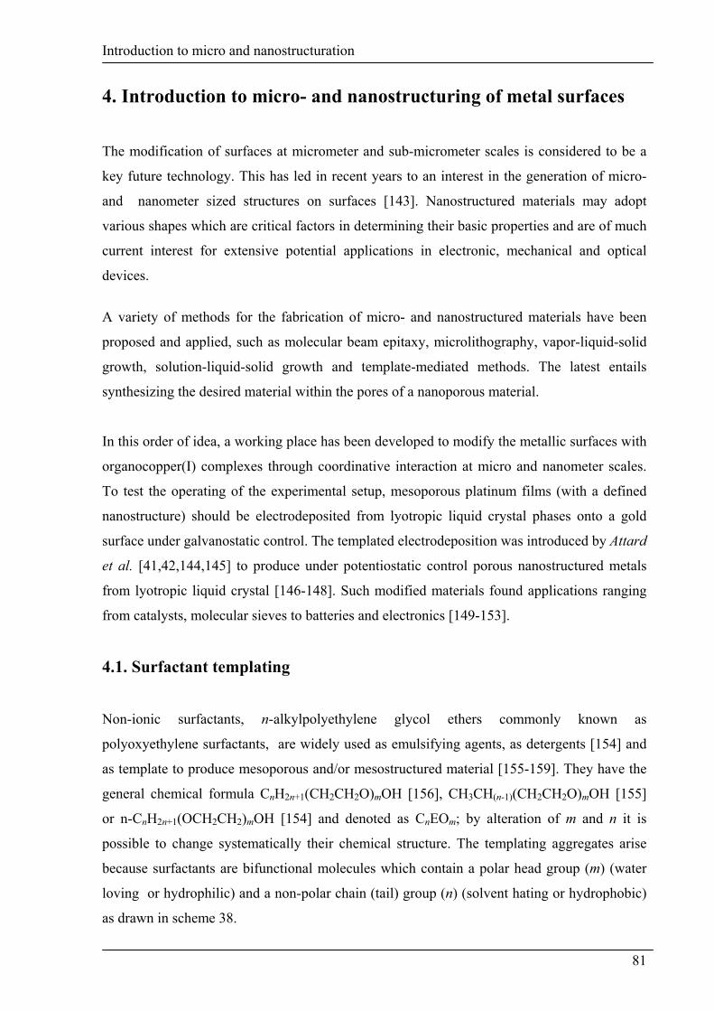
Non-ionic surfactants, n-alkylpolyethylene glycol ethers commonly known as
polyoxyethylene surfactants, are widely used as emulsifying agents, as detergents [154] and
as template to produce mesoporous and/or mesostructured material [155-159]. They have the
general chemical formula CnH2n+1(CH2CH2O)mOH [156], CH3CH(n-1)(CH2CH2O)mOH [155]
or n-CnH2n+1(OCH2CH2)mOH [154] and denoted as CnEOm; by alteration of m and n it is
possible to change systematically their chemical structure. The templating aggregates arise
because surfactants are bifunctional molecules which contain a polar head group (m) (water
loving or hydrophilic) and a non-polar chain (tail) group (n) (solvent hating or hydrophobic)
as drawn in scheme 38.
81
Introduction to micro and nanostructuration
Polyoxyethylene surfactants, when mixed with water, form micelles
above a critical concentration in water with a wide range of liquid
crystals (LC) occurring frequently at high concentrations of non-ionic
surfactants (typically more than 40 wt%) [41,144,145]. The shape of
the micelles depend highly upon surfactant concentration, electrolyte
level and temperature.
Scheme 38 The chemical structure of a non-ionic surfactants.
The amphiphilic nature of non-ionic surfactants results in molecular aggregation forming
usually a lyotropic liquid crystalline (LLC) mesophase with a well-defined structure. This
occurs because the alkyl group CnH2n+1 (oil like) of the CnEOm molecule tends to minimize
the interaction with water and also the polar EO head group (-(CH2CH2O)mOH) tends to stay
outside to form diluted water solutions. Thus the liquid crystalline phase is formed by water
molecules through polar-apolar interactions [156].
The topology of the structure can be varied in a predictable fashion. Attard et al. [144,146]
have used at high concentration, the non-ionic surfactant, octaethyleneglycol monodecyl ether
(C16EO8) to produce three liquid crystalline phases: hexagonal (HI), cubic (Ia3d) and lamellar
(Lα). Among these three phases, the hexagonal one, which exists over a wide range of
composition and temperature is the most reported.
The hexagonal phase is consisted of long cylindrical micelles of surfactants packed into a
hexagonal array with a uniform radius determined by the length of the surfactant molecules
(scheme 39). The size of pores and the thickness of metal walls can be modified by varying
the alkyl chain length of the surfactant and/or by adding a co-solvent as n-heptane to swell the
micellar surfactant rods in lyotropic liquid crystalline phase. So, the range of radii was varied
from 1.7 to 3.5 nm by using a variety of non-ionic surfactants (scheme 38) and added
hydrocarbon swelling agents, suggesting the dispersion of alkane molecules between the
surfactant micellar aggregates [160,161].
82

Introduction to micro and nanostructuration
The cubic phase does not exist at room temperature. At high concentration of surfactant the
lamellar (Lα) phase predominates and consists of surfactant molecules aggregated to form
infinite continuous planes separated by water layers [160,161].
Scheme 39 Long cylindrical micelles of surfactants.
Consequently, the nanostructures of the metal films deposite from LLC phases depend on the
structure of the lyotropic phase used. In the cases of C12EO6-H2O, C12EO8-H2O, C12EO10-
H2O, C16EO8-H2O and C16EO1-H2O systems, the sequence of the mesophase follow the
hexagonal (H1) cubic (V1) lamellar (Lα) order with increasing surfactant
concentration at room temperature. The hexagonal phase is the most stable and appears in a
range of 40-70 wt% surfactant concentrations [42,144,154,158,162].
→ →
4.2. The principle of templated electrodeposition
The principle of the templated electrodeposition consists to dissolve the metal precursor in the
hydrophilic domains of the mixture around the hexagonally packed cylindrical surfactant
micelles. In these domains the metal salts are solvated by water in the vicinity of the polar EO
head groups of the non-ionic surfactant molecules. Upon application of the potential, metal is
electrodeposited within these aqueous domains producing a metal film around the surfactant
micelles. Once the metal deposition is complete, the electrochemical cell is disassembled and
the surfactant is removed by soaking the film in water to leave an adherent nanostructured
metal film with a hexagonal array of cylindrical pores as a direct cast of the lyotropic liquid
crystalline phase (scheme 40 analog to [149,160,163,164]). Several prerequisites are required
for using this method to prepare nanostructured materials: water, choice of surfactant,
83

Introduction to micro and nanostructuration
precursor molecule and additive. Under the right conditions the precursor molecules react,
only within the aqueous domains, to build up a solid mass of product.
Scheme 40 The principle of the liquid crystal templating method.
The electrodeposition of metal by templating method usually occurs at a constant potential
where the rate of deposition is controlled by the kinetics of the process and not by mass
transport. So, the thickness of the deposited film is controlled by varying the total charge
passed during the electroplating process [164].
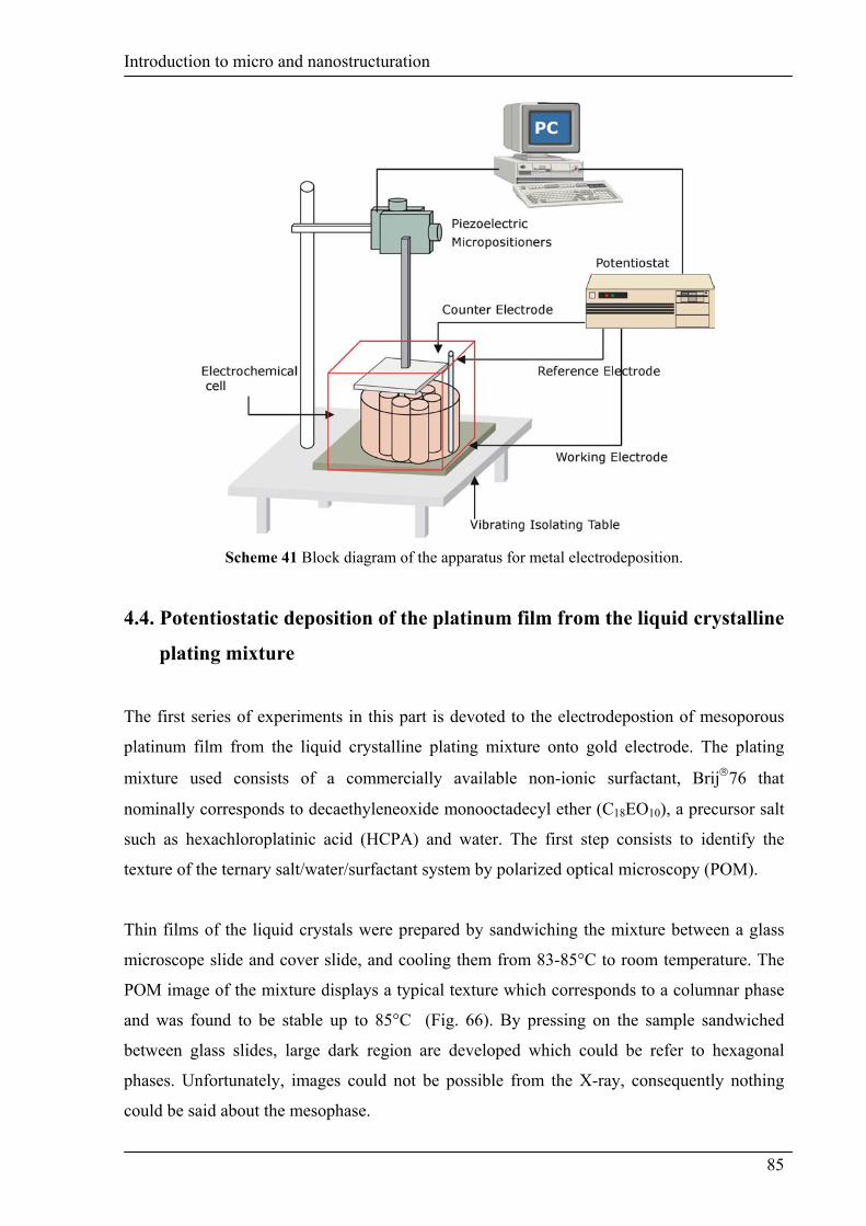
4.3. Schematic representation of the working station
The first goal of this work is to achieve galvanostatically microstructures which exhibit a
well defined nanostructures. These nanostructured surfaces will be used later for the fixation
of metal complexes. For this achievement, the basic of apparatus for metal electrodeposition
is shown in scheme 41. The working electrode is attached to the bottom of electrochemical
cell which is mounted on a stable platform. The counter electrode is fixed on a Motor
Controller which can drive up to 4 DC-motors directly from a PC with C-842 WinMove
programming software. The electrodes are connected to a Potentiostat / Galvanostat which
operates remotely from a PC via a IEEE-488 interface port with 352 SoftCorr III software.
The distance between both electrodes (WE and CE) is maintained constant during the
experiment by piezoelectric micropositioners which can move in two dimensions (y,z).
84
Introduction to micro and nanostructuration
Scheme 41 Block diagram of the apparatus for metal electrodeposition.
4.4. Potentiostatic deposition of the platinum film from the liquid crystalline
plating mixture
The first series of experiments in this part is devoted to the electrodepostion of mesoporous
platinum film from the liquid crystalline plating mixture onto gold electrode. The plating
mixture used consists of a commercially available non-ionic surfactant, Brij76 that
nominally corresponds to decaethyleneoxide monooctadecyl ether (C18EO10), a precursor salt
such as hexachloroplatinic acid (HCPA) and water. The first step consists to identify the
texture of the ternary salt/water/surfactant system by polarized optical microscopy (POM).
Thin films of the liquid crystals were prepared by sandwiching the mixture between a glass
microscope slide and cover slide, and cooling them from 83-85°C to room temperature. The
POM image of the mixture displays a typical texture which corresponds to a columnar phase
and was found to be stable up to 85°C (Fig. 66). By pressing on the sample sandwiched
between glass slides, large dark region are developed which could be refer to hexagonal
phases. Unfortunately, images could not be possible from the X-ray, consequently nothing
could be said about the mesophase.
85

Introduction to micro and nanostructuration
Fig. 66 Polarized optical microscopy image of the liquid crystal phase of C18EO10 / H2O / H2PtCl6
system (ratio 2:1:1 by weight) at room temperature.
The electrodeposition of the platinum film from the liquid crystalline plating mixture is
carried out under thermostatic and potentiostatic control. The temperature is maintained
constant at 40°C and the potential at –0.1 V for 30 min. After the deposition, the electrode is
soaked in water, after the deposition, for at least 48 h to remove the surfactant. The resulting
film was adherent, gray and shiny. Atomic force microscopy (AFM) was employed to
visualize the structure of the deposited platinum film. The morphology of the deposited film
is presented in Fig. 67.
Fig. 67 AFM image of nanostructured platinum (3-dimensional view).
86

Introduction to micro and nanostructuration
As it is shown, AFM reveals that the deposits were not uniform and present large pores which
could not be comparable whit that found Attard et al. [41] for mesoporous platinum films
electrodeposited from the hexagonal phase of a nonionic surfactant octaethyleneglycol
monohexadecyl ether (C16EO8). The pore diameters are estimated ranging 220 – 316 nm. The
use of the working place presented in the previous paragraph is more complicated for the
electorodeposition under potentiostatic control.
4.5. Galvanostatic deposition of the platinum film from the plating mixture
The galvanostatic deposition of the platinum film from the templating liquid crystal has been
carried out from the hexagonal phase formed by a mixture of a non-ionic surfactant, C18EO10
denoted Brij 76, and an aqueous solution of HCPA.
In this part, the structure and the morphology of the electrodeposited platinum films is
established by AFM. Fig. 68 represents the 3-dimensional view of the gold working electrode
without the deposit.
Fig. 68 AFM image of the gold electrode without deposit (3-dimensional view).
The ternary plating mixture is prepared from an aqueous solution of 2M HCPA which is
mixed, in a ratio 1:1, with 50 wt% of Brij 76 in water. The platinum films are deposited at a
constant current of 9 µA (Fig. 69A) and 50 µA (Fig. 69B) for about 45 min and 3 hours
respectively on a polished gold electrode. The morphology of the deposited films is
investigated over a range of length scales reported in Fig. 69. The AFM studies show a
porosity in the structure although nothing could be said presently about its regularity. It
appears that the time of deposition could play an important role with regard to the
87

Introduction to micro and nanostructuration
morphology of the film electrodeposited. The deposit obtained within a short time scale seems
to be more structured and flat (Fig 69A) than the long time deposition which present large
pores (Fig. 69B).
(A)
(B)
Fig. 69 AFM image of the electrodeposited platinum films: (A) 9 µA , 45 min and (B) 50 µA, 3 h.
88
Introduction to micro and nanostructuration
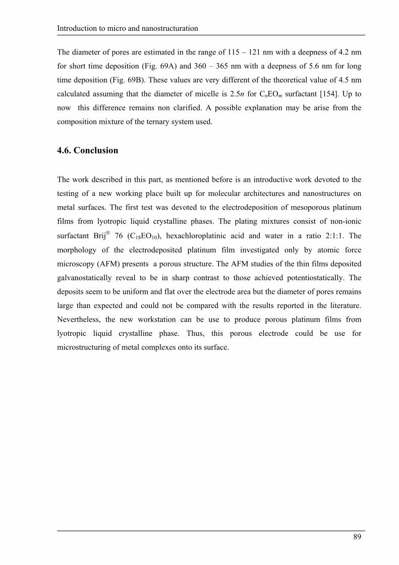
The diameter of pores are estimated in the range of 115 – 121 nm with a deepness of 4.2 nm
for short time deposition (Fig. 69A) and 360 – 365 nm with a deepness of 5.6 nm for long
time deposition (Fig. 69B). These values are very different of the theoretical value of 4.5 nm
calculated assuming that the diameter of micelle is 2.5n for CnEOm surfactant [154]. Up to
now this difference remains non clarified. A possible explanation may be arise from the
composition mixture of the ternary system used.
4.6. Conclusion
The work described in this part, as mentioned before is an introductive work devoted to the
testing of a new working place built up for molecular architectures and nanostructures on
metal surfaces. The first test was devoted to the electrodeposition of mesoporous platinum
films from lyotropic liquid crystalline phases. The plating mixtures consist of non-ionic
surfactant Brij 76 (C18EO10), hexachloroplatinic acid and water in a ratio 2:1:1. The
morphology of the electrodeposited platinum film investigated only by atomic force
microscopy (AFM) presents a porous structure. The AFM studies of the thin films deposited
galvanostatically reveal to be in sharp contrast to those achieved potentiostatically. The
deposits seem to be uniform and flat over the electrode area but the diameter of pores remains
large than expected and could not be compared with the results reported in the literature.
Nevertheless, the new workstation can be use to produce porous platinum films from
lyotropic liquid crystalline phase. Thus, this porous electrode could be use for
microstructuring of metal complexes onto its surface.
89