3 modification of the spot synthesis technique to produce
TRANSCRIPT

3 Modification of the SPOT Synthesis
39
3 Modification of the SPOT Synthesis Technique to Produce Peptides with Free C-Termini for the Study of PDZ Domain Interactions
3.1 Peptide Libraries for Proteomics Studies
Many combinatorial peptide library-based approaches have been developed to analyze the
binding or substrate specificities of protein domains (reviewed in [131]). These libraries
can be classified as either ‘display’ or ‘pool peptide libraries’. The display libraries employ
a variety of carriers for individual peptide presentation, including pins, phages, beads,
DNA binding proteins or ribosomes. The display techniques involve multiple rounds of
enrichment steps, and the identities of the selected peptides can be ascertained by DNA or
peptide sequencing. Display peptide libraries generally require isolation and sequencing of
individual peptides or the respective DNA to decode the binding motif, which is time-
consuming and costly. In contrast, the pool peptide libraries rely on the comparison of
binding affinities of different pools of randomly chosen peptides, in search for the binding
consensus motif. In the oriented peptide library method [132], soluble pools of random
peptides are ‘oriented’ towards a certain peptide motif via a central ‘fixed’ amino acid
(e.g.: xxxB1B2xxx). The soluble oriented peptide library approach still depends on peptide
sequencing, and large amounts of purified proteins are generally needed for the assay.
Recently, peptide arrays established by SPOT synthesis are reported to be useful to map
specificity, for example of antibodies, protein domains and protein kinases [133-135].
3.1.1 SPOT Synthesis
SPOT synthesis is an easy-to-handle and flexible technique for parallel chemical synthesis
on membrane supports (for detailed experimental description see [112, 113]). The most
frequent application is the synthesis of peptides on cellulose membranes, which initially
involves attachment of 9-fluorenyl-methoxycarbonyl-β-alanine (Fmoc-β-Ala-OH) to the
hydroxyl functions of a commercially available filter paper (e.g. Whatman, Maidsone,
UK). Fmoc-β-Ala-OH, other ‘amino compounds’ are used to generate either membrane
modified with aminopropylether [136] or N-modified cellulose-amino-hydroxypropyl ether

3 Modification of the SPOT Synthesis
40
(N-CAPE) [111, 137] as support. After the subsequent removal of the Fmoc-group, the free
amino group is then available for the next steps in the synthesis.
amino acid coupling
washing with DMA(3 times, 3 min)
Fmoc-deprotectionwith 20% piperidine
(20 min)
washing with DMA(5 times, 3 min)
washing with ethanol(2 times, 3 min)
staining withbromphenol blue
washing with ethanol(3 min)
washing with ether(3 min) and drying
spotting of the amino acids(double coupling, 15 min each)
side-chain group cleavageprotection
cellulose modification
spot definition
Figure 3.1 Outline of the Standard SPOT Synthesis Procedure. Cellulose modification: The surface of the cellulose membrane is first uniformly converted to an ‘amino’-group exposing support employing different ‘amino-compounds’ to accessible hydroxyl functions on a cellulose membrane. Spot definition: In the second derivatisation step, the array of spot reactors is generated by spotwise coupling of Fmoc-β-Ala-OPfp, and all residual amino functions in between spots are blocked by acetylation. Amino acid coupling: 0.2-0.3 M solutions of protected amino acid derivatives are coupled twice. After several washing steps, the Fmoc-group is cleaved with a piperidine solution. The staining with bromophenol blue (BPB) as an indicator of free amino functions helps to visualize the amino-spots on the membrane. Side-chain protection group cleavage: Final peptide deprotection is carried out with 50-90% of trifluoroacetic acid in dichloromethane with 3% triisobutylsilane and 2% water as scavengers. The detailed protocol is given in [112, 113].

3 Modification of the SPOT Synthesis
41
Synthesis areas (spots) are defined by spotting Fmoc-β-alanine-pentafluorophenyl ester
solution (Fmoc-β-Ala-OPfp) to distinct sites on the membrane. This is done either by a
pipetting robot (Abimed GmbH, Langenfeld, Germany) or manually. Blocking
(acetylating) the remaining amino functions between the spots provides up to 8000 discrete
reaction sites on a 20 x 30 cm membrane for further standard, solid phase peptide synthesis
using amino acid pentafluorophenyl esters (Fmoc-aa-OPfp). Using the Fmoc-aa-OPfp
standard protocol (Figure 3.1), the carboxyl groups are activated and the synthesized
peptides are therefore C-terminally attached to the cellulose membrane. An activation of
the amino-group for synthesizing N-terminally attached peptides would lead to a racemate
formation of ~50%.
Routinely, peptides up to a length of 20 residues, but also longer peptides [137] can be
synthesized with sufficient fidelity. The peptides can then be used for binding and
enzymatic assays directly on the cellulose membranes or cleaved from the solid support by
the use of ammonia or other reagents [111, 136].
The extended SPOT synthesis concept can be used to create libraries for various
screening options such as substitutional analyses, combinatorial libraries, length analyses,
integration of non-natural building blocks or other chemical entities. In recent years,
peptide arrays prepared by the SPOT technique have become popular tools for studying
protein-protein interactions [138].
3.1.2 Previous Method of Inverted Peptides
Some protein domains, such as PDZ domains (reviewed in [27, 139]) bind to peptides only
if the ligands have a free carboxyl-terminus (C-terminus). Unfortunately, peptides
synthesized according to the standard SPOT synthesis protocol [112, 113] (see
Chapter 3.1.1) are attached by their C-termini to the cellulose support. In principle, free
C-termini can be obtained after standard SPOT synthesis by reversing the peptide
orientation.
Methods for synthesizing free C-terminal resin-bound [140-142] and cellulose-bound
[104] peptides have been published. Yields and purities of the inverted cellulose-bound
peptides are generally low and their generation is extremely time-consuming.

3 Modification of the SPOT Synthesis
42
Figure 3.2 Outline of the SPOT Synthesis Procedure Published in Hoffmüller et al. [104]. Aminopropylether modified cellulose [136] (1) was used as support. (a) β-alanine was used as spacer and to define the spot areas, Dmab-glutamic acid as bivalent linker and HMBA as a base-labile cleavage site to yield in (2). (b) Fmoc-aa-OH as intended C-terminal amino acid was introduced as ester (3). (c) Standard SPOT synthesis and β-Ala coupling as spacer to yield the β-Ala-coupled peptide (4). (d) The Fmoc-protection-group at the N-terminus and the Dmab-protection-group at the side chain of the glutamic acid were cleaved. Subsequently, the cyclisation was performed using PyBOP and NMI to achieve the cyclic peptide (5). (e) After cleavage of the side chains, the ester bound was hydrolyzed with Li2CO3 resulting in a linear peptide with free C-terminus (6). Dmab = α-4-[N-{1-(4,4-dimethyl-2,6-dioxocyclohexyliden)- 3-methylbutyl}amino]benzyl, Fmoc = 9-fluorenyl-methyloxycarbonyl-, HMBA = 4-hydroxymethylbenzoic acid, TBTU = O-(benzotriazol-1-yl)-N,N,N',N'-tetramethyluronium tetrafluoroborate, DIEA = N,N-diisopropylethylamine, DIC = diisopropylcarbodiimide, NMI = N-methylimidazole, PyBOP = [benzotriazol-1-yloxy]tripyrrolidinophos-phonium hexafluorophosphate, NMM = N-methylmorpholine, TFA = trifluoro acetic acid. Details of the SPOT synthesis procedure are given in Hoffmüller et al. [104].

3 Modification of the SPOT Synthesis
43
Coupling problems are reflected in low yields as well as a high background of peptides
with incorrect sequences. Due to those problems, it was necessary to develop an improved
protocol, which allows the rapid and robust synthesis of large peptide arrays with free
C-termini. Short reaction times, together with high coupling efficiencies at each step
during SPOT synthesis, fewer side reactions and the possibility of automating the SPOT
synthesis process were the criteria that needed to be matched. Therefore, a robust and more
efficient protocol for the preparation of cellulose membrane-bound inverted peptide arrays
was developed in the context of this thesis. This protocol is much better suited for
extensive experimental projects mapping many different PDZ domain interactions. It is
based on the protocol published by Hoffmüller et al. [104] which was ameliorated to make
it less time consuming and more adapted for fully automatic SPOT synthesis. Using the
method of Hoffmüller et al., the following compounds had to be coupled four times for
satisfying yields: the Dmab-glutamic acid as bivalent linker, the hydroxymethylbenzoic
acid (HMBA) as base-labile cleavage site and the Fmoc-aa-OH as intended C-terminal
amino acid (Figure 3.2). Furthermore, these compounds had to be freshly activated at each
reaction-step.
3.2 Results
3.2.1 A Modified SPOT Synthesis Strategy of Cellulose Membrane-Bound
Inverted Peptides
The reaction scheme for the new SPOT synthesis of cellulose membrane-bound inverted
peptides is shown in Figure 3.3. Compared to the standard SPOT synthesis protocol [111,
112, 143], synthesis of inverted peptides was performed on a cellulose membrane carrying
a stable N-modified cellulose-amino-hydroxypropyl ether (N-CAPE) linker [111, 137]
(bold numbers 1-9 refer to Figure 3.3). Key compounds in the synthesis are the Fmoc-
amino acid 3-bromopropyl esters (Fmoc-aa-OPBr) (3), the membrane-bound
mercaptopropionyl cysteine adduct (4), the matrix-bound amino acid ester derivative (5)
and the cyclic peptide (8). Critical reaction steps are the formation of both the cleavable
ester bond and the cyclic peptide (Figure 3.3, d and g, respectively).

3 Modification of the SPOT Synthesis
44
Figure 3.3 Reaction Scheme for the Synthesis of Inverted Peptides on Cellulose Membranes. N-modified cellulose-amino-hydroxypropyl ether (N-CAPE) membrane [111, 137] (0) was used as support. (a) Coupling of Fmoc-β-Ala-OPfp in DMSO followed by Fmoc-cysteine-(Trt)-OPfp in NMP yield the bivalent linker (1); (b) Mmt-S-CH2-CH2-COOH in DMF preactivated with HATU and NMI; were used to achieve the base-labile compound (2) (c) Mmt-cleavage with dichloroacetic acid, TFA, TIBS, DCM; (d) activation of the SH-group of (4) by aqueous solution of Cs2CO3 followed by the coupling of Fmoc-aa-OPBr (3) yielding the peptide (5); (e) peptide synthesis using the standard SPOT synthesis protocol [112] to elongate the peptide (6); (f) Fmoc-β-Ala-OPfp in NMP and 2,4-dinitrophenyl-bromoacetate in NMP were coupled to the compound (7); (g) TFA, TIBS, DCM and subsequently aqueous solution of Cs2CO3 to prepare the cyclic peptide (8); (h) Used of saturated aqueous solution of Li2CO3 to obtain a linear peptide with free C-terminus (9).

3 Modification of the SPOT Synthesis
45
The inverted and N-terminally fixed peptides (9) display a free C-terminus resulting
from the reversal of the peptide orientation, and were achieved by successive thioether-
cyclisation/ester-cleavage (Figure 3.3, g-h). The crucial step during the synthesis of the
key compound (4) is the selective removal of the 4-methoxytriphenyl (Mmt) protection
group (Figure 3.3, c). The acidic mixture used in the Mmt-deprotection step has to be
adjusted carefully in order to prevent cleavage of the triphenylmethyl (Trt) protection
group of the cysteine residue.
As shown in Table 3.1, selective removal of the Mmt-group is achieved by applying a
mixture of 10% dichloroacetic acid, 0.5% trifluoroacetic acid (TFA), followed by three-
times repeated incubation of 10% dichloroacetic acid, 0.5% trifluoroacetic acid (TFA) and
5% triisobutylsilane (TIBS) in dichloromethane (DCM). The key compound (5),
containing the C-terminal amino acid, was synthesized subsequently by caesium salt-
supported S-alkylation of (4) and the coupling of Fmoc-aa-OPBr (3).
Table 3.1 Selective Cleavage of S-Mmt Group.
Acidic Conditions* Mmt Trt<0.5% TFA in DCM − −0.5 –1% TFA in DCM + −≥ 1% TFA in DCM + + +< 8% Dichloracetic acid in DCM − −8 – 15% Dichloroacetic acid in DCM + −≥ 15% Dichloroacetic acid in DCM + + +50% Acetic acid − −10% Dichloroacetic acid, 0.5% TFA in DCM + + −
Protecting Group
Footnotes: * = Mixtures contain additional 5% TIBS; % = v/v; - = no cleavage; + = cleavage; ++ = immediate and fast cleavage
Compounds (3) (except the arginine derivative) were prepared in high yields by
O-acylation of 1-bromo-3-propanol with Fmoc-amino acid fluorides [144] in a “one-pot”
reaction. The reaction is characterized by its lack of side reactions. Racemic products
could not be detected, which is in good accordance with the literature [145]. Only the
corresponding Fmoc-amino acids were observed as impurities in the range of 10-25%. The
Fmoc-arginine 1-bromo-3-propanol ester was synthesized according to standard

3 Modification of the SPOT Synthesis
46
esterification protocols [146]. Yields of the prepared Fmoc-aa-OPBr were determined by
analyzing the crude reaction products using reversed phase high performance liquid
chromatography (RP-HPLC) and electrospray ionisation-mass spectrometry (ESI-MS) as
shown in Table 3.2. Table 3.2 Yields and Characteristics of the Fmoc-Amino Acid 3-Bromopropyl Esters (3). Fmoc-aa-RR: O-(CH2)2-CH2Br
Fmoc-aa-F DCC/NMI CDI expected [M+H]+/[M+H]++ measured [M+H]+/[M+H]++
Fmoc-Ala-R 82% 432.0811/434.0793 432.0834/434.0791Fmoc-Arg(Pbf)-R - 76% 769.2271/771.2257 769.2255/771.2269Fmoc-Asn(Trt)-R 91% 43% 717.1964/719.1953 717.1986/719.2010Fmoc-Asp(OtBu)-R 89% 532.1335/534.1319 532.1354/534.1350Fmoc-Gln(Trt)-R 88% 731.2120/733.2109 731.2167/733.2137Fmoc-Glu(otBu)-R 86% 546.1491/548.1475 546.1503/548.1425Fmoc-Gly-R 81% 418.0654/420.0636 418.0654/420.0641Fmoc-His(Trt)-R 84% 740.2124/742.2113 740.2128/742.2101Fmoc-Ile-R 82% 474.1280/476.1263 474.1295/476.1285Fmoc-Leu-R 86% 77% 474.1280/476.1263 474.1281/476.1269Fmoc-Lys(Boc)-R 89% 589.1913/591.1898 589.1934/591.1940Fmoc-Met-R 83% 492.0844/494.0826 492.0819/494.0846Fmoc-Phe-R 76% 58% 508.1123/510.1107 508.1127/510.1104Fmoc-Pro-R 82% 75% 458.0967/460.0950 458.0928/460.0916Fmoc-Ser(tBu)-R 81% 504.1385/506.1369 504.1361/506.1364Fmoc-Thr(tBu)-R 79% 518.1542/520.1526 518.1523/520.1449Fmoc-Trp(Boc)-R 82% 647.1743/649.1743 647.1812/649.1785Fmoc-Tyr(tBu)-R 76% 52% 580.1699/582.1684 580.1716/582.1689Fmoc-Val-R 78% 52% 482.0162/484.0285 482.0221/484.2285
Yields ESI Mass Spectrometry Data
During the reaction between the Fmoc-aa-OPBr (3) and the membrane-bound
mercaptopropionyl cysteine adduct (4) (Figure 3.3, d) the C-terminal amino acid of the
peptide and the ester cleavage site, both necessary to reverse the peptide orientation, were
incorporated simultaneously. Due to problems caused by the ubiquitous cellulose hydroxyl
functions, we chose the S-alkylation reaction for selective formation of the ester linkage.
Coupling efficiencies of the Fmoc-aa-OPBr (3) are shown in Figure 3.4. Low coupling
efficiencies were found for the Fmoc-aa-OPBr derivatives of histidine, asparagine,
glutamine (Trt-protected side-chains) and arginine (Pbf-protected side-chain), probably
due to sterical hindrance by the bulky protection groups. However, increasing
concentrations of the critical amino acid derivatives up to 0.8 M in dimethylformamide
(DMF) and repeated coupling (3x) significantly increased the coupling efficiency (Figure
3.4). Furthermore, the thiolate formation of compound (4) is stable when activated by 10%
caesium carbonate after drying the cellulose membrane. Furthermore, no significant
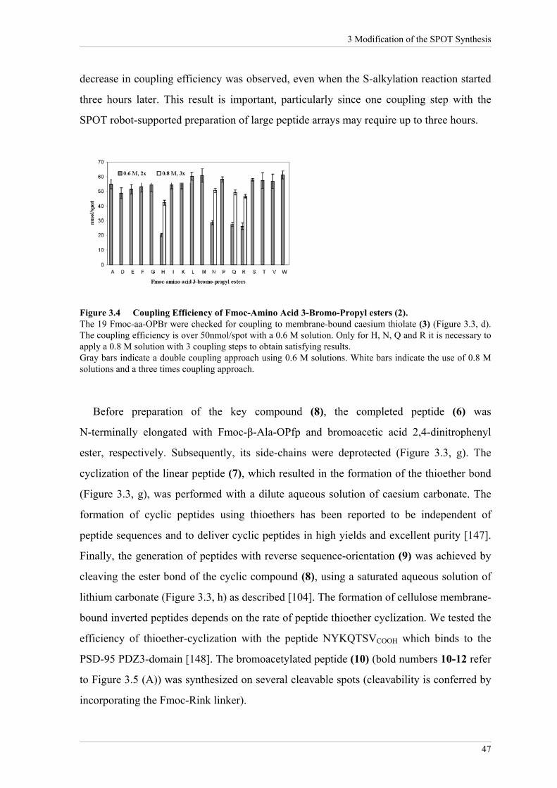
3 Modification of the SPOT Synthesis
47
decrease in coupling efficiency was observed, even when the S-alkylation reaction started
three hours later. This result is important, particularly since one coupling step with the
SPOT robot-supported preparation of large peptide arrays may require up to three hours.
Figure 3.4 Coupling Efficiency of Fmoc-Amino Acid 3-Bromo-Propyl esters (2). The 19 Fmoc-aa-OPBr were checked for coupling to membrane-bound caesium thiolate (3) (Figure 3.3, d). The coupling efficiency is over 50nmol/spot with a 0.6 M solution. Only for H, N, Q and R it is necessary to apply a 0.8 M solution with 3 coupling steps to obtain satisfying results. Gray bars indicate a double coupling approach using 0.6 M solutions. White bars indicate the use of 0.8 M solutions and a three times coupling approach.
Before preparation of the key compound (8), the completed peptide (6) was
N-terminally elongated with Fmoc-β-Ala-OPfp and bromoacetic acid 2,4-dinitrophenyl
ester, respectively. Subsequently, its side-chains were deprotected (Figure 3.3, g). The
cyclization of the linear peptide (7), which resulted in the formation of the thioether bond
(Figure 3.3, g), was performed with a dilute aqueous solution of caesium carbonate. The
formation of cyclic peptides using thioethers has been reported to be independent of
peptide sequences and to deliver cyclic peptides in high yields and excellent purity [147].
Finally, the generation of peptides with reverse sequence-orientation (9) was achieved by
cleaving the ester bond of the cyclic compound (8), using a saturated aqueous solution of
lithium carbonate (Figure 3.3, h) as described [104]. The formation of cellulose membrane-
bound inverted peptides depends on the rate of peptide thioether cyclization. We tested the
efficiency of thioether-cyclization with the peptide NYKQTSVCOOH which binds to the
PSD-95 PDZ3-domain [148]. The bromoacetylated peptide (10) (bold numbers 10-12 refer
to Figure 3.5 (A)) was synthesized on several cleavable spots (cleavability is conferred by
incorporating the Fmoc-Rink linker).

3 Modification of the SPOT Synthesis
48
Figure 3.5 Determination of the Cyclization Conditions. HPLC and MS analysis of the crude reaction product indicated that cyclization is quantitative using the condition h-3 (1 mM Cs2CO4 in DMF). Conditions h-1 and h-2 lead to incomplete cyclization of (10). Depending on the conditions used, various amounts of the linear peptide (12) were detected. The efficiency of the thioether-cyclization reaction is given using a solution of caesium carbonate (1 mM) in DMF (condition h-3). (A) The cyclic peptide (11) was generated according to Figure 3.3 with β-alanine replaced by the Fmoc-Rink linker (for details see Materials and Methods 2.2.3). (h) Conditions used for cyclization: (h-1) DMF, 3 h; (h-2) caesium thiolate, DMF, 3 h; (h-3) Cs2CO4 in DMF (1mM), 3 h; (i) TFA, water, TIBS, DCM, 3 h. (B) ESI mass spectrometry (left) and RP-HPLC (right) of the crude products resulting from the conditions h-1, h-2 and h-3 used in cyclization of the membrane bound peptide (10).

3 Modification of the SPOT Synthesis
49
Three conditions were used for the cyclization: one array was incubated with DMF
(Figure 3.5, h-1), the second was first treated with a dilute aqueous solution of caesium
carbonate and then incubated with DMF (Figure 3.5, h-2), and the third array was
incubated with a solution of caesium carbonate (1 mM) in DMF (Figure 3.5, h-3). After
3 hours the peptide products h-1, h-2 and h-3 were cleaved from the spots using TFA and
analyzed by HPLC and ESI-MS (Figure 3.5 (B)). Analysis of the reaction products clearly
demonstrated the efficiency of the thioether-cyclization reaction, using condition h-3. To
determine the influence of the peptide sequence on the cyclization/cleavage step, two
further membrane bound bromoacetylated peptides, EFHAALGSYVCOOH and
QHIDSQKKACOOH, were synthesized. During the cyclization-steps, we used a dilute
aqueous solution of caesium carbonate (5%, 12 h) and analyzed them subsequently by
ESI-MS. In both cases, quantitative and reproducible thioether-cyclization can be
observed, as shown in the spectrum of condition h-3 (1 mM caesium carbonate in DMF).
3.2.2 Evaluation of the Novel Strategy
To assess the novel method for synthesizing cellulose membrane-bound inverted peptides,
a library of 6223 C-termini of human proteins (Swiss-Prot database, release 40) was
screened. We synthesized the C-terminal peptides as 11-mers to be sensitive to a putative
interaction involving the amino acids in ligand positions below -6. This library, called
6223-Humlib (Figure 3.6), was incubated with the SNA1 PDZ domain and the signal
intensities were subsequently measured in Boehringer Light Units (BLU). The results were
compared with previously published data from Hoffmüller et al. [104]. In this publication,
a library comprising 3514 C-termini of human proteins (Swiss-Prot database, release 34)
was synthesized as 7-mer peptides and incubated under similar conditions with the SNA1
PDZ domain (called hereafter 3514-Humlib).
The BLU intensities of each spot of the two libraries (3514-Humlib and 6223-Humlib)
were determined to compare both binding experiments accurately. To deduce the peptide
sequences that could be counted as PDZ domain binders, the signal intensity of the
background was quantified using 20 spots randomly located on the cellulose membrane.
Peptide sequences showing signal intensities higher than the background intensity plus the

3 Modification of the SPOT Synthesis
50
double standard deviation were defined as PDZ domain binders (3514-Humlib:
background = 3x10³ BLU; standard deviation = 1x10³ BLU; PDZ domain binder ≥ 5x10³
BLU; 6223-Humlib: background = 9x10³ BLU; standard deviation = 1x10³ BLU; PDZ
domain binder ≥ 11x103 BLU) (A.A. Weiser and R. Volkmer-Engert, personal
communication). Taken together, we obtained signal intensities for peptide sequences that
bind to the SNA1 PDZ domain with values ranging between 5x10³ - 28x103 BLU for the
3514-Humlib and between 11x103 - 519x103 BLU for the 6223-Humlib. The first 30 spots
of each library with the highest intensities showed values ranging between 12x103 -
28x103 BLU for the 3514-Humlib and between 237x103 - 519x103 BLU for the
6223-Humlib (Table 3.3).
439
6681611
1787
3262
3507
4711
Figure 3.6 In vitro Identification of Putative SNA1 PDZ Domain Ligands. Selected region of the library of 6223 C-termini (11-mers) of human proteins from the Swiss-Prot database incubated with GST-SNA1 PDZ domain (cysteine is replaced by serine). The numbers (positioned with the first numeral above/under the corresponding spot) represent the peptide sequences showing the highest signal intensity measured in BLU (see Table 3.3 and Table 0.3 in the Appendix)

3 Modification of the SPOT Synthesis
51
Table 3.3 Evaluation of the SNA1 PDZ Domain Interactions.
6223-Humlib 3514-HumlibAcc.No. Protein description Sequence x103 BLU x103 BLU
P41595 5-hydroxytryptamine 2B receptor GDKTEEQVSYV 83 13 P28335 5-hydroxytryptamine 2C receptor SSVVSERISSV 319 10 P25100 Alpha-1D adrenergic receptor ** ADYSNLRETDI 84 28 P08913 Alpha-2A adrenergic receptor ILCRGDRKRIV 21 13 P11766 Alcohol dehydrogenase class III chi chain SGKSIRTVVKI 23 15 P50052 Type-2 angiotensin II receptor (AT2) SSLREMETFVS 361 6 P18440 Arylamine N-acetyltransferase 1 VPKHGDRFFTI 45 16 Q01814 Calcium-transporting ATPase plasma membrane, brain isoform 2 GSPIHSLETSL 283 * Q9UKP5 Adam-TS 6 precursor SSCNLAKETLL 333 * P08588 Beta-1 adrenergic receptor CRPGFASESKV 281 8 O60241 Brain-specific angiogenesis inhibitor 2 precursor EPPDGDFQTEV 248 * O60242 Brain-specific angiogenesis inhibitor 3 precursor DVQEGDFQTEV 259 * Q16581 C3A anaphylatoxin chemotactic receptor NVISERNSTTV 254 * P07766 T-cell surface glycoprotein CD3 epsilon chain precursor DLYSGLNQRRI 30 16 P22459 Voltage-gated potassium channel protein KV1.4 CSNAKAVETDV 149 14 P22460 Voltage-gated potassium channel protein KV1.5 LCLDTSRETDL 106 19 P35499 Sodium channel protein, skeletal muscle alpha-subunit TVRPGVKESLV 197 16 P53618 Coatomer beta subunit KINLSQKETSI 218 14 P81408 Cote1 protein SLNGGSRETGL 238 * P33402 Guanylate cyclase soluble, alpha-2 chain IGTMFLRETSL 359 28 O00341 Excitatory amino acid transporter 5 TIQISELETNV 270 * P23610 Factor VIII intron 22 protein LHLVLQETISP 288 6 P07148 Fatty acid-binding protein, liver DIVFKRISKRI 14 24 Q08379 Golgin-95 DENDEVKITVI 238 * Q9UI32 Glutaminase, liver isoform, mitochondrial precursor ALSKENLESMV 485 * P49683 Probable G protein-coupled receptor GPR10 HGQNMTVSVVI 238 9 P11169 Glucose transporter type 3, brain IEPAKETTTNV 332 6 Q9Y2T3 Guanine deaminase GKQVVPFSSSV 260 * P01776 IG heavy chain V-III region WAS FDVFGQGTLVT 227 12 O43711 Homeobox protein HOX-11L2 SSKVPAVTSLV 254 * P01589 Interleukin-2 receptor alpha chain precursor QRRQRKSRRTI 16 26 P15248 Interleukin-9 precursor KEKMRGMRGKI 17 15 P48049 Inward rectifier potassium channel 2 EPRPLRRESEI 195 17 P48050 Inward rectifier potassium channel 4 DNISYRRESAI 197 13 P78508 ATP-sensitive inward rectifier potassium channel 10 GSALSVRISNV 306 * Q99712 ATP-sensitive inward rectifier potassium channel 15 LRTLLLQQSNV 408 * P18084 Integrin beta-5 precursor KFNKSYNGTVD 55 13 P11801 Putative serine/threonine-protein kinase H1 VDPGARMTALQ 439 5 P01702 IG lambda chain V-I region NIG-64 FGGGTRVTVLG 128 12 P06887 IG lambda chain V-I region MEM FGTGTKVTVLR 254 8 P01705 IG lambda chain V-II region NEI FGGGTRVTVLS 230 12 P27816 Microtubule-associated protein 4 TLDSQIQETSI 128 20 P04201 MAS proto-oncogene NCNTVTVETVV 235 12 P33993 DNA replication licensing factor MCM7 VNASRTRITFV 172 28 O60669 Monocarboxylate transporter 2 QSVTSERETNI 268 * Q13614 Myotubularin-related protein 2 AQCVTPVQTVV 432 * P48645 Neuromedin U-25 precursor PRNGRRSAGFI 34 16 Q01959 Sodium-dependent dopamine transporter RQFTLRHWLKV 22 14 Q29459 Platelet-activating factor acetylhydrolase IB beta subunit ETPEEKQTTIA 238 * P35080 Profilin II ELALYLRRSDV 59 17 P25800 Rhombotin-1 QLNGTFESQVQ 261 - P47872 Secretin receptor precursor QSQGTCRTSII 147 18 P30872 Somatostatin receptor type 1 NGTCTSRITTL 258 18 P01135 Transforming growth factor alpha precursor RTACCHSETVV 210 14 P04437 T-cell receptor alpha chain V region CTL-l17 precursor FGTGTRLQVTL 334 5 P01733 T-cell receptor beta chain V region YT35 precursor TFGSGTRLTVV 519 23 Q99437 Acuolar ATP synthase 21 kDA proteolipid subunit ILQTSRVKMGD 317 *
Footnotes: * = sequence not present in Hoffmueller et a.l [104]; ** = new sequence entry in Swiss-Prot; - = non-binder; binder = background + 2x standard deviation 3514-Humlib: total: 5x10³ BLU - 28x103 BLU; 30 strong: 12x103 BLU - 28x103 BLU; 6223-Humlib: total: 10x103 BLU - 519x103 BLU; 30 strong: 237x103 BLU - 519x103 BLU;

3 Modification of the SPOT Synthesis
52
The new method produced a 20-fold increase in signal intensity but only a 3-fold
increase in the background signal, despite the usage of longer peptides. Altogether, we
obtained a total of 60 binding peptides, of which 17 sequences from the 6223-Humlib were
not represented in the 3514-Humlib. Furthermore, 3 peptide sequences were identical in
both libraries. Of the resulting 40 comparable sequences, all strong binders from the
3514-Humlib were also found as binders in the 6223-Humlib and vice versa. The only
exception was the rhombotin-1 peptide, which was detected as a strong binder
(261x103 BLU) in the 6223-Humlib, but showed no signal in the 3514-Humlib. This may
be due to different peptide length and could be excluded by additional experiments, such
as substitutional analysis or surface plasmon resonance measurements.
Comparing all sequences defined as SNA1 PDZ binder, a good correlation between
both libraries was obtained. In accordance with previously published results, strong signals
were detected for the peptide derived from the soluble α-2 chain of the guanylate cyclase
(3514-Humlib: 28x103 BLU; 6223-Humlib: 359x103 BLU) [104], from muscle sodium
channel protein type IV alpha subunit (3514-Humlib: 16x103 BLU; 6223-Humlib:
197x103 BLU) [34], from the stress-activated protein kinase-3 (3514-Humlib: sequence not
represented; 6223-Humlib: 186x103 BLU) [83], from glutaminase-L (3514-Humlib:
sequence not represented; 6223-Humlib: 485x103 BLU) [84] and a weaker signal of the
aquaporin-4 peptide (3514-Humlib: 7x10³ BLU; 6223-Humlib: 72x103 BLU) [85].
3.2.3 Analysis of novel ERBIN PDZ Domain/Ligands
The newly synthesized library of 6223 C-termini from human proteins was incubated with
the ERBIN PDZ domain (Figure 3.7 (A)). The PDZ binders were determined using the
same procedure as in Chapter 3.2.2 (background = 4x10⁴ BLU, standard deviation =
1x10⁴ BLU, PDZ domain binder ≥ 6x10⁴ BLU, signal range: 6x10⁴ - 676x104 BLU). The
sequences of the 100 strongest interacting peptides are given in Table 0.2 in the Appendix.
This list contains the C-termini of many membrane proteins and receptors including the
well-known ERBIN PDZ domain interaction partner of the armadillo repeat protein deleted
in velo-cardio-facial syndrome (ARVC; spot no.: 407) [75]. Surprisingly, an interaction
with the muscle sodium channel protein type IV alpha subunit (CIN4; spot no.: 1027) and
with the soluble α-2 chain of the guanylate cyclase (CYG4; spot no.: 1351) were found,

3 Modification of the SPOT Synthesis
53
which are both described as SNA1 PDZ domain ligands [34, 104]. We selected for further
investigations by substitutional analysis the voltage-gated potassium channel protein Kv1.4
(CIK4, peptide CSNAKAVETDVCOOH), the voltage-gated potassium channel protein
Kv1.5 (CIK5, peptide LCLDTSRETDLCOOH), the plasma membrane Ca(2+) ATPases,
isoform 1 (ATB1, peptide GSPIHSLETSLCOOH) (Figure 3.7 (B-D)) and the breakpoint
cluster region protein (BCR, peptide KRQSILFSTEVCOOH) (Figure 3.8 (A)). In those
assays, each residue of the ligand is substituted by all natural L-amino acids (cysteine
omitted), enabling the identification of the key residues of the ligand. The observed
substitution patterns for the chosen peptides were generally very similar to each other and
typical for the canonical PDZ domain/peptide interaction. In all cases, the ERBIN PDZ
domain shows a clear preference for valine in ligand position 0. However, it also tolerates
leucine or isoleucine. Position -1 shows fewer restrictions but some amino acids such as
glycine, isoleucine and valine are not tolerated. For position -2, the ERBIN PDZ domain
prefers threonine and serine, as typical for a class I PDZ domain. On the other hand,
ligands such as the receptor protein tyrosine kinase ERB2 peptide (NPEYLGLDVPVCOOH)
with a hydrophobic amino acid at position -2 (class II) can also interact with this PDZ
domain [70]. In position -3, ERBIN preferred a negatively charged amino acid such as
glutamate or aspartate, but also tolerates substitutions with small or hydrophobic amino
acids. The substitutional analysis showed less selectivity for ligand positions beyond
residue -3 (positions -4 to -9). Normally, residues N-terminal to ligand position -3 do not
contribute substantially to affinity or specificity of PDZ domains, with some exceptions
[31-33].

3 Modification of the SPOT Synthesis
54
ATB1(439)
amino acid substitution
CIK5(1018)
amino acid substitutionCIK4(1017)
amino acid substitution
407
438-439489-490
1017-1018
438-439
3340
3547
4711
1668
3214
3388
3528-3529
3454
(C)(B)
-4-3-2-1 0
-4-3-2-1 0
wt A D E F G H I K L M N P Q R S T W YVLCLDTSRETDL
wt A D E F G H I K L M N P Q R S T W YV
(A) Human C-Termini Library
(D)
wt A D E F G H I K L M N P Q R S T W YVGSPIHSLETSL
-4-3-2-1 0
GSPIHSLETSL
542
Figure 3.7 Inverted Peptide Arrays on Cellulose Membranes for Protein Binding. (A) Selected region of the library of 6223 C-termini (11-mers) of human proteins from the Swiss-Prot database incubated with the GST-labeled ERBIN PDZ domain (cysteine is replaced by serine). The 100 strongest binders are listed in Table 0.2 in the Appendix. Substitutional analyses of the peptide derivates (B) of the voltage-gated potassium channel protein Kv1.4 (CIK4), (C) of the voltage-gated potassium channel protein Kv1.5 (CIK5) and (D) of the calcium-transporting ATPase plasma membrane, isoform 1 (ATB1) incubated with GST-ERBIN-PDZ (spot numbers are given in parentheses). Each residue of the peptide ligand is substituted by 19 naturally occurring L-amino acids (cysteine omitted). All spots in the left column are identical and represent the wild type (wt) peptide (boxed in grey). All other spots represent the substitution of one amino acid against the amino acid in the respective column. Hence, each spot bears a single substitution compared to the sequence of the wild type. For example, V in position 0 can only be substituted by L and in some case by I.

3 Modification of the SPOT Synthesis
55
BCR(542)
amino acid substitution
(A)
-4-3-2-1 0
ERB
IN
IB: anti-ERBINERBIN
longer exposure
BC
R
ERB
IN
Lysa
te
+
co
tina- tina- tina- B
CR
tina-
mp.
+
com
p.
BC
Rtina
- BC
Rtina
-
+
com
p.
ysat
BCR
BC
Rtina
-
IB: anti-BCR
Lysa
te
(C) Coprecipitation
GST-ERBIN-PDZ
IB: anti-GST
HA-BCR
GST-ERBIN-PDZ
GST
BC
RW
T
Lysates
IB: anti-HA
V12
71A
GSTPD:
BC
R
(B) Pull-down
HA
-
HA
- BC
RW
T
V12
71A
BC
R
HA
-
HA
- BC
RW
T
V12
71A
BC
R
HA
-
HA
-
Figure 3.8 Characterization of BCR as Ligand of the ERBIN PDZ Domain. The interaction between the C-terminus of BCR and the ERBIN PDZ domain deduced from the 6223-Humlib could be further confirmed through a substitutional analyses and a so-called pull-down experiment. Additionally, this interaction is also detectable between endogenous ERBIN and BCR in cells. (A) Substitutional analysis of the C-terminal peptide derived from the breakpoint cluster region (BCR, spot number is given in parentheses). (B) Pull-down (PD): HEK293 cells transfected with plasmids encoding HA-BCRWT and HA-BCRV1271A were lysed and incubated with GST-ERBIN-PDZ bound to GSH-agarose beads. Proteins bound to GST fusion proteins were fractioned by SDS-PAGE and immunoblotted (IB) with an anti-HA antibody and an anti-GST antibody, respectively. As a control for the expression of the BCR proteins, direct lysates of the 293 cells were immunoblotted with an anti-HA antibody. (C) Coprecipitation of endogenous ERBIN and BCR in MKN-7 cells. The lysate of MKN-7 cells was incubated with an anti BCR antibody in the absence (top panel, lanes 4, 6) or presence of BCR peptide (+ comp.) (top panel, lanes 5, 7) followed by immunoblotting with anti-ERBIN. Expression of endogenous ERBIN and BCR was verified by immunoprecipitation with anti-ERBIN (top panel, lanes 2) and with anti-BCR (bottom panel, lane 2) and by analysis of the direct lysate with anti-ERBIN (top panel, lane 1) and with anti-BCR (bottom panel, lane 1).

3 Modification of the SPOT Synthesis
56
3.2.3.1 Measurements of Dissociation Constants
To give an overview of the affinity of the 40 strongest ERBIN PDZ domain interacting
ligands from the 6223-Humlib, the dissociation constants (Kd) were determined by surface
plasmon resonance (Biacore) measurements for selected peptides (spot no.: 407, 438,
542, 1018 and 1027). We obtained Kd values between 8.0 ± 3.1 µM for ARVC and
82.7 ± 1.3 µM for CIN4, which are typical for PDZ domain interactions (Table 3.4).
Table 3.4 Dissociation Constants of ERBIN PDZ domain/peptide complexes. Spot Sequence Kd [µM] Protein description407 DAKPQPVDSWV 8.0 ± 3.1 armadillo repeat protein deleted in velo-cardio-facial syndrome*438 GSPLHSLETSL 24.5 ± 5.0 plasma membrane calcium-transporting ATPase, isoform 1542 KRQSILFSTEV 36.0 ± 5.2 breakpoint cluster region protein
1018 LCLDTSRETDL 79.4 ± 2.8 voltage-gated potassium channel protein KV1.51027 TVRPGVKESLV 82.7 ± 1.3 sodium channel protein, skeletal muscle alpha-subunit
Footnotes: Peptide sequence with * is published in [75]. Cysteine is replaced by serine.
3.2.3.2 Verification of the BCR/ERBIN Interaction in Mammalian Cells
To confirm that the BCR peptide interaction observed in vitro may take place between
BCR and ERBIN in mammalian cells, we performed a pull-down assay by using the PDZ
domain of ERBIN fused to GST. The GST-ERBIN PDZ and the GST protein alone as a
control were incubated with lysates of HEK293 cells overexpressing BCRWT and a
C-terminal mutant BCRV1271A, both as HA (influenza hemaglutinin-epitope)-tagged
fusions. As predicted, BCRWT but not the mutant BCRV1271A bound to the ERBIN PDZ
domain (Figure 3.8 (B), lanes 1, 2), whereas the GST protein alone did not bind (Figure
3.8 (B), lanes 3, 4). Direct lysates of the 293 cells were immunoblotted with an anti-HA
antibody as a control for the expression of the BCRWT and BCRV1217A proteins. This study
was extended to substantiate the biological relevance of the interaction between full-length
ERBIN and BCR as endogenous proteins. BCR and ERBIN were precipitated with the
help of specific antibodies from MKN-7 cells where both proteins are well expressed
(Figure 3.8 (C)). Indeed, ERBIN coprecipitated with BCR (Figure 3.8 (C), lanes 4, 6).
Competition of the BCR and the ERBIN antibody with their respective antigen reduced
immunoprecipitation, supporting the specificity of the interaction (Figure 3.8 (C), lanes 3,

3 Modification of the SPOT Synthesis
57
5, 7). As a control for the expression of the BCR protein, the direct lysates of the 293 cells
were also immunoblotted with an anti-HA antibody.
3.3 Discussion
The SPOT synthesis concept has been widely used to prepare peptide arrays for proteomics
studies. The overall goal of this work was to extend the application range of the SPOT
synthesis concept by creating N-terminally fixed peptide arrays (inverted peptides),
enabling the display of free C-termini on planar cellulose supports. The new steps
introduced into the SPOT synthesis concept are the synthesis of Fmoc-aa-OPBr, the
selective cleavage of the Mmt-protection-group in presence of the Trt-protection-group,
the formation of the cleavable ester bond and thioether-cyclisation/ester-cleavage. The
developed method is now robust and suitable for proteomics studies. It could be applied to
create non-redundant statistical libraries or libraries based on database sequences. The
improved methodology, in particular with regard to automatic, robot assisted SPOT
synthesis, enables the screening of a large range of divergent peptide sequences.
The coupling problems of the previous method of Hoffmüller et al. [104], where all
compounds had to be activated stepwise, required up to four coupling-steps. With the new
method, we obtained a better signal-to-noise graduation, which is important for detecting
PDZ domain ligand interactions with low or intermediate affinities. The new method
allowed thus a better discrimination of binding strength in the micromolar range for the
investigated PDZ domains.
An obvious advantage of the improved method is due to the fact that only successfully
cyclized peptides will be reversed in their sequence-orientation. The uncyclized peptides
are released during the ester-cleavage procedure. In addition, side-reactions between
neighboring peptide sequences that perhaps occur during the cyclization-step, e.g.
cross-linking via a disulfide bridge or thioether cross-linking, do not disturb the reversal of
peptide-orientation. Such disulfide cross-linked peptides were cleaved from the membrane
during hydrolysis with saturated lithium carbonate solution. On the other hand, the
thioether cross-linked products resulted also in inverted peptides. Taken together, all these
steps are in principle additional purification steps, as evident from the RP-HPLC and MS

3 Modification of the SPOT Synthesis
58
spectra. The spectra showed inverted peptides with high purity, clearly revealing the
superiority of the improved thioether-cyclisation/ester-cleavage method.
The screen of 6223 C-termini of human proteins revealed four new peptide ligands for
the ERBIN PDZ domain: the voltage-gated potassium channel proteins Kv1.4 and Kv1.5
and the plasma membrane Ca(2+) ATPases, isoform 1, which could be potential ERBIN
PDZ domain ligands [73]. Further in vivo experiments will be needed to determine the
biological relevance of these interactions.
ERBIN is the founding member of the LRR and PDZ domain (LAP) protein family,
which is characterized by 16 LRRs at the N-terminus and one to four PDZ domains at the
C-terminus [76]. Besides the PDZ-specific interactions with ERB2, δ-catenin, ARVC or
p0071 [70, 75, 149, 150], ERBIN is known to interact with the small GTPase Ras and Rho
through the LRRs [73, 151]. The novel interaction partner of ERBIN, BCR is a
multidomain protein, containing a serine/threonine protein kinase domain, a Guanine
Nucleotide Exchange Factor (GEF) function and a GTPase-activating protein (GAP)
domain. The GEF and GAP domains can modulate the activity of Rho-type GTPases
[152, 153]. Based on these results, it could be suggested that ERBIN is a scaffolding
protein that links BCR and Rho-type GTPases through its PDZ domain and its LRRs,
respectively, into a single complex.
Taken together, these data suggest that the improved method for generating cellulose
membrane-bound inverted peptides may be a powerful tool to generate peptides with free
C-termini in a large-scale manner for proteomics studies. It is a feasible method to find
new ligands for PDZ domains and, furthermore, this technique allows the parallel
screening of different PDZ domains to determine their specificity and selectivity towards a
wide ligand sequence space.