270827-r2 pap 07 31 12 - circulation...
TRANSCRIPT

DOI: 10.1161/CIRCRESAHA.112.270827 1
Nitroxyl(HNO)-Mediated Disulfide Bond Formation Between Cardiac Myofilament Cysteines
Enhances Contractile Function
Wei Dong Gao1*, Christopher I. Murray2*, Ye Tian1,3, Xin Zhong1,3, Jenna F. DuMond4, Xiaoxu Shen1, Brian A. Stanley5, D. Brian Foster5, David A. Wink6, S. Bruce King4, Jennifer E. Van Eyk2,5, Nazareno
Paolocci5,7.
1Department of Anesthesiology and Critical Care Medicine, 2Department of Biological Chemistry, Johns Hopkins University School of Medicine; Baltimore MD, 21205 USA; 3Department of Pathophysiology,
Harbin Medical University, 150086, China; 4Department of Chemistry, Wake Forest University, Winston-Salem, NC, 27109, USA; 5Division of Cardiology, Department of Medicine, Johns Hopkins School of Medicine; Baltimore, MD, 21205, USA; 6Radiation Biology Branch, National Cancer Institute, NIH,
Bethesda, MD, 20892, USA; 7Department of Clinical Medicine, University of Perugia, Perugia, 06126 Italy.
*WDG & CIM contributed equally to this work.
Running title: HNO Modification of Myofilament Proteins Subject codes: [105] Contractile function [91] Oxidant stress [107] Biochemistry and metabolism Address correspondence to: Dr. Wei Dong Gao Department of Anesthesiology and Critical Care Medicine The Johns Hopkins University School of Medicine Zayed Tower 6208 1800 Orleans Street Baltimore, MD 21205 Tel: 410-955-7519 Fax: 410-955-0994 e-mail: [email protected]
Dr. Nazareno Paolocci Division of Cardiology Department of Medicine The Johns Hopkins University School of Medicine Traylor 911 720 Rutland Ave. Baltimore, MD 21205 Tel: 410-502-5743 Fax: 410-502-2558 e-mail: [email protected]
In June 2012, the average time from submission to first decision for all original research papers submitted to Circulation Research was 13.35 days.
by guest on June 22, 2018http://circres.ahajournals.org/
Dow
nloaded from
by guest on June 22, 2018http://circres.ahajournals.org/
Dow
nloaded from
by guest on June 22, 2018http://circres.ahajournals.org/
Dow
nloaded from
by guest on June 22, 2018http://circres.ahajournals.org/
Dow
nloaded from
by guest on June 22, 2018http://circres.ahajournals.org/
Dow
nloaded from
by guest on June 22, 2018http://circres.ahajournals.org/
Dow
nloaded from
by guest on June 22, 2018http://circres.ahajournals.org/
Dow
nloaded from
by guest on June 22, 2018http://circres.ahajournals.org/
Dow
nloaded from
by guest on June 22, 2018http://circres.ahajournals.org/
Dow
nloaded from
by guest on June 22, 2018http://circres.ahajournals.org/
Dow
nloaded from
by guest on June 22, 2018http://circres.ahajournals.org/
Dow
nloaded from
by guest on June 22, 2018http://circres.ahajournals.org/
Dow
nloaded from
by guest on June 22, 2018http://circres.ahajournals.org/
Dow
nloaded from
by guest on June 22, 2018http://circres.ahajournals.org/
Dow
nloaded from
by guest on June 22, 2018http://circres.ahajournals.org/
Dow
nloaded from
by guest on June 22, 2018http://circres.ahajournals.org/
Dow
nloaded from
DOI: 10.1161/CIRCRESAHA.112.270827 2
ABSTRACT Rationale: In the myocardium, redox/cysteine modification of proteins regulating Ca2+ cycling can affect contraction and may have therapeutic value. Nitroxyl (HNO), the one electron reduced form of nitric oxide, enhances cardiac function in a manner that suggests reversible cysteine modifications of the contractile machinery. Objective: To determine the effects of HNO modification in cardiac myofilament proteins. Methods and Results: The HNO-donor, 1-nitrosocyclohexyl acetate (NCA), was found to act directly on the myofilament proteins increasing maximum force (Fmax) and reducing the concentration of Ca2+ for 50% activation (Ca50) in intact and skinned cardiac muscles. The effects of NCA are reversible by reducing agents and distinct from those of another HNO-donor Angeli’s salt (AS), which was previously reported to increase Fmax without affecting Ca50. Using a new mass spectrometry capture technique based on the biotin switch assay, we identified and characterized the formation by HNO of a disulfide linked actin-tropomyosin and myosin heavy chain (MHC)-myosin light chain 1 (MLC1). Comparison of the NCA and AS effects with the modifications induced by each donor indicated the actin-tropomyosin and MHC-MLC1 interactions independently correlated with increased Ca2+ sensitivity and force generation, respectively. Conclusions: HNO exerts a direct effect on cardiac myofilament proteins increasing myofilament Ca2+ responsiveness by promoting disulfide bond formation between critical cysteine residues. These findings indicate a novel, redox-based modulation of the contractile apparatus which positively impacts myocardial function, providing further mechanistic insight for HNO as a therapeutic agent. Keywords: contractility, nitroxyl, redox-switch, oxidation, calcium, oxidant signalling, redox Non-standard Abbreviations: HF heart failure PTM post-translational modification redox reduction/oxidation ROS reactive oxygen species RNS reactive nitrogen species HNO nitroxyl DTT dithiothreitol AS Angeli’s salt NCA 1-nitrosocyclohexyl acetate MS mass spectrometry TM tropomyosin MH myosin heavy chain MLC1 myosin light chain 1 NEM N-ethylmaleimide biotin-HPDP N-[6-(Biotinamido)hexyl]-3´-(2´-pyridyldithio) propionamide DIGE Difference in-gel electrophoresis DTDP 2,2’-dithiodipyridine.
by guest on June 22, 2018http://circres.ahajournals.org/
Dow
nloaded from

DOI: 10.1161/CIRCRESAHA.112.270827 3
INTRODUCTION
A hallmark of acute and chronic heart failure (HF) is the loss of contractile function.1 Contraction is stimulated by an influx of intracellular Ca2+ and can be regulated by direct post-translational modification (PTM) of the myofilament proteins.2 The majority of therapies available (e.g. β agonists, phosphodiesterase inhibitors) increase intracellular Ca2+ to strengthen contraction; however, they have also been associated with long-term mortality either by increasing diastolic force, worsening diastolic dysfunction or promoting cardiac arrhythmias.3 A promising alternative approach has been the development of inotropic agents that directly increase contraction by enhancing the response of the contractile myofilament proteins to the available Ca2+.4 It has been hoped that this class of drugs would revolutionize the management of heart failure. However, recent clinical trials with Ca2+ sensitizers such as levosimendan have met with limited success.3
An area that has been overlooked in the heart until now is the possible manipulation of key
reduction/oxidation (redox) sensitive sites or “switches”. Modification of these critical residues has been shown to impact processes involved in signal transduction, transcription and metabolism5-7 leading to the assertion that similar switches may exist to regulate cardiac contraction.8 Cysteine (Cys), either individually or in pairs, forms the core of thiol-based switches that can sense the local redox-environment through the unique chemical properties of its thiol side chain. These sensors oscillate between a reduced and oxidized state, responding to reactive oxygen or nitrogen species (ROS/RNS) with a continuum of PTMs.9 Low fluxes of ROS/RNS can function as a signaling mechanism through the formation of reversibly oxidized Cys (e.g. disulfide or sulfenic acid-Cys). Higher fluxes can generate irreversibly modified Cys (e.g. sulfinic or sulfonic acid-Cys) which may lead to cellular apoptosis and eventually necrosis.9 Regulatory redox-switches have been found in components of excitation-contraction coupling where they contribute to modulate cardiac contraction,7 either reversibly, as seen with S-nitrosylation during signaling events, or more permanent oxidative modifications of the ryanodine receptor which can lead to dysfunction.10
The characterization of a similar Cys-based regulation in the myofilaments remains elusive.
Skinned cardiac muscle fibers, comprising only the contractile machinery, displayed reduced force generation when treated with oxidizing agents such as H2O2 or .O2
- likely in the presence of transition metals.11,12 Conversely, cardiac muscles superfused with donors of the thiophylic RNS agent, nitroxyl (HNO), increased force production.13 This increase in function is notably larger than can be accounted for by the associated rise in Ca2+ transient,13,14 suggesting HNO acts directly on the myofilaments, sensitizing them to Ca2+.15 HNO reacts with the thiol on Cys forming either a sulfinamide or, in the presence of an additional free thiol, a disulfide bond (Figure 1).16 The effects of HNO on myofilaments have been found to be readily reversible by the reducing agent dithiothreitol (DTT), suggesting that its action involves the formation of intra- or inter-protein disulfide bonds.17 Yet, nature and sites of HNO-induced modifications in myofilaments are currently unknown.
Our hypothesis is that HNO induces the formation of intra- or inter-protein disulfide bonds
between critical Cys in the myofilament proteins to enhance contractility. We investigated the effects of two chemically unrelated HNO donors (Angeli’s salt (AS)) and 1-nitrosocyclohexyl acetate (NCA18) on cardiac contraction in intact and skinned muscles and then identified and characterized HNO modified residues utilizing a new mass spectrometry (MS) site mapping technique.
by guest on June 22, 2018http://circres.ahajournals.org/
Dow
nloaded from

DOI: 10.1161/CIRCRESAHA.112.270827 4
METHODS Determination of force, [Ca2+]i, and steady-state force-[Ca2+]i relationships in intact cardiac muscle. See supplemental material. Steady-state force-[Ca2+] relationships in skinned cardiac and skeletal muscles. Details for muscle skinning are in the online supplement. After skinning, the muscles were activated with solutions of varied [Ca2+] while diastolic sarcomere length was kept constant. The force-[Ca2+] relationships were generated before and after treatment with NCA (2.5 µmol/L) in the same preparations. After obtaining the force-[Ca2+] relation for the control phase, the muscle was exposed to NCA in relaxing solution for 10 min. There was no increase in resting force during the treatment. Afterwards, the muscle was activated with activating solutions of varied [Ca2+] containing NCA. These activations in the presence of NCA were repeated once to ensure similar force-[Ca2+] relations were obtained. Detection of HNO modifications by biotin switch assay. HNO modified thiols were detected using a variation of the standard biotin switch protocol.19 In brief, 100 μg of isolated rat myofibrils/treatment were diluted to 0.5 μg/μL in HEN (250 mmol/L HEPES pH 7.4, 1 mmol/L EDTA and 0.1 mmol/L neocuproine) including 0.1% (w/v) SDS, treated for 10 min at 37oC and removed by acetone precipitation. Treatments included: NCA, 2.5 µmol/L; AS, 500 µmol/L; and their decomposed, inactive equivalents (reagent at rt for >96 h) in the absence of Ca2+. Additional treatments were DTT, 5 mmol/L and DEA/NO, 125 mmol/L. Remaining free thiols were blocked with 20 mmol/L N-ethylmaleimide (NEM). HNO and/or NO modified thiols were reduced using 5 mmol/L DTT or 1 mmol/L ascorbate and biotinylated with 0.8 mmol/L N-[6-(Biotinamido)hexyl]-3´-(2´-pyridyldithio) propionamide (biotin-HPDP). Excess biotin-HPDP was removed by acetone precipitation (2 volumes) and resultant pellets were washed with additional acetone. Biotinylated proteins were captured with Ultralink Immobilized Streptavidin, separated by SDS PAGE and silver stained. For MS studies, 200 µg of starting material was used and after labeling, biotinylated proteins were digested overnight with trypsin or chymotrypsin prior to capture with streptavidin (see supplemental material). Gel shift assay. 10 μg of rat myofibrils (0.5 μg/μL) were exposed to treatment or control conditions for 20 min at 37 oC. Samples were diluted to 0.1 μg/μL in LSD sample buffer, treated with 0 or 5 mmol/L DTT and separated by SDS PAGE. Proteins were visualized by silver stain or western blot and identified by MS as needed (see supplemental material). Difference in-gel electrophoresis (DIGE) analysis. NCA/AS treated purified tropomyosin (TM) or myofibril samples were labelled using the fluorescent CyDyes 2, 3, 5 according to the manufacturer's recommended protocol and analysed by SDS PAGE (see supplemental material). Statistical analysis. Student’s t-test and one-way analysis of variance (ANOVA) was used for statistical analysis of the data (Systat 10.2). A value of P<0.05 was considered to indicate significant differences between groups. Unless otherwise indicated, pooled data were expressed as mean standard error of the mean (S.E.M.).
by guest on June 22, 2018http://circres.ahajournals.org/
Dow
nloaded from

DOI: 10.1161/CIRCRESAHA.112.270827 5
RESULTS HNO donors augment force generation in isolated cardiac muscle.
In isolated intact cardiac muscle, the HNO-donor, NCA, increased force development in a dose dependent manner from 0.2 to 20 µmol/L with no significant changes in resting force at 0.5 mmol/L [Ca2+]o (Figure 2A and 2B). NCA (10 µmol/L) augmented force up to 37.7±3.8 mN/mm2 (p<0.001 vs. 8.8±1.0 mN/mm2 in control muscles). The rise in Ca2+ transient was not significantly different (0.43±0.11 vs. 0.27±0.06 µmol/L control, p=0.23) and diastolic Ca2+ only rose significantly at high doses. In the presence of NCA, twitch force remained higher at any given external Ca2+ concentration. However, the amplitude of intracellular Ca2+ transients was not different from control (Figure 2C). The force-frequency relationship was also enhanced by NCA without increasing intracellular Ca2+
concentration (Figure 2D). The disproportionate increase in force compared to changes in Ca2+ transients may be the result of
increased myofilament Ca2+ responsiveness with NCA. To test this directly, steady-state force-[Ca2+]i
relations were obtained by tetanizing the muscles in the presence of ryanodine. Steady-state force-[Ca2+]i relations in control and muscles exposed to NCA (2.5 µmol/L) are shown in Figure 3A. Both maximal Ca2+-activated force (Fmax) and [Ca2+]i required for 50% of activation (Ca50) increased significantly in muscles exposed to NCA (Table 1). Furthermore, the increased Ca2+ responsiveness persisted after removing membrane-delineated organelles with detergent (skinning), indicating that NCA acts directly on the myofilament proteins (Figure 3B and Table 1). The increases in myofilament Ca2+ sensitivity caused by HNO were completely abolished by 5 mmol/L DTT (Figure 3C), confirming that the effects of HNO are sensitive to reducing equivalents.17 Although HNO is the primary nitrogenous hydrolysis product of NCA (>50%), other products include acetic acid/sodium acetate and cyclohexanone.18 These compounds did not produce any measurable effects in the muscles (data not shown). Furthermore, 1-nitrosocyclohexyl pivalate (NCP), a compound of similar chemical structure that does not release HNO had no effects (Figure 3D).20 Together, these data suggest the positive inotropic effect of NCA is specific to HNO. HNO targets specific cysteine residues in the myofilament proteins.
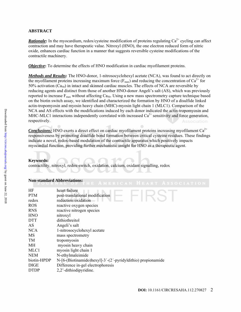
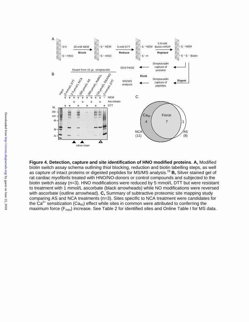
The nature of the HNO induced Cys modifications was investigated using a new variation of the biotin switch assay (Figure 4A).19 Isolated rat cardiac myofibrils were used in place of skinned fibres due to ease in preparing sufficient quantities. Mg-ATPase measurements confirmed the integrity of the myofibrils and NCA treatment resulted in a similar decrease in Ca50 observed in intact and skinned muscle fibres (Online Figure I). A change in the maximal myofibril ATPase rate was not anticipated as we have reported previously.17 For the biotin switch experiments, 5 mmol/L DTT was used to target the same HNO induced modifications which were reversed in the physiological studies. To compare HNO modification to those induced by NO, 1 mmol/L ascorbate was used to reduce S-nitrosylation groups.19 The streptavidin capture of intact biotinylated proteins revealed that HNO modified proteins could be specifically isolated and that they were distinct from those induced by treatment with the NO donor, DEA/NO (Figure 4B). HNO modifications were resistant to reduction with ascorbate while DEA/NO modifications were reversed by both reducing agents.
To map and evaluate the effects of HNO modification on individual Cys, a comparison was done
between the changes induced by NCA to those of the traditional HNO donor, AS. AS co-releases HNO and nitrite while NCA decomposes to HNO, acetate and cyclohexanone.18,21 We have previously reported that AS increased Fmax but did not affect Ca2+ sensitivity (Ca50) in cardiac muscle.17 Using the modified biotin switch technique with different donors, a comparative proteomic strategy was devised to parse the effects of the different HNO-donors; Cys modifications common to NCA and AS treatments were attributed to the increase in Fmax while sites unique to NCA were considered candidates for the decrease in
by guest on June 22, 2018http://circres.ahajournals.org/
Dow
nloaded from

DOI: 10.1161/CIRCRESAHA.112.270827 6
Ca50. A total of 12 HNO-modified Cys were identified on 8 proteins between the two treatments (Figure 4C and Table 2). Of those, 4 sites (TM Cys190, actin Cys257 and MHC Cys947 and Cys1750) were unique to NCA treatment (Online Table I). HNO induces dimeric forms of MHC-MLC1 and actin-TM.
To investigate the HNO-induced modifications, non-reducing western blot analysis was used to observe the presence of higher molecular weight species. Higher molecular weight forms of actin, TM, MHC and MLC1 were found which were reversed in the presence of 5 mmol/L DTT, indicating HNO induced inter-protein disulfide bonds (Figure 5A). MHC and MLC1 were found to be modified in a similar manner with both NCA and AS treatment as indicated by the MS analysis. The two higher molecular weight species observed were consistent with the formation of an MLC1 homodimer (25 kDa + 25 kDa = ~50 kDa) and an MHC–MLC1 heterodimer (25 kDa + 212 kDa = ~240 kDa). The formation of a higher molecular weight TM species only occurred with NCA treatment, consistent with MS analysis. A higher molecular weight form of actin, approximately 80 kDa, was observed in both NCA and AS treatment. Additionally, NCA treatment produced a loss of antibody binding for the monomeric form of actin (42 kDa). MS analysis of silver stained gel bands revealed that the actin monomer displays increased gel mobility with NCA. Analysis of other bands revealed that a similar, but less abundant, shift for actin also occurred in AS treated samples (Figure 5B and Online Table II). No molecular weight shifts were observed for any of the candidate proteins following treatment with the NO-donor.
The higher molecular weight actin and TM bands were examined by SDS-PAGE with better
resolution in that region. Comparison of purified TM and isolated myofibrils revealed a higher migration in the NCA-treated myofilament preparations (Figure 5C, left). This suggests that in myofilaments, NCA does not induce the formation of a TM homodimer but forms another disulfide linked interaction. Analysis of the higher forms of actin revealed a difference between the NCA and AS treated samples. NCA treatment of myofibrils produced a specific band at a slightly lower molecular weight than the bands observed for AS (Figure 5C, right). Comparison of the TM and actin NCA-bands suggests a linked, co-migrating species. Difference in-gel electrophoresis (DIGE) analysis revealed a distinct myofibril NCA-specific band (Cy3 - green) higher than the purified TM homodimer (Cy2 - blue) and below a series of AS-specific bands (Cy5 – red) (Figure 5D). MS analysis of the molecular weight region identified both actin and TM at that location which were lost under reducing conditions (Figure 5E and Online Table II). These findings, in combination with the site mapping results, suggest the presence of an NCA-specific heterodimer linked by the formation of a disulfide bridge between TM (Cys190) and actin (Cys257).
Other sites of modification identified in this study were considered as possible candidates for the
NCA specific increase in Ca2+ sensitivity. The two sites identified on MHC (Cys947, Cys1750) with NCA treatment were ruled out. Western blot analysis of MHC revealed no NCA-specific higher molecular weight species; only a small shift in common between the two donor treatments, determined to be an interaction with MLC1, was observed. It is possible that an intramolecular disulfide bond was formed between the residues, but considering both modifications were mapped to the linear tail region in areas not known to closely associate, it is unlikely these candidates represent meaningful sites of modification. Additional analysis into the different action of the two HNO donors revealed that the co-release of nitrite from AS had no role in inhibiting the formation of the actin-TM heterodimer. Co-incubation of NCA and nitrite did not affect heterodimer formation (Online Figure II). NCA increases Ca2+ sensitivity but not maximum force production in skeletal muscle due to the lack of dimeric MLC1 formation.
To determine if MLC1 Cys81 is involved in the increased maximum force production, the effect of HNO donors was investigated in skeletal muscle preparations. Skeletal muscle isoforms of
by guest on June 22, 2018http://circres.ahajournals.org/
Dow
nloaded from

DOI: 10.1161/CIRCRESAHA.112.270827 7
myofilament proteins contain all potential target Cys except for MLC1, which lacks the candidate site Cys81, providing a natural mutant sequence (Figure 6A). Steady-state force-[Ca2+]i relations of skeletal muscle before and after exposure to NCA (2 µmol/L) are presented in Figure 6B. Ca50 decreased significantly in the presence of NCA while Fmax remained unchanged (Fmax, 33±3.8 vs. 31.7±3.7 mN/mm2, p=NS; Ca50 0.8±0.1 vs. 1.07±0.05 µmol/L, p<0.05; Hill, 4.36±0.81 vs. 3.47±0.82, p=NS). The same insensitivity of Fmax to AS was also observed in skinned skeletal muscles (data not shown). Diagonal gel western blots revealed an absence of dimeric forms of MLC1 in NCA-treated skeletal samples but confirmed the presence of a higher molecular weight form of TM (Figure 6C and Online Figure III). These results indicate that MLC1 Cys81 is a critical residue and redox-switch for the HNO induced increase in cardiac force production. DISCUSSION
The redox-switch is emerging as a diverse signalling system for detecting and reacting to changes in the oxidative environment. Disulfide bonds have been the least appreciated candidate for these regulatory modifications.22 Here we show that two chemically unrelated HNO-donors act directly on specific myofilament proteins to form disulfide bonds between redox-switches which increase Ca2+ sensitivity and/or force. HNO-treatment resulted in the formation of an actin-TM heterodimer which correlates with the increase in Ca2+ sensitivity, and dimeric forms of MHC and MLC1 which are associated with increased force generation. These effects are reversed by treatment with reducing agents and distinct from those induced by NO. HNO action on the cardiac myofilaments.
The formation of regulatory disulfide bonds in the myofilaments that enhance contraction is a novel finding. Our data demonstrate that NCA/HNO can specifically enhance myofilament response to Ca2+ which correlates with the in vitro formation of an actin-TM heterodimer. In muscle, TM provides a physical barrier, reducing the probability of myosin S1 heads binding to the actin filament during relaxation.2 When Ca2+ binds to the troponin complex TM moves from its inactivated (blocked, B-) state over actin subdomains 1 and 2 to its Ca2+-activated (closed, C-) state over actin subdomains 3 and 4.23 This shift in position increases the probability of myosin binding to the sites on actin, further shifting TM’s position to the fully open (M-) state (Online Figure IV).23-25 Our findings indicate that HNO treatment results in the formation of a disulfide bond between the Cys residues in actin’s subdomain 4 (Cys257) and TM (Cys190),26,27 given their availability and proximity to each other during activation.28-30
The average position and movement of TM over the actin filament is a reflection of equilibrium
distribution between the B-, C- and M-states.2 In the absence of Ca2+, the distribution is ≈70/25/5%, respectively,31,32 which changes in response to Ca2+ or the presence of myosin heads.33 It is tempting to speculate that the formation of a disulfide bond between Cys190 of TM and Cys257 of actin, would tether a subpopulation of TMs in a position over actin’s subdomains 3 and 4 similar to the natural C- or M-state. This would disturb the local positional distribution of TM and may bias its local equilibrium position toward a state that is more permissive to myosin-binding; effectively priming the myofilaments for Ca2+-activation. HNO treatments were preformed in the absence of Ca2+ indicating that myofilament activation is not required for dimer formation. Only low-stoichiometry cross-linking could have a disproportionately large inotropic effect, since the effect on one TM is communicated to neighboring TMs by end-to-end interactions along the actin filament. Such local effects on TM by low-stoichiometry cross-linking do not appear to impinge on its ability to adopt the proper B-state globally, since NCA-treated muscles show normal relaxation kinetics. Increased resting force was observed for intact muscle, but not skinned preparations, and only high HNO concentrations which correlated with an increase in diastolic calcium,
by guest on June 22, 2018http://circres.ahajournals.org/
Dow
nloaded from

DOI: 10.1161/CIRCRESAHA.112.270827 8
consistent with previous findings.13,14 Alterations of TM positional equilibrium by PTMs have been demonstrated by several recent studies using C-terminus truncated troponin I.34-36
An outstanding question in the analysis is a definitive demonstration for the requirement of an
actin-TM heterodimer to influence contraction. While no other potential interactions were identified, conformation of the role of an actin-TM disulfide bond will require additional investigation including; site-directed mutagenesis via an in vivo gene-delivery approach or extraction/reconstitution of the thin filament with gelsolin, FRET techniques and 3D cryo-reconstruction of NCA treated thin filaments.32,37-39 These investigations will be the subject of future studies.
The lack of HNO-dependent increase in skeletal muscle Fmax is due to the absence of the dimeric
forms of MLC1 and MHC. Both MHC and MLC1 are essential components of the cross-bridges which form during steady-state activations. Fmax is dependent on cross-bridge turnover kinetics, the number of cross-bridges, and the force per cross-bridge.40 Therefore, alterations in the interaction between MHC and MLC1 (specifically Cys81 of MLC1) likely impacts force production. The regulatory MLC1 is positioned like a collar just below the head region on the MHC (a region called the lever arm41). It has been proposed that the swing of this lever arm produces contraction which leads to force development. MLC1 plays an essential role in stabilizing the arm.42 The orientation of these structures brings the identified residue, Cys81, in close proximity with its pair and Cys37 in MHC’s enzymatic head region (Figure S4). We therefore propose that the formation of a covalent interaction in this region, via the HNO induced disulfide bond, provides additional rigidity of the lever arm, facilitating a stronger swing of the arm, ultimately producing more force.42 Whether MHC-MLC1 heterodimer or MLC1-MLC1 homodimer are responsible is not clear as neither occurred in skeletal muscle treatments.
The slope of force-[Ca2+] relation (Hill coefficient) was not altered by NCA treatment suggesting
no change in the cooperative activation in of force/cross-bridges. Cooperative activation occurs through the transmission of Ca2+ binding to TnC across neighbouring functional units (FUs) resulting in greater cross bridge formation.2,43,44 The results indicate that NCA has little effect on Ca2+ binding to TnC, but instead suggest that it may have a greater influence on the force production of individual cross-bridges rather than the number formed. However, an NCA effect on cooperativity cannot be ruled out since not all aspects of cooperative activation are represented by changes in Hill coefficient.45 Further investigation will be required to address this possibility. HNO donors, redox chemistry and disulfide bond formation in the heart.
The formation of disulfide bonds is not specific to HNO; however, the broader circumstances of HNO derived disulfide formation appear to be unique among ROS and RNS. HNO is capable of forming disulfide bonds and has been shown to enhance in vivo cardiac inotropy;46,47 while the levels of ROS14 or NO necessary to induce disulfide bonds have been associated with wider cellular dysfunction and, in the case of myocardium, diminished contractility.48 It is likely the distinct chemistries of these oxidative species are the source of the difference.21,49 ROS can lead to rapid and transient disulfide formation via sulfenyl (thiyl) radical or sulfenic acid reactions. Both modifications can then react with nearby available thiols to form disulfides, but under conditions of persistently high oxidative stress, over-oxidation generates sulfinic and sulfonic acids which prevent this reaction. In contrast, HNO modification results in only one of two possible products, a sulfinamide or disulfide.16 The positive inotropic effects suggest the targeted chemistry of HNO is not predisposed to more harmful and permanent oxidative modifications.
A recently example of the detrimental effects of oxidative stress was presented by Canton and co-
workers who reported that left ventricular specimens obtained from explanted failing human hearts displayed greater levels of actin and TM carbonylation that correlated with contractile impairment.50 Carbonylation is an irreversible PTM likely formed by the high oxidative stress experienced over the
by guest on June 22, 2018http://circres.ahajournals.org/
Dow
nloaded from

DOI: 10.1161/CIRCRESAHA.112.270827 9
course of heart failure. In addition to carbonylation, they also found that TM participated in a disulfide cross-linked complex. The composition of this complex remains undetermined although actin was ruled out as a component. These findings highlight that differences in the levels of oxidative stress and the oxidizing agent can determine the type of PTM formed and dictate the functional or pathological outcome. Our study suggests HNO induces mildly oxidizing conditions which are manifestly different from those experienced by failing hearts contributing to contractile dysfunction.
One key observation in the current study was that treatment with HNO donors NCA and AS had distinct effects on the myofilaments. While both NCA and AS release HNO, they do so by different mechanisms and rates, yielding different by-products. AS co-releases HNO and an equal amount of nitrite upon protonation with a half life of 2.1 min at 37 oC at pH 7.4.21 Hydrolysis of NCA releases HNO, acetate and cyclohexanone with a half life that depends on the reaction conditions (t1/2 = 0.8 min in 0.1 N NaOH/MeOH and t1/2 = >2 h in pH 7.4 phosphate buffer/MeOH)18 (Online Figure V). The effect of nitrite or a possible reaction between thiol and the NCA compound have been ruled out experimentally (Online Figure II). The unique physiological effects of AS and NCA are more likely the result of their different release kinetics. The rapid release of HNO by AS is more likely to flood an area resulting in the simultaneous conversion of opposing thiols to N-hydroxysulfenamides, preventing the formation of a potential disulfide. The much slower HNO release from NCA produces a lower, more constant, concentration of HNO in the system. Low levels of HNO would favor the generation of the N-hydroxysulfenamide intermediate at only one in a pair of proximal thiols. The other thiol would remain un-modified permitting rapid reaction and disulfide formation before any rearrangement to a sulfinamide (Figure 1). Translational implications and conclusions.
Since the pharmacological management of acute HF has changed little over the past 15 years, our findings may also be of clinical interest. Current available therapies (e.g. β-agonists, phosphodiesterase inhibitors) increase intracellular [Ca2+] with negative long-term effects. HNO targets specific redox-switches to enhance the myofilament response to Ca2+, without affecting diastolic Ca2+ levels and its inotropic action is preserved in failing hearts.51 Treatment with HNO potentially offers an interesting opportunity to improve both systolic and diastolic function in HF free from the negative consequences of sustained Ca2+ manipulation.4,13,14 Clinical trials are ongoing to test this possibility in acutely decompensate HF patients.52
Oxidative modifications in the myocardium have primarily been associated with negative
functional consequences,53 but evidence suggests this may not be uniformly true. Application of the HNO donor, NCA, reproduced and exceeded the performance of previously characterized HNO-donors in cardiac tissue.17 Here we established that HNO acts directly and reversibly on the myofilament proteins to modify specific cysteine-thiols, increasing contractility. Characterization of the covalent interactions between key contractile proteins via the formation of disulfide bonds provides insight into the mechanism of this novel redox-based modulation of cardiac function. These findings provide further support for HNO-donors as a potential new class of therapeutic for the management of HF.
ACKNOWLEDGEMENTS The authors gratefully acknowledge Dr. Robert Cole and Robert O’Meally at the Johns Hopkins Mass Spectrometry and Proteomic Facility for their assistance and Dr. Jonathan Kirk for critical revision of the manuscript.
by guest on June 22, 2018http://circres.ahajournals.org/
Dow
nloaded from
DOI: 10.1161/CIRCRESAHA.112.270827 10
SOURCES OF FUNDING This work was supported by the American Heart Association (Pre-Doctoral Fellowship Grant 0815145E to CIM, Grant-in-aid AHA0855439E to WG, and Grant-in-aid 0855242E to NP), and by NIH (HL62198 to SBK; P01 HL77180-0, N01-HV-28180 and P50 HL 084946-01 to JVE; R01HL075265 and R01 HL091923 to NP) DISCLOSURES Nazareno Paolocci is a founder and stock holder of Cardioxyl Pharmaceuticals Inc. REFERENCES 1. Mudd JO, Kass DA. Tackling heart failure in the twenty-first century. Nature. 2008;451:919-928. 2. Gordon AM, Homsher E, Regnier M. Regulation of contraction in striated muscle. Physiol Rev.
2000;80:853-924. 3. Hasenfuss G, Teerlink JR. Cardiac inotropes: current agents and future directions. European
heart journal. 2011;32:1838-1845. 4. Kass DA, Solaro RJ. Mechanisms and use of calcium-sensitizing agents in the failing heart.
Circulation. 2006;113:305-315. 5. Rainwater R, Parks D, Anderson ME, Tegtmeyer P, Mann K. Role of cysteine residues in
regulation of p53 function. Mol Cell Biol. 1995;15:3892-3903. 6. Ahn SG, Thiele DJ. Redox regulation of mammalian heat shock factor 1 is essential for Hsp gene
activation and protection from stress. Genes Dev. 2003;17:516-528. 7. Burgoyne JR, Madhani M, Cuello F, Charles RL, Brennan JP, Schroder E, Browning DD, Eaton
P. Cysteine redox sensor in PKGIa enables oxidant-induced activation. Science. 2007;317:1393-1397.
8. Tocchetti CG, Stanley BA, Murray CI, Sivakumaran V, Donzelli S, Mancardi D, Pagliaro P, Gao WD, van Eyk J, Kass DA, Wink DA, Paolocci N. Playing with cardiac "redox switches": the "HNO way" to modulate cardiac function. Antioxidants & redox signaling. 2011;14:1687-1698.
9. Nagahara N, Matsumura T, Okamoto R, Kajihara Y. Protein cysteine modifications: (1) medical chemistry for proteomics. Current medicinal chemistry. 2009;16:4419-4444.
10. Hidalgo C, Donoso P. Crosstalk between calcium and redox signaling: from molecular mechanisms to health implications. Antioxidants & redox signaling. 2008;10:1275-1312.
11. MacFarlane NG, Miller DJ. Depression of peak force without altering calcium sensitivity by the superoxide anion in chemically skinned cardiac muscle of rat. Circ Res. 1992;70:1217-1224.
12. Lowe H, Baeger I, Blasig IE, Haseloff RF. Oxygen radicals attenuate the contractility of skinned muscle fibres from the pig myocardium. Die Pharmazie. 1994;49:845-849.
13. Tocchetti CG, Wang W, Froehlich JP, Huke S, Aon MA, Wilson GM, Di Benedetto G, O'Rourke B, Gao WD, Wink DA, Toscano JP, Zaccolo M, Bers DM, Valdivia HH, Cheng H, Kass DA, Paolocci N. Nitroxyl improves cellular heart function by directly enhancing cardiac sarcoplasmic reticulum Ca2+ cycling. Circ Res. 2007;100:96-104.
14. Kohr MJ, Kaludercic N, Tocchetti CG, Dong Gao W, Kass DA, Janssen PM, Paolocci N, Ziolo MT. Nitroxyl enhances myocyte Ca2+ transients by exclusively targeting SR Ca2+-cycling. Frontiers in bioscience (Elite edition). 2010;2:614-626.
15. Solaro RJ. Nitroxyl effects on myocardium provide new insights into the significance of altered myofilament response to calcium in the regulation of contractility. J Physiol. 2007;580:697.
16. Paolocci N, Jackson MI, Lopez BE, Miranda K, Tocchetti CG, Wink DA, Hobbs AJ, Fukuto JM. The pharmacology of nitroxyl (HNO) and its therapeutic potential: not just the Janus face of NO. Pharmacol Ther. 2007;113:442-458.
17. Dai T, Tian Y, Tocchetti CG, Katori T, Murphy AM, Kass DA, Paolocci N, Gao WD. Nitroxyl increases force development in rat cardiac muscle. J Physiol. 2007;580:951-960.
by guest on June 22, 2018http://circres.ahajournals.org/
Dow
nloaded from
DOI: 10.1161/CIRCRESAHA.112.270827 11
18. Sha X, Isbell TS, Patel RP, Day CS, King SB. Hydrolysis of acyloxy nitroso compounds yields nitroxyl (HNO). J Am Chem Soc. 2006;128:9687-9692.
19. Jaffrey SR, Snyder SH. The biotin switch method for the detection of S-nitrosylated proteins. Sci STKE. 2001;2001:pl1.
20. Shoman ME, DuMond JF, Isbell TS, Crawford JH, Brandon A, Honovar J, Vitturi DA, White CR, Patel RP, King SB. Acyloxy nitroso compounds as nitroxyl (HNO) donors: kinetics, reactions with thiols, and vasodilation properties. Journal of medicinal chemistry. 2011;54:1059-1070.
21. Fukuto JM, Jackson MI, Kaludercic N, Paolocci N. Examining nitroxyl in biological systems. Methods Enzymol. 2008;440:411-431.
22. Wouters MA, Fan SW, Haworth NL. Disulfides as redox switches: from molecular mechanisms to functional significance. Antioxidants & redox signaling. 2010;12:53-91.
23. Gordon AM, Regnier M, Homsher E. Skeletal and cardiac muscle contractile activation: tropomyosin "rocks and rolls". News Physiol Sci. 2001;16:49-55.
24. Vibert P, Craig R, Lehman W. Steric-model for activation of muscle thin filaments. J Mol Biol. 1997;266:8-14.
25. McKillop DF, Geeves MA. Regulation of the interaction between actin and myosin subfragment 1: evidence for three states of the thin filament. Biophys J. 1993;65:693-701.
26. McLachlan AD, Stewart M. The 14-fold periodicity in alpha-tropomyosin and the interaction with actin. J Mol Biol. 1976;103:271-298.
27. Kabsch W, Holmes KC. The actin fold. Faseb J. 1995;9:167-174. 28. Brown JH, Zhou Z, Reshetnikova L, Robinson H, Yammani RD, Tobacman LS, Cohen C.
Structure of the mid-region of tropomyosin: bending and binding sites for actin. Proc Natl Acad Sci U S A. 2005;102:18878-18883.
29. Lorenz M, Poole KJ, Popp D, Rosenbaum G, Holmes KC. An atomic model of the unregulated thin filament obtained by X-ray fiber diffraction on oriented actin-tropomyosin gels. J Mol Biol. 1995;246:108-119.
30. Lehrer SS, Ly S, Fuchs F. Tropomyosin is in a reduced state in rat cardiac muscle. J Muscle Res Cell Motil. 2011;32:63-64.
31. Xu C, Craig R, Tobacman L, Horowitz R, Lehman W. Tropomyosin positions in regulated thin filaments revealed by cryoelectron microscopy. Biophys J. 1999;77:985-992.
32. Bacchiocchi C, Lehrer SS. Ca(2+)-induced movement of tropomyosin in skeletal muscle thin filaments observed by multi-site FRET. Biophys J. 2002;82:1524-1536.
33. Galinska-Rakoczy A, Engel P, Xu C, Jung H, Craig R, Tobacman LS, Lehman W. Structural basis for the regulation of muscle contraction by troponin and tropomyosin. J Mol Biol. 2008;379:929-935.
34. Galinska A, Hatch V, Craig R, Murphy AM, Van Eyk JE, Wang CL, Lehman W, Foster DB. The C terminus of cardiac troponin I stabilizes the Ca2+-activated state of tropomyosin on actin filaments. Circ Res. 2010;106:705-711.
35. Tachampa K, Kobayashi T, Wang H, Martin AF, Biesiadecki BJ, Solaro RJ, de Tombe PP. Increased cross-bridge cycling kinetics after exchange of C-terminal truncated troponin I in skinned rat cardiac muscle. J Biol Chem. 2008;283:15114-15121.
36. Narolska NA, Piroddi N, Belus A, Boontje NM, Scellini B, Deppermann S, Zaremba R, Musters RJ, dos Remedios C, Jaquet K, Foster DB, Murphy AM, van Eyk JE, Tesi C, Poggesi C, van der Velden J, Stienen GJ. Impaired diastolic function after exchange of endogenous troponin I with C-terminal truncated troponin I in human cardiac muscle. Circ Res. 2006;99:1012-1020.
37. Lehman W, Galinska-Rakoczy A, Hatch V, Tobacman LS, Craig R. Structural basis for the activation of muscle contraction by troponin and tropomyosin. J Mol Biol. 2009;388:673-681.
38. Froehlich JP, Mahaney JE, Keceli G, Pavlos CM, Goldstein R, Redwood AJ, Sumbilla C, Lee DI, Tocchetti CG, Kass DA, Paolocci N, Toscano JP. Phospholamban thiols play a central role in
by guest on June 22, 2018http://circres.ahajournals.org/
Dow
nloaded from

DOI: 10.1161/CIRCRESAHA.112.270827 12
activation of the cardiac muscle sarcoplasmic reticulum calcium pump by nitroxyl. Biochemistry. 2008;47:13150-13152.
39. Lu X, Tobacman LS, Kawai M. Effects of tropomyosin internal deletion Delta23Tm on isometric tension and the cross-bridge kinetics in bovine myocardium. J Physiol. 2003;553:457-471.
40. Stehle R, Solzin J, Iorga B, Poggesi C. Insights into the kinetics of Ca2+-regulated contraction and relaxation from myofibril studies. Pflugers Arch. 2009;458:337-357.
41. Rayment I, Rypniewski WR, Schmidt-Base K, Smith R, Tomchick DR, Benning MM, Winkelmann DA, Wesenberg G, Holden HM. Three-dimensional structure of myosin subfragment-1: a molecular motor. Science. 1993;261:50-58.
42. Lowey S, Waller GS, Trybus KM. Skeletal muscle myosin light chains are essential for physiological speeds of shortening. Nature. 1993;365:454-456.
43. Moss RL, Razumova M, Fitzsimons DP. Myosin crossbridge activation of cardiac thin filaments: implications for myocardial function in health and disease. Circ Res. 2004;94:1290-1300.
44. Kobayashi T, Solaro RJ. Calcium, thin filaments, and the integrative biology of cardiac contractility. Annu Rev Physiol. 2005;67:39-67.
45. Razumova MV, Bukatina AE, Campbell KB. Different myofilament nearest-neighbor interactions have distinctive effects on contractile behavior. Biophys J. 2000;78:3120-3137.
46. Paolocci N, Saavedra WF, Miranda KM, Martignani C, Isoda T, Hare JM, Espey MG, Fukuto JM, Feelisch M, Wink DA, Kass DA. Nitroxyl anion exerts redox-sensitive positive cardiac inotropy in vivo by calcitonin gene-related peptide signaling. Proc Natl Acad Sci U S A. 2001;98:10463-10468.
47. Paolocci N, Katori T, Champion HC, St John ME, Miranda KM, Fukuto JM, Wink DA, Kass DA. Positive inotropic and lusitropic effects of HNO/NO- in failing hearts: independence from beta-adrenergic signaling. Proc Natl Acad Sci U S A. 2003;100:5537-5542.
48. Massion PB, Feron O, Dessy C, Balligand JL. Nitric oxide and cardiac function: ten years after, and continuing. Circ Res. 2003;93:388-398.
49. Miranda KM, Paolocci N, Katori T, Thomas DD, Ford E, Bartberger MD, Espey MG, Kass DA, Feelisch M, Fukuto JM, Wink DA. A biochemical rationale for the discrete behavior of nitroxyl and nitric oxide in the cardiovascular system. Proc Natl Acad Sci U S A. 2003;100:9196-9201.
50. Canton M, Menazza S, Sheeran FL, Polverino de Laureto P, Di Lisa F, Pepe S. Oxidation of myofibrillar proteins in human heart failure. Journal of the American College of Cardiology. 2011;57:300-309.
51. Donzelli S, Espey MG, Thomas DD, Mancardi D, Tocchetti CG, Ridnour LA, Paolocci N, King SB, Miranda KM, Lazzarino G, Fukuto JM, Wink DA. Discriminating formation of HNO from other reactive nitrogen oxide species. Free Radic Biol Med. 2006;40:1056-1066.
52. Cowart D, Aranda J, Haas G, Neutel J, Smith W, Mazhari R, Kalish V, Colucci WS. A phase I/IIA first man in saftey and tolerability study of a novel HNO donor, CLX-1020, in patents with stable congestive heart failure. JACC. 2011;57:E299-E299.
53. Seddon M, Looi YH, Shah AM. Oxidative stress and redox signalling in cardiac hypertrophy and heart failure. Heart (British Cardiac Society). 2007;93:903-907.
by guest on June 22, 2018http://circres.ahajournals.org/
Dow
nloaded from

DOI: 10.1161/CIRCRESAHA.112.270827 13
FIGURE LEGENDS Figure 1. HNO reacts with free thiols via a reactive intermediate. Following the formation of N-hydroxysulfinamide, the reaction proceeds by one of two pathways; A, isomerization to form a sulfinamide group or B, in the presence of an additional thiol formation of a disulfide bond and hydroxylamine.16 Figure 2. Effect of NCA on rat cardiac muscle. A, Raw tracings of intracellular Ca2+ transient (left) and force (right) at varied NCA concentrations. B, Pooled data of the dose-response of [Ca2+]i (left) and force development (right) to NCA (0-20 µmol/L). Note that twitch force increased significantly without increases in resting force at varied NCA concentrations (n=7-8/group). C, Effect of NCA on [Ca2+]i transient (left) and force development (right) at varied external Ca2+. At any given [Ca2+]o, twitch force increased significantly after NCA treatment while [Ca2+]i transient was not affected. * p<0.05 versus no drug (n=5 in each group). D, Effect of NCA on force-frequency relation. NCA treatment did not affect [Ca2+]i transient at any given frequencies of stimulation but increased force development at higher stimulation frequencies (* p<0.05 versus no drug, ** p<0.01 versus no drug, n=6 in each group). Figure 3. NCA acts directly on the myofilament proteins increasing Fmax and decreasing Ca50. A, Steady-state force-[Ca2+]i relationship in intact trabeculae before and after NCA (2.5 µmol/L) (n=5). B, Force-[Ca2+] relation in skinned trabeculae before and after NCA treatment (n=6). C, Reversal of NCA treatment’s effect on force-[Ca2+] in skinned muscles. The muscles were treated with DTT (5 mmol/L) for 10 min after first force-[Ca2+] was obtained in the presence of NCA treatment alone, and a second force-[Ca2+] relation was obtained in the presence of NCA+DTT treatment (n=3). D, 1-nitrosocyclohexyl pivalate (NCP), a pro-compound of NCA with similar structure that does not release HNO, did not affect force-[Ca2+] relation in skinned muscles (n=3). See Table 1. Figure 4. Detection, capture and site identification of HNO modified proteins. A, Modified biotin switch assay schema outlining thiol blocking, reduction and biotin labelling steps, as well as capture of intact proteins or digested peptides for MS/MS analysis.19 B, Silver stained gel of rat cardiac myofibrils treated with HNO/NO-donors or control compounds and subjected to the biotin switch assay (n=3). HNO modifications were reduced by 5 mmol/L DTT but were resistant to treatment with 1 mmol/L ascorbate (black arrowheads) while NO modifications were reversed with ascorbate (outline arrowhead). C, Summary of subtractive proteomic site mapping study comparing AS and NCA treatments (n=3). Sites specific to NCA treatment were candidates for the Ca2+ sensitization (Ca50) effect while sites in common were attributed to conferring the maximum force (Fmax) increase. See Table 2 for identified sites and Online Table I for MS data. Figure 5. HNO treatment induces the formation of disulfide linked dimers. A, 1 µg of rat cardiac myofibrils treated with HNO/NO donors or control compounds was separated under reducing or non-reducing conditions, western blots probed for candidate proteins identified in Table 2 (n=4). In each case the change in mobility was reversed with treatment of 5 mmol/L DTT. B, MS analysis of silver stained gel confirmed presence of monomeric actin in the NCA treatment and also revealed a similar, but less abundant, species of actin in AS treated samples (n=2) (Online Table II). C, Evaluation of the interaction between actin and TM with NCA treatment. Purified rabbit skeletal TM (0.03 µg) and isolated rat cardiac myofibrils (1 µg) were treated with NCA or AS and evaluated by 1D non-reducing western blot probing for TM (left) and actin (right) indicating a co-migrating species reversed by DTT (n=3). D, Fluorescent DIGE gel of the same samples were independently labelled; purified TM (Cy2-blue), NCA treated myofibrils (Cy3-green) and AS treated myofibrils (Cy5-red) (n=3). E, MS analysis (lower right) of the same gel region identified both actin and TM only in the non-reduced lane (n=2) (Online Table II). This analysis demonstrates a NCA specific actin-TM heterodimer linked by the formation of a disulfide bond.
by guest on June 22, 2018http://circres.ahajournals.org/
Dow
nloaded from

DOI: 10.1161/CIRCRESAHA.112.270827 14
Figure 6. MLC1 Cys81 is necessary for increase in Fmax induced by treatment with HNO donors. A, Sequence alignment comparing isoforms of rat cardiac and skeletal MLC1 in the region surrounding cardiac Cys81. B, Steady-state force–[Ca2+]i relations in before (open symbols) versus after (filled symbols) NCA treatment (2 µmol/L) from cardiac or skeletal muscles indicating loss of force increase with loss of Cys81 in skeletal isoform with NCA (n=5, each group). C, Diagonal gel shift assay (non-reduced (NR) and reduced (DTT, 100 mmol/L)) using 10 μg of skeletal or cardiac myofibrils indicating loss of higher molecular weight forms of MLC1 in skeletal HNO treated preparations while maintaining the higher molecular weight form of TM (n=3). TABLES Table 1. Effect of NCA on parameters of steady-state force-[Ca2+] relationships in intact and skinned cardiac muscles. Fmax
(mN/mm2) Ca50
(µmol/L) Hill Coefficient
(n) Intact muscles(n=5) before NCA 96±5 0.57±0.03 3.94±0.18 after NCA 123±18* 0.42±0.01* 4.92±0.84 Skinned muscles(n=6) before NCA 82±4 1.35±0.36 3.21±1.18 after NCA 94±2# 0.30±0.13# 2.39±1.02
* p<0.05 vs. before NCA in intact muscles; # p<0.05 vs. before NCA in skinned muscles. Table 2. Sites of HNO modification determined by biotin switch assay. Protein name Position of modified Cys
NC
A α-tropomyosin 190
Actin 257 myosin heavy chain 947(β)* 1750(α/β)
NC
A /
AS
myosin heavy chain 37(α) myosin light chain 1 81 Actin 12 219 α-actinin 889 myosin binding protein C 475 troponin C 35
AS
myosin heavy chain
907(α/β)
(*) denotes sites present in different isoforms of myosin heavy chain sequence.
by guest on June 22, 2018http://circres.ahajournals.org/
Dow
nloaded from

DOI: 10.1161/CIRCRESAHA.112.270827 15
Novelty and Significance What is Known?
Perturbed tissue redox conditions contribute to cardiac structural disarray and mechanical dysfunction, particularly under conditions of stress.
High fluxes of oxidizing agentscould irreversibly alter myofilaments and their regulatory proteins, reducing cardiac force generation.
Nitroxyl (HNO), a reactive nitrogen species (RNS) related to nitric oxide (NO.), increases
cardiac inotropy/lusitropy in a reversible redox-dependent manner.
What New Information Does This Article Contribute?
HNO donors act directly on specific myofilament proteins to form disulfide bonds between cysteine-based redox-switches.
HNO treatment forms actin-tropomyosin heterodimers and dimeric forms of myosin heavy chain (MHC) and myosin light chain 1 (MLC1) account for increased myofilament responsiveness to Ca2+.
HNO-induced modifications can be detected using a dithiothreitol-based biotin switch capture
technique combined with mass spectrometry (MS). Redox-switches modulate the function of channels/pumps that control cardiac Ca2+ cycling. However, evidence supporting the role of cysteine-based sensors in myofilaments is still scant, as redox-changes have been exclusively associated with reduced force generation. Here we provide evidence that HNO augments force development via reversible disulfide bond formation between specific cysteine residues in myofilaments. HNO increases Ca2+ sensitivity but not maximum force production in skeletal muscle, which lacks some sites as revealed by a new biotin-switch/MS approach. The novel idea is that redox-switches in myofilaments are dynamic in nature and undergo a continuum of redox-modifications where the functional or detrimental outcome is likely dictated by the oxidant’s nature and the redox-milieu of the myofilament compartment. The pharmacological treatment of heart failure (HF) has changed little over the past 15 years. β-agonists and phosphodiesterase inhibitors increase intracellular [Ca2+] but can have negative long-term effects. Conversely, HNO enhances myofilament Ca2+ responsiveness without affecting diastolic Ca2+ levels, and its action is preserved in failing hearts despite highly oxidizing conditions. Thus, HNO donors may improve systolic/diastolic function in HF patients, avoiding the negative consequences of sustained Ca2+ manipulation.
by guest on June 22, 2018http://circres.ahajournals.org/
Dow
nloaded from

HNO
protein S H
protein S NHOH(N-hydroxy sulfenamide)
intermediate
protein SO
NH2
Sulfinamide
protein S proteinS
NH2OH+(hydroxylamine)
Disulfide
BA
Figure 1. HNO reacts with free thiols via a reactive intermediate. Following the formation of N-hydroxysulfinamide, the reaction proceeds by one of two pathways; A, isomerization to form a sulfinamide group or B, in the presence of an additional thiol formation of a disulfide bond and hydroxylamine.16
by guest on June 22, 2018http://circres.ahajournals.org/
Dow
nloaded from

Ca transient Force
systolicdiastolic
systolicdiastolic
control, systoliccontrol, diastolicNCA, systolicNCA, diastolic
control, systoliccontrol, diastolicNCA, systolicNCA, diastolic
before NCAafter NCA
before NCAafter NCA
[Ca2+] (µmol/L) [Ca2+] (µmol/L)
Stimulation Frequency (Hz) Stimulation Frequency (Hz)
0
25
10
025
10
[Ca2+
] itra
nsie
nt (µ
mol
/L)
Developed Force (m
N/m
m2)
Developed Force (m
N/m
m2)[C
a2+] it
rans
ient
(µm
ol/L
)
Figure 2. Effect of NCA on rat cardiac muscle. A, Raw tracings of intracellular Ca2+ transient (left) and force (right) at varied NCA concentrations. B, Pooled data of the dose-response of [Ca2+]i (left) and force development (right) to NCA (0-20 µmol/L). Note that twitch force increased significantly without increases in resting force at varied NCA concentrations (n=7-8/group). C, Effect of NCA on [Ca2+]i transient (left) and force development (right) at varied external Ca2+. At any given [Ca2+]o, twitch force increased significantly after NCA treatment while [Ca2+]i transient was not affected. * p<0.05 versus no drug (n=5 in each group). D, Effect of NCA on force-frequency relation. NCA treatment did not affect [Ca2+]i transient at any given frequencies of stimulation but increased force development at higher stimulation frequencies (* p<0.05 versus no drug, ** p<0.01 versus no drug, n=6 in each group).
by guest on June 22, 2018http://circres.ahajournals.org/
Dow
nloaded from

0.0 0.5 1.0 1.5 2.0
0
25
50
75
100
125
150
[Ca2+]i (µmol/L)
control NCA
For
ce (m
N/m
m2 )
0.01 0.1 1 10 1000
20
40
60
80
100
[Ca2+] (µmol/L)
before NCA after NCA
For
ce (m
N/m
m2 )
0.01 0.1 1 10 1000.0
0.2
0.4
0.6
0.8
1.0
1.2
For
ce (%
)
[Ca2+] (µmol/L)
control NCA NCA+DTT
0.01 0.1 1 10 1000
20
40
60
80
100 before after
[Ca2+] (µmol/L)
For
ce (m
N/m
m2 )
O
O
O
N
O
O
O
N
A B
DC
Figure 3. NCA acts directly on the myofilament proteins increasing Fmax and decreasing Ca50. A, Steady-state force-[Ca2+]i relationship in intact trabeculae before and after NCA (2.5 µmol/L) (n=5). B, Force-[Ca2+] relation in skinned trabeculae before and after NCA treatment (n=6). C, Reversal of NCA treatment’s effect on force-[Ca2+] in skinned muscles. The muscles were treated with DTT (5 mmol/L) for 10 min after first force-[Ca2+] was obtained in the presence of NCA treatment alone, and a second force-[Ca2+] relation was obtained in the presence of NCA+DTT treatment (n=3). D, 1-nitrosocyclohexyl pivalate (NCP), a pro-compound of NCA with similar structure that does not release HNO, did not affect force-[Ca2+] relation in skinned muscles (n=3). See Table 1.
by guest on June 22, 2018http://circres.ahajournals.org/
Dow
nloaded from
Streptavadincapture ofproteins
Block Reduce
20 mM NEM 5 mM DTT0.8 mM
Biotin-HPDP
Replace
DigestElute
MS/MSanalysis
Streptavadincapture ofpeptides
SDS PAGE
S
S
H
HNO
S
S HNO
NEM S
S H
NEM S
S S
NEM
Biotin
A
AscorbateDTT+ ++ + + +
NEM
silver stain
Eluant from 15 µL streptavidin
++ + +++ + + ++ ++ +
200
36
116
66
21
B
Mr
C
NCA(11)
AS(8)
14 7
Ca50 Force
-
Figure 4. Detection, capture and site identification of HNO modified proteins. A, Modified biotin switch assay schema outlining thiol blocking, reduction and biotin labelling steps, as well as capture of intact proteins or digested peptides for MS/MS analysis.19 B, Silver stained gel of rat cardiac myofibrils treated with HNO/NO-donors or control compounds and subjected to the biotin switch assay (n=3). HNO modifications were reduced by 5 mmol/L DTT but were resistant to treatment with 1 mmol/L ascorbate (black arrowheads) while NO modifications were reversed with ascorbate (outline arrowhead). C, Summary of subtractive proteomic site mapping study comparing AS and NCA treatments (n=3). Sites specific to NCA treatment were candidates for the Ca2+ sensitization (Ca50) effect while sites in common were attributed to conferring the maximum force (Fmax) increase. See Table 2 for identified sites and Online Table I for MS data.
by guest on June 22, 2018http://circres.ahajournals.org/
Dow
nloaded from

Figure 5. HNO treatment induces the formation of disulfide linked dimers. A, 1 µg of rat cardiac myofibrils treated with HNO/NO donors or control compounds was separated under reducing or non-reducing conditions, western blots probed for candidate proteins identified in Table 2 (n=4). In each case the change in mobility was reversed with treatment of 5 mmol/L DTT. B, MS analysis of silver stained gel confirmed presence of monomeric actin in the NCA treatment and also revealed a similar, but less abundant, species of actin in AS treated samples (n=2) (Online Table II). C, Evaluation of the interaction between actin and TM with NCA treatment. Purified rabbit skeletal TM (0.03 µg) and isolated rat cardiac myofibrils (1 µg) were treated with NCA or AS and evaluated by 1D non-reducing western blot probing for TM (left) and actin (right) indicating a co-migrating species reversed by DTT (n=3). D, Fluorescent DIGE gel of the same samples were independently labelled; purified TM (Cy2-blue), NCA treated myofibrils (Cy3-green) and AS treated myofibrils (Cy5-red) (n=3). E, MS analysis (lower right) of the same gel region identified both actin and TM only in the non-reduced lane (n=2) (Online Table II). This analysis demonstrates a NCA specific actin-TM heterodimer linked by the formation of a disulfide bond.
by guest on June 22, 2018http://circres.ahajournals.org/
Dow
nloaded from

A
B
8070 90
Cardiac MYL3_RAT LFDRTPKGEMKITYGQCGDVLRALGQNPT
Skeletal MLE3_RAT LFDRT--GECKITLSQVGDVLRALGTNPT
***** ** *** .* ******** ***
CMr
31
21
55
36
DTTNR
31
21
55
36
Mr
MLC1
TM
[Ca2+] (µmol/L) [Ca2+] (µmol/L)
Figure 6. MLC1 Cys81 is necessary for increase in Fmax induced by treatment with HNO donors. A, Sequence alignment comparing isoforms of rat cardiac and skeletal MLC1 in the region surrounding cardiac Cys81. B, Steady-state force–[Ca2+]i relations in before (open symbols) versus after (filled symbols) NCA treatment (2 µmol/L) from cardiac or skeletal muscles indicating loss of force increase with loss of Cys81 in skeletal isoform with NCA (n=5, each group). C, Diagonal gel shift assay (non-reduced (NR) and reduced (DTT, 100 mmol/L)) using 10 μg of skeletal or cardiac myofibrils indicating loss of higher molecular weight forms of MLC1 in skeletal HNO treated preparations while maintaining the higher molecular weight form of TM (n=3).
by guest on June 22, 2018http://circres.ahajournals.org/
Dow
nloaded from
PaolocciA. Stanley, D. Brian Foster, David A. Wink, S. Bruce King, Jennifer E. Van Eyk and Nazareno
Wei Dong Gao, Christopher I. Murray, Ye Tian, Xin Zhong, Jenna F. DuMond, Xiaoxu Shen, BrianEnhances Contractile Function
Nitroxyl(HNO)-Mediated Disulfide Bond Formation Between Cardiac Myofilament Cysteines
Print ISSN: 0009-7330. Online ISSN: 1524-4571 Copyright © 2012 American Heart Association, Inc. All rights reserved.is published by the American Heart Association, 7272 Greenville Avenue, Dallas, TX 75231Circulation Research
published online July 31, 2012;Circ Res.
http://circres.ahajournals.org/content/early/2012/07/31/CIRCRESAHA.112.270827.1World Wide Web at:
The online version of this article, along with updated information and services, is located on the
http://circres.ahajournals.org/content/suppl/2012/07/31/CIRCRESAHA.112.270827.DC1Data Supplement (unedited) at:
http://circres.ahajournals.org//subscriptions/
is online at: Circulation Research Information about subscribing to Subscriptions:
http://www.lww.com/reprints Information about reprints can be found online at: Reprints:
document. Permissions and Rights Question and Answer available in the
Request Permissions in the middle column of the Web page under Services. Further information about this process isOffice. Once the online version of the published article for which permission is being requested is located, click
can be obtained via RightsLink, a service of the Copyright Clearance Center, not the EditorialCirculation Research Requests for permissions to reproduce figures, tables, or portions of articles originally published inPermissions:
by guest on June 22, 2018http://circres.ahajournals.org/
Dow
nloaded from

7
Supplemental Methods
Force and [Ca2+]i measurements in intact cardiac muscle
Rat hearts were exposed via mid-sternotomy after the animals were anesthetized with
intraperitoneal injection of pentobarbital (100 mg/kg), and were then rapidly excised and
aorta cannulated. The hearts were perfused retrogradely (~15 mL/min) with Krebs-
Henseleit (K-H) solution containing 2.3-butanedione monoxime (BDM, 20 mmol/L),
equilibrated with 95% O2 and 5% CO2. The K-H solution is composed of (in mmol/L):
NaCl 120, NaHCO3 20, KCl 5, MgCl2 1.2, glucose 10, and CaCl2 1.0, pH 7.35-7.45.
Trabeculae were quickly dissected from the right ventricles of the hearts and mounted
between a force transducer and a motor arm. The muscles were superfused with K-H
solution at a rate of ~10 mL/min and stimulated at 0.5 Hz. Force was measured using a
force transducer system (SI, Germany) and expressed in mN/mm. Sarcomere length
was measured by laser diffraction.1,2 Fura-2 potassium salt was microinjected
iontophoretically into one cell and allowed to spread throughout the whole muscle (via
gap junctions). The epifluorescence of fura-2 was measured by exciting at 380 and 340
nm. The fluorescent light was collected at 510 nm by a photomultiplier tube (R1527,
Hamamatsu). [Ca2+]i was given by (after subtraction of the autofluorescence): [Ca2+]i =
K’d(R-Rmin)/(Rmax-R), where R is the observed ratio of fluorescence (340/380), K’
d is the
apparent dissociation constant, Rmax is the ratio of 340 nm/380 nm at saturating [Ca2+],
and Rmin is the ratio of 340 nm/380 nm at zero [Ca2+]. The values of K’d, Rmax, and Rmin
were determined by in vivo calibrations.1,2 Tetanization of the trabecula was achieved by
addition of ryanodine (1.0 µmol/L) and by increasing the stimulus rate to 10 Hz briefly
(~3 sec) (isometric at sarcomere length of 2.2-2.3 µm) to obtain steady-state force-[Ca2+]
relations. Different levels of tetanized force were obtained by increasing [Ca2+] in the
perfusate (up to 20-25 mmol/L). The data was fit to the Hill equation:
8
F=Fmax[Ca2+]in/(K1/2n+[Ca2+]in), where Fmax is the maximal force. K1/2 is [Ca2+]i at half Fmax,
and n is the Hill coefficient.
Steady-state force-Ca2+ relationships in skinned cardiac and skeletal muscles
Trabeculae were skinned by ~15 min exposure to 1.0% Triton in relaxation solution
containing (mmol/L) KCl 80, HEPES 25, K2EGTA 10, creatine phosphates sodium salt
(Na2CrP) 15, Na2ATP 5, MgCl2 5.15, and leupeptin 0.5, pH 7.2. After skinning, the
muscles were activated with solutions (mmol/L: Ca2+-EGTA 10, KCl 80, HEPES 25,
Na2CrP 15, Na2ATP 5, MgCl 4.75, and leupeptin 0.5, pH 7.2) of varied [Ca2+] while
diastolic sarcomere length was kept constant. The force-pCa relationships were
generated before and after treatment with NCA (2.5 umol/L) in the same preparations.
After obtaining the force-pCa2+ relation for the control phase, the muscle was exposed to
NCA in relaxing solution for 10 min. There was no increase in resting force during the
treatment. Afterwards, the muscle was activated with activating solutions of varied [Ca2+]
containing NCA. These activations in the presence of NCA were repeated once to make
sure similar force-pCa relations were obtained.
For skeletal muscle preparations, incisions were made to the legs of the rats and
gastrocnemius muscle was dissected and placed in the dissection dish superfused with
K-H solution containing 2,3-butanedione monoxime (BDM, 20 mmol/L), equilibrated with
95% O2 and 5% CO2. Under the microscope, muscle strips (130-150 µm thick, 200-250
µm wide, 2-3 mm long) were further dissected and mounted between a force transducer
and a motor arm. The preparation was stabilized in KH (without BDM) with Ca2+ of 0.5
mM for ~10 min. The muscle was then skinned in relaxation solution containing 1.0%
Triton X-100 for 1-2 hrs. Force-Ca2+ relations were obtained in the same fashion as in
skinned cardiac muscle preparation. The stability of the preparation was tested at the
end of each experimental run at maximal activation, and the data were discarded if

9
maximal Ca2+-activated force decreased over 20% at the end of the experiment. The
experiments were performed at room temperature.
Isolation of Myofibrils
Rat myofibrillar preparations were obtained from frozen ventricles (Pel Freez Biologicals,
www.pelfreez-bio.com) minced in 20 volumes/tissue weight of 4oC relax buffer (standard
rigor buffer, SRB (mmol/L: 75 KCL, 10 imidazole pH 7.4, 2 MgCl2) plus 4
phosphocreatine, 1 ATP, 50 BDM, 1 benzamidine-HCL, 0.1 PMSF, 1 μg/mL leupeptin, 1
μg/mL pepstatin, 1.0% (v/v) Trixon X-100) and adjusted to 10 mmol/L EDTA, as
previously described.3 Minced preparations were centrifuged for 8 min at 3000xg and
the supernant was decanted. Resulting pellets were resuspended in 10 volumes of SRB
plus 1.0% Triton X-100 and subjected to 6 strokes in a Duall tissue homogenizer and
centrifuged as above. Pellets were gently resuspended and centrifuged as above twice
more in SRB including 1.0% (v/v) Triton X-100, twice in SRB lacking Triton X-100 and
once in K-60 buffer (mmol/L: 60 KCL, 20 MOPS, 2 MgCl2 pH 7.4) before being
resuspended in 5 volumes of K-60.
Myofibril ATPase Assay
Isolated myofibrils were diluted to 0.5 µg/ul in K-60 buffer and treated with 2.5 µmol/L
NCA for 10 min at room temperature. After 10 min 5 mmol/L DTT was added to some
samples. Following treatment, myofibrils were centrifuged for 5 min at 200xg and
resuspended in fresh K-60 buffer. For ATPase activity, 15 µg of myofibrils were
combined with various [Ca2+] (pCa 8 – 5.125) and 2 mmol/L ATP in a 70 µl reaction
volume and incubated for exactly 10 min at 31 oC. Reactions were halted by the addition
of 100 µl of Stop solution (3.6% (w/v) Ascorbate, 0.6 mol/L HCl, 0.3% (w/v) ammonium
molybdate and 7.2% (w/v) SDS). After allowing the signal to develop for 10 min at room

10
temperature, 100 µl of stabilizing solution (2% (w/v) Sodium Citrate, 2% (w/v) Sodium m-
Arsinite, 2% (w/v) Acetic Acid) was added. Signal was determined by measuring
absorbance at 595 nm. The amount of inorganic phosphate released during the reaction
was determined by comparison to a 5 point ammonium phosphate standard curve.
Detection of HNO modifications by biotin switch assay
HNO modified thiols were detected using a modification to the standard biotin switch
protocol.4 In brief, 100 μg of rat myofibrils/treatment were diluted to 0.5 μg/μL in HEN
(mmol/L: 250 HEPES pH 7.4, 1 EDTA and 0.1 neocuproine) including 0.1% (w/v) SDS
and exposed to a treatment for 10 min at 37oC which was subsequently removed by
acetone precipitation. Treatments included: NCA, 2.5 µmol/L; AS, 500 µmol/L; and their
decomposed, inactive equivalents which were solutions prepared and left at room
temperature for greater than 96 hours. Additional treatments were DTT, 5 mmol/L and
DEA/NO, 125 mmol/L. Remaining free thiols were blocked by resuspension in 300 μL of
HEN including 2.5% (w/v) SDS and 20 mmol/L N-ethylmaleimide (NEM), incubated for
20 min at 50oC. Excess NEM was removed by acetone precipitation. HNO and/or NO
modified thiols were reduced using 5 mmol/L DTT or 1 mmol/L ascorbate in 150 μL of
HEN including 1% (w/v) SDS and biotinylated with 0.8 mmol/L Biotin-HPDP (Thermo
Fisher Scientific, www.thermofisher.com) for one hour at room temperature. Excess
biotin-HPDP was removed by acetone precipitation (2 volumes) and resultant pellets
were carefully washed with an additional volume of acetone. Biotinylated proteins were
resuspended in 1 mL of HEN including 0.1% (w/v) SDS and captured by incubation with
15 μL of washed, packed Ultralink Immobilized Streptavidin (Thermo Fisher Scientific)
for one hour at room temperature. Beads were washed four times in 50 bead volumes
of HEN (twice including 0.1% (w/v) SDS, twice including 600 mmol/L NaCl) and twice
with Elution Buffer (mmol/L: 20 HEPES pH 7.4, 100 NaCl, 1 EDTA). Captured proteins

11
were eluted with 40 μL of EB containing 100 mmol/L DTT, mixed with 15 μL of 4X LDS
sample buffer, boiled, separated by SDS PAGE and silver stained.5 For MS studies, 200
µg of starting material was used and all subsequent volumes were doubled accordingly.
Biotinylated proteins were digested overnight with trypsin (Promega,
www.promega.com) or chymotrypsin (Roche, www.roche.com) prior to capture and
washed ten additional times with 5 mmol/L ammonium bicarbonate/20% acetonitrile
before being eluted in 100 μL of wash buffer including 100 mmol/L DTT.6
HNO donor preparation
Stock solutions of Angeli’s salt (50 mmol/L each) was prepared by dissolving powder in
10 mM NaOH and stored at -80oC for up to 2 months.7 Immediately before use, a 10x
stock was prepared in HEN buffer and applied to sample. NCA was prepared by first
diluting 1:5 in DMSO immediately before use. From that, a 10x stock solution was made
by diluting the DMSO solution 1:5000 in HEN buffer and applied to the sample.
LC/MS/MS analysis
Peptide identification by liquid chromatography/tandem mass spectrometry (LCMS/MS)
analysis was performed using an LTQ ion trap MS (Thermo Fisher Scientific,
www.thermofisher.com) interfaced with a 2D nanoLC system (Eksigent,
www.eksigent.com). Peptides were desalted on a C18 trap (75 µm x 30 mm, 5-10 µm,
120Å, YMC Gel) at 8 µL/min for 5 min with Buffer A (0.1% formic acid). After desalting,
peptides were separated on a C18 column (75 µm x 100 mm, 5 µm, 120Å, YMC ODS-
AQ, Waters, www.waters.com) with an 8 µm emitter tip (New Objective,
www.newobjective.com) using 5-60% B (90% acetonitrile in 0.1% formic acid) gradient
over 60 min at 300 nL/min. Acquired spectra were searched against the IPI rat (v3.53)
primary sequence database using Sequest (version v.27, rev. 11, Thermo Fisher

12
Scientific) utilizing the Sorcerer platform (Sage-N Research Inc.,
www.sagenresearch.com) and analyzed using Scaffold (Proteome Software Inc.,
www.proteomesoftware.com). Search parameters included peptides cleaved by trypsin
or chymotrypsin with up to 1 missed cleavage. Sequest was searched with a fragment
ion mass tolerance of 1.00 Da and a parent ion tolerance of 1.2 Da. NEM modification of
cysteine was specified in Sequest as a variable modification. Peptide identifications were
accepted if they could be established at greater than 95.0% probability as specified by
the Peptide Prophet algorithm8 and met with a visual inspection. Protein identifications
were accepted if they could be established at greater than 95.0% probability and
contained at least 1 identified peptide. Protein probabilities were assigned by the Protein
Prophet algorithm.9 Proteins that contained similar peptides and could not be
differentiated based on MS/MS analysis alone were grouped to satisfy the principles of
parsimony. A determination of a site of HNO was made when a cysteine containing
peptide was reliably identified in HNO treated samples and absent from control treated
samples and its position was mapped against the protein full sequence.
Gel shift assay
10 μg of rat myofibrils per treatment at 0.5 μg/μL in HEN were exposed to a treatment or
control conditions for 20 min at 37oC. Samples were diluted to 0.1 μg/μL in 1x LSD
sample buffer (Invitrogen, www.invitrogen.com), treated with 0 or 5 mmol/L DTT and
separated on a Bis-Tris 4-12% acrylamide 12 well, 1mm gels (Invitrogen) either using
MES or MOPS running buffer. Proteins were silver stained or transferred to
nitrocellulose and immunoblotted with primary antibodies for tropomyosin (CH1, sigma,
www.sigmaaldrich.com), actin (AC-40, sigma), myosin light chain 1 (MLM527, Abcam,
www.abcam.com) or myosin heavy chain α/β (3-48, abcam). For some silver stained gel
bands of interest, in-gel digestion was done following the established protocol.5

13
Diagonal gel shift assay
10 μg of rat cardiac or skeletal myofibrils per treatment at 0.5 μg/μL in HEN were
exposed to NCA treatment for 20 min at 37oC. Samples were diluted to 0.4 μg/μL in 1x
LSD sample buffer (Invitrogen) and separated on a Bis-Tris 4-12% acrylamide 12 well, 1
mm gels (Invitrogen) either using MOPS running buffer. After separation gel lanes were
excised using a razor blade and incubated for 20 min at 37 oC in 4 mL of 1x LDS sample
buffer contain in 100 mmol/L DTT. Gel lanes were loaded onto Bis-Tris 4-12%
acrylamide IPG well, 1.5 mm gels (Invitrogen) sealed with agarose and proteins were
separated using MOPS running buffer. Proteins were silver stained or transferred to
nitrocellulose and immunoblotted as described above.
Purification of tropomyosin
TM was purified as previously described.10
In-gel digestion
Gel slices of interest were excised from the gels cut into 1 mm3 pieces. Silver stained gel
pieces were destained in 1:1(v/v) 30 mM potassium ferricyanide and 100 mmol/L sodium
thiosulfate and washed three times with ddH2O. Gel pieces were dehydrated in 100%
acetonitrile and reswelled in 10 mmol/L DTT and incubated at 55 oC for 1 hour. After the
DTT solution was removed, a solution of 55 mmol/L iodoacetaimide was added and gel
slices were incubated at room temperature protected from light. Gel slices were then
washed 3 times with 50% (v/v) ACN, 25 mmol/L (NH4)HCO3 and then fully dehydrated in
100% ACN and dried in a speed vac. Gel pieces were reswelled in a 12.5 ng/μL trypsin
(Promega) solution containing 25 mmol/L (NH4)HCO3 and incubated at 37oC for >16
hours. Digested peptides were extracted by addition of 5% (v/v) formic acid and

14
incubation for 15 min followed by the addition of an equal volume of 100% ACN and 15
min incubation, this step was repeated and the extracts were combined. Proteins were
identified using an Orbitrap LTQ tandem mass spectrometer (Thermo Fisher) and
analyzed as described above.5
Difference in-gel electrophoresis (DIGE) analysis
Fluorescent labeling using the CyDyes (GE Healthcare, www.gehealthcare.com) was
performed according to the manufacturer's recommended protocol.11 Treated samples
were labeled as follows, 0.1 µg of purified tropomyosin (Cy2), 2 µg of NCA treated
cardiac myofibrils (Cy3), 2 µg of AS treated cardiac myofibrils (Cy5). Labeling mixtures
were incubated on ice in the dark for 30 min. The reactions were terminated by adding
0.8 μL of 10 mmol/L lysine (Sigma-Aldrich), vortexed and incubated on ice for 10 min.
Samples were then combined and mixed with LDS sample buffer and separated by 1
dimensional Bis-Tris 4-12% acrylamide 12 well, 1 mm gel (Invitrogen) using MOPS
running buffer under non-reducing conditions. After SDS–PAGE, CyDye-labeled proteins
were visualized using a TyphoonTM 9410 imager (GE Healthcare) using the
excitation/emission wavelength Cy2-488/520 nm, Cy3-532/580 nm, Cy5-633/670 nm.
Gels were scanned at 100 μm (pixel size) resolution and photomultiplier tube (PMT) was
set to 550 V using normal sensitivity. Scanned images were overlayed and intensities
were adjusted to provide optimal resolution of the bands.
References
1. Gao WD, Backx PH, Azan-Backx M, Marban E. Myofilament Ca2+ sensitivity in intact
versus skinned rat ventricular muscle. Circ Res. 1994;74:408-415.

15
2. Gao WD, Perez NG, Marban E. Calcium cycling and contractile activation in intact
mouse cardiac muscle. J Physiol. 1998;507 ( Pt 1):175-184.
3. Murphy AM, Solaro RJ. Developmental difference in the stimulation of cardiac
myofibrillar Mg2(+)-ATPase activity by calmidazolium. Pediatric research. 1990;28:46-
49.
4. Jaffrey SR, Snyder SH. The biotin switch method for the detection of S-nitrosylated
proteins. Sci STKE. 2001;2001:pl1.
5. Shevchenko A, Wilm M, Vorm O, Mann M. Mass spectrometric sequencing of
proteins silver-stained polyacrylamide gels. Analytical chemistry. 1996;68:850-858.
6. Hao G, Derakhshan B, Shi L, Campagne F, Gross SS. SNOSID, a proteomic method
for identification of cysteine S-nitrosylation sites in complex protein mixtures. Proc Natl
Acad Sci U S A. 2006;103:1012-1017.
7. Fukuto JM, Jackson MI, Kaludercic N, Paolocci N. Examining nitroxyl in biological
systems. Methods Enzymol. 2008;440:411-431.
8. Keller A, Purvine S, Nesvizhskii AI, Stolyar S, Goodlett DR, Kolker E. Experimental
protein mixture for validating tandem mass spectral analysis. Omics. 2002;6:207-212.
9. Nesvizhskii AI, Keller A, Kolker E, Aebersold R. A statistical model for identifying
proteins by tandem mass spectrometry. Analytical chemistry. 2003;75:4646-4658.
10. Pato MD, Mak AS, Smillie LB. Fragments of rabbit striated muscle alpha-
tropomyosin. I. Preparation and characterization. J Biol Chem. 1981;256:593-601.
11. Tonge R, Shaw J, Middleton B, Rowlinson R, Rayner S, Young J, Pognan F,
Hawkins E, Currie I, Davison M. Validation and development of fluorescence two-
dimensional differential gel electrophoresis proteomics technology. Proteomics.
2001;1:377-396.

2
Supplemental Figures
Online Figure I. NCA treatment of isolated myofibrils reduces pCa50 but does not affect max ATPase rate. ATPase activity was measure in isolated rat cardiac myofibrils over a range of [Ca2+] pretreated with 2.5 µmol/L NCA for 10 min or treated with NCA and then reduced with 5 mmol/L DTT (n=8). NCA treated myofibrils displayed a left shift in Ca50 compared to DTT treated (0.17±0.03 vs. 0.33±0.04 µmol/L, p=0.006). No change was observed in maximal myofibrillar ATPase rate (59.1±2.1 vs. 56.1±2.6 nmoles Pi/min/mg protein, p=0.385) or Hill coefficient (2.18±0.61 vs. 1.4±0.24, p=0.2).

3
Online Figure II. Nitrite is not implicated in the differential action of the HNO donors, NCA and AS. Gel shift assay of 1 µg of rat cardiac myofibrils treated and separated by reduced or non-reduced western blot (n=3). TM forms a higher molecular weight species (determined to be an actin-TM heterodimer) in the presence of NCA but not AS. The co-release of nitrite from AS does not inhibit this reaction as co-incubation of NCA and nitrite does not effect dimer formation.
Untre
ated
2.5
µM N
CA50
0 µM
AS
IB: tropomyosin
2.5
µM N
CA +
250
µM
NaN
O 2
250
µM N
aNO 2
DTT97
6655
36
31
Mr+ -- - - -
Untre
ated
2.5
µM N
CA50
0 µM
AS
IB: tropomyosin
2.5
µM N
CA +
250
µM
NaN
O 2
250
µM N
aNO 2
DTT97
6655
36
31
Mr+ -- - - -
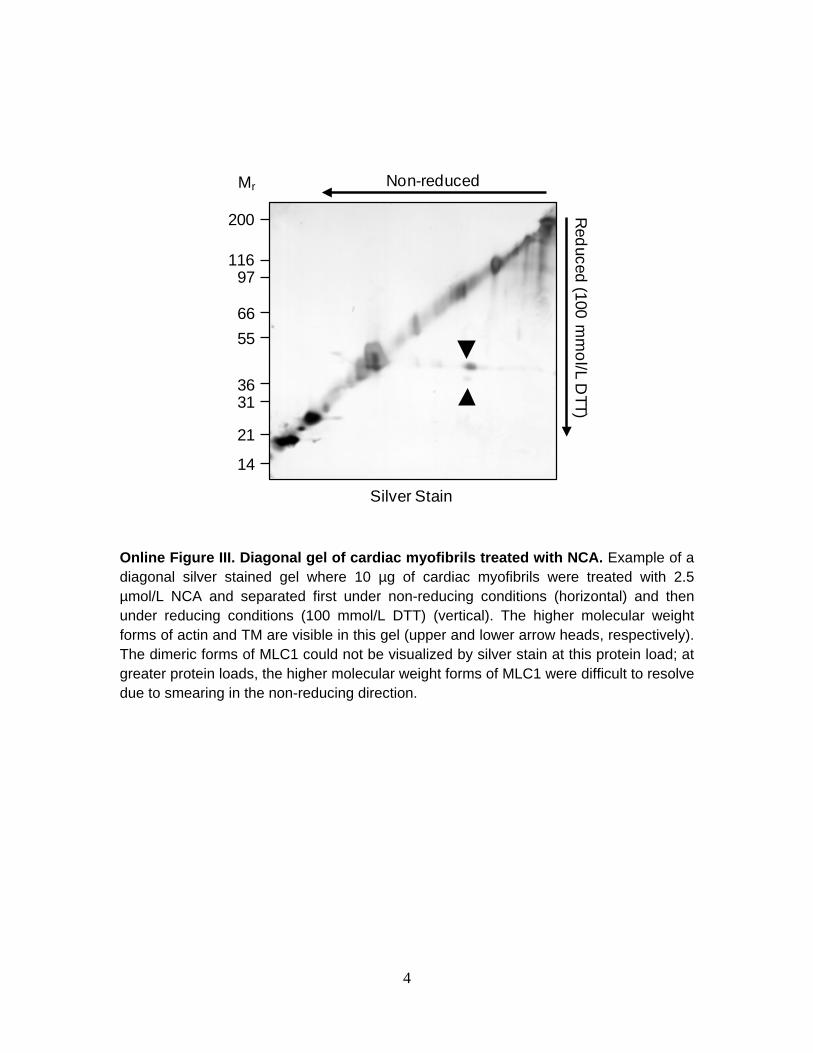
4
Non-reduced
Reduced (100 m
mol/L D
TT)
Silver Stain
200
11697
6655
3631
21
14
Mr
Online Figure III. Diagonal gel of cardiac myofibrils treated with NCA. Example of a diagonal silver stained gel where 10 µg of cardiac myofibrils were treated with 2.5 µmol/L NCA and separated first under non-reducing conditions (horizontal) and then under reducing conditions (100 mmol/L DTT) (vertical). The higher molecular weight forms of actin and TM are visible in this gel (upper and lower arrow heads, respectively). The dimeric forms of MLC1 could not be visualized by silver stain at this protein load; at greater protein loads, the higher molecular weight forms of MLC1 were difficult to resolve due to smearing in the non-reducing direction.
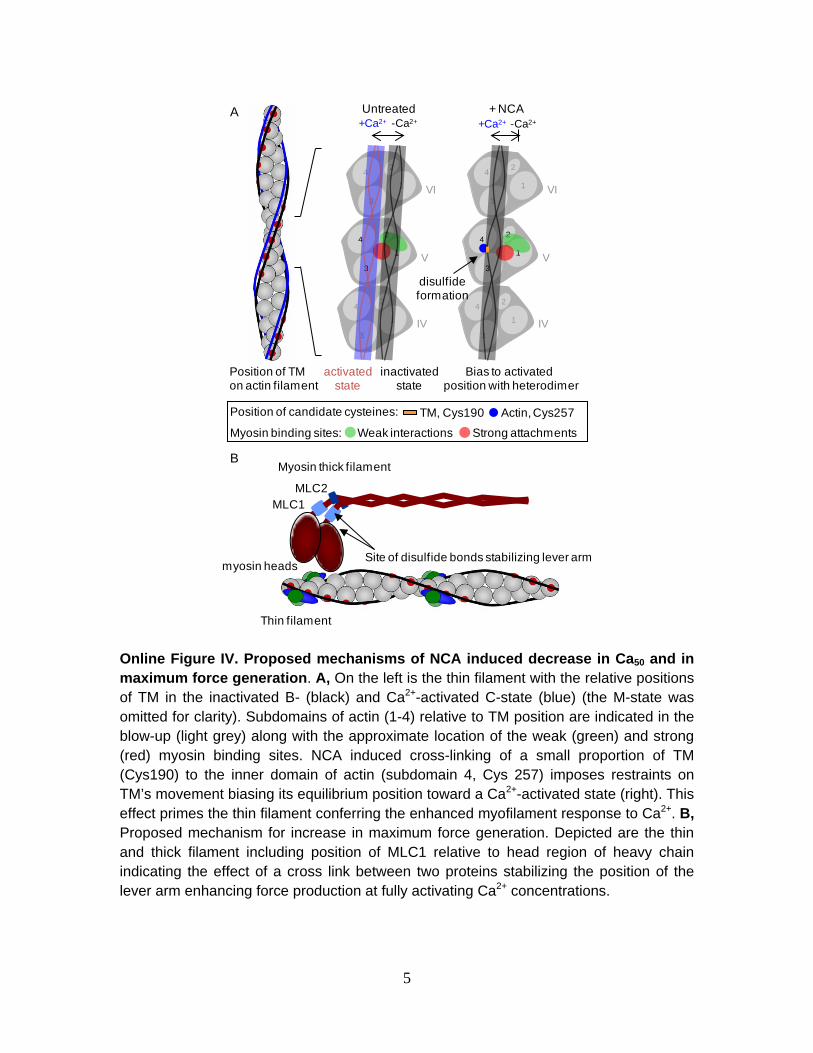
5
1
2
3
4
1
2
3
4
1
2
3
4
VI
V
IV
Position of TM on actin filament
1
2
3
4
1
2
3
4
1
2
3
4
VI
V
IV
+ NCA+Ca2+Untreated
-Ca2+ +Ca2+ -Ca2+
disulfideformation
inactivatedstate
activatedstate
Bias to activated position with heterodimer
Myosin binding sites:
Thin filament
Site of disulfide bonds stabilizing lever arm
Myosin thick filament
MLC1MLC2
A
B
myosin heads
Position of candidate cysteines:
Weak interactions Strong attachments
TM, Cys190 Actin, Cys257
Online Figure IV. Proposed mechanisms of NCA induced decrease in Ca50 and in maximum force generation. A, On the left is the thin filament with the relative positions of TM in the inactivated B- (black) and Ca2+-activated C-state (blue) (the M-state was omitted for clarity). Subdomains of actin (1-4) relative to TM position are indicated in the blow-up (light grey) along with the approximate location of the weak (green) and strong (red) myosin binding sites. NCA induced cross-linking of a small proportion of TM (Cys190) to the inner domain of actin (subdomain 4, Cys 257) imposes restraints on TM’s movement biasing its equilibrium position toward a Ca2+-activated state (right). This effect primes the thin filament conferring the enhanced myofilament response to Ca2+. B, Proposed mechanism for increase in maximum force generation. Depicted are the thin and thick filament including position of MLC1 relative to head region of heavy chain indicating the effect of a cross link between two proteins stabilizing the position of the lever arm enhancing force production at fully activating Ca2+ concentrations.

6
Online Figure V. Chemistry of NCA HNO release or reaction with thiols. A) Hydrolysis of NCA (1) results in its decomposition (2) to HNO, acetate and cyclohexane. HNO can then react with free thiols to form disulfide bonds. B) Alternatively, NCA may react with a thiol to form an organic N-hydroxysulfenamide adduct (3). Reaction of this intermediate with an additional free thiol would yield a protein disulfide releasing oxime (4) and acetate without the HNO intermediate.
O N O
O
- O H / H 2 O
1
O - N O
2 + C H 3 C O 2 H
O
+ H N O 2 R S H
R S S R + N H 2 O H
R S H
A
B O N O
3
R S O H
R S H O N
O H
O H R S S R +
N O H
+ C H 3 C O 2 H
d i s u l f i d e
d i s u l f i d e 4 , o x i m e