2.2 organ systems in animals and plants - sac
TRANSCRIPT

64 UNIT A Tissues, Organs, and Systems of Living Things
Organs Working TogetherThe star-nosed mole (Figure 2.14) may be one of the strangest-lookingcreatures on Earth, but it is also one of the most efficient predators. Itcan find and eat prey — including worms and insects — in less than one second!
The mole is built not only to be able to find and obtain food quicklybut also to escape from harm and danger quickly. The star-nosed molecan be found in eastern North America. In Canada, the star-nosedmole’s range is from Atlantic Canada to eastern Manitoba. In the U.S.,the mole ranges along the Atlantic coast to northern Florida. However,people rarely see the star-nosed mole because it lives only in marshesand wetlands. The body of the mole is elongated and covered in darkfur. This body shape is ideal for moving through the soil and the water.The dark colour of fur traps heat and keeps the mole warm while it isswimming in icy water. The limbs of the mole are strong and enable themole to dig and swim effectively.
The mole finds food by digging tunnels in the soil. While digging,the mole is able to move quickly in complex ways by kicking, brushing,and pushing dirt with its back legs. The unusual star on the nose of thismole is a touch organ, formed from 22 tentacles (Figure 2.15). Eachtentacle is covered with sensory receptors, called Eimer’s organs. Thetentacles are used to touch objects near the mole. When a mole touchessomething that may be food, it needs less than a quarter of a second toidentify it, decide if it is edible, and eat it.
Here is a summary of what youwill learn in this section:
• Organs function together toform organ systems.
• Organ systems perform one ormore functions in the humanbody.
• Organ systems work together toaccomplish movement,support, protection,communication, transport,reproduction, digestion, gasexchange, and waste removal.
• Plants have two organ systemsthat function in aninterdependent fashion.
Organ Systems in Animals and Plants
Figure 2.14 The star-nosed mole is an efficient predator because its organ systems work together.
2.2
Figure 2.15 A star-nosed moleblowing a bubble from its nose. Themole will then inhale the bubble tosmell underwater.
ist10_ch02.qxd 7/22/09 3:25 PM Page 64

65An organ consists of groups of tissues and works with other organs to form organ systems.
The star-nosed mole is a good example of how different organswork together in an organism to accomplish the many varied tasksneeded for survival. Organs that function together form organsystems, such as the nervous system or the muscular system. Eachorgan system consists of a group of organs that work together to carryout specific duties in the body. For example, for the star-nosed mole tofind food quickly, the nervous system, which for the mole includes itsstar appendage, works with its muscular system and its skeletal systemto enable the mole to move quickly and efficiently. In other words, thebrain coordinates the movements of the muscles and bones so that themole can react quickly to messages picked up by its star appendage.
A16 Quick Lab
Moving Materials
The process of digestion involves several organs.Each of the organs plays a special role in the digestiveprocess (Table 2.1). To understand the digestiveprocess and how materials move through thedigestive organs, we can use a model of the digestivesystem.
PurposeTo investigate a model of the digestive system tounderstand how materials move through the digestivetube
Procedure
1. Form small groups of three to four students.Obtain the materials from your teacher.
2. Review the function(s) of the digestive organslisted in Table 2.1.
3. Place the orange in the stocking, and attempt tomove the orange through the stocking efficiently.
4. Record the strategies that you and your groupused to move the orange from one end of thestocking to the other.
Questions
5. What problems did you encounter when you weremoving the orange from one end of the stockingto the other?
6. The orange and the stocking can be used as amodel of how digested food moves through thedigestive system. How is this model similar to themovement of materials through the digestivetube? How is this model different?
Digestive Organ Function
mouth • physical digestion through action ofteeth, tongue, and saliva
• chemical digestion of sugars usingsalivary enzymes
esophagus • movement of food in rhythmic wavesknown as peristalsis
stomach • physical digestion through churningaction and mixing with digestivejuices (acids and enzymes)
• chemical digestion of protein throughthe action of enzymes
liver • secretes bile, which breaks up fat toaid absorption, into the intestine
pancreas • secretes pancreatic juice, insulin,and enzymes into the intestine
intestines • completes chemical digestion of foodusing enzymes
• reabsorbs water• absorption of nutrients through large
surface area
rectum andanus
• storage of waste material untilelimination occurs
Table 2.1 Digestive Organs and Their Functions
• nylon stocking (open at both ends)
• an orange
Materials & Equipment
ist10_ch02.qxd 7/22/09 3:25 PM Page 65

Integumentary System Skeletal System Muscular System
Nervous SystemCirculatory SystemRespiratory SystemDigestive System
Animal Organ Systems You may have gone to a potluck dinner where every guest bringssomething that contributes to the meal. For example, someone may bringthe salad, while another person brings the main dish, and someone elsebrings the dessert. The success of the dinner depends on everyonebringing something to the dinner.
We can think of an organ system as being similar to a potluck dinner.Just as each person contributes something to the dinner, each organperforms a function in an organ system.
Biologists categorize organ systems according to their mainfunctions. There are 11 main organ systems in the human body (Figure2.16). Table 2.2 summarizes the basic functions of these organ systems.In this section, we will concentrate on the following five organ systems:integumentary, digestive, respiratory, circulatory, and excretory.
66 UNIT A Tissues, Organs, and Systems of Living Things
Figure 2.16 The 11 organ systems in the human body
ist10_ch02.qxd 7/22/09 3:25 PM Page 66

Excretory SystemEndocrine System Reproductive System Lymphatic System
67An organ consists of groups of tissues and works with other organs to form organ systems.
Organ System Organs Involved Basic Function
integumentary system skin, hair, nails, glands • covers and protects body• glands help control body temperature
skeletal system bones, cartilage • supports body• allows movement• protects the body
muscular system skeletal muscle, smooth muscle, cardiac muscle,tendons, ligaments
• works with skeletal system to provide movement• moves materials within body
digestive system mouth, esophagus, stomach, pancreas, gallbladder, liver, intestines, rectum
• ingestion • digestion• absorption of nutrients• elimination of solid wastes
respiratory system nose, mouth, trachea, lungs, bronchi, bronchioles,alveoli, diaphragm
• exchange of gases
circulatory system heart, blood vessels, blood • transportation of materials (such as oxygen,nutrients, hormones, and wastes) within body
nervous system brain, nerves, spinal cord • controls body functions• coordinates responses and activities
endocrine system glands (pituitary, hypothalamus, thyroid, adrenals),pancreas, ovaries (in females), testes (in males)
• controls growth and development• controls metabolism
excretory system skin, kidney, bladder, ureter, urethra • elimination of wastes
reproductive system ovaries, fallopian tubes, vagina, uterus (in females);testes, epididymis, vas deferens, penis, urethra (in males)
• reproduction
lymphatic system white blood cells, thymus, spleen, lymph nodes,lymph vessels
• protects body from disease• circulates fluid called lymph• absorbs and transports fats
Table 2.2 Basic Functions of Organ Systems
ist10_ch02.qxd 7/22/09 3:25 PM Page 67
The Integumentary SystemThe most visible organ system is the integumentary system. It ismade up of skin (epidermis and dermis) and accessory structures.Accessory structures include horns, antlers, hooves, quills, claws, hair,and nails. Various glands, including sweat glands, sebaceous (oil)glands, and scent glands are also part of the integumentary system.Figure 2.17 shows the human integumentary system.
Skin glands produce fluids that serve different purposes. Forexample, sweat glands secrete sweat, a clear fluid made of water andbody salts. Evaporation of sweat cools the body when it is overheated.Sebaceous glands produce oil that lubricates, waterproofs, and helpsprevent skin infections. When the sebaceous glands become pluggedwith dirt and excess oil, a blackhead forms.
The Digestive SystemIn humans, the digestive system is essentially a tube that extends fromthe mouth to the anus (Figure 2.18). The digestive system transportsnutrients through the body. In humans, the food passes from themouth, down the esophagus, into the stomach, through the small andlarge intestine, to the rectum. The major function of the digestivesystem is the absorption of nutrients. Absorption is the process bywhich food that has already been broken down passes through the wallsof the intestine into the bloodstream. Absorption takes place mainly inthe small intestine. Refer to Table 2.1 on page 65 to review the rolesthat the various organs play in human digestion.
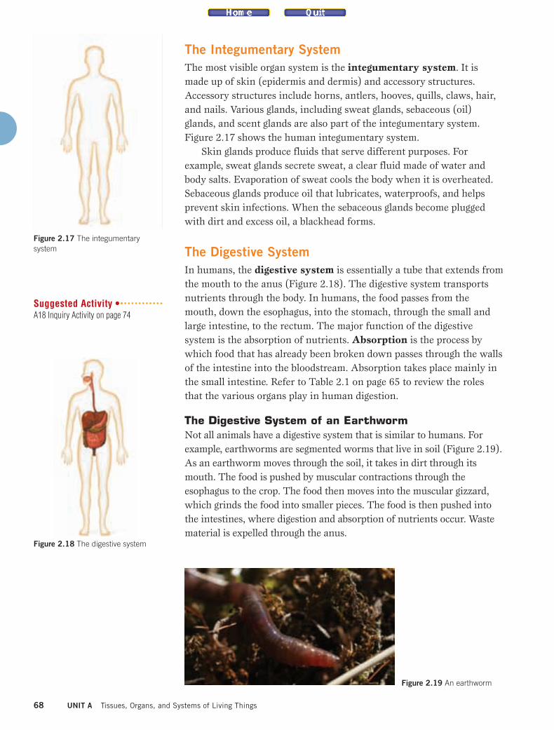
The Digestive System of an EarthwormNot all animals have a digestive system that is similar to humans. Forexample, earthworms are segmented worms that live in soil (Figure 2.19).As an earthworm moves through the soil, it takes in dirt through itsmouth. The food is pushed by muscular contractions through theesophagus to the crop. The food then moves into the muscular gizzard,which grinds the food into smaller pieces. The food is then pushed intothe intestines, where digestion and absorption of nutrients occur. Wastematerial is expelled through the anus.
68 UNIT A Tissues, Organs, and Systems of Living Things
Figure 2.17 The integumentarysystem
Figure 2.18 The digestive system
Suggested Activity •A18 Inquiry Activity on page 74
Figure 2.19 An earthworm
ist10_ch02.qxd 7/22/09 3:25 PM Page 68

69An organ consists of groups of tissues and works with other organs to form organ systems.
The Digestive System of a FishFish have a unique digestive system. For example, the yellow perch eatsinsects and other small organisms (Figure 2.20). The perch’s mouth hassmall sharp teeth that enable it to grasp its prey. Food passes from themouth down the esophagus into the stomach, where the food is brokendown. Some fish have a special pouch, called the pyloric caecum, whichfurther breaks down the food and absorbs the nutrients. Digestion iscompleted in the intestine.
The Digestive System of a Frog Adult frogs are carnivores that will eat anything that they can catch(Figure 2.21). A frog’s tongue is attached to the front of the mouth sothat it can capture flying insects effectively. It has two sets of teeth thatit uses to hold prey. When the frog swallows, it closes its eyes andpushes its eyes downward. This action causes pressure on the roof ofthe mouth, which forces the food to move into the gullet. The foodtravels down the esophagus to the stomach and then to the intestines.Waste materials exit the body through an opening called the cloaca.
The Respiratory SystemEach cell in your body requires oxygen to carry out various lifeprocesses including growth, movement, and reproduction. Oxygen isalso required to break down food to produce energy: this chemicalprocess is known as cellular respiration.
The function of the respiratory system is to obtain oxygen andrelease carbon dioxide. When you inhale, you take in air through eitheryour nose or mouth. The air passes down the trachea into the bronchusto the bronchioles. The bronchioles empty into the alveoli, which aresurrounded by thin-walled blood vessels. The alveoli are the sites of gasexchange. Figure 2.22 shows the organs involved in the humanrespiratory system.
Figure 2.20 Yellow perch Figure 2.21 North American bullfrog
During Reading
Figure 2.22 The respiratory system
A Venn Diagram SynthesizesSimilarities and Differences
Every living creature has a
digestive system. Create a triple
Venn diagram for the earthworm,
the fish, and the frog. In the
overlapping part of the circles, put
the features or actions of the
digestive systems that are similar.
In the outer parts of the circles,
put the features or actions that
are different.
ist10_ch02.qxd 7/22/09 3:25 PM Page 69

BreathingYour lungs are housed in your chest cavity, which is enclosed by theribs, chest muscles, and the diaphragm. When you inhale, your rib cagerises and your diaphragm contracts and moves downward, whichincreases the size of your chest cavity. An increase in the volume of thecavity causes a decrease in the internal air pressure in the cavity.Because the internal air pressure of the cavity is less than the airpressure in the environment, air rushes into your lungs to equalize thepressure.
When you exhale, your rib cage lowers and your diaphragm relaxesand moves upward, decreasing the size of your chest cavity. Thedecrease in the volume of the cavity causes an increase in the internalair pressure in the cavity. Since the internal air pressure is higher thanthe pressure in the environment, air moves out of your lungs. Figure2.23 shows the movement of the diaphragm during breathing.
The Circulatory SystemThe circulatory system is the blood’s transportation system (Figure2.24). The circulatory system includes the heart, blood, and bloodvessels. The heart acts as a pump to transport and regulate the flow ofblood through a series of blood vessels: arteries, veins, and capillaries.
Arteries are thick-walled vessels that carry blood away from theheart to the tissues. The thickened muscular walls of the arteries allowthem to withstand the force of the blood that is pumped from the heart.Veins carry blood back to the heart. The blood flowing through theveins is at a lower pressure than that in the arteries. Therefore, veinshave thinner walls than arteries. Veins also contain valves so that theblood does not flow backward. Arteries do not contain valves becausethe blood flow is pushed along by the blood pumped by the heart. Anetwork of capillaries connects veins and arteries.
70 UNIT A Tissues, Organs, and Systems of Living Things
airinhaled
inhalation
rib cagerises
diaphragm
rib cagelowers
airexhaled
exhalation
diaphragm
Figure 2.23 During inhalation, thechest cavity expands as the rib cagerises and the diaphragm contracts.During exhalation, the rib cage lowersand the diaphragm relaxes, whichdecreases the size of the chest cavity.
Figure 2.24 The circulatory system
Suggested Activities •• A19 Quick Lab on page 76• A20 Quick Lab on page 76
ist10_ch02.qxd 7/22/09 3:25 PM Page 70

CapillariesCapillaries are the smallest blood vessels in your body; they areabout one cell thick. Oxygen (O2) and carbon dioxide (CO2)flow in and out of capillaries by the process of diffusion(Figure 2.25). Diffusion is the movement of a substance froman area of high concentration to an area of low concentration.If the blood has more oxygen than the tissues, oxygen willdiffuse across the capillary walls and enter the tissues. Carbondioxide and other wastes are also removed from tissues bydiffusion. If the tissues have more carbon dioxide than theblood, the carbon dioxide diffuses across the capillary wallsand enters the blood. The blood then carries the carbondioxide to the lungs, where it is released as you exhale.
The Excretory System The excretory system consists of the kidneys, ureters, urinary bladder,urethra, and skin (Figure 2.26). This system filters waste products fromthe blood and maintains the proper levels of water and electrolytes in thebody. As blood flows through your kidneys, wastes such as urea, carbondioxide, and water are removed by filters called nephrons. These wastesform a fluid called urine. The urine moves out of the kidneys down theureters to the urinary bladder, where it is stored until it can beeliminated. Elimination occurs when urine travels through the urethraand out of the body. The skin is considered to be part of the excretorysystem because it excretes water, salts, and urea in sweat.
71
Learning Checkpoint
1. What organs in the digestive system are common to the earthworm, perch,
and frog?
2. Name one structure that is unique to the digestive system of the earthworm,
perch, and frog.
3. What is the diaphragm, and how is it involved in breathing?
4. Explain the role of diffusion in the process of gas exchange.
5. Explain how the excretory system eliminates waste.
capillary
O2
CO2
alveolus
An organ consists of groups of tissues and works with other organs to form organ systems.
Figure 2.26 The excretory system
Figure 2.25 Gas exchange betweena capillary and the membrane of analveolus
ist10_ch02.qxd 7/22/09 3:25 PM Page 71

Plant Organ SystemsA plant has two organ systems: a shoot system and a root system(Figure 2.27). The shoot system is everything that is above ground: thestem, leaves, buds, flowers, and fruits. The root system is everythingunderground, as well as aerial roots even though they are above ground.
To understand the interdependence between the shoot and rootsystem, consider how water is transported through the plant. Both theroots and the shoots play a role in moving water through a plant.
A plant’s roots can push water up the stem. However, the roots canonly push the water a few metres and many plants are over 100 m tall.Water enters the root hairs and travels to the xylem. Once the water isin the xylem, it is moved against gravity up the stem to the leavesthrough transpiration. Transpiration is the evaporation of waterthrough the stomata in the leaves. As each water molecule evaporates, itcreates a transpiration pull on the adjacent water molecules, whichpulls the water up the xylem to the leaves. Once the water reaches theleaf, the transpiration pull is enough to move the water from the xyleminto the ground tissue. The leaves lose a high proportion of the waterbecause of evaporation through the stomata. This evaporationmaintains the transpiration pull, and water is continuously drawn upthe stem. Figure 2.28 shows the direction of water movement.
The organs of a plant also work together to ensure that the plantsurvives changes in the environment. For example, some specializedcells record changes in the exposure to light. When the length ofdaylight increases, chemical messages are delivered to tissues tostimulate the production of a flower. Sometimes, in times of droughtand excessive heat, a plant may decrease its production of leaves.
72 UNIT A Tissues, Organs, and Systems of Living Things
flow of water
Figure 2.28 Water in atree flows from the rootsto the leaves.
Figure 2.27 A tomato plant’s organsystems
shoot
root
The tobacco mosaic virus isresponsible for severe damage tomany Ontario crops. The viruscauses changes to a plant’s shootsystem including the formation ofa mosaic pattern on the leaves.The damage to the leaves stressesthe plant and results in stuntedplant growth. The study of thisvirus has helped scientists to learnabout diseases of plant organsystems and viruses. Learn moreabout which Ontario food cropsare affected by this virus and howthis virus affects Ontario foodcrops. Report back to the class.Begin your research atScienceSource.
Take It Further
ist10_ch02.qxd 7/22/09 3:25 PM Page 72

73An organ consists of groups of tissues and works with other organs to form organ systems.
A17 Skill Builder Activity
There are some important terms that are used indissection. You will learn these terms while dissectinga vegetable.
Procedure
1. Obtain a cucumber, and cut out two holes in oneside. These holes represent the eyes. The top ofthe cucumber is known as the anterior, or cranial.The other end of the cucumber is the posterior, orcaudal. Refer to Figure 2.29(a).
2. The front-facing side of the cucumber is theventral side. The back side of the cucumber iscalled the dorsal side. We can think of the ventralside as the stomach side. Refer to Figure 2.29(b).
3. Locate the anterior end, and use a scalpel tomake a shallow cut along the ventral side of thecucumber to the posterior end. This is known as asagittal cut. If you cut the cucumber all the waythrough, you would make a sagittal section.
4. Make a shallow cut that is midway on the ventralside. Extend the cut from left to right. This type ofcut is known as transverse. If you were to cut all ofthe way through the cucumber, you would make atransverse section of the cucumber.
5. Make a sketch of your cucumber, and label withthe terms that you have learned.
6. Clean up your work area. Make sure to follow yourteacher’s directions for safe disposal of materials.Wash your hands thoroughly.
Dissection Essentials
CAUTION: If you are allergic to plants or pollen, let yourteacher know. To avoid injury, use proper techniqueswhen using the scalpel.
• cucumber
• paper towel
• paper
• pen and/or pencil
• scalpel
Materials & Equipment
dorsal ventral
posterior (caudal)
transverse section
sagittal section
anterior (cranial)
Figure 2.29 A view of acucumber showing (a) theanterior and posterior end and(b) the orientation of thetransverse and sagittalsections. (a) (b)
ist10_ch02.qxd 7/22/09 3:25 PM Page 73

A18 Inquiry Activity
An animal is able to process and absorb nutrients inthe food using its digestive system. Digestive systemsvary in animals. Biologists have found that particularanimals, such as the earthworm, perch, and frog, aregood representatives of the increasing complexity indigestive systems.
In this activity, you will study these three digestivesystems through dissection. You may do the dissectionwith preserved specimens of an earthworm and perchor use a virtual dissection program. You may choose todo only one dissection, or you may do all three tocompare the systems.
QuestionHow does the digestive system of the earthworm, theperch, and the frog accomplish the process ofdigestion?
Procedure
Part 1 — Digestive System of theEarthworm
1. Since the organs are small, it is helpful if you arefamiliar with their position in the earthworm beforeyou begin your dissection. Complete a diagram ofthe earthworm digestive system based on Figure2.30. When you are finished with your diagram,complete a virtual dissection of an earthworm byfollowing the instructions in the program, or obtaina preserved specimen of an earthworm, dissectiontools, and dissection pan. Rinse your specimenwith water, and pat dry.
2. Using the hand lens, examine the externalstructure of the earthworm so that you can identifythe prostomium, clitellum, setae, and anus. Theprostomium is in front of the mouth. The clitellumlooks like a saddle and is on the dorsal side of theearthworm. The setae are tiny bristles found onthe ventral side. The anus is found on the ventralside of the last segment of the worm.
3. Place the earthworm so that the dorsal side isfacing up. Using your scissors, make a shallowcut on the dorsal side from the clitellum to theprostomium.
The Digestive System of an Animal
SKILLS YOU WILL USE� Adapting or extending
procedures� Interpreting data/information to
identify patterns or relationships
CAUTION: To avoid injury, use proper techniques whenusing the scalpel.
• paper towels
• preserved specimens ofearthworm and perch
• hand lens
• scalpel or dissectingscissors
• probe
• dissecting tray
• dissecting pins
• forceps
• virtual dissectionprogram for earthworm, perch, and frog
• pen and/or pencil
• paper
Skills References 2, 6
Materials & Equipment
74 UNIT A Tissues, Organs, and Systems of Living Things
gizzard esophagus
mouthclitellum
anus
setae
cropintestine
Figure 2.30 The external and internal anatomy of the earthworm
Key ActivityDI
ist10_ch02.qxd 7/22/09 3:25 PM Page 74

75An organ consists of groups of tissues and works with other organs to form organ systems.
A18 Inquiry Activity (continued)
4. Separate the tissue, and use dissecting pins to pinthe body wall down to the tray. You may need tocut through the tissue that holds the body wall.
5. Locate the mouth, esophagus, crop, gizzard,intestine, and anus using Figure 2.30.
6. Clean up your work area. Make sure to follow yourteacher’s directions for safe disposal of materials.Wash your hands thoroughly.
Part 2 — Digestive System of the Perch
7. Complete a diagram of the perch digestive systembased on Figure 2.31. When you are finished withyour diagram, complete a virtual dissection of aperch or obtain a preserved specimen of a perch,dissection tools, and dissection pan. Rinse yourspecimen with water, and pat dry.
8. Observe the external structure of the perch. Notethe position and number of fins. Find the lateralline, and locate the gill cover and anal opening.
9. Examine the mouth of the perch.
10. Create a flap through the muscle wall. Make anincision from the bottom of the gill cover along theventral side to the anal opening. Continue theincision up from the anal opening to the lateralline and then along that line to the head of thefish. Finish your flap by extending your incisionback to the base of the gill cover.
11. Lift the flap of muscle wall to look at the organs ofthe perch. If you have a female perch, the areamay be filled with eggs. If this is the case, youshould remove the mass of eggs beforeproceeding. If the perch is male, the testes will besmaller and lighter in colour. Locate the liver (lightbrown), gall bladder (olive colour), esophagus,stomach, pyloric caeca, and intestines.
12. Clean up your work area. Make sure to follow yourteacher’s directions for safe disposal of materials.Wash your hands thoroughly.
Part 3 — Digestive System of the Frog
13. Complete a virtual dissection of a frog. Identify themouth parts, liver, gall bladder, stomach,pancreas, small and large intestine, and cloaca.
Analyzing and Interpreting
14. How is the mouth specialized?
15. Explain how the structure of the intestines isrelated to their role in digestion.
16. Why do you think the gall bladder is located soclose to the liver? Explain your answer.
Skill Practice
17. Describe one problem that you encountered inperforming the dissection, and explain how yousolved the problem.
Forming Conclusions
18. How is the digestive system of the worm, theperch, and the frog each suited to its habitat?
stomachesophagus
gill cover
spiny dorsal fin
lateral line
swim bladderanuspyloric caeca
intestinesliver
heartgall bladder
Figure 2.31 The external and internal anatomy of the perch
ist10_ch02.qxd 7/22/09 3:25 PM Page 75

76 UNIT A Tissues, Organs, and Systems of Living Things
A19 Quick Lab
When you breathe, you move about 500 mL of air in andout of your lungs. Usually we are not aware of ourbreathing. What can you learn about how you breathe ifyou concentrate on your breathing?
PurposeTo observe the movements of your body as youbreathe and to count the number of breaths that youtake when you breathe normally
Procedure
1. Work in pairs. One partner sits in a chair andbreathes normally. The other person observes andrecords any breathing movements that occur inthe chest, shoulders, and abdomen.
2. While breathing normally, your partner counts thenumber of breaths that you take in one minuteand records the number.
3. Change places with your partner, and repeat steps1 and 2.
Questions
4. Explain how the chest and abdomen changeduring breathing.
5. Explain why the number of breaths per minutemay change when exercising.
A Look at Breathing
• pen and/or pencil • stopwatch
Materials & Equipment
A20 Quick Lab
Heart disease is a major cause of death in Canada.There are several known risk factors for heart disease,including high blood pressure, high blood cholesterol,stress, being overweight, diabetes, excessive alcoholconsumption, smoking, physical inactivity, andunhealthy diets.
PurposeTo research the risk factors associated with heartdisease
Procedure
1. Work in a group of 3–4 students.
2. Each member of the group should select one ofthe risk factors for heart disease to research.
3. ScienceSource Research to learn about heartdisease and how your chosen risk factor increasesthe risk of heart disease. Record your informationin a table.
4. Share your information with your group so thatevery member will understand the relationshipbetween risk factors and heart disease.
Questions
5. Describe any common features that exist betweenthe risk factors discussed in your group. Does thissuggest that there is a common approach toreducing the risk of heart disease?
6. Your research focusses on the risk factors thatcan be controlled. Describe one way in whichsociety influences an individual’s ability to controlhis or her risk factors for heart disease.
Inquiring about Heart Disease
ist10_ch02.qxd 7/22/09 3:25 PM Page 76

Key Concept Review1. Define and give an example of an organ
system.
2. What organ system is involved intransporting materials around the body?
3. Name and describe the function of twoorgans of the digestive system.
4. Name the organ system involved in breathing.
5. Describe the role of muscle tissue in thedigestive system of the earthworm.
6. Name and describe the function of threeaccessory structures of the integumentarysystem.
7. Look at the organs and job descriptions givenin the following table. Match each organ to itsproper job description.
8. List the two organ systems that are found inplants.
Connect Your Understanding9. Explain why the crop and gizzard are
important parts in the digestive system of theearthworm.
10. Why is it important to maintain a healthyintegumentary system?
11. Using the star-nosed mole as an example,write a paragraph that explains how organsinteract with each other to help accomplishthe tasks needed for survival.
12. Write a paragraph that explains how chestmuscles, ribs, and the diaphragm worktogether to help you to breathe efficiently.
13. The circulatory system is a transportationsystem. Use an analogy of a roadway toexplain how this system functions.
14. There is a puppet master controlling thepuppets shown below. Is there an “organmaster” controlling the actions of all theorgans in the body? Explain your answerusing an example.
Reflection15. Choose an organ system. Identify two
questions you have about how that organsystem works in your body.
16. Describe three facts that you found mostinteresting in this section that you did notknow before.
For more questions, go to ScienceSource.
2.2 CHECK and REFLECT
77An organ consists of groups of tissues and works with other organs to form organ systems.
Question 14
Organ Job Description
heart • filters and cleans blood
teeth • controls whole body
intestines • grinds food
skin • breaks down food and absorbsnutrients
kidney • exchanges gases
esophagus • covers and protects surface
bladder • pumps blood
brain • stores urine
lungs • passes food from the mouth to thestomach
Organs and Their Job Descriptions
ist10_ch02.qxd 7/22/09 3:25 PM Page 77