213 nm ultraviolet photodissociation on peptide anions
TRANSCRIPT

HAL Id: hal-01344628https://hal.archives-ouvertes.fr/hal-01344628
Submitted on 12 Jul 2016
HAL is a multi-disciplinary open accessarchive for the deposit and dissemination of sci-entific research documents, whether they are pub-lished or not. The documents may come fromteaching and research institutions in France orabroad, or from public or private research centers.
L’archive ouverte pluridisciplinaire HAL, estdestinée au dépôt et à la diffusion de documentsscientifiques de niveau recherche, publiés ou non,émanant des établissements d’enseignement et derecherche français ou étrangers, des laboratoirespublics ou privés.
213 nm Ultraviolet Photodissociation on PeptideAnions: Radical-Directed Fragmentation Patterns
Mohammad A Halim, Marion Girod, Luke Macaleese, Jérôme Lemoine,Rodolphe Antoine, Philippe Dugourd
To cite this version:Mohammad A Halim, Marion Girod, Luke Macaleese, Jérôme Lemoine, Rodolphe Antoine, et al..213 nm Ultraviolet Photodissociation on Peptide Anions: Radical-Directed Fragmentation Patterns.Journal of The American Society for Mass Spectrometry, Springer Verlag (Germany), 2016, 27 (3),pp.474-486. �10.1007/s13361-015-1297-5�. �hal-01344628�
1
213 nm Ultraviolet Photodissociation on Peptide Anions:
Radical-Directed Fragmentation Patterns
Mohammad A. Halim,1 Marion Girod,2 Luke MacAleese,1 Jérôme Lemoine,2 Rodolphe
Antoine,1 Philippe Dugourd1
1Institut Lumière Matière, Université Lyon 1 – CNRS, Université de Lyon, 69622 Villeurbanne
Cedex, France
2Institut des Sciences Analytiques, Université Lyon 1 – CNRS, Université de Lyon, 69622
Villeurbanne Cedex, France
Correspondance to: Philippe Dugourd, Email : [email protected]
Citation : 213 nm Ultraviolet Photodissociation on Peptide Anions: Radical-Directed Fragmentation Patterns. M. A. Halim, M. Girod, L. MacAleese, J. Lemoine, R. Antoine and P. Dugourd. J. Am. Soc. Mass Spectrom. 27, 474-486 (2016). http://dx.doi.org/10.1007/s13361-015-1297-5

2
Abstract
Characterization of acidic peptides and proteins is greatly hindered due to lack of suitable analytical
techniques. Here we present the implementation of 213 nm Ultraviolet photodissociation (UVPD)
in high-resolution quadrupole-Orbitrap mass spectrometer in negative polarity for peptide anions.
Radical-driven backbone fragmentation provides 22 distinctive fragment ion types, achieving the
complete sequence coverage for all reported peptides. Hydrogen-deficient radical anion not only
promotes the cleavage of Cα–C bond but also stimulates the breaking of N–Cα and C–N bonds.
Radical-directed loss of small molecules and specific side-chain of amino acids are detected in
these experiments. Radical containing side-chain of amino acids (Tyr, Ser, Thr, and Asp) may
possibly support the N–Cα backbone fragmentation. Proline comprising peptides exhibit the
unusual fragment ions similar to reported earlier. Interestingly, basic amino acids such as Arg and
Lys also stimulated the formation of abundant b and y ions of the related peptide anions. Loss of
hydrogen atom from the charge-reduced radical anion and fragment ions are rationalized by time-
dependent density functional theory (TDDFT) calculation, locating the potential energy surface
(PES) of ππ* and repulsive πσ* excited states of a model amide system.
Keywords: Photo-fragmentation, Radical Anions, UVPD, Peptide, TDDFT

3
Introduction
Alternative to collision [1-3] and electron [4, 5] based techniques, photon-based methods have
emerged as new powerful approaches for characterizing peptides, polysaccharides and proteins [6-
12]. Among them, ultraviolet photodissociation (UVPD) leads to intense fragmentation patterns.
In this method, protein and peptide cations predominately dissociate to a/x ions and less frequently
to c/z and b/y ions. Different wavelengths such as 157, 193, 220, and 280 nm have been
implemented in UVPD. Above and at 280 nm, specific fragmentation have been reported following
excitation of aromatic residues in peptides or proteins [13]. The number of fragment ions increases
as the wavelength decreases from 280 nm to 213 nm [13, 14].
Another efficient and popular wavelength 193 nm has been implemented in hybrid linear ion trap-
orbitrap mass spectrometer for characterizing different peptide and proteins in positive polarity.
Wide-ranging fragmentation yields a/x, b/y, c/z, y-1, v, w and d ions and thus provides nearly
complete sequence coverage. Whole protein characterization has been achieved by this technique
implementing direct infusion and/or chromatographic time scale [15, 16]. Along with common
fragment ions, Madsen et al also observed some uncommon fragment ions such as a+2, c-1 and
z+1 [17]. This study disclosed that fragmentation patterns varied with the protonation state of the
peptide. When protonation takes place at N-terminus, cleavage of Cα-C bond occurred, however,
N-Cα cleavage is favored with C-terminus protonation.
Thompson et al employed vacuum photodissociation at 157 nm on singly protonated peptide ions
to elucidate the unusual backbone cleavage [18]. Cui et al further revealed that basic residues in
the C-terminal yields to x, v and w fragment ions whereas N-terminal produces a and d fragments
ions [19]. Moreover, a+1 and x+1 radical ions are identified from the charge localized N- and C-
terminals, respectively. Secondary radical elimination of hydrogen atom are detected from a+1 and
x+1 ions to produce a and x ions, respectively. Satellite ions such as d, v and w are formed due to
part of side chain elimination. b, c and z fragment ions are also noticed but are less frequent than a
and x ions. Hydrogen/deuterium exchange experiments further confirmed that both backbone
amide and side-chain β-carbon hydrogens can undergo elimination to yield a and x ions [20].
Implementing time-resolved photodissociation at 157 nm revealed some unusual but stable x+2
fragment ion compared to less common a+2 ion [21]. They proposed that addition of one hydrogen
4
to x+1 and a+1 radical ions can yield x+2 and a+2 ions. Migration and transfer of hydrogen atom
to radical ions have also been witnessed in ECD studies [22], [23].
However, most of these experiments were conducted on peptide and protein cations and very few
were directed on negative polarity. It is assumed that around 50% of naturally occurring peptides
are acidic and prone to yield negative ions. Kjeldsen et al reported Cα-C backbone fragmentation
by EDD (electron detachment dissociation) for peptide and observed more C-terminal species (x
ions) than N-terminal fragments (a• ions) [24]. Comparison of negative electron transfer
dissociation (NETD) and UVPD for peptide anion disclosed that NETD usually produce simple set
of a/x ions [25]. In NETD, along with a/x ions various neutral losses are observed from entire or
partial side-chain cleavage of amino acids [26]. Activated ion negative electron transfer
dissociation (AI-NETD) of doubly charged peptide ions also generates some hydrogen loss from a
and x fragment ions [27].
Some previous electron photo-detachment dissociation (EPD) studies were performed with UV
lasers on peptides and small proteins in negative polarity [28, 29]. Antoine et al investigated the
electron photo-detachment dissociation of peptides using 262 nm with a linear ion trap [30].
Formation of [M-2H]-• radical anion from the precursor ion was documented in this experiment.
a/x and c/z fragment ions were observed [28]. Comparative studies between EDD and EPD
revealed significantly different fragment ion distributions in which EPD fragment ions are typically
produced from tryptophan and histidine resides whereas in EDD backbone dissociation are favored
[28]. However, EDD on small proteins including ubiquitin and melittin suggests that basic resides
may promote the formation of a/x fragment ions [31].
Radical containing peptides promote characteristic fragmentation pattern in mass spectrometry [32,
33]. Radical peptides are classified into two categories: hydrogen-deficient and hydrogen-rich
radicals [34]. The former type is typically formed in UVPD, EDD and NETD routes whereas the
later one is generated from ECD/ETD [8, 24, 35-37]. Recently, formation of hydrogen-deficient
species from the hydrogen-rich radical cation in ECD received great attention due to extensive
fragmentation and wide-spread side-chain loss [33, 38]. Radical migration in hydrogen-deficient
peptide radical promotes extensive neutral loss and allows remote backbone dissociation [33, 39].
5
Here, we present the implementation of 213 nm UVPD in a Thermo Scientific Q Exactive hybrid
quadrupole-orbitrap mass spectrometer in negative polarity for peptide anions. We observed
distinctive Cα–C, N–Cα and C-N backbone fragmentations from the hydrogen-deficient radical
anions. Radical-driven extensive neutral loss is likewise evident in these experiments. Moreover,
series of hydrogen-deficient and hydrogen-rich fragments are observed.
Material and Methods
Photodissociation Mass Spectrometry
All experiments were performed on a hybrid quadrupole-orbitrap Q-Exactive® mass spectrometer
(Thermo Fisher Scientific, San Jose, CA, USA) equipped with a HESI ion source. Three small
peptides YTIAALLSPYS, DYKDDDDK and RGDSPASSKP were used without any further
purification. Peptides samples were prepared at 1 µM concentration in 50/49/1 (v/v/v)
acetonitrile/water/ammonium hydroxide and directly infused to MS at a flow rate of 5 µL/min. All
spectra were acquired using a mass range of 100-1500 m/z and resolving power of 140000 at m/z
400. The AGC (Automatic Gain Control) target for MS/MS was set to 1x10⁶ and the maximum
injection time was set at 250 ms. The isolation width was 2 Th. When required, the identification
of fragment ions was confirmed by fragmentation of a single isotope (selection width 0.4 Th). The
HCD collision energy was set to the minimum 2 eV in order to avoid collisions and provide
photofragmentation spectra free of CID contamination. Different HCD (High Collision
Dissociation) trapping times including 100, 500, 1000 and 2000 ms (2, 10, 20, 40 laser shots,
respectively) were considered. All experiments were performed on 5 microscans mode with
averaging 200 scans.
For UVPD experiments, BrillantB Nd:YAG (Quantel, Les Ulis, France) laser was employed.
Details of the set-up are given elsewhere [14]. In brief, the 5th harmonic (λ=213 nm) with a
repetition rate of 20 Hz was used. The hybrid quadrupole-orbitrap Q-Exactive® mass spectrometer
was modified to permit the laser irradiation of peptide ions. The laser beam passes through lenses,
diaphragms and then is introduced in the HCD cell using two dichroic mirrors. A UV grade fused-
silica window was fitted on the back of the HCD cell to allow penetration of a laser beam. The
laser beam energy irradiating the ions was ~1 mJ/ pulse. The laser was slightly off axis so as to
avoid photofragmentation in the C-trap.

6
Manual analysis of UVPD data was performed with the aid of ChemCalc software [40]. Peak lists
of three peptides were generated for all six major UVPD ion types (a, b, c, x, y, and z). Fragments
mass tolerance was set to 20 ppm.
Computation
All calculations were conducted with the Gaussian 09 software package [41]. Optimization and
subsequent vibrational frequency calculation on the model amide system [CH3CONHCH3] were
performed using density functional theory employing Becker’s (B3) [42] exchange functional
combining Lee, Yang, and Parr’s (LYP) [43] correlation functional. Gaussian basis set 6-311+G
(2d,p) was considered. Natural bond orbitals (NBO) [44, 45] calculations were computed at the
same level of theory. For calculating the excited state properties, time-dependent density functional
theory (TDDFT) [46] was employed with the B3LYP/6-311+G(2d,p) level of theory in gas phase.
For TDDFT calculation, 20 excited states were considered.
Result and Discussion
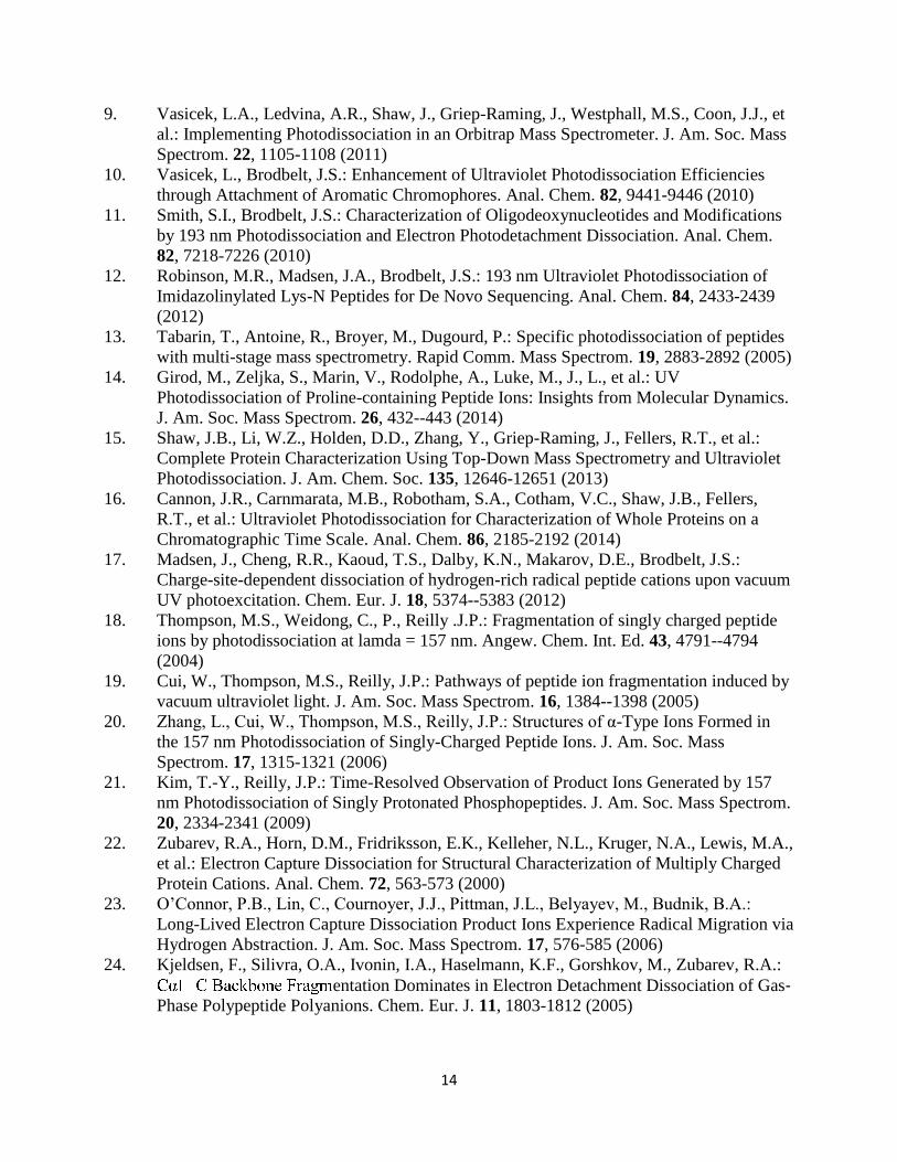
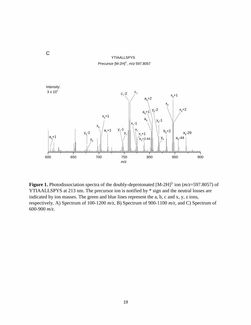
The Photodissociation of Peptide 1 (YTIAALLSPYS)
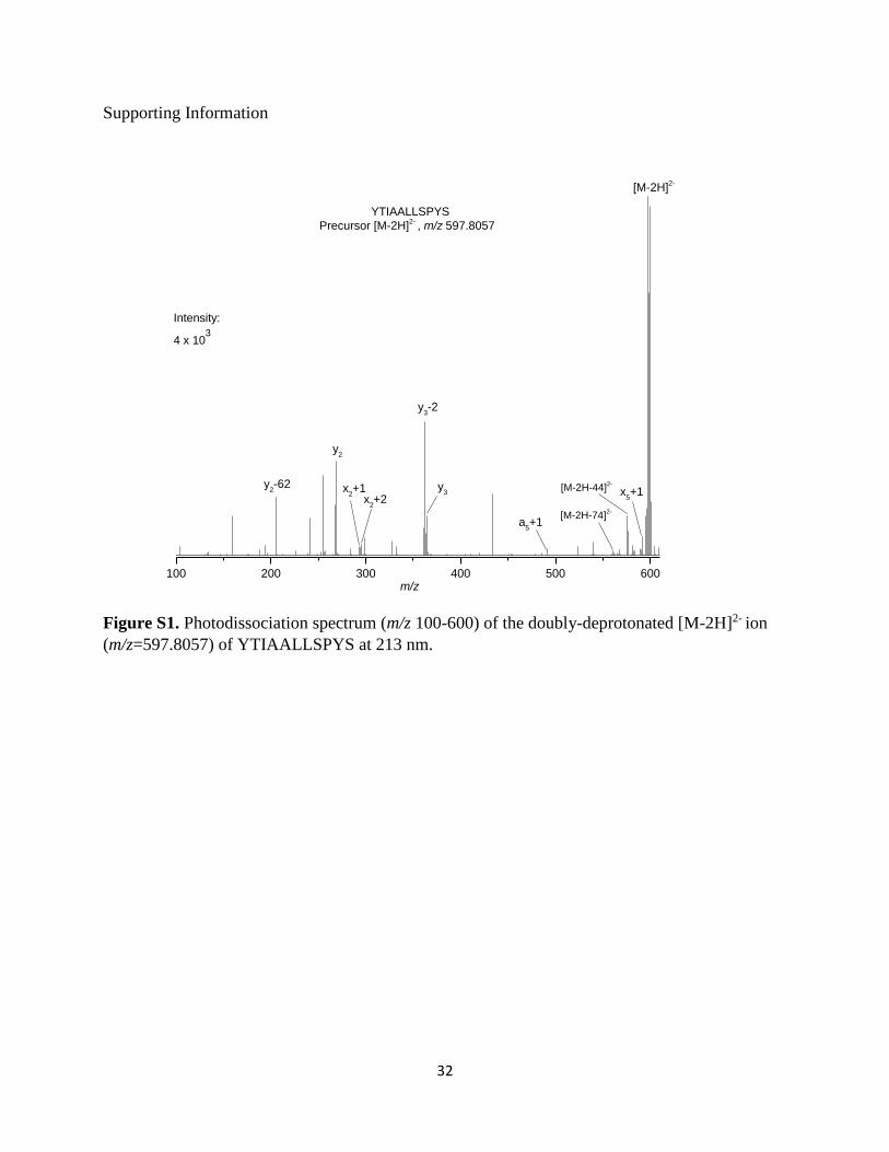
The photodissociation spectrum of the doubly-deprotonated [M-2H]2- (m/z 597.8057) of this
peptide is presented in Figure 1A. Exact masses and assignments of fragment ions of this peptide
are summarized in Table 1. Similar to previous studies, the characteristic [M-2H]-• charge-reduced
radical species is detected at m/z 1195.6094 Da. This radical species is typically generated from
photo-induced electron detachment from the selected peptide precursor. Intense neutral losses are
detected from this radical species (Table 2 and Figure 2). Similar neutral losses are also
demonstrated in previous studies [30, 31, 47-49]. The CH3 radical (15.0242 Da) loss appears at m/z
1180.5852 from the side-chain of Ala [50]. Neutral loss of CO (27.9947 Da) and CH3CH2 (28.9995
Da) are noticed at m/z 1167.6147 and 1166.6099 Da, respectively. Removal of CH3CH2 can be
used to distinguish the side chain loss of Ile (28.9995 Da) or Leu (43.0542 Da) [51]. Loss of CH2O
(30.0100 Da) and CH2OH (31.0178 Da) are also observed from the side chain of Ser. NETD study
on serine (Ser) containing peptide witnessed the loss of CH2O when Ser is not phosphorylated [26].
The peak at m/z 1151.5829 can be assigned to the loss of C2H4O (44.0265 Da) from Thr side-chain

7
[26, 50]. The sequential loss (61.9998 Da) of CO2 and H2O is also identified at m/z 1133.6099.
Radical elimination of a C3H8ON from the Thr residue may lead to the fragment ion detected at
m/z 1121.5759. Loss of tyrosylate groups from the side chain of Tyr (107.0472 and 106.0406 Da)
is identified at m/z 1088.5622 and 1089.5688 Da, respectively. The phenoxy group of the tyrosylate
produces an oxygen radical, which induces the cleavage of Cα-Cβ side-chain of the tyrosine residue
and promotes the formation of O=C6H4=CH2 (exact mass 106.0413 Da) ion [8, 50, 52]. Two
relatively weak peaks at m/z 1139.5855 and 1123.5910 can be assigned for the side-chain and
related ion loss (56.0239 and 72.0184 Da) from Leu or Ile [26, 51-53]. Combined losses of
tyrosylate and C2H4O from Tyr and Thr appear at m/z 1045.5419 and 1044.5346 respectively.
Zooms of Figure 1A are shown in Figure 1B-1C and S1. Selected fragment ions from the single
isotope selection of the doubly-deprotonated [M-2H]2- precursor ions are shown in Figure S2. For
peptide 1, a series of radical (an+1)-. fragment ions is observed for n=5, 6, 7, 8 and 9 These ions
correspond to the elemental composition of an ions plus one hydrogen atom (explaining the +1 in
the notation) and are radicals (dot in the notation).This nomenclature is in agreement with the one
proposed recently by Chu et al [54] excepted that we do not include the hydrogen symbol (H) after
the number of losses or gains. Homolytic cleavage between the Cα and the carbonyl C from the
precursor ion induced the formation of these radical ions, as shown in Scheme 1. Classical (an)-
fragment ions are detected for n=8 and 9. These ions may mainly arise from the fragmentation of
the doubly-deprotonated [M-2H]2- precursor ion. However, they can also be produced by secondary
H elimination from the radical (an+1)-. fragment ions [19]. Abundant a ions are favored by aromatic
amino acids and in this case it is due to Tyr residue in N-terminal [28, 52]. An unusual fragment
such as (a8+2)- is additionally identified at m/z 805.4815 and which may be due to the presence of
Pro residue [14, 17]. Detection of (a+2)- is also reported by Madsen et al in a high-throughput
UVPD study in negative polarity for complex proteomic sample [55]. Two peaks at m/z 871.5031
and 856.4917 correspond to the loss of CH3CH2 (28.9995 Da from Ile) and C2H4O (44.0265 Da
from Thr) from (a9)- ion. Radical (xn+1)-. ions are also formed via homolytic cleavage of the Cα -
carbonyl C bond, complementary to (an+1)-. ions (Scheme 1). Series of radical (xn+1)-. ions are
noticed at n= 2, 5, 6, 7, 8, and 10 whereas (xn)- ions are detected at n= 6, 7, 8 and 9. Two unusual
fragment types such as (xn+2)- for n=2, 8 and radical (xn-1)-. for n=7 and 9 appear for peptide 1.
(x2+2)- ion detected at m/z 295.0924 is close to Pro residue [14]. Kim and Reilly found xn+2
fragment ions at 157 nm UVPD and concluded that some x+1 radical ions may take one hydrogen

8
to form these new ions [21]. (xn+2)- ions are also detected at 193 nm UVPD [55]. The proposed
fragmentation pathway for the formation of (x2+2)- ion is presented in Scheme 2.The formation of
two (xn-1)-. ions are likewise owing to the radical elimination of hydrogen atom from the
corresponding xn ions. Shaw et al also observed some (xn-1)-. ions in activated ion negative electron
transfer dissociation [27]. Moreover, classical fragmentation of the Cα - C bond with proton
transfers from the charge-reduced [M-2H]-. radical species also yields to the formation of (xn-1)-.
ions. Indeed, these ions will contain the initial radical site and the negative charge. Fragmentation
is then observed after electron photo-detachment.
Series of (yn)- ions are detected at n=2, 3, 6, 7, and 8. Radical (yn-1)-. ions are also observed at the
positions n=6, 7, 8, and 10. These ions arise from the homolytic cleavage of the C-N bond from
the precursor ion (Scheme 3). However, complementary (bn+1)-. radical ions are not detected.
Fragmentation of the C-N bond from the charge-reduced [M-2H]-. radical species may also leads
to the formation of the (yn-1)-. ions, if the charge and the radical site after electron loss are located
on the C-terminal side. As a general statement, the abundance of fragment ions results from both
direct fragmentation of the precursor ions and fragmentation of the charge-reduced radical ions
obtained after electron loss (EPD). (yn-1)-. radical ions could also be formed by H elimination from
the (yn)- ions. Three new (yn-2)- ions are detected for this peptide at n=3, 8, and 9 positions and
could be formed by H elimination from the (yn-1)-. ions. The fragmentation of the C-N bond close
to the Pro residue can also explain the formation of the (y3-2)- fragment ion [14]. Once again, these
fragment ions could also arise from the homolytic cleavage of C-N bond fragmentation from the
charge-reduced [M-2H]-. radical species. One (b8+2)- fragment ion is detected at m/z 833.4757 for
this peptide due to the presence of the Pro residue [14]. A neutral loss of 44.0264 Da corresponds
to C2H4O of Thr observed at m/z 789.4493 from (b8+2)- (Figure 1A).
c/z ions are less abundant for this peptide. Two (cn)- ions are detected at n=7 and 9 positions.
Moreover, two (cn-1)-. ions at n=9,10 positions and (cn-2)- ions at n=7, 10 sites are observed.
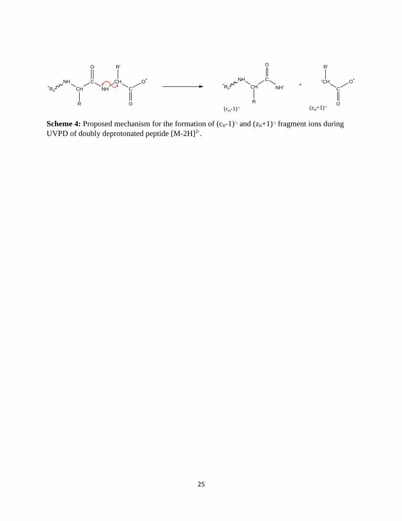
Radical (cn-1)-. ions could be produce via the homolytic cleavage of the N-Cα bond from the
precursor ion (Scheme 4). Hydrogen abstraction from c ions are also detected in ECD [22, 56, 57].
The formation of the (cn-2)- ions could be explained by the radical induced fragmentation of the N-
Cα bond from the charge-reduced [M-2H]-. radical species after electron loss.

9
The Photodissociation of Peptide 2 (DYKDDDDK)
The photodissociation spectrum of the doubly-deprotonated [M-2H]2- (m/z 505.1906) of peptide
DYKDDDDK is presented in Figure 3 and S3. Exact masses and assignments of fragment ions of
this peptide are summarized in Table 3. Intense neutral losses are also evident from this peptide
(Table S1). Loss of H2O from the charge-reduced radical species [M-2H]-• is detected at m/z
992.3709. Losses of one, two and three CO2 are identified at m/z 966.3913, 922.4019, and
878.4116, respectively. Madsen et al observed one and two CO2 loss at 193 nm UVPD of singly
and multiply charged peptide anions [49]. Abundant CO2 loss was moreover demonstrated in
electron detachment dissociation for peptide and protein [29, 30]. Elimination of several CO2 is a
common feature related to aspartic and glutamic acid residues in NETD, Al-NETD, EDD and
UVPD [27, 30]. The UVPD spectrum showed losses of 27.9955 Da from [M-2H]-• that can be
attributed to CO, similar to peptide 1. Loss of CO from radical species is also found in an earlier
ECD study [58]. The peaks at m/z 903.3321 and 904.3394 correspond to the losses of tyrosylate
groups of Tyr (107.0491 and 106.0418 Da) from the [M-2H]-•. Radical C3H6O2N (88.0371 Da)
group elimination from the aspartic amino acid yields to the ion detected at m/z 922.3441. The ion
observed at m/z 938.3961, can be assigned to the loss of C3H4O2 (71.9851 Da) from Asp residue
[26]. Loss of Lys residue (100.0736 Da) is also detected at m/z 910.3076. Moreover, a loss of
71.0713 Da (C4H9N) observed for the ion at m/z 939.3099 is from the Lys residue [26]. A combined
loss of CO2 and H2O appears at m/z 948.3803.
A complete series of (an)- fragment ion is observed for this peptide for n=2-7. (an+1)-. ions are
detected for n=4, 5, 6 and 7. These ions are formed via homolytic cleavage from the precursor ion
(Scheme 1). Radical (an-1)-. ions are detected for n= 3, 5 and 6. Fragmentation of the Cα - C bond
from the charge-reduced radical species [M-2H]-• is involved to produce these series. Secondary
radical elimination of hydrogen atom from (an)- ions could also yield to the formation of these ions.
A complete series of (xn)- fragment ions is detected at n=2-7 similar to complementary (an)
- ions.
Two radical (xn+1)-. ions (n=3 and 6) are detected at m/z 402.1380 and 760.2859, respectively.
Moreover, two (xn+2)- ions (n=2 and 6), which are formed by addition of one extra hydrogen atom
to (xn+1)- ions are detected. Additionally, (x7-1)- ion is observed at m/z 921.3364. Same
fragmentation mechanisms are proposed for the formation of these ions than for the peptide 1

10
described previously. A distinctive peaks at m/z 886.3281 corresponds to the loss of one and two
H2O molecules from (x7)-, respectively.
Two (bn)- fragment ions are observed at n=1 and 7 sites whereas very abundant radical (bn-1)-. ions
are detected for n= 1, 3-7. These ions would come from the fragmentation of the C-N bond from
the charge-reduced [M-2H]-• radical species. Several (yn)- ions appear at n= 3-6 positions. Some
(yn-1)-. ions at n=3, 6, 7 sites are also detected (formed via the mechanism proposed Scheme 3) as
well as (y7-2)- ion. Specific radical induced fragmentation of the [M-2H]-• radical species is then
also observed, after electron loss, for this peptide.
Cleavage of N-Cα bonds produces series of c and z ions. Four (cn)- ions and (cn-1)-. radical ions are
noticed at n=4-7 positions. These ions arise from the homolytic cleavage of the N-Cα bond from
the precursor ion (Scheme 4). However, complementary (zn+1)-. radical ions are not detected. (zn)-
ions are detected from 2, 3, 6 and 7 positions. Interestingly, complete series of radical (zn-1)-. ions
(n=2-7) is observed for this peptide. Classical fragmentation of the N-Cα bond with proton transfers
from the [M-2H]-• radical species is proposed for the formation of these ions as well as the (cn-1)-.
series. Compared to first peptide, abundance of c and z ions is noticeable for this peptide and may
be due to the presence of five Asp residues. Removal of one H2O, one CO2 and combined CO2 and
H2O from (z2)- ion are detected at m/z 225.0868 199.1074, and 181.0967, respectively. Previous
studies also noticed the losses of H2O and CO2 from z ion when peptide contained Asp residues
[54]. Combinations of backbone cleavages and neutral losses are listed in Table S1.
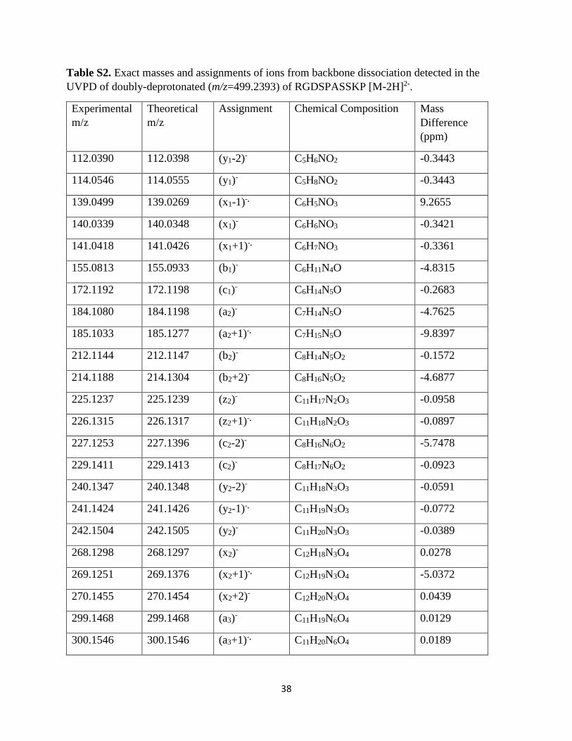
The Photodissociation of Peptide 3 (RGDSPASSKP)
The photodissociation spectrum of the doubly-deprotonated [M-2H]2- (m/z 499.2393) of peptide
RGDSPASSKP is presented in Figure 4 and S4. Exact masses and assignments of fragment ions
of this peptide are summarized in Table S2. Intense neutral losses are summarized in Table S3.
The loss of H2O from the charge-reduced radical species [M-2H]-• (m/z 998.4767) is noticed at m/z
980.4673 (Figure 4A). There are three Ser residues in this peptides and loss of CH2O (30.0095 Da)
at m/z 968.4672 can be attributed to the side chain of Ser. The loss of 60.0540 Da observed for the
peak at m/z 938.4227 corresponds to the C2H6ON group of the Ser residue. Loss of CO2 (exact
mass 43.9895 Da) from the carboxyl group located in C-terminal or side chain of aspartic acid
11
appears at m/z 954.4872. Two distinctive peaks at m/z 899.3982 and 912.4072 correspond to the
losses of 99.0785, and 86.0695 Da from the arginine side chain [26, 53]. Loss of 88.0498 Da which
is detected at m/z 910.4269 is related to the side chain of Asp [59].
Nearly complete series of (an)- fragment ions is observed for this peptide for n=2-9 whereas (an-1)-
. ions are detected for n= 6 and 9. Radical (an+1)-. ions are detected for n=2-9 (Scheme 1). Addition
of one hydrogen to (an+1)-. radical ions (similar as shown in Scheme 2 for the xn+1 ions) which
yield (an+2)- is also prevalent for n=3-5,7-9 positions. (an+2)- ions are also observed for Proline
containing peptides [14, 60] and explain the formation of (a4+2)-. and (a9+2)- ions. An almost
complete series of (xn)- fragment ions is detected at n=1-4, 6-9 similar to the complementary (an)
-
ions. Four (xn-1)-. ions are observed for n= 1, 4, 7-9 sites. Moreover, (xn+1)-. ions are detected for
n=1-4, 6, 7, and 9. Three (xn+2)- ions (n=2, 3, 6 and 7) are also formed via H addition on the (xn+1)-
. ions.
(bn)- and (yn)
- fragments ions are predominant in this peptides, which may be due to the presence
of basic Arg and Lys amino acids [61]. (bn)- ions are identified for n=1-5, 8, and 9 positions only
missing n=6 and 7 related to Ala-Ser and Ser-Ser amide bonds. (bn+1)-. ions are detected for n= 4,
5, and 9 (Scheme 3). Three (bn-1)-. ions are observed at n= 3, 8 and 9. Representative (bn+2)- ions
appear at 2, 4, 9 positions in which two sites (4 and 9) are closed to the Pro residues. (b2+2)- ion
could be explain by the H addition on the (bn+1)-. ion. Complete sequence of (yn)- ions are found
(n= 1-3, 5-9) whereas (yn-1)-. ions are noticed for n=2, 5-9. Distinctive (yn-2)- ions are detected for
n=1, 2, 6-9.
Homolytic cleavage and fragmentation, associated with proton transfers, of N-Cα bonds is also
noticeable. Full sequence of (cn)- ions located for n=1-3, 5-9 and (cn-1)-. ions are noticed at n= 3,
6-9. Fragment (cn-2)- ions are detected for n= 2, 3, 6-8. Similar to peptide 2, complete series of (zn)-
ions (n=2-9) are generated from this peptide. (zn-1)-. ions are also observed for n=3,7-9. Moreover,
(zn+1)-. ions are detected for n=2, 4, 6, 7 and 9 (Scheme 4).
Photo-induced Hydrogen loss at 213 nm
A general trend is observed for those three peptides with series of backbone cleavages leading to
ions deficient in hydrogen. All three peptides produce the distinctive doubly-deprotonated [M-2H]-

12
• charge-reduced radical species upon irradiation of the monoisotopic precursor ion [M-2H]2-, along
with hydrogen loss from the charge-reduced radical species as shown in (Figure 5). Time-
dependent density functional theory (TDDFT) calculation has been performed on a model amide
system to elucidate the role of πσ* excited state in the photodissociation of peptide. The potential
energy surface of the model amide system, π, π*, and σ* molecular orbitals are displayed in Figure
6. The lowest ππ*, πσ* and electronic group state (S0) are shown with respect to the N-H stretching
coordinate of the model amide. The ππ* excitation is observed for the amide system at 215 nm
(5.75 eV) which relates with our UVPD experiment at 213 nm. The diffuse and polar character of
σ* orbital is observed which is similar to the previous studies on pyrrole/indole system [62, 63].
The shallow barrier with respect to N-H stretch indicates the repulsive nature of this state [62]. For
this amide system, the ππ* surface is above the πσ* surface which may allow the fast internal
crossing from the ππ* to the πσ* states and lead to H atom dissociation [63-65]. The ππ* excitation-
induced amide hydrogen loss then provides a general route for the formation of hydrogen-deficient
ions in 213 nm UVPD. Repetition of this mechanism with absorption of several photons can lead
to fragments displaying multiple H-loss. Moreover, the ππ* excitation-induced amide hydrogen
loss may yield a nitrogen-centered amide anion intermediate and stimulate the wide-spread
backbone fragmentation. However, details theoretical calculation are sought to elicit the
mechanism of radical-driven side-chain loss and backbone fragmentation at 213 nm
photodissociation on peptide and protein anions. Similar mechanism can also arise on other bonds
from aromatic cycles or COO chromophore groups.
Conclusion
The key features of these experiments can be summarized as follows: (1) Extensive sequence
specific side-chain losses are observed for all three peptides. (2) Near complete series of classical
backbone cleavages (a/x, b/y, c/z) are observed. (3) Unusual fragment ions including (x+1)-., (x+2)-
, (x-1)-., (y-1)-., (y-2)-, (z-1)-., (z+1)-., (z+2)- and (a-1)-., (a+1)-., (a+2)-, (b-1)-., (b+1)-., (b+2)-, (c-1)-
., (c-2)- are consistently observed in these experiments and further confirmed by selecting single
isotopic peak of the precursor ions. Some of these ions are coming from homolytic cleavages of
the backbone from the precursor doubly charged ion. Classical fragmentation of backbone bonds
concerted with proton transfers and homolytic cleavages are also observed for the charge-reduced
[M-2H]-. radical species after electron photo-detachment. Radical-induced specific fragment ions

13
are then produced in these experiments of UVPD in the negative mode. Some of these ions may
also result from secondary H eliminations. (4) Hydrogen-deficient ions may result from ππ*
excitation-induced amide hydrogen loss. This ππ* excitation is reached upon absorption of a photon
at 213 nm. The present study outlines the difficulty to interpret and systematically analyze the
wealth of fragmentation produced by irradiation of peptide and protein anions at the onset of the
amide bond absorption band, which may be different from VUV excitation.
Acknowledgements
The research leading to these results has received funding from the European Research Council
under the European Union's Seventh Framework Programme (FP7/2007-2013 Grant agreement
N°320659)
Supporting Information: Figures S1-S4 and Table S1-S3 are included in the supporting file.
References:
1. Zhurov, K.O., Fornelli, L., Wodrich, M.D., Laskay, U.A., Tsybin, Y.O.: Principles of
electron capture and transfer dissociation mass spectrometry applied to peptide and
protein structure analysis. Chem. Soc. Rev. 42, 5014-5030 (2013)
2. McLuckey, S.A.: Principles of collisional activation in analytical mass spectrometry. J,
Am. Soc. Mass Spectrom. 3, 599-614 (1992)
3. Wells, J. M., McLuckey, S. A.: Collision‐induced dissociation (CID) of peptides and
proteins. Methods in Enzymol. 402, 148-185 (2005)
4. Zubarev, R.A., Kelleher, N.L., McLafferty, F.W.: Electron capture dissociation of
multiply charged protein cations. A nonergodic process. J. Am. Chem. Soc. 120, 3265-
3266 (1998)
5. Syka, J.E.P., Coon, J.J., Schroeder, M.J., Shabanowitz, J., Hunt, D.F.: Peptide and protein
sequence analysis by electron transfer dissociation mass spectrometry. Proc Natl Acad Sci
USA. 101, 9528-9533 (2004)
6. Brodbelt, J.S.: Shedding Light on the Frontier of Photodissociation. J. Am. Chem. Soc.
22, 197-206 (2011)
7. Brodbelt, J.S.: Photodissociation mass spectrometry: new tools for characterization of
biological molecules. Chem. Soc. Rev. 43, 2757-2783 (2014)
8. Antoine, R., Lemoine, J., Dugourd, P.: Electron Photodetachment Dissociation For
Structural Characterization Of Synthetic And Bio-Polymer Anions. Mass Spectrom. Rev.
33, 501-522 (2014)
14
9. Vasicek, L.A., Ledvina, A.R., Shaw, J., Griep-Raming, J., Westphall, M.S., Coon, J.J., et
al.: Implementing Photodissociation in an Orbitrap Mass Spectrometer. J. Am. Soc. Mass
Spectrom. 22, 1105-1108 (2011)
10. Vasicek, L., Brodbelt, J.S.: Enhancement of Ultraviolet Photodissociation Efficiencies
through Attachment of Aromatic Chromophores. Anal. Chem. 82, 9441-9446 (2010)
11. Smith, S.I., Brodbelt, J.S.: Characterization of Oligodeoxynucleotides and Modifications
by 193 nm Photodissociation and Electron Photodetachment Dissociation. Anal. Chem.
82, 7218-7226 (2010)
12. Robinson, M.R., Madsen, J.A., Brodbelt, J.S.: 193 nm Ultraviolet Photodissociation of
Imidazolinylated Lys-N Peptides for De Novo Sequencing. Anal. Chem. 84, 2433-2439
(2012)
13. Tabarin, T., Antoine, R., Broyer, M., Dugourd, P.: Specific photodissociation of peptides
with multi-stage mass spectrometry. Rapid Comm. Mass Spectrom. 19, 2883-2892 (2005)
14. Girod, M., Zeljka, S., Marin, V., Rodolphe, A., Luke, M., J., L., et al.: UV
Photodissociation of Proline-containing Peptide Ions: Insights from Molecular Dynamics.
J. Am. Soc. Mass Spectrom. 26, 432--443 (2014)
15. Shaw, J.B., Li, W.Z., Holden, D.D., Zhang, Y., Griep-Raming, J., Fellers, R.T., et al.:
Complete Protein Characterization Using Top-Down Mass Spectrometry and Ultraviolet
Photodissociation. J. Am. Chem. Soc. 135, 12646-12651 (2013)
16. Cannon, J.R., Carnmarata, M.B., Robotham, S.A., Cotham, V.C., Shaw, J.B., Fellers,
R.T., et al.: Ultraviolet Photodissociation for Characterization of Whole Proteins on a
Chromatographic Time Scale. Anal. Chem. 86, 2185-2192 (2014)
17. Madsen, J., Cheng, R.R., Kaoud, T.S., Dalby, K.N., Makarov, D.E., Brodbelt, J.S.:
Charge-site-dependent dissociation of hydrogen-rich radical peptide cations upon vacuum
UV photoexcitation. Chem. Eur. J. 18, 5374--5383 (2012)
18. Thompson, M.S., Weidong, C., P., Reilly .J.P.: Fragmentation of singly charged peptide
ions by photodissociation at lamda = 157 nm. Angew. Chem. Int. Ed. 43, 4791--4794
(2004)
19. Cui, W., Thompson, M.S., Reilly, J.P.: Pathways of peptide ion fragmentation induced by
vacuum ultraviolet light. J. Am. Soc. Mass Spectrom. 16, 1384--1398 (2005)
20. Zhang, L., Cui, W., Thompson, M.S., Reilly, J.P.: Structures of α-Type Ions Formed in
the 157 nm Photodissociation of Singly-Charged Peptide Ions. J. Am. Soc. Mass
Spectrom. 17, 1315-1321 (2006)
21. Kim, T.-Y., Reilly, J.P.: Time-Resolved Observation of Product Ions Generated by 157
nm Photodissociation of Singly Protonated Phosphopeptides. J. Am. Soc. Mass Spectrom.
20, 2334-2341 (2009)
22. Zubarev, R.A., Horn, D.M., Fridriksson, E.K., Kelleher, N.L., Kruger, N.A., Lewis, M.A.,
et al.: Electron Capture Dissociation for Structural Characterization of Multiply Charged
Protein Cations. Anal. Chem. 72, 563-573 (2000)
23. O’Connor, P.B., Lin, C., Cournoyer, J.J., Pittman, J.L., Belyayev, M., Budnik, B.A.:
Long-Lived Electron Capture Dissociation Product Ions Experience Radical Migration via
Hydrogen Abstraction. J. Am. Soc. Mass Spectrom. 17, 576-585 (2006)
24. Kjeldsen, F., Silivra, O.A., Ivonin, I.A., Haselmann, K.F., Gorshkov, M., Zubarev, R.A.:
entation Dominates in Electron Detachment Dissociation of Gas‐Phase Polypeptide Polyanions. Chem. Eur. J. 11, 1803-1812 (2005)

15
25. Shaw, J.B., Madsen, J., Xu, H., Brodbelt, J.S.: Systematic comparison of ultraviolet
photodissociation and electron transfer dissociation for peptide anion characterization. J.
Am. Soc. Mass Spectrom. 23, 1707--1715 (2012)
26. Rumachik, N.G., McAlister, G.C., Russell, J.D., Bailey, D.J., Wenger , C.D., Coon, J.J.:
Characterizing peptide neutral losses induced by negative electron-transfer dissociation
(NETD). J. Am. Soc. Mass Spectrom. 23, 718--727 (2012)
27. Shaw, J.B., Kaplan, D.A., Brodbelt, J.S.: Activated Ion Negative Electron Transfer
Dissociation of Multiply Charged Peptide Anions. Anal. Chem. 85, 4721-4728 (2013)
28. Larraillet, V., Vorobyev, A., Brunet, C., Lemoine, J., Tsybin, Y.O., Antoine, R., et al.:
Comparative Dissociation of Peptide Polyanions by Electron Impact and Photo-Induced
Electron Detachment. J. Am. Soc. Mass Spectrom. 21, 670-680 (2010)
29. Larraillet, V., Antoine, R., Dugourd, P., J., L., me: Activated-electron photodetachment
dissociation for the structural characterization of protein polyanions. Anal. Chem. 81,
8410--8416 (2009)
30. Antoine, R., Joly, L., Tabarin, T., Broyer, M., Dugourd, P., Lemoine, J.: Photo-induced
formation of radical anion peptides. Electron photodetachment dissociation experiments.
Rapid Comm. Mass Spectrom. 21, 265-268 (2007)
31. Ganisl, B., Valovka, T., Hartl, M., Taucher, M., Bister, K., Breuker, K.: Electron
Detachment Dissociation for Top-Down Mass Spectrometry of Acidic Proteins. Chem.
Eur. J. 17, 4460-4469 (2011)
32. Turecek, F., Julian, R.R.: Peptide Radicals and Cation Radicals in the Gas Phase. Chem.
Rev. 113, 6691-6733 (2013)
33. Moore, B.N., Ly, T., Julian, R.R.: Radical Conversion and Migration in Electron Capture
Dissociation. J. Am. Chem. Soc. 133, 6997-7006 (2011)
34. Zubarev, R.: Peptide radical cations: gender determines dissociation chemistry. Mass
spectrom. (Tokyo, Japan). 2, S0004-S0004 (2013)
35. Coon, J.J., Shabanowitz, J., Hunt, D.F., Syka, J.E.P.: Electron Transfer Dissociation of
Peptide Anions. J. Am. Soc. Mass Spectrom. 16, 880-882 (2005)
36. Oh, H.B., Moon, B.: Radical-Driven Peptide Backbone Dissociation Tandem Mass
Spectrometry. Mass Spectrom. Rev. 34, 116-132 (2015)
37. Sohn, C.H., Chung, C.K., Yin, S., Ramachandran, P., Loo, J.A., Beauchamp, J.L.:
Probing the Mechanism of Electron Capture and Electron Transfer Dissociation Using
Tags with Variable Electron Affinity. J. Am. Chem. Soc. 131, 5444-5459 (2009)
38. Kalli, A., Hess, S.: Electron Capture Dissociation of Hydrogen-Deficient Peptide Radical
Cations. J. Am. Soc. Mass Spectrom. 23, 1729-1740 (2012)
39. Sun, Q., Nelson, H., Ly, T., Stoltz, B.M., Julian, R.R.: Side Chain Chemistry Mediates
Backbone Fragmentation in Hydrogen Deficient Peptide Radicals. J. Proteom. Res. 8,
958-966 (2009)
40. Patiny, L., Borel, A.: ChemCalc: A Building Block for Tomorrow’s Chemical
Infrastructure. J. Chem. Inf. Model. 53, 1223-1228 (2013)
41. Frisch, M., Trucks, G., Schlegel, H.B., Scuseria, G., Robb, M., Cheeseman, J., et al.:
Gaussian 09, Revision A. 02, Gaussian. Inc., Wallingford, CT. 200, (2009)
42. Becke, A.D.: Density-functional exchange-energy approximation with correct asymptotic
behavior. Phys. Rev. A. 38, 3098 (1988)
43. Lee, C., Yang, W., Parr, R.G.: Development of the Colle-Salvetti correlation-energy
formula into a functional of the electron density. Phys. Rev. B. 37, 785 (1988)

16
44. Reed, A.E., Weinstock, R.B., Weinhold, F.: Natural population analysis. J. Chem. Phys.
83, 735-746 (1985)
45. Reed, A.E., Curtiss, L.A., Weinhold, F.: Intermolecular interactions from a natural bond
orbital, donor-acceptor viewpoint. Chem. Rev. 88, 899-926 (1988)
46. Runge, E., Gross, E.K.: Density-functional theory for time-dependent systems. Phys. Rev.
Lett. 52, 997 (1984)
47. Han, X., Jin, M., Breuker, K., McLafferty, F.W.: Extending Top-Down Mass
Spectrometry to Proteins with Masses Greater Than 200 Kilodaltons. Science. 314, 109-
112 (2006)
48. Yoo, H.J., Ning, W., Shuyi, Z., Hangtian, S., Kristina, H.: Negative-ion electron capture
dissociation: Radical-driven fragmentation of charge-increased gaseous peptide anions. J.
Am. Chem. Soc. 133, 16790--16793 (2011)
49. Madsen, J., Kaoud, T.S., Dalby, K.N., Brodbelt, J.S.: 193-Nm Photodissociation of Singly
and Multiply Charged Peptide Anions for Acidic Proteome Characterization. Proteom. 11,
1329--1334 (2011)
50. Bowie, J.H., Brinkworth, C.S., Dua, S.: Collision-induced fragmentations of the (M-H)−
parent anions of underivatized peptides: An aid to structure determination and some
unusual negative ion cleavages. Mass Spectrom. Rev. 21, 87-107 (2002)
51. Han, H., Xia, Y., McLuckey, S.A.: Ion Trap Collisional Activation of c and z• Ions
Formed via Gas-Phase Ion/Ion Electron-Transfer Dissociation. J. Proteom. Res. 6, 3062-
3069 (2007)
52. Zhang, L., Reilly, J.P.: Radical-Driven Dissociation of Odd-Electron Peptide Radical Ions
Produced in 157 nm Photodissociation. J. Am. Soc. Mass Spectrom. 20, 1378-1390
(2009)
53. Papayannopoulos, I.A.: The interpretation of collision-induced dissociation tandem mass
spectra of peptides. Mass Spectrom. Rev. 14, 49-73 (1995)
54. Chu, I. K., Siu, C. K., Lau, J. K. C., Tang, W. K., Mu, X., Lai, C. K., et al.: Proposed
nomenclature for peptide ion fragmentation. Int. J. Mass Spectrom. (In press)
doi:10.1016/j.ijms.2015.07.021 (2015).
55. Madsen, J. A., Xu, H., Robinson, M. R., Horton, A. P., Shaw, J. B., Giles, D. K., et al.:
High-throughput database search and large-scale negative polarity liquid
chromatography–tandem mass spectrometry with ultraviolet photodissociation for
complex proteomic samples. Mol. Cell. Proteom. 12, 2604-2614 (2013).
56. Fung, Y.M.E., Dominic, C.T.W.: Experimental and theoretical investigations of the loss
of amino acid side chains in electron capture dissociation of model peptides. J. Am. Soc.
Mass Spectrom. 16, 1523--1535 (2005)
57. Tureček, F., Syrstad, E.A.: Mechanism and Energetics of Intramolecular Hydrogen
Transfer in Amide and Peptide Radicals and Cation-Radicals. J. Am. Chem. Soc. 125,
3353-3369 (2003)
58. Cooper, H.J., Hudgins, R.R., Håkansson, K., Marshall, A.G.: Characterization of amino
acid side chain losses in electron capture dissociation. J. Am. Soc. Mass Spectrom. 13,
241-249 (2002)
59. Harrison, A.G., Tu, Y.P.: Ion chemistry of protonated aspartic acid derivatives. J. Mass
Spectrom. 33, 532-542 (1998)
17
60. Kim, T.-Y., Valentine, S.J., Clemmer, D.E., Reilly, J.P.: Gas-Phase Conformation-
Specific Photofragmentation of Proline-Containing Peptide Ions. J. Am. Soc. Mass
Spectrom. 21, 1455-1465 (2010)
61. Summerfield, S.G., Whiting, A., Gaskell, S.J.: Intra-ionic interactions in electrosprayed
peptide ions. Int. J. Mass. Spectrom. Ion Processes. 162, 149-161 (1997)
62. Sobolewski, A.L., Domcke, W., Dedonder-Lardeux, C., Jouvet, C.: Excited-state
hydrogen detachment and hydrogen transfer driven by repulsive (1)pi sigma* states: A
new paradigm for nonradiative decay in aromatic biomolecules. Phys. Chem. Chem.
Phys. 4, 1093-1100 (2002)
63. Ashfold, M.N.R., King, G.A., Murdock, D., Nix, M.G.D., Oliver, T.A.A., Sage, A.G.: pi
sigma* excited states in molecular photochemistry. Phys. Chem. Chem. Phys. 12, 1218-
1238 (2010)
64. Ashfold, M.N.R., Cronin, B., Devine, A.L., Dixon, R.N., Nix, M.G.D.: The role of pi
sigma* excited states in the photodissociation of heteroaromatic molecules. Science. 312,
1637-1640 (2006)
65. Sage, A.G., Nix, M.G.D., Ashfold, M.N.R.: UV photodissociation of N-methylpyrrole:
The role of pi sigma* states in non-hydride heteroaromatic systems. Chem. Phys. 347,
300-308 (2008)
18
Tables and Figures
200 400 600 800 1000 1200
Y T I A A L L S P Y S
1 10
10 1
m/z
[M-2H]-.
-44
-28
-(44+18)
-74
[M-2H]2-
1195.6094
597.8057
-106
Intensity:
1x10⁵
A
900 950 1000 1050 1100
YTIAALLSPYS
m/z
c10
-1
c10
-2
x10
x10
+1
y10
-1x
9x
9-1
c9
y9-2
a9+1
a9
Precursor [M-2H]2-, m/z 597.8057
-(106+44)
-(107+44)
-106
-107
c9-1
-(106+73)-(107+73)
B
Intensity:
6 x 10³
19
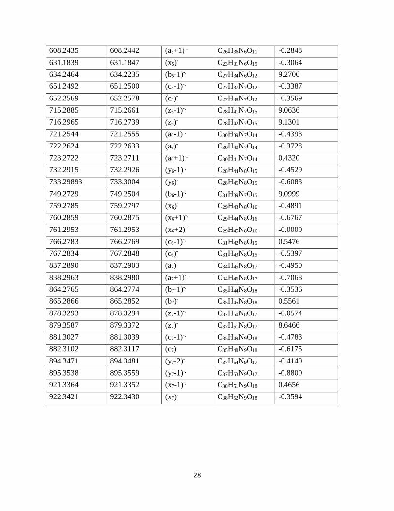
600 650 700 750 800 850 900
YTIAALLSPYS
m/z
x8+1
x8
b8+2
y8-1
y8-2
a8+2
a8+1
a8
x7+1
x7
x7-1
c7c
7-2
y7
y7-1a
7+1
x6
a6+1
Precursor [M-2H]2-, m/z 597.8057
a9-29
a9-44b
8+2-44
x8+2
y8
C
Intensity:
3 x 10³
y6-1
y6
x6+1
Figure 1. Photodissociation spectra of the doubly-deprotonated [M-2H]2- ion (m/z=597.8057) of
YTIAALLSPYS at 213 nm. The precursor ion is notified by * sign and the neutral losses are
indicated by ion masses. The green and blue lines represent the a, b, c and x, y, z ions,
respectively. A) Spectrum of 100-1200 m/z, B) Spectrum of 900-1100 m/z, and C) Spectrum of
600-900 m/z.

20
Figure 2. Side-chain losses detected from peptide 1, YTIAALLSPYS at 213 nm.
21
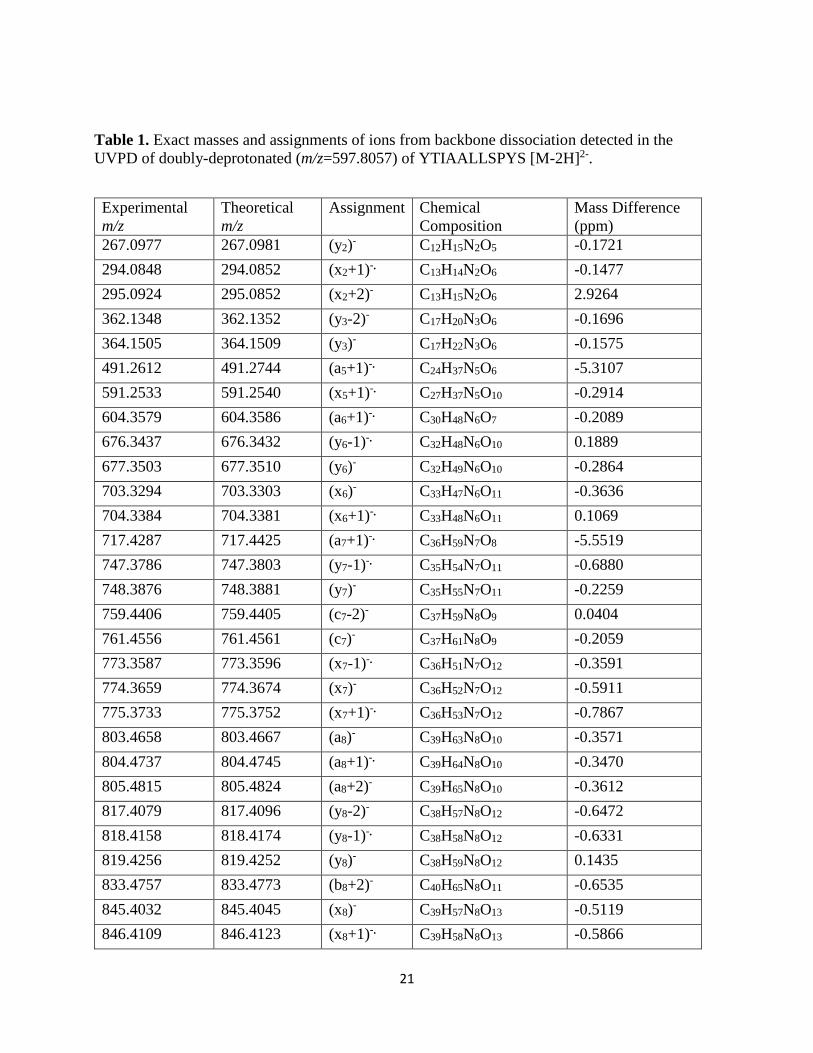
Table 1. Exact masses and assignments of ions from backbone dissociation detected in the
UVPD of doubly-deprotonated (m/z=597.8057) of YTIAALLSPYS [M-2H]2-.
Experimental
m/z
Theoretical
m/z
Assignment Chemical
Composition
Mass Difference
(ppm)
267.0977 267.0981 (y2)- C12H15N2O5 -0.1721
294.0848 294.0852 (x2+1)-. C13H14N2O6 -0.1477
295.0924 295.0852 (x2+2)- C13H15N2O6 2.9264
362.1348 362.1352 (y3-2)- C17H20N3O6 -0.1696
364.1505 364.1509 (y3)- C17H22N3O6 -0.1575
491.2612 491.2744 (a5+1)-. C24H37N5O6 -5.3107
591.2533 591.2540 (x5+1)-. C27H37N5O10 -0.2914
604.3579 604.3586 (a6+1)-. C30H48N6O7 -0.2089
676.3437 676.3432 (y6-1)-. C32H48N6O10 0.1889
677.3503 677.3510 (y6)- C32H49N6O10 -0.2864
703.3294 703.3303 (x6)- C33H47N6O11 -0.3636
704.3384 704.3381 (x6+1)-. C33H48N6O11 0.1069
717.4287 717.4425 (a7+1)-. C36H59N7O8 -5.5519
747.3786 747.3803 (y7-1)-. C35H54N7O11 -0.6880
748.3876 748.3881 (y7)- C35H55N7O11 -0.2259
759.4406 759.4405 (c7-2)- C37H59N8O9 0.0404
761.4556 761.4561 (c7)- C37H61N8O9 -0.2059
773.3587 773.3596 (x7-1)-. C36H51N7O12 -0.3591
774.3659 774.3674 (x7)- C36H52N7O12 -0.5911
775.3733 775.3752 (x7+1)-. C36H53N7O12 -0.7867
803.4658 803.4667 (a8)- C39H63N8O10 -0.3571
804.4737 804.4745 (a8+1)-. C39H64N8O10 -0.3470
805.4815 805.4824 (a8+2)- C39H65N8O10 -0.3612
817.4079 817.4096 (y8-2)- C38H57N8O12 -0.6472
818.4158 818.4174 (y8-1)-. C38H58N8O12 -0.6331
819.4256 819.4252 (y8)- C38H59N8O12 0.1435
833.4757 833.4773 (b8+2)- C40H65N8O11 -0.6535
845.4032 845.4045 (x8)- C39H57N8O13 -0.5119
846.4109 846.4123 (x8+1)-. C39H58N8O13 -0.5866
22
847.4204 847.4201 (x8+2)- C39H59N8O13 0.1174
900.5049 900.5195 (a9)- C44H70N9O11 -5.8453
901.5120 901.5273 (a9+1)-. C44H71N9O11 -6.1741
930.4916 930.4937 (y9-2)- C44H69N9O13 -0.8223
944.5335 944.5331 (c9-1)-. C45H72N10O12 0.1744
945.5378 945.5409 (c9)- C45H73N10O12 -1.2759
957.4795 957.4807 (x9-1)-. C45H67N9O14 -0.4833
958.4859 958.4886 (x9)- C45H68N9O14 -1.0864
1032.5470 1032.5492 (y10-1)-. C48H76N10O15 -0.8721
1059.5334 1059.5440 (x10)- C49H75N10O16 -4.2870
1060.5395 1060.5440 (x10+1)-. C49H76N10O16 -1.8463
1106.5866 1106.5886 (c10-2)- C54H80N11O14 -0.8114
1107.5938 1107.5964 (c10-1)-. C54H81N11O14 -1.0757

23
Table 2. Exact masses and assignments of neutral loss detected in the UVPD of doubly-
deprotonated (m/z=597.8057) of YTIAALLSPYS [M-2H]2- .
Experimental
m/z
Theoretical
m/z
Assignment Chemical
Composition
Mass
Difference(ppm)
1195.6094 1195.6119 [M-2H]-• C57 H85 N11 O17 -1.0065
1180.5852 1180.5885 [M-2H-CH3]- C56 H82 N11 O17 -1.3313
1167.6147 1167.6170 [M-2H-CO]-• C56 H85 N11 O16 -0.9306
1166.6099 1166.5728 [M-2H-CH3CH2]- C55 H80 N11 O17 14.9551
1165.6009 1165.6047 [M-2H-CH2O]-• C56 H83 N11 O16 -1.5532
1164.5942 1164.5935 [M-2H-CH2OH]- C56 H82 N11 O16 0.2703
1151.5829 1151.6221 [M-2H-C2H4O]-• C55 H81 N11 O16 -15.7951
1150.5783 1150.6143 [M-2H-COOH]- C56 H84 N11 O15 -14.5355
1139.5855 1139.5504 [M-2H-C4H8]-• C53 H78 N11 O17 14.1289
1133.6099 1133.6116 [M-2H-(CO2+H2O)]-• C56 H83 N11 O14 -0.6445
1123.5910 1123.5312 [M-2H-C4H11N]-• C53 H75 N10 O17 24.1551
1121.5759 1121.5513 [M-2H-(C3H8ON)]- C54 H77 N10O16 9.9354
1089.5688 1089.5712 [M-2H-(OC6OH4=CH2)]-• C50 H79 N11 O16 -0.9454
1088.5622 1088.5633 [M-2H-(HOC6H4=CH2)]- C51 H78 N11 O16 -0.4512
1045.5419 1045.5449 [M-2H-(OC6OH4=CH2+C2H4O)]-• C48 H75 N11 O15 -1.2420
1044.5346 1044.5371 [M-2H-(HOC6H4=CH2+C2H4O) ]-• C48 H74 N11 O15 -1.0060
987.5129 987.5271 [z10-CHO]-• C47H73N9O14 -5.7445
986.5053 986.5193 [z10-1-CHO]- C47H72N9O14 -5.6699
871.5031 871.5162 [a9-CH3CH2]- • C43H69N9O10 -5.2771
856.4917 856.5291 [a9-C2H4O]- C43H70N9O9 -15.0806
789.4493 789.4869 [b8+2-C2H4O]- C39H65N8O9 -15.1617
597.8057 597.8059 [M-2H]2- C57 H85 N11 O17 -0.1139
575.7925 575.8111 [M-2H-CO2]2- C56 H85 N11 O15 -7.4861
205.0700 205.0972 [y2-CO2+H2O]- C11H13N2O2 -10.9548

24
Scheme 1: Proposed mechanism for the formation of (an+1)-. and (xn+1)-. fragment ions during
UVPD of doubly deprotonated peptide [M-2H]2-.
O
N
O
HH
H
H H
H
OH
HH
NO
H
H HO
H
H O
H
O
N
O
HH
H
H H
H
OH
HH
NO
H
H HO
H
H O +H
(x2+1)- (x2+2)-
m/z = 294.0848 m/z=295.0924
C13H14N2O6C13H15N2O6
Scheme 2: Proposed mechanism for the formation of (x2+2)- product ion from the (x1+1)-.
fragment ions during the UVPD of the doubly deprotonated YTIAALLSPYS peptide.
Scheme 3: Proposed mechanism for the formation of (bn+1)-. and (yn-1)-. fragment ions during
UVPD of doubly deprotonated peptide [M-2H]2-.
-Rn
NH
CH
C
NH
CH
R
C
O-
O
R'O
-Rn
NH
CH.
R
.C
NH
CH
C
O-
O
R'O
+
(an+1)-. (xn+1)-.
-Rn
NH
CH
C
NH
CH
R
C
O-
O
R'O
+ .NH
CH
C
O-
O
R'
-Rn
NH
CH
C.
R
O
(bn+1)-.(yn-1)-.
25
Scheme 4: Proposed mechanism for the formation of (cn-1)-. and (zn+1)-. fragment ions during
UVPD of doubly deprotonated peptide [M-2H]2-.
-Rn
NH
CH
C
NH
CH
R
C
O-
O
R'O
+.CH
C
O-
O
R'
-Rn
NH
CH
C
NH.
R
O
(cn-1)-. (zn+1)-.

26
200 400 600 800 1000
D Y K D D D D K
1 7
7 1
m/z
[M-2H]- .
[M-2H]2-
-18
-44
-88
-62
-28
[M-2H-18]2-
505.1906
1010.3812
-107
71
-100
Internsity:
1 x 10⁵
A
700 750 800 850 900
DYKDDDDK
m/z
c7
c7-1
x6+1
M-2H-132
x6
y6
y6-1
b6-1
a7+1
a7
b7-1
x7-62
x7-36
a6
a6-1
a7+1-18
a7-44
x6-45
a7-18
Precursor [M-2H]2-,m/z 505.1906
y7-1 y
7-2
z7
b7
x6+2
a6+1
z6
z6-1
Intensity:
2x 10³
B
Figure 3. Photodissociation spectra of the doubly-deprotonated [M-2H]2- ion (m/z=505.1906) of
DYKDDDDK at 213 nm (the precursor ion is signified by * and neutral losses are indicated by
ion masses). The green and blue lines represent the a, b, c and x, y, z ions, respectively.

27
Table 3. Exact masses and assignments of ions from backbone dissociation detected in the
UVPD of doubly-deprotonated (m/z=505.1906) of DYKDDDDK [M-2H]2-.
Experimental
m/z
Theoretical m/z Assignment Chemical
Composition
Mass
Difference(ppm)
113.0339 113.0113 (b1-1)-. C4H3NO3 9.1566
114.0179 114.0191 (b1)- C4H4NO3 -0.4551
242.1134 242.0903 (z2-1)-. C10H14N2O5 9.3508
243.0975 243.0981 (z2)- C10H15N2O5 -0.2527
249.0869 249.0875 (a2)- C12H13N2O4 -0.2630
251.0926 251.1032 (a2+2)- C12H14N2O4 -4.2771
286.1034 286.1039 (x2)- C11H16N3O6 -0.2139
288.1197 288.1196 (x2+2)- C11H18N3O6 0.0724
357.1405 357.1172 (z3-1)-. C14H19N3O8 9.3899
358.1252 358.1250 (z3)- C14H18N3O8 0.0647
374.1324 374.1438 (y3-1)-. C14H22N4O8 -4.5965
375.1509 375.1516 (y3)- C14H23N4O8 -0.2818
376.1976 376.1747 (a3-1)-. C18H24N4O5 9.2708
377.1818 377.1825 (a3)- C18H25N4O5 -0.2723
401.1300 401.1309 (x3)- C15H21N4O9 -0.3442
402.1380 402.1387 (x3+1)-. C15H22N4O9 -0.2656
404.1926 404.1696 (b3-1)-. C19H24N4O6 9.2932
471.1592 471.1363 (z4-2)- C18H23N4O11 9.2294
472.1673 472.1442 (z4-1)-. C18H24N4O11 9.3202
490.1778 490.1785 (y4)- C18H28N5O11 -0.3057
491.1933 491.1864 (y4+1)-. C18H29N5O11 2.7890
492.1965 492.2094 (a4)- C22H30N5O8 -5.2316
493.2165 493.2173 (a4+1)-. C22H31N5O8 -0.3199
516.1568 516.1578 (x4)- C19H26N5O12 -0.3898
519.2194 519.2043 (b4-1)-. C23H29N5O9 6.0706
536.2224 536.2231 (c4-1)-. C23H32N6O9 -0.2609
537.2301 537.2309 (c4)- C23H33N6O9 -0.3274
587.1941 587.1711 (z5-1)-. C22H30N5O14 9.2746
605.2049 605.2055 (y5)- C22H33N6O14 -0.2439
606.2282 606.2287 (a5-1)-. C26H34N6O11 -0.1597
607.2356 607.2364 (a5)- C26H35N6O11 -0.3272
28
608.2435 608.2442 (a5+1)-. C26H36N6O11 -0.2848
631.1839 631.1847 (x5)- C23H31N6O15 -0.3064
634.2464 634.2235 (b5-1)-. C27H34N6O12 9.2706
651.2492 651.2500 (c5-1)-. C27H37N7O12 -0.3387
652.2569 652.2578 (c5)- C27H38N7O12 -0.3569
715.2885 715.2661 (z6-1)-. C28H41N7O15 9.0636
716.2965 716.2739 (z6)- C28H42N7O15 9.1301
721.2544 721.2555 (a6-1)-. C30H39N7O14 -0.4393
722.2624 722.2633 (a6)- C30H40N7O14 -0.3728
723.2722 723.2711 (a6+1)-. C30H41N7O14 0.4320
732.2915 732.2926 (y6-1)-. C28H44N8O15 -0.4529
733.29893 733.3004 (y6)- C28H45N8O15 -0.6083
749.2729 749.2504 (b6-1)-. C31H39N7O15 9.0999
759.2785 759.2797 (x6)- C29H43N8O16 -0.4891
760.2859 760.2875 (x6+1)-. C29H44N8O16 -0.6767
761.2953 761.2953 (x6+2)- C29H45N8O16 -0.0009
766.2783 766.2769 (c6-1)-. C31H42N8O15 0.5476
767.2834 767.2848 (c6)- C31H43N8O15 -0.5397
837.2890 837.2903 (a7)- C34H45N8O17 -0.4950
838.2963 838.2980 (a7+1)-. C34H46N8O17 -0.7068
864.2765 864.2774 (b7-1)-. C35H44N8O18 -0.3536
865.2866 865.2852 (b7)- C35H45N8O18 0.5561
878.3293 878.3294 (z7-1)-. C37H50N8O17 -0.0574
879.3587 879.3372 (z7)- C37H51N8O17 8.6466
881.3027 881.3039 (c7-1)-. C35H49N9O18 -0.4783
882.3102 882.3117 (c7)- C35H48N9O18 -0.6175
894.3471 894.3481 (y7-2)- C37H54N9O17 -0.4140
895.3538 895.3559 (y7-1)-. C37H53N9O17 -0.8800
921.3364 921.3352 (x7-1)-. C38H51N9O18 0.4656
922.3421 922.3430 (x7)- C38H52N9O18 -0.3594

29
100 200 300 400 500 600 700 800 900 1000
R G D S P A S S K P
1 9
9 1
[M-2H]2-
m/z
-30
-18
[M-2H]-.
-44
-60-86
-88
Intensity:
8 x 10⁵
-99
A
499.2393
998.4767
600 650 700 750 800 850 900
Precursor [M-2H]2-,m/z 499.2393
m/z
c9+1
c9
c9-1
b9+2
b9+1
b9
b9-1
x9+1
x9
a9
a9+1
a9-1
y9
y9-1
y9-2
z9+1
z9
z9-1
x8
x8-1
y8
y8-1
y8-2
c8
c8-1
c8-2
Intensity:
2 x 10⁵
c6
c6-1
c6-2
x6+1
x6+2
x6
a7+2
a7+1
a7
z7
z7+1
z7+2
z7-1
y7
y7-2
c7
c7-2
c7-1
x7
x7+1
x7+2
x7-1 a
8
a8+1
a8+2
b8b
8-1
z8-2
z8-1
B RGDSPASSKP
Figure 4. Photodissociation spectra of the doubly-deprotonated [M-2H]2- ion (m/z=499.2393) of
RGDSPASSKP at 213 nm (the precursor ion is signified by * and neutral losses are indicated by
ion masses). The green and blue lines represent the a, b, c and x, y, z ions, respectively.

30
Figure 5: Photodissociation spectra of the doubly-deprotonated [M-2H]2- ion of three peptides.
Loss of hydrogen is observed from the characteristic [M-2H]-• charge-reduced radical at single
isotope selection of the doubly-deprotonated [M-2H]2- precursor ions.

31
Figure 6: Potential Energy Surface of the lowest ππ*, πσ* and electronic ground state (S0) as a
function of the NH stretch reaction coordinate. The optimization, natural bond orbital (NBO) and
TD-DFT calculations have been performed at B3LYP/6-311+G(2d,p) level of theory.
32
Supporting Information
100 200 300 400 500 600
YTIAALLSPYS
m/z
x2+2
x2+1 y
3
a5+1
x5+1
Precursor [M-2H]2-
, m/z 597.8057
[M-2H-44]2-
[M-2H-74]2-
[M-2H]2-
y2
y3-2
y2-62
Intensity:
4 x 10³
Figure S1. Photodissociation spectrum (m/z 100-600) of the doubly-deprotonated [M-2H]2- ion
(m/z=597.8057) of YTIAALLSPYS at 213 nm.

33
Figure S2: Selected fragment ions from the single isotope selection of the doubly-deprotonated
[M-2H]2-precursor ions. a/x, b/y and c ions are from peptide 1 and z ion is from peptide 2.

34
510 540 570 600 630 660
Intensity:
2 x 10³
DYKDDDDK
m/z
c5
c5-1
b5-1
x5
a5+1
a5
a5-1
y5
z5-1
a5+1-45
y5-1-45
c4
c4-1
b4-1
x4
Precursor [M-2H]2-, m/z 505.1906
A
120 160 200 240 280 320 360 400 440 480
DYKDDDDK
m/z
b1
b1-1
z2
z2-1
a2
x2
x3+1
x3
b3-1
a3
y3
z4-1
z4-2
y4+1
y4
a4+1
Precursor [M-2H]2-, m/z=505.1906z
2-18
z3-1-17
x3+1-88
[M-2H-44]2-
M-2H-2x18]2-
z2-44
z2-62
z3-1
z3-17
x2+2
a2+2
Intensity:
2 x 10³
B
Figure S3. Photodissociation spectra of the doubly-deprotonated [M-2H]2- ion (m/z=505.1906) of
DYKDDDDK at 213 nm.

35
100 150 200 250 300 350 400 450 500 550m/z
Intensity:
1 x 10³
y1
y1-2
x1+1
x1
x1-1
b1
c2
c2-2
z2
z2+1
b2+2
b2
a2
a2+1
c1
y2
y2-1
y2-2
x2+1
x2
a3 a
3+1
a3+2
z3
z3-1
y6
y6-1y
6-2
z6+2
z6+1
z6
a6+1
a6
x5
b5+1
b5
c5
c5+1
z4+1
z4
b4+2
b4
x4+1
x4
x4-1
z5+1
z5
y5
y5-1
a5+2
a5+1
a5
y3
c3
c3-1
c3-2
b3
a4
a4+1
x3+2
x3+1
x3
Precursor [M-2H]2-,m/z 499.2393
RGDSPASSKP
Figure S4. Photodissociation spectra of the doubly-deprotonated [M-2H]2- ion (m/z=499.2393) of
RGDSPASSKP at 213 nm.

36
Table S1. Exact masses and assignments of neutral loss detected in the UVPD of doubly-
deprotonated (m/z=505.1906) of DYKDDDDK [M-2H]2-
Experimental
m/z
Theoretical
m/z
Assignment Chemical
Composition
Mass
Difference
(ppm)
1010.3812 1010.3829 [M-2H]-• C41H58N10O20 -0.6656
992.3709 992.3718 [M-2H-H2O]- C41H56N10O19 -0.3674
982.3857 982.3874 [M-2H-CO]- • C40H58N10O19 -0.6780
966.3913 966.3925 [M-2H-CO2]- C40H58N10O18 -0.4866
948.3803 948.3819 [M-2H-(CO2+H2O)]- C40H56N10O17 -0.6824
939.3099 939.3088 [M-2H-C4H9N]- • C37H49N9O20 0.4453
938.3961 938.3612 [M-2H-C3H4O2]- C38H54N10O18 14.0692
922.4019 922.4027 [M-2H-2CO2]- C39H58N10O16 -0.3296
922.3441 922.3425 [M-2H-C3H6O2N]- C38H52N9O18 0.6608
910.3076 910.2823 [M-2H-C5H12N2]- C36H46N8O20 10.1959
878.4116 878.4128 [M-2H-3CO2]- C38H58N10O14 -0.5074
904.3394 904.3405 [M-2H-(OC6H4=CH2)]- C34H52N10O19 -0.4238
903.3321 903.3326 [M-2H-(HOC6H4=CH2)]- C34H51N10O19 -0.2201
886.3281 886.3213 [x7-2H2O]- C38H48N9O16 2.7382
878.3516 878.3526 [x7-CO2]- C37H52N9O16 -0.4247
860.3502 860.3421 [x7-(H2O+CO2)]- C37H50N9O15 3.2927
820.2853 820.2869 [a7+1-H2O]- • C34H44N8O16 -0.6650
819.2776 819.2791 [a7-H2O]- C34H43N8O16 -0.6267
793.2991 793.2999 [a7-CO2]- C33H45N8O15 -0.3062
714.2574 714.2815 [x6-COOH]- • C28H42N8O14 -9.7102
563.2456 563.2460 [y5-COO]- C25H35N6O9 -0.1707
559.1989 559.1994 [y5-1-COOH]- • C21H31N6O12 -0.1883

37
505.1908 505.1912 [M-2H]2- C41H58N10O20 -0.1402
496.1855 496.1859 [M-2H-H2O]2- C41H56N10O19 -0.1555
491.1933 491.1937 [M-2H-CO]2-• C39H58N10O17 -0.1776
487.1801 487.1814 [M-2H-2H2O]2- C41H54N10O18 -0.5527
483.1955 483.1971 [M-2H-CO2]2- C40H58N10O18 -0.6334
341.1216 341.0979 [z3-NH3]- C14H17N2O8 9.5572
340.1138 340.0901 [z3-1-NH3]- • C14H16N2O8 9.5451
314.1347 314.1585 [x3+1-2CO2]- • C13H22N4O5 -9.5739
225.0868 225.0869 [z2-H2O]- C10H13N2O4 -0.0780
199.1074 199.1077 [z2-CO2]- C9H15N2O3 -0.1447
181.0967 181.0971 [z2-(H2O+CO2)]- C9H13N2O2 -0.1873
38
Table S2. Exact masses and assignments of ions from backbone dissociation detected in the
UVPD of doubly-deprotonated (m/z=499.2393) of RGDSPASSKP [M-2H]2-.
Experimental
m/z
Theoretical
m/z
Assignment Chemical Composition Mass
Difference
(ppm)
112.0390 112.0398 (y1-2)- C5H6NO2 -0.3443
114.0546 114.0555 (y1)- C5H8NO2 -0.3443
139.0499 139.0269 (x1-1)-. C6H5NO3 9.2655
140.0339 140.0348 (x1)- C6H6NO3 -0.3421
141.0418 141.0426 (x1+1)-. C6H7NO3 -0.3361
155.0813 155.0933 (b1)- C6H11N4O -4.8315
172.1192 172.1198 (c1)- C6H14N5O -0.2683
184.1080 184.1198 (a2)- C7H14N5O -4.7625
185.1033 185.1277 (a2+1)-. C7H15N5O -9.8397
212.1144 212.1147 (b2)- C8H14N5O2 -0.1572
214.1188 214.1304 (b2+2)- C8H16N5O2 -4.6877
225.1237 225.1239 (z2)- C11H17N2O3 -0.0958
226.1315 226.1317 (z2+1)-. C11H18N2O3 -0.0897
227.1253 227.1396 (c2-2)- C8H16N6O2 -5.7478
229.1411 229.1413 (c2)- C8H17N6O2 -0.0923
240.1347 240.1348 (y2-2)- C11H18N3O3 -0.0591
241.1424 241.1426 (y2-1)-. C11H19N3O3 -0.0772
242.1504 242.1505 (y2)- C11H20N3O3 -0.0389
268.1298 268.1297 (x2)- C12H18N3O4 0.0278
269.1251 269.1376 (x2+1)-. C12H19N3O4 -5.0372
270.1455 270.1454 (x2+2)- C12H20N3O4 0.0439
299.1468 299.1468 (a3)- C11H19N6O4 0.0129
300.1546 300.1546 (a3+1)-. C11H20N6O4 0.0189

39
301.1625 301.1624 (a3+2)- C11H21N6O4 0.0128
311.1483 311.1481 (z3-1)-. C14H21N3O5 0.0602
312.1561 312.1559 (z3)- C14H22N3O5 0.0622
326.1466 326.1339 (b3-1)-. C12H18N6O5 5.1204
327.1418 327.1417 (b3)- C12H19N6O5 0.0392
329.1826 329.1825 (y3)- C14H25N4O5 0.0464
342.1527 342.1526 (c3-2)- C12H20N7O5 0.0356
343.1605 343.1604 (c3-1)-. C12H21N7O5 0.0416
344.1683 344.1682 (c3)- C12H22N7O5 0.0235
355.1618 355.1618 (x3)- C15H23N4O6 0.0325
356.1694 356.1696 (x3+1)-. C15H24N4O6 -0.0825
357.1764 357.1774 (x3+2)- C15H25N4O6 -0.4032
386.1791 386.1788 (a4)- C14H24N7O6 0.1063
387.1868 387.1866 (a4+1)-. C14H25N7O6 0.0760
388.1947 388.1945 (a4+2)- C14H26N7O6 0.0982
399.1880 399.1879 (z4)- C17H27N4O7 0.0306
400.1959 400.1958 (z4+1)-. C17H28N4O7 0.0366
414.1739 414.1737 (b4)- C15H24N7O7 0.0923
415.1818 415.1815 (b4+1)-. C15H25N7O7 0.0984
416.1896 416.1894 (b4+2)- C15H26N7O7 0.1004
441.1861 441.1859 (x4-1)-. C18H27N5O8 0.0634
442.1940 442.1938 (x4)- C18H28N5O8 0.0856
443.2018 443.2016 (x4+1)-. C18H29N5O8 0.0674
470.2251 470.2251 (z5)- C20H32N5O8 -0.0073
471.2330 471.2329 (z5+1)-. C20H33N5O8 0.0391
483.2319 483.2316 (a5)- C19H31N8O7 0.1209
484.2337 484.2394 (a5+1)-. C19H32N8O7 -2.2936

40
485.2429 485.2472 (a5+2)- C19H32N8O7 -1.7511
486.2441 486.2438 (y5-1)-. C20H35N6O8 0.1202
487.2521 487.2516 (y5)- C20H34N6O8 0.1746
511.2155 511.2265 (b5)- C20H31N8O8 -4.4115
512.2232 512.2343 (b5+1)-. C20H32N8O8 -4.4982
513.2310 513.2309 (x5)- C21H33N6O9 0.0558
528.2534 528.2530 (c5)- C20H34N9O8 0.1476
529.2625 529.2609 (c5+1)-. C20H35N9O8 0.6619
553.2618 553.2607 (a6-1)-. C22H35N9O8 0.3675
554.2689 554.2687 (a6)- C22H36N9O8 0.0871
555.2766 555.2765 (a6+1)-. C22H37N9O8 0.0407
567.2784 567.2778 (z6)- C25H39N6O9 0.2213
568.2723 568.2857 (z6+1)-. C25H40N6O9 -5.4128
569.2802 569.2935 (z6+2)- C25H41N6O9 -5.3623
582.2892 582.2887 (y6-2)- C25H40N7O9 0.1973
583.2971 583.2966 (y6-1)-. C25H41N7O9 0.1993
584.3047 584.3044 (y6)- C25H42N7O9 0.1247
597.2752 597.2745 (c6-2)- C23H37N10O9 0.2712
598.2831 598.2823 (c6-1)-. C23H38N10O9 0.3014
599.2907 599.2901 (c6)- C23H39N10O9 0.2348
610.2845 610.2837 (x6)- C26H40N7O10 0.3367
611.2918 611.2915 (x6+1)-. C26H41N7O10 0.1087
612.2993 612.2993 (x6+2)- C26H42N7O10 0.0098
641.3017 641.3007 (a7)- C25H41N10O10 0.4064
642.3091 642.3085 (a7+1)-. C25H42N10O10 0.2309
643.3171 643.3164 (a7+2)- C25H43N10O10 0.2813
653.3027 653.3021 (z7-1)-. C28H43N7O11 0.2682

41
654.3105 654.3099 (z7)- C28H44N7O11 0.2419
655.3183 655.3177 (z7+1)-. C28H45N7O11 0.2318
656.3124 656.3255 (z7+2)- C28H46N7O11 -5.2971
669.3214 669.3208 (y7-2)- C28H45N8O11 0.2625
670.3293 670.3286 (y7-1)-. C28H46N8O11 0.2806
671.3371 671.3364 (y7)- C28H47N8O11 0.2827
684.3073 684.3065 (c7-2)- C26H42N11O11 0.2960
685.3149 685.3143 (c7-1)-. C26H43N11O11 0.2254
686.3226 686.3222 (c7)- C26H44N11O11 0.1911
695.3001 695.3000 (x7-2)- C29H43N8O12 0.0089
696.3084 696.3079 (x7-1)-. C29H44N8O12 0.2022
697.3161 697.3157 (x7)- C29H45N8O12 0.1719
698.3236 698.3235 (x7+1)-. C29H46N8O12 0.0529
699.3314 699.3313 (x7+2)- C29H47N8O12 0.0145
728.3337 728.3327 (a8)- C28H46N11O12 0.3909
729.3417 729.3406 (a8+1)-. C28H47N11O12 0.4413
730.3494 730.3484 (a8+2)- C28H48N11O12 0.3909
755.3087 755.3198 (b8-1)-. C29H45N11O13 -4.4984
756.3160 756.3277 (b8)- C29H46N11O13 -4.6901
767.3457 767.3212 (z8-2)- C32H47N8O14 9.8827
768.3531 768.3290 (z8-1)-. C32H48N8O14 9.7395
769.3361 769.3368 (z8)- C32H49N8O14 -0.2837
771.3389 771.3385 (c8-2)- C29H47N12O13 0.1272
772.3465 772.3464 (c8-1)-. C29H48N12O13 0.0647
773.3546 773.3542 (c8)- C29H49N12O13 0.1514
784.3483 784.3477 (y8-2)- C32H50N9O14 0.2452
785.3564 785.3555 (y8-1)-. C32H51N9O14 0.3359

42
786.3650 786.3634 (y8)- C32H52N9O14 0.6647
811.3353 811.3348 (x8-1)-. C33H49N9O15 0.2049
812.3433 812.3426 (x8)- C33H50N9O15 0.2796
825.3753 825.3505 (z9-1)-. C34H51N9O15 10.0083
826.3604 826.3583 (z9)- C34H52N9O15 0.8524
827.3673 827.3661 (z9+1)-. C34H53N9O15 0.4672
841.3696 841.3692 (y9-2)- C34H53N10O15 0.1791
842.3775 842.3770 (y9-1)-. C34H54N10O15 0.2053
843.3848 843.3848 (y9)- C34H55N10O15 -0.0146
855.4221 855.4199 (a9-1)-. C34H57N13O13 0.8960
856.4281 856.4277 (a9)- C34H58N13O13 0.1396
857.4357 857.4355 (a9+1)-. C34H59N13O13 0.0689
858.4437 858.4433 (a9+2)- C34H60N13O13 0.1436
868.3570 868.3563 (x9-1)-. C35H52N10O16 0.3043
869.3647 869.3641 (x9)- C35H53N10O16 0.2418
870.3723 870.3719 (x9+1)-. C35H54N10O16 0.1349
883.4153 883.4148 (b9-1)-. C35H57N13O14 0.2124
884.4231 884.4226 (b9)- C35H58N13O14 0.1902
885.4307 885.4304 (b9+1)-. C35H59N13O14 0.1236
886.4387 886.4383 (b9+2)- C35H60N13O14 0.1861
900.4183 900.4413 (c9-1)-. C35H60N14O14 -9.2759
901.4257 901.4492 (c9)- C35H61N14O14 -9.4716
902.4337 902.4569 (c9+1)-. C35H62N14O14 -9.3808

43
Table S3. Exact masses and assignments of neutral loss detected in the UVPD of doubly-
deprotonated (m/z=499.2393) of RGDSPASSKP [M-2H]2-.
Experimental
m/z
Theoretical
m/z
Assignments Chemical
Composition
Mass
Difference(ppm)
998.4767 998.4776 [M-2H]-• C40H66N14O16 -0.3643
980.4673 980.4670 [M-2H-H2O]- C40H64N14O15 0.1216
968.4672 968.4670 [M-2H-CH2O]-• C39H64N14O15 0.0611
954.4872 954.4877 [M-2H-CO2]- C39H66N14O14 -0.2218
938.4227 938.4326 [M-2H-C2H6ON]- C38H60N13O15 -4.0060
912.4072 912.4057 [M-2H-C3H8N3]- C37H57N11O16 0.5365
899.3982 899.3979 [M-2H-C4H9N3]- C36H57N11O16 0.1210
910.4269 910.4377 [M-2H-C3H6O2N]- C37H60N13O14 -4.3368