2.1 mammalian liver: structure and function -...
TRANSCRIPT
Review of literature
3
2.1 Mammalian liver: structure and function
The liver is the most vital organ in the mammalian body and performs all important
functions that impact all body systems. The liver has lobular structure and lies in the
abdominal cavity below diaphragm. The circulatory system of the liver is different
from that of other organs. Roughly 75% of the blood entering in liver through the
portal vein is the venous blood returning back from the small intestine, stomach,
pancreas, and spleen. From this portal venous blood all nutrients along with drugs and
other potentially harmful substances are absorbed. The remaining 25% of the arterial
blood received by liver is the oxygenated blood being carried from the pulmonary
system to the liver by the hepatic artery. The blood contents of the hepatic artery as
well as hepatic portal vein empty into sinusoids. Sinusoidal blood moves towards the
central vein of each lobule and empties its content. Hepatic veins carry deoxygenated
blood from liver to the inferior vena cava (Fawcett, 1994; Malarkey et al., 2005).
2.1.1 Architecture of liver
The liver is covered with a connective tissue capsule (Glisson’s capsule) except at a
region where blood vessels and hepatic/bile ducts enter and/or leave the organ.
Branches of the connective tissue extend throughout the liver as septae. This
connective tissue provides a network, support and the highway along which lymphatic
vessels, bile ducts and afferent blood vessels can traverse across the liver. The
parenchyma of the liver divides into small units called lobules with the help of
connective tissue sheet (Bhunchet and Wake, 1998).
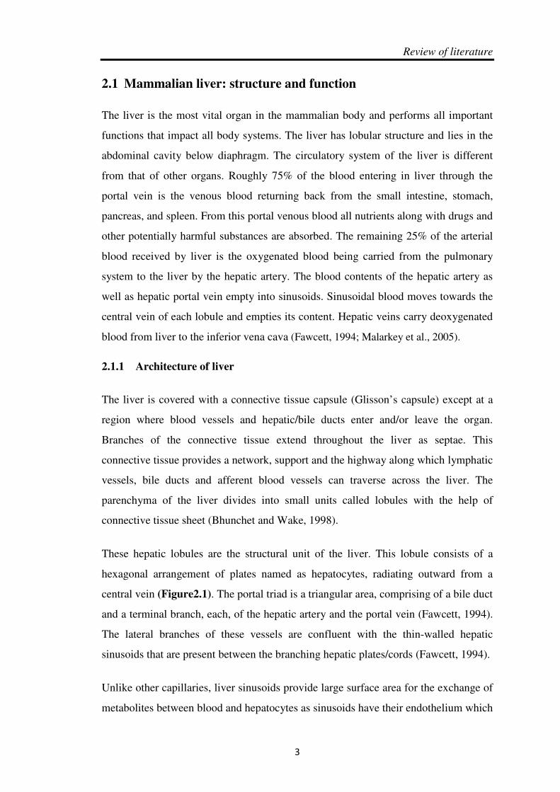
These hepatic lobules are the structural unit of the liver. This lobule consists of a
hexagonal arrangement of plates named as hepatocytes, radiating outward from a
central vein (Figure2.1). The portal triad is a triangular area, comprising of a bile duct
and a terminal branch, each, of the hepatic artery and the portal vein (Fawcett, 1994).
The lateral branches of these vessels are confluent with the thin-walled hepatic
sinusoids that are present between the branching hepatic plates/cords (Fawcett, 1994).
Unlike other capillaries, liver sinusoids provide large surface area for the exchange of
metabolites between blood and hepatocytes as sinusoids have their endothelium which

Review of Literature
4
lacks the basal membrane. The sub-endothelial space called as the space of Disse or
per sinusoidal space, separates endothelium from the hepatocytes plates (Grisham,
1962).
CCCC
Figure 2.1: (a) and (b) Showing liver architecture. Reproduced from (Duncan et al., 2009).
(c) Showing cells of liver and blood flow through portal vessels. Reproduced
from (Tanaka M, 2011).
Review of literature
5
2.1.2 Cells of liver
The hepatocytes are liver parenchymal cells which consist of 60-80% of liver cell
population. Whereas, the cells such as kupffer cells, stellate cells, biliary epithelial
cells, sinusoidal endothelial cells and lymphocytes are non-parenchymal cells and
comprise the rest 20-40% of the liver cell population (Racanelli and Rehermann,
2006).
Hepatocytes compose a major fraction of hepatic cell population. They are
polyhedral in shape, arranged in single-cell cords or plates. These hepatocytes are
linked together via intercellular adhesion complexes and tight junctions. Their one
side faces the persinusoidal space, while the other faces the bile canaliculi and
covered with microvilli (Bioulac-Sage, 2007). Binucleated cells with large polyploid
nuclei are commonly seen in adult liver (Abel T, 2013). Hepatocytes are responsible
for most of the liver functions such as metabolism, detoxification, synthesis, and
storage of nutrients, carbohydrates, fats, and vitamins. They are also involved in
secretory and excretory functions along with other hepatic cells. These functions are
performed by different hepatocytes residing in different zones of hepatic lobules. This
zonation has been correlated with the direction blood flow and help in carrying
metabolites (Duncan et al., 2009). Most of the hepatic toxins cause necrosis and
damage, which varies with different zonal of hepatic lobules. Thus hepatocytes
present in zone near central vein are more prone to the injury in comparison to the
cells near portal triad region (Michalopoulos and DeFrances, 1997).
Kupffer cells are resident macrophages in liver with largest population. They are
attached to the luminal surface of the sinusoidal endothelium (Figure2.1 c)
(McCuskey and McCuskey, 1990). These cells are essential for the phagocytosis of
foreign particles, infecting organism as well as cytokines products (Taub, 2004).
Hepatic stellate cells are located within persinusoidal space of Disse, in the recesses
between hepatocytes. These cells are associated with several functions such as
secretion of cytokines, storage of vitamin A and synthesis of hepatic extracellular
matrix. They gets activated during liver injury and play a key role in progression of
fibrosis (Wang Y, 2013; Yi HS, 2013).

Review of Literature
6
Biliary epithelial cells, line the bile duct in portal triads and are also known as the
cholangiocytes. The connecting duct between bile duct and bile canaliculi (canal of
Hering) along with hepatocytes are also lined by these biliary cells. They are involved
in modifying bile composition by altering solute and water content (Katsuda T, 2013).
Also, it has now become evident that during liver transplantation,
the biliary epithilium is an important target for leucocytes of the graft recipient
(Scholz et al., 1997).
Endothelial cells are largest group of non-parenchymal cells of liver and line the
intrahepatic circulatory vessels of liver and provide a large surface area for nutrient
absorption. They form a pathogenic and selective barrier during separation of
hepatocytes from sinusoidal blood by exchange of molecules (Racanelli and
Rehermann, 2006; Taub, 2004).
Lymphocytes are present every where in the liver parenchymal sinusoids. These
lymphocytes are also a part of innate immune system and selectively rich in NKT
cells and natural killer cells providing defence against invading pathogens (Taub,
2004).
2.1.3 Liver as a secretory organ
Bile salts are required for emulsification of dietary fats prior to their absorption and
digestion. These bile salts are present in bile juice that is produced by the liver and
secreted into duodenum by common bile duct. Many endogenous and exogenous
compounds are known to be metabolized and excreted into bile through liver. Limited
amount of these compounds were reabsorbed in the small intestine and eliminated by
the kidney. For example, recycling of iron while globins chains get catabolized and
their components are reused. However, hemoglobin also contains a porphyrin called
heme that must be eliminated. Heme is converted into free bilirubin inside phagocytic
cells, which is released into plasma and secreted by the liver. It also involved in bile
excretion (Jones, 1977).

Review of literature
7
2.1.4 Liver as an immuno-organ
Liver serves as a primary hematopoietic organ during the fetal stage whereas in post-
natal stages, liver retains the important immunological functions with some new ones
(Crispe, 2009). These functions belong to both innate and adaptive immunity
branches. For example, production of acute phase proteins, non-specific phagocytosis
& cell killing, disposal of inflammatory wastes and non-specific immunoregulation
are some of the innate immune functions. On the other hand, the adaptive immune
responses of T-cells are reviewed extensively such as deletion of activated T-cells,
induction of tolerance to ingested and self-antigens, extrathymic proliferation of T-
cells and specific immunoregulation (Crispe, 2009).The liver with the substantial
population of immunologically active cells are responsible for all above mentioned
functions. These cells are B and T lymphocytes, kupffer cells, natural killer (NK)
cells, NK cells expressing T-cell receptor, stellate cells and dendritic cells (Parker,
2012).
Liver receives blood from both – the portal system and the systemic circulation. Portal
venous blood contains the antigens and microbial products along with digestive
products. Liver protects the body from endotoxemia by neutralizing these LPS
molecules from portal veins. In normal physiological conditions, systemic blood
doesn’t contain detectable amounts of LPS but portal blood shows a level up to
1ng/ml. The LPS receptors which are present in hepatic sinusoids, effectively remove
these molecules and thus, systemic blood is not exposed to them. There are many
immuno stimuli other then LPS that liver is exposed to. Liver is potent immunological
organ (Racanelli and Rehermann, 2006) thus provide innate immunity by NK cells
and NKT cells present in it while Kupffer cells act as resident phagocytic cells. They
potentially respond to innate immune signals though their expression of TLR4, CD14,
and MD2, which renders them LPS responsive (Paik et al., 2003).
2.1.5 Liver as a metabolic organ
Hepatocytes are associated with metabolic process in the body. They also play a
critical role in synthesizing molecules that support homeostasis and regulating energy
balances (Felber and Golay, 1995).

Review of Literature
8
Carbohydrate metabolism
Critical concentrations of glucose must be maintained inside body. This is an
important function of the liver. Hepatocytes are residence for different metabolic
pathways and enzymes that regulate blood glucose level (Chanda et al., 1995). Excess
glucose enters into the blood after intake of food and rapid synthesis of glycogen via
glycogenesis occurs in liver. Whereas when glucose concentration decreases in blood,
the liver activates another pathway which leads to depolymerization of glycogen
(glycogenolysis) into glucose. When hepatic glycogens reserves become exhausted,
during prolong starvation, hepatocyte activates gluconeogenesis in order to complete
glucose demand of body (Chanda and Mehendale, 1996).
Fat metabolism
Lipid metabolism is predominantly carried out by the liver. Triglycerides oxidized to
produce energy inside liver. The hepatocytes are site for breakdown of many fatty
acids into acetoacetate. Liver also associated with synthesis of lipoproteins,
cholesterol, and phospholipids. Excess carbohydrates and proteins get converted into
fatty acids and triglyceride in liver, and later on exported and stored in adipose tissue
(Kulkarni SR, 2013).
Protein metabolism
The hepatocytes are responsible for synthesis of most of the plasma proteins such as
albumin and clotting factors necessary for blood coagulation. Liver is also involved in
deamination and transamination of amino acids. Liver is also associated with
conversion of the non-nitrogenous part of those molecules to glucose or lipids. Liver
is responsible for synthesis of urea from ammonia which is very toxic and may result
in diseases associated with central nervous system (Rebelato, 2013).
2.2 Liver: pathological condition
Beside its main function such as storage and distribution of nutrients, liver also function
as detoxifying organ. The liver takes up chemical substances ingested through the food
and absorbed through the gastrointestinal tract. Doing so, however, harbours the danger
that the substances which are degraded and/or eliminated by liver lead to tissue damage
(Ramadori et al., 2008). Most of these substances are compatible with cellular

Review of literature
9
metabolic processing thus utilized by body intact or can be transformed further into
components of cells and tissues. But substances which are not processed further or
metabolized completely enter into portal circulation and processed by hepatocyte so that
they can be excreted out of body. These un-metabolized substances known as
xenobiotics are processed by enzymes such as members of the family of cytochrome
P450 enzymes. This detoxification process generates free radicals and reactive
intermediate reacts with DNA and proteins, forming adducts and causing hepatocyte
damage and/or cell death.
Liver get exposed to several compounds such as acetaminophenon, carbon
tetrachloride, thioacetamide and various other chemical carcinogens in the process of
detoxification. Depending upon the quantity of toxic compounds, an acute, or a chronic
injury occurs to the liver. They commonly damage hepatocytes (main functional cell of
liver) and under chronic condition death occurs (Fausto, 2000; GK., 2011).
2.2.1 The liver and affecting diseases
Being the main detoxifying and metabolizing organ, liver is prone to a number of
diseases. Severity of diseases depends on the onset and duration of pathological
conditions. On the bases of it diseases can be categorised into acute and chronic. They
are characterised into, hepatic steatosis, jaundice, hepatitis, fibrosis, cirrhosis,
cholestasis and cancer (hepatocellular carcinoma and cholangiocarcinoma). Some
common pathological conditions are described below.
Hepatic steatosis is conditions were triglycerides accumulated in hepatic cells increase
abnormally leading to fatty liver disease. The hepatocytes maintain triglyceride content
of body through normal processes of uptake, synthesis, and esterification of fatty acids.
Beside many metabolic, nutritional and genetic cause of high triglyceride level in body
it majorly due to excessive alcohol intake (AFLD) or obesity-related causes (NAFLD).
Severe steatosis or fatty liver associated with inflammation known as steatohepatitis
(Day and James, 1998; den Boer et al., 2004; Reddy and Rao, 2006).
Jaundice is characterized by overload of bile pigment on liver cells and condition is
also known as hyperbilirubinemia. It may also result from disturbances in uptake,
transport, conjugation, and biliary excretion mechanisms of bilirubin by the liver

Review of Literature
10
cells. It can be caused due to damage, injury, or infection to the liver cells. However,
failure of biliary excretion and cholestasis (intra-hepatic or extra-hepatic) may lead to
jaundice (Sherlock, 1967).
Hepatitis is characterized by inflammation of the liver. It can be caused by various
viruses, drugs/toxins, alcohol, and autoimmunity conditions. Hepatitis viruses such as
A, B, C, D and E, are commonly known viruses. These viruses differ in their modes
of transmission. Hepatitis A and E viruses are transmitted by contaminated food and
water. Whereas hepatitis viruses B, C and D are get spread through potential contact
with infected body fluids. Hepatitis can be acute or chronic depending upon its
severity. Viruses of type B and C can cause chronic hepatitis while all other are
associated with acute hepatitis. Chronic hepatitis leads to liver cirrhosis, which
ultimately leads to hepatocellular carcinoma. Hepatitis may lead to cholestasis in
some cases (Ferrell, 2000; Popper, 1972; Waris and Siddiqui, 2003).
Fibrosis is characterized by encapsulation or replacement of injured liver tissue by a
collagenous scar. It occurs during normal wound-healing response, resulting in an
abnormal continuation of fibrogenesis. This includes deposition and production of
connective tissue/matrix protein (Bataller and Brenner, 2005).
Cirrhosis is an advanced stage of liver fibrosis. It involves distortion of the hepatic
vasculature. Cirrhosis can be defined as widespread hepatic fibrosis with nodule
formation. It leads to the distortion of hepatic vessel; impair blood supply, with scar in
the space of Disse and endothelial fenestrations are lost. The impaired hepatocyte
function, exhaustive proliferation and division, are common feature of cirrhosis. The
chronic liver diseases conditions that result in cirrhosis include alcohol abuse,
hepatitis B and C, autoimmune hepatitis. It also caused due to continued exposure to
certain toxins/drugs that lead to hepatocyte necrosis/apoptosis (Schuppan and Afdhal,
2008; Sherlock, 1968).
2.3 Liver regeneration
In normal condition the liver exhibits low level of cell turnover, but it maintains the
rate of proliferation in response to toxic injury and infection. As soon as any damage
or injury occurs, loss of liver cells especially hepatocyte take place. A rapid
Review of literature
11
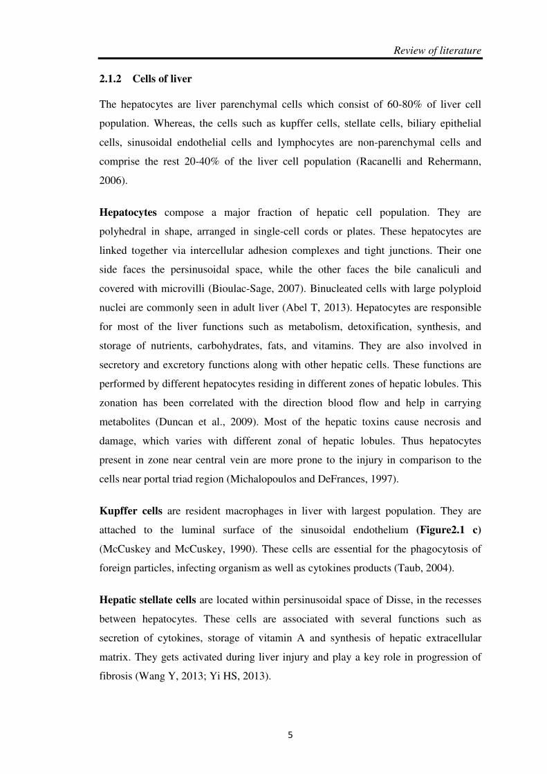
regenerative response is elicited from all cell types in the liver to restore its pristine
state (Alison et al., 2009). Proliferation of differentiated hepatocytes or activation of
progenitor cells occurs soon after any damage to the liver which leads to restoration of the
original liver mass (Figure 2.2).
The immense regenerative capacity of the liver was first defined in Greek mythology and
clearly shown by Higgins and Anderson (Higgins, 1931). The two-thirds partial
hepatectomy model of regeneration was established in rodents by surgical removal of
liver lobes. The original liver mass is restored approximately in 1 week after surgery. This
two-thirds partial-hepatectomy model in murine was well established in our laboratory.
Liver regeneration is usually misunderstood because the parts of the liver that are
removed do not grow back, unlike the regeneration of limbs in amphibian models. In this
type of regenerative models, remaining hepatocyte divide to satisfy body demand. No
progenitor cells are involved in this type of liver regeneration (Bucher, 1963). It is a
compensatory process because the size of the liver is determined by the demands of the
organism. Once the original mass of the liver has been restored by rapid division and
proliferation, cell proliferation stops and cells enter into normal quiescent state (Taub,
2004; Michalopoulos and DeFrances, 1997).
Figure 2.2: Liver regeneration is triggered by partial hepatectomy or liver damage. Reproduced from (Taub, 2004).

Review of Literature
12
2.3.1 The pathways of liver regeneration
There are two basic pathways associated with liver regeneration. (1) The hepatocyte-
driven regeneration, involves proliferation of terminally differentiated cells. This pathway
involves replication of existing hepatocytes induced by acute liver damage. (2) The liver
progenitor/stem cell-driven (LPS) regeneration pathway is induced by chronic liver
injury. It involves activation, replication, and differentiation of intrahepatic progenitor
cells into functional hepatocytes (Figure 2.3). Impaired and exhausted pool of
hepatocytes leads to activation of LPS pathway. There are many reports suggesting
involvement of liver progenitor cell/stem cells in liner regeneration but factors associated
with them are much remains to be understood (Viebahn and Yeoh, 2008).
Figure 2.3: Different pathways showing liver regeneration after acute and chronic liver injury. Reproduced from (Viebahn and Yeoh, 2008).
The present study is based on the chemically induced liver injury model in order to
generate acute liver injury model. This particular form of liver regeneration follows the
hepatocyte-driven regenerative pathway. In this case hepatocytes proliferate in order to
re-establish liver normal function and mass. Therefore in present study we primarily
focus on liver regeneration involving hepatocyte driven pathway in the proceeding
sections.
2.3.2 Molecular mechanism of liver regeneration
The liver is composed of cells such as hepatocytes, Kupffer cells, sinusoidal endothelial
cells (SECs), hepatic stellate cells (HSCs), and stem cells. These cells form complex
networking and interactions inside the liver. These networks are functionally connected
with the extrahepatic organs in order to regulate the functions and behaviour of liver cells.
Hepatocyte

Review of literature
13
Many molecules such as insulin, glucagon, T3, norepinephrine, somatostatin, and growth
factors (EGF) are involved in regulating liver function (Taub, 2004; Viebahn and Yeoh,
2008).
The activated signalling pathways during liver regeneration may be divided into three
phases: initiation phase, proliferation phase, and termination phase. The priming phase
involves triggering of hepatocytes and nonparenchymal cells (Kupffer cells, SECs, HSCs)
present in quiescent phase to enter into cell cycle with the help of cytokines such as TNF-
alpha and IL-6 (Fujiyoshi, 2011; Webber et al., 1998). This preparatory stage of
hepatocyte is important not only for hepatocyte proliferation but also due to the changes
associated with patterns of hepatocyte gene expression in order to maintain their
homeostatic functions (Michalopoulos, 2007).
The proliferation phase involves hepatocytes division and cell cycle progression. This
step is essential for restoration of liver volume and function (Fausto et al., 1995). Growth
factors are also actively involved in regulating process of priming and proliferation
(Figure 2.4). The hepatocyte turns off and returns to quiescence stage during termination
phase. While in case of inappropriate increase in cell numbers it may undergo apoptosis
(Sakamoto et al., 1999). The entire process of restoration of liver mass takes one or more
weeks in rodents and around fifteen days in humans. However the architectural
reorganization, takes several weeks to complete (Michalopoulos, 2007).
Figure 2.4: Intercellular interactions and initiation and proliferation in liver regeneration. Reproduced from (Fujiyoshi, 2011).

Review of Literature
14
2.3.2.1 Cell proliferation during regeneration
By serial culture experiments of hepatocye (Block et al., 1996; Overturf et al., 1997) it
was evident that they are long-lived, rarely dividing adult cells, with high ploidy and
complex functional role. Besides its remarkable capacity to proliferative and divide,
the hepatocytes simultaneously continue to perform all the necessary metabolic and
homeostatic functions and help liver to maintain them (Michalopoulos and
DeFrances, 1997).
Hepatocytes are the first cells to divide after any injury to the liver. It has been
reported that timing of cell cycle progression differs in mice and rats. In case of any
treatment/partial hepatectomy, DNA synthesis in rats begins after 12 hours while
DNA synthesis in mice occurs later at 36-40 hour. This happens due to a longer G1
phase in mice and it varies with type of strains. Similar pattern of DNA synthesis in
mice were seen in case of direct damage of liver cell by chemical compounds
(Bucher, 1963).
2.3.2.2 Networks of cell-to-cell signalling during regeneration
Under normal physiological conditions, hepatocytes have a long life span and divide
rarely. But under stress situation such as partial hepatectomy, toxic injury induced by
thioacetamide, carbon tetra chloride, galectosamine and viral infection, liver
regeneration is triggered by stimulatory and inhibitory signals for cell division and
growth. This process of hepatocyte division is regulated by interaction between
various signalling molecules which help in restoration of cell number and mass
(Figure 2.5) (GK., 2010). These signalling pathways has been categorize into two
types (1) cytokine-dependent (2) growth factor-dependent pathways (3) metabolic
pathway (Taub, 2004). It has been evident from many reports that mitogens or growth
factors release after stimulation (injury/partial hepatectomy), induce hepatocyte
proliferation (Moolten and Bucher, 1967). It also reported that progression of cell
cycle largely depend on growth factor signalling (Fujiyoshi, 2011).
These proliferative signals have been categorised into two groups, (1) complete
mitogens or direct mitogens include HGF, EGF, TGFα, HB-EGF and amphiregulin.
(2) Auxiliary signals only enhance the effects of complete mitogens; it includes
substances like TNF-α, IL-6, VEGF, norepinephrine, bile acids, serotonin,

Review of literature
15
complement proteins, estrogens, and insulin (GK, 2010). Coordination between the
metabolic requirements of the body and regenerative responses in terms of cell
proliferation is an important aspect of the overall process of regeneration. However,
the nutrient and energy levels and there link with mitogenic signals is still obscure
(Fausto et al., 2006).
The most potent growth factor (HGF) derived from activated hepatic stellate cells
(HSCs) and promotes proliferation and DNA synthesis of hepatocytes (Huh et al.,
2004). An inactive precursor known as pro-HGF stored in the extracellular matrix,
secretes HGF (Kim et al., 1997). HGF promotes hepatocyte proliferation by tyrosine
mediated pathways. This signaling is transmitted to its downstream pathway mainly
via phosphatidylinositol kinase (PI3-K/PDK 1/Akt) pathway (Kim et al., 1997). It also
involves Ras-Raf-MEK pathways (Okano et al., 2003) and contributes to liver
regeneration by promoting DNA synthesis, cell cycle progression, cell proliferation
and cell growth.
TGF-alpha is also stimulates DNA synthesis directly in autocrine as well as endocrine
fashion as it is secreted by hepatocyte itself. Its production and secretion by
hepatocyte is promoted by HGF. Molecules like MET and EGFR responsible for
activating multiple intracellular signalling pathways, which in turn regulate
transcription factors involved in hepatocyte proliferation. The MAPK pathway also
play key role in activation of cytokine and growth-factor signals which ultimately
leading to hepatocyte proliferation (Fausto et al., 2006). Another important link
appears between TACE (which cleaves membrane-bound TGFα) and TNF-α. TNF-α
activate TACE and thus leads to downstream signalling through EGFR (Argast et al.,
2004). There are reports suggesting that pathways which play essential role in acute
liver injury are IL-6/STAT3 pathway and PI3-K/PDK1/Akt (Figure 2.6).

Review of Literature
16
Figure 2.5: Signalling interactions between various hepatic cell types during liver regeneration. Modified from (Michalopoulos, 2007; Taub, 2004).
Figure 2.6: Showing cell proliferation is mediated by IL-6/STAT3 pathway and cell size is mediated by PI3-K/PDK1/Akt pathway. Modified from (Fujiyoshi, 2011).
Once the number and size of liver cells has been regained these signalling pathways
are need to be terminated and stop the process of regeneration. TGFβ1 act as a key
player in the termination of regeneration process and marked as a suppressor of
hepatocyte proliferation (Houck and Michalopoulos, 1989). Plasma level of TGFβ1 is
high during initial phase of regeneration (as a result of matrix remodelling). It is
produced by stellate cells and inactivated by binding to alpha-2-macroglobulin
(LaMarre et al., 1991). The regeneration process requires both TGFβ1 and activin for

Review of literature
17
its termination (another mito-inhibitor for hepatocytes) because proliferating
hepatocytes become resistant to its mito-inhibitory effects by down-regulating the
expression of its receptors (Chari et al., 1995).
2.3.2.3 Patterns of gene expression and liver regeneration
Drug induced hepatotoxicity causes sensible amount of mortality and morbidity.
But due to insufficient information regarding mechanism of drug induced liver
injury it becomes a major concern in drug development. Over the last decade,
several gene expression studies using microarray technology have focussed on the
regeneration phenomenon that follows drug induced liver injury in rodents (Togo
et al., 2004; Xu et al., 2005). The most important changes in expression profile of
genes occur during the priming phase of regeneration. It helps in the
categorization of expression patterns into immediate-early and delayed-early genes
(Haber et al., 1993). More than 100 genes which are not expressed in normal liver
get induced during regenerative process; responsible for entry of hepatocytes into
cell cycle and play an essential role in fulfilling metabolic demand of cells while
dividing (Michalopoulos, 2007). Genes associated with priming phase of
regeneration include genes for transcription factors, stress and inflammatory
responses. These genes regulate entry of liver cells into cell-cycle. Genes involved
in cytoskeletal and ECM remodelling is also activated during priming phase (Su et
al., 2002). The G1/S transition, involves a set of differentially expressed genes
with many diverse functions (Satyanarayana et al., 2004). Data analyses of
expression pattern of genes with respect to different phases of regeneration, shown
up-regulation of genes involved in protein synthesis, cytoskeletal organization and
DNA replication while down-regulation of steroid and lipid metabolism genes
(Fukuhara et al., 2003).
Although microarray technology provides a global view of gene expression pattern
that are either and/or both hepatocytes and non-parenchymal cells. Few efforts
have been made to study gene expression of every liver cell (Haber et al., 1993).
Most recent studies have monitored gene expression changes in both Kupffer cells
(Xu CS, 2012) and sinusoidal endothelial cells (Xu CS, 2011) especially during
liver regeneration.
Review of Literature
18
2.4 Liver progenitor cells
The capacity of mature liver cells to proliferate in response to any type of chemical or
mechanical injury is remarkable. However, when normal proliferative responses of
hepatocyte become impaired, in the case of exhaustive proliferation, the contribution of
hepatic progenitor cells plays a critical role. A population of small portal zone cells, first
seen in rats, with a high nuclear/cytoplasmic ratio and an ovoid nucleus, known as “oval
cells”(Shinozuka et al., 1978).These cells have high proliferative power and, differentiate
into hepatocytes upon migration into the lobule. The proliferating epithelial cells in the
periportal region are also referred to as oval cells. But it remains unclear whether or not
the oval cells in different species have common characteristics. The lack of appropriate
cell surface markers is a major problem in characterizing, isolating and identify the oval
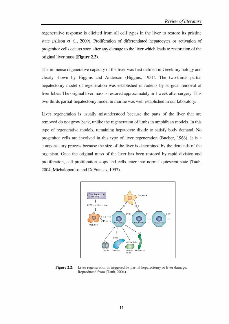
cell compartment (Tanaka M, 2011). Liver progenitor cells or oval cells activation,
differentiation and relationships between the two principal epithelial cell types in liver
and their progenitors were summarized below (Figure 2.7).
A number of recent studies support the contribution of bone marrow derived
hematopoietic stem cells and their ability to transdifferentiate into or fuse with
hepatocytes or oval cells. But have not led to conclusive paradigms behind this act (GK.,
2011). It must be noted that both biliary cells in the canals of Hering and hepatocytes in
the periportal areas, under normal conditions function as mature biliary cells and
hepatocytes, respectively.
Figure 2.7: Progenitor lineage relationships in adult liver Oval cells can give rise to hepatocytes and biliary epithelial cells in chronic liver injury. Reproduced from (Tanaka M, 2011).

Review of literature
19
2.5 Model systems to study liver regeneration
Owing to its powers of regeneration, the liver has been extensively used to understand the
cellular and molecular mechanisms. These understandings provide a base which in turn
has led to advance technique of in vivo ‘‘tissue engineering’’. These techniques enable
complete restoration of liver architecture and re-establishment of the specific functions of
the liver after various types of liver injury. These advances have been achieved primarily
by partial hepatectomy as an important liver regeneration model system in rodent. In
recent years, hepatic injury model systems simulate various pathophysiological situations
that are important in developing and evaluating improved pharmacological therapeutics.
By studying the interaction between cell damage, recovery, and regeneration, the drug-
induced models of liver regeneration have assumed growing importance with a greater
clinical relevance.
There are multiple factors including physical & chemical, which can induce liver damage
and regeneration. Depending upon the methods for inducing regeneration, the
experimental models of liver regeneration can be broadly classified as: surgical models
and pharmacological models (Palmes and Spiegel, 2004; Tunon et al., 2009) as discussed
below. The various other models based on the cell type that is induced to proliferate:
hepatocyte-mediated and LPS-dependent models for liver regeneration (Santoni-Rugiu et
al., 2005), as already explained.
2.5.1 Surgical animal models
Partial hepatectomy is commonly used surgical model of liver regeneration in rodents.
Report suggest that while removal of varying fractions of rat liver (9%, 34%, 43% and
68% of the total liver mass) showed that the hepatocytes respond maximally and
synchronously in 68% (or two-third) partial hepatectomy (Bucher and Swaffield,
1964). Another common technique of surgical model is partial devascularization by
portal branch ligation. But due to better reproducibility of result, two-third partial
hepatectomy has, become a widely utilized method to induce liver regeneration, in
order to understand mechanism involves in liver regeneration. It has been well
optimized in our laboratory previously. Liver regeneration following partial
hepetectomy is a process of compensatory hyperplasic. It involves replication of all
the mature cell types of the remaining liver. Experimental conditions need

Review of Literature
20
standardization for surgical removal of liver such as partial hepatectomy as well as
portal branch ligation. These experimental variations during surgery can affect the
survival of animals as well as onset of regeneration process.
2.5.2 Pharmacological animal models
Mammalian liver is prone to a large number of toxins and drugs, and get influenced
by them in various manners. In contrast to surgical animal models, chemically
induced liver injury models are easier to work upon and provide a better way to study
many related liver diseases in humans. In these models, regenerative response often
varies with dose and mode of administration of drug. A particular amount of drug
dose induced acute liver failure while repeated doses can induce chronic injury such
as liver cirrhosis (Palmes and Spiegel, 2004).
It must be noted that different drugs can have different effects on the liver tissue
depending on their metabolic process and the mechanisms of regeneration which
differ to a large extent. In these kind of toxic models the processes of liver injury and
repair are overlapped and/or closely associated. Effects of some commonly employed
chemicals related to liver damage and injury are discussed below. These chemicals
have similar mode of action to induce centrilobular hepatic damage. The hepatocytes
nearer the portal triads receive oxygenated blood and nutrients thus are less prone to
injury, while more distal hepatocytes receiving less oxygen and nutrients, and are
more prone to ischaemic or nutritional damage (Rahman et al., 2002).The mode of
action of these chemicals depends upon the bioactivation by liver cytochrome P-450
system. Thus, these hepatocytes surrounding the central vein become the prime
targets for the damage induced by such chemicals (Sturgill and Lambert, 1997).
Carbon tetrachloride (CCl4), a classical hepatotoxicant is used to induce liver injury
by breakdown of its metabolic byproduct through cytochrome P-450 enzyme.
Trichloromethyl is a highly reactive metabolite of CCl4 and damages hepatocyte by
triggering lipid peroxidation. Kupffer cells get activated by free redicals and help in
progression of injury by release of cytokines. Acute, reversible liver injury can be
induced by a single oral, intraperitoneal, or subcutaneous dose of CCl4 during fibrosis
(Rao et al., 1997; Taniguchi et al., 2004; Weber et al., 2003).

Review of literature
21
Thioacetamide (TA), a classical hepatotoxicant is commonly used to induce acute/
chronic liver injury depending on dose and time. Thioacetamide generally induces
centrilobular necrosis. Thioacetamide undergoes bioactivation by cytochrome P2E1
into TA sulfoxide and finally to TA-S,S-dioxide which damage to liver tissue
(Mangipudy and Mehendale, 1998) detailed in section below.
D-galactosamine induces hepatotoxicity by causing intracellular deficiency of uridine
metabolites. Other factors such as endotoxinaemia are also associated with it. It is
responsible for inducing fulminant liver failure and liver damage. Liver regeneration
induced by D-galactosamine is weaker as compared to CCl4 induced liver
regeneration (Kalpana et al., 1999; Keppler et al., 1968; Reutter et al., 1968).
Acetaminophen is most commonly used drug to induce liver intoxication causing
acute liver failure. Normally, acetaminophen undergoes biotransformation in the liver
by a combination of glucuronidation and sulphation. They are excreted from body via
kidney. An overdose of acetaminophen may block its breakdown pathway thus
cytochrome P-450 oxidase system helps in acetaminophen metabolism (LM, 2013).
Ethanol is a hepatotoxicant which damages hepatocyte. It is mainly associated with
liver steatosis and necrosis. Different modes of administration cause variation in
results (Palmes and Spiegel, 2004). Long-term exposure may leads to activation of
progenitor cells and involvement of LPS-driven pathway (Roskams et al., 2003;
Smith et al., 1996).
2.5.2.1 Thioacetamide as a hepatotoxicant
Tissue injury and repair is also a compensatory cell proliferation and regeneration
phenomenon stimulated by chemicals/drugs in order to overcome acute tocxicity and
maintain tissue structure and function. This complex process is regulated by cellular
signalling network involving initiation of multiple chemokines, cytokines, growth
factors which leads to promitogenic gene expression and cell division. Tissue repair
also involves regeneration of extracellular matrix and angiogenesis in order to restore
structure and function of liver (Chanda and Mehendale, 1996). The toxicokinetic
study of these compounds is important as its toxicity depends on bioactivation into a
reactive intermediate. Thioacetamide undergoes two-step bioactivation in the presence

Review of Literature
22
of cytochrome (CYP2E1) to produce a highly reactive metabolite that initiates
centrilobular necrosis (al-Bader et al., 2000; Wasser and Tan, 1999). TA has several
advantages over other chemicals such as it has a relatively short half-life (Porter et al.,
1979; Porter and Neal, 1978), well known ability to cause acute liver injury (Witzmann et
al., 1996), and provides a large window of time between survival and death (Mangipudy
et al., 1995; Mehendale, 2005). Liver injury begins with formation of reactive
intermediate thioacetamide sulfoxide (TASO). It further metabolizes to thioacetamide-
S,S-dioxide (TASO2) (Figure 2.8). Thioacetamide-S,S-dioxide is a highly reactive
metabolite and initiates necrosis by covalently binding to liver macromolecules like
proteins and DNA (Hunter et al., 1977).
Thioacetamide is known for its high specificity for liver as a target organ. But there are
some reports suggesting its association with some disorders related to brain, for instance,
encephalopathy and marginal but transient renal injury (Barker and Smuckler, 1974). An
extensive time course studies of TA in rat and mice suggests that it provides larger
window of time for studying changes that occurs during phase of injury and
recovery/regeneration (Mehendale, 1995). Several reports emphasize the existence of
two-stage model of toxicity and thioacetamide also follows the same pathway (Figure
2.9). Stage I involves initiation of injury through well-established mechanisms of
bioactivation followed by detoxification processes, while stage II involves
progression/regression phase corresponding with the absence/presence of compensatory
tissue repair, respectively. The lower doses of thioacetamide initiate injury which leads to
tissue recovery (Rao et al., 1997). However, high doses of thioacetamide inhibit
compensatory tissue repair leading to progression of liver injury and death of animal
(Calabrese and Mehendale, 1996).
Figure 2.8: Mechanism of TA toxicity. Reproduced from (Chilakapati et al., 2005).

Review of literature
23
Figure 2.9: Two-stage model of toxicity.Reproduced from (Mangipudy et al., 1995; Rao et al., 1997)
The conclusive evidence from several studies employing two strategies followed by drug
to induced liver injury: (1) antimitosis studies where tissue repair was deliberately
inhibited (2) autoprotection and heteroprotection.Toxicants such as CCl4 (Thakore and
Mehendale, 1991), thioacetamide (Mangipudy et al., 1995), and acetaminophen (Dalhoff
et al., 2001) function as autoprotection and heteroprotection in combination or separately
(Chanda et al., 1995) to induce liver injury. Most of these chemicals such as piperazine
(dipin), and, 3, 5-diethoxycarbonyl-1, 4-dihydrocollidine (DDC) are known to induce
liver regeneration in rodents but by progenitor or oval cells mediated pathway.
2.6 Chromatin and Epigenetics
2.6.1 Chromatin
The chromatin is the association of DNA (approximately 2 meters long in a human
diploid cell) wrapped around protein (histones). Thus, it confines the large amount of
eukaryotic DNA within nuclear limits of around 10µm diameter. The term ‘chromatin’
was first defined in 1880 by Walther Flemming, as a stainable material in nuclei during
study of mitotic events using aniline dyes. The chromatin organization has hierarchical
levels of complexity and is highly dynamic. Such level of compaction is provided by
DNA-protein association or chromatin (Kornberg, 1974, 1977; Kornberg and Thomas,
1974).

Review of Literature
24
The hierarchical level of compaction between DNA and chromosomal proteins is such
that DNA becomes inaccessible to enzymatic machinery of cell and thus, DNA-
dependent cellular events such as replication and transcription get affected. This
repressive effect of compact chromatin on cellular events like replication and
transcription (Felsenfeld, 1996; Kornberg and Lorch, 1991) is temporally and spatially
altered by dynamic nature of chromatin. There are several enzymatic machinery and
protein complexes within the cell which helps in unwinding of chromatin making DNA
assessable for many cellular events. Thus, depending on the cellular needs two alternative
states of chromatin exist: (1) a condensed heterochromatic state, and, (2) an open
euchromatic state. The heterochromatin has differential/darker staining property whereas
euchromatin shows even/lighter staining pattern. The heterochromatinized form of DNA
remains transcriptionally inactive while euchromatinized DNA is assessible to
transcriptional machinery and transcriptionally active (Watson, 2008). Each of these
aspects of chromatin would now be discussed in detail.
2.6.1.1 Levels of organization
The organization of chromatin is highly complex and dynamic involving high level of
folding and compaction. The ‘nucleosome’ is the basic repeating structural unit of
chromatin representing first level of organization. A nucleosome is composed of a
core histone octamer with ~200bp of DNA. This is stabilized by linker histone H1. A
continuous string of nucleosomes, linked together by linker DNA to form the 11nm
chromatin fibre (beads-on-a-string model) (Allis, 2007). Further folding and
compaction of fiber gives rise to the 30nm fibre which has been proposed to be in two
alternative confirmations: solenoid and zigzag (Watson et al., 2008). This 30nm fibre
further forms ‘supersolenoid’ which is 300nm chromatin fibre, and lastly the 700nm
fibre comprising the tertiary structure of chromatin (Allis et al., 2007).
2.6.1.1.1 First level of organization: structure and organization of nucleosomes
Nucleosome is the fundamental structural unit of chromatin (Lorch et al., 1999;
Luger, 2003). It comprises of ~200bp of DNA wrapped over a core histone octamer.
Approximately 146-147bp of DNA forms the nucleosome core particle along with
eight histone proteins and remaining 53-54bp of DNA (linker DNA) connects the
adjacent core particles.

Review of literature
25
The length of linker DNA varies with species and cell types (Watson et al., 2008),
thus it becomes important for variation in gene regulation. Histone H1 is the linker
histone which provides stability to the nucleosome structure. The availability of
crystal structure of nucleosome, at a resolution of 2.8Å (Luger et al., 1997) and 1.9Å
(Davey et al., 2002) helped in providing the structural details of nucleosome. The core
histone octamer consists of two copies each of histones H2A, H2B, H3 and H4
(Kornberg, 1974; Kornberg and Thomas, 1974; Luger et al., 1997). The DNA in
nucleosome core is wrapped in the form of ~1.67 superhelical turns with a pitch of
25.9Å and a radius of ~41.9Å, assuming a molecular mass of ~206kDa (Richmond
and Davey, 2003).
Histones are small proteins with molecular weight of (11-22kDa) that constitute
major protein component of eukaryotic chromatin. They are one of the most
conserved eukaryotic proteins and have been recognized as a distinct class of proteins
with unique properties (van Holde and Zlatanova, 1996). These proteins are basic in
nature, rich in amino acids lysine and arginine, with positive charge at physiological
pH referred in (Kossel, 1928). This positive charge is unevenly distributed across
histone structure. N-terminal of all histones and C-terminal of H2A, H3 and H1
contain a high concentration of basic residues (Isenberg, 1979).
Five major types of histones are present in all eukaryotic somatic cells: H1 (the linker
histone) and H2A, H2B, H3 and H4 (the core histones). Histones are classified on the
bases of their lysine to arginine content ratio. Out of all the histone types, H1 is the
most variant form while core histones are evolutionary most conserved proteins
(Wolffe and Pruss, 1996).
All the core histones contain three different regions: a conserved histone fold
(globular) domain, α-helical histone fold extension unique for each histone, and the
labile N- and C- terminal tail (Arents et al., 1991; Arents and Moudrianakis, 1995).
The histone folds are involved in histone-histone and histone-DNA interactions.The
α-helix regions of a histone fold domain help in formation of heterodimer (H3 with
H4 and H2A with H2B). Following heterodimerization of the histones, loop segments
are paired to form β-bridges and with the help of their two positively charged amino
acids it makes contact with minor grooves of DNA. This interaction wraps DNA
around the histone octamer. In a nucleosome, eight β-bridges (of four heterodimers),
Review of Literature
26
and four paired ends of α-helices provide contact sites for 12 out of 14 helical turns of
nucleosomal DNA (Wolffe and Kurumizaka, 1998). Remaining two DNA turns are
bound by additional helix loop segments of histone H3 (Luger et al., 1997).
Heterodimerization between domains of different histones have been reported to be
called as handshake motif (Ramakrishnan, 1995).
These histone tails domains (at N-termini of all core histones and at C-terminal of
histone H2A) protrude outside the nucleosome core and do not have any defined
conformational structure. Histone tails contain sites for covalent, post-translational
modifications and thus, provide a binding/docking platform to diverse range of
protein factors. These modifications and/or bound factors modulate the DNA
accessibility to enzymatic machinery by altering the chromatin. These modifications
at specific amino acid residues on histone tails are discussed below.
The linker histone H1 interacts with about 20bp of DNA and binds the nucleosome at
entry and exit points of DNA (Hansen, 2002). The resultant structure named as
chromatosome, consisting of total 167bp of DNA, core histone octamer and linker
histone H1. Similar to core histones, H1 can also be post-translationally modified at
specific amino acids, resulting in chromatin structural & functional alteration (Happel
and Doenecke, 2009; Khorasanizadeh, 2004; Wolffe and Guschin, 2000). It is
associated with almost every nucleosome in mammalian cell and exchanges rapidly in
euchromatin as well as heterochromatin (Lever et al., 2000; Misteli et al., 2000).
2.6.1.1.2 Higher orders of chromatin organization
A number of nucleosome core particles are joined together with the help of linker
DNA and give rise to 11nm fibre, called as ‘beads-on-a-string’ model. This is the
form of chromatin seen at interphase stage (Allis et al., 2007). A series of folding and
compaction strategies are adapted to form the higher order chromatin structure and
accommodate the DNA in nuclei. The nucleosomal array is further arranged to form a
30nm fibre. This 30nm chromatin fibre can be either in solenoid form or zig-zag form
(Tremethick, 2007).
In the solenoid model, nucleosomes are coiled in a superhelical array around a central
axis, with six to eight nucleosomes per turn of the coil. Whereas, in zig-zag model,
nucleosomes are arranged in two rows connected to each other by cross-linking linker
Review of literature
27
DNA (Robinson et al., 2006). These 30nm chromatin fibres are also known as
secondary level of chromatin organization (Watson et al., 2008).
A 300nm supersolenoid form of chromatin fibre results from looping of 30nm fibres
on a central proteinaceous nuclear scaffold which consist of various non-histone
proteins. A further compaction of 30nm fibres leads to the formation of 700nm fibre,
which represent the diameter of one sister chromatid in a metaphase chromsosome.
The mechanism of 700nm fibre formation is not yet fully understood, and hence
remains elusive (Carruthers et al., 1998; Woodcock et al., 1993).
2.6.1.2 States of chromatin
Euchromatin and heterochromatin represent two structurally and functionally different
states of chromatin. The euchromatin is an open form of chromatin that allows DNA
accessibility for various cellular functions like transcription and replication. On the
other hand, heterochromatin is condensed form of chromatin which prevents DNA
accessibility to cellular enzymatic machinery. These two states of chromatin can be
differentiated by cytological staining methods where heterochromatin stains darkly
and euchromatin stains lightly, by virtue of their folded and open forms, respectively
(Grigoryev, 2001). Euchromatin and heterochromatin domains are interspersed along
the chromosome, and are demarcated by insulator or boundary elements (Bi and
Broach, 2001). These two states of chromatin are achieved by a number of cellular
mechanisms, discussed below.
2.6.2 Epigenetics
Although the cells within a multicellular organism contain identical DNA sequences,
varied cellular phenotypes are observed. This phenotypic variation is a result of
underlying differences in gene expression. Hence, the function of gene is supposed to
be regulated by more than just changes in DNA sequence. All these mechanisms
which bring about changes in gene expression, independent of DNA sequences are
covered under ‘epigenetics’ which was first named by Conrad Waddington and
describes as how genotypes give rise to phenotypes (Waddington, 1957). Robin
Holliday defined epigenetics as "the study of the mechanisms of temporal and spatial
control of gene activity during the development of complex organisms” (Holliday,
1990). It is also defined as DNA-independent heritable changes in gene expression

Review of Literature
28
leading to differences in cellular phenotype (Bird, 2007). Thus, it represents a
connecting link between genotype and phenotype.
Being a main physiological substrate for all genetic process, chromatin must be
involved in all these epigenetic mechanisms. Various epigenetic mechanisms include:
Chromatin remodelling (ATP dependent and ATP independent-histone post-
translational modifications), DNA methylation, histone variant composition, and non-
coding RNA. Chromatin remodelling mechanisms are broadly categorized as ATP-
dependent and ATP-independent, depending on requirement of energy. ATP-
dependent chromatin remodelling includes nucleosome sliding, histone displacement
and histone replacement by variant forms. On the other hand, ATP-independent
chromatin remodelling includes histone post-translational modifications mediated
changes in chromatin. In DNA methylation a methyl group is added at position 5 of
cytosine pyrimidine ring (5-Me-C) (Klose and Bird, 2006). This is another
mechanism that results in repressed chromatin state or inhibits gene expression.
Replacement of histones by their variants (Jin et al., 2005; Kamakaka and Biggins,
2005) and non-coding RNA mediated (Zaratiegui et al., 2007) chromatin alterations
are yet other mechanisms that govern gene expression. All these mechanisms are
described below with emphasis on post-translational histone modifications,
considering the focus of present study.
2.6.2.1 ATP-dependent chromatin remodelling
The chromatin remodelers are multi-protein complexes that utilize energy from ATP
hydrolysis to alter the histone-DNA interactions (Flaus and Owen-Hughes, 2004;
Saha et al., 2006; Smith and Peterson, 2005). These protein complexes have a
characteristic subunit with DNA-dependent ATPase activity. These subunits are
essential for catalytic function to these complexes. There are five different families of
remodeling complexes identified and classified on the basis of their ATPase subunit
composition: SWI/SNF, ISWI, NURD/Mi-2/CHD, INO80 and SWR1. These
complexes differ in having unique protein domains in their catalytic ATPase region
and also having different set of recruited subunits (Cairns, 2007; Vaquero et al.,
2003). Remodelers can affect the chromatin in different ways as (Figure2.10): by
facilitating sliding or moving of histone octamer (Hamiche et al., 1999; Langst et al.,
1999; Whitehouse et al., 1999), ejection or complete displacement of octamer (Boeger

Review of literature
29
et al., 2004), or removal of H2A-H2B dimer (Bruno et al., 2003; Yang et al., 2007)
and exchange of histone dimer (Mizuguchi et al., 2004) and lastly histone dimers can
be replaced with histone variant in order to alter chromatin structure. The various
mechanisms in combination can provide structural and functional diversity to a single
chromatin remodelling complex (Figure 2.11) (Morrison and Shen, 2009).
Figure 2.10: Different Mechanisms of ATP-dependent Chromatin Remodeling. Reproduced from (Gaspar-Maia A, 2011).
Figure 2.11: Distinct mechanisms which in combination can generate structural and functional diversity for a single chromatin remodeling complex. Reproduced from (Morrison and Shen, 2009).
2.6.2.2 DNA methylation
DNA methylation is an epigenetic mark associated with repressed chromatin.
Eukaryotic DNA gets methylated on cytosine residues at 5 position of pyrimidine ring
(5-Me-C). This reaction is catalyzed by cytosine methyltransferase enzymes. This

Review of Literature
30
modifying enzyme use S-adenosyl methionine as a donor of methyl group. DNA
methyltransferases (DNMT) are categorized as: de novo DNMTs help in establishing
new methylation marks and maintenance following replication (De Carvalho DD,
2010). In mammalian cells DNA methylation usually occurs at cytosine residues in
CpG dinucleotides, also known as CpG islands (CGI). DNA methylation represents
condensed form of chromatin leading to inhibition of gene expression (Klose and
Bird, 2006). These CGI normally remain unmethylated, except for inactive X-
chromosome, imprinted genes, and some germ cell specific genes (Cedar and
Bergman, 2009). The heterochromatinization of methylated region of DNA can be
achieved by two ways: (1) recruitment of DNA binding factors is inhibited due to
exposed cytosine in the major groove of DNA after modification. (2) Methylated
cytosine recognized by methyl-CpG binding proteins (MBPs) that can further recruit
transcriptional co-repressor proteins to silence transcription (Klose and Bird, 2006;
De Carvalho et al., 2010).
2.6.2.3 Non-coding RNAs
Non-coding RNA (ncRNA) is another mechanism to regulate gene expression by
silent transcription and post-translational mechanism. Gene silencing mediated by
ncRNA is referred to as RNA interference. These ncRNAs are also termed as siRNA,
miRNA etc, depending on their mechanism of action. Distinct transcriptional and
post-transcriptional mechanisms of ncRNA mediated gene expression regulation are
known. These ncRNAs mediate various other nuclear events such as X-chromosome
inactivation, imprinting, and heterochromatin formation that involve mediated
transcriptional regulation (Zaratiegui et al., 2007).
2.6.2.4 Post-translational modifications on histone tails
The ‘ATP-independent chromatin remodelling’ is defined as change/alteration of
chromatin state by various enzyme complexes without hydrolysis of ATP. These
epigenetic mechanisms include covalent post-translational modifications of histone tails
mostly on amino acid residue of N-terminal and C-terminals of these histone tails, and at
times globular domains as well. A number of such modifications are known such as,
acetylation, methylation, phosphorylation, ADP-ribosylation, ubiquitinylation,
sumoylation, propionylation, butyrylation, proline isomerization, glycosylation,

Review of literature
31
citrullination, biotinylation etc. These modifications are histone site-specific in nature but,
same amino acid site can have more than one modification e.g. histone H3 lysine 9 can be
both acetylated and methylated. There are various enzymes available which write these
modifications which are read by various reader molecules followed by removal by so
called eraser molecules (Kouzarides, 2007). The modification made on amino acid
residues of histone tails by modification specific enzymes provide specific charge and
help in altering DNA-histone interaction that indeed alters the packaging of chromatin
and in turn, expression of genes. Also, these modifications provide docking sites for
various non-histone proteins that lead to different functional consequences (Wang et al.,
2007). The combinatorial interactions between histone modifications or so called cross-
talk results in varied outcomes. Histone code hypothesis is proposed for these mutual
communications between histone modifications (Fischle et al., 2003; Jenuwein and Allis,
2001). Out of the variety of modifications that can take place on histones, only the
modifications relevant to present thesis are described below:
Acetylation is the most common and exhaustively studied histone modification, which
was first reported in early 1960s (Allfrey et al., 1964). The histone acetyltransferase
(HATs) enzymes catalyze the transfer of acetyl group from acetyl-CoA, to the ε-amino
group of lysine residues on the histone tails. Reverse of this reaction is carried out by
histone deacetylases (HDACs). Most HATs and HDACs are not specific for a particular
lysine residue, and can act on multiple sites (Roth et al., 2001). Acetylation neutralizes the
positive charge on histones, leading to weakening of the DNA-histone interaction. It
allows the chromatin to open for binding of several DNA binding proteins such as those
involved in transcription, to be recruited (Lee et al., 1993). Histone acetylation has been
known as mark for transcriptional activation (Grunstein, 1997; Hebbes et al., 1988;
Struhl, 1983). Histone acetylation has been reported in various other cellular activities as
well such as cell cycle progression (Clarke et al., 1999), chromosome dynamics (Ekwall
et al., 1997), DNA recombination (Schlissel, 2000), and DNA repair & apoptosis (Ikura
et al., 2000).
Acetylated histone tails provides a docking site for various chromatin interacting
machinery because of certain protein domain’s capability to bind acetylated histones e.g.,
bromodomain (Berger, 2002; Zeng and Zhou, 2002). On the other hand, histone
deacetylation has been linked with transcriptional repression in turn represent close
chromatin form.

Review of Literature
32
Histone Phosphorylation is another most extensively studied histone modification. It
phosphorylates hydroxyl group of serine, threonine, or tyrosine residue on histones
tails by transferring phosphate group from ATP to these residue. Various kinases
catalyze this reaction and reversal is carried out by phosphatases. It was first observed
in 1960s (Gutierrez and Hnilica, 1967; Kleinsmith et al., 1966; Ord and Stocken,
1966).
Histone Phosphorylation adds negative charge and alters the histone-DNA interaction
thus, destabilize the chromatin structure. Histone H3 phosphorylation has been correlated
with chromosome condensation during mitosis (Hanks et al., 1983; Hirano, 2000).
Histone H3 phosphorylation at serine 10 has been shown to inhibit H3K9 methylation
and it promotes H3K9/14 acetylation by GCN5 (HAT). In turn, H3acK9 stimulates
H3phS10 (Latham and Dent, 2007). Thus, it represents another example of
communication between histone modifications.
Phosphorylation at H3S10 basically involved in two structurally opposed processes:
transcriptional activation which requires decondensation of chromatin fiber and
chromosome compaction during cell division. H3S10 phosphorylation initiates during
G2/M transition. It reaches its peak at metaphase, followed by a decrease till telophase.
This phosphorylation also interferes with the binding of HP1 to methylated H3K9 during
mitosis and suggests the mutual antagonistic role of the two modifications (Fischle et al.,
2003; Gurley et al., 1974).
Histone methylation is defined as transfer of methyl group from S-adenosyl-L-
methionine to lysine and arginine resdues on Histones. Histone methyltransferases
(HMTs) catalyze this reaction and its reversal is carried out by histone demethylases
(HDMs). HMTs can be lysine methyltransferases (HKMTs) or arginine
methyltransferases (PRMTs). These methyle transferases such as HMTs and HDMs are
highly site specific (Bannister et al., 2005; Zhang and Reinberg, 2001). Only histone H3
and H4 tail domains have been reported to undergo methylation (H3K79 is an exception),
which can be mono-, di- or tri-methylation for lysine and mono-and di-methylation for
arginine, which adds further to complexity of histone modification array.
Histone methylation may have both transcriptional activation and repression effects.
The protein domains that bind to methylated histones are: MBT, tudor, chromo- and

Review of literature
33
PHD domains. H3me3K4, H3me2K36 are correlated with active transcription state
whereas, H3me2K9, H3me3K27 and H4me3K20 are observed in repressed
chromatin state (Volkel and Angrand, 2007).
H3me3K4 is found to be localized at 5’ end of active genes and is associated with
initiated form of RNA polymerase II. H3me2K4 is found to be localized at middle
and H3me1K4 at 3’end of an active gene. Their functions depend on the
downstream signalling by recruiting chromatin modifying proteins (Li et al., 2007;
Santos-Rosa et al., 2002).
H3me2K36 has been reported to accumulate at 3’end of active genes, which is
recognized by chromodomain of EAF3 protein. This recruitment of deacetylase
provides a hyperacetylated environment within the coding region of gene which
results in suppression of inappropriate initiation from cryptic start sites within the
coding region.H3me2K36 shows a positive effect if present in coding region and
negative effect when present in promoter region of the gene (Joshi and Struhl,
2005).
Methylation at H3K9 is shown to be involved in silencing of euchromatin genes as
well as formation of silent heterochromatin. It leads to binding of chromodomain,
HP1 to the promoter of repressed genes. HP1 in turn, recruits deacetylase and
methytransferase activity containing proteins that result in gene silencing. Similar
to H3K36 methylation, H3K9 methylation results in different outcome depending
on the site of binding. H3K9 methylation, when present in promoter region, has
negative effects whereas, it has positive effects when present in coding regions
(Kouzarides, 2007).
Methylation at H3K27 is associated with transcriptional silencing of inactive X-
chromosome, repression of homeotic genes and also associated with genome
imprinting. The writers and readers of this modification belong to PcG protein
complex. And it is known that PcG and Trx-G are involved in maintenance of
silencing and expression, respectively, of homeobox, HOX, or homeotic genes
(Ringrose and Paro, 2004).

Review of Literature
34
2.6.2.5 Incorporation of histone variants
The histone variants, as described earlier also contribute to chromatin alteration. In
contrast to canonical histones, they are synthesized and assembled into
nucleosomes in replication-independent manner (Talbert PB, 2010) . Exchange
and replacement of histones with their variants provides another mechanism of
altering chromatin. Histone variants can be universal i.e. found in all eukaryotes or
lineage specific. H3.3 variant of histone H3 helps in transcription activation.
H2A.Z and H2.X are found to be involved in transcription and DNA repair,
respectively (Talbert PB, 2010).