2018 aanp program final
TRANSCRIPT
1 | P a g e
AANPAMERICAN ASSOCIATIONOF NEUROPATHOLOGISTS
June 7 - 10, 2018Hyatt Regency Louisville
Louisville, Kentucky
This activity is provided by the American Association of Neuropathologists.
2 | P a g e
TABLE OF CONTENTSPresident’s Welcome ................................................................................................................ 3Save the Date ............................................................................................................................ 4AANP Organization.................................................................................................................... 5Committees............................................................................................................................... 6CME Information....................................................................................................................... 7Disclosure Information ............................................................................................................. 9General Information ............................................................................................................... 12Exhibitors and Sponsors.......................................................................................................... 14Hyatt Regency Floor Plans ...................................................................................................... 18Meeting at a Glance................................................................................................................ 19Thursday Special Course ......................................................................................................... 23
Biographies and Presentation Information................................................................. 24Overview: Scientific Sessions.................................................................................................. 39
Friday Platforms 1-4 (Abstracts 1-32)......................................................................... 40Friday Posters (Abstracts 33-107)............................................................................... 42Saturday Platforms 5-8 (Abstracts 108-139) .............................................................. 46Saturday Posters (Abstracts 140-218) ........................................................................ 48
Endowed Lectureships............................................................................................................ 52Parisi Lecture............................................................................................................... 53DeArmond Lecture ...................................................................................................... 56Saul R. Korey Lecture................................................................................................... 59Matthew T. Moore Lecture ......................................................................................... 63
Award for Meritorious Contributions to Neuropathology ..................................................... 65What Every Neuropathologist Needs to Know....................................................................... 72Diagnostic Slide Session.......................................................................................................... 76Sunday Presidential Symposium............................................................................................. 89
Biographies and Presentation Information................................................................. 902017 Donations …………………………………………………………………………………………………………………96Abstract Author Index ………………………………………………………………………………………………………97Notes..................................................................................................................................... 104

3 | P a g e
The American Association of Neuropathologists8156-E S. Wadsworth Blvd., Suite 197
Littleton, Colorado 80128Phone: 720-372-0888; Fax: 303-568-0406
Email: [email protected]
Dear Colleagues:
As the President of the American Association of Neuropathologists (AANP), it gives me greatpleasure to welcome you to Louisville, Kentucky for the AANP’s 94th Annual Meeting.
The AANP has a proud history of being the only association exclusively focused on the clinical andscientific practice of neuropathology in the United States. Since the 1920s, the AANP has beenworking to advance the science, teaching and training of the diseases of the nervous system and thepractice of neuropathology. Most recently, we have entered the era of genomics and precisiondiagnostics. In an effort to stay relevant in this rapidly evolving field, the 94th Annual Meeting servesto provide the latest insights into cutting-edge science and groundbreaking research.
This year’s meeting includes a one-day Special Course on Thursday dedicated to the Neuropathologyof Aging and an Update on Molecular Analysis of Brain Tumors. During the two days of scientificplatform and poster sessions, Friday and Saturday, you have the opportunity to match your needsand interests with a variety of topics. Saturday evening will include the Diagnostic Slide Session, andSunday concludes with a half-day Presidential Symposium focusing on Emerging Technologies inNeuropathology.
As a community that genuinely enjoys time together, you will have ample opportunity to socializewith old friends and forge new neuropathology connections with colleagues from around the world.I also want to thank our exhibitors. Their attendance at this meeting is deeply appreciated. Pleasevisit the exhibit space and explore a variety of high-quality products and services.
As I approach the last days in my term as President, I want to thank each of you for attending theAANP’s 94th Annual Meeting and for bringing your expertise, experience, questions and commentsto this event. I hope you leave this conference with innovative ideas, strengthened connections anda renewed spirit.
Sincerely,
Elizabeth J. Cochran, MDPresidentThe American Association of Neuropathologists

4 | P a g e

5 | P a g e
AMERICAN ASSOCIATION OFNEUROPATHOLOGISTS
AANP OFFICE8156-E S. Wadsworth Blvd., Suite 197, Littleton, CO 80128
Phone: 720-372-0888; Fax: 303-568-0406Email: [email protected]
OFFICERSElizabeth J. Cochran, MD, Medical College of Wisconsin, President
Daniel J. Brat, MD, PhD, Northwestern University Feinberg School of Medicine, Vice PresidentMatthew Frosch, MD, PhD, Harvard Medical School, President Elect
R. Ross Reichard, MD, Mayo Clinic, Vice President ElectDouglas Anthony, MD, PhD, Brown University, Vice President for Professional Affairs
Charles L. White III, MD, University of Texas Southwestern, Interim Secretary-TreasurerEdward Lee, MD, PhD, University of Pennsylvania, Assistant Secretary-Treasurer
OTHER EXECUTIVE COUNCIL MEMBERSEileen H. Bigio, MD, Northwestern University Feinberg School of Medicine
Rebecca D. Folkerth, MD, New York City Office of the Chief Medical ExaminerJohn (Jack) Lee, MD, PhD, NorthShore University HealthSystem
Thomas J. Montine, MD, PhD, Stanford University School of MedicineSuzanne Z. Powell, MD, Houston Methodist HospitalArie Perry, MD, University of California San Francisco
ARCHIVISTMichael N. Hart, MD, University of Wisconsin School of Medicine
OFFICIAL JOURNALJournal of Neuropathology and Experimental Neurology
John (Jack) Lee, MD, PhD, Editor-in-ChiefEileen S. Healy, Managing Editor
E-mail: [email protected] page: http://www.jneuropath.com
DIAGNOSTIC SLIDE SESSIONCaterina Giannini, MD, PhD, Mayo Clinic, Moderator
Rebecca D. Folkerth, MD, New York City Office of the Chief Medical Examiner, Manager
COUNCILORS TO THE INTERNATIONAL SOCIETY OF NEUROPATHOLOGYAlexander Judkins, MD, Children’s Hospital Los Angeles
George Perry, PhD, The University of Texas at San AntonioArnulf H. Koeppen, MD, Albany Stratton Veterans Affairs Medical Center
E. Tessa Hedley-Whyte, MD, Massachusetts General Hospital; Harvard Medical SchoolAdekunle Adesina, MD, PhD, Texas Children’s Hospital

6 | P a g e
AANP COMMITTEES
Awards CommitteeFausto Rodriquez, MD (Chair)
Malek Abedalthagafi, MDEwa Borys, MD
Sonika Dahiya, MDCheng-Ying Ho, MD, PhD, FRCPC
Kyle Hurth, MD, PhDKarra Jones, MD, PhD
Mirna Lechpammer, MD, PhDNorman L. Lehman, MD, PhDJames W. Mandell, MD, PhD
Maria Martinez-Lage, MDCarrie Ann Mohila, MDDavid Nauen, MD, PhDHilary Nickols, MD, PhD
Edward Plowey, MD, PhDAditya Raghunathan, MD
Maria Rivera-Zengotita, MDMatija Snuderl, MDJoshua Sonnen, MD
Education CommitteeEdward B. Lee, MD, PhD (Chair)
Jennifer Baccon, MD, PhDRati Chkheidze, MD*Sonika Dahiya, MDBrent T. Harris, MDJesse L. Kresak, MD
Qinwen Mao, MD, PhDAngelica Oviedo, MD
Anat Stemmer-Rachamimov, MDRachael Vaubel, MD, PhD*
Cynthia T. Welsh, MDCharles L. White III, MD
Qian Wu, MDGabrielle A. Yeaney, MD
Constitution CommitteeSteven L. Carroll, MD, PhD (Chair)
Kevin Bieniek, PhD*Alexander Z. Feldman, MD*
James Hackney, MDRyan Miller, MD, PhD
Membership CommitteePeter Pytel, MD (Chair)
Sarah E. Bach, MDAnne Buckley, MD, PhDJohn DeWitt, MD, PhD*
Leslie Hamilton, MDMelike Pekmezci, MD
Jose Velazquez Vega, MD*
Nominating CommitteeArie Perry, MD (Chair)Suzanne Z. Powell, MD
Thomas J. Montine, MD, PhDAnthony T. Yachnis, MD
Neuropathology FellowshipProgram Directors Committee
(Sub-Committee of ProfessionalAffairs)
Suzanne Z. Powell, MD (Chair)Matthew P. Anderson, MD, PhD
Sandra Camelo-Piragua, MDUmberto De Girolami, MD
Kenneth Fallon, MDMary Fowkes, MD, PhD
Matthew Frosch, MD, PhDMarjorie Grafe, MD, PhD
Leslie Hamilton, MDRobert Hammond, MD
Christine M. Hulette, MDC. Dirk Keene, MD, PhD
Maria Beatriz S. Lopes, MDQinwen Mao, MD, PhD
Janna Neltner, MDJoseph E. Parisi, MD
Arie Perry, MDEdward Stopa, MD
Juan C. Troncoso, MDHannes Vogel, MDCindy Welsh, MD
Charles L. White III, MDClayton Wiley, MD, PhDThomas Wisniewski, MDAnthony T. Yachnis, MD
William H. Yong, MD
Professional Affairs CommitteeDouglas Anthony, MD, PhD (Chair)
Leomar Y. Ballester, MD, PhD*Meghan Driscoll, MD*
Steven Dubner, MDJennifer Libien, MD, PhDBradley Miller, MD, PhD
Mariarita Santi-Vicini, MD, PhD
Program CommitteeJulia Kofler, MD (Chair)
Adenkunle Adesina, MD, PhDThomas Beach, MDIvana Delalle, MD
Sean Ferris, MD, PhD*Miguel Guzman, MD
Kimmo J. Hatanpaa, MD, PhDAnne Hiniker, MD, PhD
Nancy C. Kois, MD, FCAPMichael Lawlor, MD
Giselle Y. Lopez, MD, PhD*Han Lee, MD
Michelle Madden Felicella, MDBrent Orr, MD, PhD
Richard Perrin, MD, PhDCharles Specht, MD
Website Committee (Ad Hoc ofProfessional Affairs)
Douglas Anthony, MD, PhD (Chair)Henry G. Brown, MD, PhD
John Crary, MD, PhDSonika Dahiya, MD
Kar-Ming Fung, MD, PhDMurat Gokden, MD
Luis F. Gonzalez-Cuyar, MDEdward Lee, MD, PhD
Brian Moore, MDHilary Nickols, MD, PhD
J. Stephen Nix, MD*Michael Punsoni, MDJingxin Qiu, MD, PhD
Charles L. White, III, MD
*Junior Member

7 | P a g e
CME INFORMATION
TARGET AUDIENCEThe educational design of the AANP’s Annual Meeting addresses the needs of physicians and scientists in thefield of neuropathology who are involved in the diagnosis and/or treatment of patients with neurologicaldisorders.
STATEMENT OF NEEDThe purpose of this activity shall be to advance medical and scientific knowledge, understanding, andcompetence in the practice of neuropathology. The practice of neuropathology is understood to includediagnosis of diseases of the nervous system, scientific investigation into their causes, and teaching ofneuropathology principles to colleagues and trainees.
LEARNING OBJECTIVESUpon completion of this activity, participants should be able to:1. Describe new advancements in mechanisms and etiologies of neurologic diseases.2. Discuss recent advances in neurologic disease research.3. Evaluate new methodologic and diagnostic knowledge that can improve patient care in neuropathology.
DISCLAIMERParticipants have an implied responsibility to use the newly acquired information to enhance patient outcomesand their own professional development. The information presented in this activity is not meant to serve as aguideline for patient management. Any procedures, medications, or other courses of diagnosis or treatmentdiscussed in this activity should not be used by clinicians without evaluation of patient conditions and possiblecontraindications on dangers in use, review of any applicable manufacturer’s product information, andcomparison with recommendations of other authorities.
PHYSICIAN ACCREDITATION STATEMENTThe American Association of Neuropathologists is accredited by the Accreditation Council for ContinuingMedical Education (ACCME) to provide continuing medical education for physicians.
PHYSICIAN CREDIT DESIGNATIONThe American Association of Neuropathologists designates this live educational activity for a maximum of 25.25AMA PRA Category 1 CreditsTM. Physicians should claim only the credit commensurate with the extent of theirparticipation in the activity.
MAINTENANCE OF CERTIFICATIONThe 94th Annual Meeting of the American Association of Neuropathologists will offer both Continuing MedicalEducation (CME) and American Board of Pathology (ABPath) Continuous Certification, formerly Maintenanceof Certification (MOC), Part II: Lifelong Learning and Self-Assessment. Participation in the live activity andsuccessful completion of the corresponding evaluation component for each eligible session enablesparticipants to earn up to a maximum of 16.25 SAM credits.

8 | P a g e
CME INFORMATION (Continued)
CME AND SAM CREDITInstructions to Receive CME CreditIn order to receive credit for this activity, the participant must complete the CME evaluations and creditapplications for sessions attended, which are made available through the AANP Meeting App or by using thefollowing link http://eventmobi.com/aanp2018.
Instructions to Receive SAM CreditShortly after the 94th Annual Meeting, the evaluation component for SAM credit will launch atwww.neuropath-education.org. Participants will need to use their Dayspring website log-in to gain access toeach evaluation component and must have attended the live session held at the 2018 Annual Meeting inLouisville, KY. Each SAM costs $25.00 unless you previously purchased the SAMs bundle.
To purchase the SAMs bundle visit this link: www.neuropath.org/sams-bundle. Please note there is a one totwo-week delay in unlimited access being set up on your Dayspring account.
The chart below outlines which sessions are offered for CME credit and the maximum number of credit hoursa physician can earn for each educational activity being accredited for AMA PRA Category 1 CreditTM at thisyear’s Annual Meeting. The chart also outlines the SAM credit available for each session.
Activity CME Credit Hours SAM Credit Hours
Special Course 6.75 6.75
Scientific Sessions 8.0 0
Korey Lecture 1.0 1.0
DeArmond Lecture 1.0 1.0
Parisi Lecture 1.0 1.0
Moore Lecture 0.75 0.75
What Every Neuropathologist Needs toKnow
1.0 0
Diagnostic Slide Session 3.0 3.0
Presidential Symposium 2.75 2.75
Total 25.25 16.25
CONTACT INFORMATIONFor any questions regarding the accreditation of this meeting, please contact AANP’s CME Coordinator, SarahPorter, via e-mail at: [email protected], or via phone at: 303-557-0859 x84.

9 | P a g e
DISCLOSURE INFORMATION
Disclosure of Commercial Support:This activity is supported by an educational, in-kind donation of microscopes, provided by Nikon Instruments, Inc.
Disclosure of R13 Grant:Funding for this conference was made possible, in part by 1R13AG059336-01 from National Institute on Aging. The viewsexpressed in written conference materials or publications and by speakers and moderators do not necessarily reflect theofficial policies of the Department of Health and Human Services; nor does mention by trade names, commercial practices,or organizations imply endorsement by the U.S. Government.
Disclosure of Unlabeled Use:This educational activity may contain discussion of published and/or investigational uses of agents that are not indicatedby the FDA. The American Association of Neuropathologists does not recommend the use of any agent outside of thelabeled indications.
The opinions expressed in this educational activity are those of the faculty and do not necessarily represent the views ofany organization associated with this activity. Please refer to the official prescribing information for each product fordiscussion of approved indications, contraindications and warnings.
Disclosure of Conflict of Interest:The American Association of Neuropathologists requires instructors, planners, managers and other individuals who are ina position to control the content of this activity to disclose all relevant financial relationships with commercial intereststhey may have as related to the content of this activity. All identified conflicts of interest are thoroughly vetted by AANPfor fair balance, scientific objectivity of studies mentioned in the materials or used as the basis for content, andappropriateness of patient care recommendations.
Planners and Managers
The following planners and managers have nothing to disclose:
Adekunle Adesina, Douglas Anthony, Jennifer Baccon, Eileen Bigio, Daniel Brat, Rati Chkheidze, Elizabeth Cochran, SonikaDahiya, Ivana Delalle, Sean Ferris, Rebecca Folkerth, Matthew Frosch, Caterina Giannini, Miguel Guzman, Brent Harris,Kimmo Hatanpaa, Anne Hiniker, Julie Kofler, Nancy Kois, Jesse Kresak, Edward Lee, Giselle Lopez, Michelle MaddenFelicella, Qinwen Mao, Thomas Montine, Brent Orr, Angelica Oviedo, Richard Perrin, Arie Perry, Sarah Porter, SuzannePowell, R. Ross Reichard, Charles Specht, Anat Stemmer-Rachamimov, Rachel Vaubel, Cynthia (Cindy) Welsh, CharlesWhite, Qian Wu, Gabrielle Yeaney

10 | P a g e
DISCLOSURE INFORMATION (Continued)
The following planners/managers reported the following financial relationships or relationships to products or devicesthey or their spouse/ partner have with commercial interests related to the content of this CME Activity.
Thomas Beach Consultant/Independent Contractor: GlaxoSmithKline, Prothena, Roche,Ventana, Avid Radiopharmaceuticals, Navidea BiopharmaceuticalsGrant/Research Support: Avid Radiopharmaceuticals, NavideaBiopharmaceuticals
Mike Lawlor Consultant/Independent Contractor: Wave Life Sciences, Dynacure,Advisory Board - Audentes Therapeutics, Solid Biosciences, IchorionTherapueticsGrant/Research Support: Audentes Therapeutics, Solid Biosciences,Ichorion Therapeutics
Han Lee Stock Shareholder (Spouse/Partner): GileadEmployee (Spouse/Partner): Theravance
John (Jack) Lee Consultant/Independent Contractor: Holds joint patents on a drug fortreatment of Alzheimer's disease/senile dementia and anxiety disorderswith others and Cornelli Consulting, Milan, Italy (No royalties associated –patent tied to research in early stages)
Faculty, Authors, Content Developers
The following faculty, authors, and content developers have nothing to disclose:
Kenneth Aldape, Leomar Ballester, Laurent Benayoun, Mercia Bezerra Gondim, Kevin Bieniek, Melissa M. Blessing,Christopher Borck, Daniel Brat, Sandra Camelo-Piragua, Cheng-Hsuan (Jason) Chiang, P.J. Cimino, Elizabeth Cochran, KyleConway, Shannon Coy, John Crary, Travis Danielsen, Philip De Jager, Suzanne de la Monte, Ivana Delalle, PhediasDiamandis, Dennis Dickson, Brittany Dugger, Rebecca Folkerth, Andrew Gao, Bernardino Ghetti, Jason Gregory, PengCheng Han, Fadi Hanna Al-Shaikh, Brian Harding, Cynthia Hawkins, Lili-Naz Hazrati, Bertrand Huber, Cristiane Ida, BryanIorgulescu, Vanya Jaitly, Karra Jones, Missia Kohler, Olga Krasnozhen-Ratush, Edward Lee, Julieann Lee, Norman Lehman,Benjamin Liechty, Hsiang-Chih Lu, Jian-Qiang Lu, Qinwen Mao, Maria Martinez-Lage, Phillip McMullen, II, Rupal Mehta,David Meredith, Steven A. Moore, Elizabeth Mormino, E. Kelly Mrachek, David Nauen, Peter Nelson, Janna Neltner, AiviNguyen, Brent Orr, Jose Otero, Doreen Palsgrove, Megan Parilla, Melike Pekmezci, Richard Perrin, Joanna Phillips, R. RossReichard, Fausto Rodriguez, Matthew Rose, Shahram Saberi, Julie Schneider, Nima Sharifai, Anne Shepler, Shu-HsienSheu, Matija Snuderl, Raymond Sobel, David Solomon, Isaac Solomon, Thor Stein, Mario Suva, Diana Thomas, RachaelVaubel, Angela Viaene, Maria Adelita Vizcaino Villalobos, Jamie Walker, Christopher William

11 | P a g e
DISCLOSURE INFORMATION (Continued)
The following faculty, authors, content developers reported the following financial relationships or relationships toproducts or devices they or their spouse/ partner have with commercial interests related to the content of this CMEActivity.
Thomas Beach Consultant/Independent Contractor: GlaxoSmithKline, Prothena,Roche, Ventana, Avid Radiopharmaceuticals, NavideaBiopharmaceuticalsGrant/Research Support: Avid Radiopharmaceuticals, NavideaBiopharmaceuticals
Lee Cooper Grant/Research Support: Ventana Medical Systems, Inc.
Matthias Holdhoff Honoraria: Celgene, AbbVie - Advisory Board Participation
Craig Horbinski Consultant/Independent Contractor: Eisai, AbbVie
Michael Lawlor Consultant/Independent Contractor: Wave Life Sciences, Dynacure,Advisory Board - Audentes Therapeutics, Solid Biosciences, IchorionTherapueticsGrant/Research Support: Audentes Therapeutics, Solid Biosciences,Ichorion Therapeutics
Mirna Lechpammer Employee (Spouse/Partner): Pfizer
Felix Sahm Grant/Research Support: Agilent, IlluminaSpeaker's Bureau: Roche, Illumina, Medac
Clayton Wiley Clinical Evaluation Board: Omnyx
Content Reviewers
The following content reviewers have nothing to disclose:
Rati Chkheidze, Sonika Dahiya, Qinwen Mao, Ashley Marostica, Angelica Oviedo, Rachael Vaubel

12 | P a g e
GENERAL INFORMATION
LOCATIONHyatt Regency Louisville
311 S. 4th StreetLouisville, KY 40202
All meeting sessions will be held at the Hyatt Regency Louisville.
All platform presentations and general sessions (Special Course, Korey Lecture, DeArmond Lecture, ParisiLecture, Moore Lecture, Business Meetings, Diagnostic Slide Session, and Presidential Symposium) will be heldin the Regency Ballroom and Regency South Ballroom of the hotel on the second floor.
All poster sessions will be held in Gulfstream-Hialeah and Keeneland on the second floor.
REGISTRATION DESKTop of the Escalators
Wednesday, June 6 4:00 pm – 8:00 pm
Thursday, June 7 7:00 am – 5:00 pm
Friday, June 8 7:00 am – 5:00 pm
Saturday, June 9 7:00 am – 5:00 pm
Sunday, June 10 7:00 am – 12:00 pm
PRE-REGISTRATION PICK-UPAttendees pre-registered and pre-paid for the meeting will have their name badge, program booklet, and theJune 2018 issue of the Journal of Neuropathology and Experimental Neurology (JNEN), inclusive of AnnualMeeting abstracts, ready for pick-up at the AANP Registration Desk, located at the Top of the Escalators, on thesecond floor. On-site registration and additional tickets for the Annual Reception will be available at theregistration desk. Registration receipts are available upon request.
NAME BADGE REQUIREMENTYour name badge is required for admittance to any function of the Association, including the Special Course, allFriday, Saturday and Sunday sessions, and the Thursday evening Annual Reception.
MICROSCOPE VIEWING ROOMMulti-headed microscopes will be available in Downs on the second floor of the hotel.
Location: Downs
Thursday, June 7 7:00 am – 7:30 pm
Friday, June 8 7:00 am – 7:30 pm
Saturday, June 9 7:00 am – 7:30 pm
Sunday, June 10 7:00 am – 10:30 am

13 | P a g e
GENERAL INFORMATION (Continued)
SPECIAL MEETINGS BY INVITATIONDay/Date Meeting Time/Location
Wednesday,June 6
Neuropathology Fellowship Program DirectorsCommittee Meeting
4:30 pm – 6:30 pmGulfstream-Hialeah, 2nd Floor
Education Committee Meeting 6:30 pm – 9:30 pmKentucky Suite, 2nd Floor
Thursday,June 7
Awards Committee Meeting #1 5:30 pm – 6:00 pmOaklawn, 2nd Floor
Executive Council Meeting 7:00 pm – 10:00 pmPimlico, 1st Floor
Friday,June 8
Trainee Luncheon and Networking Event**Open to all Trainees and Travel Award Winners
11:45 am – 2:00 pmPark Suite/Kentucky Suite, 2nd Floor
Website Committee Meeting 12:30 pm – 1:30 pmAqueduct, 1st Floor
Awards Committee Meeting #2 5:30 pm – 6:30 pmOaks, 2nd Floor
Professional Affairs Committee Meeting 5:30 pm – 7:00 pmSaratoga, 1st Floor
Saturday,June 9
JNEN Editorial Board Meeting 7:00 am – 8:00 amKentucky Suite, 2nd Floor
Awards Committee Meeting #3 6:00 pm – 8:00 pmBelmont, 1st Floor
Presidential Reception 6:00 pm – 8:00 pmThe Spire
Sunday,June 10
DSS Founders Breakfast 7:00 am – 8:00 amAqueduct, 1st Floor
ANNUAL RECEPTIONThe annual reception will be held from 5:30 pm to 7:30 pm, Thursday, June 7 in the Kentucky Suite/Park Suite on the second floor ofthe Hyatt Regency. Registrants and guests of the AANP are welcome to attend. Additional tickets are $20 each for guests of AANPattendees, and may be purchased at the time of registration, or at the door.
Kentucky Suite/Park Suite
Thursday, June 7, 2018 5:30 pm – 7:30 pm
TRAINEE LUNCHEONTrainees and Travel Award winners are invited to attend the 2018 Trainee Luncheon and Networking Event on Friday, June 8 in ParkSuite/Kentucky Suite (2nd Floor), hosted by Dr. Suzanne Powell. Lunch will be provided, followed by dessert. The agenda is postedbelow.
2018 Trainee Luncheon AgendaRoom: Park Suite
I. 11:45 am – 12:15 pm: Welcome & LunchII. 12:15 pm – 12:45 pm: Travel Awards Recognition
Room: Kentucky SuiteIII. 12:45 pm – 2:00 pm: Networking Event & DessertIV. 1:30 pm – 2:00 pm: Mingle with Executive Council

14 | P a g e
EXHIBITORS & SPONSORS
Thank you to our 2018 exhibitors and sponsors! Please visit the exhibit booths in the Regency Ballroom Foyer.
Location: Regency Ballroom Foyer
Thursday, June 7 7:00 am – 5:30 pm
Friday, June 8 7:00 am – 5:30 pm
Saturday, June 9 7:00 am – 5:30 pm
EXHIBITORS
Oxford University Press is the proud publisher of the Journal ofNeuropathology & Experimental Neurology (JNEN). OUP also publishes someof the most renowned and respected medicine books and journals in theworld. Visit our stand for promotional items and more information about yourAANP member benefits to publishing in JNEN. Note: Copies of the JuneEdition of the JNEN have been provided by Oxford University Press.
Booth #1
Booth #2
Elsevier is a world-leading provider of information solutions that enhance theperformance of science, health, and technology professionals, empoweringthem to make better decisions, deliver better care, and sometimes makegroundbreaking discoveries that advance the boundaries of knowledge andhuman progress. Elsevier provides web-based, digital solutions — amongthem ScienceDirect, Scopus, Elsevier Research Intelligence and ClinicalKey —and publishes over 2,500 journals, including The Lancet and Cell, and morethan 33,000 book titles, including a number of iconic reference works. Elsevieris part of RELX Group plc, a world-leading provider of information solutionsfor professional customers across industries.
Nikon Instruments Inc. is a world leader in the development and manufactureof optical and digital imaging technology for biomedical and clinicalapplications. Now in its 100th year, Nikon provides imaging systems that offeroptimal versatility, performance and productivity. Cutting-edge instrumentsinclude microscopes, digital imaging products and software.
Booth #3
Booth #4
Although there is substantial evidence from neuroimaging studies that thebrain of a child with autism is undergoing abnormal development, little isknown about the underlying cellular, molecular and genetic mechanisms thatlead to the onset of autistic symptoms. The only way to answer questionsrelated to the fundamental genetic and neuropathological aspects of autismspectrum disorder is to study brain tissue from individuals with autismspectrum disorder. The Autism BrainNet is a collaboration of 4 differentresearch organizations to increase the number of brains available toresearchers for study.
Studies of postmortem brain tissue will lead the way to better prevention andtreatment of autism spectrum and related neurodevelopmental disorders. Tolearn more go to www.autismbrainnet.org.
15 | P a g e
Roche provides innovative research and clinical diagnostics solutions forlaboratories to be more productive and for healthcare providers to makefaster, more confident therapy decisions for patients. Our VENTANAportfolio for slide-based tissue diagnosis of cancer and infectious diseaseincludes comprehensive solutions for digital pathology and workflow,companion diagnostics, high medical value assays, automated slide stainingand state-of-the-art sample tracking. These solutions help anatomicalpathology labs improve workflow efficiency, deliver enhanced medical valueand ensure safety for the user and the patient.
Roche Diagnostics Corporation9115 Hague Road Indianapolis, IN 46256 United States800-428-5074www.usdiagnostics.roche.comwww.ventana.com
Booth #5
Booth #6
EntroGen is a Los Angeles-based biotechnology company with a primary focuson molecular diagnostics in the areas of hematology and oncology. EntroGenhas a growing commercial portfolio of real-time PCR and NGS based tests,with many of its products being used to guide and monitor targeted therapiesfor various malignancies. Through a network of distributors, EntroGen’sfootprint reaches every corner of the globe to deliver reliable and innovativetools to researchers and clinicians.
Foundation Medicine is a world-leading molecular insights company,connecting physicians and their patients to the latest cancer treatmentapproaches and making precision medicine a reality for thousands.Our portfolio of genomic tests is the leading platform for comprehensivegenomic profiling. We interrogate more than 300 genes across all four classesof genomic alterations to help understand the genomic makeup of a patient'stumor.
Booth #7
SPONSORS
Therapath's experienced neuropathology team provides services tophysicians throughout the United States and Canada. Founded in 2004,Therapath provides complete analysis of skeletal muscle and peripheralnerve biopsies. We accept samples Monday through Saturday. Thecompany also offers expert diagnostic services for the examination ofbrain and spinal cord tissues removed at autopsy at outside institutions.www.therapath.com
Meeting LanyardSponsor

16 | P a g e

17 | P a g e

18 | P a g e
THE HYATT REGENCY FLOOR PLANS

19 | P a g e
2018 MEETING AT A GLANCE
2018 SPECIAL COURSENeuropathology of Aging & Update on Molecular Analysis of Brain Tumors
Co-Directors: Elizabeth J. Cochran, MD & Daniel J. Brat, MD, PhD
Thursday, June 7, 2018
Time: Regency Ballroom
7:00 am – 8:00 am CONTINENTAL BREAKFAST – Regency Ballroom Foyer
Neuropathology of Aging
8:00 am – 8:15 amWelcome and CME Pre-Test
8:15 am – 9:00 amPET Imaging of Amyloid and Tau in Human Aging
Elizabeth Mormino, PhDStanford University, Stanford, CA
9:00 am – 9:45 am
Primary Age-Related Tauopathy (PART): Concepts, Neuropathology, Advances &Questions
John F. Crary, MD, PhDIcahn School of Medicine at Mount Sinai, New York, NY
9:45 am – 10:30 am
Hippocampal Sclerosis of Aging (HS-Aging) and Cerebral Age-Related TDP-43 withSclerosis (CARTS)
Peter T. Nelson, MD, PhDUniversity of Kentucky, Lexington, KY
10:30 am – 11:00 am REFRESHMENT BREAK – Regency Ballroom Foyer
11:00 am – 11:45 amNeuropathology in Aged: Perspectives from Rush Cohorts
Julie A. Schneider, MD, MSRush University Medical Center, Chicago, IL
11:45 am – 12:30 pm Neuropathology of Aging: Panel Discussion
12:30 pm – 1:30 pm LUNCH ON OWN
Update on Molecular Analysis of Brain Tumors
1:30 pm – 2:15 pmMolecular Analysis of Ependymoma
Kenneth Aldape, MDCenter for Cancer Research, National Cancer Institute, Bethesda, MD
2:15 pm – 3:00 pmIDH-Mutant Gliomas
Daniel J. Brat, MD, PhDNorthwestern University, Chicago, IL
3:00 pm – 3:30 pm REFRESHMENT BREAK – Regency Ballroom Foyer
3:30 pm – 4:15 pmDiffuse Gliomas of Childhood
Cynthia Hawkins, MD, PhD, FRCPCUniversity of Toronto, Toronto, ON, Canada
4:15 pm – 5:00 pmMeningioma - Molecular Pathogenesis and Current Biomarkers
Felix Sahm, MDUniversity of Heidelberg, Heidelberg, Germany
5:00 pm – 5:10 pm Closing Remarks and CME Post-Test
5:30 pm – 7:30 pmANNUAL RECEPTION – Kentucky Suite & Park SuiteAll Attendees Welcome

20 | P a g e
2018 MEETING AT A GLANCE
2018 ABSTRACTS AND NAMED LECTURES, DAY 1Director: Elizabeth J. Cochran, MD
Friday, June 8, 2018
7:00 am – 8:00 amCONTINENTAL BREAKFAST
Regency Ballroom FoyerGulfstream-Hialeah &
Keeneland
Regency South Ballroom Regency Ballroom
Posters #33-107Friday, June 8
8:00 am – 5:00 pm
8:00 am – 10:00 am
PLATFORM 1Developmental/Pediatric
Abstracts #1-8
PLATFORM 2Tumors: Adult Diffuse
GliomasAbstracts #9-16
10:00 am – 10:30 amREFRESHMENT BREAK
Regency Ballroom Foyer
Regency Ballroom
10:30 am – 11:30 am
PARISI LECTUREThe Genomic Architecture of Aging-RelatedNeuropathologies: Spotlight on Microglia
Philip L. De Jager, MD, PhDColumbia University Medical Center, New York, NY
11:30 am – 11:45 amMERITORIOUS AWARD
Honoring Hannah C. Kinney, MDPresented by Rebecca D. Folkerth, MD
11:45 am – 12:45 pmBUSINESS MEETING I
All Members Welcome
12:45 pm – 2:00 pm LUNCH ON OWN
Regency South Ballroom Regency Ballroom
2:00 pm – 4:00 pm
PLATFORM 3Neurodegenerative:
AlzheimerAbstracts #17-24
PLATFORM 4Tumors: Other Glial,Neuroepithelial and
EmbryonalAbstracts #25-32
4:00 pm – 4:45 pmPOSTER VIEWING & REFRESHMENT BREAK
Gulfstream-Hialeah & Keeneland; Regency Ballroom Foyer
Regency Ballroom
4:45 pm – 5:45 pm
DEARMOND LECTUREDysregulated Metabolism in the Pathogenesis of
Alzheimer’s Disease: Type 3 Diabetes
Suzanne M. de la Monte, MD, MPHWarren Alpert Medical School of Brown University,
Providence, RI

21 | P a g e
2018 MEETING AT A GLANCE
2018 ABSTRACTS AND NAMED LECTURES, DAY 2Director: Elizabeth J. Cochran, MD
Saturday, June 9, 2018
7:00 am – 8:00 amCONTINENTAL BREAKFAST
Regency Ballroom FoyerGulfstream-Hialeah &
Keeneland
Regency Ballroom Regency South Ballroom
Posters #140-218Saturday, June 9
8:00 am – 5:00 pm
8:00 am – 10:00 am
PLATFORM 5CTE/Trauma
Abstracts #108-115
PLATFORM 6Muscle/Other
Abstracts #116-123
10:00 am – 10:30 amREFRESHMENT BREAK
Regency Ballroom Foyer
Regency Ballroom
10:30 am – 11:30 am
KOREY LECTUREReading Tea Leaves:
Patterns of Injury in the Pediatric Nervous SystemRebecca D. Folkerth, MD
NYC Office of Chief Medical Examiner, New York, NY
11:30 am – 11:45 amMERITORIOUS AWARD
Honoring Brian N. Harding, MA, DPhil, BM, BCh, FRCPathPresented by David W. Ellison, MD, PhD
11:45 am – 12:45 pmBUSINESS MEETING IIAll Members Welcome
12:45 pm – 2:00 pm LUNCH ON OWN
Regency Ballroom Regency South Ballroom
2:00 pm – 4:00 pm
PLATFORM 7Neurodegenerative:
FTLD/Lewy body/PrionAbstracts #124-131
PLATFORM 8Tumors: Other
Abstracts #132-139
4:00 pm – 4:45 pmPOSTER VIEWING & REFRESHMENT BREAK
Gulfstream-Hialeah & Keeneland; Regency Ballroom Foyer
Regency Ballroom
4:45 pm – 5:15 pm
What Every Neuropathologist Needs to Know:What Practical Information Every Neuropathologist
Needs to Know About the US Legal SystemR. Ross Reichard, MD
Mayo Clinic, Rochester, MN
5:15 pm – 5:45 pm
What Every Neuropathologist Needs to Know:Update on c-IMPACT-NOW
Daniel J. Brat, MD, PhDNorthwestern University, Chicago, IL
Regency Ballroom
8:00 pm – 11:00 pmDIAGNOSTIC SLIDE SESSION
11 Cases Moderated by Caterina Giannini, MD, PhDand Rebecca D. Folkerth, MD

22 | P a g e
2018 MEETING AT A GLANCE
2018 PRESIDENTIAL SYMPOSIUMEmerging Technologies in Neuropathology
Co-Directors: Elizabeth J. Cochran, MD & Daniel J. Brat, MD, PhD
Sunday, June 10, 2018
Time: Regency Ballroom
7:00 am – 8:00 amCONTINENTAL BREAKFASTRegency Ballroom Foyer
8:00 am – 8:05 amWelcome and CME Pre-Test
Elizabeth J. Cochran, MDMedical College of Wisconsin, Milwaukee, WI
8:05 am – 8:30 amEvolution of Diagnostic Techniques in Pathology: Moving Beyond the Microscope
Elizabeth J. Cochran, MDMedical College of Wisconsin, Milwaukee, WI
8:30 am – 9:15 am
MOORE LECTUREDeciphering Single-Cell Regulatory Programs in Adult and Pediatric Gliomas
Mario L. Suvà, MD, PhDHarvard Medical School, Boston, MA
9:15 am – 10:00 amTeleneuropathology for Intraoperative Consultation
Clayton A. Wiley, MD, PhDUniversity of Pittsburgh, Pittsburgh, PA
10:00 am – 10:30 am AANP AWARDS PRESENTATIONS
10:30 am – 10:45 amREFRESHMENT BREAKRegency Ballroom Foyer
10:45 am – 11:30 amLiquid Biopsy in Cancers of the Central Nervous System
Matthias Holdhoff, MD, PhDJohn Hopkins University School of Medicine, Baltimore, MD
11:30 am – 12:15 pmDeep Learning Neural Networks
Lee Cooper, PhD, MSEmory University School of Medicine, Atlanta, GA
12:15 pm – 12:30 pm INSTALLATION OF NEW OFFICERS AND ADJOURNMENT

23 | P a g e
AANPAMERICAN ASSOCIATIONOF NEUROPATHOLOGISTS
Special CourseThursday, June 7, 2018
Learning Objectives:1. Summarize recent advances in neurodegenerative disease neuropathology.2. Articulate advancements in the molecular analysis of brain tumors.

24 | P a g e
SPECIAL COURSE
PET Imaging of Amyloid and Tau in Human AgingTime: 8:15 am – 9:00 amElizabeth Mormino, PhD, Assistant Professor, Department of Neurology, Stanford
I. Learning Objectives
At the end of this activity learners should be able to:
1. Describe the prevalence of Alzheimer disease (AD) neuropathology (amyloid and tau) among individualswithout clinical symptoms.
2. Explain how molecular imaging can be used to investigate AD pathophysiology.3. Summarize imaging predictors of early cognitive decline.
II. Abstract & Relevant References
The accumulation of beta-amyloid (Aβ) begins at least a decade before the clinical onset of Alzheimer’s disease (AD) dementia, providing an opportunity to understand early disease processes in clinically normal (CN) olderindividuals. Work across multiple research groups has shown that CN older individuals with abnormal levels of Aβ show significant cognitive decline and risk of progression to clinical impairment compared to Aβ- CN. The ability to measure the brain accumulation of the other pathological hallmark of AD, the aggregation of Tau, has recentlybecome available and integrated into studies of aging. Importantly, Tau PET elevation in a set of restricted brainregions--the medial temporal lobe and inferior temporal cortex—have been found in Aβ+ CN and are associated with short term memory decline. The opportunity to investigate the pathophysiological processes of AD inclinically asymptomatic individuals provides an ideal opportunity to test whether disease-modifying treatmentsapplied during the preclinical stage of AD will prevent the clinical manifestation of AD dementia.
References:
1. Sperling, Mormino, Johnson, 2014. PMID: 254429392. Jack et al, 2018. PMID: 295386473. Donohue et al, 2017. PMID: 286095334. Nelson et al, 2012. PMID: 224878565. Jansen et al, 2017. PMID: 25988462

25 | P a g e
III. Faculty Biography
Dr. Beth Mormino is a neuroscientist that applies imaging techniques in clinical populations to understanddisease progression and the neural correlates of behavioral and cognitive changes that occur in disease. Herprimary research focus is on the intersection between Alzheimer’s disease (AD) and human aging, and herresearch program is focused on the use of multimodality imaging to improve the ability to monitor diseaseprogression, detect pathological changes as early as possible, and to understand mechanisms underlying thebehavior manifestations of neurodegenerative diseases.
Dr. Mormino completed a PhD in Neuroscience at UC Berkeley in the laboratory of Dr. William Jagust, where sheperformed some of the initial studies applying Amyloid PET with the tracer PIB to clinically normal olderindividuals. This initial work provided evidence that the pathophysiological processes of Alzheimer’s diseasebegin years before clinical symptoms and are associated with subtle changes to brain regions critical formemory. During her postdoctoral fellowship with Drs. Reisa Sperling and Keith Johnson at MassachusettsGeneral Hospital she used multimodal imaging techniques to understand longitudinal cognitive changes amongindividuals classified as preclinical AD. In 2017, Dr. Mormino joined the faculty at Stanford University in thedepartment of Neurology and Neurological Sciences. Her recent work has focused on the quickly developingfield of Tau PET imaging, a critical technology for understanding the earliest accumulation of this hallmarkpathological feature of AD.

26 | P a g e
SPECIAL COURSE
Primary Age-Related Tauopathy (PART): Concepts, Neuropathology,Advances and QuestionsTime: 9:00 am – 9:45 amJohn F. Crary, MD, PhD, Associate Professor, Department of Pathology, Fishberg Department of Neuroscience, FriedmanBrain Institute, Ronald M. Loeb Center for Alzheimer’s Disease, Icahn School of Medicine at Mount Sinai
I. Learning Objectives
At the end of this activity learners should be able to:
1. Summarize the key neuropathological features of primary age-related tauopathy (PART).2. Distinguish primary age-related tauopathy from Alzheimer disease and other tauopathies.3. Assess the clinical implications of primary age-related tauopathy in patients.
II. Abstract & Relevant References
Recent consensus criteria have been advanced by a large group of neuropathologists defining a new category ofAlzheimer disease-type (AD) neuropathologic change. Individuals with this disorder, now termed primary age-related tauopathy (PART), develop AD-type neurofibrillary tangles (NFT) in the absence of amyloid-beta peptidecontaining-plaques. Patients with PART may have normal cognition, amnestic mild cognitive impairment (aMCI),or an amnestic dementia. The prevalence of PART dementia is unclear, with estimates ranging from 1-7%. Theobjective of this lecture is to review the clinical, neuropathological, molecular and genetic features that distinguishPART from other tauopathies. The lecture will also explore the controversial relationship between PART and AD.
References:
1. Crary JF, Trojanowski JQ, Schneider JA, Abisambra JF, Abner EL, Alafuzoff I, Arnold SE, Attems J, Beach TG,Bigio EH, Cairns NJ, Dickson DW, Gearing M, Grinberg LT, Hof PR, Hyman BT, Jellinger K, Jicha GA, Kovacs GG,Knopman DS, Kofler J, Kukull WA, Mackenzie IR, Masliah E, McKee A, Montine TJ, Murray ME, Neltner JH,Santa-Maria I, Seeley WW, Serrano-Pozo A, Shelanski ML, Stein T, Takao M, Thal DR, Toledo JB, Troncoso JC,Vonsattel JP, White CL, 3rd, Wisniewski T, Woltjer RL, Yamada M, Nelson PT. Primary age-related tauopathy(PART): a common pathology associated with human aging. Acta neuropathologica. 2014;128(6):755-66. Epub2014/10/29. doi: 10.1007/s00401-014-1349-0. PubMed PMID: 25348064; PMCID: 4257842.
2. Crary JF. Primary age-related tauopathy and the amyloid cascade hypothesis: the exception that proves therule? J Neurol Neuromedicine. 2016;1(6):53-7. PubMed PMID: 27819070; PMCID: PMC5094182.
3. Besser LM, Crary JF, Mock C, Kukull WA. Comparison of symptomatic and asymptomatic persons with primaryage-related tauopathy. Neurology. 2017. doi: 10.1212/WNL.0000000000004521. PubMed PMID: 28916532.
4. Kovacs GG, Ferrer I, Grinberg LT, Alafuzoff I, Attems J, Budka H, Cairns NJ, Crary JF, Duyckaerts C, Ghetti B,Halliday GM, Ironside JW, Love S, Mackenzie IR, Munoz DG, Murray EM, Nelson PT, Takahashi RH, TrojanowskiJQ, Ansorge O, Arzberger T, Baborie A, Beach TG, Bieniek KF, Bigio EH, Bodi I, Dugger BN, Feany M, Gelpi E,Gentleman S, Giaccone G, Hatanpaa KJ, Heale R, Hof PR, Hofer M, Hortobagyi T, Jellinger K, Jicha GA, Ince P,Kofler J, Kövari E, Kril JJ, Mann DM, Matej R, McKee AC, McLean C, Milenkovic I, Montine TJ, Murayama S, LeeEB, Rahimi J, Rodriguez RD, Rozemuller A, Schneider JA, Schultz C, Seeley W, Seilhean D, Smith C, Tagliavini F,Takao M, Thal DR, Toledo JB, Tolnay M, Troncoso JC, Vinters HV, Weis S, Wharton SB, White CLI, WisniewskiT, Woulfe JM, Yamada M, Dickson DW. Aging-related tau astrogliopathy (ARTAG): harmonized evaluationstrategy. Acta neuropathologica. 2015;DOI 10.1007/s00401-015-1509-x.
5. Jellinger KA, Alafuzoff I, Attems J, Beach TG, Cairns NJ, Crary JF, Dickson DW, Hof PR, Hyman BT, Jack CR, Jr.,Jicha GA, Knopman DS, Kovacs GG, Mackenzie IR, Masliah E, Montine TJ, Nelson PT, Schmitt F, Schneider JA,Serrano-Pozo A, Thal DR, Toledo JB, Trojanowski JQ, Troncoso JC, Vonsattel JP, Wisniewski T. PART, a distinct

27 | P a g e
tauopathy, different from classical sporadic Alzheimer disease. Acta neuropathologica. 2015;129(5):757-62.Epub 2015/03/18. doi: 10.1007/s00401-015-1407-2. PubMed PMID: 25778618.
6. Nelson PT, Trojanowski JQ, Abner EL, Al-Janabi OM, Jicha GA, Schmitt FA, Smith CD, Fardo DW, Wang WX,Kryscio RJ, Neltner JH, Kukull WA, Cykowski MD, Van Eldik LJ, Ighodaro ET. "New Old Pathologies": AD, PART,and Cerebral Age-Related TDP-43 With Sclerosis (CARTS). Journal of neuropathology and experimentalneurology. 2016;75(6):482-98. Epub 2016/05/23. doi: 10.1093/jnen/nlw033. PubMed PMID: 27209644.
III. Faculty Biography
Dr. John F. Crary received his MD-PhD training at SUNY Downstate Medical Center, Brooklyn, NY. He trained inanatomic and neuropathology at Columbia University Medical Center, New York, NY. He is now a practicingneuropathologist and associate professor at the Icahn School of Medicine at Mount Sinai, New York, NY, wherehe directs an NIH-funded translational research program focused on tauopathies. Dr. Crary has published widelyon neurodegenerative disease, but is best known for his work on primary age-related tauopathy. He is a memberof the Rainwater Charitable Foundation’s Tau Consortium. He has received a number of awards, including theSUNY Chancellor’s Award for Student Excellence, American Academy of Neurology Medical Student Prize forExcellence in Neurology. He is an Alexander Saint-Amand Scholar and Louis V. Gerstner Jr. Scholar. He serves onthe editorial board of Acta Neuropathologica, is a member of the Chronic Effects of Neurotrauma ConsortiumData Monitoring Committee and is a frequent ad hoc member of the Cellular and Molecular Neurodegenerationstudy section.
28 | P a g e
SPECIAL COURSE
Hippocampal Sclerosis of Aging (HS-Aging) and Cerebral Age-RelatedTDP-43 with Sclerosis (CARTS)Time: 9:45 am – 10:30 amPeter T. Nelson, MD, PhD, Professor of Pathology, University of Kentucky
I. Learning Objectives
At the end of this activity learners should be able to:1. Describe neuropathologic features of HS-Aging/CARTS.2. Discuss the overlap of features between HS-Aging/CARTS and other neuropathologic disorders and how to
differentiate between these entities.3. Outline evidence for TDP-43 as a biomarker for HS-Aging/CARTS.
II. Abstract & Relevant References
Hippocampal sclerosis of aging (HS-Aging), which we refer to using the terminology Cerebral Age-Related TDP-43with Sclerosis (CARTS), is a subtype of dementia-related disease. The published literature on HS-Aging/CARTSprovides strong evidence of a prevalent, high-morbidity, and under-appreciated brain disease of aging. A fullunderstanding of this pathology-defined disease requires an appreciation of the broader field of clinical-pathological studies in advanced age, a field which has seen dramatic changes in recent years. The currentdefinition of HS-Aging are as follows: neuronal loss and gliosis in the hippocampal formation that is out ofproportion to Alzheimer’s disease (AD)-type, or any other type, of hippocampal pathology. HS-Aging is alsostrongly associated with TDP-43 pathology. HS-Aging pathology appears to be most prevalent in the oldest-old:autopsy series indicate that 5-30% of nonagenarians’ brains harbor HS-Aging pathology. Among prior studies,differences in study design have contributed to the study-to-study variability in reported disease prevalence. Thepresence of HS-Aging pathology correlates with significant cognitive impairment which is often misdiagnosed asAD clinically. The antemortem diagnosis is further confounded by other diseases linked to hippocampal atrophy(and which often have TDP-43 pathology), including frontotemporal lobar degeneration and AD. Recent advancescharacterizing the neurocognitive profile of HS-Aging patients have begun to provide clues that may help identifyliving individuals with HS-Aging pathology. Structural brain imaging studies of research subjects followed toautopsy reveal hippocampal atrophy that is substantially greater in people with eventual HS-Aging pathology,compared to those with AD pathology alone. An overview of genetics is presented. We also help convey strategiesfor differential diagnosis in the context of brain aging, which is often complicated by “mixed” pathology subtypes.We conclude with a discussion of why an overhaul of terminology is required, and why CARTS may serve as animprovement over any terminology that includes “hippocampal sclerosis”.
References:
1. Hippocampal Sclerosis in Older Patients: Practical Examples and Guidance With a Focus on Cerebral Age-Related TDP-43 With Sclerosis.Cykowski MD, Powell SZ, Schulz PE, Takei H, Rivera AL, Jackson RE, Roman G, Jicha GA, Nelson PT.Arch Pathol Lab Med. 2017 Aug;141(8):1113-1126. doi: 10.5858/arpa.2016-0469-SA. Epub 2017 May 3.PMID: 28467211
2. "New Old Pathologies": AD, PART, and Cerebral Age-Related TDP-43 With Sclerosis (CARTS).Nelson PT, Trojanowski JQ, Abner EL, Al-Janabi OM, Jicha GA, Schmitt FA, Smith CD, Fardo DW, Wang WX,Kryscio RJ, Neltner JH, Kukull WA, Cykowski MD, Van Eldik LJ, Ighodaro ET.J Neuropathol Exp Neurol. 2016 Jun;75(6):482-98. doi: 10.1093/jnen/nlw033. Epub 2016 May 21. Review.PMID: 27209644
3. Hippocampal Sclerosis of Aging Can Be Segmental: Two Cases and Review of the Literature.

29 | P a g e
Ighodaro ET, Jicha GA, Schmitt FA, Neltner JH, Abner EL, Kryscio RJ, Smith CD, Duplessis T, Anderson S, PatelE, Bachstetter A, Van Eldik LJ, Nelson PT.J Neuropathol Exp Neurol. 2015 Jul;74(7):642-52. doi: 10.1097/NEN.0000000000000204. Review.PMID: 26083567
4. Hippocampal sclerosis of aging is a key Alzheimer's disease mimic: clinical-pathologic correlations andcomparisons with both alzheimer's disease and non-tauopathic frontotemporal lobar degeneration.Brenowitz WD, Monsell SE, Schmitt FA, Kukull WA, Nelson PT.J Alzheimers Dis. 2014;39(3):691-702. doi: 10.3233/JAD-131880. PMID: 24270205
5. Hippocampal sclerosis of aging, a prevalent and high-morbidity brain disease.Nelson PT, Smith CD, Abner EL, Wilfred BJ, Wang WX, Neltner JH, Baker M, Fardo DW, Kryscio RJ, Scheff SW,Jicha GA, Jellinger KA, Van Eldik LJ, Schmitt FA.Acta Neuropathol. 2013 Aug;126(2):161-77. doi: 10.1007/s00401-013-1154-1. Epub 2013 Jul 18. Review.PMID: 23864344
6. Hippocampal sclerosis in advanced age: clinical and pathological features.Nelson PT, Schmitt FA, Lin Y, Abner EL, Jicha GA, Patel E, Thomason PC, Neltner JH, Smith CD, Santacruz KS,Sonnen JA, Poon LW, Gearing M, Green RC, Woodard JL, Van Eldik LJ, Kryscio RJ.Brain. 2011 May;134(Pt 5):1506-18. doi: 10.1093/brain/awr053. PMID: 21596774
7. Hippocampal sclerosis and TDP-43 pathology in aging and Alzheimer disease.Nag S, Yu L, Capuano AW, Wilson RS, Leurgans SE, Bennett DA, Schneider JA.Ann Neurol. 2015 Jun;77(6):942-52. doi: 10.1002/ana.24388. Epub 2015 Apr 22. PMID: 25707479
8. Differential clinicopathologic and genetic features of late-onset amnestic dementias.Murray ME, Cannon A, Graff-Radford NR, Liesinger AM, Rutherford NJ, Ross OA, Duara R, Carrasquillo MM,Rademakers R, Dickson DW.Acta Neuropathol. 2014 Sep;128(3):411-21. doi: 10.1007/s00401-014-1302-2. Epub 2014 Jun 5.PMID: 24899141
9. Prevalence, laterality, and comorbidity of hippocampal sclerosis in an autopsy sample.Zarow C, Weiner MW, Ellis WG, Chui HC.Brain Behav. 2012 Jul;2(4):435-42. doi: 10.1002/brb3.66. Epub 2012 Jun 26. PMID: 22950047
10. Clinical and neuropathological characteristics of hippocampal sclerosis: a community-based study.Leverenz JB, Agustin CM, Tsuang D, Peskind ER, Edland SD, Nochlin D, DiGiacomo L, Bowen JD, McCormickWC, Teri L, Raskind MA, Kukull WA, Larson EB.Arch Neurol. 2002 Jul;59(7):1099-106.PMID: 12117357
III. Faculty Biography
Pete Nelson is the Director of the Neuropathology Division of the Pathology Department, University of KentuckyMedical Center, and he also directs the brain bank and performs brain autopsies for the Neuropathology Core ofthe University of Kentucky Alzheimer’s Disease Center. Pete is an experimental neuropathologist interested inneurodegenerative diseases. Following a PhD in Dr. Clif Saper’s lab focusing on animal models ofneurodegeneration, Pete was trained in neurodegenerative disease neuropathology by Dr. John Trojanowski andin molecular biology by Dr. Zissimos Mourelatos at the University of Pennsylvania. Pete’s work at the Universityof Kentucky has provided insights into the associative impact of pathology in the aged brain, and how geneticsmay play a role in neurodegenerative diseases. Pete contributed to papers on primary age-related tauopathy(PART), cerebral age-related TDP-43 with sclerosis (CARTS), and brain arteriolosclerosis. Pete also helped producescholarship defining the neuropathologic features underlying diabetes (it’s not Alzheimer’s disease!), and thedisease substrates that underlie subjective memory complaints and MCI (also often not Alzheimer’s disease!).Teasing apart the features that delineate each AD mimicking disease provides an exciting opportunity to obtainnew disease-modifying or disease-prevention strategies. In addition to duties as a neuropathologist, Pete is anexperimental researcher focusing on the molecular neurochemistry of the human brain — in health and inneurodegenerative disease — particularly in the context of RNA biology. Pete’s career is motivated partly by hisown grandmother, Sylvia “Tib” Becker, who died with Alzheimer's disease.
30 | P a g e
SPECIAL COURSE
Neuropathology in Aged: Perspectives from Rush CohortsTime: 11:00 am – 11:45 amJulie A. Schneider, MD, MS, The Deborah R. And Edgar D. Jannotta Presidential Professor of Pathology and NeurologicalSciences, Associate Director, Rush Alzheimer's Disease Center, Rush University Medical Center
I. Learning Objectives
At the end of this activity learners should be able to:
1. List the common mixed and single pathologies that underlie Alzheimer’s dementia in older persons.2. Describe clinical and pathologic factors associated with resistance and resilience to AD dementia in aging.3. Discuss implications of pathologic heterogeneity underlying Alzheimer’s dementia for public health and treatment
of dementia in aging.
II. Abstract & Relevant References
“Alzheimer’s disease”, considered the most common cause of dementia in aging, is a term often used toencompass both a clinical syndrome as well as the pathologic basis of this syndrome (amyloid plaques and tauneurofibrillary tangles); however new guidelines propose nomenclature that separates the pathologic diseasefrom the clinical syndrome, the latter of which may be caused by non-AD or multiple brain pathologies.Alzheimer’s disease now refers to the specific pathologies of plaques and tangles, whereas the clinical syndromeof slowly progressive memory loss evolving into impairment of other cognitive domains is referred to asAlzheimer’s dementia.1 The underlying pathologic basis of the clinical syndrome of Alzheimer’s dementiaespecially in the old – is often confirmed to be Alzheimer’s disease, however it is quite commonly the result ofmixed or multiple comorbid pathologies. This is recognized in current criteria for the pathologic diagnosis of AD.2
Moreover, it is well known that Alzheimer’s disease can be present in older persons without dementia or evenany cognitive impairment. Thus the aging brain may be one without significant pathology, or have mild, moderate,severe pathology, or single or many pathologies and the person may or may not exhibit symptoms from thesepathologies. Prospective longitudinal studies have shown that older persons who die with Alzheimer’s dementiaare often confirmed to have a pathologic diagnosis of Alzheimer’s disease.3,4 In an important minority, thedementia can be attributed to vascular or other degenerative diseases. In addition, in those confirmed to have apathologic diagnosis of AD, studies show that less than half will have only AD pathology underlying their dementiasyndrome. The most common co-morbidities are vascular(macroinfarcts, microinfarcts, atherosclerosis,arteriolosclerosis, and cerebral amyloid angiopathy), TDP-43 pathology, hippocampal sclerosis, and Lewy bodies.These pathologies each add to the likelihood of a dementia diagnosis during life. While some have unique clinicalsigns and symptoms when presenting in isolation; these may overlap and/or be overshadowed by those causedby the AD pathology, making it difficult to determine which pathologies are contributing to the dementia.Moreover, all of these pathologies can impair episodic memory, the clinical hallmark of Alzheimer’s disease. Theaging brain is rarely without any pathologies3 and in about a third of older persons there is a pathologic diagnosisof AD3,5 in spite of normal cognition. This is often referred to as “reserve”. Older persons often have not onlyAlzheimer;s Disease pathology but also “subclinical” vascular pathology, TDP-43 pathology, hippocampal sclerosis,and Lewy bodies. The more severe pathology and the greater numbers of pathologies in a brain, the more likelya person will exhibit dementia. Other life style, behavioral, medical and genetic factors also are likely to play arole not only in the level of pathology (resistance) but your resilence to that pathology.6
References:
1. Jack CR Jr., Bennett DA, Blennow K, Carrillo MC, Dunn B, Haeberlein SB, Holtzman DM, Jagust W, Jessen F,Karlawish J, Liu E, Molinuevo JL, Montine T, Phelps C, Rankin KP, Rowe CC, Scheltens P, Siemers E, SnyderHM, Sperling R; Contributors. NIA-AA Research Framework: Toward a biological definition of Alzheimer'sdisease. Alzheimers Dement. 2018 Apr;14(4):535-562.

31 | P a g e
2. Montine TJ, Phelps CH, Beach TG, et al. National Institute on Aging-Alzheimer's Association guidelines forthe neuropathologic assessment of Alzheimer's disease: a practical approach. Acta neuropathologica2012;123:1-11.
3. Kapasi A, DeCarli C, Schneider JA. Impact of multiple pathologies on the threshold for clinically overtdementia. Acta Neuropathol. 2017 Aug;134(2):171-186.
4. Rahimi J, Kovacs GG. Prevalence of mixed pathologies in the aging brain. Alzheimers Res Ther. 2014 Nov21;6(9):82. doi: 10.1186/s13195-014-0082-1. eCollection 2014.
5. Petrie EC1, Cross DJ, Galasko D, et al. Preclinical evidence of Alzheimer changes: convergent cerebrospinalfluid biomarker and fluorodeoxyglucose positron emission tomography findings. Arch Neurol. 2009May;66(5):632-7. doi: 10.1001/archneurol.2009.59.
6. Arenaza-Urquijo EM, Vemuri P. Resistance vs resilience to Alzheimer disease: Clarifying terminology forpreclinical studies. Neurology. 2018 Mar 28.
III. Faculty Biography
Dr. Julie A. Schneider is the The Deborah R. And Edgar D. Jannotta Presidential Professor of Pathology(Neuropathology) and Neurological Sciences, and Associate Director at the Rush Alzheimer’s Disease Center, atRush University Medical Center. She completed her Neurology residency at the University of Chicago andNeuropathology fellowship at Emory University in Atlanta and is board certified in both specialties. Dr. Schneideris certified in Geriatric Neurology, and has a Master’s Degree in Clinical Research with a focus in Epidemiology.She is the Neuropathology Core Leader of the Rush Alzheimer’s Disease Center and the senior neuropathologistfor multiple studies including the Religious Orders Study, Rush Memory and Aging Project, and Rush MinorityAging Research Study, Rush Latino Core, and NCRAD (National Cell Repository for Alzheimer’s disease). Dr.Schneider has provided peer review for over 25 journals; has been invited to multiple journal editorial boards; andhas provided numerous grant peer reviews for the National Institutes of Health, Alzheimer’s Association, and over5 other funding agencies. She has served on numerous scientific and external national and international advisoryboards for academia and industry; and has presented findings from her research both nationally andinternationally. Dr. Schneider has extensive experience with clinical-pathologic epidemiologic studies of aging anddementia and has over 250 peer-reviewed publications and 4 book chapters. She also has extensive experiencecollaborating with researchers, participating in multicenter grants and initiatives, and partnerships with industryto advance science. The foundation of Dr. Schneider’s research is the exploration of pathologic factors in theclinical expression of cognitive and motor decline in aging. Her work focuses on Alzheimer’s and mixedpathologies, vascular pathologies, and their role in cognitive decline, and transitions from normality to MCI, clinicalAD and other dementias. More recently, Dr. Schneider has been investigating TDP-43 and hippocampal sclerosispathologies in aging and dementia, and linking pathology with neuroimaging to inform on disease pathogenesisand potential biomarkers. Finally, she collaborates with numerous investigators in studies linking risk factors (e.g.genetic, diet and life style, neurobehavioral, and health related), neuroimaging and peripheral biomarkers, andother biochemical/molecular factors with neuropathology to ascertain mechanisms of susceptibility, resistanceand resilience to age-related cognitive impairment.
32 | P a g e
SPECIAL COURSE
Molecular Analysis of EpendymomaTime: 1:30 pm – 2:15 pmKenneth Aldape, MD, Center for Cancer Research, National Cancer Institute, Bethesda, MD
I. Learning Objectives
At the end of this activity learners should be able to:1. Explain the role of integrated molecular and histological criteria in the classification of ependymoma.2. Identify the major subgroups of supratentorial and posterior fossa ependymoma.3. Describe recently defined molecular platforms and immunohistochemical tests that can be used to delineate
ependymoma subgroups.
II. Abstract & Relevant References
Ependymoma is a histologically defined intrinsic tumor that involves the three major anatomic compartments(supratentorial brain, posterior fossa, and spinal cord) of the central nervous system and affects both children andadults. The current standard for diagnosis is primarily histological, with the recent addition of one molecularsubgroup within supratentorial ependymomas. The consensus on the prognostic ability of histopathologicalgrading criteria to risk-stratify patients has not been reached, and the use of molecular or tumor-specificimmunohistochemical markers to subgroup tumors is only now emerging. Recent advances in the biologicalcharacterization of ependymal tumors have demonstrated the existence of multiple clinically, demographically,and molecularly distinct entities, with distinct subgroups within each anatomic compartment, identified by specificgenomic/epigenomic events. These findings offer new opportunities to create a precise, reliable, and objectiveplatform for stratification of ependymoma patients, and the potential for altering therapeutic decisions based onmolecular features. This talk will discuss the current consensus on the molecular subgroups of intracranialependymoma (WHO Grade II/III) in children and adults, to place recent molecular findings into a diagnosticcontext.
References:
1. Godfraind C, Kaczmarska JM, Kocak M, Dalton J, Wright KD, Sanford RA, Boop FA, Gajjar A, Merchant TE,Ellison DW. Distinct disease-risk groups in pediatric supratentorial and posterior fossa ependymomas. ActaNeuropathol. 2012;124:247–257
2. Johnson RA, Wright KD, Poppleton H, Mohankumar KM, Finkelstein D, Pounds SB, Rand V, Leary SE, White E,Eden C, et al. Cross-species genomics matches driver mutations and cell compartments to modelependymoma. Nature. 2010;466:632–636.
3. Mack SC, Witt H, Piro RM, Gu L, Zuyderduyn S, Stutz AM, Wang X, Gallo M, Garzia L, Zayne K, et al.Epigenomic alterations define lethal CIMP-positive ependymomas of infancy. Nature. 2014
4. Pajtler KW, Witt H, Sill M, Jones DT, Hovestadt V, Kratochwil F, Wani K, Tatevossian R, Punchihewa C, JohannP, et al. Molecular classification of ependymal tumors across all CNS compartments, histopathologicalgrades, and age groups. Cancer Cell. 2015;27:728–743.
5. Bayliss J, Mukherjee P, Lu C, Jain SU, Chung C, Martinez D, Sabari B, Margol AS, Panwalkar P, Parolia A,Pekmezci M, McEachin RC, Cieslik M, Tamrazi B, Garcia BA, La Rocca G, Santi M, Lewis PW, Hawkins C, MelnickA, David Allis C, Thompson CB, Chinnaiyan AM, Judkins AR, Venneti S. Lowered H3K27me3 and DNAhypomethylation define poorly prognostic pediatric posterior fossa ependymomas. Sci Transl Med. 2016 Nov23;8(366).

33 | P a g e
III. Faculty Biography
Kenneth Aldape is currently the Chief of the Laboratory of Pathology at the Natiuonal Cancer Institute.
Previously he held faculty positions at the University of California, San Francisco, and MD Anderson Cancer
Center and Princess Margaret Cancer Centre. He focusses on genomics and molecular diagnostics of primary
brain tumors. As a neuropathologist, is involved in the integration of biologic tumor signatures into clinical use
for brain tumor classification.
34 | P a g e
SPECIAL COURSE
IDH-Mutant GliomasTime: 2:15 pm – 3:00 pmDaniel J. Brat, MD, PhD, Magerstadt Professor and Chair, Department of Pathology, Northwestern Feinberg School ofMedicine
I. Learning Objectives
At the end of this activity learners should be able to:
1. Describe the markers that are currently used to establish the diagnosis of IDH-mutant diffusely infiltratingastrocytomas.
2. Describe the markers that are currently used to establish the diagnosis of oligodendroglioma.3. Explain the gaps that remain in our grading scheme following the reclassification of diffuse gliomas in the
WHO 2016.
II. Abstract & Relevant References
Molecular-genetic analysis is now an integral component of contemporary surgical neuropathology. . A criticalconceptual shift that materialized with the publication of the revised 4th edition of the WHO classification of CNStumors was the incorporation of molecular alterations into the diagnosis of specific neoplastic entities, indistinction to the prior practice of recognizing them as mere association. In this new era, diagnostic impressionsfrom microscopic examination of H&E stained slides are interpreted in the setting of molecular-genetic testing.The classification of gliomas has been re-structured with the discovery of isocitrate dehydrogenase (IDH) 1/2mutations in the vast majority of lower grade infiltrating gliomas and secondary glioblastomas (GBM), with IDH-mutant astrocytomas further characterized by TP53 and ATRX mutations. Whole-arm 1p/19q co-deletion inconjunction with IDH mutations now define oligodendrogliomas, which are also enriched for CIC, FUBP1, PI3K,NOTCH1, and TERT-p mutations. This lecture will summarize the current understanding of IDH-mutant gliomas.
References:
1. Cancer Genome Atlas Research N, Brat DJ, Verhaak RG, et al. Comprehensive, Integrative Genomic Analysisof Diffuse Lower-Grade Gliomas. N Engl J Med. 2015;372(26):2481-2498.
2. Eckel-Passow JE, Lachance DH, Molinaro AM, et al. Glioma Groups Based on 1p/19q, IDH, and TERTPromoter Mutations in Tumors. N Engl J Med. 2015;372(26):2499-2508.
3. Ceccarelli M, Barthel FP, Malta TM, et al. Molecular Profiling Reveals Biologically Discrete Subsets andPathways of Progression in Diffuse Glioma. Cell. 2016;164(3):550-563.
4. Louis DN, Perry A, Reifenberger G, et al. The 2016 World Health Organization Classification of Tumors of theCentral Nervous System: a summary. Acta Neuropathol. 2016;131(6):803-820.
5. Aoki K, Nakamura H, Suzuki H, et al. Prognostic relevance of genetic alterations in diffuse lower-gradegliomas. Neuro Oncol. 2018;20(1):66-77.
6. Weller M, Weber RG, Willscher E, et al. Molecular classification of diffuse cerebral WHO grade II/III gliomasusing genome- and transcriptome-wide profiling improves stratification of prognostically distinct patientgroups. Acta Neuropathol. 2015;129(5):679-693.
7. Cimino PJ, Zager M, McFerrin L, Wirsching HG, Bolouri H, Hentschel B, von Deimling A, Jones D, ReifenbergerG, Weller M, Holland EC. Multidimensional scaling of diffuse gliomas: application to the 2016 World HealthOrganization classification system with prognostically relevant molecular subtype discovery. ActaNeuropathol Commun. 2017;5(1):39

35 | P a g e
III. Faculty Biography
Dr. Brat received his MD and PhD from the Mayo Medical and Graduate Schools and completed Residency inAnatomic Pathology and a Fellowship in Neuropathology at Johns Hopkins Hospital. Dr. Brat’s is a practicingsurgical neuropathologist with special expertise in neoplastic diseases. He also directs an NIH-funded basic andtranslational research lab that investigates mechanisms of glioma progression, including the contributions ofhypoxia, genetics, tumor microenvironment and stem cells, using Drosophila and mice to model the humandisease. He also has an interest in the in silico investigation of brain tumors and has used large-scale clinical andmolecular databases, such as The Cancer Genome Atlas (TCGA), to address fundamental questions in humanglioma behavior. He has over 18 years of experience in brain tumor research and has written more than 200 peer-reviewed manuscripts and reviews. Dr. Brat has served in leadership positions that oversee clinical practice andinvestigation in Oncology and Pathology, including the TCGA Glioblastoma and Lower Grade Gliomas WorkingGroups; the College of American Pathologists (CAP) Glioma Guidelines Committee; the Executive Council of theAmerican Association of Neuropathologists; the Board of Directors for the Society of Neuro-oncology; the WHOCommittee for Classification of Brain Tumors; and the AJCC Expert Panel. He is a member of the American Societyfor Clinical Investigation.

36 | P a g e
SPECIAL COURSE
Diffuse Gliomas of ChildhoodTime: 3:30 pm – 4:15 pm
Cynthia Hawkins, MD, PhD, FRCPC, University of Toronto, Toronto, ON, Canada
I. Learning Objectives
At the end of this activity learners should be able to:1. Approach the diagnosis of pediatric gliomas using a combination of morphologic and molecular diagnostic
techniques.2. Describe the spectrum and relative frequency of molecular alterations in pediatric glioma.3. Explain the difference between pediatric-type and adult type low grade gliomas and its significance in the
management of pediatric gliomas.
II. Abstract & Relevant References
This lecture will provide an update on the molecular alterations found in low and high grade pediatric diffusegliomas including: 1) the association with particular morphologies; 2) the prognostic implications; 3) the relativefrequencies of these alterations; and 4) the differences from adult diffuse gliomas. It will also provide an approachto molecular investigation based on clinical features and morphologic appearance.
References:
1. Mackay et al. Integrated Molecular Meta-Analysis of 1,000 Pediatric High-Grade and Diffuse Intrinsic PontineGlioma. Cancer Cell. 32(4):520-537. 2017
2. Lassaletta et al. Therapeutic and Prognostic Implications of BRAF V600E in Pediatric Low-Grade Gliomas. J ClinOncol. 35(25):2934-2941, 2017
3. Lassaletta et al. An integrative molecular and genomic analysis of pediatric hemispheric low-grade gliomas:an update. Child's nervous system. 32(10): 1789-97, 2016
4. Ryall et al. Targeted detection of genetic alterations reveal the prognostic impact of H3K27M and MAPKpathway aberrations in paediatric thalamic glioma. Acta neuropathologica communications. 4(1): 93, 2016
5. Solomon et al. Diffuse Midline Gliomas with Histone H3-K27M Mutation: A Series of 47 Cases Assessing theSpectrum of Morphologic Variation and Associated Genetic Alterations. Brain Pathology. 26:569-80, 2016
6. Pratt et al. Circumscribed/non-diffuse histology confers a better prognosis in H3K27M-mutant gliomas. ActaNeuropath. 135(2): 299-301, 2018
7. Huse et al. Polymorphous low grade neuroepithelial tumor of the young (PLNTY): an epileptogenic neoplasmwith oligodendroglioma like components, aberrant CD34 expression, and genetic alterations involving theMAP kinase pathway. Acta Neuropath. 133:417-29, 2017
8. Qaddoumi et al. Genetic alterations in uncommon low grade neuroepithelial tumors: BRAF, FGFR1, and MYBmutations occur at high frequency and align with morphology. Acta Neuropath 131:833-45, 2016
III. Faculty Biography
Dr. Cynthia Hawkins is a Neuropathologist and Senior Scientist at the Hospital for Sick Children and a Professor ofLaboratory Medicine and Pathobiology at the University of Toronto, in Toronto, Canada. Her research focuses onmolecular pathogenesis and therapeutics for pediatric gliomas as well as identification and clinicalimplementation of novel prognostic and therapeutic markers for pediatric brain tumors.

37 | P a g e
SPECIAL COURSE
Meningioma - Molecular Pathogenesis and Current BiomarkersTime: 4:15 pm – 5:00 pmFelix Sahm, MD, University of Heidelberg, Heidelberg, Germany
I. Learning Objectives
At the end of this activity learners should be able to:1. Recall the most frequently mutated genes in meningiomas and their association with histological subtypes.2. Explain the concept of accumulation of molecular aberrations during meningioma progression.3. Summarize the limitations of mutational and cytogenetic profiling as potential adjuncts in grading.
II. Abstract & Relevant References
Molecular markers are not yet included in the diagnostic approach to meningioma. However, there areassociations between molecular aberrations and histological subtypes, WHO grade, and risk of recurrence. Theseemerging findings may result in an “integrated” approach to meningioma diagnostics.
The talk will first review the molecular concept of meningioma progression: Accumulation of molecularaberrations, including deletions of chromosomal arm 1p, chromosome 10, homozygous deletion of CDKN2A, TERTpromoter mutations, and others are accompanied by increasing aggressiveness [1-4].
Then, the correlation of molecular findings and histological presentation will be summarized: AKT1/TRAF7 andSMO mutations in meningothelial meningioma, KLF4/TRAF7 in secretory meningioma, SMARCE1 in clear cellmeningioma, BAP1 in rhabdoid meningioma, gain of chromosome 5 in angiomatous meningioma [5-9]. This partwill also review the genomic features characteristic for meningiomas induced by radiation (NF2 fusions, doublestrand breaks) and progestin-associated meningioma (PIK3CA) [10, 11].
Subsequently, the potential and limitations of these molecular aberrations to assist in grading and identifyingnovel treatment targets will be discussed [12, 13].
Finally, more recent developments on the role of DNA methylation in identifying subgroups of meningioma withdistinct risk of recurrence will be presented [14, 15].
References:
1. Perry, A., D.H. Gutmann, and G. Reifenberger, Molecular pathogenesis of meningiomas. J Neurooncol, 2004.70(2): p. 183-202.
2. Mawrin, C. and A. Perry, Pathological classification and molecular genetics of meningiomas. J Neurooncol,2010. 99(3): p. 379-91.
3. Louis, D.N., et al., The 2016 World Health Organization Classification of Tumors of the Central NervousSystem: a summary. Acta Neuropathol, 2016. 131(6): p. 803-20.
4. Peyre, M., et al., De novo and secondary anaplastic meningiomas: a study of clinical and histomolecularprognostic factors. Neuro Oncol, 2017.
5. Clark, V.E., et al., Genomic analysis of non-NF2 meningiomas reveals mutations in TRAF7, KLF4, AKT1, andSMO. Science, 2013. 339(6123): p. 1077-80.
6. Brastianos, P.K., et al., Genomic sequencing of meningiomas identifies oncogenic SMO and AKT1 mutations.Nat Genet, 2013. 45(3): p. 285-9.
7. Shankar, G.M., et al., Germline and somatic BAP1 mutations in high-grade rhabdoid meningiomas. NeuroOncol, 2017. 19(4): p. 535-545.

38 | P a g e
8. Smith, M.J., et al., Loss-of-function mutations in SMARCE1 cause an inherited disorder of multiple spinalmeningiomas. Nat Genet, 2013. 45(3): p. 295-8.
9. Abedalthagafi, M.S., et al., Angiomatous meningiomas have a distinct genetic profile with multiplechromosomal polysomies including polysomy of chromosome 5. Oncotarget, 2014. 5(21): p. 10596-606.
10. Peyre, M., et al., Progestin-associated shift of meningioma mutational landscape. Ann Oncol, 2017.11. Agnihotri, S., et al., Therapeutic radiation for childhood cancer drives structural aberrations of NF2 in
meningiomas. Nat Commun, 2017. 8(1): p. 186.12. Sahm, F., et al., TERT Promoter Mutations and Risk of Recurrence in Meningioma. J Natl Cancer Inst, 2016.
108(5).13. Yuzawa, S., et al., Clinical impact of targeted amplicon sequencing for meningioma as a practical clinical-
sequencing system. Mod Pathol, 2016. 29(7): p. 708-16.14. Olar, A., et al., Global epigenetic profiling identifies methylation subgroups associated with recurrence-free
survival in meningioma. Acta Neuropathol, 2017. 133(3): p. 431-444.15. Sahm, F., et al., DNA methylation-based classification and grading system for meningioma: a multicentre,
retrospective analysis. Lancet Oncol, 2017. 18(5): p. 682-694.
III. Faculty Biography
Dr. Sahm graduated from University Heidelberg Medical School in 2009 after studying in Heidelberg, Boston, andLondon. He conducted the research for his MD thesis in the Department of Neurooncology Heidelberg (ProfWolfgang Wick) in the group of Prof Michael Platten, working on glioma-promoting effects of tryptophandegradation (Opitz, Litzenburger, Sahm et al., Nature 2011). He subsequently started his residency in theDepartment of Neuropathology led by Prof Andres von Deimling at the University Hospital Heidelberg, being alsoaffiliated with the Clinical Cooperation Unit Neuropathology at the German Cancer Research Center (DKFZ) aspostdoc researcher. In 2016, he was board-certified in Neuropathology, which is an independent discipline inGermany and requires a minimum residency of six years. In parallel, he pursued his Habilitation under thementorship of Prof Andreas von Deimling. This was completed by being awarded the venia legendi in ClinicalNeuropathology by the Medical Faculty Heidelberg in 2017. He currently serves as consultant and head ofmolecular diagnostics at the Dept. of Neuropathology Heidelberg, providing molecular testing by panelsequencing and DNA methylation array for diagnostic routine and several clinical trials with collaboration partnersProfs Wolfgang Wick, Michael Platten, and Stefan Pfister. His research focus is molecular pathology of glioma andmeningioma, with special interest in identifying novel clinically meaningful subgroups, their respective markersand developing innovative diagnostic tools.

39 | P a g e
AANPAMERICAN ASSOCIATIONOF NEUROPATHOLOGISTS
Overview: Scientific SessionsFriday, June 8, 2018 &Saturday, June 9, 2018
All abstracts of the papers presented in this program are published in the June 2018 issue ofthe Journal of Neuropathology and Experimental Neurology.

40 | P a g e
FRIDAY PLATFORMS 1 & 2
Platform Session 1:Developmental/PediatricRegency South BallroomModerators: David Nauen, MD, PhD; BrianHarding, DPhil, FRCPath
Platform Session 2:Tumors: Adult Diffuse GliomasRegency BallroomModerators: Joanna Phillips, MD, PhD; Maria Martinez-Lage, MD
8:00 am –8:15 am
1GABAergic Neuronal Deficiency Patterns and Type 2Potassium-Chloride Co-transporter immaturity in HumanFocal Cortical DysplasiaPengcheng Han, Cynthia Welsh, Michael Smith, RobertSchmidt, Steven Carroll
9Prospective genomic testing in newly diagnosed glioblastomafacilitates trial analysis and biomarker discoveryDavid Meredith, Mehdi Touat, Adrian Dubuc, Sarah Gaffey, JackGeduldig, Shakti Ramkissoon, John de Groot, Evanthia Galanis,Mary Welch, Burt Nabors, Isabel Arrilaga-Romany, E. AntonioChiocca, Sandro Santagata, David Schiff, Manmeet Ahluwalia,Howard Colman, Jan Drappatz, Brian Alexander, Patrick Wen,Keith Ligon
8:15 am –8:30 am
2Focused ion beam – scanning electron microscopy revealsdistinct structural features and putative contacts of CA1pyramidal neuron primary ciliaShu-Hsien Sheu, Pichet Adstamongkonkul, Amalia Pasolli,Song Pang, Justin Houser, Tomas Kirchhausen, HaraldHess, David Clapham
10Capture-Based 1,200 Gene NGS Panel as a Comprehensive Toolfor Classifying Gliomas on FFPE Tissue or Intraoperative SmearPreparationsMegan Parilla, John Collins, Sabah Kadri, Sushant Patil, JeremySegal, Carrie Fitzpatrick, Peter Pytel
8:30 am –8:45 am
3Altered Gene Expression by Dentate Gyrus Granule Cellsin the Development of EpilepsyDavid Nauen, Begum Choudhury, Andrew Wilson, Anne Li,Alaleh Azhir, Ben Langmead
11Protein-based molecular profiling of gliomas using massspectrometryUgljesa Djuric, Jennifer Kao, Mike Papaioannou, Ihor Batruch,Almos Klekner, Ken Aldape, Phedias Diamandis
8:45 am –9:00 am
4In situ visualization of protein interactions revealsCdc42's coordination of cytoskeletal pathways duringdendrite morphogenesisNima Sharifai, Daichi Kamiyama, Akira Chiba
12DNA methylation of circulating tumor educated leukocytes asa biomarker of IDH1/2 mutation in diffuse gliomasAndreas Kloetgen, Jonathan Serrano, Seema Patel, ChristopherBowman, Guomiao Shen, David Zagzag, Matthias Karajannis,John Golfinos, Dimitris Placantonakis, Aristotelis Tsirigos,Andrew Chi, Matija Snuderl
9:00 am –9:15 am
5Blockade of potassium channel KCa3.1 in the post-treatment of neonatal hypoxic-ischemic brain injuryMirna Lechpammer, Verónica Martínez Cerdeño, HilaryGonzales, Yen Tran, Philip Huebner, Heike Wulff, RobertBerman
13Recurrent Unusual Patterns in Clinical Molecular Profiling ofAdult Diffuse GliomasCristiane Ida, Cinthya Zepeda-Mendoza, Corinne Praska, JessicaBalcom, Kirsten Swanson, Emily Barr Fritcher, Joseph Parisi,Mark Jentoft, Katherine Geiersbach, Aditya Raghunathan,Caterina Giannini, Benjamin Kipp, Robert Jenkins
9:15 am –9:30 am
6Genetic mapping of diversity among developingbrainstem motor neuron subtypes at single cellresolutionMatthew Rose, Alon Gelber, Max Tischfield, Alan Tenney,Alicia Nugent, Phillip Ang, Sarah Izen, Matthew Bauer,Wentao Huang, Rahul Satija, Orit Rozenblatt-Rosen, AvivRegev, Elizabeth Engle
14Methylation-dependent suppression of Tissue Factor is a keycontributor to the reduced malignancy of IDH1 mutant gliomasDusten Unruh, Snezana Mirkov, Denise Scholtens, Jann Sarkaria,C. James, Craig Horbinski
9:30 am –9:45 am
7Complex Ependymal Remnants In Early-onset TetheredCord SyndromeMercia Bezerra Gondim, Laurie Ackerman, DanielFulkerson, Nucharin Supakul, Natasha Gibson, AlexanderVortmeyer
15Increased HOXA5 expression provides a selective advantagefor gain of whole chromosome 7 in proneural glioblastomaP.J. Cimino, Youngmi Kim, Hua-Jun Wu, Jes Alexander, Hans-Georg Wirsching, Frank Szulzewsky, Ken Pitter, Tatsuya Ozawa,Jiguang Wang, Julio Vazquez, Sonali Arora, Raul Rabadan, RossLevine, Franziska Michor, Eric Holland
9:45 am –10:00 am
8Anti-Myelin Proteolipid Protein Antibodies Bind Cell
Surface Proteins on Developing Neurons and Inhibit TheirDifferentiationRaymond Sobel, Mary Jane Eaton, Julian Hinojoza
16Neuroradiology Concordance With Neuropathology InPredicting 1p/19q-Codeletion In IDH-Mutant Lower-GradeGliomasEdward Kelly Mrachek, Prem Batchala, Sohil Patel, Camilo Fadul,David Schiff, Eli Williams, Maria-Beatriz Lopes
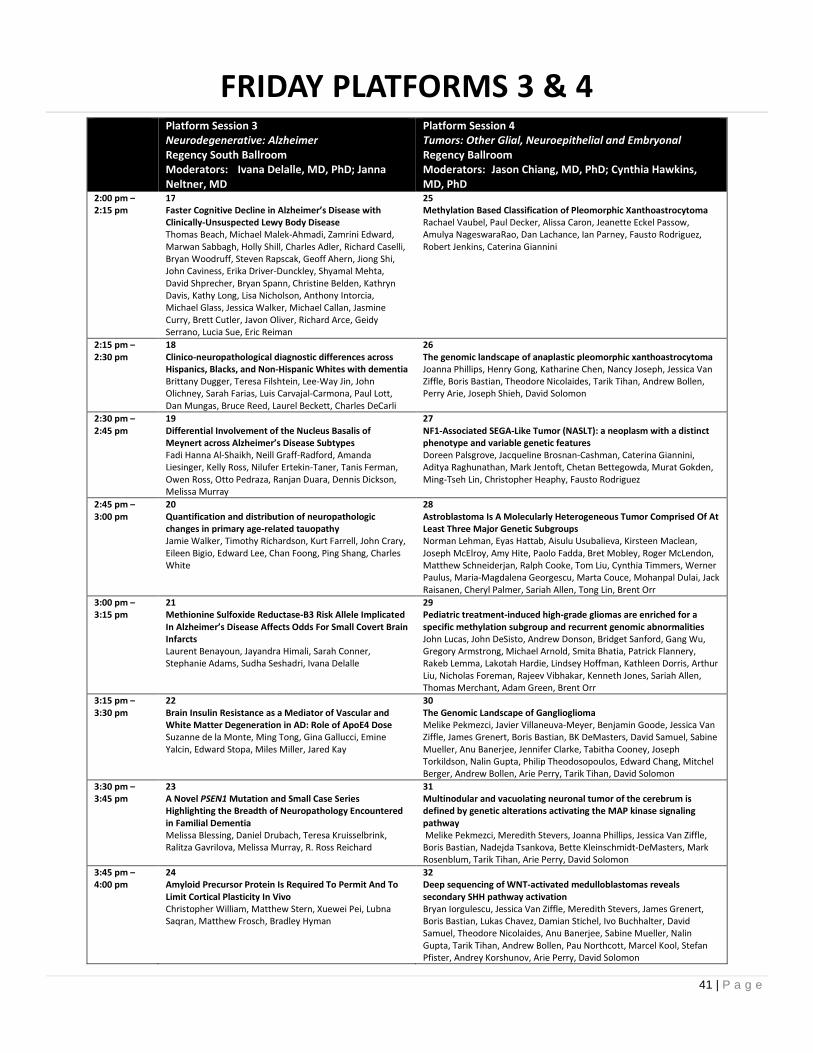
41 | P a g e
FRIDAY PLATFORMS 3 & 4Platform Session 3Neurodegenerative: AlzheimerRegency South BallroomModerators: Ivana Delalle, MD, PhD; JannaNeltner, MD
Platform Session 4Tumors: Other Glial, Neuroepithelial and EmbryonalRegency BallroomModerators: Jason Chiang, MD, PhD; Cynthia Hawkins,MD, PhD
2:00 pm –2:15 pm
17Faster Cognitive Decline in Alzheimer’s Disease withClinically-Unsuspected Lewy Body DiseaseThomas Beach, Michael Malek-Ahmadi, Zamrini Edward,Marwan Sabbagh, Holly Shill, Charles Adler, Richard Caselli,Bryan Woodruff, Steven Rapscak, Geoff Ahern, Jiong Shi,John Caviness, Erika Driver-Dunckley, Shyamal Mehta,David Shprecher, Bryan Spann, Christine Belden, KathrynDavis, Kathy Long, Lisa Nicholson, Anthony Intorcia,Michael Glass, Jessica Walker, Michael Callan, JasmineCurry, Brett Cutler, Javon Oliver, Richard Arce, GeidySerrano, Lucia Sue, Eric Reiman
25Methylation Based Classification of Pleomorphic XanthoastrocytomaRachael Vaubel, Paul Decker, Alissa Caron, Jeanette Eckel Passow,Amulya NageswaraRao, Dan Lachance, Ian Parney, Fausto Rodriguez,Robert Jenkins, Caterina Giannini
2:15 pm –2:30 pm
18Clinico-neuropathological diagnostic differences acrossHispanics, Blacks, and Non-Hispanic Whites with dementiaBrittany Dugger, Teresa Filshtein, Lee-Way Jin, JohnOlichney, Sarah Farias, Luis Carvajal-Carmona, Paul Lott,Dan Mungas, Bruce Reed, Laurel Beckett, Charles DeCarli
26The genomic landscape of anaplastic pleomorphic xanthoastrocytomaJoanna Phillips, Henry Gong, Katharine Chen, Nancy Joseph, Jessica VanZiffle, Boris Bastian, Theodore Nicolaides, Tarik Tihan, Andrew Bollen,Perry Arie, Joseph Shieh, David Solomon
2:30 pm –2:45 pm
19Differential Involvement of the Nucleus Basalis ofMeynert across Alzheimer’s Disease SubtypesFadi Hanna Al-Shaikh, Neill Graff-Radford, AmandaLiesinger, Kelly Ross, Nilufer Ertekin-Taner, Tanis Ferman,Owen Ross, Otto Pedraza, Ranjan Duara, Dennis Dickson,Melissa Murray
27NF1-Associated SEGA-Like Tumor (NASLT): a neoplasm with a distinctphenotype and variable genetic featuresDoreen Palsgrove, Jacqueline Brosnan-Cashman, Caterina Giannini,Aditya Raghunathan, Mark Jentoft, Chetan Bettegowda, Murat Gokden,Ming-Tseh Lin, Christopher Heaphy, Fausto Rodriguez
2:45 pm –3:00 pm
20Quantification and distribution of neuropathologicchanges in primary age-related tauopathyJamie Walker, Timothy Richardson, Kurt Farrell, John Crary,Eileen Bigio, Edward Lee, Chan Foong, Ping Shang, CharlesWhite
28Astroblastoma Is A Molecularly Heterogeneous Tumor Comprised Of AtLeast Three Major Genetic SubgroupsNorman Lehman, Eyas Hattab, Aisulu Usubalieva, Kirsteen Maclean,Joseph McElroy, Amy Hite, Paolo Fadda, Bret Mobley, Roger McLendon,Matthew Schneiderjan, Ralph Cooke, Tom Liu, Cynthia Timmers, WernerPaulus, Maria-Magdalena Georgescu, Marta Couce, Mohanpal Dulai, JackRaisanen, Cheryl Palmer, Sariah Allen, Tong Lin, Brent Orr
3:00 pm –3:15 pm
21Methionine Sulfoxide Reductase-B3 Risk Allele ImplicatedIn Alzheimer’s Disease Affects Odds For Small Covert BrainInfarctsLaurent Benayoun, Jayandra Himali, Sarah Conner,Stephanie Adams, Sudha Seshadri, Ivana Delalle
29Pediatric treatment-induced high-grade gliomas are enriched for aspecific methylation subgroup and recurrent genomic abnormalitiesJohn Lucas, John DeSisto, Andrew Donson, Bridget Sanford, Gang Wu,Gregory Armstrong, Michael Arnold, Smita Bhatia, Patrick Flannery,Rakeb Lemma, Lakotah Hardie, Lindsey Hoffman, Kathleen Dorris, ArthurLiu, Nicholas Foreman, Rajeev Vibhakar, Kenneth Jones, Sariah Allen,Thomas Merchant, Adam Green, Brent Orr
3:15 pm –3:30 pm
22Brain Insulin Resistance as a Mediator of Vascular andWhite Matter Degeneration in AD: Role of ApoE4 DoseSuzanne de la Monte, Ming Tong, Gina Gallucci, EmineYalcin, Edward Stopa, Miles Miller, Jared Kay
30The Genomic Landscape of GangliogliomaMelike Pekmezci, Javier Villaneuva-Meyer, Benjamin Goode, Jessica VanZiffle, James Grenert, Boris Bastian, BK DeMasters, David Samuel, SabineMueller, Anu Banerjee, Jennifer Clarke, Tabitha Cooney, JosephTorkildson, Nalin Gupta, Philip Theodosopoulos, Edward Chang, MitchelBerger, Andrew Bollen, Arie Perry, Tarik Tihan, David Solomon
3:30 pm –3:45 pm
23A Novel PSEN1 Mutation and Small Case SeriesHighlighting the Breadth of Neuropathology Encounteredin Familial DementiaMelissa Blessing, Daniel Drubach, Teresa Kruisselbrink,Ralitza Gavrilova, Melissa Murray, R. Ross Reichard
31Multinodular and vacuolating neuronal tumor of the cerebrum isdefined by genetic alterations activating the MAP kinase signalingpathwayMelike Pekmezci, Meredith Stevers, Joanna Phillips, Jessica Van Ziffle,Boris Bastian, Nadejda Tsankova, Bette Kleinschmidt-DeMasters, MarkRosenblum, Tarik Tihan, Arie Perry, David Solomon
3:45 pm –4:00 pm
24Amyloid Precursor Protein Is Required To Permit And ToLimit Cortical Plasticity In VivoChristopher William, Matthew Stern, Xuewei Pei, LubnaSaqran, Matthew Frosch, Bradley Hyman
32Deep sequencing of WNT-activated medulloblastomas revealssecondary SHH pathway activationBryan Iorgulescu, Jessica Van Ziffle, Meredith Stevers, James Grenert,Boris Bastian, Lukas Chavez, Damian Stichel, Ivo Buchhalter, DavidSamuel, Theodore Nicolaides, Anu Banerjee, Sabine Mueller, NalinGupta, Tarik Tihan, Andrew Bollen, Pau Northcott, Marcel Kool, StefanPfister, Andrey Korshunov, Arie Perry, David Solomon

42 | P a g e
FRIDAY POSTERS #33-#51
Friday, June 8, 2018Time: Poster #: Gulfstream-Hialeah & Keeneland
8:00 am –5:00 pm
33 Prevalence of Constitutional Mismatch Repair Deficiency in Saudi Arabian HGG cohort andsuccessful use of Nivolumab in an affected childMalak Abedalthagafi, Musa AlHarbi, Nahla Mobark
34 Malignant Glioneuronal Neoplasm with Divergent Differentiation and Histone H3 K27M Mutation:Case ReportJiancong Liang, Devang Pastakia, John Wellons, Christine Fuller
35 Sporadic Pilocytic Astrocytoma with Novel Neurotrophic Receptor Tyrosine Kinase 2 (NTRK2)AlterationRebecca Yoda, Siobhan Pattwell, Sonali Aurora, Eric Konnick, P.J. Cimino
36 ATRX, IDH, p53 and H3F3A-G34 Integrated Analysis in Hemispheric Glioblastomas in YoungPatientsElena Martinez-Saez, Jessica Camacho, Teresa Moliné, Rosa Somoza, Regina Mayor, Francisco RamonMartinez-Ricarte, Joan Seoane, Javier Hernandez-Losa, Santiago Ramon y Cajal
37 Supratentorial Clear Cell Ependymoma Associated with RELA Fusion and Long Term SurvivalErnest Nelson, MacLean Nasrallah, Angela Viaene, Phyllis Faust, Cristina Antonescu, Marilyn Li, LeaSurrey, Minjie Luo, Eun-Sook Cho, Leroy Sharer
38 Primary diffuse leptomeningeal gliomatosis initially diagnosed as T.B meningitisMariam Youssef, KO Riley, Marcus Bredel, Mina Lobbous, DJ Israel, JR Hackney
39 Expression of PRAME is increased in K27M mutant gliomas: Identification of a potential target forimmunotherapyMarissa Spino, James Stafford, Luis Chiriboga, Briana Zeck, Vladislav Sviderskiy, Andrew Chi, RichardPossemato, Matija Snuderl
40 Cerebellar glioblastoma: a clinicopathologic series of 16 casesAndrew Gao, Maryam Abdollahi, Hidehiro Okura, Cynthia Hawkins, Michael Cusimano, David Munoz
41 Thyroid Transcription Factor-1 (TTF-1) Expression in Glioblastoma with Primitive NeuronalComponent (GB-PN)Mustafa Goksel, Manoj Kumar, Murat Gokden
42 Rosette-Forming Glioneuronal Tumor (RFGT) of the Temporal LobeMurat Gokden, Amy Joiner, Brandon Evans, Kremer Nicholas
43 High-grade Diffuse Astrocytoma with Epithelioid Features, BRAF V600E and IDH-1 (R132H)MutationsUrooba Nadeem, Rohan Samant, Hazem Ahmed, Murat Gokden
44 Immunohistochemical Assessment of Tumor-Associated Microglia/Macrophages and T Cells in CNSNeoplasmsM. Adelita Vizcaino, Charles G. Eberhart
45 Differential microRNA expression in NF1-associated high- and low-grade gliomaJames Nix, Eddie Imada, Heather Ames, Luigi Marchionni, Laura Smithson, David Gutmann, FaustoRodriguez
46 Novel insight into mechanisms of tumorigenesis of glioblastoma on the background of Ollierdisease: A case reportMaliha Khara, Julian Spears, Lyne Noel de Tilly, David Munoz
47 The AURKA Inhibitor Alisertib Is Synergistic With Irinotecan And Carboplatin Against GlioblastomaCellsCory Zumbar, Paul King, Muge Sak, Aisulu Usubalieva, Xiaohui Li, Caroline Mifsud, Joseph McElroy,Eric Burton, Norman Lehman
48 Histone H3F3A G34R-mutant Infiltrating Glioma with Rapid Development of Diffuse ExtracranialBony MetastasesAshley Holloman, Kevin Fisher, Hao Wu, Vidya Mehta, Sergio Islas, Suzanne Bull, Xuli Wu, NileshDesai, Guillermo Aldave Orzaiz, Holly Lindsay, Timothy Lotze, Adekunle Adesina, Carrie Mohila
49 INSM1 over-expression in CDK4-amplified glioblastomaHeather Ames, Fausto Rodriguez, Charles Eberhart, Jody Hooper, John Laterra
50 IDH-Mutant Or IDH-Wildtype? Unusual Subclonal IDH Mutation In A GlioblastomaMaria Martinez-Lage, Deborah Forst, Pamela Jones, Dora Dias-Santagata, Jochen Lennerz
51 A case of adult intraventricular low-grade glial neoplasm with epithelioid features and TERT andBRAFV600E mutationsAmir Banihashemi, Mellissa Umphlett, Clare Bryce, Mary Fowkes
Posters are not offered for CME credit

43 | P a g e
FRIDAY POSTERS #52-#70
Friday, June 8, 2018Time: Poster #: Gulfstream-Hialeah & Keeneland8:00 am –5:00 pm
52 Infantile High-grade Glioma with Novel Translocation Recurring as Ganglion Cell TumorMartin Powers, Christina Di Loreto, Lawrence Hansen, Denise Malicki
53 Against the odds: genome-wide analysis of glioblastomas with ultra-long survivalTimothy Richardson, Matija Snuderl, Jonathan Serrano, Dwight Oliver, Charles White, Jack Raisanen, BruceMickey, Kimmo Hatanpaa
54 An unusual case of recurrent epilepsy associated rosette-forming glioneuronal tumor with anaplastictransformation in the absence of therapyAaron Halfpenny, Sean Ferris, Marjorie Grafe, Randy Woltjer, Nathan Selden, Kellie Nazemi, Arie Perry,David Solomon, Stephen Moore, Helen Lawce, Lora Lucas, Chris Corless, Matthew Wood
55 Prognostic Implications of MAPT Expression in Primary Brain TumorsMeaghan Morris, Antionette Price, Charles Eberhart
56 Polymorphous low-grade neuroepithelial tumor of the young: How much is PLNTY?Jason Gregory, Steven Roper, Anthony Yachnis, Marie Rivera-Zengotita
57 A high-grade glioneuronal tumor with SOS1 amplificationBarbara Vidal, Christine Bryke, Nancy Hsu, Ronnie Alterman, Hemant Varma
58 Dysembryoplastic Neuroepithelial Tumor-Like Neoplasm (DNET) of Septum Pellucidum: MolecularCharacterization of Primary & Recurrent TumorsMohammad Khan, Kevin Kawachi, Jean Lopategui, Serguei Bannykh, Warren Tourtellotte, Xuemo Fan
59 Histologic, Immunohistochemical, and Molecular Features of Pituicytomas and Atypical PituicytomasAngela Viaene, Edward Lee, Jason Rosenbaum, MacLean Nasrallah
60 Next-Generation Sequencing Uncovers Novel Glioma Stem Cell-Associated MutationsIgor Katsyv, Jessica Tome-Garcia, Ethan Ellis, Maya Strahl, Wissam Hamou, Jane Houldsworth, RaymundYong, Bin Zhang, Robert Sebra, Nadejda Tsankova
61 Next Generation Sequencing in Clinical Practice: A Prospective Study of 316 Diffuse Astrocytomas. Doesit Replace Other Ancillary Studies?Thomas Pearce, Christopher Suciu, Ronald Hamilton
62 False negative rate of ATRX immunohistochemistry in gliomasWesley Samore, Maria Martinez-Lage
63 Utilizing Histology to Define a Digital Signature for Cell Infiltration in GlioblastomaMelissa Umphlett, Vu Nguyen, Rajarsi Gupta, Dimitris Samaras, Heather Bell, Joel Saltz, Nadejda Tsankova
64 A Case of Widely Metastatic Glioblastoma with Multiple Genetic MutationsMelissa Umphlett, Stephanie Shea, Amy Chan, Adilia Hormigo, Jane Houldsworth, Mary Fowkes, NadejdaTsankova, Raymund Yong
65 Relationship between Ki67 and clinical outcome in PFA pediatric ependymomasNishant Tiwari, Debra Hawes, Sriram Venneti, Alexander Judkins
66 High-grade Infiltrating Gliomas of the Spinal Cord in Adults: Clinico-pathological and Genomic Evaluationof 16 casesMohammed Ali Alvi, Michael A. Paolini, Cristiane Ida, Panagiotis Kerezoudis, Sandy Goncalves, MohamadBydon, Aditya Raghunathan
67 BRAF activating mutations involving the β3-αC loop in V600E-negative anaplastic pleomorphicxanthoastrocytomaDrew Pratt, Sandra Camelo-Piragua, Kathryn McFadden, Denise Leung, Rajen Mody, Arul Chinnaiyan, CarlKoschmann, Sriram Venneti
68 A Novel Targetable RAF1-QKI Fusion in Adult Spinal Pleomorphic Xanthoastrocytoma without BRAFV600E MutationElena Daoud, Timothy Richardson, Divya Mella, Edward Pan, Anna Schwarzbach, Charles White, III, KimmoHatanpaa
69 Pre-treatment Monocyte-to-Lymphocyte Ratio in Pediatric Gliomas with Histone H3 K27M MutationSeema Patel, Shiyang Wang, Jonathan Serrano, Sharon Gardner, Matija Snuderl
70 In vivo PiB-PET neuroimaging correlates with autopsy β-amyloidosis in both late-onset and autosomaldominant Alzheimer diseaseNamita Sinha, Aihong Zhou, Erin Franklin, Chengjie Xiong, John Morris, Randall Bateman, TammieBenzinger, Nigel Cairns
Posters are not offered for CME credit

44 | P a g e
FRIDAY POSTERS #71-#88
Friday, June 8, 2018Time: Poster #: Gulfstream-Hialeah & Keeneland
8:00 am –5:00 pm
71 White matter small vessel pathology in Alzheimer’s diseaseSara Nasrabady, James Goldman, Adam Brickman
72 A 3D High Resolution Model of Brain ConnectivityClio Gonzalez Zacarias, Celia Williams, Michael Bienkowski, Hong Wei Dong, Kristi Clark, Carol AnnMiller
73 Patient-derived induced pluripotent stem cells from brain donors for the study of Alzheimer’sdisease and other neurodegenerative disordersDerek Oakley, Naomi Klickstein, Bradley Hyman, Matthew Frosch
74 Associations of APOE in the Honolulu Asia Aging StudyMargaret Flanagan, Steven Edland, Laura Hemmy, Brenna Cholerton, Thomas Montine, Lon White
75 Neuronal identity of the dentate gyrus in AD: ex vivo neuroimaging and histologyFarshid Sepehrband, Linda Szymanski, Nishant Tiwari, Kristi Clark, Carol Miller
76 Hereditary, early-onset, and rapidly progressive Alzheimer Disease, associated with thep.Leu381Phe mutation in the Presenilin 1 geneJose Bonnin, Bernardino Ghetti, Holly Garringer, Francine Epperson, Rose Marie Richardson, GeraldCampbell, Benjamin Gelman, Jill Mokry, Brendan Lee, Paolo Moretti
77 Suppression of Non-Specific Bielschowsky Silver Staining by Pretreatment with PotassiumPermanganate and Oxalic AcidAnthony Intorcia, Geidy Serrano, Lucia Sue, Thomas Beach
78 Neuropathologic Associations of APOE in the Nun StudyMargaret Flanagan, Steven Edland, Laura Hemmy, Joyce Meints, Brenna Cholerton, Kelvin Lim,Thomas Montine, Lon White
79 Early-Onset Alzheimer Disease in a Puerto Rican Man with the G206A Mutation in Presenilin 1(PSEN1)Jose Bonnin, Holly Garringer, Francine Epperson, Rose Marie Richardson, Bernardino Ghetti
80 Increasing the Value of Autopsies in Patients with Brain Tumors in the Molecular EraJared Ahrendsen, Mariella Filbin, Susan Chi, Peter Manley, Karen Wright, Pratiti Bandopadhayay,Mark Kieran, Keith Ligon, Sanda Alexandrescu
81 Novel BRAF Alteration in Desmoplastic Infantile GangliogliomaMelissa Blessing, Patrick Blackburn, Jessica Balcom, Chandra Krishnan, Michael Zimmermann, RoryJackson, Asha Nair, Christopher Zysk, Kevin Halling, Benjamin Kipp, Cristiane Ida
82 Two Cases of Pineal Anlage TumorKathryn Scherpelz, Nicholas Vitanza, Sarah Leary, Ralph Ermoian, Bonnie Cole
83 Dentate Gyrus Dysplasia: Prevalence in Unselected Autopsy PopulationsDouglas Miller, Kristen Scheitler, Deiter Duff, Jeffery Holloway, C Stacy
84 Inhibitory Interneurons in Hemimegalencephaly: A Survey of 9 CasesSeth Lummus, Jimena Andersen, Sergiu Pasca, Bette Kleinschmidt-DeMasters, Hannes Vogel
85 Temporal and anatomical expression of Rab5 subfamily of GTPases in the central nervous systemKwok-Ling Kam, Paige Parrack, Camille Milton, Marcellus Banworth, Louisa Williams, SheejaAravindan, Guangpu Li, Kar-ming Fung
86 Neuropathology of CHARGE Syndrome: Report of Two CasesChristopher Borck, Kristen Landi, Nori Williams, Rebecca Folkerth
87 Eosinophilic Cytoplasmic Astrocytic Inclusions in Pediatric Patients with Epilepsy: A Report of 2CasesElmira Vaziri Fard, Manish Shah, Gretchen von Allmen, Rajan Patel, Leomar Ballester, MeenakshiBhattacharjee
88 Mice Expressing c-MYC in Neural Precursors Develop Choroid Plexus and Ciliary Body TumorsMorgan Shannon, Ryann Fame, Kevin Chau, Neil Dani, Monica Calicchio, Hart Lidov, SandaAlexandrescu, Maria Lehtinen
Posters are not offered for CME credit

45 | P a g e
FRIDAY POSTERS #89-#107
Friday, June 8, 2018Time: Poster #: Gulfstream-Hialeah & Keeneland
8:00 am –5:00 pm
89 Neuropathological characterization of a long term survivor with RCDP type IJosh Klonoski, Valarie McMurtry, Joshua Sonnen, Jessica Comstock, Theodore Wilson
90 Acquired neuropathological lesions in infants dying from congenital heart disease from 2000-2017Leigh Rettenmaier, Patricia Kirby, Marco Hefti
91 Immune Myopathy with Large-Histiocyte-Related Myofiber NecrosisNamita Sinha, Ziad Alhumayd, Cindy Ly, Robert Bucelli, Robert Schmidt, Alan Pestronk
92 Detection of Mitochondrial Adaptations to Exercise in a Muscle BiopsyDavid Priemer, Alexander Vortmeyer
93 A Case of Sarcoid Associated Necrotizing Myopathy With Immune Mediated FeaturesYasir Al-Khalili, Osama Elkadi
94 Brachial Plexopathy Caused by Endoneurial IgM Deposition in a Patient with Waldenstrom'sMacroglobulinemiaSean Ferris, Giselle López, Marta Margeta
95 GRANULOMATOUS MYOSITIS AS SOLE MANIFESTATION OF GRAFT VERSUS HOST DISEASEEwa Borys, Stefan Pambuccian
96 Mitochondrial DNA Depletion Syndrome 12A: A Cause of Mitochondrial Disease with SevereNeurological SymptomsHeidi Reinhard, Debra Byler, Roger Ladda, Charles Specht
97 Novel RYR1 Mutation in Congenital Muscular DystrophyChristina Di Loreto, Martin Powers, Lawrence Hansen, Denise Malicki
98 Plectinopathy Resulting In Epidermolysis Bullosa Simplex With Muscular Dystrophy And PyloricStenosisLindsey Lowder, Jose Velazquez Vega, Pia Mendoza, Stewart Neill, Matthew Schniederjan
99 Single-cell transcriptome analysis of medulloblastomaJohn DeWitt, Volker Hovestadt, Mariella Filbin, Laure Bihannic, Mckenzie Shaw, Andrew Groves, KyleSmith, Jennifer Hadley, Amar Gajjar, Giles Robinson, Lisa Mayr, Irene Slavc, Liliana Goumnerova, KeithLigon, Mario Suva, Paul Northcott, Bradley Bernstein
100 A Case of Reversible Infantile Respiratory Chain Deficiency Presenting with hypotonia,hyperammonemia and failure to thriveJessenia Guerrero, Helio Pedro, Sarah Parisotto, Ada Baisre
101 Adult orbital xanthogranulomatous disease: A rare histiocytic disease of the orbit with variablesystemic manifestationsRajnish Bharadwaj, Kathryn Scherpelz, C. Keene, Joanne Ho, Arash Amadi, Gordana Juric-Sekhar, LuisGonzalez-Cuyar
102 Endogenous Aspergillus Endophthalmitis: A Case Study of Inpatient Phthisis Bulbi With EnucleationDue To Fungal InfectionEdward Kelly Mrachek, Steven Newman, James Mandell
103 Unusual Overexpression of CD34 in Mature Orbital Adipose Tissue May Pose Challenge in theDiagnosis of Orbital Spindle Cell/Pleomorphic LipomasRati Chkheidze, Kyle Molberg, Elena Lucas, Sunati Sahoo, Charles White, Robert Hogan, Bret Evers
104 Bilateral Acanthamoeba Panophthalmitis: a Rare and Unique CaseRati Chkheidze, Dominick Cavuoti, Robert Hogan, Bret Evers
105 PRES: Review of Histological FeaturesJustin Honce, Nicholas Willard
106 Segmental Arterial Mediolysis as a Cause of Intracranial Dissecting Aneurysm: Pathological Findingsin Two Surgical CasesHajime Miyata, Junko Kawamura, Junta Moroi, Tatsuya Ishikawa, Toshibumi Kinoshita, Yasuji Yoshida
107 An Experimental White Matter Infarct Model: Persistent Motor Paralysis by a LaserPhotothrombotic PyramidotomyMin-Cheol Lee, Kyung-Hwa Lee, Hanlim Song, Hyoung-Ihl Kim
Posters are not offered for CME credit
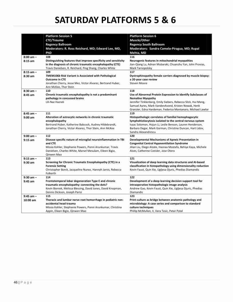
46 | P a g e
SATURDAY PLATFORMS 5 & 6
Platform Session 5CTE/TraumaRegency BallroomModerators: R. Ross Reichard, MD; Edward Lee, MD,PhD
Platform Session 6Muscle/OtherRegency South BallroomModerators: Sandra Camelo-Piragua, MD; RupalMehta, MD
8:00 am –8:15 am
108Distinguishing features that improve specificity and sensitivityin the diagnosis of chronic traumatic encephalopathy (CTE)Travis Danielsen, R. Reichard, Ping Shang, Charles White
116Neurogenic features in mitochondrial myopathiesJian-Qiang Lu, Adnan Mubaraki, Chuanzhu Yan, John Provias,Mark Tarnopolsky
8:15 am –8:30 am
109TMEM106b Risk Variant is Associated with PathologicalOutcome in CTEJonathan Cherry, Jesse Mez, Victor Alvarez, Bertrand Huber,Ann McKee, Thor Stein
117Dystrophinopathy female carriers diagnosed by muscle biopsy:a 20-year case reviewSteven Moore
8:30 am –8:45 am
110Chronic traumatic encephalopathy is not a predominantpathology in concussed brainsLili-Naz Hazrati
118Use of Abnormal Protein Expression to Identify Subclasses ofNemaline MyopathyJennifer Tinklenberg, Emily Siebers, Rebecca Slick, Hui Meng,Samuel Ayres, Mark VandenAvond, Kristen Nowak, HenkGranzier, Edna Hardeman, Federica Montanaro, Michael Lawlor
8:45 am –9:00 am
111Alteration of astrocytic networks in chronic traumaticencephalopathyBertrand Huber, Katherine Babcock, Audrey Hildebrandt,Jonathan Cherry, Victor Alvarez, Thor Stein, Ann McKee
119Histopathologic correlates of familial hemophagocyticlymphohistiocytosis isolated to the central nervous systemIsaac Solomon, Hojun Li, Leslie Benson, Lauren Henderson,Barbara Degar, Mark Gorman, Christine Duncan, Hart Lidov,Sandra Alexandrescu
9:00 am –9:15 am
112Disease specific nature of microglial neuroinflammation in TBIand CTEMissia Kohler, Stephanie Powers, Ponni Arunkumar, TravisDanielsen, Charles White, Marsel Mesulam, Eileen Bigio,Qinwen Mao
120Developmental Mechanisms of Apneic Presentation inCongenital Central Hypoventilation SyndromeJilian Liu, Diego Alzate, Hasnaa Mostafa, Behiye Kaya, MicheleAlves, Catherine Czeisler, Jose Otero
9:15 am –9:30 am
113Screening for Chronic Traumatic Encephalopathy (CTE) in aForensic SettingChristopher Borck, Jacqueline Nunez, Hannah Jarvis, RebeccaFolkerth
121Visualization of deep learning data structures and AI-basedclassification in histopathology using dimensionality reductionKevin Faust, Quin Xie, Ugljesa Djuric, Phedias Diamandis
9:30 am –9:45 am
114Frontotemporal lobar degeneration Type E and chronictraumatic encephalopathy: connecting the dots?Kevin Bieniek, Melissa Blessing, David Jones, David Knopman,Dennis Dickson, Joseph Parisi
122Development of a deep learning decision support tool forintraoperative histopathologic image analysisAndrew Gao, Kevin Faust, Quin Xie, Ugljesa Djuric, PhediasDiamandis
9:45 am –10:00 am
115Thoracic and lumbar nerve root hemorrhage in pediatric non-accidental head traumaMissia Kohler, Stephanie Powers, Ponni Arunkumar, ChristinaAppin, Eileen Bigio, Qinwen Mao
123Print-culture as bridge between anatomic pathology andmicrobiology: A case series and comparison to standardculture techniquesPhillip McMullen, II, Vera Tesic, Peter Pytel

47 | P a g e
SATURDAY PLATFORMS 7 & 8
Platform Session 7Neurodegenerative: FTLD/Lewy body/PrionRegency BallroomModerators: John Crary, MD, PhD; Richard Perrin, MD,PhD
Platform Session 8Tumors: OtherRegency South BallroomModerators: Craig Horbinski, MD, PhD; Matija Snuderl, MD
2:00 pm –2:15 pm
124Molecular phenotyping of human neurons with TDP-43pathology reveals decondensation of transposableelementsElaine Liu, Jenny Russ, Alexandre Amlie-Wolf, Edward Lee
132Metabolomic Analysis of CSF Differentiates Primary andMetastatic CNS Tumors from Non-Neoplastic DiseaseLeomar Ballester, Lu Gurangong, Soheil Zorofchian, VenkataVantaku, Vasanta Putluri, Yuanqing Yan, Octavio Arevalo-Espejo,Roy Riascos-Castaneda, Arun Sreekumar, Yoshua Esquenazi,Nagireddy Putluri, Jay-Jiguang Zhu
2:15 pm –2:30 pm
125Antisense RNA foci and poly-GR dipeptide repeat proteinsare unique in repeat-expanded C9orf72 ALS/FTDShahram Saberi, Jennifer Stauffer, Jie Jiang, Sandra DiazGarcia, Amy Taylor, Derek Schulte, Takuya Ohkubo,Cheyenne Schloffman, Marcus Maldonado, MichaelBaughn, Maria Rodriguez, Don Pizzo, Don Cleveland, JohnRavits
133Whole genome DNA methylation signatures for diagnosis ofbrain metastasesOlga Krasnozhen-Ratush, Jonathan Serrano, Matija Snuderl
2:30 pm –2:45 pm
126GRN Mutations Cause Unique CA1 PathologyQinwen Mao, Missia Kohler, Tamar Gefen, Jayson Wilson,Zachary Parton, Haibin Xia, Rosa Rademakers, EmilyRogalski, Sandra Weintraub, Marek-Marsel Mesulam, EileenBigio
134CSF cytology in the evaluation of primary and metastatic CNSmalignanciesTatiana Belousova, Vanya Jaitly, Octavio Arevalo-Espejo, RoyRiascos-Castaneda, Jing Liu, Yoshua Esquenazi, MeenakshiBhattacharjee, Leomar Ballester
2:45 pm –3:00 pm
127Neuropathology of Progressive Supranuclear Palsy –Experience from 20-years of Brain Banking for CurePSPDennis Dickson
135Integrated Liquid Biopsy Analysis for Pediatric Brain TumorPatients Using Detection of ctDNA and Circulating Tumor CellsKaicen Zhu, Katherine Barnett, Guomiao Shen, HusseinMohammed, Tania Panicucci-Roma, Jonathan Serrano, DavidHarter, Jeffrey Wisoff, Amanda Yaun, Shiyang Wang, SharonGardner, Matija Snuderl
3:00 pm –3:15 pm
128Evidence of Novel Pathologic Protein Misfolding in theHuman Amygdala -- a Proteomics StudyPeter Nelson, Haining Zhu, Jozsef Gal
136Telomere alterations in NF1-associated solid tumorsFausto Rodriguez, Mindy Graham, Jacqueline Brosnan-Cashman,Christine Davis, M. Adelita Vizcaino, Doreen Palsgrove, CaterinaGiannini, Murat Gokden, Jaishri Blakeley, Allan Belzberg,Christopher Heaphy
3:15 pm –3:30 pm
129Gain-of-function of the ATXN1-CIC co-repressor complexdrives the cerebellar pathology in spinocerebellar ataxiatype 1 (SCA1)Hsiang-Chih Lu, Maxime Rousseaux, Tyler Tschumperlin,Elizabeth Lackey, Vitaliy Bondar, Ying-Wooi Wan, QiuminTan, Carolyn Adamski, Jillian Friedrich, Kirk Twaroski, WeiliChen, Jakub Tolar, Christine Henzler, Ajay Sharma,Aleksandar Bajić, Tao Lin, Lisa Duvick, Roy Sillitoe, Huda Zoghbi, Harry Orr
137Whole Exome Sequencing of Clinically AggressiveMeningiomas Reveals Mutational Signatures Associated withDNA Mismatch Repair and AgingBenjamin Liechty, Sylvia Eisele, Stephen Kelly, VarshiniVasudevaraja, Ramona Bledea, Peter Wu, Jonathan Serrano,Leah Katz, Joshua Silverman, Donato Pacione, Stephen Russell,Chandra Sen, John Golfinos, Andrew Chi, Matija Snuderl
3:30 pm –3:45 pm
130Dominantly Inherited prion protein cerebral amyloidosis: areappraisalBernardino Ghetti, Pedro Piccardo, Gianluigi Zanusso
138Multiplexed Immunofluorescence Reveals Potential PD-1/PD-L1 Pathway Vulnerabilities in CraniopharyngiomaShannon Coy, Rumana Rashid, Jia-Ren Lin, Ziming Du, AndrewDonson, Todd Hankinson, Nicholas Foreman, Peter Manley,Mark Kieran, David Reardon, Peter Sorger, Sandro Santagata
3:45 pm –4:00 pm
131Automated digital solutions for quantitativeneuropathologyKangway Chuang, Ziqi Tang, Michael Keiser, Charles DeCarli,Laurel Beckett, Lee-Way Jin, Brittany Dugger
139LEF1 Immunohistochemistry in the Evaluation ofMedulloblastoma SubgroupsDiana Thomas, Geoffrey Murdoch, Ronald Hamilton, Brent Orr

48 | P a g e
SATURDAY POSTERS #140-#160
Saturday, June 9, 2018Time: Poster #: Gulfstream-Hialeah & Keeneland
8:00 am –5:00 pm
140 Pediatric Primary CNS NTRK-Associated Fibroblastic Neoplasm: A Case ReportMatthew Torre, Nicholas Jessup, Jason Hornick, Sanda Alexandrescu
141 Dural tail in meningioma – Is it a radiological guide for curative resection?Dewa Pakshage Chula Kanishka Ananda Lal
142 CNS involvement in Peripheral T-cell lymphoma, a rare presentation.Mariam Youssef, Deniz Peker, JR Hackney
143 Evaluating Circulating Tumor DNA from the Cerebrospinal Fluid of Patients with Melanoma andLeptomeningeal DiseaseLeomar Ballester, Isabella Glitza Oliva, Dzifa Douse, Melissa Chen, Chieh Lan, Lauren Haydu, JasonHuse, Sinchita Roy-Chowdhuri, Rajalakshmi Luthra, Ignacio Wistuba, Michael Davies
144 Malignant progression in extraventricular neurocytoma arising from the VIIIth cranial nerve: Acase report, literature review, and debateYasuo Sugita, Takuya Furuta, Satoru Komaki, Koichi Ohshima, Kiyohiko Sakata, Motohiro Morioka
145 Corpus Callosum ChordomaDavid Priemer, Lorenzo Rinaldo, Terence Burns, Aaron Cohen-Gadol, Daniel Brat, Caterina Giannini,Alexander Vortmeyer
146 ATRX Mutation in Pineal Parenchymal Tumor of Intermediate DifferentiationMichelle Nagurney, Christopher Farrell, Zi-Xuan Wang, Mark Curtis
147 Spindle Cell Oncocytoma with Papillary FeaturesKymberly Gyure, Rahul Singh, Joseph Voelker, M. Beatriz Lopes
148 Choroid Plexus Papilloma (CPP) with Neuropil-like (NPL) DifferentiationJohn Block, Christine Fuller, Rongsheng Cai, Rahgu Ramakrishnaiah, Murat Gokden
149 Pituitary Adenoma with Ganglion Cell Differentiation (PA-G)Michael Occidental, Jennifer McCarty, John Day, Murat Gokden
150 Calcifying pseudoneoplasm of the neuraxis: a histologic characterization of 38 casesMichael Paolini, Matthew Ball, Mai Lan Ho, Hannah Monahan, Aditya Raghunathan
151 Primary Intracranial Nerve Sheath Myxoma within the Cavernous SinusNichole Allen, Gary Pearl, Aaron Wagner, Naren Ramakrishna
152 Clinicopathologic and Genetic Features of a Meningioma with Unusual MorphologyJared Woods, Matthew Torre, Alexandra Golby, Eudocia Lee, Shyam Tanguturi, William Denison,Zachary Spigelman, Umberto De Girolami
153 Intravascular Large B-cell Lymphoma (IVL) with Endocrine Organ InvolvementAnna Tart, Raga Palagiri, Jeanette Ramos, Vikas Koppurapu, Jennifer Forsyth, Anusha Jillella,Harmeen Goraya, Murat Goraya
154 Clinical and Immunohistochemical Analysis of Clinically Non-functional Pituitary NeuroendocrineTumorsJonathan Lavezo, Merav Frankel, Brunilda Balliu, James Pan, Andrew Hoffman, Robert Dodd,Griffith Harsh, Laurence Katznelson, Hannes Vogel
155 A Case of STAT6 Positive Intraventricular Hemangiopericytoma In An AdultColin Kanach, Osama Elkadi
156 Molecular Alterations in Primary and Metastatic Central Nervous System LymphomasSoheil Zorofchian, Hanadi El-Achi, Yoshua Esquenazi, Leomar Ballester
157 CNS High-Grade Neuroepithelial Tumor with BCOR Alteration: Heterogeneous Histologic FeaturesExpand the Morphologic SpectrumJosé Velázquez Vega, Matthew Schniederjan, Arie Perry, David Solomon
158 Epstein-Barr Virus-Associated Smooth Muscle Tumor: A Case with Unusual HistologyJosé Velázquez Vega, Lindsey Lowder, Matthew Schniederjan
159 Papillary Tumor of The Pineal Region: A Case Report Including Detailed IHC and MolecularEvaluationAstin Powers, Martha Quezado, Cara Monroe, Edjah Nduom, Abhik Ray-Chaudhury, Mark Gilbert,Mark Raffeld, Terri Armstrong, Sonja Crandon, Elizabeth Vera
160 Synaptophysin-positive Solitary Fibrous Tumor/Hemangiopericytoma Occurring in theParasellar/Cavernous RegionShalini Mukhi, Emilie Rouah
Posters are not offered for CME credit

49 | P a g e
SATURDAY POSTERS #161-#181
Saturday, June 9, 2018Time: Poster #: Gulfstream-Hialeah & Keeneland
8:00 am –5:00 pm
161 Adenocarcinoma arising from a recurrent intracranial teratomaDaniel Shepherd, Stefan Pambuccian, Ewa Borys
162 Clinicopathological and molecular analysis of multinodular and vacuolating neuronal tumorsSeong-Ik Kim, Euno Choi, Jae Kyung Won, Chun Kee Chung, Sung-Hye Park
163 A case report of necrotizing myopathy, likely paraneoplastic in origin, arising in the setting of highgrade serous carcinoma of the ovary.Nathan Clement, Anat Stemmer-Rachamimov
164 Lymphomatoid granulomatosis, grade 3Josh Klonoski, Valarie McMurtry, Frederic Clayton, Cheryl Palmer
165 SOX-10 Expression Aids in the Differential Diagnosis of Endolymphatic Sac TumorsAnne Shepler, Julia Kofler
166 Where Is My Hemangioblastoma – Case Report: Gliosis Radiographically Mimicking A ParaspinousNeoplasmKanish Mirchia, Joseph Fullmer
167 Loss of H3K27me3 Expression Distinguishes MPNST from ANNUBP but Lacks PrognosticSignificanceErik Williams, Ann Belan Larque, Tareq Juratli, Vikram Deshpande, Ivan Chebib, G. Petur Nielsen,Anat Stemmer-Rachamimov
168 Medulloepithelioma: a case report with mosaic loss of chromosomes 19 and 2Suash Sharma, Ravindra Kolhe, Ian Heger, Amyn Rojiani
169 Polymorphous low-grade neuroepithelial tumor of the young: a case report with genomic findingsSuash Sharma, Ravindra Kolhe, Cole Giller
170 Ventricle-Predominant Primary CNS Lymphomas: A Series of 5 CasesMatthew Ball, Adam Wood, Jonathan Morris, Aditya Raghunathan
171 Metastatic urothelial carcinoma to the brain and infiltrating into the overlying dura and invadingthe skullKristyn Galbraith, Joseph Fullmer
172 Detection of the MYD88 p.L265P mutation in the CSF of a Patient with Central Nervous SystemLymphomaSoheil Zorofchian, Lu Guangrong, Jay-Jiguang Zhu, Dzifa Y. Duose, Chieh Lan, Yoshua Esquenazi,Leomar Y. Ballester
173 Plurihormonal pituitary neuroendocrine tumor with Pit1 and SF-1 coexpression: A novel entityLavezo Jonathan, Erna Forgo, Larry Katznelson, Griffith Harsh, Hannes Vogel
174 An Unusual Location For An Uncommon Adult CNS TumorNishant Tiwari, Kyle Hurth, Anna Mathew
175 Histiocytic Neoplasm Harboring a MAP2K1 Gene Mutation Presenting as a Dural Mass. A CasePresentationDiana Castro, Eli Diamond, Christine Moung, Marc Rosenblum, James Liu, Ada Baisre
176 An aggressive primary melanocytic neoplasm of the spinal cord with molecular characterization.Rajnish Bharadwaj, Daniel Gallego, Ashley Eckel, Lincoln Pac, Jing Zhang, Eric Konnick, DesireeMarshall
177 Molecular and histological findings in Multinodular and Vacuolating Glioneuronal Tumor (MVNT)suggest the entity is a clonal neoplasmAivi Nguyen, Jason Rosenbaum, Suyash Mohan, MacLean Nasrallah
178 Application of Raman Techniques for the Diagnosis of Skull Base TumorsGordana Juric-Sekhar, Kseniya Shin, Andrew Francis, Jason Barber, Laligam Sekhar, Dan Fu
179 Spinal Myoepithelial Carcinoma with T12 Spine Fracture and Spinal Cord Compression: A CaseReportZhiyan Fu, Jiang Qian
180 Tau Inclusions in Pick Disease Associated with DeltaK280 MAPT MutationKathy Newell, Manuel Schweighauser, Masami Masuda-Suzukake, Therése Klingstedt, HollyGarringer, Peter Nilsson, Bernardino Ghetti, Michel Goedert
181 CNS Synucleinopathy in two siblings with Action Myoclonus Renal Failure Syndrome (AMRF)Graham Lobel, Camilo Toro, Nahid Tayebi, Edwards Nancy, Virginia Kimonis, Abhik Ray Chaudhury
Posters are not offered for CME credit
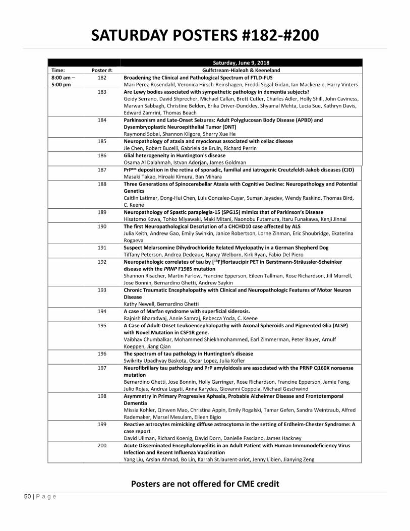
50 | P a g e
SATURDAY POSTERS #182-#200
Saturday, June 9, 2018Time: Poster #: Gulfstream-Hialeah & Keeneland
8:00 am –5:00 pm
182 Broadening the Clinical and Pathological Spectrum of FTLD-FUSMari Perez-Rosendahl, Veronica Hirsch-Reinshagen, Freddi Segal-Gidan, Ian Mackenzie, Harry Vinters
183 Are Lewy bodies associated with sympathetic pathology in dementia subjects?Geidy Serrano, David Shprecher, Michael Callan, Brett Cutler, Charles Adler, Holly Shill, John Caviness,Marwan Sabbagh, Christine Belden, Erika Driver-Dunckley, Shyamal Mehta, Lucia Sue, Kathryn Davis,Edward Zamrini, Thomas Beach
184 Parkinsonism and Late-Onset Seizures: Adult Polyglucosan Body Disease (APBD) andDysembryoplastic Neuroepithelial Tumor (DNT)Raymond Sobel, Shannon Kilgore, Sherry Xue He
185 Neuropathology of ataxia and myoclonus associated with celiac diseaseJie Chen, Robert Bucelli, Gabriela de Bruin, Richard Perrin
186 Glial heterogeneity in Huntington's diseaseOsama Al Dalahmah, Istvan Adorjan, James Goldman
187 PrPres deposition in the retina of sporadic, familial and iatrogenic Creutzfeldt-Jakob diseases (CJD)Masaki Takao, Hiroaki Kimura, Ban Mihara
188 Three Generations of Spinocerebellar Ataxia with Cognitive Decline: Neuropathology and PotentialGeneticsCaitlin Latimer, Dong-Hui Chen, Luis Gonzalez-Cuyar, Suman Jayadev, Wendy Raskind, Thomas Bird,C. Keene
189 Neuropathology of Spastic paraplegia-15 (SPG15) mimics that of Parkinson’s DiseaseHisatomo Kowa, Tohko Miyawaki, Maki Mitani, Naonobu Futamura, Itaru Funakawa, Kenji Jinnai
190 The first Neuropathological Description of a CHCHD10 case affected by ALSJulia Keith, Andrew Gao, Emily Swinkin, Janice Robertson, Lorne Zinman, Eric Shoubridge, EkaterinaRogaeva
191 Suspect Melarsomine Dihydrochloride Related Myelopathy in a German Shepherd DogTiffany Peterson, Andrea Dedeaux, Nancy Welborn, Kirk Ryan, Fabio Del Piero
192 Neuropathologic correlates of tau by [18F]flortaucipir PET in Gerstmann-Sträussler-Scheinkerdisease with the PRNP F198S mutationShannon Risacher, Martin Farlow, Francine Epperson, Eileen Tallman, Rose Richardson, Jill Murrell,Jose Bonnin, Bernardino Ghetti, Andrew Saykin
193 Chronic Traumatic Encephalopathy with Clinical and Neuropathologic Features of Motor NeuronDiseaseKathy Newell, Bernardino Ghetti
194 A case of Marfan syndrome with superficial siderosis.Rajnish Bharadwaj, Annie Samraj, Rebecca Yoda, C. Keene
195 A Case of Adult-Onset Leukoencephalopathy with Axonal Spheroids and Pigmented Glia (ALSP)with Novel Mutation in CSF1R gene.Vaibhav Chumbalkar, Mohammed Shiekhmohammed, Earl Zimmerman, Peter Bauer, ArnulfKoeppen, Jiang Qian
196 The spectrum of tau pathology in Huntington’s diseaseSwikrity Upadhyay Baskota, Oscar Lopez, Julia Kofler
197 Neurofibrillary tau pathology and PrP amyloidosis are associated with the PRNP Q160X nonsensemutationBernardino Ghetti, Jose Bonnin, Holly Garringer, Rose Richardson, Francine Epperson, Jamie Fong,Julio Rojas, Andrea Legati, Anna Karydas, Giovanni Coppola, Michael Geschwind
198 Asymmetry in Primary Progressive Aphasia, Probable Alzheimer Disease and FrontotemporalDementiaMissia Kohler, Qinwen Mao, Christina Appin, Emily Rogalski, Tamar Gefen, Sandra Weintraub, AlfredRademaker, Marsel Mesulam, Eileen Bigio
199 Reactive astrocytes mimicking diffuse astrocytoma in the setting of Erdheim-Chester Syndrome: Acase reportDavid Ullman, Richard Koenig, David Dorn, Danielle Fasciano, James Hackney
200 Acute Disseminated Encephalomyelitis in an Adult Patient with Human Immunodeficiency VirusInfection and Recent Influenza VaccinationYang Liu, Arslan Ahmad, Bo Lin, Karrah St.laurent-ariot, Jenny Libien, Jianying Zeng
Posters are not offered for CME credit

51 | P a g e
SATURDAY POSTERS #201-#218
Saturday, June 9, 2018Time: Poster #: Gulfstream-Hialeah & Keeneland
8:00 am –5:00 pm
201 Neuropathological Findings in a Case of Aicardi–Goutières Syndrome with IFIH-1 MutationAhmed Gilani, Kelley Capocelli, Eric Wartchow, Bette Kleinschmidt-DeMasters
202 Dual Pathology in Rasmussen EncephalitisAhmed Gilani, Eric Prince, Bette Kleinschmidt-DeMasters
203 A case report of presumed adult-onset Rasmussen's encephalitis.Nathan Clement, Matthew Frosch
204 Anti-GABA-A receptor antibody-associated encephalitis in an adult and pediatric case: radiologic,biopsy, and CSF findingsAivi Nguyen, Neena Cherayil, Ana Cristancho, Mariarita Santi, Eric Lancaster, MacLean Nasrallah
205 Behcet’s Disease of the Central Nervous System (CNS)Susan de la Monte, Edward Stopa, Galam Khan, Justin Remer, Katarina Dakay
206 Fatal Granulomatous Amebic Encephalitis In a Heart Transplant Patient: A Correlation BetweenClinical, Radiographic, and Autopsy Findings.William Harrison, Bruce Leckey, Christine Hulette
207 Non-tubercular mycobacterial spinal cord abscesses in an HIV+ male due to M. HaemophilumBette Kleinschmidt-DeMasters, Kellie Hawkins, Carlos Franco-Paredes
208 Congenital rubella syndrome (CRS)--lest we forgetBette Kleinschmidt-DeMasters
209 Pseudorabies Outbreak in Two Catahoula Cur Hunting Dogs in LouisianaTiffany Peterson, Fabio Del Piero
210 Brainbow labeling in human ex vivo brain slices for high-throughput morphological analysisShu-Hsien Sheu, Jonathan Ting, Pichet Adstamongkonkul, Ian Boothby, Leonie Hoyo, Ed Lein, JeffLichtman, David Clapham
211 Trigeminal Amyloidoma: A Report of Two CasesAmy Swanson, Rachael Vaubel, Michael Link, Jamie Van Gompel, John Wald, Ellen McPhail, CaterinaGiannini
212 Validation of THK 5351 PET ligand to detect astrogliosis in vivoShigeo Murayama, Renpei Sengoku, Kenji Ishii, Yuko Saito
213 A novel in-frame BRAF deletion in a case of central nervous system Rosai-Dorfman diseaseTimothy Richardson, Megan Wachsmann, Dwight Oliver, Zahidur Abedin, Dennis Burns, JackRaisanen, Benjamin Greenberg, Kimmo Hatanpaa
214 Histopathology of the Cat Somatosensory Cortex After Chronic Electrical StimulationNishant Tiwari, Ryan Long, Carol Miller, Douglas McCreery
215 Relapsing Multifocal Necrotizing Encephalopathy in a 60-year-old PatientDiana Thomas, Kiruba Dharaneeswaran, Clayton Wiley, Julia Kofler
216 Detecting Astrocytopathy with mAb DE-R-11Diana Thomas, Stephanie Bissel, Geoffrey Murdoch
217 Rare/Unsual Pathologies in Epilepsy Surgical Resections, and Their Implications for GeneticDisease Identification in Some Cases.Meenakshi Bhattacharjee, Gretchen Von Allmen, Manish Shah, David Sandberg, Elliott Friedman,Nitin Tandon
218 A novel FIJI workflow demonstrates dynamic changes in postnatal respiratory nuclei innervationby Nkx2.2- and Olig3-derived neurons.Jillian Liu, Julie Stephens, Catherine Czeisler, Jose Otero
Posters are not offered for CME credit

52 | P a g e
AANPAMERICAN ASSOCIATIONOF NEUROPATHOLOGISTS
Endowed LectureshipsFriday, June 8, 2018
I. Parisi LectureII. DeArmond Lecture
Saturday, June 9, 2018I. Saul R. Korey Lecture
Sunday, June 10, 2018I. Matthew T. Moore Lecture

53 | P a g e
ENDOWED LECTURESHIP
PARISI LECTUREhe Parisi Lecture was established in 2007. The lecture was named the Parisi Lectureship in honor of one of theAmerican Association of Neuropathologists’ exceptional members, Dr. Joseph E. Parisi. He has published seminalneuropathological studies on a wide range of diseases affecting the nervous system, with particular focus on
neurodegenerative diseases and multiple sclerosis. He has held virtually every office of the Society, including President,and has served on several AANP committees. In 2006, his dedication and generosity were recognized with the Award forMeritorious Contributions to Neuropathology. He is considered by many the heart and soul of the association and a manworth emulating.
We are pleased to have Philip L. De Jager join our list of distinguished speakers.
2008 Claudia Lucchinetti The Spectrum of CNS Inflammatory Demyelinating Diseases: FromPathology to Pathogenesis
2009 Hans Lassmann Inflammation Induced Mitochondrial Injury: A Major Mechanism ofNeurodegeneration
2010 Joseph Dalmau Autoimmune Synaptic Encephalitis2011 Steven S. Scherer Molecular Pathologies at the Nodes of Ranvier2012 Bruce D. Trapp Neuronal Damage in Multiple Sclerosis2013 Albee Messing GFAP: Friend or Foe2014 Clayton Wiley Human Parechovirus Encephalitis2015 Bruce T. Lamb The Role of Innate Immunity in Neurodegenerative Disease
Pathogenesis2016 Bette
Kleinschmidt-DeMastersCNS White Matter Disorders with Viral Causation and Association
2017 Sean J. Pittock Autoimmune Gliopathies: A Journey of Discovery2018 Philip L. De Jager The Genomic Architecture of Aging-Related Neuropathologies:
Spotlight on Microglia
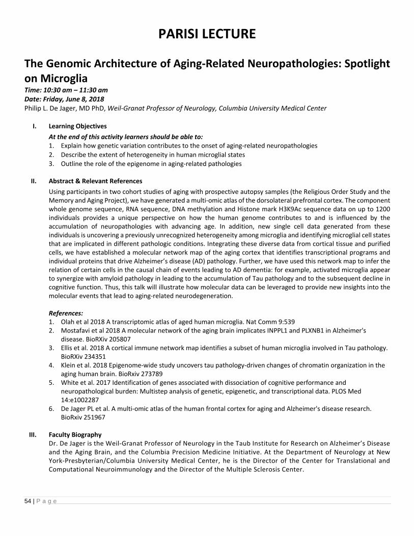
54 | P a g e
PARISI LECTURE
The Genomic Architecture of Aging-Related Neuropathologies: Spotlighton MicrogliaTime: 10:30 am – 11:30 amDate: Friday, June 8, 2018Philip L. De Jager, MD PhD, Weil-Granat Professor of Neurology, Columbia University Medical Center
I. Learning Objectives
At the end of this activity learners should be able to:1. Explain how genetic variation contributes to the onset of aging-related neuropathologies
2. Describe the extent of heterogeneity in human microglial states
3. Outline the role of the epigenome in aging-related pathologies
II. Abstract & Relevant References
Using participants in two cohort studies of aging with prospective autopsy samples (the Religious Order Study and theMemory and Aging Project), we have generated a multi-omic atlas of the dorsolateral prefrontal cortex. The componentwhole genome sequence, RNA sequence, DNA methylation and Histone mark H3K9Ac sequence data on up to 1200individuals provides a unique perspective on how the human genome contributes to and is influenced by theaccumulation of neuropathologies with advancing age. In addition, new single cell data generated from theseindividuals is uncovering a previously unrecognized heterogeneity among microglia and identifying microglial cell statesthat are implicated in different pathologic conditions. Integrating these diverse data from cortical tissue and purifiedcells, we have established a molecular network map of the aging cortex that identifies transcriptional programs andindividual proteins that drive Alzheimer’s disease (AD) pathology. Further, we have used this network map to infer therelation of certain cells in the causal chain of events leading to AD dementia: for example, activated microglia appearto synergize with amyloid pathology in leading to the accumulation of Tau pathology and to the subsequent decline incognitive function. Thus, this talk will illustrate how molecular data can be leveraged to provide new insights into themolecular events that lead to aging-related neurodegeneration.
References:1. Olah et al 2018 A transcriptomic atlas of aged human microglia. Nat Comm 9:5392. Mostafavi et al 2018 A molecular network of the aging brain implicates INPPL1 and PLXNB1 in Alzheimer's
disease. BioRXiv 2058073. Ellis et al. 2018 A cortical immune network map identifies a subset of human microglia involved in Tau pathology.
BioRXiv 2343514. Klein et al. 2018 Epigenome-wide study uncovers tau pathology-driven changes of chromatin organization in the
aging human brain. BioRxiv 2737895. White et al. 2017 Identification of genes associated with dissociation of cognitive performance and
neuropathological burden: Multistep analysis of genetic, epigenetic, and transcriptional data. PLOS Med14:e1002287
6. De Jager PL et al. A multi-omic atlas of the human frontal cortex for aging and Alzheimer's disease research.BioRxiv 251967
III. Faculty BiographyDr. De Jager is the Weil-Granat Professor of Neurology in the Taub Institute for Research on Alzheimer’s Diseaseand the Aging Brain, and the Columbia Precision Medicine Initiative. At the Department of Neurology at NewYork-Presbyterian/Columbia University Medical Center, he is the Director of the Center for Translational andComputational Neuroimmunology and the Director of the Multiple Sclerosis Center.

55 | P a g e
“The goal of my work as a clinician-scientist is to apply modern methods of human immunology, statisticalgenetics, epigenomics and systems biology to the understanding of common neurodegenerative diseases,” saysDr. De Jager, who also serves as Chief of the Division of Neuroimmunology, which focuses on characterizing andtargeting the neuroimmunologic component of neurodegenerative diseases.
“Our new division seeks to provide innovative, compassionate care to patients with immune dysfunction thattargets the brain and spinal cord,” says Dr. De Jager. “We also lead transformative, rigorous human researchstudies to first understand and then to target the role of the immune system in neurodegenerative diseases suchas ALS, Alzheimer’s, Parkinson’s, and multiple sclerosis.”
After graduating from Yale University with a degree in French literature and Molecular Biophysics andBiochemistry, he received a PhD in neurogenetics from The Rockefeller University and his medical degree fromWeill Cornell Medicine, followed by an MMSc in clinical investigation at Harvard Medical School and MIT. He thenpursued a residency in neurology and additional training in clinical neuroimmunology at the MassachusettsGeneral Hospital and Brigham and Women’s Hospital. Prior to joining Columbia, Dr. De Jager was on the facultyof Harvard Medical School.

56 | P a g e
ENDOWED LECTURESHIP
DEARMOND LECTURE
he DeArmond Lecture was established in recognition of Stephen J. DeArmond’s excellent leadership andorganization of the scientific program for the 2006 International Congress of Neuropathology. This successfulmeeting garnered significant support intended for the future advancement of the mission of the American
Association of Neuropathologists. To continue these intended goals and recognize Dr. DeArmond’s contributions, theAmerican Association of Neuropathologists has honored him by establishing the DeArmond Lecture. Dr. DeArmond is aleading authority on prion disease, where his work has been fundamental in demonstrating mechanisms of transmissionand routes to therapeutics. The DeArmond Lecture focuses on honoring those making major advances in the field ofneurodegeneration and aging with a particular emphasis on translating these findings to patient care.
We are pleased to have Suzanne M. de la Monte join our list of distinguished speakers.
2008 Virginia M.Y. Lee TDP-43, A New Class of Proteinopathies in NeurodegenerativeDiseases
2009 Rudy Tanzi Decoding Alzheimer’s Disease Gene by Gene2010 Todd Golde Alzheimer’s Disease: Models and Therapeutics2011 Beverley L. Davidson Emerging Therapies for Neurogenetic Diseases2012 Krystof Bankiewicz New Therapies for Parkinson Disease2013 Stanley Prusiner A Unifying Role for Prions in Neurodegenerative Diseases2014 Dale Bredesen Prionic Loops, Dependence Receptors, and a New Approach to
Alzheimer’s Disease2015 William W. Seeley Frontotemporal Dementia: Onset and Spread2016 Eric J. Huang FTD and ALS: Genes, Circuits and Therapeutic Targets2017 David C. Van Essen Structure, Function, Connectivity, Development and Evolution
of Human Cerebral Cortex2018 Suzanne M. de la
MonteDysregulated Metabolism in the Pathogenesis of Alzheimer’sDisease: Type 3 Diabetes

57 | P a g e
DEARMOND LECTURE
Dysregulated Metabolism in the Pathogenesis of Alzheimer’s Disease:Type 3 DiabetesTime: 4:45 pm – 5:45 pmDate: Friday, June 8, 2018Suzanne M. de la Monte, MD, MPH, Warren Alpert Medical School of Brown University, Providence, RI
I. Learning Objectives
At the end of this activity learners should be able to:
1. Summarize evidence demonstrating links between impaired energy metabolism in the brain andneurodegeneration, particularly Alzheimer’s disease.
2. Explain how insulin resistance and other diabetes-type effects may contribute to major brain pathologies inAD.
3. Consider potential cofactor roles of systemic diseases in the pathogenesis of AD and other neurodegenerativediseases.
II. Abstract & Relevant References
The metabolic basis of AD is fundamentally linked to insulin resistance and deficiency in the brain. In AD,dysregulation of glucose and lipid metabolism, impairments in insulin signaling, activation of pro-inflammatorycytokines, and compromises to mitochondrial function are all reminiscent of abnormalities that occur in systemicinsulin resistance diseases including, diabetes mellitus (DM), non-alcoholic fatty liver disease (NAFLD), obesity,and metabolic syndrome (MetS). Over the past 12 years, many of AD’s salient neuropathological features,including amyloid-beta (Aβ42) accumulations, tau hyperphosphorylation, impairments in neuronal plasticity, and cholinergic deficits have been correlated with perturbations in brain insulin and insulin-like growth factorsignaling. Moreover, experimental data show that brain insulin resistance compromises oligodendrocyte survivaland myelin maintenance, providing a mechanism for leukoaraiosis and white matter atrophy in AD. Similarly,microvascular disease, a universal consequence of insulin resistance, is associated with brain hypo-perfusion,which likely exacerbates concurrent effects of neurodegeneration, and contributes to break-down of the blood-brain barrier due to disruption of glial-vascular networks. The findings that DM, obesity, NAFLD, and MetSincrease risk for cognitive impairment and brain insulin resistance suggest that systemic insulin resistance diseasesmay promote neurodegeneration. Finally, recent evidence suggests that in sporadic AD unrelated to DM, MetS,NAFLD, or obesity, metabolic hormones and inflammatory indices are simultaneously altered in blood and CSF inthe early stages of disease.
References:
1. de la Monte SM. Insulin Resistance and Neurodegeneration: Progress Towards the Development of NewTherapeutics for Alzheimer's Disease. Drugs. 2017;77(1):47-65.
2. Steen E, Terry BM, Rivera EJ, Cannon JL, Neely TR, Tavares R, et al. Impaired insulin and insulin-like growthfactor expression and signaling mechanisms in Alzheimer's disease--is this type 3 diabetes? J Alzheimers Dis.2005;7(1):63-80.
3. Tong M, Dominguez C, Didsbury J, de la Monte SM. Targeting Alzheimer's Disease Neuro-MetabolicDysfunction with a Small Molecule Nuclear Receptor Agonist (T3D-959) Reverses Disease Pathologies. JAlzheimers Dis Parkinsonism. 2016;6(3):238-44.
4. Reger MA, Watson GS, Frey WH, 2nd, Baker LD, Cholerton B, Keeling ML, et al. Effects of intranasal insulinon cognition in memory-impaired older adults: modulation by APOE genotype. Neurobiol Aging.2006;27(3):451-8.

58 | P a g e
5. de la Monte SM, Wands JR. Alzheimer's disease is type 3 diabetes-evidence reviewed. J Diabetes Sci Technol.2008;2(6):1101-13.
6. Claxton A, Baker LD, Hanson A, Trittschuh EH, Cholerton B, Morgan A, et al. Long-acting intranasal insulindetemir improves cognition for adults with mild cognitive impairment or early-stage Alzheimer's diseasedementia. J Alzheimers Dis. 2015;44(3):897-906.
7. Freiherr J, Hallschmid M, Frey WH, 2nd, Brunner YF, Chapman CD, Holscher C, et al. Intranasal insulin as atreatment for Alzheimer's disease: a review of basic research and clinical evidence. CNS Drugs.2013;27(7):505-14.
8. Duffy AM, Holscher C. The incretin analogue D-Ala2GIP reduces plaque load, astrogliosis and oxidative stressin an APP/PS1 mouse model of Alzheimer's disease. Neuroscience. 2013;228:294-300.
9. Beydoun MA, Beydoun HA, Gamaldo AA, Teel A, Zonderman AB, Wang Y. Epidemiologic studies ofmodifiable factors associated with cognition and dementia: systematic review and meta-analysis. BMCPublic Health. 2014;14:643.
10. Wallin A, Nordlund A, Jonsson M, Blennow K, Zetterberg H, Ohrfelt A, Stalhammar J, Eckerstrom M, CarlssonM, Olsson E, Gothlin M, Svensson J, Rolstad S, Eckerstrom C, Bjerke M: Alzheimer's disease--subcorticalvascular disease spectrum in a hospital-based setting: Overview of results from the Gothenburg MCI anddementia studies. J Cereb Blood Flow Metab 2016, 36:95-113
11. Grammas P: Neurovascular dysfunction, inflammation and endothelial activation: implications for thepathogenesis of Alzheimer's disease. J Neuroinflammation 2011, 8:26.
12. Talbot K, Wang HY, Kazi H, Han LY, Bakshi KP, Stucky A, Fuino RL, Kawaguchi KR, Samoyedny AJ, Wilson RS,Arvanitakis Z, Schneider JA, Wolf BA, Bennett DA, Trojanowski JQ, Arnold SE: Demonstrated brain insulinresistance in Alzheimer's disease patients is associated with IGF-1 resistance, IRS-1 dysregulation, andcognitive decline. J Clin Invest 2012, 122:1316-38
III. Faculty Biography
Suzanne de la Monte, MD MPH is a Professor in Pathology and Laboratory Medicine, Neurology, & Neurosurgeryat the Alpert Medical School of Brown University and Rhode Island Hospital. She obtained an A.B. from CornellUniversity, M.D. from the Weill Medical College of Cornell University, and MPH from the Blumberg School of PublicHealth at Johns Hopkins. She received residency training in Anatomical and Pediatric Pathology at Hopkins andfellowship training in Neuropathology at the Massachusetts General Hospital. Dr. de la Monte launched her careeras a physician-scientist while on the faculty at MGH and Harvard Medical School. In 2000, she joined the facultyat Rhode Island Hospital and Brown where she conducts basic, translational, and clinical research on mechanismsand consequences of brain insulin resistance and metabolic dysfunction, which she refers to as ‘brain diabetes’.Her lab also investigates how various lifestyle exposures cause neurodegeneration associated with brain insulinresistance, and evaluates novel approaches for therapeutic targeting of brain metabolic defects. Dr. de la Monte’sresearch is funded by the NIH. She is the recipient of a MERIT award from the NIAAA and has published over 300peer-reviewed articles. Extra-curricular interests include golf, spin biking, theater, classical music, and freestylePaleo cooking.

59 | P a g e
ENDOWED LECTURESHIP
SAUL R. KOREY LECTUREhe Korey Lecture was established by Dr. Robert D. Terry in honor of Dr. Saul R. Korey, the founder and first Chair ofthe Department of Neurology at Albert Einstein College of Medicine. Dr. Korey’s vision of an interdisciplinaryapproach to the study of neurological diseases by basic and clinical scientists has inspired generations of colleagues
and trainees. Dr. Terry, a close collaborator and colleague of Dr. Korey, was the first recipient of the prestigious PotamkinPrize for Pick’s and Alzheimer’s Disease in 1988, in recognition of his seminal observations of the pathological changes inAlzheimer’s disease. Dr. Terry generously contributed a portion of the prize funds to endow the Korey Lectureship, to beadministered by the American Association of Neuropathologists, with the lecturer to be chosen annually by the Presidentin conjunction with the Nominating Committee and the Chair of the Program Committee. Dr. Terry has summarized thequalities of the Korey Lecturer as someone who has “…been an active member of the Association…a working MD orMD/PhD neuropathologist…responsible for diagnostic work as well as teaching and research. The lecture should be aimedat the members of the Association, and the lecturer might well serve as a role model for younger members.”
We are pleased to have Rebecca D. Folkerth join our list of distinguished speakers.
Year Lecturer Title1989 Nicholas K. Gonatas MG-60, a Novel Sialoglycoprotein of Medial Cisternae of the Neuronal
Golgi Apparatus: Implications and Applications1990 Henry M. Wisniewski Amyloidosis in Alzheimer’s Disease and the Spongiform
Encephalopathies1991 Robert D. Terry Alzheimer’s Disease as Seen by a Lucky Morphologist1992 Henry de Forest
WebsterFormation and Regeneration of Myelin
1993 Kunihiko Suzuki Molecular Genetics of Tay-Sachs and Related Disorders: The Legacy ofSaul Korey
1994 No Lecture XIIth International Congress (Toronto)1995 Blas Frangione Amyloid Genes and Chaperones in Alzheimer Disease1996 Floyd Gilles The 3R’s of Neuro-oncology – Recording, Reliability and Reporting1997 Donald L. Price The Role of Neuropathologists in the Analyses of Models of
Neurodegenerative Disease1998 Sandra H. Bigner Molecular Genetics of Medulloblastoma1999 William F. Hickey Key Participants in the Initiation of Inflammation in the Central
Nervous System2000 Mary E. Case Neuropathology and Forensic Pathology: A Natural Synergism2001 Paul H. Kleihues Molecular Biology of Brain Tumors2002 James E. Goldman Astrocytes, Intermediate Filaments, Cellular Stress and
Neuropathology2003 Samuel K. Ludwin Pathology and Pathogenesis in Multiple Sclerosis2004 James M. Powers The Road Not Taken2005 Bernardino Ghetti Deciphering Hereditary Presenile Dementias: Neuropathology at the
Crossroads of Neuropsychiatry and Molecular Genetics2006 Donna M. Ferriero Molecular Mechanisms of Hypoxic-Ischemic Injury in the Developing
Nervous System2007 Dennis W. Dickson Neuropathology and Genetics of Parkinsonism2008 David N. Louis Brain Tumor Classification: Little Steps and Big Jumps

60 | P a g e
2009 Stephen J. DeArmond Mechanisms of Neurodegeneration in Prion Disease Originating fromthe Neuronal Plasma Membrane
2010 Peter C. Burger A Long-Term Perspective on Pediatric CNS Tumors2011 Hans H. Goebel Protein Aggregate Myopathies2012 Michael Norenberg Astrocyte Pathobiology2013 Harry Vinters Gain and Pain from Cerebral Microvessels – Adventures in Vascular
Neuropathology2014 Thomas J. Montine Alzheimer’s Disease and Related Dementias2015 Matthew Frosch Working at the Crossroads of Neurodegeneration and
Cerebrovascular Disease2016 Eileen H. Bigio The FTLD-ALS Connection2017 Eliezer Masliah Disease Modifying Therapeutical Approaches for Synucleinopathies of
the Aging Population2018 Rebecca D. Folkerth Reading Tea Leaves: Patterns of Injury in the Pediatric Nervous System

61 | P a g e
SAUL R. KOREY LECTURE
Reading Tea Leaves: Patterns of Injury in the Pediatric Nervous SystemTime: 10:30 am – 11:30 amDate: Saturday, June 9, 2018Rebecca D. Folkerth, MD, Neuropathologist, New York City Office of the Chief Medical Examiner, Clinical AssociateProfessor of Forensic Medicine, NYU School of Medicine
I. Learning Objectives
At the end of this activity learners should be able to:1. Identify constellations of general autopsy and nervous system findings consistent with inflicted injury in children2. Describe how to use nervous system autopsy prosection techniques to obtain diagnostically important specimens
for complete case analysis3. Identify the “gray zones” between traumatic injury and other causes of nervous system abnormalities in children4. Explain pitfalls in interpretation, documentation, and testimony in pediatric medicolegal cases
II. Abstract & Relevant References
Child abuse is, sadly, a fixed feature of the human condition, having been described through the ages and acrossall cultures. The nervous system is frequently affected, both directly by the mechanical injury itself, and indirectlyby supervening hypoxia/ischemia, the latter related to several factors, including resuscitation. Thus,neuropathologists must discern the subtle and overlapping patterns of these direct and indirect insults, as well asthe features of underlying natural disease. A solid understanding of the range of non-traumatic neuropathologyin the immature brain is therefore essential.Bringing to bear her longtime interest and experience in study of the developing brain, Dr. Folkerth will reviewthe elements of specimen analysis most helpful in child abuse cases. She will also emphasize the importance ofcontext, collaboration, and intellectual honesty in making observations and interpretations that may eventuallysurface in the courtroom setting.
References:
1. Dolinak D, Reichard R. An Overview of Inflicted Head Injury in Infants and Young Children, With aReview of B-amyloid Precursor Protein Immunohistochemistry. Arch Pathol Lab Med 2006;130(5):712-7
2. Gill JR, Goldfeder LB, Armbrustmacher V, Coleman A, Mena H, Hirsch CS. Fatal Head Injury in ChildrenYounger Than 2 Years in New York City and an Overview of the Shaken Baby Syndrome. Arch PatholLab Med 2009; 133:619-27
3. Forbes BJ, Levin AV. Chapter 17. Abusive Head Trauma/Shaken Baby Syndrome. In: Reynolds J andOlitsky S, eds. Pediatric Retina (Berlin and Heidelberg, Springer-Verlag), 2010: 409-22
4. Matshes EW, Evans RM, Pinckard JK, Joseph JT, Lew EO. Shaken Infants Die of Neck Trauma, Not ofBrain Trauma. Acad Forensic Pathol 2011;1(1):82-91
5. Johnson MW, Stoll L, Rubio A, Troncoso J, Pletnikova O, Fowler DR, Li L. Axonal Injury in YoungPediatric Head Trauma: A Comparison Study of B-amyloid Precursor Protein (B-APP)Immunohistochemical Staining in Traumatic and Nontraumatic Deaths. J Forensic Sci.2011;56(5):1198-1205
6. Gill JR, Andrew T, Gilliland MGF, Love J, Matshes E, Reichard RR. National Association of MedicalExaminers Position Paper: Recommendations for the Postmortem Assessment of Suspected HeadTrauma in Infants and Young Children. Acad Forensic Pathol 2013;4(2):206-13
7. Snyder VS, Hansen LA. A Conceptual Overview of Axonopathy in Infants and Children with AllegedlyInflicted Head Trauma. Acad Forensic Pathol 2016;6(4):608-21
62 | P a g e
8. Folkerth RD, Nunez J, Georgievskaya Z, McGuone D. Neuropathologic Examination in SuddenUnexpected Deaths in Infancy and Childhood: Recommendations for Highest Diagnostic Yield andCost-Effectiveness in Forensic Settings. Acad Forensic Pathol 2017;7(2):182-99
III. Faculty Biography
Rebecca D. Folkerth, MD, is a Neuropathologist with wide diagnostic experience in the brain abnormalities ofinfants and children. Prior to joining the New York City Office of the Chief Medical Examiner as the agency’sNeuropathologist in 2016, Dr. Folkerth was Director of Neuropathology at Brigham and Women’s Hospital,affiliated with Harvard Medical School, in Boston. For nearly 3 decades, she also served as a Consultant inNeuropathology at Boston Children’s Hospital, participating with her mentor, Dr. Hannah C. Kinney, in a long-running NIH-funded program project analyzing the cellular basis of brain damage in prematurely born infants. Shealso was a member of an NIH-supported international investigation into maternal alcohol exposure and other keyfactors in the risk of sudden infant death and stillbirth, results of which are being compiled and publishedcurrently.
More recently, Dr. Folkerth has been part of a national consortium, also NIH-funded, studying traumatic braininjury. She serves as part of the working group that has developed the consensus criteria for the neuropathologicdiagnosis of chronic traumatic encephalopathy (CTE). Her interests since joining the OCME include theneuropathology of abusive head trauma, as well as in long-term survivors of traumatic brain injury; brainabnormalities in individuals with cerebral palsy; and developmental and acquired lesions in sudden unexpecteddeath in infancy and childhood (SUID/SUDC), and in sudden unexpected death in epilepsy (SUDEP).

63 | P a g e
ENDOWED LECTURESHIP
MATTHEW T. MOORE LECTURE
n 1970, Dr. Matthew T. Moore made a contribution to the AANP to establish the Moore Award, which is given annuallyto recognize the “Best Paper on Clinico-Pathological Correlation Presented at the Annual Meeting.” In 1987, RechelleFishman, a former patient of Dr. Moore, bequeathed $75,000 to the Moore Award Fund. Dr. Moore requested thatthis bequest be used to establish a “Rachelle Fishman-Matthew Moore Distinguished Lectureship” (later shortened
to just the “Moore Lectureship”), which is “to be given by a distinguished lecturer, on a subject which represents theleading edge of advanced research in neuropathological subjects of contemporary interest. The lecture is to take placeon the day of the Presidential Address.” In 1988, it was decided that this Lectureship would replace the “DistinguishedLectureship” that had been sponsored each year by the Association. The Moore Lecturer is selected annually by thePresident in conjunction with the Nominating Committee and the Chair of the Program Committee.
We are pleased to have Mario L. Suvà join our list of distinguished speakers.
Year Lecturer Title
1990 Robert H. Horvitz The Genetic Control of GABAergic and Serotonergic Neuronal Differentiation andof Programmed and Pathological Cell Death in a Nematode Nervous System
1991 Charles Janeway Induction, Mediation and Continuation of Immune Responses
1991 Ramzi S. Contran Cytokine-Endothelial Interactions in inflammation, Immunity and Vascular Injury
1992 D. Carleton Gajdusek The genetic Control of Spontaneous Generation of Infectious Amyloids: Kuru-CJD-GSS-Scrapie-BSE
1995 Leroy Hood Deciphering the Human Genome: Implications for Medicine of the 21st Century
1996 Martin Raff Programmed Cell Death--Mechanisms and Social Controls
1998 James Eberwine Single Cell Molecular Neuropathology
1999 Richard T. Johnson Viral Pathogenesis, an Overview
2000 Seymour Benzer The Neuropathology of Drosophila
2001 Dennis Choi Ischemia-Induced Perturbations in Neuronal Ionic Homeostasis
2002 J. William Langston, MPTP: Its impact on Parkinson's Disease Research
2003 Carolyn C. Meltzer Future of PET in the Study of Neurological Disease
2004 Henry L. Paulson Toward Understanding the Pathogenesis of Repeat Expansion Diseases
2005 Peter St. George Hyslop Molecular Genetics and Biology of Alzheimer Disease Generate Clues forTherapeutics
2006 Keith L. Ligon Stem and Progenitor Cell Insights into Gliomas: Novel Origins, Markers and Targets
2008 William Mobley Trafficking Trophic Signals to Prevent Neurodegeneration
2009 Donald W. Cleveland From Charcot to Lou Gehrig: Mechanisms and Treatment of ALS
2011 Mark Gilbert RTOG: Clinical Trials and the Increasing Role of Neuropathology
2012 Kevin P. Campbell Mechanistic and Molecular Insights into the Pathogenesis of Glycosylation –Deficient Muscular Dystrophy
2013 Bradley Hyman How does Alzheimer’s Disease know Neuroanatomy?
2014 David N. Louis WHO’s Next? Guidelines for the Next WHO Classification of Brain Tumors
2015 Eric C. Holland Brain Tumors in Mouse and Man
2016 Ted M. Dawson Unlocking the Secrets of Parkinson’s
2017 M. Beatriz S. Lopes An Update of the WHO Classification of Tumors of the Pituitary Gland, 4th Edition2018 Mario L. Suvà Deciphering Single-Cell Regulatory Programs in Adult and Pediatric Gliomas

64 | P a g e
MATTHEW T. MOORE LECTURE
Deciphering Single-Cell Regulatory Programs in Adult and PediatricGliomasTime: 8:30 am – 9:15 pmDate: Sunday, June 10, 2018Mario L. Suvà, MD, PhD, Assistant Professor of Pathology, Massachusetts General Hospital; Associate Member, The BroadInstitute of MIT and Harvard; Co-Director, Cancer Program, Harvard Stem Cell Institute
I. Learning Objectives
At the end of this activity learners should be able to:1. Identify new methods to profile of single-cells.2. Discuss research findings related to single-cell genomic studies in human gliomas.3. Discuss the cancer stem cell model in gliomas.
II. Abstract & Relevant References
Tumor heterogeneity is a major barrier to therapy. Three dominant factors govern tumor heterogeneity: (I)genetic alterations shape cancer cells programs and their evolution; (II) developmental programs, such asprograms of adult stem cell and their differentiation into specialized cell types, ascribe cancer cells with specificfunctional properties; (III) the composition of the tumor micro-environment and its interaction with malignantcells provides further heterogeneity to the tumor ecosystem. In this lecture, we dissect these influences in humandiffuse gliomas studying patient samples with single-cell genomic technologies. We discuss comprehensive single-cell data that help redefine malignant cell lineages, cancer stem cell programs, genetic heterogeneity, and dissectthe transcriptional programs of microglia, macrophages, and T cell in clinical gliomas. We will also review thetoolbox available in the field of single-cell genomics and the strengths and limitations of different protocols.Overall, we will provide technological insights in the field of single-cell genomics and deep characterization ofgliomas biology, with important implications for their management.
References:1. Patel et al, Science 20142. Tirosh et al, Nature 20163. Venteicher et al, Science 20174. Filbin et al, Science 20185. Suvà et al, Cell 2014
III. Faculty BiographyMario Suvà is a scientist in the Department of Pathology and the Center for Cancer Research at MassachusettsGeneral Hospital (MGH) and at the Broad Institute. Suvà’s expertise is in neuro-oncology and single-cell genomics.Suvà's laboratory focuses on diffuse gliomas in adults and children. A particular effort of the laboratory is ondissecting the heterogeneity of patient tumors and relating transcriptional and genetic programs of individualcancer cells. Suvà directed pioneering studies characterizing glioblastoma, oligodendroglioma, astrocytoma andpediatric gliomas with single-cell genomic technologies, shedding light on tumor heterogeneity, tumorclassification, glioma cell lineages, cancer stem cell programs, tumor evolution and the composition of the tumormicroenvironment.
Dr. Suvà obtained his M.D and Ph.D. in Lausanne, Switzerland, studying cancer stem cells in gliomas and sarcomas.He did his post-doctoral research at MGH with Brad Bernstein and David Louis, applying chromatin analysis andfunctional approaches to identify master regulators of glioma stem cell programs.

65 | P a g e
AANPAMERICAN ASSOCIATIONOF NEUROPATHOLOGISTS
Award for MeritoriousContributions to Neuropathology
Friday, June 8, 201811:30 am – 11:45 amHonoring Hannah C. Kinney, MDPresented by: Rebecca D. Folkerth, MD
Saturday, June 9, 201811:30 am – 11:45 amHonoring Brian N. Harding, MA, DPhil, BM, BCh, FRCPathPresented by: David Ellison, MD, PhD

66 | P a g e
AWARD FOR MERITORIOUS CONTRIBUTIONS TONEUROPATHOLOGY
he Award for Meritorious Contributions to Neuropathology recognizes a member who has made significantcontributions to the advancement of knowledge in neuropathology and provided service to the American Associationof Neuropathologists. Each recipient of the award is nominated by the President, in conjunction with the NominatingCommittee and with the approval of the Executive Council. The qualities of outstanding scientific achievement and
service are embodied in this year’s recipients, Dr. Hannah C. Kinney and Dr. Brian N. Harding. They join the rich roster ofdistinguished former award recipients.
Year Recipient1999 Lucy B. Rorke2000 William R. Markesbery2001 John J. Kepes
Henry de Forest Webster2002 Dikran S. Horoupian
Fusahiro IkutaKurt A. Jellinger
2003 Bernardino F. Ghetti2004 Michael N. Hart2005 E. Tessa Hedley-Whyte
Suzanne S. Mirra2006 Joseph E. Parisi
Jeannette J. Townsend2007 James M. Powers
Cedric S. Raine2008 Kinuko Suzuki
Margaret G. Norman2009 Peter C. Burger
Pierluigi GambettiNicholas K. Gonatas
2010
2011
2012
2013
2014
2015
2016
2017
Stephen J. DeArmondSamuel K. LudwinWilliam W. SchlaepferLeroy R. SharerBernd W. ScheithauerDonald L. PriceReid HeffnerDawna ArmstrongFloyd GillesFrançoise GrayJohn Q. TrojanowskiBette K. Kleinschmidt-DeMastersDennis W. DicksonBarbara J. CrainRonald C. KimHarry V. Vinters
2018 Hannah C. KinneyBrian N. Harding
Year Recipient1959 Armando Ferraro
Arthur Weil1960 Joseph H. Globus
George B. Hassin1968 Abner Wolf
Paul I. YakovlevHarry M. Zimmerman
1970 Webb E. Haymaker1971 James W. Kernohan1972 George A. Jervis1979 Raymond D. Adams
David CowenMatthew T. Moore
1981 Richard Lindenberg1983 Orville T. Bailey1984 Margaret Murray1985 Kenneth M. Earle
Nathan MalamudLeon Roizin
1986 Martin G. Netsky1987 No Award Presented1988 Edward P. Richardson, Jr.
F. Stephen Vogel1989 Lucien J. Rubinstein
Robert D. Terry1991 Lysia K. S. Forno1992 John Moossy
Gabriele M. ZuRhein1993 Peter W. Lampert
Elias E. Manuelidis1994
1995
19971998
Murray B. BornsteinSamuel P. HicksLowell W. LaphamAmico BignamiAsao HiranoHenryk M. WisniewskiRichard L. DavisWolfgang Zeman

67 | P a g e
AWARD FOR MERITORIOUS CONTRIBUTIONS TONEUROPATHOLOGY
Biography: Hannah C. Kinney, MD
Dr. Kinney is Emeritus (2018) Professor of Pathology at Harvard Medical School, Boston, MA, anda pediatric neuropathologist at Children’s Hospital Boston. Her laboratory performs research indevelopmental disorders of the human brain in early life, with a focus upon brain disordersleading to sudden death in fetuses (stillbirth), infants, and children. Her research includes studiesof the brainstem and hippocampus in the sudden infant death syndrome (SIDS), the hippocampusand temporal lobe in sudden unexplained death in childhood (SUDC), and the cerebral cortex,white matter (periventricular leukomalacia), thalamus, and brainstem in premature infants,among other disorders of the developing human brain.
In 1970 Dr. Kinney graduated from Duke University, Durham, NC, with a BA degree (major inhistory), and from Case Western Reserve University School of Medicine, Cleveland, OH, in 1974.
She subsequently performed a pediatric residency at the Children’s Hospital of Philadelphia, PA, and an anatomic pathology residencyat Duke University Medical School, Durham, NC. Dr. Kinney is board-certified in pediatrics, anatomic pathology, and neuropathology.She came to Children’s Hospital Boston and Harvard Medical School, Boston, MA, for specialized training in pediatric neuropathologyand developmental neuroscience in 1981, and subsequently rose through the academic ranks to Professor of Pathology in 2003.
In her career, Dr. Kinney served as the Director of the Christopher James Murphy SIDS Laboratory and oversaw research into SIDSbrainstems in search of underlying neural mechanisms of sudden death, as well as potential biomarkers of brainstem pathology inliving infants at risk for SIDS. She is served as the Director of a NICHD-funded program project to study the role of the brainstem inSIDS though integrated and multidisciplinary studies of human brainstem pathology and models. This program project involved ~50investigators and post-graduate level students at Harvard, Dartmouth, University of Iowa, Rady Children’s Hospital, San Diego, CA, andthe medical examiner’s system of San Diego County. Dr. Kinney served as the Director of the Developmental Biology and PathologyCenter of the Safe Passage Study which oversees alcohol-related research in the developing human brain and placenta. ThisNICHD/NIAAA-funded study addressed the question of the potential links among prenatal exposure to maternal drinking and smoking,SIDS, stillbirth, and fetal alcohol spectrum disorders (FASD) in a prospective analysis of 12,000 pregnancies, with infants followed tothe end of the first postnatal year, the period of SIDS risk. In the last five years of her career, Dr. Kinney served as the Director andthen Associate Director of the comprehensive Program in Sudden and Unexpected Death in Pediatrics (SUDP) that she co-foundedwith the pediatrician and palliative care physician, Dr. Richard D. Goldstein at Boston Children’s Hospital in 2012. This programprovides comprehensive care and pathology case review to families who have lost an infant under 3 years to sudden and unexpecteddeath due to natural causes.
Dr. Kinney’s publication record includes 148 original articles, 37 reviews, 18 chapters, and 4 tributes. She has received multiple awardsfor her contributions to SIDS and developmental brain research, as well as invitations for honorary SIDS lectureships. Her awardsinclude a MERIT Award from the NICHD (2000-2010), and Moore Award (1987) and Weil Award (Honorable Mention, 1986) from theAmerican Association of Neuropathologists, Award for Excellence in Giving, CJ Foundation for SIDS (2014), Award for “SIDS Researcherof the Year”, American SIDS Institute, (2014), and Distinguished Researcher, International Society of Perinatal and Infant Deaths forOutstanding Contributions to SIDS Research (2016). The SIDS Symposium at the 50th Annual Congress of the Federation of SouthAfrican Societies of Pathology (FSASP) in Somerset West, South Africa, in September 2010, was dedicated to her. Dr. Kinney hasmentored many medical students, neuropathology residents and doctoral and post-doctoral fellows in developmental neuropathologyand neuroscience over her professional career.

68 | P a g e
Dr. Hannah C. Kinney “Contributions to the Field”, written by Rebecca D. Folkerth, MD
Dr. Hannah Chase Kinney, best known as the originator of the Serotonin Hypothesis in the Sudden Infant Death Syndrome (SIDS), wasborn in Cleveland, Ohio, and went to college at Duke University, majoring in History. She then returned to Cleveland to the CaseWestern Reserve University School of Medicine, graduating in 1974. Following a full residency in Pediatrics at the Children’s Hospitalof Philadelphia, she returned to Duke for Anatomic Pathology and Neuropathology training, the latter with Dr. Peter Burger. She cameto Children’s Hospital in Boston in 1981, for additional clinical training with Dr. Floyd Gilles, and research with Dr. Richard Sidman.Since her appointment to the faculty at Children’s and Harvard Medical School in 1984, her trajectory has been one of continuedexcellence in the field of developmental neuropathology, having been continuously funded by the NIH. She was promoted to Professorof Pathology in 2003, in recognition of her furtherance of the field of research into SIDS and its biologic underpinnings directly in thehuman brain, as well as for her role in training a generation of neuroscientists and neuropathologists. In 2012, she spearheaded thecreation of Robert’s Program at Children’s Hospital Boston, for comprehensive clinical, pathologic, and genetic investigation of suddenunexpected death in pediatrics.
Additional interests of Professor Kinney include the neuroanatomic substrates of coma and persistent vegetative state; the spectrumof gray and white matter abnormalities in preterm infants; neuropathology of stillbirth; genomics and proteomics of developmentalbrain disease; and most recently, imaging-neuropathologic correlation in pursuit of in vivo biomarkers for SIDS and hippocampaldysgenesis.
Professor Kinney has been a dedicated member of the Association since 1983, serving on the Awards Committee, including 2 years asits Chair, and on the Professional Affairs Committee, and as an AANP Councilor to the International Society of Neuropathology. Shehas presented a great deal of her research at the Annual Meeting, and her laboratory has received the Moore Award (5 times, of which4 were honorable mentions), and the Weil Award (2 honorable mentions). Hannah has been an editorial board member and reviewerfor the Journal of Neuropathology and Experimental Neurology her entire career. In fact, she has no fewer than 28 original articlesand 3 review articles published in the Journal! Nearly every one of these papers involved trainees, generously welcomed to her lab atall levels, from college students on summer break, to medical students, to post-docs in PhD and MD programs. Hannah is a real “piedpiper” in the sense of getting individuals to follow her into our field!
When not marveling at the brain, Hannah likes to write poetry, paint, and spend time with her husband, son and daughter-in-law, andgrandson on the Chesapeake River.

69 | P a g e
Biography: Brian N. Harding, MA, DPhil, BM, BCh, FRCPath
Although both sides of my family hale from various eastern European countries, I am like my parentsa Londoner. I was borne at St George’s Hospital at Hyde Park Corner (now the Lanesborough Hotel!).My father had been a medical student there, then surgical houseman (intern) during the Blitz. I wasalso a medical student and intern there, and a trainee pathologist.
Before my clinical training I spent over six years in Oxford, first as an undergraduate at WorcesterCollege earning a BA in Animal Physiology in 1969, and then, after winning a research scholarship, 3years as a postgraduate in the Anatomy department. It was my particular good fortune to besupervised by TPS (Tom) Powell, one of the most illustrious neuroanatomists of his day – in his earlycareer he had supplied the anatomic substrate for Vernon Mountcastle’s groundbreaking discoveryof the columnar organization of the cortex.
This turned out to be pivotal to my career. Tom was an inspiring presence, with a fierce objectivity,piercing intelligence and scientific and moral integrity, once remarking to me that “there was more
science fiction than science fact in the literature”. His quiet yet coruscating wit did not suffer fools gladly, which did not commend himto some of his contemporaries. Perhaps this was partly the reason he was never given a Professorship in Oxford, despite being FRS.But the output from his group was prodigious. In that period, electron-microscopy was in vogue and I was tasked with examining theultrastructure and connexions of primate thalamic nuclei, comparing the intralaminar centre median nucleus of Luys with theventrolateral relay nucleus. Furnished with long ribbons of ultrathin serial sections (up to 200 thanks to the virtuosity of our techs), Iwas able to reconstruct complex formations of axo- and dendro-dendritic and reciprocal synapses and uncover their triadic nature,which formed the basis of my DPhil thesis (1976). There was enough material for 4 papers, but Tom’s Welsh parsimony inclinedtowards a monograph, unpopular with most journals, but it formed a single issue of Phil Trans Roy Soc.
Following my clinical studies, it was Tom Powell who suggested a career in neuropathology, and a return to Oxford to join DrOppenheimer’s department; but I preferred to remain in London. Despite the initial enthusiasm at St George’s for my request to learnneuropathology, there was little practical support. I had become acquainted with William Blackwood, erstwhile editor of Greenfield’sNeuropathology, recently retired from the chair at the Institute of Neurology, Queen Square, and he introduced me to his successor.Leo Duchen had migrated from the Institute of Psychiatry to Queen Square with his assistant, Francesco Scaravilli, and they took on 2trainee neuropathologists, myself and Seth Love. Both Leo and Francesco were great mentors and superb clinician scientists, but Iincreasingly gravitated towards the pediatric material presided over by a feisty Hungarian/Jewish holocaust survivor, Magda Erdohazi,who worked both at Queen Square and at the adjacent Great Ormond Street Hospital for Children (GOSH). Following her retirement,I was appointed to the joint senior lectureship which I held until 1994, when because of the increasing work load it was recast into afulltime position at GOSH.
The 1980s was an exciting time for the neuropathologist at GOSH, a tertiary referral center and the leading children’s hospital in theUK. Imaging was just beginning to make an impact on diagnosis, metabolic studies were becoming increasingly sophisticated, but theexplosion in genetics had not quite begun; there was still a considerable clinical need and opportunity for morphologic studies anddiagnosis. With an increasing consultation practice across Europe and further afield I was exposed to a huge range of both systemicand neurologic disease, as evidenced by the extensive catalogue of illustrations in the pediatric sections of “Neuropathology, areference text” (eds Ellison and Love) almost entirely furnished from my own material. It was a superb teaching archive, whichattracted many UK neuropathology trainees to spend short periods in my lab, and it was extensively mined for the lectures andseminars I gave at the Royal College of Pathologists, the British Neuropathological Society, EuroCNS and IPPA, among others. It wasalso invaluable in writing for Greenfield’s Neuropathology, with which I have had a long association, contributing 8 chapters over 5editions.
During much of my tenure at GOSH I ran the pediatric neuropathology service single-handed, encompassing surgical biopsy,neuromuscular and epilepsy surgery and autopsy and my research interests emerged from clinical experience mostly with metabolicdegenerative and developmental disorders and epilepsy, rather than neoplasia. Published reports of metabolic errors ran the gamutfrom urea cycle through leukodystrophies to glycosylation disorders, but the large series of Leigh syndrome and Alpers’ disease wereperhaps the most informative, clarifying the clinico-pathologic phenotypes particularly for Alpers’ where there had previously beenmuch confusion regarding this hepato-cerebral mitochondrial disorder. My interest in Alpers’ was sparked when Dr Erdohazi made anaked eye diagnosis at brain cutting, a feat I was lucky enough to repeat 10 years later in a rare example with adult onset.

70 | P a g e
A pediatric neuropathologist encounters their fair share of rarities, Fowler’s and Fazio-Londe disease, zoster embryopathy, pseudo-Torch and migrational disorders, to name a few of mine. Some others never made it to the journals, but reside in book chapters, forexample my collection of dentato-olivary anomalies, although one group became a published case series and later proved familial.And some remained on the shelf when I migrated across the “pond”.
In the last part of my London career I was fortunate to recruit a very talented trainee, who later became my colleague: Tom Jacqueswho now runs the GOSH service and has recently become the editor of Neuropathology and applied Neurobiology. Thus, I did not feeltoo guilty when at the suggestion of Lucy Rorke and Jeff Golden, I jumped ship and landed up at CHOP where I have been extremelyhappy for nearly 10 years now, and privileged to have both of them, Alex Judkins and Mariarita Santi as wonderful colleagues, butespecially fortunate that Mariarita Santi has been my friend and close collaborator throughout this whole period. And it has allowedme to continue my interest in SMA and rare brainstem malformations, and orphan diseases such as Amish cerebro-renal disease, nowunderpinned by genetic diagnosis. And it has enabled me to solve an old mystery, a micro-lissencephaly with multinucleated neurons,which had sat on the shelf for nearly 20 years; DNA analysis by my collaborator Stephanie Bielas at Ann Arbor, found a deletion ofcitron kinase, a central actor in cytokinesis.
Philadelphia with its exuberant artistic and culinary culture has been good to me, not least because I have been able to indulge twinpassions for music and collecting ceramics. There are many musicians in my family, and for as long as I can remember music has beenan essential part of my life. So with both the world famous Philadelphia orchestra and the superb Philadelphia Chamber Music Societyon my doorstep, and frequent visits to the Metropolitan opera, I do not miss the London scene too much. My interest in ceramicsdates back nearly 40 years. There is a vibrant crafts community in Philadelphia with which I have become closely involved, while mymainly British collection has considerably enlarged and now includes some American examples. In Philadelphia, fortunately, I have thespace to house them.

71 | P a g e
Dr. Brian N. Harding “Contributions to the Field”, written by David W. Ellison, MD, PhD
Dr. Brian Harding was born and grew up in London, England, although both sides of his family came originally from Eastern Europe.He went to Oxford University to study medicine, and as is usual for those trained at Oxford or Cambridge took an intercalated degreein a medical science subject – animal physiology in Brian’s case. Unusually for British medics at that time, Brian then spent a furtherthree years studying for a DPhil degree before beginning the clinical element of his medical undergraduate training. His postgraduatestudies in the Oxford Anatomy department were mentored by Dr. Tom Powell, one of the most illustrious neuroanatomists of his time.The field’s focus at that time was electron microscopy, and Brian was given projects on the ultrastructure and connections of primatethalamic nuclei, comparing the intralaminar nucleus of Luys with the ventrolateral relay nucleus. His thesis was transcribed into amonograph, which was published as a single issue of the Philosophical Transactions of the Royal Society, Dr. Powell’s favored journal.Having completed his clinical training at St. George’s Hospital in London, Brian stayed in London to complete his house officer jobs andsubsequent training in histopathology and neuropathology. One of the best places to train in neuropathology was the National Hospitalfor Neurology and Neurosurgery at Queen Square. In the 70’s and 80’s, the department was run by a series of distinguishedneuropathologists, many of whom edited Greenfield’s Neuropathology, and Brian was one of two trainees mentored in the early 80’sby Professor Leo Duchen (Editor for editions 4 & 5). The other trainee was Seth Love. During his training, Brian increasingly gravitatedtowards the pediatric neuropathology service, inspired by Dr. Magda Erdohazi, who had appointments at Queen Square and GreatOrmond Street Hospital for Children (GOSH).
Brian was appointed to a joint position at Queen Square and GOSH upon Dr. Erdohazi’s retirement and ran the service at GOSH single-handed for many years, until the arrival of his successor, Dr. Thomas Jacques. During that time, Brian encountered a wide range ofrarities, many of which he shared with the neuropathology community in publications and at conferences. His research interestsbecame focused on metabolic, degenerative and developmental disorders, and he provided advice to many British colleagues whowelcomed his expertise when evaluating challenging cases. If Brian was not publishing his experience with pediatric neuropathologyin scientific papers, he was contributing chapters on the subject in learned textbooks. He had a long association with Greenfield’sNeuropathology, contributing a peerless eight chapters over five editions! Brian edited Developmental Neuropathology, with Dr. JeffGolden, and wrote for all three editions of Neuropathology, a reference atlas of the CNS. All his chapters were beautifully illustratedwith material he had seen at GOSH.
In 2009, upon retiring from the National Health Service, Dr. Harding made the journey to Philadelphia, taking up a position as Professorof Pathology and Laboratory Medicine at the Children's Hospital of Philadelphia and the University of Pennsylvania School of Medicine.Here, he worked alongside Drs. Lucy Rorke, Jeff Golden, Mariarita Santi, and Alex Judkins. At CHOP, Brian has maintained his interestsin SMA and rare brainstem malformations and has even encountered new entities, such as Amish cerebro-renal disease.
Those who know Brian outside neuropathology see a cultured enthusiast of the Arts and a sociable and knowledgeable friend, alwaysready with an interesting story or piece of news – whether from his life or from the media. He is an excellent pianist and a devotee ofthe Philadelphia Orchestra and Metropolitan Opera. He is also extremely knowledgeable about ceramic art and has an impressivecollection in his apartment, which is displayed mainly on the floor, prompting visitors to circle pieces with admiration and sometrepidation. Brian has loved his time in Philadelphia but, in his cosmopolitan way will be returning to London in two years, where hewill surely enjoy the Arts scene and be ready to entertain visiting neuropathologists with news of friends and developments in thefield.

72 | P a g e
AANPAMERICAN ASSOCIATIONOF NEUROPATHOLOGISTS
What Every NeuropathologistNeeds to Know
Saturday, June 9, 2018

73 | P a g e
WHAT EVERY NEUROPATHOLOGIST NEEDS TOKNOW
What Practical Information Every Neuropathologist Needs to KnowAbout the US Legal SystemTime: 4:45 pm – 5:15 pmR. Ross Reichard, MD, Mayo Clinic, Rochester, MN
I. Learning Objectives:
At the end of this activity, learners should be able to:
1. Define the types of legal proceedings a pathologist might participate.2. Illustrate the medicolegal role of pathology results.3. Clarify the intersection of quality, peer review and the law.4. Define how a pathologist may be a witness.
II. Abstract and Relevant Resources
The legal system of the United States is complex, with nuances that are particular to its many jurisdictions. Theneuropathologist may professionally interact with the legal system in both criminal and civil proceedings as eithera fact or expert witness. The nature of the legal issue at hand will define the pathologist’s role and determinewhat actions are required and/or requested. The pathology report (findings) may be critical in particular types ofcases (e.g. neuropathology of alleged child abuse) or immaterial, even when clinically unexpected. Theinteractions between the pathologist, as a witness, and the attorneys, judge and jury will be determined by thetype of legal case and venue in which their testimony is taken (e.g. jury trial, video deposition, affidavit). Peerreview is intended to improve patient care and the confidentiality and protection of these activities is determinedat the state level, but the intersection between quality endeavors and the law maybe blurry. Despite thecomplexity of the legal system, the types of situations a pathologist will be involved are relatively few and thatwill define their role.
References:
1. Davis, GG. Pathology and Law: A practical guide for the pathologist. New York, NY: Springer-Verlag; 2004.
III. Faculty Biography:
Dr. Ross Reichard is an Associate Professor of Pathology at the Mayo Clinic College of Medicine where he holdsseveral leadership roles including Vice Chair of Quality for the Department of Laboratory Medicine and Pathology,Vice Chair of Practice for the Division of Anatomic Pathology and Chief Medical Examiner for the Southern MNRegional Medical Examiner’s Office. His particular areas of interest are forensic neuropathology,neurodegenerative diseases, and traumatic brain injury, particular pediatric traumatic brain injury.

74 | P a g e
WHAT EVERY NEUROPATHOLOGIST NEEDS TOKNOW
What Every Neuropathologist Should Know: Update on c-IMPACT-NOWTime: 5:15 pm – 5:45 pmDaniel J. Brat, MD, PhD, Magerstadt Professor and Chair, Department of Pathology, Northwestern Feinberg School ofMedicine
I. Learning Objectives:At the end of this activity, learners should be able to:1. Explain the gaps in the WHO classification that are addressed by cImpact-Now.2. Describe the correct use of the Not Otherwise Specified (NOS) designation in WHO.3. State which genetic markers in an IDH-wildtype diffusely infiltrating astrocytoma would qualify it for a grade
IV designation.
II. Abstract and Relevant ResourcesWorld Health Organization (WHO) central nervous system tumor classification represents the primary source ofupdates on diagnostic classes, grades and criteria. However, recent and ongoing advances in molecularpathogenesis warrant more rapid integration into clinical practice between WHO updates. To accomplish this,cIMPACT-NOW (the Consortium to Inform Molecular and Practical Approaches to CNS Tumor Taxonomy – NotOfficial WHO) was established in 2016. Since then, cIMPACT-NOW has convened three separate workingcommittees to address classification and grading questions and challenges. My lecture will cover the progress thatthese working committees have made in on their specific topics.
References:1. Louis DN, Aldape K, Brat DJ, et al. Announcing cIMPACT-NOW: the Consortium to Inform Molecular and
Practical Approaches to CNS Tumor Taxonomy. Acta Neuropathol. 2017;133(1):1-3.2. Louis DN, Wesseling P, Paulus W, Giannini C, Batchelor TT, Cairncross JG, Capper D, Figarella-Branger D,
Lopes MB, Wick W, van den Bent M. cIMPACT-NOW update 1: Not Otherwise Specified (NOS) and NotElsewhere Classified (NEC). Acta Neuropathol. 2018;135:481-484.
3. Louis DN, Giannini C, Capper D, Paulus W, Figarella-Branger D, Lopes MB, Batchelor TT, Cairncross JG, vanden Bent M, Wick W, Wesseling P. cIMPACT-NOW update 2: diagnostic clarifications for diffuse midlineglioma, H3 K27M-mutant and diffuse astrocytoma/anaplastic astrocytoma, IDH-mutant. Acta Neuropathol.2018;135:639-642.
4. Wijnenga MMJ, Dubbink HJ, French PJ, et al. Molecular and clinical heterogeneity of adult diffuse low-gradeIDH wild-type gliomas: assessment of TERT promoter mutation and chromosome 7 and 10 copy numberstatus allows superior prognostic stratification. Acta Neuropathol. 2017;134(6):957-959.
5. Aibaidula A, Chan AK, Shi Z, et al. Adult IDH wild-type lower-grade gliomas should be further stratified.Neuro Oncol. 2017;19(10):1327-1337.
6. Aoki K, Nakamura H, Suzuki H, et al. Prognostic relevance of genetic alterations in diffuse lower-gradegliomas. Neuro Oncol. 2018;20(1):66-77.
7. Weller M, Weber RG, Willscher E, et al. Molecular classification of diffuse cerebral WHO grade II/III gliomasusing genome- and transcriptome-wide profiling improves stratification of prognostically distinct patientgroups. Acta Neuropathol. 2015;129(5):679-693.

75 | P a g e
III. Faculty Biography:Dr. Brat received his MD and PhD from the Mayo Medical and Graduate Schools and completed Residency inAnatomic Pathology and a Fellowship in Neuropathology at Johns Hopkins Hospital. Dr. Brat’s is a practicingsurgical neuropathologist with special expertise in neoplastic diseases. He also directs an NIH-funded basic andtranslational research lab that investigates mechanisms of glioma progression, including the contributions ofhypoxia, genetics, tumor microenvironment and stem cells, using Drosophila and mice to model the humandisease. He also has an interest in the in silico investigation of brain tumors and has used large-scale clinical andmolecular databases, such as The Cancer Genome Atlas (TCGA), to address fundamental questions in humanglioma behavior. He has over 18 years of experience in brain tumor research and has written more than 200 peer-reviewed manuscripts and reviews. Dr. Brat has served in leadership positions that oversee clinical practice andinvestigation in Oncology and Pathology, including the TCGA Glioblastoma and Lower Grade Gliomas WorkingGroups; the College of American Pathologists (CAP) Glioma Guidelines Committee; the Executive Council of theAmerican Association of Neuropathologists; the Board of Directors for the Society of Neuro-oncology; the WHOCommittee for Classification of Brain Tumors; and the AJCC Expert Panel. He is a member of the American Societyfor Clinical Investigation.

76 | P a g e
AANPAMERICAN ASSOCIATIONOF NEUROPATHOLOGISTS
Diagnostic Slide SessionSaturday, June 9, 2018
Learning Objectives:
1. Generate a differential diagnosis of degenerative disorders affecting whitematter.
2. Describe recently recognized neoplasms and other cellular proliferativedisorders affecting the nervous system, and their underlying genetic bases.
3. Outline the work-up of vacuolating myopathic disorders and their geneticcauses.
4. Distinguish among individual tau disorders causing neurodegeneration.5. Recognize substrates of cystic lesions affecting the nervous system.
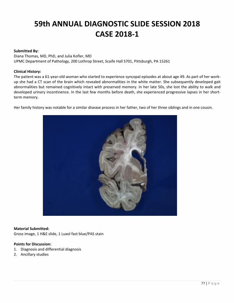
77 | P a g e
59th ANNUAL DIAGNOSTIC SLIDE SESSION 2018CASE 2018-1
Submitted By:Diana Thomas, MD, PhD, and Julia Kofler, MDUPMC Department of Pathology, 200 Lothrop Street, Scaife Hall S701, Pittsburgh, PA 15261
Clinical History:The patient was a 61-year-old woman who started to experience syncopal episodes at about age 49. As part of her work-up she had a CT scan of the brain which revealed abnormalities in the white matter. She subsequently developed gaitabnormalities but remained cognitively intact with preserved memory. In her late 50s, she lost the ability to walk anddeveloped urinary incontinence. In the last few months before death, she experienced progressive lapses in her short-term memory.
Her family history was notable for a similar disease process in her father, two of her three siblings and in one cousin.
Material Submitted:Gross image, 1 H&E slide, 1 Luxol fast blue/PAS stain
Points for Discussion:1. Diagnosis and differential diagnosis2. Ancillary studies

78 | P a g e
59th ANNUAL DIAGNOSTIC SLIDE SESSION 2018CASE 2018-2
Submitted By:Julieann Lee1, Sean Ferris1, David Solomon1, Dimitri Trembath2, Arie Perry1
1. Neuropathology, University of California, San Francisco, CA.2. Neuropathology, The University of North Carolina at Chapel Hill, Chapel Hill, NC.
Clinical History:The patient is a 25-year-old man who presented with left hand numbness and headaches. He was found to have a 4.7 cmheterogeneously enhancing intra-axial mass in the left cerebellar hemisphere, with mass effect on the fourth ventricle. Abiopsy was performed.
Material submitted:H&E slide from biopsy of cerebellar mass
Points for discussion:1. Differential diagnosis2. Immunohistochemical and molecular evaluation

79 | P a g e
59th ANNUAL DIAGNOSTIC SLIDE SESSION 2018CASE 2018-3
Submitted By:Karra A. Jones, MD, PhD and Steven A. Moore, MD, PhDThe University of Iowa, Department of Pathology, Iowa City, IA 52242
Clinical History:2-year-old male with mild global developmental delay, frequent falls, and oropharyngeal dysphagia. Neurologic examrevealed a positive Gowers’ sign and abnormal gait consistent with a compensated Trendelenburg. His family history waspositive for a maternal uncle with unknown neuromuscular disease requiring the use of a wheelchair since age 12. Theserum CK was elevated in the range of 1300-1400 U/L.
Genetic testing prior to muscle biopsy included a non-diagnostic microarray, normal DMD del/dup testing and completesequencing, normal GAA enzymatic assay, and negative congenital hypotonia next generation sequencing panel.
Material Submitted:H&E stained cryosection of muscle
Points for Discussion:1. Differential diagnosis2. Approach to diagnostic testing

80 | P a g e
59th ANNUAL DIAGNOSTIC SLIDE SESSION 2018CASE 2018-4a
Submitted By:Anne Shepler, MD and Julia Kofler, MDUniversity of Pittsburgh Medical Center Presbyterian HospitalDivision of Neuropathology200 Lothrop Street, Room S701 Scaife HallPittsburgh, PA 15213
Clinical History:The patient was a 37-year-old lawyer with no family history of dementia, who at age 33 began to have performance issues atwork with inattentiveness to detail and a lack of concern for deadlines. He became progressively abulic and socially withdrawnwith a loss of interest in his hobbies and personal appearance. By age 34, he began to choke frequently on food and was notedby his wife to have developed a nasal voice and bilateral ptosis. While he could remember his two young children’s names, hewas unable to care for them. He also developed reduplicative paramnesia wherein he insisted that there were two “2B”apartments in his apartment building where another woman (with his wife’s name) lived with two children. While he did notget agitated if corrected, he returned to this delusion repeatedly.
On neurological examination, he showed no lateralizing sensory or motor signs and muscle strength was normal. Nofasciculations were present. His reflexes were brisk and symmetric with no sustained clonus or Babinski sign. He was fluentwith intact comprehension, repetition, naming, reading, and writing. He often answered questions too quickly with impulsiveerrors as he would not wait of the conclusion of the question; he was undisturbed when he made a wrong answer. Other thanptosis and a mild weakness of the orbicularis oculi, the cranial nerve examination was unremarkable. Initial MRI and CT scanswere normal. A PET scan revealed bilateral frontal diminution of glucose utilization, worse on the right than the left, withextension to the right caudate. An EMG was negative.
Autopsy findings:The brain weight was 1160 grams (fresh). Neuropathologic examination was notable for moderate atrophy of the frontal lobeand caudate and mild atrophy of the temporal and parietal lobes.
Material Submitted:One H&E slide of frontal cortex
Points for Discussion:1. Differential diagnosis2. Useful immunohistochemical stains

81 | P a g e
59th ANNUAL DIAGNOSTIC SLIDE SESSION 2018CASE 2018-4b
Submitted By:Aivi T. Nguyen and Edward B. LeeHospital of the University of Pennsylvania3400 Spruce StreetPhiladelphia, PA 19104
Clinical History:The decedent was a 72-year-old right handed female who presented for evaluation of incoordination and frequent falls.She complained of multiple episodes of imbalance and demonstrated increasing forgetfulness, anosmia, andmicrographia. She was evaluated by multiple neurologists and eventually treated with carbidopa/levodopa with noimprovement. Physical exam was remarkable for masked facies, impaired extraocular movements with jerky saccades,axial rigidity, moderate bradykinesia, and a narrow-based gait with short strides, absent arm swing, and no posturalcontrol. Over the course of five years she developed severe cognitive defects, including word finding difficulties andmemory decline, and was eventually transferred to hospice where she died.
Autopsy Findings:The brain was examined 14 hours post-mortem and weighed 1147 grams. Gross findings included diffuse cerebral atrophy,severely atrophic hippocampus and amygdala, ex-vacuo hydrocephalus, and substantia nigra depigmentation.
Material Submitted:1 H&E section of the hippocampus
Points for Discussion:1. Differential diagnoses2. Useful immunohistochemical/additional stains3. Revised differential diagnoses after additional stains and biochemical work-up

82 | P a g e
59th ANNUAL DIAGNOSTIC SLIDE SESSION 2018CASE 2018-5
Submitted By:E. Kelly S. Mrachek, M.D., M. Beatriz S. Lopes, M.D., Ph.D.University of Virginia Health SystemDepartment of Pathology, Division of NeuropathologyBox 800214Charlottesville, VA 22908-0214
Clinical History:The patient is a 6-year-old male who presented with a two-week history of headaches with recent nausea and vomiting. HisPCP diagnosed strep throat, and treatment with antibiotics failed. While getting additional labs drawn, the patient had acuteonset of left sided facial drooping, and was brought to the ER. MRI of the brain with and without contrast showed a 5.2 cmavidly enhancing, extra-axial mass in the left cerebellopontine angle with extension into the internal auditory canal. A leftretrosigmoid craniotomy and gross total resection were performed at the University of Virginia.
Material Submitted:MRI image of brain demonstrating a left CPA mass, and an H&E section of the mass.
Points for Discussion:1. What is the final integrated histomolecular diagnosis of this left CPA mass?2. What is the most common molecular genetic alteration in lesions of this type?

83 | P a g e
59th ANNUAL DIAGNOSTIC SLIDE SESSION 2018CASE 2018-6
Submitted By:Jason A. Gregory, CPT USA, MD, Meggen Walsh, MD and Jesse Lee Kresak, MDUniversity of FloridaDepartment of Pathology, Immunology, and Laboratory Medicine1600 SW Archer RdPO Box 100275Gainesville, FL 32610
Clinical History:A twenty-year-old male presents for esophagogastroduodenoscopy for persistent dysphagia with a specific chiefcomplaint of “I’m unable to chew because I can’t open my mouth and my teeth hurt.” He has a history of hypertension,obstructive sleep apnea, and a congenital musculoskeletal disorder. During the procedure, the patient was noted to haveairway instability with subsequent desaturation. Resuscitative efforts were unsuccessful and the patient expired.
Autopsy findings:General autopsy revealed a thin man of short stature weighing 75 lbs. He had various musculoskeletal abnormalitiesincluding scoliosis, pes planus, and asymmetrical muscular atrophy. The brain weighed 1,560 grams. Gross examinationrevealed an enlarged brainstem with a markedly stenotic aqueduct. The medulla appeared ovoid rather than the normal“papilionaceous” shape. Internal architecture of the brainstem appeared distorted. The dentate nucleus of the cerebellumwas difficult to delineate grossly. The remainder of the cerebrum and cerebellum appeared normal.
Material Submitted:H&E-stained section of the medulla
Points for Discussion:1. Differential diagnosis and ancillary studies2. Pathogenesis

84 | P a g e
59th ANNUAL DIAGNOSTIC SLIDE SESSION 2018CASE 2018-7
Submitted By:Kyle Conway, Theodore Brown, David Gordon, John Kennedy, and Sriram VennetiUniversity of Michigan Department of Pathology1301 Catherine St.Ann Arbor, MI 48109
Clinical History:The decedent is a 50-year-old female who presented with hemiparesis, expressive dysphasia, headaches, visualdisturbances, and paresthesias. Imaging showed multifocal frontal and temporal lesions crossing the corpus callosum. Afrontal lobe biopsy at this time demonstrated only reactive gliosis and perivascular inflammation. Several months later,she presented to the emergency department with acute worsening of symptoms. Repeat imaging showed findingsconsistent with an ischemic stroke. Serologic workups for autoimmune disease and infection were negative. She wasdischarged, and her neurologic condition worsened over the course of a year. She passed way after an acute deteriorationin neurologic status.
Autopsy findings:Gross autopsy findings included multiple areas of softened parenchyma in the left frontal, right fronto-parietal, rightsuperior parietal, left mid parietal, and left occipital lobes. The lumina of the carotid arteries were grossly narrowed(approximately 0.1 cm in diameter).
Material Submitted:1. MRI images2. Gross photographs of cortex and base of brain3. H&E stained slide of cortical lesion4. Trichrome stained slide of carotid artery
Points for Discussion:1. Explain the differential diagnosis of this clinical history and workup2. Understand the classification and subtyping for this disease

85 | P a g e
59th ANNUAL DIAGNOSTIC SLIDE SESSION 2018CASE 2018-8
Submitted By:Angela N. Viaene, MacLean P. Nasrallah, and Zissimos MourelatosHospital of the University of Pennsylvania, Department of Pathology and Laboratory Medicine, Division ofNeuropathology, 6th Floor Founders, 3400 Spruce Street, Philadelphia, PA 19104
Clinical History:A 38-year-old female with no significant past medical history presented with headache, nausea and photophobia for threedays. She subsequently developed visual loss and aura in both eyes which prompted imaging studies. A large,predominantly cystic left parietal mass measuring 6.4 x 4.3 x 5.2 cm associated with irregular and nodular peripheralenhancement and areas of hemorrhage around its periphery was seen on brain MRI. There was mass effect including 7-8mm rightward midline shift and partial effacement of the basal cisterns. The patient underwent left craniotomy for gross-total resection of the enhancing mass.
Material Submitted:1. One representative H&E slide2. Representative pre-operative MRI image
Points for Discussion:1. Differential diagnosis2. Molecular findings

86 | P a g e
59th ANNUAL DIAGNOSTIC SLIDE SESSION 2018CASE 2018-9
Submitted By:Missia Kohler, M.D., Jamie Walker, M.D., Ph.D., Qinwen Mao, M.D., Ph.D., and Eileen Bigio, M.D.Northwestern University Feinberg School of MedicineDepartment of Pathology710 North Fairbanks CtChicago, IL 60611
Clinical History:The patient initially presented in 2009 at the age of 72 to neuropsychiatry with a two-year history of minor memoryproblems. His cognitive exam revealed memory issues that were primarily amnestic, moderate executive dysfunction anda MoCA score of 20/30. His sensory and motor exam were unremarkable. His MRI demonstrated more atrophy than
expected for his age. His APOE allele status was ε4/ε4. In 2016, he was still driving, but experienced bowel and bladderincontinence and was having a “hard time fixing things” in the home. By 2017, he was displaying more word-findingdifficulties and required instructions for most daily tasks. In the autumn of 2017, he had a few falls and hospitalizationsfor infections. Thereafter, he rapidly declined, and died at the age of 80 in October of 2017.
Material Submitted:One H&E stained slide
Points for Discussion:1. Differential diagnosis2. Immunohistochemical work-up

87 | P a g e
59th ANNUAL DIAGNOSTIC SLIDE SESSION 2018
CASE 2018-10Submitted By:M. Adelita Vizcaino M.D.1, Verena Staedtke M.D.2, Allan Belzberg, M.D.2, Fausto J. Rodriguez M.D.1
1. Division of Neuropathology2. Department of Neurology and Neurosurgery, Johns Hopkins University School of Medicine
Clinical History:An 11-year-old boy was previously healthy until approximately the year prior, when the family noticed that he was walkingon the lateral aspect of his right foot, resulting in callus formation and closed pressure ulcer. During physical examination,it was noted that the patient had a high arch of the right foot as well as a 3-cm. leg length discrepancy. Several subtle café-au-lait spots were also identified. Subsequent radiographs demonstrated multiple stress fractures, which prompted MRIof the spine and lower extremities demonstrating enlargement of the right lumbosacral plexus and right sciatic nerveextending distally down the thigh, progressing to include the tibial nerve and common peroneal nerve to the bifurcationwith continued involvement of the deep and superficial peroneal nerve branches to the foot. At the time of surgicalintervention for orthopedic corrections, a biopsy of the tibial nerve was performed.
Material Submitted:Virtual H&E image of tibial nerve biopsy
Points for Discussion:1. Differential Diagnosis2. Pathogenesis3. Role of molecular genetics
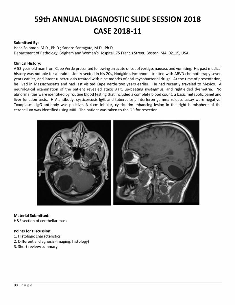
88 | P a g e
59th ANNUAL DIAGNOSTIC SLIDE SESSION 2018
CASE 2018-11Submitted By:Isaac Solomon, M.D., Ph.D.; Sandro Santagata, M.D., Ph.D.Department of Pathology, Brigham and Women’s Hospital, 75 Francis Street, Boston, MA, 02115, USA
Clinical History:A 53-year-old man from Cape Verde presented following an acute onset of vertigo, nausea, and vomiting. His past medicalhistory was notable for a brain lesion resected in his 20s, Hodgkin’s lymphoma treated with ABVD chemotherapy sevenyears earlier, and latent tuberculosis treated with nine months of anti-mycobacterial drugs. At the time of presentation,he lived in Massachusetts and had last visited Cape Verde two years earlier. He had recently traveled to Mexico. Aneurological examination of the patient revealed ataxic gait, up-beating nystagmus, and right-sided dysmetria. Noabnormalities were identified by routine blood testing that included a complete blood count, a basic metabolic panel andliver function tests. HIV antibody, cysticercosis IgG, and tuberculosis interferon gamma release assay were negative.Toxoplasma IgG antibody was positive. A 4-cm lobular, cystic, rim-enhancing lesion in the right hemisphere of thecerebellum was identified using MRI. The patient was taken to the OR for resection.
Material Submitted:H&E section of cerebellar mass
Points for Discussion:1. Histologic characteristics2. Differential diagnosis (imaging, histology)3. Short review/summary

89 | P a g e
AANPAMERICAN ASSOCIATIONOF NEUROPATHOLOGISTS
Presidential SymposiumSunday, June 10, 2018
Learning Objectives:1. Describe recent technological advances in methods of pathological diagnosis.
2. Explain how to implement and validate a digital pathology system.
3. Outline the role of machine learning in diagnostic pathology.

90 | P a g e
PRESIDENTIAL SYMPOSIUM
Evolution of Diagnostic Techniques in Pathology: Moving Beyond theMicroscopeTime: 8:05 am – 8:30 amElizabeth J. Cochran, M.D., Medical College of Wisconsin, Milwaukee, WI
I. Learning Objectives:At the end of this activity, learners should be able to:
1. List the sequence of developments that led to use of light microscopic evaluation of tissue as the primary
mode of disease diagnosis in pathology.
2. Compare recently developed techniques of tissue evaluation that are alternatives and/or adjuncts to
evaluation of light microscopic sections on glass slides by pathologists.
3. Define computational pathology, including an explanation of how it may change the practice of pathology.
II. Abstract and Relevant ResourcesFor the past 150 years, diagnostic pathologists have focused upon the evaluation of normal and abnormal tissuemorphology as seen upon examination of thin tissue sections on glass slides through the light microscope.However, new technologies are emerging that provide alternatives to this approach; the development of digitalpathology and computational analyses has supported this growth. These new techniques range from nextgeneration sequencing to new methods of identifying morphology, and many require computer system analysisof the large volume complex datasets that are produced. As these new tools mature and are brought into use inclinical medicine, the role of the pathologist is evolving to include integration of molecular and computationaldata in addition to the traditional role of histopathologist.
References:
1. Eschbacher JM, Georges JF, Belykh E, et al., Immediate Label-Free Ex Vivo Evaluation of Human Brain TumorBiopsies With Confocal Reflectance Microscopy. J Neuropath Exp Neurol December 2017; 76(12): 1008-1022.
2. Granter SR, Beck AH, Papke DJ., Deep Learning, and the Future of the Human Microscopist. Arch Pathol LabMed 2017 141:619-623.
3. Hariri LP. In Vivo Microscopy Will the Microscope Move From Our Desk Into the Patient? Arch Pathol Lab Med.June, 2015; 139: 719-720.
4. Kut C, Chaichana KL, Xi J, et al., Detection of human brain cancer infiltration ex vivo and in vivo usingquantitative optical coherence tomography. Sci Transl Med 2015 Jun 17;7(292): 292ra100.
5. Louis DN, Feldman M, Carter AB, et al., Computational Pathology, A Path Ahead. Arch Pathol Lab Med 2016;140: 41-50.
6. Majno G, Joris I. Microscope in the history of pathology. Virchows Arch 1973 360: 273-286.7. Van den Tweel JG, Taylor CR. A brief history of pathology. Virchows Arch 2010; 457: 3-10.8. Kalderon AE The evolution of microscope design from its invention to the present days. Am J Surg Path 1983;
7 (1): 95-102.

91 | P a g e
III. Faculty Biography:
Dr. Cochran obtained her M.D. degree from Rush Medical College in Chicago, Illinois in 1982. She completedresidency training in anatomic pathology at Northwestern University in Chicago, Illinois, followed by aneuropathology fellowship with Drs. Pierluigi Gambetti and Uros Roessmann at Case Western Reserve UniversityInstitute of Pathology in Cleveland, Ohio. She joined the faculty of Rush University Medical Center in 1989 as anassistant professor, and she helped to establish the Alzheimer’s disease brain bank at Rush. She wasneuropathology core leader in the Rush NIH-funded Alzheimer’s disease center and Religious Order Study grants,participating in research on Alzheimer’s disease and aging. Over the last decade, she has focused on education ofmedical students and residents, first directing the Rush pathology residency program, medical student pathologycourse and clerkships, at Rush, while continuing to run the diagnostic neuropathology and autopsy services. In2010, she joined the faculty of the Medical College of Wisconsin in Milwaukee, Wisconsin as Professor ofPathology and is currently director of the autopsy and neuropathology services and of medical student educationin pathology.
92 | P a g e
PRESIDENTIAL SYMPOSIUM
Teleneuropathology for Intraoperative ConsultationTime: 9:15 am – 10:00 amClayton A. Wiley, MD, PhD, University of Pittsburgh Medical Center, Pittsburgh, PA
I. Learning Objectives
At the end of this activity learners should be able to:1. Describe how telepathology evolved to its current stage2. Recognize and anticipate the challenges of implementing telepathology3. Describe how to avoid problems inherent in rendering pathological diagnoses remotely4. Recognize many legal issues related to telepathology are in flux with great variation between states5. Recognize that telepathology can successfully distribute medical expertise to regions of need.
II. Abstract & Relevant References
Telepathology has been under development for close to half a century. With the advent of the Internet andavailability of rapid information transmission, the pace of development has quickened. While technological andsecurity hurdles have been surpassed, there are substantial challenges associated with the practice of medicinethat need to be surmounted in order for telepathology to fully replace onsite intraoperative consultation. Beingvirtually present is not the same as being present. Many practical and unrecognized assumptions need to beaddressed before telepathology can be successfully integrated into patient care. Human adoption of any newtechnology is always subject to substantial pushback, however, the advantages of telepathology now outweighdisadvantages and will drive adoption system wide.
References:
1. Wiley CA, Murdoch G, Parwani A, Cudahy T, Wilson D, Payner T, Springer K, Lewis T (2011) Interinstitutionaland interstate teleneuropathology. Journal of pathology informatics.2:21.
2. Horbinski C, Fine JL, Medina-Flores R, Yagi Y, Wiley CA (2007) Telepathology for intraoperativeneuropathologic consultations at an academic medical center: a 5-year report. J Neuropathol ExpNeurol.66(8):750-9.
3. Hiemenz MC, Leung ST, Park JY (2014) Crossing boundaries: a comprehensive survey of medical licensinglaws and guidelines regulating the interstate practice of pathology. The American journal of surgicalpathology.38(3):e1-5.
4. Williams BJ, Bottoms D, Treanor D (2017) Future-proofing pathology: the case for clinical adoption of digitalpathology. Journal of clinical pathology.
5. Cheng CL, Azhar R, Sng SH, Chua YQ, Hwang JS, Chin JP, Seah WK, Loke JC, Ang RH, Tan PH (2016) Enablingdigital pathology in the diagnostic setting: navigating through the implementation journey in an academicmedical centre. Journal of clinical pathology.69(9):784-92.
6. Meyer J, Pare G (2015) Telepathology Impacts and Implementation Challenges: A Scoping Review. Archivesof pathology & laboratory medicine.139(12):1550-7.
7. https://thesource.americantelemed.org/blogs/jessica-washington/2016/09/12/american-telemedicine-association-issues-clinical-guidelines-for-telepathology.

93 | P a g e
III. Faculty Biography:
Dr. Wiley did his undergraduate training at the University of Chicago and his MD/PhD training in Neurosciences atthe University of California San Diego. This was followed by Anatomical Pathology residency at University ofCalifornia San Francisco and Neuropathology fellowship back at UCSD. In 1985 Dr. Wiley was appointed AssistantProfessor of Pathology at UCSD where he advanced to the rank of full professor. In 1993 he was recruited to theUniversity of Pittsburgh Medical Center as Director of the Division of Neuropathology and its fellowship program.The Division has been involved in telepathology for 25 years.
Throughout his professional career he has been actively involved in educating physician scientists at both pre- andpostgraduate stages. From 1997 to 2012 Dr. Wiley served as Director of the Pittsburgh Medical Scientist TrainingProgram and Associate Dean in the University of Pittsburgh School of Medicine. Throughout this time, he wasactively involved in the National Association of MD/PhD Programs and the MD/PhD Section of the GREAT groupin the AAMC where he served as President and Chair respectively. He also served on the AAMC Council ofAcademic Societies Task Force on Dual Degree Programs. Dr. Wiley has maintained an active NIH funded researchprogram investigating the pathogenesis of viral mediated neurodegeneration. He has published over 250 peer-reviewed publications and was elected a Fellow of the American Association for the Advancement of Science in1997. Currently his research is focused on the role of innate and adaptive immunity in protecting the brain fromviral infections.

94 | P a g e
PRESIDENTIAL SYMPOSIUM
Liquid Biopsy in Cancers of the Central Nervous SystemTime: 10:45 am – 11:30 pmMatthias Holdhoff, MD, PhD, Associate Professor of Oncology and Neurosurgery, Sidney Kimmel Comprehensive CancerCenter, Johns Hopkins University School of Medicine
I. Learning Objectives
At the end of this activity learners should be able to:
1. Describe current concepts and potential applications of ‘liquid biopsy’ in oncology.2. Discuss the potential role of ‘liquid biopsy’ as a non- or minimally invasive tool in CNS cancers.3. Explain the specific hurdles in the development of blood- or CNS-based markers in CNS cancers.
II. Abstract & Relevant References
The development and clinical application of ‘liquid biopsies’ is a promising topic in oncology. In this lecture, wewill review current concepts of circulating biomarkers in cancers with specific focus on the potential role of liquidbiopsies in cancers of the central nervous system. Specific challenges for development of such markers in braincancers will also be discussed.
References:
1. Bettegowda C et al. Detection of Circulating tumor DNA in early- and late stage human malignancies. SciTransl Med. 2014 Feb 19;6(224):224ra24
2. Westphal M, Lamszus K. Circulating biomarkers for gliomas. Nature Reviews Neurology 2015;11:556-566.3. Wang Y, et al. Detection of tumor-derived DNA in cerebrospinal fluid of patients with primary tumors of the
brain and spinal cord. Proc Natl Acad Sci U S A. 2015 Aug 4;112(31):9704-94. Pentsova EI et al. Evaluating Cancer of the Central Nervous System Through Next-Generation Sequencing of
Cerebrospinal Fluid. J Clin Oncol. 2016 Jul 10;34(20):2404-15.
III. Faculty Biography:
Dr. Holdhoff is an associate professor of oncology and neurosurgery at The Sidney Kimmel Comprehensive Cancercenter at Johns Hopkins. He is a clinical translational investigator in neuro-oncology and a clinical expert inmalignant gliomas and central nervous system lymphomas. His research focuses on early phase clinical trials andbiomarker research in cancers of the central nervous system. Dr. Holdhoff received his medical degree at FreieUniversität Berlin and completed his dissertation at Charité University Medicine Berlin. He completed his internalmedicine residency training at Johns Hopkins Bayview Medical Center and a medical oncology fellowship at TheJohns Hopkins Hospital, during which he also pursued research training within the Ludwig Center for CancerGenetics and Therapeutics.

95 | P a g e
PRESIDENTIAL SYMPOSIUM
Deep Learning Neural NetworksTime: 11:30 am – 12:15 pmLee Cooper, PhD, MS, Emory University School of Medicine, Atlanta, GA
I. Learning Objectives
At the end of this activity learners should be able to:
1. Summarize the basic components of a deep learning neural network2. Contrast the differences between engineered and adaptive approaches to solving machine learning
problems3. Explain how deep learning can be used to solve a problem like predicting clinical outcomes
II. Abstract & Relevant References
This lecture will discuss machine learning and the development and application of predictive models in digitalpathology. We will discuss advanced in an area called deep learning that can create powerful models that canlearn to identify important patterns in histology and perform diagnostic tasks with human-level accuracy. We willshow how this technology can be used to build advanced survival models that integrate histology and genomicsto accurately predict the clinical outcomes of patients diagnosed with glioma.
References:
1. P Mobadersany, S Yousefi, M Amgad, DA Gutman, JS Barnholtz-Sloan, JE Velazquez-Vega, DJ Brat, LADCooper, Predicting cancer outcomes from histology and genomics using convolutional networks, PNAS,
115(13), pp. 2970-9, March 20182. U Djuric, G Zadeh, K Aldape, P Diamandis, Precision histology: how deep learning is poised to revitalize
histomorphology for personalized cancer care, Precision Oncology, 22, June 20173. LAD Cooper, EG Demicco, JH Saltz, RT Powell, A Rao, AJ Lazar, PanCancer insights from The Cancer Genome
Atlas: the pathologist’s perspective, Journal of Pathology (Annual Review), 244(5), pp. 512-24, April 20184. S Webb, Deep learning for biology, Nature, 554, pp. 555-7, February 20185. Y LeCun, Y Bengio, G Hinton, Deep Learning, Nature, 521, pp. 436-44, May 2015
III. Faculty Biography:
Lee Cooper, PhD is an engineer and an Assistant Professor of Biomedical Informatics at Emory University. Hereceived a PhD in Electrical and Computer Engineering in 2009 from Ohio State University, where he beganresearch in digital pathology in 2005. His research is funded by the Informatics Technology for Cancer Researchprogram of the National Cancer Institute and seeks to develop software algorithms and infrastructure foranalyzing genomic and digital pathology data.

96 | P a g e
2017 DONATIONS
Special thank you to the members that contributed generous donationsto the AANP in 2017.
John Andrews
Dennis Burns
Matthew Cykowski
James Dollar
Peter Dowling
Jonathan Fratkin
Matti Haltia
Margaret Jones
Nancy Kois
Hirofumi Kusaka
Qinwen Mao
Douglas Miller
Sverre Mork
Kathy Newell
Randal Nixon
Arie Perry
Gerald Reis
Ralf Schober
Jane Uyehara-Lock
Karen Weidenheim
William Yong

97 | P a g e
ABSTRACT AUTHOR INDEX
Abdollahi, Maryam 40 Appin, Christina 115, 198 Belan Larque, Ann 167 Bryke, Christine 57
Abedalthagfi, Malak 33 Aravindan, Sheeja 85 Belden, Christine M 17, 183 Bucelli, Robert 91, 185
Abedin, Zahidur 213 Arce, Richard C 17 Bell, Heather 63 Buchhalter, Ivo 32
Ackerman, Laurie 7 Arevalo-Espejo,Octavio
132, 134 Belousova, Tatiana 134 Bull, Suzanne C 48
Adams, Stephanie L 21 Armstrong, Gregory T. 29 Belzberg, Allan 136 Burns, Terence C 145
Adamski, Carolyn J. 129 Armstrong, Terri 159 Benayoun, Laurent 21 Burns, Dennis K 213
Adesina, Adekunle M 48 Arnold, Michael 29 Benson, Leslie A 119 Burton, Eric C 47
Adler, Charles H 17, 183 Arora, Sonali 15 Benzinger, Tammie 70 Bydon, Mohamad 66
Adorjan, Istvan 186 Arrilaga-Romany,Isabel C
9 Berger, Mitchel 30 Byler, Debra L 96
Adstamongkonkul,Pichet
2, 210 Arunkumar, Ponni 112, 115 Berman, Robert F 5 Cai, Rongsheng 148
Ahern, Geoff L 17 Aurora, Sonali 35 Bernstein, Bradley E 99 Cairns, Nigel 70
Ahluwalia, Manmeet 9 Ayres, Samuel 118 Bettegowda, Chetan 27 Calicchio, Monica L 88
Ahmad, Arslan 200 Azhir, Alaleh 218 Bezerra Gondim,Mercia J
7 Callan, Michael 17, 183
Ahmed, Hazem M 43 Babcock, Katherine 111 Bharadwaj, Rajnish 101, 176,194
Camacho, Jessica 36
Ahrendsen, Jared T. 80 Baisre, Ada 100, 175 Bhatia, Smita 29 Camelo-Piragua,Sandra
67
Al Dalahmah, Osama 186 Bajić, Aleksandar 129 Bhattacharjee,Meenakshi B
87, 134, 217 Campbell, Gerald 76
Aldape, Ken 11 Balcom, Jessica R 13, 81 Bieniek, Kevin F. 114 Capocelli, Kelley 201
Aldave Orzaiz,Guillermo
48 Ball, Matthew K. 150, 170 Bienkowski, Michael 72 Caron, Alissa 25
Alexander, Brian M 9 Ballester, Leomar Y 87, 132,134, 143,156, 172
Bigio, Eileen H. 20, 112,115, 126,198
Carroll, Steven L 1
Alexander, Jes 15 Balliu, Brunilda 154 Bihannic, Laure 99 Carvajal-Carmona, Luis 18
Alexandrescu, Sanda 80, 88,119, 140
Bandopadhayay,Pratiti
80 Bird, Thomas D. 188 Caselli, Richard J 17
AlHarbi, Musa 33 Banerjee, Anu 30, 32 Bissel, Stephanie 216 Castro, Diana 175
Alhumayd, Ziad 91 Banihashemi, Amir 51 Blackburn, Patrick R 81 Caviness, John N 17, 183
Al-Khalili, Yasir 93 Bannykh, Serguei 58 Blakeley, Jaishri 136 Cavuoti, Dominick 104
Allen, Sariah J 28, 29 Banworth, Marcellus J 85 Bledea, Ramona 137 Chan, Amy 64
Allen, Nichole 151 Barber, Jason K 178 Blessing, Melissa M 23, 81, 114 Chang, Edward 30
Alterman, Ronnie 57 Barnett, Katherine 135 Block, John B 148 Chau, Kevin F 88
Alvarez, Victor E. 109, 111 Barr Fritcher, Emily G 13 Bollen, Andrew W. 26, 30, 32 Chavez, Lukas 32
Alves, Michele 8 Bastian, Boris C. 26, 30, 31,32
Bonnin, Jose
Boothby, Ian
76, 79, 192,197210
Chebib, Ivan 167
Alvi, Mohammed Ali 66 Batchala, Prem P. 16 Borck, Christopher C. 86, 113 Chen, Katharine 26
Alzate, Diego 8 Bateman, Randall 70 Borys, Ewa 95, 161 Chen, Weili 129
Amadi, Arash J. 101 Batruch, Ihor 11 Bowman, ChristopherJ.
12 Chen, Melissa 143
Ames, Heather 45, 49 Bauer, Matthew R 6 Brat, Daniel J 145 Chen, Jie 185
Amlie-Wolf, Alexandre 124 Bauer, Peter 195 Bredel, Marcus 38 Chen, Dong-Hui 188
Andersen, Jimena 84 Baughn, Michael 125 Brickman, Adam M 71 Cherayil, Neena 204
Ang, Phillip 6 Beach, Thomas G 17, 77, 183 Brosnan-Cashman,Jacqueline A.
27, 136 Cherry, Jonathan 109, 111
Antonescu, Cristina R. 37 Beckett, Laurel A. 18, 131 Bryce, Clare 51 Chi, Andrew S. 12, 39

98 | P a g e
Chi, Susan N. 80 de Bruin, Gabriela 185 Duvick, Lisa 129 Foreman, Nicholas K. 29, 138
Chi, Andrew 137 De Girolami, Umberto 152 Eaton, Mary Jane 120 Forgo, Erna 173
Chiba, Akira 4 de Groot, John F 9 Eberhart, Charles G. 44, 49, 55 Forst, Deborah A 50
Chinnaiyan, Arul 67 de la Monte, SuzanneM.
22, 205 Eckel, Ashley 176 Forsyth, Jennifer A 153
Chiocca, E. Antonio 9 DeCarli, Charles 18, 131 Eckel Passow,Jeanette
25 Fowkes, Mary 51, 64
Chiriboga, Luis 39 Decker, Paul 25 Edland, Steven D. 74, 78 Francis, Andrew T 178
Chkheidze, Rati 103, 104 Dedeaux, Andrea 191 Edward, Zamrini 17 Franco-Paredes, Carlos 207
Cho, Eun-Sook 37 Degar, Barbara A 119 Eisele, Sylvia 137 Frankel, Merav 154
Choi, Euno 162 Del Piero, Fabio 191, 209 El-Achi, Hanadi 156 Franklin, Erin 70
Cholerton, Brenna 74, 78 Delalle, Ivana 21 Elkadi, Osama R. 93, 155 Friedman, Elliott 217
Choudhury, Begum 218 DeMasters, BK 30 Ellis, Ethan 60 Friedrich, Jillian 129
Chuang, Kangway V 131 Denison, William 152 Engle, Elizabeth C 6 Frosch, Matthew P 24, 73, 203
Chumbalkar, Vaibhav 195 Desai, Nilesh K 48 Epperson, Francine 76, 79, 192,197
Fu, Dan 178
Chung, Chun Kee 162 Deshpande, Vikram 167 Ermoian, Ralph P. 82 Fu, Zhiyan 179
Cimino, P.J. 15, 35 DeSisto, John 29 Ertekin-Taner, Nilufer 19 Fulkerson, Daniel 7
Clapham, David 2, 210 DeWitt, John C 99 Esquenazi, Yoshua 132, 134,156, 172
Fuller, Christine 34, 148
Clark, Kristi 72, 75 Dharaneeswaran,Kiruba
215 Evans, Brandon C 42 Fullmer, Joseph M 166, 171
Clarke, Jennifer 30 Di Loreto, Christina M. 52, 97 Evers, Bret M. 103, 104 Funakawa, Itaru 189
Clayton, Frederic 164 Diamandis, Phedias 11, 121,122
Fadda, Paolo 28 Fung, Kar-ming 85
Coppola, Giovanni 197 Diamond, Eli L. 175 Fadul, Camilo E. 16 Furuta, Takuya 144
Corless, Chris 54 Dias-Santagata, Dora 50 Fame, Ryann M 88 Futamura, Naonobu 189
Couce, Marta 28 Diaz Garcia, Sandra 125 Fan, Xuemo 58 Gaffey, Sarah C 9
Coy, Shannon 138 Dickson, Dennis W 19, 114,127
Farias, Sarah T. 18 Gajjar, Amar 99
Crandon, Sonja 159 Djuric, Ugljesa 11, 121,122
Farlow, Martin R 192 Gal, Jozsef 128
Crary, John F. 20 Dodd, Robert 154 Farrell, Kurt 20 Galanis, Evanthia 9
Cristancho, Ana G 204 Dong, Hong Wei 72 Farrell, Christopher 146 Galbraith, Kristyn 171
Curry, Jasmine R 17 Donson, Andrew 29, 138 Fasciano, Danielle 199 Gallego, Daniel 176
Curtis, Mark T 146 Dorn, David 199 Faust, Phyllis L. 37 Gallucci, Gina 22
Cusimano, Michael D 40 Dorris, Kathleen 29 Faust, Kevin 121, 122 Gao, Andrew F. 40, 122,190
Cutler, Brett D 17, 183 Drappatz, Jan 9 Ferman, Tanis J 19 Gardner, Sharon 69, 135
Czeisler, Catherine 3, 8 Driver-Dunckley, Erika 17, 183 Ferris, Sean 54, 94 Garringer, Holly J. 76, 79,180, 197
Dakay, Katarina B 205 Drubach, Daniel 23 Filbin, Mariella G. 80, 99 Gavrilova, Ralitza H 23
Dani, Neil 88 Du, Ziming 138 Filshtein, TeresaJenica
18 Geduldig, Jack E 9
Danielsen, Travis 108, 112 Duara, Ranjan 19 Fisher, Kevin E 48 Gefen, Tamar 126, 198
Daoud, Elena V. 68 Dubuc, Adrian M 9 Fitzpatrick, Carrie 10 Geiersbach, KatherineB
13
Davies, Michael 143 Duff, Deiter J 83 Flanagan, Margaret E. 74, 78 Gelber, Alon 6
Davis, Kathryn J 17 Dugger, Brittany N 18, 131 Flannery, Patrick 29 Gelman, Benjamin 76
Davis, Christine 136 Dulai, Mohanpal S 28 Folkerth, Rebecca D. 86, 113 Georgescu, Maria-Magdalena
28
Davis, Kathryn J. 183 Duncan, Christine N 119 Fong, Jamie C 197 Geschwind, Michael 197
Day, John D 149 Duose, Dzifa 143, 172 Foong, Chan 20

99 | P a g e
Ghetti, Bernardino 76, 79,130, 180,192, 193,197
Hackney, JR 38, 142,199
Holland, Eric 15 Jinnai, Kenji 189
Giannini, Caterina 13, 25, 27,136, 145,211
Hadley, Jennifer 99 Holloman, Ashley M 48 Joiner, Amy K 42
Gibson, Natasha 7 Halfpenny, Aaron 54 Holloway, Jeffery 83 Jonathan, Lavezo 173
Gilani, Ahmed 201, 202 Halling, Kevin C 81 Honce, Justin M 105 Jones, Kenneth 29
Gilbert, Mark 159 Hamilton, Ronald L 61, 139 Hooper, Jody E 49 Jones, Pamela S 50
Giller, Cole 169 Hamou, Wissam 60 Horbinski, Craig 14 Jones, David T. 114
Glass, Michael J 17 Han, Pengcheng 1 Hormigo, Adilia 64 Joseph, Nancy M. 26
Glitza Oliva, Isabella 143 Hankinson, Todd C. 138 Hornick, Jason 140 Judkins, Alexander R. 65
Goedert, Michel 180 Hanna Al-Shaikh, FadiS
19 Houldsworth, Jane 60, 64 Juratli, Tareq 167
Gokden, Murat 27, 41, 42,43, 136,148, 149
Hansen, Lawrence A. 52, 97 Houser, Justin 2 Juric-Sekhar, Gordana 101, 178
Goksel, Mustafa 41 Hardeman, Edna 118 Hovestadt, Volker 99 Kadri, Sabah 10
Golby, Alexandra 152 Hardie, Lakotah 29 Hoyo, Leonie 210 Kam, Kwok-Ling 85
Goldman, James E 71, 186 Harrison, William T 206 Hsu, Nancy 57 Kamiyama, Daichi 4
Golfinos, John G. 12, 137 Harsh, Griffith 154, 173 Huang, Wentao 6 Kanach, Colin J 155
Goncalves, Sandy 66 Harter, David 135 Huber, Bertrand 109, 111 Kao, Jennifer 11
Gong, Henry 26 Hatanpaa, Kimmo J 53, 68, 213 Huebner, Philip A 5 Karajannis, Matthias 12
Gonzales, Hilary A 5 Hattab, Eyas M 28 Hulette, Christine M 206 Karydas, Anna M 197
Gonzalez Zacarias, Clio 72 Hawes, Debra 65 Hurth, Kyle 174 Katsyv, Igor 60
Gonzalez-Cuyar, Luis F. 101, 188 Hawkins, Cynthia 40 Huse, Jason 143 Katz, Leah 137
Goode, Benjamin 30 Hawkins, Kellie 207 Hyman, Bradley T 24, 73 Katznelson, Laurence 154, 173
Goraya, Harmeen 153 Haydu, Lauren 143 Ida, Cristiane M 13, 66, 81 Kawachi, Kevin 58
Goraya, Murat 153 Hazrati, Lili-Naz 110 Imada, Eddie Luidy 45 Kawamura, Junko 106
Gorman, Mark P 119 Heaphy, ChristopherM.
27, 136 Intorcia, Anthony J 17, 77 Kay, Jared 22
Goumnerova, Liliana 99 Hefti, Marco M 90 Iorgulescu, Bryan 32 Kaya, Behiye 8
Grafe, Marjorie 54 Heger, Ian 168 Ishii, Kenji 212 Keene, C. Dirk 101, 188,194
Graff-Radford, Neill R 19 Hemmy, Laura S. 74, 78 Ishikawa, Tatsuya 106 Keiser, Michael J. 131
Graham, Mindy K. 136 Henderson, Lauren A 119 Islas, Sergio 48 Keith, Julia 190
Granzier, Henk L 118 Henzler, Christine 129 Israel, DJ 38 Kelly, Stephen 137
Green, Adam L. 29 Hernandez-Losa, Javier 36 Izen, Sarah 6 Kerezoudis, Panagiotis 66
Greenberg, BenjaminM
213 Hess, Harald 2 Jackson, Rory A 81 Khan, Mohammad 58
Gregory, Jason A 56 Hildebrandt, Audrey 111 Jaitly, Vanya 134 Khan, Galam 205
Grenert, James P. 30, 32 Himali, Jayandra J 21 James, C. David 14 Khara, Maliha 46
Groves, Andrew 99 Hinojoza, Julian 120 Jarvis, Hannah 113 Kieran, Mark W. 80, 138
Guangrong, Lu 172 Hirsch-Reinshagen,Veronica
182 Jayadev, Suman 188 Kilgore, Shannon 184
Guerrero, Jessenia 100 Hite, Amy 28 Jenkins, Robert B 13, 25 Kim, Youngmi 15
Gupta, Nalin 30, 32 Ho, Joanne 101 Jentoft, Mark E 13, 27 Kim, Hyoung-Ihl 107
Gupta, Rajarsi 63 Ho, Mai Lan 150 Jessup, Nicholas 140 Kim, Seong-Ik 162
Gurangong, Lu 132 Hoffman, Lindsey M. 29 Jiang, Jie 125 Kimonis, Virginia 181
Gutmann, David H 45 Hoffman, Andrew 154 Jillella, Anusha 153 Kimura, Hiroaki 187
Gyure, Kymberly A. 147 Hogan, Robert N. 103, 104 Jin, Lee-Way 18, 131 King, Paul D 47

100 | P a g e
Kinoshita, Toshibumi 106 Leary, Sarah E. S. 82 Liu, Jing 134 Mayor, Regina 36
Kipp, Benjamin R 13, 81 Lechpammer, Mirna 5 Liu, James K. 175 Mayr, Lisa 99
Kirby, Patricia A 90 Leckey, Bruce 206 Liu, Yang 200 McCarty, Jennifer 149
Kirchhausen, Tomas 2 Lee, Edward B. 20, 59, 124 Lobbous, Mina 38 McCreery, Douglas 214
Kleinschmidt-DeMasters, Bette K
31, 84,201, 202,207, 208
Lee, Brendan 76 Lobel, Graham 181 McElroy, Joseph 28, 47
Klekner, Almos 11 Lee, Kyung-Hwa 107 Long, Kathy E 17 McFadden, Kathryn 67
Klickstein, Naomi 73 Lee, EudociaLee, Min-Cheol
152107
Long, Ryan 214 McKee, Ann C. 109, 111
Klingstedt, Therése 180 Legati, Andrea 197 Lopategui, Jean 58 McLendon, Roger E 28
Kloetgen, Andreas 12 Lehman, Norman L 28, 47 Lopes, Maria-BeatrizS.
16, 147 McMullen, II, Phillip 123
Klonoski, Josh 89, 164 Lehtinen, Maria K 88 Lopez, Oscar 196 McMurtry, Valarie 89, 164
Knopman, David S. 114 Lein, Ed 210 López, Giselle Y 94 McPhail, Ellen D. 211
Koenig, RichardGabriel
199 Lemma, Rakeb 29 Lott, Paul 18 Mehta, Shyamal 17, 183
Koeppen, Arnulf 195 Lennerz, Jochen 50 Lotze, Timothy E 48 Mehta, Vidya 48
Kofler, Julia 165, 196,112, 115,126, 198,215
Leung, Denise 67 Lowder, Lindsey 98, 158 Meints, Joyce 78
Kolhe, Ravindra 168, 169 Levine, Ross 15 Lu, Hsiang-Chih 129 Mella, Divya 68
Komaki, Satoru 144 Li, Marilyn M. 37 Lu, Jian-QiangLucas, John T.
11629
Mendoza, Pia 98
Konnick, Eric 35, 176 Li, Xiaohui 47 Lucas, Lora 54 Meng, Hui 118
Kool, Marcel 32 Li, Guangpu 85 Lucas, Elena 103 Merchant, Thomas E. 29
Koppurapu, Vikas S 153 Li, Hojun 119 Lummus, Seth 84 Meredith, David M 9
Korshunov, Andrey 32 Li, Anne 218 Luo, Minjie 37 Mesulam, Marsel 112, 126,198
Koschmann, Carl 67 Liang, Jiancong 34 Luthra, Rajalakshmi 143 Mez, Jesse 109
Kowa, Hisatomo 189 Libien, Jenny 200 Ly, Cindy 91 Michor, Franziska 15
Krasnozhen-Ratush,Olga A
133 Lichtman, Jeff 210 Mackenzie, Ian R. 182 Mickey, Bruce E 53
Krishnan, Chandra 81 Lidov, Hart G W 88, 119 Maclean, Kirsteen H 28 Mifsud, Caroline S 47
Kruisselbrink, Teresa 23 Liechty, Benjamin L 137 Maldonado, Marcus 125 MIhara, Ban 187
Kumar, Manoj 41 Liesinger, Amanda M 19 Malek-Ahmadi,Michael
17 Miller, Miles C. 22
Lachance, Dan 25 Ligon, Keith L 9, 80, 99 Malicki, Denise M. 52, 97 Miller, Carol Ann 72, 75, 214
Lackey, Elizabeth P. 129 Lim, Kelvin 78 Mandell, James W 102 Miller, Douglas C 83
Ladda, Roger L 96 Lin, Ming-Tseh 27 Manley, Peter E. 80, 138 Milton, Camille K 85
Lal, Dewa PakshageChula KanishkaAnanda
141 Lin, Tong 28 Mao, Qinwen 112, 115,126, 198
Mirchia, Kanish 166
Lan, Chieh 143, 172 Lin, Tao 129 Marchionni, Luigi 45 Mirkov, Snezana 14
Lancaster, Eric 204 Lin, Jia-Ren 138 Margeta, Marta 94 Mitani, Maki 189
Landi, Kristen 86 Lin, Bo 200 Marshall, Desiree A. 176 Miyata, Hajime 106
Langmead, Ben 218 Lindsay, Holly B 48 Martínez Cerdeño,Verónica
5 Miyawaki, Tohko 189
Laterra, John J 49 Link, Michael J. 211 Martinez-Lage, Maria 50, 62 Mobark, Nahla 33
Latimer, Caitlin S. 188 Liu, Jillian 3, 8 Martinez-Ricarte,Francisco Ramon
36 Mobley, Bret C 28
Lavezo, Jonathan L 154 Liu, Tom 28 Martinez-Saez, Elena 36 Mody, Rajen 67
Lawce, Helen 54 Liu, Arthur 29 Masuda-Suzukake,Masami
180 Mohammed, Hussein 135
Lawlor, Michael W 118 Liu, Elaine Y. 124 Mathew, Anna J 174 Mohan, Suyash 177

101 | P a g e
Mohila, Carrie A 48 Nguyen, Vu 63 Parton, Zachary 126 Quezado, Martha 159
Mokry, Jill A. 76 Nguyen, Aivi T 177, 204 Pasca, Sergiu 84 Rabadan, Raul 15
Molberg, Kyle 103 Nicholas, KremerNicholson, Lisa R
4217
Pasolli, Amalia 2 Rademaker, Alfred 198
Moliné, Teresa 36 Nicolaides, Theodore 26, 32 Pastakia, Devang J 34 Rademakers, Rosa 126
Monahan, Hannah R. 150 Nielsen, G. Petur 167 Patel, Seema 12, 69 Raffeld, Mark 159
Monroe, Cara 159 Nilsson, Peter 180 Patel, Sohil H. 16 Raghunathan, Aditya 13, 27, 66,150, 170
Montanaro, Federica 118 Nix, James S 45 Patel, Rajan P 87 Raisanen, Jack M 28, 53, 213
Montine, Thomas J. 74, 78 Noel de Tilly, Lyne 46 Patil, Sushant 10 Ramakrishna, Naren 151
Moore, Stephen 54 Northcott, Paul 32, 99 Pattwell, Siobhan 35 Ramakrishnaiah,Rahgu H
148
Moore, Steven A. 117 Nowak, Kristen 118 Paulus, Werner 28 Ramkissoon, Shakti 9
Moretti, Paolo 76 Nugent, Alicia A 6 Pearce, Thomas M 61 Ramon y Cajal,Santiago
36
Morioka, Motohiro 144 Nunez, Jacqueline 113 Pearl, Gary 151 Ramos, Jeanette M 153
Moroi, Junta 106 Oakley, Derek H 73 Pedraza, Otto 19 Rapscak, Steven Z 17
Morris, Meaghan O 55 Occidental, Michael A 149 Pedro, Helio 100 Rashid, Rumana 138
Morris, John 70, 170 Ohkubo, Takuya 125 Pei, Xuewei 24 Raskind, Wendy 188
Mostafa, Hasnaa 8 Ohshima, Koichi 144 Peker, Deniz 142 Ravits, John 125
Moung, Christine 175 Okura, Hidehiro 40 Pekmezci, Melike 30, 31 Ray Chaudhury, Abhik 159, 181
Mrachek, Edward KellySteven
16, 102 Olichney, John M. 18 Perez-Rosendahl,Mari
182 Reardon, David A. 138
Mubaraki, Adnan 116 Oliver, Javon C 17 Perrin, Richard J. 185 Reed, Bruce 18
Mueller, Sabine 30, 32 Oliver, Dwight 53, 213 Perry, Arie 26, 30, 31,32, 54, 157
Regev, Aviv 6
Mukhi, Shalini V 160 Orr, Brent A 28, 29, 139 Pestronk, Alan 91 Reichard, R. Ross 23, 108
Mungas, Dan 18 Orr, Harry T. 129 Peterson, Tiffany A 191, 209 Reiman, Eric M 17
Munoz, David G. 40, 46 Otero, Jose 3, 8 Pfister, Stefan 32 Reinhard, Heidi L 96
Murayama, ShigeoMurdoch, Geoffrey H.
212139, 216
Ozawa, Tatsuya 15 Phillips, Joanna J. 26, 31 Remer, Justin 205
Murray, Melissa E 19, 23 Pac, Lincoln 176 Piccardo, Pedro 130 Rettenmaier, Leigh A 90
Murrell, Jill R 192 Pacione, Donato 137 Pitter, Ken 15 Riascos-Castaneda,Roy
132, 134
Nabors, Burt 9 Palagiri, Raga D 153 Pizzo, Don 125 Richardson, Timothy E. 20, 53, 68,213
Nadeem, Urooba 43 Palmer, Cheryl A 28, 164 Placantonakis,Dimitris
12 Richardson, RoseMarie
76, 79,192, 197
NageswaraRao,Amulya
25 Palsgrove, Doreen N. 27, 136 Possemato, Richard 39 Riley, KO 38
Nagurney, Michelle 146 Pambuccian, Stefan 95, 161 Powers, Martin P. 52, 97 Rinaldo, Lorenzo 145
Nair, Asha A 81 Pan, Edward 68 Powers, Stephanie 112, 115 Risacher, Shannon L 192
Nancy, Edwards 181 Pan, James 154 Powers, Astin Shea 159 Rivera-Zengotita,Marie L
56
Nasrabady, Sara E 71 Pang, Song 2 Praska, Corinne E 13 Robertson, Janice 190
Nasrallah, MacLean P. 37, 59,177, 204
Panicucci-Roma, Tania 135 Pratt, Drew 67 Robinson, Giles W 99
Nauen, David 218 Paolini, Michael A. 66, 150 Price, Antionette 55 Rodriguez, Fausto 25, 27, 45,49, 136
Nazemi, Kellie 54 Papaioannou, Mike 11 Priemer, David S 92, 145 Rodriguez, Maria 125
Nduom, Edjah 159 Parilla, Megan 10 Prince, Eric 202 Rogaeva, Ekaterina 190
Neill, Stewart 98 Parisi, Joseph E 13, 114 Provias, John 116 Rogalski, Emily 126, 198
Nelson, Ernest 37 Parisotto, Sarah 100 Putluri, Nagireddy 132 Rojas, Julio C 197
Nelson, Peter T 128 Park, Sung-Hye 162 Putluri, Vasanta 132 Rojiani, Amyn 168
Newell, Kathy L 180, 193 Parney, Ian 25 Pytel, Peter 10, 123 Roper, Steven N 56
Newman, Steven A 102 Parrack, Paige 85 Qian, Jiang 179, 195 Rose, Matthew F 6

102 | P a g e
Rosenbaum, Jason N 59, 177 Saykin, Andrew J 192 Shea, Stephanie 64 Spears, Julian 46
Rosenblum, Mark K 31 Scheitler, Kristen M 83 Shen, Guomiao 12, 135 Specht, Charles S 96
Rosenblum, Marc 175 Scherpelz, Kathryn P. 82, 101 Shepherd, Daniel 161 Spigelman, Zachary 152
Ross, Kelly M 19 Schiff, David 9, 16 Shepler, Anne M 165 Spino, Marissa 39
Ross, Owen A 19 Schloffman, Cheyenne 125 Sheu, Shu-Hsien 2, 210 Sreekumar, Arun 132
Rouah, Emilie 160 Schmidt, Robert E 1, 91 Shi, Jiong 17 St.laurent-ariot, Karrah 200
Rousseaux, MaximeW. C.
129 Schneiderjan,Matthew J
28, 98,157, 158
Shieh, Joseph T.C. 26 Stacy, C Christopher 83
Roy-Chowdhuri,Sinchita
143 Scholtens, Denise 14 Shiekhmohammed,Mohammed
195 Stafford, James 39
Rozenblatt-Rosen, Orit 6 Schulte, Derek 125 Shill, Holly A 17, 183 Stauffer, Jennifer 125
Russ, Jenny 124 Schwarzbach, Anna 68 Shin, Kseniya 178 Stein, Thor D. 109, 111
Russell, Stephen 137 Schweighauser,Manuel
180 Shoubridge, Eric 190 Stemmer-Rachamimov, Anat
163, 167
Ryan, Kirk 191 Sebra, Robert 60 Shprecher, David 17, 183 Stephens, Julie 3
Sabbagh, Marwan N 17, 183 Segal, Jeremy 10 Siebers, Emily 118 Stern, Matthew A 24
Saberi, Shahram 125 Segal-Gidan, Freddi 182 Sillitoe, Roy V. 129 Stevers, Meredith 31, 32
Sahoo, Sunati 103 Sekhar, Laligam N 178 Silverman, Joshua 137 Stichel, Damian 32
Saito, Yuko 212 Selden, Nathan 54 Singh, Rahul 147 Stopa, Edward G. 22, 205
Sak, Muge 47 Sen, Chandra 137 Sinha, Namita 70, 91 Strahl, Maya 60
Sakata, Kiyohiko 144 Sengoku, Renpei 212 Slavc, Irene 99 Suciu, Christopher G 61
Saltz, Joel 63 Seoane, Joan 36 Slick, Rebecca A 118 Sue, Lucia I 17, 77, 183
Samant, Rohan S 43 Sepehrband, Farshid 75 Smith, Michael T 1 Sugita, Yasuo 144
Samaras, Dimitris 63 Serrano, Jonathan 12, 53, 69,133, 135,137
Smith, Kyle S 99 Supakul, Nucharin 7
Samore, Wesley 62 Serrano, Geidy E 17, 77, 183 Smithson, Laura J 45 Surrey, Lea F. 37
Samraj, Annie N. 194 Seshadri, Sudha 21 Snuderl, Matija 12, 39, 53,69, 133,135, 137
Suva, Mario 99
Samuel, David 30, 32 Shah, Manish N 87, 217 Sobel, Raymond A. 120, 184 Sviderskiy, Vladislav 39
Sandberg, David I 217 Shang, Ping 20, 108 Solomon, David A. 26, 30, 31,32, 54, 157
Swanson, Kirsten E 13
Sanford, Bridget 29 Shannon, Morgan L 88 Solomon, Isaac H 119 Swanson, Amy A. 211
Santagata, Sandro 9, 138 Sharer, Leroy R. 37 Somoza, Rosa 36 Swinkin, Emily 190
Santi, Mariarita 204 Sharifai, Nima 4 Song, Hanlim 107 Szulzewsky, Frank 15
Saqran, Lubna 24 Sharma, Ajay 129 Sonnen, Joshua 89 Szymanski, Linda J 75
Sarkaria, Jann 14 Sharma, Suash 168, 169 Sorger, Peter K. 138 Takao, Masaki 187
Satija, Rahul 6 Shaw, Mckenzie 99 Spann, Bryan 17 Tallman, Eileen F 192

103 | P a g e
Tan, Qiumin 129 Usubalieva, Aisulu 28, 47 Welborn, Nancy 191 Xie, Quin 121, 122
Tandon, Nitin 217 Van Gompel, Jamie J. 211 Welch, Mary R 9 Xiong, Chengjie 70
Tang, Ziqi 131 Van Ziffle, Jessica 26, 30, 31,32
Wellons, John Clifton 34 Xue He, Sherry 184
Tanguturi, Shyam 152 VandenAvond, Mark 118 Welsh, Cynthia T 1 Yachnis, Anthony T 56
Tarnopolsky, Mark 116 Vantaku, Venkata R 132 Wen, Patrick Y 9 Yalcin, Emine B. 22
Tart, Anna K 153 Varma, Hemant 57 White, Lon R. 74, 78 Yan, Chuanzhu 116
Tayebi, Nahid 181 Vasudevaraja, Varshini 137 White, III, Charles L. 20, 53, 68,103, 108,112
Yan, Yuanqing 132
Taylor, Amy 125 Vaubel, Rachael A 25, 211 Wiley, Clayton A. 215 Yaun, Amanda 135
Tenney, Alan P 6 Vaziri Fard, Elmira 87 Willard, Nicholas 105 Yoda, Rebecca 35, 194
Tesic, Vera 123 Vazquez, Julio 15 William, ChristopherM
24 Yong, Raymund 60, 64
Theodosopoulos,Philip
30 Velazquez Vega, José 98, 157,158
Williams, Eli S. 16 Yoshida, Yasuji 106
Thomas, Diana L 139, 215,216
Venneti, Sriram 65, 67 Williams, Celia 72 Youssef, Mariam 38, 142
Tihan, Tarik 26, 30, 31,32
Vera, Elizabeth 159 Williams, Louisa 85 Zagzag, David 12
Timmers, Cynthia 28 Viaene, Angela N. 37, 59 Williams, Nori 86 Zamrini, Edward 183
Ting, Jonathan T 210 Vibhakar, Rajeev 29 Williams, Erik A 167 Zanusso, Gianluigi 130
Tinklenberg, Jennifer A 118 Vidal, Barbara 57 Wilson, Theodore 89 Zeck, Briana 39
Tischfield, Max A 6 Villaneuva-Meyer,Javier
30 Wilson, Jayson 126 Zeng, Jianying 200
Tiwari, Nishant 65, 75,174, 214
Vinters, Harry V. 182 Wilson, Andrew 218 Zepeda-Mendoza,Cinthya J
13
Tolar, Jakub 129 Vitanza, Nicholas A. 82 Wirsching, Hans-Georg
15 Zhang, Bin 60
Tome-Garcia, Jessica 60 Vizcaino, M. Adelita 44, 136 Wisoff, Jeffrey 135 Zhang, Jing 176
Tong, Ming 22 Voelker, Joseph L. 147 Wistuba, Ignacio I 143 Zhou, Aihong 70
Torkildson, Joseph 30 Vogel, Hannes 84, 154,173
Woltjer, Randy 54 Zhu, Haining 128
Toro, Camilo 181 Von Allmen, Gretchen 87, 217 Won, Jae Kyung 162 Zhu, Jay-Jiguang 132, 172
Torre, Matthew 140, 152 Vortmeyer, AlexanderO
7, 92, 145 Wood, Matthew 54 Zhu, Kaicen 135
Touat, Mehdi 9 Wachsmann, Megan 213 Wood, Adam J. 170 Zimmerman, Earl 195
Tourtellotte, Warren 58 Wagner, Aaron 151 Woodruff, Bryan K 17 Zimmermann, MichaelT
81
Tran, Yen P 5 Wald, John T. 211 Woods, Jared K. 152 Zinman, Lorne 190
Tsankova, Nadejda 31, 60, 63,64
Walker, Jessica E 17 Wright, Karen D. 80 Zoghbi, Huda Y. 129
Tschumperlin, Tyler 129 Walker, Jamie M. 20 Wu, Hua-Jun 15 Zorofchian, Soheil 132, 156,172
Tsirigos, Aristotelis 12 Wan, Ying-Wooi 129 Wu, Gang 29 Zumbar, Cory T 47
Twaroski, Kirk 129 Wang, Jiguang 15 Wu, Hao 48 Zysk, Christopher D 81
Ullman, David Ian 199 Wang, Shiyang 69, 135 Wu, Xuli 48
Umphlett, Mellissa 51, 63, 64 Wang, Zi-Xuan 146 Wu, Peter 137
Unruh, Dusten 14 Wartchow, Eric 201 Wulff, Heike 5
Upadhyay Baskota,Swikrity
196 Weintraub, Sandra 126, 198 Xia, Haibin 126

104 | P a g e
NOTES

105 | P a g e
NOTES