2018-2019...approaches, biotin-switch methods to identify deacetylase substrates, and site-specific...
TRANSCRIPT

1

2
2018-2019
UNDERGRADUATE
RESEARCH PROGRAM Department of Chemistry and Biochemistry
Brigham Young University
The Department of Chemistry and Biochemistry has a long tradition of
undergraduate involvement in research with our faculty. Students gain valuable
experience as they join graduates and undergraduates in ongoing programs.
For more information about the research described in this booklet,
talk directly to the professor or visit chem.byu.edu/faculty.

3
Contents Undergraduate Research Awards (URA) ....................................................................................................... 5
Y-Chem Society ............................................................................................................................................. 6
Research Facilities ......................................................................................................................................... 8
FACULTY RESEARCH PROFILES ...................................................................................................................... 9
Joshua L. Andersen, PhD ......................................................................................................................... 10
Merritt B. Andrus, PhD ............................................................................................................................ 12
Matthew C. Asplund, PhD ....................................................................................................................... 14
Daniel E. Austin, PhD ............................................................................................................................... 16
Steven L. Castle, PhD ............................................................................................................................... 17
Kenneth A. Christensen, PhD .................................................................................................................. 19
Daniel H. Ess, PhD ................................................................................................................................... 23
Paul B. Farnsworth, PhD ......................................................................................................................... 24
Steven R. Goates, PhD............................................................................................................................. 26
Jaron C. Hansen, PhD .............................................................................................................................. 30
Roger G. Harrison, PhD ........................................................................................................................... 32
Jeremy A. Johnson, PhD .......................................................................................................................... 34
Matthew R. Linford, PhD ......................................................................................................................... 38
David J. Michaelis, PhD ........................................................................................................................... 40
Dr. James D. Moody ................................................................................................................................ 42
Daniel N. Mortensen, PhD ...................................................................................................................... 46
Matt A. Peterson, PhD ............................................................................................................................ 50
J.C. Price, PhD .......................................................................................................................................... 53
Joshua L. Price, PhD ................................................................................................................................ 55
Paul B. Savage, PhD ................................................................................................................................ . 59
Eric T. Sevy, PhD ...................................................................................................................................... 61
Kara J. Stowers, PhD................................................................................................................................ 63
Pam M. Van Ry, PhD ............................................................................................................................... 65
Richard K. Watt, PhD ............................................................................................................................... 67
Barry M. Willardson, PhD ........................................................................................................................ 70
Brian F. Woodfield, PhD ..........................................................................................................................72
Adam T. Woolley, PhD............................................................................................................................74
Walter F. Paxton, PhD.............................................................................................................................49James E. Patterson.................................................................................................................................47
Ryan Kelly, PhD.......................................................................................................................................36
Steven W. Graves, PhD...........................................................................................................................28
David V. Dearden, PhD............................................................................................................................20

4
5
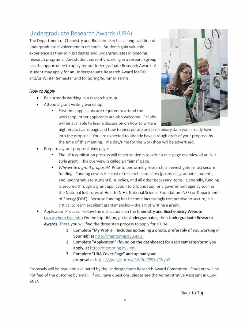
Undergraduate Research Awards (URA) The Department of Chemistry and Biochemistry has a long tradition of
undergraduate involvement in research. Students gain valuable
experience as they join graduates and undergraduates in ongoing
research programs. Any student currently working in a research group
has the opportunity to apply for an Undergraduate Research Award. A
student may apply for an Undergraduate Research Award for Fall
and/or Winter Semester and for Spring/Summer Terms.
How to Apply
Be currently working in a research group.
Attend a grant writing workshop:
First time applicants are required to attend the
workshop; other applicants are also welcome. Faculty
will be available to lead a discussion on how to write a
high-impact aims page and how to incorporate any preliminary data you already have
into the proposal. You are expected to already have a rough draft of your proposal by
the time of this meeting. The day/time for the workshop will be advertised.
Prepare a grant proposal aims page:
The URA application process will teach students to write a one-page overview of an NIH-
style grant. This overview is called an “aims” page.
Why write a grant proposal? Prior to performing research, an investigator must secure
funding. Funding covers the cost of research associates (postdocs, graduate students,
and undergraduate students), supplies, and all other necessary items. Generally, funding
is secured through a grant application to a foundation or a government agency such as
the National Institutes of Health (NIH), National Science Foundation (NSF) or Department
of Energy (DOE). Because funding has become increasingly competitive to secure, it is
critical to learn excellent grantsmanship—the art of writing a grant.
Application Process: Follow the instructions on the Chemistry and Biochemistry Website
(www.chem.byu.edu) On the top ribbon, go to Undergraduates, then Undergraduate Research
Awards. There you will find the three step process to apply for a URA.
1. Complete "My Profile" (Includes uploading a photo, preferably of you working in
your lab) at http://mentoring.byu.edu
2. Complete "Application" (found on the dashboard) for each semester/term you
apply, at http://mentoring.byu.edu
3. Complete "URA Cover Page" and upload your
proposal at https://goo.gl/forms/lPW0reZP5YqT2ryh2
Proposals will be read and evaluated by the Undergraduate Research Award Committee. Students will be
notified of the outcome by email. If you have questions, please see the Administrative Assistant in C104
BNSN.
Back to Top

6
Y-Chem Society
Y-Chem is the student chapter of the Central Utah Section of
the American Chemical Society and is designed to help BYU students
succeed in a challenging scientific environment. Though the focus is
on chemistry and biochemistry, students of every major are
welcome.
Y-Chem is run by a group of students who are passionate
about chemistry. They strive to share their love of science with
others, while helping them with challenges they may encounter
along the way. In addition to their efforts in planning events, they
are good resources for questions that students may have. They also
work closely with several professors.
One of Y-Chem’s main purposes is to help students succeed in their chosen discipline.
Accordingly, many of the activities are directed to this end. Some examples of past activities include
fundraising to sponsor students attending national meetings, graduate school preparation, and tours of
academic and industrial science laboratories.
Another important purpose of the club is community outreach. The students are passionate
about science and want to help kindle that flame in others. Y-Chem members receive the opportunity to
be trained to perform chemistry “magic shows.” Once trained, these members perform dazzling
chemistry demonstrations both on and off campus for a variety of audiences. They also participate in
judging science fairs, as well as Undergraduate Research Night and the BYU major fair. In addition to
these activities, Y-Chem also puts on an annual community outreach event called Open Lab Day. During
Open Lab Day, Y-Chem members engage with junior high and high school students by helping them
perform exciting science experiments.
The international chemistry community is relatively tight-knit. It is quite possible that today’s
classmates will become tomorrow’s colleagues and employers. Y-Chem sponsors several social activities
each year in order to promote networking amongst peers. In addition to meeting and associating with
people on similar career paths, Y-Chem offers its members opportunities to interact with professors.
These professors often become valuable contacts and can offer excellent career advice.
Back to Top

7
Y-Chem’s activities aren’t all business. Y-Chem also seeks to have activities that are just plain
fun—like the annual Nerd Dance.
You are invited to join Y-Chem. It provides a great opportunity to associate with peers and
professors as well as to learn, grow, and serve. Y-Chem strives to be as beneficial as possible and they are
always excited to hear what suggestions members have so that they can better serve them.
To join or to find more information, please visit ychem.byu.edu or contact me at
Sincerely,
Jacob Shaner, President
Back to Top

8
Research Facilities
Research activities occupy more than 50 percent of a
192,000-square-foot building. The University library, where the
science collection includes more than 500,000 volumes and about
9,000 journal subscriptions, is located about 150 yards away.
Major equipment available in the department includes NMR
(200, 300, and 500 MHz); mass spectrometry (high-resolution,
quadrupole, ion cyclotron resonance, ToF-SIMS, and MALDI); X-ray
diffraction (powder and single crystal); spectrophotometry (IR,
visible, UV); lasers (YAG, gas, excimer, Ti-sapphire and dye);
separations—including capillary column GC/MS, ion, and
supercritical fluid chromatography; capillary electrophoresis;
particle size analyzers; environmental chambers; ICP; thermodynamics (calorimeters of all types,
including temperature and pressure scanning, titration, flow, heat conduction, power compensation,
combustion, and metabolic); and molecular biology (DNA synthesizer and sequencer, phosphorimager,
tissue culture facility, recombinant DNA facility, fluorescence activated cell sorter, and ultracentrifuges).
All computing facilities are fully networked, including computational chemistry and laboratory
workstations as well as office personal computers, with convenient connection to supercomputing
facilities and the internet. Fully staffed shops for glassblowing, machining, and electronics also serve
research needs.
Back to Top

9
FACULTY RESEARCH PROFILES
Back to Top

10
Joshua L. Andersen, PhD
Biochemistry
E111 BNSN, 422-7193
Email: [email protected]
Mechanisms of Chemoresistance in Cancer
Nearly half of us will be afflicted with cancer in our lifetime and for the
majority of patients, cytotoxic chemotherapy is the primary treatment
option. The goal of these treatments is to induce tumor cell death. However, these therapies are often
ineffective because tumor cells possess the dynamic ability to subvert cell death and become
chemoresistant. With this in mind, our research combines molecular and proteomics approaches to
understand the mechanisms by which tumor cells gain resistance to cell death and chemotherapy, with
the ultimate goal of developing therapeutic approaches to overcome chemoresistance and improve
clinical outcomes for patients.
Our recent work has focused on the dynamic phospho-binding protein 14-3-3ζ and its role in
promoting tumor cell growth and survival. As part of this effort, we have harnessed 14-3-3ζ proteomics as
a tool to identify novel tumor cell survival mechanisms. This work uncovered a mechanism by which
tumor cells “switch on” a catabolic process called autophagy, which allows the tumor cell to survive
through periods of stress commonly encountered in vivo. Another interest of our lab is to understand
how acetylation of non-histone proteins regulates apoptotic/survival signaling in tumor cells. Since the
experimental tools to study acetylation are limited, a critical part of our early effort has been in
developing new tools to understand acetylation. These include comparative acetyl-proteomics
approaches, biotin-switch methods to identify deacetylase substrates, and site-specific acetyl-lysine
antibodies. Using these tools, we recently uncovered a mechanism by which the pro-tumor activity of 14-
3-3ζ is governed by acetylation. We now have the ability to “turn off” 14-3-3ζ activity in tumor cells by
modulating the enzymes that govern 14-3-3ζ acetylation, which we are now evaluating as a potential
therapeutic approach in breast cancer.
Your research in my laboratory will expose you to current issues in the cancer field, and will teach
you the molecular and biochemical tools to solve pressing research questions. Dedicated students will
also get the chance to be a part of the larger scientific community through research presentations at
Back to Top

11
international meetings, and preparing and submitting research manuscripts to peer-reviewed journals. I
encourage interested and highly motivated undergraduates to apply for research opportunities in our
laboratory—there is no requirement for previous research experience or years in school.
References
1. Weerasekara VK, Panek DJ, Broadbent DG, Mortenson JB, Mathis AD, Logan GN, Prince JT,
Thomson DM, Andersen JL. Metabolic stress-induced rearrangement of the 14-3-3z interactome
promotes autophagy via a ULK1-and AMPK-regulated 14-3-3z interaction with phosphorylated
Atg9. Molecular and Cellular Biology. Epub ahead of print.
2. Mortenson JB, Heppler LN, Banks CJ, Weerasekara VK, Whited MD, Piccolo SR, Johnson WE,
Thompson JW, Andersen JL. "Deacetylation of Lysines within the 14-3-3ζ Binding Pocket." Journal
of Biological Chemistry; 290 (20):12487-12496.
3. Johnson SE, Lindblom KR, Robeson A, Stevens RD, Ilkayeva OR, Newgard CB, Kornbluth
S, Andersen JL. (2013) Metabolomics profiling reveals a role for caspase-2 in
lipoapoptosis. Journal of Biological Chemistry 17; 288(20):14463-75
Back to Top

12
Merritt B. Andrus, PhD
Organic & Biomolecular Chemistry
C410 BNSN, 422-8178
Email: [email protected]
Natural Product Synthesis
Efforts in our lab are focused on methods for the synthesis of biologically-
active natural products that possess unique structures and potential for
combinatorial library construction and screening. New methods include metal-catalyzed couplings and
condensations to assemble key intermediates. Libraries of structural variants are then made and used to
probe receptor binding and improve activity.
Recent work includes the synthesis of the polyene stipiamide, a new agent to treat multidrug
resistance (MDR); geldanamycin A, a large anticancer macrocycle; englerin A, a terpene based anticancer
agent; and resveratrol, a small disease prevatative stilbene.
Resveratrol, a simple, yet very important target, is the suspected causative agent of the “French
Paradox.” Diets rich in foods that contain this material, grapes in particular, lead to lower rates of cancer
and heart disease. New coupling methods and strategies developed to produce this material will now be
used to produce structural variants for various screens. New targets now include F4-4, an antiviral lignin
Back to Top

13
natural product that inhibits herpes and shingle infection, and simplified analogs of englerin A. General
synthetic methods with broad application are also under development using new ligands for asymmetric
styryl Diels-Alder and aldol transformations.
Dedicated undergraduate students including beginning students are welcome to participate in all
aspects of the work.
References
1. “Design and Synthesis of Terpene Based Englerin A Mimics Using Chromium Oxide Mediated
Remote CH2 Oxidation” Acerson, M. J.; Bingham, B. S.; Allred, C. A.; Andrus, M. B. Tetrahedron
Lett. 2015, 56, 3277–3280.
2. “Selective Esterification of the Polyphenol Resveratrol at the 4’-Position” Acerson, M. J.; Andrus,
M. A. Tetrahedron Lett. 2014, 55, 757–760.
3. “Quantum Mechanical Transition-State Analysis Reveals the Precise Origin of Stereoselectivity in
Chiral Quaternary Cinchonidinium Phase-Transfer Catalyzed Enolate Allylation” Cook, T. C.;
Andrus, M. B.; Ess, D. E. Org. Lett. 2012, 14, 5836–5839.
Back to Top

14
Matthew C. Asplund, PhD
Physical Chemistry
C309 BNSN, 422-5275
Email: [email protected]
Organometallic Photochemistry
The development of short-pulsed lasers, from 10 femtoseconds (10 x 10-15
s) to nanoseconds (10-9 s) has allowed for unprecedented information into
the dynamics of chemical reactions. With pulses of light this short we can easily measure the spectra of
chemical intermediates in condensed phase (primarily liquid solution) chemical reactions. A first photon
(usually in the visible or UV region of the spectrum) begins the reaction, and the intermediates can be
monitored on a number of time scales in the infrared to give structural detail. We have used this
instrumentation to study a class of organometallic intermediates important in chemical catalysis. The
reaction begins when a photon of UV light causes one ligand to dissociate from a metal center to form a
metallic radical. On a very short time-scale, this unsaturated metal center forms a complex with a
neighboring solvent molecule. Over time, this complex exchanges with other solvent molecules until it
finally decomposes after 5-10 seconds. By following the infrared spectrum of the complex, we can
measure the dynamics and binding energy of these weak complexes, and compare them with quantum
chemical calculations.
Model Ring Formation Reactions
One area of particular interest in my lab is reactions involving organometallic species involved in
the formation of new carbon-carbon bonds and the formation of rings. An interesting class of reactions is
labeled Pauson-Khand reactions. In its most general form, it is the reaction of an alkene, and alkyne a
carbonyl to form a 5-membered cyclopenteneone ring.
Back to Top

15
The reaction proceeds thermally, and in order to follow the reaction with time-resolved
spectroscopic methods, we use a variant of the reactant that combines the alkene and alkyne in the same
molecule.
The reaction mechanism shows that the first step is the removal of a CO from the Mo(CO)6,
followed by formation of a complex between the Mo(CO)5 and the complex, followed by formation of the
ring. We are trying to establish which part of the ligand attaches to the metal first.
Bi-metal catalyst systems
One of the difficulties in current catalytic systems is that they usually require use of a rare and
expensive metal atom. There is tremendous interest in using bimetallic systems where the two atoms act
cooperatively to give reactions that are similar to rare metals. While there are many catalytic reaction
studies that have established the viability of this approach, there is little known about the details of the
reactions. We are applying our transient infrared spectroscopy to these bi-metallic systems to try to
understand how these cooperative systems drive chemistry.
Back to Top

16
Daniel E. Austin, PhD
Analytical Chemistry
C310 BNSN, 422-1551
Email: [email protected]
Chemistry and Biology of High-Velocity Impacts: Simulating Space Processes
We are developing experimental tools to explore high-velocity impacts—particularly those of
molecules, sub-micron particles, and microorganisms—on surfaces. In such impacts, the kinetic energy
of the impacting species is converted to vibrational modes, which can drive both physical/biological
changes and chemical reactions. As an example of a project in this area, we are building an ultra-fast
rotor that allows molecule-surface impacts at 2-4 km/s. This is faster than can be studied using any
other technique. The resulting molecular fragmentation is studied using gas chromatography mass
spectrometry. This project has application to the study of planetary exospheres using mass
spectrometers on flyby or orbiting spacecraft. We are also studying the impact survivability of bacterial
spores, with the goal of exploring possible transfer of bacteria in space. Finally, we are looking at high-
velocity impacts of aerosols and icy grains to gain understanding of space measurements of particulates.
Undergraduate students receive training in building scientific equipment, particularly vacuum
systems and mass spectrometers. Any chemistry, physics, or engineering students who have completed
their first two years of undergraduate study are invited to join.
Back to Top

17
Steven L. Castle, PhD
Organic & Biomolecular Chemistry
C411 BNSN, 422-1780
Email: [email protected]
Synthetic Organic Chemistry, Peptide Chemistry
Our research focuses on the total synthesis of complex bioactive natural
products and peptides. The structures of such compounds serve as
inspiration for the invention of new organic reactions and processes. Additionally, studies of their
bioactivity can increase our understanding of their modes of action, potentially leading to the design and
development of new therapeutic agents. Some of our recent synthetic targets are shown below.
The new reactions that we develop in the course of synthesizing a target compound are fully
investigated with respect to scope and mechanism. It is our aim to develop widely applicable processes
that deliver complex products from simple starting materials in a minimum number of steps. We also
believe that it is important to understand how these processes operate.
We frequently synthesize structural
analogues of the target natural products or
peptides. This allows us to elucidate the modes of
action of these compounds, often in collaboration
with biological and biochemical research groups.
We are also engaged in finding new ways to
stabilize peptides to proteolytic degradation,
thereby increasing their potential as drugs.
Students in our group receive rigorous
training in the techniques of organic synthesis and
structure determination. In addition, they learn the more general, widely applicable skills of strategic
planning and problem solving. Furthermore, in the course of presenting their research in verbal and
written formats, they acquire valuable communications skills. Prior to joining our group, students should
have completed Chem 351, 352, and 353/354 (concurrent enrollment in 352 and 353/354 is acceptable).
Back to Top

18
References
1. Cai, Y.; Jalan, A.; Kubosumi, A.R.; Castle, S. L. “Microwave-Promoted Tin-Free Iminyl Radical
Cyclization with TEMPO Trapping: A Practical Synthesis of 2-Acylpyrroles.” Org. Lett. 2015, 17,
288-491.
2. Ma, Z.; Jiang, J.; Luo, S.; Cai, Y.; Cardon, J.M.; Kay, B.M.; Ess, D.H.; Castle, S.L. “Selective Access to
E- and Z-ΔIle-Containing Peptides via a Stereospecific E2 Dehydration and an O → N Acyl
Transfer.” Org. Lett. 2014, 16, 4044-4047.
3. Ma, B.; Banerjee, B.; Litvinov, D.N.; He, L.; Castle, S.L. “Total Synthesis of the Antimitotic Bicyclic
Peptide Celogentin C.” J. Am. Chem. Soc. 2010, 132, 1159-1171.
Back to Top

19
Kenneth A. Christensen, PhD
Biochemistry, Analytical Chemistry E205 BNSN, 422-0249 Email: [email protected] Website: https://christensenlab.byu.edu
My lab works in the fields of biochemistry and bioanalytical chemistry. We develop methods that apply optical spectroscopy, flow cytometry, time-lapse microscopy, and other current analytical and biophysical techniques to questions in biochemistry, biophysics, cell and microbiology.
A current area of research in my lab grew out of our discovery several years ago that the anthrax toxin receptors capillary morphogenesis
gene protein 2 (ANTRX2/CMG2) and tumor endothelial marker 8 (ANTRX1/TEM8) were involved in angiogenesis in pathological conditions in the eye and in tumor models. Our current work focuses on identifying and characterizing critical extracellular ligands of the CMG2 and TEM8, which are thought to be extracellular matrix proteins. In addition, we are also identifying intracellular interactors via proteomics using mass spectrometry. Together, the lab is trying to address a critical barrier to progress in this field by identifying the role these cell surface receptors play in angiogenesis and developing a model that can be tested using traditional biochemical approaches. In parallel with these efforts, we also have some small molecule and peptide scaffolds that are being tested for activity in a corneal neovascularization animal model and for other pathologies of angiogenesis.
A second project focuses on measuring and monitoring glucose metabolism in eukaryotic parasites. For example, in Trypanosoma brucei (the causative agent of Human African Trypanosomiasis), the sole source for generating ATP during the infectious lifecycle stage of the African trypanosome occurs exclusively in a unique peroxisome-like compartment called the glycosome. We are developing and using both recombinant protein-based FRET sensors and peptide-targeted small molecule sensors to quantitatively measure intraglycosomal pH, glucose, and ATP levels in live parasites. We are interested in the mechanisms the organism uses for regulation of pH, glucose, ATP production, and other important metabolites. Since glycolysis is key to parasite survival, inhibiting glycolysis in the glycosome could be an excellent targeted therapeutic approach for treatment of African Trypanosomiasis. A high-throughput screen to identify small molecules that block glucose uptake using our FRET sensors was recently funded. Other parasites of interest are Leishmania donovoni and Trypanosoma cruzi.
Note: I am willing to work with beginning students.
Selected References
1. Rogers, M.S., Christensen, K.A., Wigelsworth, D.J., Collier, R.J., and D’Amato, R.J., “Mutant AnthraxToxin B-moiety (Protective Antigen) Inhibits Angiogenesis and Tumor Growth”, Cancer Research, 67,9980-9985 (2007)
2. Lin, S., Voyton, C., Morris, M.T., Ackroyd, P.C., Morris, J.C., and Christensen, K.A., “pH Regulation inGlycosomes of Procyclic Form Trypanosoma brucei”, Journal of Biological Chemistry, In Press (2017),DOI: 10.1074/jbc.M117.784173
3. Gordhan, H., Milanes, J., Qiu, Y., Golden, J., Christensen, K., Morris, J., and Whitehead, D., “A targeteddelivery strategy for the development of potent trypanocides”, Chemical Communications, In Press(2017), DOI: 10.1039/C7CC03378H
Back to Top

20
David V. Dearden, PhD
Analytical/Physical Chemistry
C104B BNSN, 422-2355
Email: [email protected]
Our group uses one of the most powerful types of mass spectrometry,
combined with molecular modeling using high-end supercomputers, to
develop methods for characterizing molecule-sized devices. My goal is to
give students a real taste of the kind of work done by researchers on the
cutting edge of science, culminating in publication and/or presentation of the results. Our group has an
excellent track record placing undergraduates who desire additional training in some of the world’s top
graduate programs. Projects will be selected based on the student’s level of preparation. All of our work
includes strong possibilities for collaboration with other groups working in related areas.
Tertiary Structure from Mass Spectrometry: A “CRAFTI” New Method
Tertiary structure (the way a molecule is folded, resulting in its overall shape) is extremely
important to molecular function in such diverse areas as biochemistry, catalysis, and the assembly and
function of molecular nanomachines. Therefore, it is important to develop ways to determine tertiary
structure, and to do so with very small samples. Although mass spectrometry is a powerful, sensitive
technique for characterizing the atomic composition and connectivity of atoms within molecules, it
usually yields no information about tertiary structure. We recently invented a new technique for
obtaining tertiary structural information using Fourier transform ion cyclotron resonance mass
spectrometry; we call the technique "CRAFTI" (from cross sectional areas by Fourier transform ion
cyclotron resonance). Interested students will explore the strengths and limitations of this new
technique, and develop supramolecular chemistry applications for it, supported by funding from the
National Science Foundation. Students who are at least concurrently registered for Chem 461/462 will be
most successful in these projects, although motivated students with less preparation (as little as Chem
111 or Chem 105) can also do excellent experimental work. Commitments of about 10 hours per week
are generally required to make meaningful progress on experimental projects.
Back to Top

21
A Picture is Worth a Thousand Words: Molecular Modeling
Visualization and modeling of molecular systems is an essential part of our research. Software
packages such as SPARTAN, ECCE, and NWChem will be used to model the same host-guest complexes
we are studying experimentally. All of these software packages have intuitive, graphical user interfaces
that make them easy to operate. Goals of the modeling projects include determination of low-energy
structures and energies for the complexes, dynamic simulation of the complexes, and calculation of
vibrational frequencies that can then be used as input to statistical mechanics programs. Much of this
work is computationally very demanding and will require use of campus supercomputers. No prior
knowledge of either modeling methods or computer operating systems is needed, but students will need
to learn to be comfortable with UNIX. Students taking Chem 351/352 have sufficient background to carry
out these projects successfully, and motivated students who are at earlier stages of their preparation will
Back to Top

22
also be able to make important, significant contributions. Again, to make meaningful progress on these
projects students will need to commit to about 10 hours per week.
The undergraduates involved in this work will have full access to our state-of-the-art equipment.
We have a well-equipped research lab centered around a Fourier transform ion cyclotron resonance
(FTICR) mass spectrometer with a 4.7-Tesla superconducting magnet and an external ion source equipped
with electrospray and sonic spray ionization modules (Bruker model APEX 47e). All of this equipment is
computer-interfaced and script-controllable, allowing very versatile experiments to be designed and
performed. For students with good mechanical or programming skills interested in building instruments,
we currently have ongoing needs for instrument control and data analysis software development.
References
1. "Quantitative Collision Cross-Sections from FTICR Linewidth Measurments: Improvements inTheory and Experiment," Anupriya; Gustafson, E.; Mortensen, D. N.; Dearden, D. V. J. Am. Soc.Mass Spectrom. 2017, in press.
2. "Collision Cross Sections for 20 Protonated Amino Acids: Fourier Transform Ion CyclotronResonance and Ion Mobility Results," Anupriya; Jones, C. A.; Dearden, D. V. J. Am. Soc. MassSpectrom. 2016, 27, 1366-1375.
3. "Effects of Kinetic Energy and Collision Gas on Measurement of Cross Sections by FourierTransform Ion Cyclotron Resonance Mass Spectrometry," Yang, F.; Jones, C. A.; Dearden, D. V. Int.J. Mass Spectrom. 2015, 378, 143-150.
4. "Linewidth Pressure Measurement: a New Technique for High Vacuum Characterization," Jones,C. A.; Dearden, D. V. J. Am. Soc. Mass Spectrom. 2015, 26, 323-329.
5. "Binding of α,ω-Alkyldiammonium Ions by Cucurbit[n]urils in the Gas Phase," Yang, F.; Jones, C.A.; Selvapalam, N.; Ko, Y. H.; Kim, K.; Dearden, D. V. Supramolecular Chem. 2014, 26, 684-691.
6. "Collision Cross Sectional Areas from Analysis of Fourier Transform Ion Cyclotron Resonance LineWidth: A New Method for Characterizing Molecular Structure," Yang, F.; Voelkel, J.; Dearden, D.V. Anal. Chem. 2012, 84, 4851-4857.
Back to Top

23
Daniel H. Ess, PhD
Computational Chemistry
C403 BNSN, 422-9164
Email: [email protected]
Inorganic and Organometallic Theory and Computation
My group uses and develops quantum-chemistry and molecular dynamics
methods to discover mechanisms, reactivity principles, and selectivity for
experimentally important chemical reactions related to catalysis, energy,
and organic synthesis. My group emphasizes making predictions and designing catalysts that are then
realized in the laboratory. This naturally leads to close collaboration with experimental groups in
academia and industry. My group publishes several top-tier publications each year and undergraduates
are very often co-authors. Current areas of research involve: (1) Computational catalyst design with
industrial application. (2) Computational studies of alkane C-H functionalization reactions. (3)
Computational studies on multinuclear catalysis. (4) Computational tools development and testing.
Undergraduate students in my lab have a range in backgrounds, from a computer science minor to no
programming experience. To be successful all you need to do is have a desire to learn inorganic and
organic chemistry, develop computer skills, and work hard (15-20 hours per week in Fall/Winter and 30-
40 hours per week in Spring/Summer).
Back to Top

24
Paul B. Farnsworth, PhD
Analytical Chemistry
C211A BNSN, 422-6502
Email: [email protected]
Mass spectrometry is an exceptionally powerful analytical technique that
plays a critical role in all chemistry sub-disciplines, and is also having a
tremendous impact on the biological sciences. Research in my group
focuses on ion sources for a variety of mass spectrometers. We seek to
understand the fundamental processes that make existing ion sources work, and to use that
understanding to develop sources that are more sensitive and versatile. Our current projects fall into two
general categories: elemental mass spectrometry and ambient ionization mass spectrometry.
Elemental Mass Spectrometry
Ions from a plasma (a very hot gas) can be extracted into a
mass spectrometer, which sorts and counts them to give an
extremely sensitive indication of what atoms were present in the
plasma. The picture at the left is of an inductively coupled plasma
aimed at the entrance of a mass spectrometer. This combination,
called ICP-MS, is capable of detecting elements in solution at the parts per quadrillion level. That is
roughly equivalent to detecting a small grain of salt dissolved in an Olympic swimming pool. We are
using lasers to understand what happens to the ions as they pass from the plasma, which is at
atmospheric pressure, to the mass spectrometer, which is under high vacuum. There are serious
problems with instruments that rely on this transformation from a plasma to an ion beam because the
transformation is poorly understood. The goal of our research is to provide the fundamental insights into
ion extraction behavior that will allow the construction of improved instruments.
Ambient Ionization Sources for Mass Spectrometry
DESI (desorption electrospray ionization) and DART (direct analysis in real time) are two of a
family of techniques that produce ions at atmospheric pressure and room temperature. The techniques
make it possible to perform very rapid analysis of a variety of materials, including pharmaceuticals, paper,
Back to Top

25
and plant tissues. A sample can be placed in one of the ambient sources with no sample preparation, and
it is possible to detect very small quantities of chemicals on the surface of the sample. We are currently
collaborating with the J. C. Price research group to use DESI for chemical imaging of mouse tissues. The
mice have been fed deuterated water, and our chemical images tell us how fast material in the mouse’s
diet is incorporated into the tissue. We are essentially creating maps of metabolic rates.
It is a goal in my research to have each student take ownership of a project or part of a project,
develop that project to the point that the student can give a presentation at a national meeting, and in
many cases write a paper for the peer-reviewed literature. We have excellent facilities and a close-knit
research group composed of a mix of graduate and undergraduate students.
References
1. Nicholas Taylor, Kyli N. McKay-Bishop, Ross L. Spencer and Paul B. Farnsworth, “A novel approach
to understanding the effect of matrix composition on analyte emission in an inductively coupled
plasma.” Journal of Analytical Atomic Spectrometry, 29, 644-656 (2014)
2. Charlotte Reininger, Kellie Woodfield, Joel D. Keelor, Adam Kaylor, Facundo M. Fernandez, and
Paul B. Farnsworth, “Absolute number densities of helium metastable atoms determined by
atomic absorption spectrometry in helium plasma-based discharges used as ambient
desorption/ionization sources for mass spectrometry,” Spectrochimica Acta part B, 100, 98-104
(2014)
3. Lance M. Moses, Wade C. Ellis, Derick D. Jones and Paul B. Farnsworth, “Fluorescence imaging of
ion distributions in an inductively coupled plasma with laser ablation sample introduction,”
Spectrochimica Acta, part B, 105, 47 - 59 (2015).
4. Wade C. Ellis, Charlotte R. Lewis, Anna P. Openshaw, and Paul B. Farnsworth, “The Ef-fects of
Added Hydrogen on Noble Gas Discharges used as Ambient Desorption/Ionization Sources for
Mass Spectrometry,” Journal of the American Society for Mass Spectrometry,” 27, 1539-1549
(2016).
Back to Top

26
Steven R. Goates, PhD
Analytical Chemistry
C311 BNSN, 422-2227
Email: [email protected]
Lasers have opened up new realms of study in
virtually every field of science. My students and I
employ lasers and spectroscopic techniques in a
variety of studies, some of which are described
below. The work is challenging, but I do not expect that students will enter my
program with any background in this field of research. An eagerness to learn, self-
motivation, and a readiness to tackle unfamiliar problems are the
primary qualifications.
Spectroscopy at surfaces and interfaces
Unusual effects can be achieved with lasers. Among these
effects are non-linear wave generation in which new beams are
generated when two or more laser beams converge on a sample.
An example is sum frequency spectroscopy (SFS) pictured in
Figure 1. Another experiment of this type is coherent anti-Stokes
Raman spectroscopy (CARS). Not only are such experiments
impossible to do with ordinary light, but they reveal details about
chemical structure at surfaces and interfaces (where much of the
action in chemistry takes places).
Supersonic Jet Spectroscopy
Supersonic jet spectroscopy is a high resolution technique which we have shown can be used to
selectively detect compounds of interest in complex samples containing hundreds to thousands of
substances. Supersonic jet streams are formed when gases at high pressures are expanded through a
small nozzle into a vacuum; molecules in the resulting gas jet begin to move at supersonic speeds and, in
Figure 1. Schematic depiction of an SFS experiment. Two laser beams at different colors are mixed at the sample, generating a new beam at a third color.
ν1
ν2
ν2 + ν1
Back to Top

27
the process, give up their thermal energy to forward motion. Ideally, a jet of very cold, but isolated
molecules result, and the spectrum becomes much sharper and more detailed.
This detail allows unambiguous identification of the compounds in even a very
complex sample.
We have used supercritical fluids (which flow like gases but dissolve
substances like liquids) as carriers to enable analysis of large molecules by
supersonic jet spectroscopy. Large molecules can be introduced into a supersonic
jet by dissolving them in a supercritical fluid and then supersonically expanding the
solution. This approach has allowed us to do detailed analysis of compounds in
fossil fuel-related pollutants. Our current interests lie in applying this approach to the selective detection
of biologically important natural products.
Raman and Laser-Induced Fluorescence Spectroscopy of Mobile Phase Behavior
We have long had an interest in
understanding the physical processes involved in
chromatographic separations. At present we are
particularly interested in what happens to the
mobile phase in solvating gas chromatography
(SGC) during the course of a separation. SGC is a
high-speed separations method in which the mobile
phase is in a supercritical state (i.e. above its critical
pressure and temperature) at the head of the column, but converts to a gas at the end of the column. To
take full advantage of this separation method it is important to determine how the mobile phase changes
along the column and at what point it converts to a gas. We are using Raman spectroscopy to answer
these questions because Raman allows the mobile phase to be probed without disrupting its flow.
Beyond this particular study, we believe Raman will prove to be a valuable probe of a number of mobile
and stationary phase changes in real chromatography columns.
Figure 2. Image of a supersonic jet.
Back to Top

28
Steven W. Graves, PhD
Biochemistry
C212 BNSN, 422-2148
Email: [email protected]
Serum Proteomics, Peptidomics and Lipidomics of Various Diseases,
Including Preterm Birth, Preeclampsia, Alzheimer’s Disease, and
Endometriosis
For several years those in my laboratory have developed and applied one
or more serum “omics” methods that typically incorporate capillary liquid chromatography coupled to
mass spectrometry. Using this method we can interrogate ≈7,000-8,000 molecules in a small volume of
blood. We have found quantitative differences in low abundance serum peptides, small proteins and
lipids in pregnant women who later experience complications compared with women having
uncomplicated pregnancies. We have identified several molecules, weeks prior to preterm labor, which
are quantitatively different in women who will deliver early. Different sets of “markers” allow us to
predict 85-90% of those pregnant women who will develop this complication. We have conducted other
studies using this same approach and have found several biomarkers that predict which pregnant women
will later develop a very serious complication of pregnancy termed preeclampsia. Using combinations of
our biomarkers we can predict ≈90% of women who will later develop preeclampsia. More recently, we
used our serum proteomic approach to study Alzheimer’s disease (AD) and have found dozens of
biomarkers that allow for the correct diagnosis of AD and even to distinguish very early stage disease.
Some of these biomarkers may even allow for the prediction of those who might develop this serious
disease. Other markers appear to be specific to women or men with AD which may help to explain
gender differences in AD. Currently, we are evaluating serum peptide and lipid markers for
endometriosis, a complication affecting up to 10% of women of child bearing age that results in chronic
or intermittent severe pain and frequently infertility in these women. Currently, the diagnosis requires
surgery but preliminary data from our “omics” approach suggests that biomarker panels can diagnosis
endometriosis biochemically without surgery.
Back to Top

29
References
1. Anand S, Alvarez TMB. Johnson WE, Esplin MS, Merrell K, Porter TF, Graves SW. Serum biomarkers
predictive of preeclampsia. Biomarkers in Medicine 2015;9:563-575.
2. Kedia K, Nichols CA, Thulin CD, Graves SW. Novel ‘omics’ approach to study low abundance, low
molecular weight components in a complex biological tissue: Regional differences between chorionic
and basal plates of human placenta. Anal Bioanal Chem (epub 09-2015) 2015;407:8543-8556.
3. Shah DJ, Rohlfing F, Anand S, Johnson WE, Alvarez MTB, Cobell J, King J, Young SA, Kauwe JSK, Graves
SW. Discovery and Subsequent Confirmation of Novel Serum Biomarkers Diagnosing Alzheimer’s
Disease. J Alzheimers Dis (epub 09-2015) 2016;49:317-327
4. Anand S, Young SA, Esplin MS, Peaden B, Tolley HD, Porter TF, Varner MW, D’Alton ME, Jackson BJ,
Graves SW. Detection and confirmation of serum lipid biomarkers for preeclampsia using direct
infusion mass spectrometry. J Lipid Res 2016;57:687-696.
5. Kedia K, Smith SF, Wright AH, Barnes JM, Tolley HD, Esplin MS, Graves SW. Global “omics” evaluation
of human placental responses to preeclamptic conditions. Amer J Obstet Gynecol 2016;215:238.e1-
238.e20.
6. Kang H, Roeder BL, Martin HM, Anand S, Cook AC, Lau CY, Buckmiller EM, Brown KK, Eggett DL,
Graves SW. A lipidomic approach reveals colostrum and milk biomarkers predictive of production-
related metabolic disease. J Agri Life Sci 2017;3 (In press)
7. Anand S, Barnes JM, Young SA, Garcia DM, Tolley HD, Kauwe JSK, Graves SW. Discovery and
confirmation of diagnostic serum lipid biomarkers for Alzheimer’s disease using direct infusion mass
spectrometry. J Alzheimer’s Dis 2017;59:277-290.
Back to Top

30
Jaron C. Hansen, PhD
Analytical Chemistry
C307 BNSN, 422-4066
Email: [email protected]
Atmospheric Chemistry and Renewable Energy
Research in the Hansen group is divided into three elements: (1) Kinetics
and Spectroscopy of Environmentally Important Reactions (2) Air Sampling
Campaigns (3) Biofuel/Alternative Energy. Our group couples together
computational and experimental studies to investigate the kinetics and mechanisms of important
atmospheric reactions. Laboratory studies are complimented by air sampling campaigns designed to
investigate the sources of air pollution. We utilize an environmental chamber to aid in the interpretation
of our air sampling campaign studies. We also have an active research element in our group that studies
the conversion of biomass into energy. Details about his group’s efforts in element 1 are included below.
Interested students are encouraged to contact Dr. Hansen with questions about his research in Air
Sampling Campaigns and Biofuel/Alternative Energy. Undergraduate students with at least two years of
classwork are often utilized as research assistants in his group.
Kinetics and Spectroscopy of Earth’s Atmosphere
Organic peroxy radicals (RO2) play an important role in the atmospheric oxidation and in the
combustion of hydrocarbons and are important precursors in the formation of smog. They are formed as
a result of the addition of molecular oxygen to alkyl radicals. In polluted environments, the most
important loss mechanism for RO2 radicals is by reaction with NO:
RO2 + NO → RO + NO2 (1)
→ RONO2 (2)
The dominate product pathway oxidizes NO to NO2 , ultimately resulting in the formation of tropospheric
ozone and production of an alkoxy radical (RO). For larger alkyperoxy radicals a substantial fraction of the
product yield is stable alkyl nitrate species (RONO2). Our group has proposed the idea that RO2 radicals
can form loosely bound van der Waals complexes with water vapor (as well as other polar species). These
complexes are of interest to atmospheric/environmental chemists because their formation has been
associated with an increase in their reactivity.
Back to Top
31
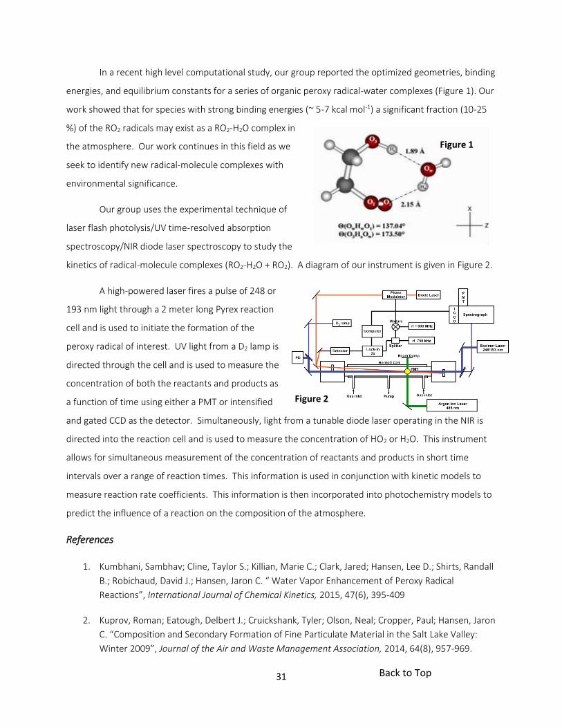
In a recent high level computational study, our group reported the optimized geometries, binding
energies, and equilibrium constants for a series of organic peroxy radical-water complexes (Figure 1). Our
work showed that for species with strong binding energies (~ 5-7 kcal mol-1) a significant fraction (10-25
%) of the RO2 radicals may exist as a RO2-H2O complex in
the atmosphere. Our work continues in this field as we
seek to identify new radical-molecule complexes with
environmental significance.
Our group uses the experimental technique of
laser flash photolysis/UV time-resolved absorption
spectroscopy/NIR diode laser spectroscopy to study the
kinetics of radical-molecule complexes (RO2-H2O + RO2). A diagram of our instrument is given in Figure 2.
A high-powered laser fires a pulse of 248 or
193 nm light through a 2 meter long Pyrex reaction
cell and is used to initiate the formation of the
peroxy radical of interest. UV light from a D2 lamp is
directed through the cell and is used to measure the
concentration of both the reactants and products as
a function of time using either a PMT or intensified
and gated CCD as the detector. Simultaneously, light from a tunable diode laser operating in the NIR is
directed into the reaction cell and is used to measure the concentration of HO2 or H2O. This instrument
allows for simultaneous measurement of the concentration of reactants and products in short time
intervals over a range of reaction times. This information is used in conjunction with kinetic models to
measure reaction rate coefficients. This information is then incorporated into photochemistry models to
predict the influence of a reaction on the composition of the atmosphere.
References
1. Kumbhani, Sambhav; Cline, Taylor S.; Killian, Marie C.; Clark, Jared; Hansen, Lee D.; Shirts, Randall
B.; Robichaud, David J.; Hansen, Jaron C. “ Water Vapor Enhancement of Peroxy Radical
Reactions”, International Journal of Chemical Kinetics, 2015, 47(6), 395-409
2. Kuprov, Roman; Eatough, Delbert J.; Cruickshank, Tyler; Olson, Neal; Cropper, Paul; Hansen, Jaron
C. “Composition and Secondary Formation of Fine Particulate Material in the Salt Lake Valley:
Winter 2009”, Journal of the Air and Waste Management Association, 2014, 64(8), 957-969.
Figure 1
Figure 2
Back to Top

32
Roger G. Harrison, PhD
Inorganic Chemistry
C104A BNSN, 422-8096
Email: [email protected]
Molecular Binding and Encapsulation
The supramolecular interactions between a host molecule and a guest molecule allow for such
interactions as selective binding, chiral recognition, and separation. Our introduction into the field of
host-guest complexes came with the discovery in our group of a metal-assembled capsule, consisting of
two synthesized cup-shaped molecules brought together by metal ions. Along with the capsules, we have
formed metal-resorcinarene complexes with various metal ions such as iron, cobalt and copper. We are
now pursuing with interest host molecules that will
selectively bind one enantiomer preferentially over
another. We are also exploring the synthesis and
properties of larger host molecules and their ability to
encapsulate water contaminants. Students working on
this project learn to synthesize and characterize organic
and inorganic compounds and use them to bind or
encapsulate other molecules.
Separations of Contaminants Using Macrocyclic
Compounds
Another related area of research we are pursuing is the application of cavitands in separations.
Small quantities of molecules are harmful to us as water contaminants or unwanted substances in our
body. Students in our group use ion chromatography to detect and quantify anions, cations,
pharmaceuticals and biological compounds. To do this they pack columns with cavitands and perform
separations using an ion chromatography instrument. Students become experts in separation techniques
and use their skills to analyze molecules in tap water, river water, and biological fluids.
Cavitand molecule with alanines along its
upper rim.
Back to Top

33
Metal Compounds Designed to Combat the Flu
The flu virus has a protein channel used for proton movement that is essential for viral
replication. Students in our lab design, synthesize, characterize and test metal containing
compounds for blocking the protein channel. We have found neutral copper compounds are
able to block the channel and inhibit viral growth. Students working on this project learn organic
synthesis techniques and how to characterize compounds by NMR, MS, ICP, and IR. This is a
collaborative project with the Busath lab, whose students test the compounds for viral activity.
Nanomaterial Synthesis and Properties
Materials with subunits in the nanometer range are being
studied for their semiconductor and energy transfer properties.
Members of our group synthesize nanoparticles, nanoprisms, and
nanoplates made of ZnO and investigate their light absorption and
emission properties, as well as gas adsorption. Students on this project
synthesize new nanomaterials and while characterizing them, learn to
operate many instruments, such as XRD, SEM, TEM, ICP, Uv-vis and NMR.
References
1. N. Li, F. Yang, H. A. Stock, D. V. Dearden, J. D. Lamb, R. G. Harrison “Cavitands with chiral
substituents: resorcinarene-based cavitands with amino acid functional groups” Org. Biomol.
Chem. 2012, 10, 7392-7401.
2. T. Panahi, D. J. Weaver, J. D. Lamb, R. G. Harrison “A new approach for trace analysis of guanidine
compounds in surface water with resorcinarene-based ion chromatography columns” Analyst,
2016, 141, 939-946.
3. N. Gordon, K. McGuire, S. Wallentine, G. Mohl, J. Lynch, R. G. Harrison, D. Busath “Divalent
copper complexes as influenza A inhibitors”. Antiviral Research, 2017, submitted.
4. J. M. Hancock, W. M. Rankin, T. M. Hammad, J. K. Salam, K. Chesnel, R. G. Harrison “Optical and
magnetic properties of ZnO nanoparticles doped with Co, Ni, and Mn and synthesized at low
temperature” J. Nanosci. Nanotech. 2015, 15, 3809-3815.
Prisms made of ZnO
Back to Top

34
Jeremy A. Johnson, PhD
Physical Chemistry
C312 BNSN, 422-0245
Email: [email protected]
Creating Ultrafast Spectroscopy
Light can be a wonderful tool to measure all sorts of fascinating material
properties, but there is one important truth all spectroscopists keep in
mind: light only cares about the optical properties of a material! In order to use light to learn about a
whole host of material properties, the radiation must couple to the material property of interest. But
oftentimes the optical properties are coupled to many material properties and understanding what we
see can be difficult. Therefore, making measurements more “selective” to the property or dynamics of
interest is crucial.
“Selectivity” in spectroscopy can be achieved in a number of ways. Perhaps the most
straightforward is by simply changing the wavelength (color) of electromagnetic radiation we use, from x-
rays to radio waves. In the Johnson Spectroscopy Lab, we focus on experiments using ultraviolet, visible,
and infrared radiation. In addition we have a strong emphasis on using terahertz (THz) radiation, an
exciting region of the electromagnetic spectrum that lies just beyond the infrared, with wavelengths from
3 mm to 30 m corresponding to frequencies from 0.1 to 10 THz (1 THz = 1012 Hz). These frequencies are
associated with the time scales of atomic vibrations in solids, the lifetimes of excited electronic carriers in
some materials, electronic spin which gives rise to magnetism, and other dynamic properties we can
study in solids, liquids, and gases. New high field THz sources are under development.
In typical “pump/probe” experiments to measure time-dependent laser-induced dynamics,
thousands to even millions of laser shots are used to record the sample response. This requires the
sample to return to exactly the same state after every single laser shot. In the Johnson spectroscopy lab
Back to Top

35
we are also developing what is called "single-shot probe" measurements, where all dynamics are
recorded in a single laser shot. This opens up new possibilities to study irreversible dynamics central to
laser processing of surfaces, light induced damage, and ultrafast phase transitions. Additionally, single-
shot measurements can even expedite the collection of typical pump/probe data in normal, reversible
measurements.
Using Ultrafast Spectroscopy
Ultimately, spectroscopy is a tool to study and control systems of interest. We study materials
and processes that have promise to be used as ultrafast switches in the next generation of computing
devices, as well as nanoparticles and layered hetero-structures with interesting properties relevant for
energy production and catalysis.
We use high field THz pumping in tandem with single-shot probing to excite and control quantum
mechanical modes coupled to macroscopic properties. We also use excitation light with wavelengths
from the UV to IR to investigate and influence carrier dynamics, surface states, and energy flow in
nanomaterials, which we can probe with optical light or THz radiation.
Note: I am willing to work with beginning students.
References
1. J. A. Johnson, F. D. J. Brunner, S. Grübel, A. Ferrer, S. L. Johnson, T. Feurer, Distortion-free
enhancement of terahertz signals measured by electro-optic sampling. II. Experiment, J. Opt. Soc.
Am. B 31, 1035 (2014).
2. T. Kubacka, J. A. Johnson, M. C. Hoffmann, C. Vicario, S. de Jong, P. Beaud, S. Grübel, S-W. Huang,
L. Huber, L. Patthey, Y-D Chuang, J. J. Turner, G. Dakovski, W-S. Lee, W. Schlotter, R. G. Moore, C.
Hauri, S. M. Koohpayeh, V. Scagnoli, G. Ingold, S. L. Johnson, U. Staub, Direct view of spin
dynamics in a large magnitude coherent eletromagnon, Science 343, 1333 (2014).
3. J. A. Johnson, A. A. Maznev, J. Cuffe, J. K. Eliason, A. J. Minnich, T. Kehoe, C. M. Sotomayor Torres,
G. Chen, K. A. Nelson, Direct Measurement of Room-Temperature Nondiffusive Thermal
Transport Over Micron Distances in a Silicon Membrane, Phys. Rev. Lett. 110, 025901 (2013).
Back to Top

36
Ryan Kelly, PhD
C363 BNSN
422-1949
Mass spectrometry (MS)-based proteomics and metabolomics analyses enable the quantification of hundreds or thousands of biomolecules within biological system, providing critical information for understanding cellular structure, function and pathology. However, due to limitations in analytical sensitivity, samples comprising thousands or millions of cells are typically required for such in-depth biochemical measurements, which can lead to a blurry picture of the biological system that fails to differentiate multiple cell types, tissue structures and their microenvironments. In addition, each measurement can take hours or days to complete, which leads to a high cost per analysis.
Our research group focuses on developing improved methods and instrumentation for MS-based biochemical measurements. Specifically, we strive to extract the maximum amount of biochemical information from the smallest samples possible to address questions in biology that cannot be answered using existing approaches. This requires overcoming shortcomings and minimizing sample losses across the entire workflow, including sample isolation, preparation, separation, ionization and mass spectrometry.
Tools of the trade
Some of the instruments and techniques that we use and/or strive to improve are:
Sample isolation – Using laser capture microdissection, fluorescence-activated cell sorting and microfluidics approaches to isolate tissues or cells of interest while excluding unwanted background material
Sample preparation – Developing microfluidic and robotic systems such as our recent Nanodroplet Processing in One Pot for Trace Samples (nanoPOTS) system to efficiently convert raw biological material from ultrasmall samples including single cells into ready-to-analyze biomolecules
Separations – Miniaturizing and improving nanoscale liquid chromatography and capillary electrophoresis separations to effectively deliver biomolecules to the mass spectrometer
Ionization – Optimizing nanoelectrospray ionization to efficiently convert solution-phase biomolecules into gas-phase ions for analysis by MS
Mass spectrometry – Ensuring optimal performance for commercial and custom MS instrumentation
Applications
We collaborate with researchers at a variety of institutions to address otherwise intractable problems in biology and biomedicine. For example, we are working with Prof. Rosalie Sears, Co-director or the Brendan-Colson Center for Pancreatic Care at the Oregon Health & Science University, to understand what causes certain cells within pancreatic ductal adenocarcinoma tissues to undergo a transition from epithelial to neuroendocrine-like phenotype, and why these changes are associated with increased resistance to treatment. This requires us to map protein expression across tissues with high spatial resolution, and we are funded by the National Cancer Institute to develop the required technology.
We are also working to isolate and analyze extremely rare circulating tumor cells from the blood of cancer patients to track disease progression and responses to therapies with a minimally invasive assay.
Back to Top

37
Recent Publications
1. Liang, Y.; Zhu, Y. Dou, M.; Xu, K.; Chu, R. K.; Chrisler, W. B.; Zhao, R.; Hixson, K. K.; Kelly, R. T.
Spatially resolved proteome profiling of <200 cells from tomato fruit pericarp by integrating
laser-capture microdissection with nanodroplet sample preparation, Anal. Chem. 2018, in press.
2. Dou, M.; Zhu, Y.; Liyu, A.; Liang, Y.; Piehowski, P. D.; Zhao, R.; Moore, R. J.; Qian, W.-J.; Kelly,R. T. Nanowell-mediated two-dimensional liquid chromatography enables deep proteomeprofiling of <1000 mammalian cells, Chem. Sci., 2018, DOI: 10.1039/C8SC02680G.
3. Zhu, Y.; Dou, M.; Piehowski, P. D.; Liang, Y.; Wang, F.; Chu, R. K.; Chrisler, W. B.; Smith, J. N.;Schwarz, K. N.; Shen, Y.; Shukla, A. K.; Moore, R. J.; Smith, R. D.; Qian, W.-J.; Kelly, R. T.Spatially resolved proteome mapping of laser capture microdissected tissue withautomated sample transfer to nanodroplets, Molecular & Cellular Proteomics, 2018, DOI:10.1074/mcp.TIR118.000686.
4. Zhu, Y.; Clair, G.; Chrisler, W. B.; Shen, Y.; Shukla, A. K.; Moore, R. J.; Smith, R. D.; Ansong,C.; Kelly, R. T. Proteomic analysis of single mammalian cells enabled by microfluidicnanodroplet sample preparation and ultrasensitive nanoLC-MS, Angew. Chem. Int. Ed.,2018, DOI: 10.1002/anie.201802843.
5. Moser, T. H.; Mehta, H.; Park, C.; Kelly, R. T. Shokuhfar, T.; Evans, J. E. The Role ofIrradiation History in Liquid Cell Transmission Electron Microscopy, Science Advances, 2018,4, eaaq1202.
6. Zhu, Y.; Piehowski, P. D.; Zhao, R.; Chen, J.; Shen, Y.; Moore, R. J.; Shukla, A. K.; Petyuk, V.;Campbell-Thompson, M.; Mathews, C. E.; Smith, R. D.; Qian, W.-J.; Kelly, R. T. Nanodropletprocessing platform for deep and quantitative proteome profiling of 10–100 mammaliancells, Nat. Commun., 2018, 9, 882.
7. Orton, D. J.; Tfaily, M. M.; Moore, R. J.; LaMarche, B. L.; Zheng, X.; Fillmore, T. L.; Chu, R. K.;Weitz, K. K.; Monroe, M. E.; Kelly, R. T.; Smith, R. D.; Baker, E. S. A Customizable FlowInjection System for Automated, High Throughput and Time Sensitive Ion MobilitySpectrometry and Mass Spectrometry Measurements, Anal. Chem., 2018, 90, 737–744.
8. Zhu, Y.; Zhao, R.; Piehowski, P. D.; Moore, R. J.; Shen, Y.; Lim, S.; Orphan, V.; Pasa-Tolic, L.;Qian, W.-J.; Smith, R. D.; Kelly, R. T. Sub-Nanogram Proteomics: Impact of LC ColumnSelection, MS Instrumentation and Data Analysis Strategy on Proteome Coverage for TraceSamples, Int. J. Mass Spectrom., 2018, 427, 4–10 (featured on cover).
Back to Top

38
Matthew R. Linford, PhD
Analytical Chemistry
C306 BNSN, 422-1699
Email: [email protected]
Research in Synthetic and Analytical Chemistry on Surfaces
Students who work in my group have the opportunity to learn about
many different areas of science while they focus on our primary interests:
surface functionalization and characterization. We currently have projects that involve the development
of new materials for chromatography (separation science) and chromatography sample preparation, i.e.,
new materials for solid phase microextraction (SPME), thin layer chromatography (TLC), and high
performance liquid chromatography (HPLC). We are also doing advanced surface characterization of glass
surfaces, and developing new coatings for an industrial partner.
One of the reasons that students are exposed to many different ideas while they work in my
group is because my work overlaps two different regions of chemistry: the synthetic side as well as the
analytical side. On the synthetic end we have prepared surfaces with different reactive functional groups,
such as epoxides or carboxyl groups, and attached DNA to them. We are also using or are planning to use
different polymerization methods, including ring opening metathesis polymerization, atom transfer
radical polymerization, and conventional radical polymerization to grow polymers from surfaces. This
polymer work should fit in nicely with the new methods we have developed for patterning silicon surfaces
with micron and even nanometer sized features. It should allow us to create polymeric features on
surfaces with these tiny dimensions for nanotechnology.
On the analytical end, my students use a number of instruments and methods to characterize our
new materials, and also to characterize other materials we get by collaboration. Tools that we use include
X-ray photoelectron spectroscopy (XPS), time-of-flight secondary ion mass spectrometry (ToF-SIMS),
ellipsometry, wetting, scanning electron microscopy (SEM), and atomic force microscopy (AFM). While
most undergraduate students are not familiar with these methods before they join my group, within a
few months they have usually developed a good sense for the type of information that these tools can
provide and have become users of more than one of them. We have also developed an increasingly
strong emphasis in chemometrics in my group. An important branch of chemometrics uses advanced data
Back to Top

39
processing/statistical tools to extract information from large data sets. Two such tools we use are
Principal Components Analysis (PCA) and Partial Least Squares (PLS). These tools are important for ToF-
SIMS characterization of fuels (coal and biomass) and cancer tissue.
References
1. David S. Jensen, Vipul Gupta, Rebecca E. Olsen, Alex T. Miller, Robert C. Davis, Daniel H. Ess, Zihua
Zhu, Michael A. Vail, Andrew E. Dadson, Matthew R. Linford. Functionalization/passivation of
porous graphitic carbon with di-tert-amylperoxide. J. Chrom. A. 2011, 1218, 8362 – 8369.
2. Wiest, L.A.; Jensen, D.S.; Hung, C.-H.; Olsen, R.E.; Davis, R.C.; Vail, M.A.; Dadson, A.E.; Nesterenko,
P.N.; Linford, M.R. “Pellicular Particles with Spherical Carbon Cores and Porous
Nanodiamond/Polymer Shells for Reversed-Phase HPLC.” Analytical Chemistry 2011, 83(14),
5488-5501.
3. Song, J.; Jensen, D.S.; Hutchison, D.N.; Turner, B.; Wood, T.; Dadson, A.; Vail, M.A.; Linford, M.R.;
Vanfleet, R.; Davis, R.C. “Carbon Nanotube-Templated Microfabrication of Porous Silicon-Carbon
Materials with Application to Chemical Separations.” Advanced Functional Materials 2011, 21(6),
1132 – 1139.
Back to Top

40
David J. Michaelis, PhD
Organic, Inorganic, & Biomolecular Chemistry
C409 BNSN, 422-9416
Email: [email protected]
Electrophilic Catalysis with Heterobimetallic M-Ti Complexes
Nature often uses metals such as iron, copper, or cobalt in the active sites
of enzymes in order to enable difficult reactions. In many instances, two or
more metals are present that can cooperate to lower the barriers for reactions and enable faster
reactivity. In organic synthesis, however, catalysts containing only a single transition metal are generally
employed. In our laboratory, we are designing transition metal complexes containing two different metals
as catalysts for organic synthesis. The second metal is specifically designed to interact with the
catalytically active metal in such a way as to accelerate the overall rate of the reaction. Using this
strategy, we are developing catalysts with unprecedented reactivity and exploring the development of
new types of reactions that don’t work with traditional single-metal catalysts. Many of the complexes that
we synthesize are air and water sensitive, and thus much of the chemistry performed for this project
takes place in an inert atmosphere glove box. Students on this project learn organic and inorganic
synthesis, air-free reaction techniques, and spectroscopic techniques such as NMR, Mass spectrometry,
and X-ray crystallography.
Polymer-Incarcerated Nanoparticle Catalysts for Organic Synthesis
A second focus of our research program seeks to design, synthesize, and utilize polymer-supported
transition metal nanoparticles for catalytic applications. In these studies, we seek to modify the reactivity
and selectivity of the catalysts by changing the structure of the polymer support. Polystyrene is a highly
versatile polymer that can be easily modified to control the strength of the contact between the
nanoparticle and the polymer in order to influence nanoparticle reactivity. We desire to modify this
interaction by changing the steric and electronic properties of the polymer to rationally influence the
magnitude of interaction and hence finely tune the electronic properties of the metal particles. Early
published results from our laboratory have confirmed that the electronic structure of the supporting
polymer can have a significant impact on catalytic activity in nitroarene reduction reactions. One of the
main goals of this project is to understand the nature of the polymer-nanoparticle interaction in order to
Back to Top

41
rationally design new supports that enhance catalytic activity in organic reactions. We are also seeking to
employ these catalysts broadly in organic synthesis where they can serve as highly selective, tunable, and
recoverable/recyclable heterogeneous catalyst.
α-Helical Peptide Scaffolds as Modular, Tunable, Enzyme-Like Catalysts for Multistep Synthesis
The enormous breadth of chemical reactions performed in biological systems can be attributed
to nature’s ability to construct highly ordered arrangements of catalytic functional groups, or enzyme
active sites. In addition, many organisms have evolved the ability to assemble polyketide synthases (PKSs)
or multienzyme complexes that are capable of performing multistep synthesis in a linear
fashion. Chemists have tried to mimic nature’s efficiency by constructing multifunctional catalysts or by
designing multicomponent reactions. In this project, we are investigating the use of short helical
peptides to scaffold multiple organic catalysts (transition metals, organocatalysts, Lewis acids) in close
proximity in order to facilitate enzyme-like catalysis. This template approach will provide a new strategy
for designing and optimizing catalysts that takes advantage of substrate preorganization and proximity to
improve catalytic activity. The helical scaffold will also make possible the design and construction of
multifunctional catalysts capable of performing multistep synthetic processes. Our catalysts are
synthesized using microwave-accelerated solid phase peptide synthesis, and students on this project
learn to analyzed their catalysts and reaction using HPLC, NMR, mass spectrometry, and circular
dichroism. We also perform kinetic experiments to quantify the reactivity of our enzyme-like catalysts.
Note: I am willing to take beginning students in my lab.
References
1. Walker, W. K.; Kay, B. M.; Michaelis, S. A.; Anderson, D. L.; Smith, S. J.; Ess, D. H.; Michaelis, D. J.
“Origin of Fast Catalysis in Allylic Amination Reactions Catalyzed by Pd–Ti Heterobimetallic
complexes.” J. Am. Chem. Soc. 2015, 137, 7371–7378
2. Udumula, V.; Tyler, J. H.; Davis, D. A.; Wang, H.; Linford, M. R.; Minson, P. S.; Michaelis, D. J. “A
Dual Optimization Approach to Bimetallic Nanoparticle Catalysis: Impact of M1:M2 Ratio and
Supporting Polymer Structure on Reactivity.” ACS Catal. 2015, 5, 3457–3462
3. Walker, W. K.; Anderson, D. L.; Stokes, R. W.; Smith, S. L.; Michaelis, D. J. “Allylic Aminations with
Hindered Secondary Amine Nucleophiles Catalyzed by Heterobimetallic Ti–Pd Complexes.” Org.
Lett. 2015, 17, 752–755.Back to Top

42
Dr. James D. Moody
Biochemistry
C201 BNSN
801-422-6272
Protein engineering to accelerate scientific discovery
Currently we are working to develop generalizable protein engineering-based methods to facilitate
protein structure determination by X-ray crystallography.
Moody laboratory approach
X-ray crystallography allows us to determine the structure of proteins at the atomic level, helping us to
understand how protein dysfunction causes disease, develop new treatments, and engineer new protein-
based tools. Unfortunately, X-ray crystallography is only useful for those proteins that can be induced to
form ordered crystals; about 20-30% of all known proteins.1 Recently we engineered a variant of pyruvate
formate-lyase activating enzyme (PFL-AE-H), a radical SAM enzyme, for facile crystallization.2, 3 We
observed that while the engineered PFL-AE variant formed at least 4 different crystal packing
arrangements (lattices), all of these shared a conserved screw axis. Screw axes can be thought of as
ordered fibers composed of stacked copies of the protein. Since this screw axis is common among 4
different crystal lattices, we propose that it forms first during crystal nucleation and serves to dictate the
packing arrangement of the rest of the crystal. Currently we are investigating fusing proteins of interest
to engineered screw axis fibers to pre-order them and nucleate crystal formation. It is our hope that new
protein crystallization methods like this one will enable structure determination of a much greater
percentage of known proteins and greatly accelerate scientific discovery and disease treatment.
ProteinCrystallography ProteinModeling
ProteinEngineering
Aminoacidcross-linking
Back to Top

43
PFL-AE-H in its crystal lattice 4 distinct PFL-AE-H lattices share a screw axis
Synthetic screw axes could pre-order proteins of interest
In the Moody lab you will learn computational protein modeling and design, molecular biology
techniques, protein biochemistry, and macromolecular X-ray crystallography. If you’re interested, I’d love
to talk with you! We welcome dedicated, hard working students with all levels of experience, including
beginning students. Be prepared to dedicate at least 15 hours per weak to the research to make
meaningful progress.
To learn more about radical SAM enzymes and some of the first proteins we aim to crystallize, keep
reading!
Radical SAM enzymes create highly reactive organic radicals and use them to accomplish a huge variety of
high-energy chemical transformations in substrate molecules, nucleic acids, and other proteins.4
Back to Top

44
Radical SAM activation mechanism
Right now we’re studying the interactions between a radical SAM enzyme, glycerol dehydratase activating
enzyme (GD-AE), and its substrate, B12-independent glycerol dehydratase (GD).4 We’ve modeled the
complex and are working to determine its atomic structure using chemical crosslinking, mass
spectrometry, protein engineering, and crystallography. This will help us understand the role and
mechanism of the unusual ferredoxin domain of GD-AE and the mechanism by which the GD substrate
peptide receives the radical modification and is re-inserted into the GD active site.
Homology model of GD-AE bound to GD
2.ElectrontransfertoSAM
3.SAMcleavageandradicalgenera on
1.Res ngstate
+
e-
•
2+ 1+
2+
+
4.H-atomabstrac onfromsubstrate
•
2+
[4Fe4S]cluster
SAM
substratepep de
5’-dAdo
pep deradical
Metradical
glyceroldehydrataseac va ngenzyme
glyceroldehydratasehomo-dimer
ferredoxindomain
[4Fe4S]clusters
exposed re-inserted
substratepep de
Back to Top

45
Using the same approaches we’re also studying the interactions between the FeFe-hydrogenase
maturases HydE, HydF, and HydG. Structural characterization of complexes of these enzymes will provide
clues to the identity of the elusive HydE substrate and the mechanism by which these maturases
assemble the complex 2-iron subcluster of FeFe-hydrogenase.4
References
1. Dale GE, Oefner C, D'Arcy A. The protein as a variable in protein crystallization. J Struct Biol. 2003Apr;142(1):88-97.
2. Moody JD, Shisler K, Horitani M, Lawrence M, Galambas A, Whittenborn E, Hoffman B, Drennan C,Broderick JB. The structural basis of [4Fe4S] valence localization in pyruvate formate-lyaseactivating enzyme. Manuscript in Preparation.
3. Moody JD, Lawrence M, Whittenborn E, Drennan C, Broderick JB. Cis-peptides are structurallyconserved in a subset of radical SAM enzymes. Manuscript in Preparation.
4. Broderick JB, Duffus BR, Duschene KS, Shepard EM. Radical S-adenosylmethionine enzymes. ChemRev. 2014 Apr 23;114(8):4229-317.
HydFDimer
HydE HydG
Back to Top

46
Daniel N. Mortensen, PhD
Professional Faculty
E114 BNSN, 422-7123
Email: [email protected]
Biological Mass Spectrometry and Cell Sorting Core Facility
Biological systems can be incredibly complex and diverse. In the human
body alone there are approximately 200 different types of cells and
nearly 20,000 different proteins. And neither of these numbers
accounts for the vast number of metabolites, fast, salts, and other types of chemicals found in the
body. Small components of biological systems often play a significant role in the function and
regulation of these systems. Therefore, understanding how these complex systems operate
requires the ability to detect and identify small components with high levels of accuracy and
precision.
Our lab provides instrumentation and analysis tools for analyzing a diverse range of biological
samples. We have two mass spectrometers, which are used to identify and quantify the amounts
of proteins, metabolites, lipids, and other chemicals in biological systems. We also have a flow
cytometry cell sorter, which is used to analyze what types of cells are present in a sample and to
separate different types of cells into separate containers. Separating cells allows specific types of
cells to be cultured and analyzed independent of any other cell line that may be present within a
sample. We serve users from across campus who have a wide array of research interests,
including the determination of disease development mechanisms and the origins of aging and the
development of clinical screens for hypertensive diseases and Alzheimer’s.
Undergraduate research assistants are primarily needed to assist users with cell sorting.
Dedicated students will gain marketable skills in operating and maintaining cell sorter
instrumentation, as well as learn the fundamentals of cell staining and separation. I encourage
interested undergraduates to apply for research opportunities in our lab. There is no requirement
for previous research experience or years in school, but a research interest in biochemistry or
other biological sciences is preferred.
Back to Top

47
James E. Patterson, PhD
Physical Chemistry
C303 BNSN, 422-1481
Email: [email protected]
Materials Science is a very important area of current research. This
interdisciplinary field involves aspects of chemistry, physics, and
engineering. Our interest lies mainly in establishing the molecular basis for
bulk properties of materials. In many cases, the properties of the material
that are the most important are the surface and interface properties. This is particularly important with
composite materials, where two different materials are directly in contact with each other, and in
mechanical processes such as lubrication and adhesion.
Response of Materials to Mechanical Stress
We are also interested in how materials respond to mechanical stress at the molecular level. We
use nonlinear spectroscopy, specifically sum-frequency generation (SFG) and second harmonic
generation (SHG), to probe the surfaces of materials before and after they are subjected to mechanical
deformation. With these techniques, we are able to identify spectroscopic signatures of mechanical stress
due to molecular-level changes at the surface of the material. These approaches have great potential for
use in nondestructive testing and materials state awareness applications.
Molecular Basis for Adhesion
Because SFG can probe buried interfaces as well as free surfaces, we can investigate the
molecular structure of bonded materials. In a bonded system, two surfaces are held in mechanical
contact by a layer of adhesive. Unfortunately, a full molecular basis for adhesive interactions has not been
developed, primarily because of a lack of molecular level information on such systems. We want to
systematically investigate bonded systems, such as polymers on solid substrates and industrial adhesive
materials, to understand how changes in the molecular structure affect the strength of the adhesive
interactions. Initially we will investigate static systems to establish links between structure and function.
When we have a better understanding of the interactions, we plan to interface our SFG spectroscopy
Back to Top

48
system to dynamic strength testing equipment to observe the molecular level dynamics of adhesive
failure.
We also want to understand the formation of adhesive bonds. Scientific questions we want to
address are: What are the chemical and structural changes that take place as an adhesive cures? How do
changes in the environment affect this bond formation? We also want to investigate aging phenomena.
How does the structure of the adhesive interface change over time, leading to bond failure? The results
of this research program will be applicable to other fields such as materials science and mechanical
engineering.
The Interface of Science
Our research focuses on interfacial systems, but we are also interested in exploring interfaces of
science. Other fields we could explore include mechanisms of chromatographic separation,
biocompatible materials, interfacial properties of nanomaterials, heterogeneous atmospheric chemistry,
lubrication and others. Such projects will most likely involve collaboration with other members of the
department and groups in other departments both on campus and at other universities. With the new
spectroscopic tools available to us, we are excited at the prospects of exploring a wide variety of
interfacial systems. Our group is open to beginning students who have done well in their freshman
courses, as well as more advanced students.
References
1. S.C. Averett, A.R. Calchera, J.E. Patterson; Polarization and Phase Characteristics of Non-
resonant Sum Frequency Generation Response from a Silicon (111) Surface. Optics Letters,
40, 2015, 4879-4882.
2. A.D. Curtis, A.R. Calchera, M.C. Asplund, J.E. Patterson; Observation of Sub-Surface Phenyl
Rings in Polystyrene with Vibrationally Resonant Sum-Frequency Generation. Vibrational
Spectroscopy, 68, 2013, 71-81.
3. A.R. Calchera, A.D. Curtis, J.E. Patterson; Plasma Treatment of Polystyrene Thin Films Affects
More than the Surface. ACS Applied Materials & Interfaces, 4, 2012, 3493-3499.
4. E.R. Mansfield, D.S. Mansfield, J.E. Patterson, T.A. Knotts, IV; Effects of Chain Grafting
Positions and Surface Coverage on Conformations of Model RPLC Stationary Phases. Journal
of Physical Chemistry C, 116, 2012, 8456-8464.
Back to Top

49
Walter F. Paxton, PhD Chemistry
E111 BNSN, 422-4917
Email: [email protected]
Soft Nanotechnology. My research involves i) understanding the self-
assembly of stimuli-responsive polymers into dynamic nanostructures.
Materials like this can form assemblies that respond to environmental stimuli
– like changes in temperature, pH, or chemical potential. We’re going to use
these materials to ii) control the chemical and electronic properties at liquid-
solid interfaces and iii) use biocatalysis – enzymes or membrane proteins – to
change the properties and morphologies of these self-assembly nanomaterials.
Designing and Controlling the Self-Assembly of Stimuli-Responsive Polymers. Amphiphilic molecules,
like surfactants and lipids, spontaneously organize into aggregated nanostructures, like micelles and
vesicles. Polymer amphiphiles offer a rich library of self-assembling molecules that may be finely tuned
for specific applications. Incorporating functional groups that are responsive to pH, temperature, or
chemical potential makes it possible to change the properties and shape of these assemblies. Students
interested in this area will develop skills designing, synthesizing, and interrogating stimuli-responsive nano-
structures, which will be useful for a range of applications from sensors to drug delivery.
Tuning the Chemical and Electronic Properties of Interfaces. Some polymer amphiphiles readily adsorb
and fuse to hydrophilic surfaces to form layered structures with tunable thicknesses on the nanometer scale.
These adsorbed layers may be used as models of cell surfaces and also allow tuning the chemical properties
of interfaces. Understanding how these adsorbed layers change the electronic properties of interfaces can
be especially important for developing new kinds of sensors. Students interested in this area will develop
expertise in forming supported polymeric and hybrid (lipid/polymer) bilayers and measuring their
electronic properties using electrochemical methods, like cyclic voltammetry.
Integrating Biological Function in Stimuli-Responsive Vesicles
and Bilayers. We are also interested in creating materials that
change their shape to favor the dynamic transformation between
micelles, bilayers, and other structures depending on their local
environment. Stimuli-responsive polymers coupled to hetero-
geneous catalysts, including enzymes, make it possible to translate
chemical signals into physical changes to self-assembled nano-
structures. Clever designs of such systems would enable the
development of biohybrid “cyborg materials” that respond via
catalysis to stimuli that are (otherwise) unrelated to the normal
stimuli these materials are sensitive to. Such materials would find
tremendous value as self-actuating nanomaterials.
References. 1. Paxton et al. “Self-Assembly/Disassembly of Giant Double-Hydrophilic Polymersomes at Biologically-Relevant pHs,” Chem.
Commun. 2018, accepted.
2. Paxton et al. “Adsorption and Fusion of Hybrid Lipid/Polymer Vesicles onto 2D and 3D Surfaces,” Soft Matter, 2018, under
review.
3. Paxton et al. “Capable Crosslinks: Polymersomes Reinforced with Catalytically Active Metal-Ligand Bonds,” Chem. Mater.
2015, 27, 4808–4813.
Schematic of a biohybrid “cyborg material” consisting of a localized biotic catalyst – an enzyme or a membrane transport protein (purple) – bound to a bilayer membrane prepared from abiotic stimuli-responsive polymers.
Back to Top

50
Matt A. Peterson, PhD
Organic & Biomolecular Chemistry
C463 BNSN, 422-6843
Email: [email protected]
Students in this group gain valuable hands-on experience in all aspects of
modern synthetic organic chemistry. These research projects also provide
excellent training for students preparing for careers in the pharmaceutical
industry. Projects are currently open in each of the areas listed below.
Nucleoside-Based Enediynes
Naturally occurring enediynes are among the most potent antitumor compounds currently
known. Unfortunately, selectivity levels are low, and general cytotoxicities are a major problem. We have
been intrigued by the possibility that nucleoside-fused or nucleoside-linked enediynes might selectively
target rapidly dividing tumors due to the tumor's enhanced need for building blocks for DNA or RNA. The
enhanced uptake of nucleosides by rapidly dividing tumors could potentially lead to a more selective
targeting of tumor cells and thereby reduce problems associated with general cytotoxicity. We have
successfully synthesized numerous analogues, and a majority of the compounds tested demonstrate
promising activities (IC50 values in the low uM range) against murine models of leukemia, mammary
carcinoma, and several lines of human T-lymphocytes.
Transition State Analogue Inhibitors
Cytidine triphosphate synthetase (CTP synthetase; EC 6.3.4.2) and cytidine deaminase (CDA; EC
3.5.4.5) are two key enzymes involved in de novo synthesis and salvage pathways in pyrimidine
biosynthesis. CTP synthetase catalyzes the conversion of uridine triphosphate (UTP) to CTP. This is the
key step in the only de novo pathway for the biosynthesis of cytidine. CTP may be incorporated into RNA
or undergo dephosphorylation to give cytidine diphosphate (CDP), a substrate for ribonucleotide
reductase. The product of reduction (dCDP) is converted to dCTP which is incorporated into DNA.
Salvage pathways for uridine include hydrolysis of cytidine and cytidine monophosphate. These
Back to Top

51
hydrolyses are catalyzed by CDA and CMP deaminase respectively. The activity of CTP synthetase has
been shown to correlate with cellular proliferation in malignant tissues and may be linked to the process
of malignant transformation. High levels of CTP synthetase activity have been reported in several
malignant tissues including rat hematoma, human renal cell carcinoma, Hodgkin’s disease, non-Hodgkin
lymphomas, and acute lymphoblastic leukemia. Efficient CTP synthetase inhibitors could act as potent
antineoplastic agents in these and other forms of cancer. CDA has been implicated in the development of
resistance to commonly used cytosine-based antitumor agents such as arabinosyl cytosine (araC), 5-
azacytidine (AZA), and gemcitibine. Inibitors of CDA could potentiate the activities of these currently used
antitumor agents by prolonging their lifetimes in vivo. Compounds IV mimic the transition states involved
in the CTP synthetase—and CDA-catalyzed reactions and could act as competitive inhibitors of these
important enzymes.
HIV Integrase Inhibitors
HIV integrase (IN) is one of three enzymes encoded by HIV. IN is a 32 kDa protein consisting of
three distinct domains (N-terminus, catalytic core, and C-terminus), and plays a key role in incorporating
viral DNA into host DNA. This is accomplished via a three-step process referred to collectively as
integration. The first step in integration involves cleavage of a GT dinucleotide from both 3'-ends of
double stranded viral DNA and is called 3'-end processing. The 3'-end processed duplex next undergoes a
concerted transesterification reaction with a phosphodiester linkage in host DNA, a process referred to as
strand transfer. In the final step, the unmatched AC dinucleotides on both 5'-ends and associated gaps
are repaired by a cellular enzyme. The first two steps, 3'-end processing and strand transfer, are catalyzed
by IN. IN is a magnesium metalloenzyme, and an active site Mg2+ is involved in both 3'-end processing
and strand transfer. Although IN was identified as a promising antiviral target over a decade ago, only
recently has a viable candidate for clinical inhibition of IN been discovered. The relentless emergence of
drug-resistant HIV strains demands continual addition of antiviral compounds to our arsenal of
chemotherapeutic agents. We have recently embarked on a drug discovery program aimed at
discovering inhibitors of HIV integrase. We utilize state-of-the-art molecular modeling software (FlexX
and CombiFlexX, Sybyl 6.9, Tripos, Inc.) to perform high-throughput screening of vast (> 50,000)
compound libraries in order to identify promising leads for laboratory synthesis. Vast combinatorial
libraries are docked at strategic points indicated by the X-ray crystal structure of IN. Compounds
identified by the docking studies share in common a nucleoside(tide) scaffold and should bind tightly to
Back to Top

52
critical residues at or near the active site of IN. We will utilize our expertise in nucleoside chemistry to
synthesize the most promising compounds indicated by the in silico high-throughput screen.
Back to Top

53
J.C. Price, PhD
Biochemistry
E113 BNSN, 422-6040
Email: [email protected]
My research explores mechanisms used by living cells to control the
synthesis and degradation of protein. Specifically, we use stable isotopes to
label newly-synthesized molecules with a time-dependent tag. With a mass
spectrometer, the time-dependent stable isotope enrichment and relative concentration of many
molecules can be measured even within a complex mixture. This allows us to calculate synthesis and
degradation rates for many molecules in the body as it responds to stimuli. This allows us to perform
experiments that survey broad sections of the proteome, and compare against DNA, RNA or small
molecules produced by enzymes within the cell. We have successfully used this technique in many
different biosynthetic systems from "cell free" environments to humans. Currently, we are focused on
understanding post-transcriptional control of the proteome composition within cells, focusing on the
changes associated with aging or disease as well as how protein degradation is regulated to maintain
homeostasis. If you find these research questions interesting please come talk to me, I am always open to
working with motivated students.
Changes in protein homeostasis associated with aging and disease
A healthy cell controls very carefully the expression and degradation of individual proteins so that
the concentration doesn’t change over time. We are studying how changes in cellular control of protein
concentration (protein homeostasis) are associated with breast cancer and aging. Our goal is to identify if
we can prevent these changes in protein homeostasis. This may allow us to treat cancer and prevent age-
associated diseases.
Maintenance of proteome homeostasis through protein degradation
Many of today’s most devastating diseases can be identified as diseases of protein
homeostasis. Parkinson’s, Alzheimer’s, Huntington’s, diabetes and other diseases all exhibit cellular
deposits of aggregated protein. These aggregates are often highly resistant to degradation and may
indicate a dysfunction within the catabolic machinery of the cell. Continuous protein catabolism is critical
in the presence of constitutive transcription and translation, yet these processes are poorly
Back to Top

54
understood. It has recently been shown that the cell employs thousands of proteins (ubiquitin ligases,
targeted proteases, proteasome, etc.) to guide the process of protein degradation. Thus, the complexity
of the regulatory structure for removing a protein from the cell may be comparable to producing the
protein in the first place. Our current work is focused on identifying the substrates for cellular proteases
and understanding how targeted proteolytic processing is used by the cell.
References
1. Price JC, Khambatta CF, Li KW, Bruss MD, Shankaran M, Dalidd M, Floreani NA, Roberts LS, Turner
SM, Holmes WE, and Hellerstein MK, 2012 “The effect of long-term calorie restriction on hepatic
proteostasis and mitochondrial dynamics in mice” Mol. Cell. Proteomics, 11.12: 1801-1814
2. Price JC, Holmes WE, Li KW, Floreani NA, Neese RA, Turner SM, Hellerstein MK, 2011
“Measurement of human plasma proteome dynamics with 2H2O and liquid chromatography
tandem mass spectrometry.” Analytical Biochem., 420(1):73-83
3. Price JC, Guan S, Burlingame A, Prusiner SB, Ghaemmaghami S., 2010, “Analysisof proteome
dynamics in the mouse brain.” Proc. Natl. Acad. Sci. USA, 107: 14508-13
Back to Top

55
Joshua L. Price, PhD
Organic & Biomolecular Chemistry
C406 BNSN, 422-3689
Email: [email protected]
Chemical Biology, Protein Folding and Structure
We are broadly interested in how proteins and polypeptides fold and adopt
the beautiful three-dimensional conformations that ultimately give rise to
their diverse functions. We want to understand how modifying protein side-chains (via glycosylation,
phosphorylation, or with unnatural polymers like polyethylene glycol) changes the thermodynamic stability
and folding kinetics of the modified protein.
Our motivation for this work derives from the increasing promise of therapeutic proteins as
treatments for conditions that are difficult to address with conventional small molecule therapies (cancer,
chronic inflammatory and auto-immune disorders, anemia, neutropenia, etc.). Despite many recent
successes, several problems continue to limit the
usefulness of proteins as drugs: (1) they must be
injected to avoid digestion by gastrointestinal
proteases; (2) they are quickly cleared from blood
via kidney filtration and proteolysis by serum
proteases; and (3) they can adopt non-functional
unfolded or misfolded conformations, which can
then self-associate to form aggregates, sometimes
leading to undesired side effects, including
immune responses.
Increasing protein thermodynamic
stability could address these problems because
thermodynamic stabilization increases the
population of the pharmacologically-active folded
state, while decreasing the populations of the
Figure 1. (A) Ribbon diagram of the WW domain from the
human protein Pin 1 (PDB: 1PIN, ref. 22). β-strands are shown
in blue, reverse turns in gray. (B) Stick representation of
reverse turn 1 in the WW domain. Main-chain hydrogen
bonds represented by black dashes; the i, i+3, and i+5
positions are highlighted in red. Protein 6-F,T, and PEGylated
protein 6PEG-F,T differ in the group attached to Asni+3. These
differences affect the melting temperature Tm of each protein
as shown. Tm values are given as mean ± standard error for
10 μM protein in 20 mM sodium phosphate buffer, pH 7 (ref.
19). All structures were rendered in Pymol.
Back to Top

56
protease-sensitive unfolded ensemble and/or aggregation-prone misfolded states. My research group is
interested in developing reliable strategies for increasing protein stability.
PEGylation Project
One potentially useful strategy is to attach an ethylene oxide oligomer (i.e. polyethylene glycol or
PEG) to a protein, typically by reacting a functionalized PEG electrophile with one or more nucleophilic
side chain groups on the protein surface (this approach is hereafter called PEGylation). The bulky size of
the attached PEG can block proteins from self-associating to form aggregates, can shield immunogenic
epitopes on the protein surface, and can prevent the PEGylated protein from being filtered out of the
bloodstream by the kidneys. We believe these beneficial effects could be further enhanced if PEGylation
consistently led to increases in protein thermodynamic stability. However, little is known about the
conditions under which PEGylation of a protein is energetically favorable.
We are currently working on uncovering the fundamental principles that allow PEGylation to
increase protein thermodynamic stability. We are working to understand which secondary structures
(sheets, turn, or helices) are most amenable to PEG-based stabilization and whether favorable interactions
between PEG and nearby protein side-chains can increase this stabilizing effect. We are always willing to
talk about research with undergraduate students; beginning students are welcome to apply!
PEGylation References
1. Lawrence, P. B.; Gavrilov, Y.; Matthews, S. S.; Langlois, M. I.; Shental-Bechor, D.; Greenblatt, H.
M.; Pandey, B. K.; Smith, M. S.; Paxman, R.; Torgerson, C. B.; Merrell, J. P.; Ritz, C.; Prigozhin, M.
B.; Levy, Y.; Price, J. L. J. Am. Chem. Soc. 2014, 136, 17547–17560.
2. Pandey, B. K.; Smith, M. S.; Price, J. L. “Cysi–Lysi+3–Lysi+4 Triad: A General Approach for PEG
Based Stabilization of α-Helical Proteins.” Biomacromolecules 2014, 15, 4643–4647.
3. Chao, S. -H.; Matthews, S. S.; Paxman, R.; Aksimentiev, A.; Gruebele, M.; Price, J. L. “Two
Structural Scenarios for Protein Stabilization by PEG.” J. Phys. Chem. B. 2014, 118, 8388-8395.
Noncovalent Interactions Project
Ground-breaking studies from physical organic chemistry have provided important insights about
the key role played by non-covalent interactions in protein folding and conformational stability. These
include the hydrophobic effect, along with non-covalent interactions, including salt bridges, hydrogen
Back to Top

57
bonding, cation-π, anion-π, π-π, CH-π, and n to π* interactions. Understanding these interactions is crucial
to developing methods for predicting protein secondary, tertiary, quaternary structure from primary
sequence, especially for proteins with no known function and with little homology with known proteins.
Computational chemistry and structural biologists have made substantial progress toward this end, but
much remains to be done. Many investigations have evaluated non-covalent interactions between two
amino acid residues via double mutant cycle analysis, in which the two residues of interest are mutated to
a non-interacting surrogate (typically Ala) first individually, and then in combination. Comparing the
stability, binding affinity, or some other measurable property of each variant then provides an estimate of
the strength of the interaction between the two residues, independent of the intrinsic effect of individually
mutating each residue to Ala. This approach has yielded important insights into the impact of non-covalent
interactions on protein folding and stability but is limited by the assumption that the two residues of
interest interact exclusively with each other and not with any other nearby amino acids.
This assumption is useful to a first approximation but seems unlikely to be generally true given the
structural complexity of most proteins. Instead, one might expect the strength of some binary non-covalent
interactions to be substantially influenced by one or more additional nearby amino acid side chains. Indeed,
computational predictions, bioinformatic analyses, and experimental work on small-molecule model
systems suggest that this kind of cooperative or synergistic coupling of three groups (i.e., a ternary
interaction) is feasible within the complicated architecture of proteins. Experimentally assessing the impact
of such ternary interactions involves measuring of the strength of a binary non-covalent interaction when
a third residue of interest is present vs. when the third residue is replaced by a non-interacting surrogate.
This approach combines two double mutant cycles and is sometimes called triple mutant box analysis.
Previous experimental studies have used triple mutant boxes or analogous approaches to assess
the impact of nearby residues on salt-bridges, hydrogen-bonds, and carbohydrate-π interactions within
proteins. Despite these promising precedents, there are few studies aimed at identifying cooperativity and
ternary interactions involving other important non-covalent interactions within proteins, including anion-
π, cation-π, π–π, and n to π* interactions. Our lab has extensive training in assessing the impact of non-
covalent interactions on protein conformational stability using and triple mutant box analysis, which
uniquely positions us to pursue a detailed fundamental understanding of these ternary cooperative effects,
thereby providing important insights that are essential for further development of computational tools to
predict and analyze protein structure and folding; for design of new protein-based catalysts, therapeutics,
and nanostructured biomaterials; and for discovery of small-molecule inhibitors of enzymatic activity,
Back to Top

58
receptor function, and protein-protein interactions. Our recent progress toward these ends is described in
the publications cited below.
Noncovalent Interactions References
1. Smith, M.S.; Lawrence, E.E.K; Billings, W.M.; Larsen, K.S.; Bécar, N.A.; Price, J.L.* “An anion-π
Interaction Strongly Stabilizes the β-sheet Protein WW.” ACS Chem. Biol. 2017, 12, 2535–2537.
2. Smith, M.S.; Billings, W.M.; Whitby, F.G.; Miller, M.B.; Price, J.L.* “Enhancing a long-range salt bridge
with intermediate aromatic and nonpolar amino acids.” Org. Biomol. Chem. 2017, 15, 5882—5886.
We are always willing to talk about research with undergraduate students; beginning students are welcome
to apply!
Back to Top

59
Paul B. Savage, PhD
Organic & Biomolecular Chemistry
C404A BNSN, 422-4020
Email: [email protected]
The ability to synthesize complex molecules enables organic chemists to
influence, and in some cases control, biological processes. Our research
group prepares new compounds and studies their interactions in multiple
biological settings.
Development of Antibacterial Agents, Control of Their Cell Selectivity
Continuing emergence of drug-resistant bacteria has become a major health concern and may
lead to untreatable infections in a vast number of people and animals. As a means of controlling bacterial
growth without causing bacterial resistance, organisms ranging from bacterial to mammals produce
peptide antibiotics that disrupt bacterial membranes. We have been interested in mimicking the
antibacterial activities of these peptides using cationic steroid antibiotics developed in our laboratory.
This research has led to preparation of multiple series of new potent antibiotics (e.g. structures 1 and 2).
These compounds rapidly kill a broad spectrum of bacteria (both Gram-negative and -positive),
demonstrate selectivity for prokaryotic cells, and are unlikely to induce formation of resistant strains. We
are currently using these compounds to study how small molecules can be used to disrupt bacterial
membranes. We are also working to improve the potency and cell selectivity of the antibiotics. Research
on this project spans a number of disciplines. Studies involving titrations to determine binding constants
are performed, new compounds are synthesized, and bacterial susceptibilities are measured.
Back to Top

60
Stimulation of Natural Killer T Cells and Generation of Conjugate Vaccines
As the immune systems of higher organisms become better understood, the abilities of relatively
small molecules to cause potent immunological responses become clear. An aspect of innate immunity in
mammals governed by interactions with glycolipids is currently being elucidated. Association of
glycolipids with a protein, termed CD1d, on antigen presenting cells is followed by binding of the
glycolipid-CD1d complex with a T cell receptor on natural killer T (NKT) cells. Depending upon the
structure of the glycolipid, the NKT cells can release a variety of potent chemical messengers. Release of
these chemical messengers, called cytokines, can cause a strong up-regulation of the immune system (T
helper 1 mediated). Responses from stimulation of NKT cells can be harnessed to improve the
effectiveness of vaccines. We are preparing carbohydrate-based vaccines containing bacterial antigens,
conjugating these on self-assembling protein nanoparticles and using NKT cell responses to give strong
memory responses to the bacterial antigens.
Recent Papers
Hashemi, M. M.; Rovig, J.; Bateman, J.; Holden, B. S.; Modzelewski, T.; Gueorguieva, I.; von Dyck, M.;
Bracken, R.; Genberg, C.; Deng, S.; Savage, P. B. J. Antimicrob. Chemother. 2018, 73, 143-150.
Preclinical testing of a broad-spectrum antimicrobial endotracheal tube coated with an innate immune
synthetic mimic.
Hashemi, M. M.; Rovig, J.; Holden, B. S.; Taylor, M. F.; Weber, S; Wilson, J.; Hilton, B.; Zaugg, A. L.;
Ellis, S. W.; Yost, C. D., Finnegan, P. M.; Kistler, C. K.; Berkow, E. L.; Deng, S.; Lockhart, S. R.;
Peterson, M.; Savage, P. B. J. Antimicrob. Chemother. 2018, 73, 1537-1545. Ceragenins are active
against drug-resistant Candida auris clinical isolates in planktonic and biofilm forms.
Polonskaya, Z.; Deng, S.; Sarka, A.; Kain, L.; Comellas-Aragones, M.; McKay, C.; Kaczanowska, K.;
Holt, M.; McBride, R.; Palomo, V.; Self, K.; Taylor, S.; Irimia, A.; Mehta, S. R.; Dan, J. M.; Brigger, M.;
Crotty, S.; Schoenberger, S. P.; Paulson, J. C.; Wilson, I. A.; Savage, P. B.; Finn, M. G.; Teyton, L. J.
Clin. Invest. 2017, 127, 1491-1504. Nanomolar affinity anti-glycan antibody generation is controlled by
T cells.
Deng, S.; Kain, L.; Pereira, C. S.; Mata, S.; Macedo, M. F.; Bendelac, A.; Teyton, L.; Savage, P. B.
Chem. Sci. 2017, 8, 2204-2208. Psychosine variants as antigens for natural killer T cells.
Back to Top

61
Eric T. Sevy, PhD
Physical Chemistry
C361 BNSN, 422-7235
Email: [email protected]
We are interested in studying events involving highly excited molecules
with a “chemically significant” amount of energy. Highly excited molecules
are of great importance, due to their reactivity; however, they are often
extremely difficult to study as a result of their complexity. Reactants are
produced using laser pumping techniques after which we observe the outcome of either a bimolecular
collisional energy transfer event, or a unimolecular or bimolecular reaction. The goal of our studies is to
understand these chemically significant events in a quantum state resolved fashion with detail that was,
until recently, only dreamed of. We use novel high resolution spectroscopic techniques (~0.0003 cm-1) to
study the amount of energy distributed in the various energy states (vibration, rotation, and translation)
of molecules after a reaction or collision. Current projects can be divided into three general categories:
Collisional Energy Transfer
Collisional Energy Transfer is one of the key steps in the Lindemann mechanism for unimolecular
reactions. Collisional deactivation competes with chemical reaction by removing enough energy to bring
the reactant species below threshold. By studying the final rotational and vibrational quantum states as
well as the translational energy distributions of simple collision partners, we can establish the probability
of transferring a specific amount and type of energy. The results from this quantum state picture can be
converted into a probability distribution function, which provides information about the transition state
and potential energy surface of the interaction.
Photo-Induced Chemical Reaction Dynamic and Kinetics
Using similar techniques, it is possible to track the products of a photodissociation process with
quantum state resolution. Because the molecules used to study collisional energy transfer have such a
large amount of energy (~5 eV), they are literally ready to explode into molecular and atomic fragments
when the collision event takes place. Unimolecular decomposition is thus in competition with collisional
energy transfer. By probing the molecular fragments, it is possible to follow the course of these photo-
Back to Top

62
induced chemical reactions with detail never before observed. It is possible to extract not only the
reaction rate, but also learn a great deal about fundamental properties of chemical reactions.
Combustion Chemistry
The combustion of methane is of considerable importance in the generation of energy; thus, it
has received considerable attention. This apparently simple chemical reaction is actually not so simple.
The kinetics of the reaction of methyl radicals with oxygen atoms, the key step in the overall combustion
process, has been studied extensively; however, a consensus has yet to be reached in our understanding
of this important reaction. Some of the controversy is potentially tied to methyl radical production.
Understanding the photodissociation dynamics of methyl radical precursors, particularly the partitioning
of energy among the various quantum states, is of utmost importance if a completely clear picture is to
be obtained for the reaction of CH3 with O (3P). It is highly improbable that various methods of CH3
production produce radicals with the same characteristics; thus, the outcome of subsequent reactions
will also, most likely, be different. In addition to performing a detailed quantum state resolved study of
methyl radical formation, we are also interested in studying the subsequent chemical reactions.
References
1. J.A. Johnson, K. Kim, M. Mayhew, D.G. Mitchell, and E.T. Sevy. "Rotationally resolved IR-diode
laser studies of ground state CO2 by collisions with vibrationally excited pyridine." Journal of
Physical Chemistry A 112, (2008).
2. J.A. Johnson, A.M. Duffin, B.J. Hom, K.E. Jackson, and E.T. Sevy. "Quenching of highly vibrationally
excited pyrimidine by collisions with CO2." Journal of Chemical Physics 128, 054304 (2008).
3. D.G. Mitchell, A.M. Johnson, J.A. Johnson, K.A. Judd, K. Kim, M. Mayhew, A. L. Powell, and E.T.
Sevy. "Collisional relaxation of the three vibrationally excited difluorobenzene isomers by
collisions with CO2: Effect of donor vibrational mode." Journal of Physical Chemistry A 112, 1157-
1167 (2008).
Back to Top

63
Kara J. Stowers, PhD
Inorganic Chemistry, Organic Chemistry, Physical Chemistry
C405 BNSN, 422-0835
Email: [email protected]
Welcome to the Stowers Laboratory!
We expect students to be hardworking and dependable, committed
to learning to do hard things, and enthusiastic about research. It's
okay if you feel like you don't know enough about chemistry to work
with us - many students felt the same way when they got started. It would be great if you could
attend group meetings in order to get to know the lab and the projects. We typically like students
who are going to commit to being around over a summer. Chemistry students are preferred, but
we've had mechanical and chemical engineers, and other majors who have worked with us. The
following are three active areas of research in the group:
1) Inorganic Synthesis of Heterogeneous Catalysts
We have found that many catalysts are sensitive to the types of preparations and additives in
order to increase efficiency for a particular reaction. Synthesizing and characterizing
nanoparticle* catalysts and supported nanoparticle catalysts will allow us to determine how
molecules interact with the surface and ultimately lead to designing better selectivity and
reactivity. Students on this project will synthesize inorganic catalysts and determine the catalyst
structure by a variety of techniques including microscopy and spectroscopy. We use these
catalysts in a flow reactor where gases pass over the surface and are recombined into new
chemicals, which are detected by an online spectrometer.
2) Mechanistic Studies on Catalyst Surfaces
We use a stainless steel chamber at ultra high vacuum in order to probe how organic molecules
react at a metal surface without the competition of air or water molecules that usually cover
surfaces. By using X-ray photoelectron spectroscopy we can find out information regarding
concentration, oxidation state and elemental composition of intermediates on the surface. A
heating ramp allows us to find out how the reactions react and desorb from the surfaces to form
or break bonds. We use this model catalyst as a means for designing new catalysts, designing
Back to Top

64
new reactions, or better understanding known reactions at a metal surface. Students working on
this project will operate and become familiar with an ultra-high vacuum chamber, in-situ XPS
and temperature-programmed reaction spectroscopy, as well as computational analysis.
3) Fine Chemical Synthesis using Heterogeneous Catalysts
The interface between solids and liquids are interesting and can be tuned with the inclusion of
heterogeneous surfaces that can act as acids or bases. We are interested in what kinds of
chemical bonds can be broken or formed at the liquid solid interface in the context of fine
chemicals. Catalysts that we have currently used include silver and copper nanoparticles and
molybdenum-based metal oxides. Students working on this project will learn bench-top isolation
techniques and characterize products using NMR or gas chromatography coupled with a flame
ionization detector or a mass spectrometer.
Sample publications:
1. Park, J. L.; Balijepalli, S. K.; Argyle, M. A.; Stowers, K. J. "Low Temperature Oxidative
Dehydrogenation of Ethane by Ce-Modified NiNb Catalysts" Ind. Eng. Chem. Res., 2018, 57, 5234-
5240
2. Zhou, Z.; Anderson, C. M.; Butler, S. K.; Thompson, S. K.; Whitty, K. J.; Shen, T.-C.;
Stowers, K. J. “Stability and efficiency of CO2 capture using linear amine polymer
modified carbon nanotubes.” J. Mat. Chem. A, 2017, 5, 10486-10494.
3. Stowers, K. J.; Biener, M.M.; Biener J.; Friend, C. M.; Madix, R. J. Facile Ester Synthesis
on Ag-Modified Nanoporous Au: Oxidative Coupling of Ethanol and 1-Butanol under
UHV Conditions. Cat. Lett., 2015, 6, 1217-1223.
4. Wang, L.C.; Stowers, K. J.; Zugic, B.; Baumer, M.; Biener, M.M.; Biener J.; Friend, C.
M.; Madix, R. J. Methyl ester synthesis catalyzed by nanoporous gold: from 10-9 Torr to 1
atm Cat. Sci. Tech., 2015, 5, 1299-1306.
Back to Top

65
Pam M. Van Ry, PhD
Biochemistry
E221 BNSN, 422-1540
Email: [email protected]
Identification of Protein Therapies for Muscular Dystrophy
The muscular dystrophies are a group of progressive degenerative muscle wasting diseases that
vary in age of onset, phenotype, cause, severity and life span. Many of the treatment options for these
diseases have not resulted in substantial quality of life treatment options desperately needed for patients
and families. The goal of my lab is to identify protein therapies for several different types of muscular
dystrophies. Once a therapy is identified, known endpoint markers for specific muscular dystrophies will
be used to determine efficacy and mechanism.
Dysferlinopathy is caused by mutations in the DYSF gene (encoding dysferlin protein) and is
characterized by the following: delayed removal of necrotic muscle fibers, loss of calcium sensitivity
leading to signaling mis-regulation, increased inflammatory infiltrate, muscle atrophy, malformation of
transverse tubule structure and defective membrane repair. The role dysferlin plays in membrane
resealing is well established, however research shows that dysferlin has multiple roles in skeletal muscle,
many of which still need to be elucidated. The lack of treatment options for dysferlinopathy patients
requires increased research to decrease disease progression.
Galectin-1 is expressed in diverse tissues intra and extracellularly with tissue and pathogenic
specificity. Glectin-1 is known to modulate the immune system, tumor progression, regeneration of
central nervous system after injury, hypoxia, and vascularization. Research has shown that mdx mice
treated with recombinant Galectin-1 display improved sarcolemma stability, reduced muscle pathology,
improved muscle repair, and increased angiogenesis. These results indicate Galectin-1 is an exciting, new
biologic therapy for the treatment of DMD. Since Galectin-1 has been shown to possess a diverse set of
beneficial activities, we believe this biologic could be used to modify disease progression not only in DMD
but also in other types of muscular dystrophy. My lab will test the hypothesis that Galectin-1 will improve
the removal of necrotic muscle fibers, provide a network for the stabilization of stress-induced calcium
Back to Top

66
signaling and formation in transverse tubules membrane, improves muscle strength and modulate
immune response in mouse models of dysferlinopathy and other muscular dystrophies.
To accomplish the goals of my lab, students will learn to use a variety of histological,
biomolecular, biochemical and physiological techniques, including immunohistochemistry, bright field
skeletal muscle histology, polymerase chain reactions, cell and tissue culture, protein purification and
quantification, along with evaluation of cardiac and skeletal muscle physiological techniques. As a student
in my lab you will learn about current issues in the field of muscle disease. By learning the techniques
used in my lab, students will be able to formulate and solve important research questions which will have
translational impact on patients and families. Students showing dedication will also get the opportunity to
present data at local and international meetings, prepare, write and submit manuscripts to peer-reviewed
journals. Research experience is a valued tool for any student to obtain setting them apart from their
colleagues. I encourage undergraduate students to apply for an opportunity to gain research experience
in my laboratory. I welcome all motivated undergraduates showing an interest in gaining research
experience.
References
1. Van Ry, P.M., Minogue, P., Hodges, B.L., and Burkin, D.J. (2014). Laminin-111 improves muscle repairin a mouse model of merosin-deficient congenital muscular dystrophy. Hum Mol Genet 23, 383-396.
2. Van Ry, P.M., Wuebbles, R.D., Keys, M., and Burkin, D.J. (2015). Galectin-1 protein therapy preventspathology and improves muscle function in the mdx mouse model of Duchenne muscular dystrophy.Mol Ther.
3. Lewon, Matthew, Peters, C.M., Van Ry, P.M., Burkin, D.J., Hunter, K.H., and Hayes, L.J., (May, 2017).Evaluation of the behavioral characteristics of the mdx mouse model of duchenne muscular dystrophythrough operant conditioning procedures. Behavioral Processes.
4. Van Ry, P.M., Fontelonga, T., Barraza-Flores, P., Sarathy, A., Nunes, A., and Burkin, D.J. (June, 2017).ECM-Related Myopathies and Muscular Dystrophies: Pros and cons of Protein Therapies.Comprehensive Physiology.
Back to Top

67
Richard K. Watt, PhD
Biochemistry, Inorganic Chemistry
C210 BNSN, 422-1923
Email: [email protected]
BIOINORGANIC CHEMISTRY
Biological systems require trace amounts of transition metal ions to sustain
life. Transition metal ions are required at the active sites of many enzymes for
catalytic activity. In fact, transition metals catalyze some of the most
energetically demanding reactions in biology. Unfortunately, these highly
reactive metal ions also catalyze reactions that are dangerous for biological
systems, especially if the metal ion is free in solution. For this purpose, biology
has evolved elaborate transition metal ion handling systems to bind and sequester transition metal ions in non-
reactive environments to prevent these dangerous reactions from occurring. The Watt lab focuses on how iron is
properly moved throughout the body.
A healthy individual possesses iron trafficking systems to absorb iron from the diet, transport iron in the
bloodstream and deliver iron to cells that require iron. The failure or inhibition of these iron trafficking systems
results in free iron that is a potent catalyst to form reactive oxygen species or oxidative stress.
The Watt lab studies diseases where iron trafficking is disrupted and oxidative stress is elevated. Such conditions
include Alzheimer’s disease, Parkinson’s disease, kidney disease, Diabetes along with other conditions.
Anemia of Chronic Inflammation Caused by Hepcidin
Hepcidin is an iron regulatory hormone induced by inflammation that degrades the iron transport protein
ferroportin. Hepcidin causes a condition known as anemia of chronic inflammation. Ferroportin is required to
transport iron into the bloodstream from the intestinal cells that absorb iron from the diet. Ferroportin also exports
iron from the liver, and spleen into the bloodstream where transferrin binds iron and delivers iron to the bone
marrow for red blood cell synthesis. The Watt lab has identified hepcidin inhibitors that prevent hepcidin
production and stabilize ferroportin. Studies in rats show that iron delivery to the bone marrow is restored using
these hepcidin inhibitors.
Caption – Inflammation produces hepcidin that binds
to and degrades ferroportin. This stops iron delivery
to the bone marrow. Inflammation also blocks EPO
production and secretion from the kidneys.
Combined these effects decrease red blood cell
synthesis. Erythropoie n
RedBloodCellstoBloodstream
IronHemoglobinRedBloodCells
Bloodstream
-Liver-Spleen-Diet
Iron
Transferrin-Iron
Ferropor n
IronSources
KidneyProduces
Erythropoie n(EPO)
EPOSecre onInflamma on
Hepcidin
Back to Top

68
Caption– FerroMobilin Drug effect.
FerroMobilin drugs block hepcidin
production so ferroportin is not
degraded. Iron is released from the
liver and loads transferrin for iron
delivery to the bone marrow.
Depending on the cytokines that
triggered the inflammatory process
EPO may be present or ESA drugs may
be required to stimulate red blood cell
synthesis.
Inhibitors of Iron Binding Proteins
The Watt lab has focused on metabolites that build up in diseases with oxidative stress. We identified metabolites
that disrupt iron loading into ferritin and transferrin. In Chronic kidney disease, serum phosphate levels increase
because the kidneys are not properly filtering phosphate from the bloodstream. We demonstrated that elevated
phosphate inhibits iron loading into ferritin and transferrin by forming insoluble iron phosphate complexes. We are
now focusing on other elevated metabolites to determine if they also disrupt normal iron loading or release of iron
from ferritin or transferrin.
Caption: As Fe3+ is
exported from the cell
into the bloodstream
it encounters a variety
of serum molecules
that can react with
Fe3+ and form
complexes that are
not substrates for
loading into apo
transferrin. This work
shows that citrate and
albumin can prevent
these dangerous side
reactions and mediate
iron delivery to apo
transferrin to prevent
the formation of non-
transferrin bound iron.
ESADrug
RedBloodCellstoBloodstream
IronHemoglobinRedBloodCells
Bloodstream
-Liver-Spleen-Diet
Iron
Transferrin-Iron
Ferropor n
IronSources
KidneyProduces
Erythropoie n(EPO)
EPOSecre on
Inflamma on
HepcidinFerromobilinDrugs
Back to Top

69
Alzheimer’s Disease
Iron dysregulation is intimately connected to Alzheimer’s disease (AD) but the direct connections are not clear. A
new hypothesis relating to homocysteine disrupting iron loading into ferritin might explain the elevated cytosolic
iron and oxidative stress. The inability to load iron into ferritin results in elevated cytosolic iron which upregulates
expression of the Amyloid Precursor Protein (APP). Homocysteine also inhibits the phosphatase that
dephosphorylates tau leading to elevated hyper-phosphorylated tau and tau tangles. In collaboration with Dr.
Jonathan Wisco in the BYU PDBio department, we are testing this hypothesis.
Diagnostics
For each of the situations outlined above, we are developing point of care diagnostic methods to evaluate known
biomarkers. The goals of the diagnostics research are two-fold. First, we are modifying and developing new
methods related to antibody detection methods to provide increased sensitivity for this type of analysis. We also
focus on particular biomarkers that give diagnostic information to aid clinical practitioners identify the most
beneficial and effective treatment.
Selected Publications
1. Matias, C., Belnap, D. W., Smith, M. T., Stewart, M. G., Torres, I. F., Gross, A. J., Watt, R. K., Citrateand albumin facilitate transferrin iron loading in the presence of phosphate, J. Inorg. Biochem.,168 (2017) 107–113.
2. Swensen, A. C., Finnell, J. G., Matias, C, Gross, A. J., Prince, J. T., Watt, R. K., Price, J. C., Wholeblood and urine bioactive Hepcidin-25 determination using liquid chromatography massspectrometry. Analytical Biochemistry (2017), 517, 23-30.
3. Watt, R. K., A Unified Model for Ferritin Iron Loading by the Catalytic Center: Implications forControlling “Free Iron” during Oxidative Stress. ChemBioChem (2013), 14, 415-419.
4. Hilton, R. J., Andros, N. D., Watt, R. K., The Ferroxidase Center is Essential for Ferritin Iron Loadingin the Presence of Phosphate and Minimizes Side Reactions that Form Fe(III)-Phosphate Colloids.BioMetals (2012) 25 (2), 259-273.
5. Hilton, R. J., Seare, M. C., Andros, N. D., Kenealley, Z., Watt, R. K., Phosphate Inhibits In Vitro Fe3+Loading into Transferrin by Forming a Soluble Fe(III)-Phosphate Complex: A Potential Non-Transferrin Bound Iron Species. J. Inorg. Biochem. (2012) 110, 1-7.
6. Watt, R.K., The many faces of the octahedral protein ferritin (Invited Review), BioMetals, (2011)24 (3), 489-500.
7. Orihuela, R., Fernández, B., Atrian, S., Watt, R. K., Domínguez-Vera, J. M., Capdevila, M. Ferritinand Metallothionein: Dangerous Liaisons. Chem. Comm. (2011) 28, 47(44). 12155-7.
Back to Top

70
Barry M. Willardson, PhD
Biochemistry
C204A, 104C BNSN, 422-2785
Email: [email protected]
Mechanisms of Assembly of Signaling Complexes
Most cellular functions are performed by proteins associated together into
complexes. These protein complexes often require the help of other
proteins, called chaperones, to bring the complexes together. This is
certainly the case for protein complexes involved in cell signaling processes. Our work has focused on
one of these signaling complexes, the G protein heterotrimer. It is through the G protein complex and its
associated receptors and effectors that cells detect hormones, neurotransmitters, chemokines and
sensory signals such as odorants, taste molecules and even photons of light. G proteins regulate almost
every aspect of cellular physiology and as a result more than a third of current therapeutic drugs target G
protein signaling pathways.
Our lab has described the initial steps in the assembly of the G protein complex. This process
begins with the association of the G protein subunit (G) with the G protein subunit (G) into the G
dimer. G is an obligate dimer, meaning that neither subunit is stable in the cell without the other. As a
result, G and G must be brought together by chaperones. The way in which chaperone-mediated G
assembly occurs is as follows. At some point during or after translation, the nascent G subunit binds to
the cytosolic chaperonin complex (CCT). CCT is a large protein-folding machine made up of a double-ring
structure with eight different chaperonin subunits in each ring. Like other proteins folded by CCT, G
associates in the folding cavity in the center of the ring and is folded into a near-native
conformation. However, G cannot achieve its native fold and release from CCT without G nor can G
associate with G until it is released from CCT. This conundrum is resolved by the CCT co-chaperone,
phosducin-like protein 1 (PhLP1). PhLP1 binds G in the CCT folding cavity and initiates the release of G
from CCT. Once released, G is able to bind G in the PhLP1-G complex and form the stable G
dimer. The G protein subunit then associates with G, forming the active G heterotrimer and
simultaneously releasing PhLP1. All four of the typical G subunits are assembled with their 12
associated G subunits by this same mechanism involving CCT and PhLP1.
Back to Top

71
The atypical G5 subunit forms an obligate dimer with regulators of G protein signaling (RGS)
proteins of the RGS7 subfamily. These dimers have a different function than G dimers. They turn off G
protein signaling in neurons by accelerating the rate of GTP hydrolysis on the G subunit. We have found
that CCT and PhLP1 also assist in the assembly of these G5-RGS complexes. In fact, the conditional
deletion of the PhLP1 gene in the rod photoreceptor cells of mice results in the loss of the G5-RGS9
dimer from these cells in addition to the loss of G dimers. Consequently, G protein-dependent
responses to light by rod photoreceptors were diminished and their recovery was slow. These findings
have confirmed the importance of PhLP1 in G and G5-RGS dimer formation in vivo.
Some recent work has focused on the structural mechanism by which CCT and PhLP1 mediate
G assembly. We have isolated two intermediates in the process of G assembly, the G-CCT and the
PhLP1-G-CCT complexes and have determined their structures by cryo-EM in collaboration with the lab
of Jose M. Valpuesta at the Centro National de Biotecnologia in Madrid, Spain. The cryo-EM structures
show that G folds into a near-native conformation within the CCT folding cavity and associates with the
apical domain of the CCT subunit. PhLP1 binds G from above the folding cavity, disrupting its
interaction with CCT and allowing PhLP1-G to release from CCT for association with G.
My lab typically brings on undergraduates at the beginning of their junior biochemistry courses,
but talented sophomores have occasionally joined in the lab as well. Successful students can expect to
co-author a top-tier publication and compete for acceptance in the best graduate or medical schools.
References
1. Plimpton, R. L.*, Cuéllar, J.*, Lai, C. W. J., Aoba, T., Makaju, A., Franklin, S., Mathis, A. D., Prince, J.
T., Carrascosa, J. L., Valpuesta, J. M. and Willardson, B. M. (2015) “Structures of the G-CCT and
PhLP1-G-CCT Complexes Reveal a Molecular Mechanism for G protein Subunit Folding and
Dimer Assembly”. Proc. Natl. Acad. Sci. U.S.A. 112, 2413-2418 *Equal contribution
2. Tracy, C. M., Gray A. J., Cuellar, J., Shaw, T.S., Howlett, A.C., Taylor, R.M., Prince, J.T., Ahn, N.G.,
Valpuesta, J.M. and Willardson, B.M. (2014) “Programmed cell death protein 5 interacts with the
chaperonin CCT to regulate -tubulin folding.” J. Biol. Chem. 289, 4490-4502. *Selected as paper
of the week
3. Lai, C. W. J., Kolesnikov, A.V., Frederick, J.M., Blake, D.R., Li, J., Stewart, J., Chen, C.-K., Barrow,
J.R., Baehr, W., Kefalov, V.J. and Willardson, B.M. (2013) “Phosducin-like protein 1 is essential for
G protein assembly and signaling in retinal rod photoreceptors.” J. Neurosci. 33, 7941-7951.
*Selected in Faculty of 1000
Back to Top

72
Brian F. Woodfield, PhD
Physical Chemistry
C304A BNSN, 422-2093
Email: [email protected]
Chemical Thermodynamics
While commercial specific heat apparatuses using relaxation methods
exist, our custom designed and built instruments are capable of accuracies
and precisions approaching, and even exceeding, 0.1%. This type of accuracy and precision allows us to
study a wide range of interesting and relevant topics in solid-state physics and chemical thermodynamics.
Shown below is an example of our measurements on a bulk sample of MnO and a sample of the collosal
magnetoresister La1-xSrxMnO3.
Currently, our primary research interest is in the Energetics of Nanomaterials, which is funded by
the Department of Energy. Our focus in this research project is to understand the fundamental driving
forces governing the stability of materials as their particle sizes reach the nanoscale. We have done
extensive work on high quality samples of the TiO2 polymorphs of anatase and rutile with sizes of 7 nm
and on the magnetic material CoO.
Fisher-Tropsch Catalysis
We have created a Fisher Tropsch research focus in collaboration with the Catalysis Group in
Chemical Engineering. We have applied our proprietary solvent deficient precipitation method to
Back to Top

73
synthesize a series of industrial viable and state-of-the-art alumina catalyst supports and Fe and Co Fisher
Tropsch catalysts. These supports and catalysts have tunable properties and perform better than any
catalysts currently reported in the literature. We continue to focus our work on innovating in the catalysis
area using our proprietary solvent deficient method.
Synthesis of Nanoparticles
We have recently developed an elegantly simple process
that allows us to make a nearly unlimited array of well-defined
inorganic nanoparticles that have controlled sizes from 1 nm to
bulk. The particles are highly crystalline with well-defined shapes
(usually spherical but also rods). We can synthesize them with
chemical and phase purities as high as 99.9999%, we can control
the particle size distribution to approximately ±10%, and we
project with confidence that we can make industrial size quantities
with manufacturing costs significantly less than any other current
technique. The types of particles we can make are, in general, metal oxides, but the process allows us to
control the oxidation state so we can make high, medium, and low oxidation state oxides and metals. We
can make oxides of all of the transition metals, lanthanides, and actinides, and any stoichiometric
combination of any number of these metals. We can include group I and group II metals in combination
with the transition metals. Consequently, we have the ability to make an almost innumerable array of
nanomaterials (single metal and multi-metal) with well-controlled physical properties, purity, oxidation
state, size and size distribution using a process that is fast, reliable, and inexpensive. Table 1 gives
examples of some of the materials we have synthesized, and below are some representative TEM images
for NiO, Y2O3, and CoO powders.
Back to Top

74
Adam T. Woolley, PhD
Analytical/Materials Chemistry
C305 BNSN, 422-1701
Email: [email protected]
My group works at the interface between chemistry, engineering and biology.
Thus, students receive broad technical training and are well positioned to
contribute in these key research fields. A common theme in my research is
the interrelationship between biological molecules and miniaturization. We
are utilizing miniaturization tools to analyze for clinically-relevant biomolecules, and we are also applying
DNA in forming nanoscale materials.
3D Printed Integrated Microfluidic Systems for Preterm Birth
Risk Assessment
Preterm birth (PTB) is a serious issue, with approximately
10% of pregnancies resulting in a preterm delivery, frequently
coupled with complications that lead to poor outcomes and
increased medical costs. We are developing 3D printed microfluidic
systems that integrate various analytical processes in a single
microchip (Fig. 1). These devices will provide high-throughput,
point-of-care screening from a finger prick quantity of blood to
assess risk of a preterm delivery, weeks before contractions begin.
Biotemplated Nanofabrication of Electronics
My group is leading an interdisciplinary team
whose objective is to explore bottom-up methods for the
fabrication of nanoscale electronic systems. We fold DNA
into controlled nanoscale designs that can be converted
into functional electronic elements after purification and
metallization (Fig. 2). We are presently applying these Figure 2. DNA origami are formed with seeding
nanoparticles, purified to remove defects, and
functionalized with inorganic electronic materials.
Figure 1. Software design concepts are converted into 3D-printed integrated microfluidic devices in <1 h. Testing these devices in analyzing preterm birth biomarkers guides subsequent design improvement. Our integrated microfluidic assay for preterm birth biomarkers in blood will allow risk assessment to guide medical intervention.
Back to Top

75
methods in making conductive metal nanowires and metal-semiconductor junctions with linewidths as
small as 10 nm.
Rapid Blood Infection Determination
We are developing methods for detecting bacterial infections in blood in less than one hour, in
collaboration with a group of biologists and engineers. A schematic of the proposed system is shown in
Figure 3. Our focus is on the capture and fluorescent labeling of nucleic acid material from bacteria. We are
developing microfluidic systems with
solid supports designed to selectively
capture nucleic acid sequences from
pathogenic organisms in blood. The
retained nucleic acids will then be
labeled fluorescently for subsequent
single-molecule detection.
References
1. Sahore, V.; Sonker, M.; Nielsen, A.V.; Knob, R.; Kumar, S.; Woolley, A.T. Automated Microfluidic
Devices Integrating Solid-Phase Extraction, Fluorescent Labeling and Microchip Electrophoresis
for Preterm Birth Biomarker Analysis. Anal. Bioanal. Chem. 410, 933-941 (2018).
2. Gong, H.; Woolley, A.T.; Nordin, G.P. 3D printed high density, reversible, chip-to-chip
microfluidic interconnects. Lab Chip 18, 639-647 (2018).
3. Uprety, B.; Jensen, J.K.; Aryal, B.; Davis, R.C.; Woolley, A.T.; Harb, J.N. Directional Growth of
DNA-functionalized Nanorods to Enable Continuous, Site-specific Metallization of DNA Origami
Templates. Langmuir 33, 10143-10152 (2017).
4. Knob, R.; Hanson, R.L.; Tateoka, O.B.; Wood, R.L.; Guerrero-Arguero, I.; Robison, R.A.; Pitt, W.G.;
Woolley, A.T. Sequence-specific sepsis-related DNA capture and fluorescent labeling in monoliths
prepared by single-step photopolymerization in microfluidic devices. J. Chromatogr. A. 1562, 12-
18 (2018).
Figure 3. Schematic of integrated pathogen detection cartridge showing each
component.
Back to Top