2017 acc expert consensus decision pathway for ... report of the american college of cardiology task...
TRANSCRIPT

J O U R N A L O F T H E A M E R I C A N C O L L E G E O F C A R D I O L O G Y V O L . 6 9 , N O . 1 0 , 2 0 1 7
ª 2 0 1 7 B Y T H E A M E R I C A N C O L L E G E O F C A R D I O L O G Y F O U N D A T I O N I S S N 0 7 3 5 - 1 0 9 7 / $ 3 6 . 0 0
h t t p : / / d x . d o i . o r g / 1 0 . 1 0 1 6 / j . j a c c . 2 0 1 6 . 1 2 . 0 0 6P U B L I S H E D B Y E L S E V I E R
ACC CLINICAL DOCUMENT
2017 ACC Expert Consensus DecisionPathway for Transcatheter Aortic ValveReplacement in the Management ofAdults With Aortic Stenosis
A Report of the American College of Cardiology Task Force on Clinical Expert Consensus DocumentsWriting Catherine M. Otto, MD, FACC, Co-Chair
Committee Dharam J. Kumbhani, MD, SM, FACC, Co-ChairKaren P. Alexander, MD, FACCJohn H. Calhoon, MD, FACC
This document was approved by the American College Clinical Policy App
The American College of Cardiology requests that this document be cited
Kaul S, Lee JC, Ruiz CE, Vassileva CM. 2017 ACC expert consensus decision
adults with aortic stenosis: a report of the American College of Cardiology
2017;69:1313–46.
Copies: This document is available on the World Wide Web site of the Am
please contact Elsevier Reprint Department via fax (212) 633-3820 or e-mail
Permissions: Multiple copies, modification, alteration, enhancement, and
permission of the American College of Cardiology. Requests may be compl
author-agreement/obtaining-permission).
Milind Y. Desai, MD, FACCSanjay Kaul, MD, FACCJames C. Lee, MDCarlos E. Ruiz, MD, PHD, FACCChristina M. Vassileva, MD, FACC
Task Force onClinical ExpertConsensusDocuments
James L. Januzzi, JR, MD, FACC, Chair
Luis C. Afonso, MBBS, FACC
Brendan M. Everett, MD, FACCJonathan Halperin, MD, FACCAdrian Hernandez, MD, FACCWilliam Hucker, MD, PHDHani Jneid, MD, FACCDharam J. Kumbhani, MD, SM, FACCEva M. Lonn, MD, FACCJoseph Marine, MD, FACCJames K. Min, MD, FACCPamela B. Morris, MD, FACCRobert Piana, MD, FACCJohn Puskas, MD, FACCKarol E. Watson, MD, FACCBarbara S. Wiggins, PHARMD, AACC
roval Committee on behalf of the Board of Trustees in December 2016.
as follows: Otto CM, Kumbhani DJ, Alexander KP, Calhoon JH, Desai MY,
pathway for transcatheter aortic valve replacement in the management of
Task Force on Clinical Expert Consensus Documents. J Am Coll Cardiol
erican College of Cardiology (www.acc.org). For copies of this document,
/or distribution of this document are not permitted without the express
eted online via the Elsevier site (http://www.elsevier.com/about/policies/

Otto et al. J A C C V O L . 6 9 , N O . 1 0 , 2 0 1 7
2017 ECD Pathway for TAVR in AS Management M A R C H 1 4 , 2 0 1 7 : 1 3 1 3 – 4 6
1314
TABLE OF CONTENTS
PREFACE . . . . . . . . . . . . . . . . . . . . . . . . . . . . . . . . . . . . . 1314
1. INTRODUCTION . . . . . . . . . . . . . . . . . . . . . . . . . . . . . 1315
2. METHODS . . . . . . . . . . . . . . . . . . . . . . . . . . . . . . . . . . 1315
3. ASSUMPTIONS AND DEFINITIONS . . . . . . . . . . . . . 1316
4. PATHWAY SUMMARY GRAPHIC . . . . . . . . . . . . . . . 1316
Figure 1. TAVR Decision Pathway Outline . . . . . . . . . 1317
5. DESCRIPTION AND RATIONALE . . . . . . . . . . . . . . . 1318
5.1. Pre-TAVR Patient Selection and Evaluation . . . . 1318
Table 1. Checklist for Pre-TAVR Patient Selectionand Evaluation . . . . . . . . . . . . . . . . . . . . . 1318
5.1.1. Shared Decision-Making and theHeart Valve Team . . . . . . . . . . . . . . . . . . . . . 1319
5.1.2. Initial Assessment . . . . . . . . . . . . . . . . . . . . 1319Figure 2. Pre-TAVR Considerations by the
Heart Valve Team . . . . . . . . . . . . . . . . 1320
5.1.3. Functional Assessment . . . . . . . . . . . . . . . . 1321
5.1.4. Risk Categories . . . . . . . . . . . . . . . . . . . . . . 1322
5.1.5. Integrated Benefit-Risk of TAVR andShared Decision-Making . . . . . . . . . . . . . . . 1322
5.2. TAVR Imaging Assessment . . . . . . . . . . . . . . . . . 1323
Table 2. Checklist for TAVR ImagingAssessment . . . . . . . . . . . . . . . . . . . . . . . 1323
5.2.1. General Principles and TechnicalConsiderations . . . . . . . . . . . . . . . . . . . . . . . 1324
5.2.2. Preprocedural Evaluation . . . . . . . . . . . . . . 1324Table 3. Typical CT Specific Measurements
for TAVR . . . . . . . . . . . . . . . . . . . . . . . . . 1325Figure 3. Imaging for TAVR . . . . . . . . . . . . . . . . 1326
5.2.3. Periprocedural Evaluation . . . . . . . . . . . . . . 1328
5.2.4. Long-Term Postprocedural Evaluation . . . 1329
5.3. TAVR Procedure . . . . . . . . . . . . . . . . . . . . . . . . . . 1329
5.3.1. Preprocedural Planning . . . . . . . . . . . . . . . . 1329
Table 4. Checklist for TAVR Procedure . . . . . . . 1330
5.3.2. Procedural Details . . . . . . . . . . . . . . . . . . . . 1332
Table 5. TAVR Procedural Complications andManagement . . . . . . . . . . . . . . . . . . . . . 1333
5.4. Post–TAVR Clinical Management . . . . . . . . . . . . 1335
Table 6. Checklist for Post–TAVR ClinicalManagement . . . . . . . . . . . . . . . . . . . . . 1335
5.4.1. Immediate Postprocedure Management . . 1336
5.4.2. Long-Term Follow-Up . . . . . . . . . . . . . . . . . 1336
6. DISCUSSION AND IMPLICATIONS OF
PATHWAY . . . . . . . . . . . . . . . . . . . . . . . . . . . . . . . . . . 1337
APPENDIX 1
Author Relationships With Industry and Other Entities(Relevant) . . . . . . . . . . . . . . . . . . . . . . . . . . . . . . . . . . . 1340
APPENDIX 2
Peer Reviewer Relationships With Industry andOther Entities (Comprehensive) . . . . . . . . . . . . . . . . . 1341
APPENDIX 3
Abbreviations . . . . . . . . . . . . . . . . . . . . . . . . . . . . . . . . 1346
PREFACE
The American College of Cardiology (ACC) develops anumber of clinical policy documents to provide memberswith guidance on clinical topics. Although clinicalpractice guidelines remain the primary mechanism foroffering evidence-based recommendations, such guide-lines may contain gaps in how to make clinical de-cisions, particularly when equipoise is present in a topic.Expert consensus documents are intended to provideguidance for clinicians in areas where evidence maybe limited or new and evolving, or where there is alack of sufficient data to fully inform clinical decision-making.
In an effort to increase the effect of ACC clinicalpolicy on patient care, an ACC Presidential Task Forcewas formed in 2014 to examine processes of the ACC’sclinical documents. The main recommendation of theTask Force was a new focus on concise decision path-ways and/or key points of care, instead of the traditionallonger documents. The Task Force also establishedcriteria for identifying high-value clinical topics to beaddressed, as well as an innovative approach to col-lecting stakeholder input through a roundtable or thinktank meeting. To complement the new focus on briefdecision pathways and key points, expert consensusdocuments were rebranded as Expert Consensus Deci-sion Pathways.
Although decision pathways have a new format, theymaintain the same goal of expert consensus documentsto develop clinical policy based on expert opinion inareas which important clinical decisions are notadequately addressed by the available existing trials.Expert Consensus Decision Pathways are designed tocomplement the guidelines and bridge the gaps in

J A C C V O L . 6 9 , N O . 1 0 , 2 0 1 7 Otto et al.M A R C H 1 4 , 2 0 1 7 : 1 3 1 3 – 4 6 2017 ECD Pathway for TAVR in AS Management
1315
clinical guidance that remain. In some cases, topicscovered by Expert Consensus Decision Pathways will beaddressed subsequently by ACC/American Heart Associ-ation (AHA) guidelines as the evidence base evolves. Thewriting groups are charged with developing algorithmsthat are more actionable and can be implemented intotools or applications to accelerate the use of these doc-uments at the point of care. Decision pathways are notintended to provide a single correct answer, but toencourage clinicians to ask certain questions andconsider important factors as they come to their owndecision on a treatment plan for their patients. Theremay be multiple pathways that can be taken for treat-ment decisions, and the goal is to help clinicians make amore informed decision.
James L. Januzzi, Jr, MD, FACCChair, ACC Task Force on Clinical Expert
Consensus Documents
1. INTRODUCTION
Transcatheter aortic valve replacement (TAVR) is a newand transformational technology for patients with severeaortic valvular stenosis. Although currently approved foruse in patients with severe symptomatic aortic stenosis(AS) who are at intermediate to high surgical risk or areinoperable, it is likely that it will be utilized outside ofclinical trials in lower-risk surgical candidates in thefuture. Since the first U.S. Food and Drug Administrationapproval in 2011, over 50,000 patients have undergoneTAVR in the United States alone. Multiple studies havedocumented favorable outcomes using a wide spectrumof endpoints, including survival, symptom status, qualityof life, and need for repeat hospitalizations. The imple-mentation of TAVR into the flow of patient care is com-plex, involving a Heart Valve Team and consideration ofseveral key factors, such as clinical site selection, oper-ator and team training and experience, patient selectionand evaluation, procedural performance and complica-tion management, and postprocedural care. Collaborativestakeholder involvement is required in the successfulmanagement of this high-risk patient population withextensive coexistent medical conditions. The intent ofthis clinical expert consensus pathway is to provideadditional details and practical guidance about TAVRwith point-of-care checklists and algorithms. These havebeen separated into 4 sections: 1) preprocedure evalua-tion of the patient being considered for TAVR; 2) imagingmodalities and measurements; 3) key issues in perform-ing the TAVR procedure; and 4) recommendations forpatient follow-up after TAVR.
This Clinical Decision Pathway Checklist builds on therecommendations in the 2014 AHA/ACC Guideline for theManagement of Patients With Valvular Heart Disease (1).
This pathway starts at the point where a patient with se-vere AS has an indication for AVR and is being consideredfor TAVR based on the indication for AVR and choice ofvalve type (Sections 3.2.3 and 3.2.4 in the guideline [1]).Echocardiographic assessment of AS severity has beenperformed before making the decision that AVR isneeded. Thus, echocardiography is not discussed in detailin this document; readers are referred to recent reviewpapers on this topic for additional information. The cur-rent document only addresses TAVR for native valveaortic stenosis; valve-in-valve procedures are notaddressed. Many aspects of management of TAVR pa-tients are undergoing rapid change, necessitating generalrecommendations, for example, in the choice of agent,dose, and duration of antithrombotic therapy after TAVR.Readers are urged to use these checklists as a startingpoint, revising them as needed to match institutionalprotocols and updating details as new clinical databecome available.
2. METHODS
The 2014 AHA/ACC Guideline for the Management ofPatients With Valvular Heart Disease provides specificrecommendations on timing of aortic valve replacement(AVR) in adults with aortic valve stenosis (Section 3.2.3 inthe guideline [1]). That guideline also provides recom-mendations (Section 3.2.4) on the choice between surgi-cal aortic valve replacement (SAVR) and TAVR based onthe published evidence addressing this issue (2014Valvular Heart Disease Guideline, Data Supplement 9 [1]).In the current document, the data review and commen-tary start at the point when a patient is considered tomeet an indication for an intervention for AS and may bea candidate for the TAVR procedure. The central role ofthe Heart Valve Team in decision-making at each stepalong the way is highlighted. To provide an easy-to-follow checklist format, the Writing Committeereviewed currently available checklists from their ownand other major institutions as a starting point. Afteragreeing upon a construct comprising 4 sections (asmentioned in the previous text), available evidence wascollated and, where necessary, supplemented by “bestpractices” recommendations. Guideline documentsrelating to the management of valvular heart disease (1)and echocardiographic and computed tomography (CT)assessment of the aortic valve (2,3) were preferentiallyconsidered for the relevant sections. The 2012 ACCF/AATS/SCAI/STS Expert Consensus Document on Trans-catheter Aortic Valve Replacement was also used as avaluable reference for this document (4).
The work of the Writing Committee was supportedexclusively by the ACC without commercial support.Writing Committee members volunteered their time to
Otto et al. J A C C V O L . 6 9 , N O . 1 0 , 2 0 1 7
2017 ECD Pathway for TAVR in AS Management M A R C H 1 4 , 2 0 1 7 : 1 3 1 3 – 4 6
1316
this effort. Conference calls of the Writing Committeewere confidential and were attended only by committeemembers and ACC staff. A formal peer reviewprocess was completed consistent with ACC policy andincluded expert reviewers nominated by the ACC (seeAppendix 2). A public comment period was also held toobtain further feedback. Following reconciliation of allcomments, this document was approved for publicationby the ACC Clinical Policy Approval Committee.
3. ASSUMPTIONS AND DEFINITIONS
To limit inconsistencies in interpretation, specific as-sumptions and definitions were considered by the WritingCommittee in the development of this document.
1. The most important first step is the accurate diagnosisand staging of AS. All patients being considered forTAVR should have severe symptomatic AS (Stage D).Severe AS is defined as detailed in the 2014 AHA/ACCGuideline for the Management of Patients WithValvular Heart Disease, Section 3.1 (1), based on inte-gration of data on valve anatomy, valve hemody-namics, hemodynamic consequences, and patientsymptoms. Symptomatic severe high-gradient AS(Stage D1) is characterized by valve hemodynamicswith an aortic velocity of 4.0 m/s or higher, corre-sponding to a mean transaortic gradient of 40 mm Hgor higher. Typically, aortic valve area is #1.0 cm2 withan indexed aortic valve area of #0.6 cm2/m2, but it maybe larger, with mixed stenosis and regurgitation. StageD2 severe symptomatic low-flow low-gradient severeAS with a low left ventricular (LV) ejection fraction (EF)(<50%) is defined by a severely calcified valve withreduced systolic opening and an aortic valve area#1.0 cm2. Aortic velocity is <4.0 m/s at rest but in-creases to at least 4.0 m/s on low-dose dobutaminestress echocardiography. Stage D3 severe symptomaticlow-flow low-gradient severe AS with a normal LVEF isdefined as an aortic valve area #1.0 cm2 with an aorticvelocity <4.0 m/s and mean gradient <40 mm Hg.Diagnosis of Stage D3 severe AS is challenging, withkey features including an indexed aortic valve areaof #0.6 cm2/m2, a stroke volume index <35 ml/m2,confirmation of hemodynamics when the patient isnormotensive, and no other explanation for patientsymptoms.
2. These algorithms assume that patients being consid-ered for TAVR are adults with calcific valvular AS.TAVR for congenital AS, rheumatic valve disease, orisolated aortic regurgitation (AR) has not been studiedin clinical trials.
3. A central component for TAVR consideration is theunderlying risk for SAVR. Our discussions assume riskstratification based on the 2014 AHA/ACC Guidelinefor the Management of Patients With Valvular HeartDisease, Section 2.5 (1). This integrated assessmentcombines the Society of Thoracic Surgeons (STS) Pre-dicted Risk of Mortality score, frailty, main organsystem dysfunction, and procedure-specific impedi-ments. The STS Predicted Risk of Mortality riskcalculator is the first step in this assessment, withclassification into 3 initial categories of risk based onthe STS score: <4% (low risk), 4% to 8% (intermediaterisk), and >8% (high risk). An assessment of frailty isalso central to the decision-making process. Frailty,however, is difficult to define precisely and can befairly subjective. Recommendations for frailtytesting are provided in this document. The impor-tance of considering other major organ systeminvolvement is reviewed and the key procedure-specific impediments are outlined. Risk calculatorsspecific to the TAVR procedure are still in theirnascent stages but are expected to become progres-sively important as this technology and its indicationscontinue to evolve.
4. The document also assumes that the Heart Valve Teamwill be involved with all aspects of the decision-makingand delivery of this complex technology. Althoughsome important aspects for initial assessment of allpatients are discussed, a further assumption for themajority of this document is that the patient beingconsidered has already been determined to have anindication for AVR. The checklists and algorithmsprovided here are intended to provide a starting pointfor institution-specific checklists, which will neces-sarily be much more detailed than the broad outlinesprovided here. Some sections of these checklists, suchas monitoring after anesthesia, depend on institution-specific protocols, with only the central elements be-ing listed here. In addition, procedural details willchange with newer technology, which will requirecontinuous updating of these protocols along withcontinuous quality improvement at the institutionallevel.
4. PATHWAY SUMMARY GRAPHIC
Figure 1 provides a framework for managing a potentialTAVR candidate by outlining key steps in patient selec-tion and evaluation, imaging modalities and measure-ments, issues in performing the TAVR procedure, andrecommendations for post-TAVR management.

FIGURE 1 TAVR Decision Pathway Outline
J A C C V O L . 6 9 , N O . 1 0 , 2 0 1 7 Otto et al.M A R C H 1 4 , 2 0 1 7 : 1 3 1 3 – 4 6 2017 ECD Pathway for TAVR in AS Management
1317

Otto et al. J A C C V O L . 6 9 , N O . 1 0 , 2 0 1 7
2017 ECD Pathway for TAVR in AS Management M A R C H 1 4 , 2 0 1 7 : 1 3 1 3 – 4 6
1318
5. DESCRIPTION AND RATIONALE
5.1. Pre-TAVR Patient Selection and Evaluation
Table 1 outlines the key steps in pre-TAVR patient selec-tion and evaluation.
TABLE 1 Checklist for Pre-TAVR Patient Selection and Evaluation
Key Steps Essential Elements Additional Details
5.1.1 Approach to Care
Shared decision making , Heart Valve Team
, Referring physician, Patient input, Family input
, Cardiology: general, Cardiology: interventional, Cardiology/radiology: imaging, CT surgeon, CV anesthesiologist, Valve clinic care coordinators
5.1.1 Goals of Care
Live longer, feel better , Life expectancy, Patient preferences and values, Goals and expectations, End of life construct
, Life table estimates, Symptoms and/or survival
, What complications to avoid?, Ideas about end of life?
5.1.2 Initial Assessment
AS symptoms and severity , Symptoms, AS severity
, Intensity, acuity, Echocardiography and other imaging (see Imaging Checklist)
Baseline clinical data , Cardiac history, Physical examination and labs, Chest irradiation, Dental evaluation, Allergies, Social support
, Prior cardiac interventions, Routine blood tests, PFTs, Access issues, other cardiac effects, Treat dental issues before TAVR, Contrast, latex, medications, Recovery, transportation, postdischarge planning
Major CV comorbidity , Coronary artery disease, LV systolic dysfunction, Concurrent valve disease, Pulmonary hypertension, Aortic disease, Peripheral vascular disease
, Coronary angiography, LV ejection fraction, Severe MR or MS, Assess pulmonary pressures, Porcelain aorta (CT scan), Prohibitive re-entry after previous open heart surgery (CT scan), Hostile chest, See imaging for PVD
Major non–CV comorbidity , Malignancy, Gastrointestinal and liver disease,
bleeding
, Kidney disease
, Pulmonary disease
, Neurological disorders
, Remote or active, life expectancy, IBD, cirrhosis, varices, GIB—ability to take antiplatelets/anticoagulation
, eGFR <30 cc/min/1.73m2 or dialysis
, Oxygen requirement, FEV1 <50% predicted or DLCO <50% predicted
, Movement disorders, dementia
5.1.3 Functional Assessment
Frailty and disability , Frailty assessment
, Nutritional risk/status
, Gait speed (<0.5 m/s or <0.83 m/s with disability/cognitive impairment), Frailty (Not frail or frail by assessments)
, Nutritional risk status (BMI <21 kg/m2, albumin <3.5 mg/dl, >10-lbweight loss in past year, or #11 on MNA)
Physical Function , Physical function and endurance, Independent living
, 6-min walk <50 m or unable to walk, Dependent in $1 activities
Cognitive Function , Cognitive impairment, Depression, Prior disabling stroke
, MMSE <24 or dementia, Depression history or positive screen
Futility , Life expectancy, Lag-time to benefit
, <1 year life expectancy, Survival with benefit of <25% at 2 years
Continued on the next page

TABLE 1 Continued
5.1.4 Overall Procedural Risk
Risk categories , Low risk , STS-PROM <4% and, No frailty and, No comorbidity and, No procedure specific impediments
, Intermediate risk , STS-PROM 4%–8% or, Mild frailty or, 1 major organ system compromise not to be improved postoperatively or, A possible procedure-specific impediment
, High risk , STS-PROM >8% or, Moderate-severe frailty or, >2 major organ system compromises not to be improved postoperatively or, A possible procedure-specific impediment
, Prohibitive risk , PROMM >50% at 1 year or, $3 major organ system compromises not to be improved postoperatively or, Severe frailty, Severe procedure-specific impediments
5.1.5 Integrated Benefit-Risk of TAVR and Shared Decision-Making
No current indicationfor AVR
, AS not severe or, No AS symptoms or other indication
for AVR
, Periodic monitoring of AS severity and symptoms, Re-evaluate when AS severe or symptoms occur
AVR indicated but SAVRpreferred over TAVR
, Lower risk for surgical AVR, Mechanical valve preferred, Other surgical considerations
, SAVR recommended in lower-risk patients, Valve durability considerations in younger patients, Concurrent surgical procedure needed (e.g., aortic root replacement)
TAVR candidate withexpected benefit > risk
, Symptom relief or improved survival, Possible complications and expected
recovery, Review of goals and expectations
, Discussion with patient and family, Proceed with TAVR imaging evaluation and procedure
Severe symptomatic AS butbenefit < risk (futility)
, Life expectancy <1 year, Chance of survival with benefit
at 2 years <25%
, Discussion with patient and family, Palliative care inputs, Palliative balloon aortic valvuloplasty in selected patients
AS ¼ aortic stenosis; AVR ¼ aortic valve replacement; BMI ¼ body mass index; CT ¼ computed tomography; CV ¼ cardiovascular; DLCO ¼ diffusing capacity of the lung for carbonmonoxide; eGFR ¼ estimated glomerular filtration rate; FEV1 ¼ forced expiratory volume in 1 s; GIB ¼ gastrointestinal bleeding; IBD ¼ inflammatory bowel disease; LV ¼ leftventricular; MMSE ¼ mini mental state examination; MNA ¼ mini nutritional assessment; MR ¼ mitral regurgitation; MS ¼ mitral stenosis; PFT ¼ pulmonary function test;PROMM ¼ predicted risk of mortality or major morbidity; PVD ¼ peripheral vascular disease; SAVR ¼ surgical aortic valve replacement; STS-PROM ¼ predicted risk of mortality;TAVR ¼ transcatheter aortic valve replacement.
J A C C V O L . 6 9 , N O . 1 0 , 2 0 1 7 Otto et al.M A R C H 1 4 , 2 0 1 7 : 1 3 1 3 – 4 6 2017 ECD Pathway for TAVR in AS Management
1319
5.1.1. Shared Decision-Making and the Heart Valve Team
The management of patients with severe AS who are beingconsidered for TAVR is best achieved by a multidisci-plinary, collaborative Heart Valve Team that includes car-diologists with expertise in valvular heart disease,structural interventional cardiologists, imaging special-ists, cardiovascular surgeons, cardiovascular anesthesiol-ogists, and cardiovascular nursing professionals (1)(Table 1). Patient management relies on a shareddecision-making approach based on a comprehensive un-derstanding of the risk-benefit ratio of different treatmentstrategies and integration of patient preferences andvalues. Shared decision-making involves education of thepatient, his or her family, and the referring physician abouttreatment alternatives. Patient goals and expectationsshould be established early in this process in the context ofa discussion of life expectancy, anticipated improvementin symptoms or survival, and end-of-life constructs, whenappropriate. This enables an exchange about the promiseof TAVR as well as the realities of advanced age, alterna-tives to intervention, and palliative care options (Figure 2).
The specific tasks for the Heart Valve Team are to: 1)review the patient’s medical condition and the severity of
the valve abnormality; 2) determine which interventionsare indicated, technically feasible, and reasonable; and 3)discuss benefits and risks of these interventions with thepatient and family, keeping in mind their values andpreferences. The Heart Valve Team should emphasizethat the purpose of valvular intervention is to improvesymptoms and/or prolong survival, while minimizingadverse outcomes associated with the intervention.
5.1.2. Initial Assessment
5.1.2.1. Aortic Stenosis Symptoms and Severity
The initial assessment of the patient includes evaluationof AS symptoms, disease severity, and standard clinicaldata as well as determination of major cardiovascular andnoncardiovascular comorbidities. Echocardiographicmeasures of AS severity should be reviewed, diseaseseverity confirmed, and additional imaging performed asindicated (see Section 5.2).
5.1.2.2. Baseline Clinical Data
Baseline clinical data includes physical examination,standard blood tests, pulmonary function tests, and

FIGURE 2 Pre-TAVR Considerations by the Heart Valve Team
Otto et al. J A C C V O L . 6 9 , N O . 1 0 , 2 0 1 7
2017 ECD Pathway for TAVR in AS Management M A R C H 1 4 , 2 0 1 7 : 1 3 1 3 – 4 6
1320
carotid ultrasound, when indicated. Any previous re-actions to contrast agents or latex, as well as medicationallergies, should be documented. Dental evaluation isrecommended with treatment of any acute issues prior toTAVR to avoid prosthetic valve endocarditis. Evaluationof social support should be considered, particularly withrespect to transportation and recovery.
5.1.2.3. Major Cardiovascular Comorbidity
Previous cardiac surgical procedures or transcatheterinterventions should be reviewed, as these may bepertinent to the intervention being planned. Diagnostictests aid in evaluating major cardiovascular comorbid-ities that might affect treatment decisions. Coronaryangiography is indicated in all patients, because coro-nary artery disease is common in patients undergoingTAVR (40% to 75%) (5). Concurrent coronary revascu-larization may be needed, particularly if multivessel orleft main coronary disease is present, although it is un-clear if 30-day mortality is influenced by revasculariza-tion status. Until more definitive randomized data are
available, the Heart Valve Team should decide whetherto revascularize before TAVR on a case-by-case basisusing the individual patient’s anatomic, clinical, andphysiological characteristics. In a post hoc analysis of thePARTNER (Placement of Aortic Transcatheter Valves) 2Atrial—which enrolled a lower-risk cohort than did thePARTNER 1A trial (high-risk cohort)—revascularizationwith PCI or coronary artery bypass graft in addition toTAVR did not increase the risk of death or disablingstroke at 2-year follow-up compared with TAVR or SAVRalone, respectively (6).
Other conditions that might increase procedural risk orlimit the benefit of the procedure include LV systolic ordiastolic dysfunction, severe mitral regurgitation (MR) ormitral stenosis, and severe pulmonary hypertension, allof which can be evaluated by echocardiography. Althoughlow EF has traditionally been identified as a risk markerfor poor outcomes after TAVR, recent studies suggestlow flow—defined as stroke volume index less than35 mL/m2
—may also be associated with poor outcomespost–TAVR regardless of EF (7,8). Therefore, both stroke

J A C C V O L . 6 9 , N O . 1 0 , 2 0 1 7 Otto et al.M A R C H 1 4 , 2 0 1 7 : 1 3 1 3 – 4 6 2017 ECD Pathway for TAVR in AS Management
1321
volume index and EF should be considered for patientselection in TAVR because these patients have poor out-comes regardless of management strategy. The presenceof significant mitral valve (MV) disease in patients withsevere AS can complicate the decision for TAVR andwarrants careful consideration. The prevalence ofmoderate-to-severe MR in published registries and ran-domized trials is approximately 20%, with a high preva-lence of primary MV disease. Important comorbiditiesthat predict poor outcomes after TAVR in patients withsignificant MR include primary MV disease, atrial fibril-lation (AF), pulmonary hypertension, and reduced EF (1).Secondary MR does tend to improve following TAVR inmany patients (9).
Some low-risk candidates for AVR have anatomicalfactors that increase the risk of surgery. These includeprior mediastinal irradiation, chest wall abnormalities,and previous surgical procedures, which result in bypassgrafts or vital mediastinal structures being fused to theundersurface of the sternum. In addition to post-treatment scarring from prior irradiation, other effectsof radiation on the heart reduce the benefits of aorticvalve interventions, including concurrent MV disease,coronary artery disease, myocardial dysfunction, andpericardial involvement. The presence of a “porcelainaorta” is a relative contraindication for SAVR, so TAVR ispreferred in patients with this anatomy (10). The anatomyand size of peripheral vessels and the presence ofatherosclerosis are important in decision-making aboutaccess routes for TAVR and may influence the decision toproceed with SAVR versus TAVR (see Sections 5.2 and 5.3for further details).
5.1.2.4. Major Noncardiovascular Comorbidity
Patients should be evaluated for major noncardiovascularcomorbidities, including active malignancy with limitedlife expectancy; gastrointestinal disease, such as inflam-matory bowel disease, cirrhosis, and varices; activegastrointestinal bleeding with limited ability to take an-tiplatelet and anticoagulant agents; severe chronic kidneydisease (estimated glomerular filtration rate <30 mL/minor dialysis); severe pulmonary disease (oxygen depen-dence, forced expiratory volume in 1 s <50% predicted, ordiffusing capacity of the lungs for carbon monoxide <50%predicted), and neurological disorders such as movementdisorders and dementia (e.g., Mini Mental State Exami-nation score <24). A very prevalent and important co-morbidity is chronic lung disease, which remains anindependent predictor of poor outcomes post–TAVR. Pa-tients with oxygen-dependent chronic obstructive pul-monary disease and very low values of forced expiratoryvolume in 1 s (<30% predicted) have poor life expectancy,independent of severity of AS. The utility of TAVR in suchpatients should be carefully considered.
5.1.3. Functional Assessment
5.1.3.1. Frailty and Disability
A comprehensive evaluation includes assessments offrailty, physical function, independence in activities ofdaily living (e.g., feeding, bathing, dressing, trans-ferring, and toileting), and cognitive function (11). Anevaluation should start with screening for indepen-dence, cognitive function, and slow walking speed (gaitspeed—3 timed trials over a 5-m distance). Those withgait speed >0.83 m/s and preserved cognition and in-dependence are likely not frail, but those with gaitspeed <0.5 m/s or with gait speed <0.83 m/s withdisability or cognitive impairment need further evalua-tion. Additional assessment can be informed by quali-tative rating scales like the Canadian Study of Healthand Aging Scale, performance-based assessmentslike the “Up and Go” test and chair stands, deficitaccumulation summary measures like the RockwoodFrailty Index, or frailty phenotype scales like the Car-diovascular Health Study Frailty Scale or EdmontonFrail Scale (12–18). Nutritional deficiency (body massindex <21 kg/m2 or albumin <3.5 g/dL), risk formalnutrition (score #11 on Mini Nutritional Assess-ment), or weight loss (>10 lb decline in 1 year) add in-formation on energy intake and consumption (19). Thepatient can be classified as not frail, prefrail, or frailwith varying severity as an aggregate clinical assess-ment based on tests performed (20).
5.1.3.2. Physical Functioning
In addition, the 6-min walk test should be utilized toassess the physical functioning and endurance of thepatient (21). This test provides predictive information onthe likely benefit, long-term mortality, and functionaloutcomes of patients undergoing TAVR. Independence inbasic activities of daily living also informs baseline func-tional ability and can provide information on post-procedural care needs. These tests are ideally performedin an outpatient setting because results may differ in aninpatient admission setting.
5.1.3.3. Cognitive Function
Cognitive function should be assessed using validatedtools to screen for prior disabling stroke, cognitiveimpairment or dementia, and depression. The MiniMental State Examination can be used to identify thosewith dementia, with scores <24 being abnormal (22).Although cognitive function following TAVR is preservedin most (23), assessment can establish baseline cognitivereserve prior to the procedure. Depression is a confounderof cognitive performance; thus, a history followed by avalidated tool such as the Center for EpidemiologicStudies Depression Scale is warranted (24).

Otto et al. J A C C V O L . 6 9 , N O . 1 0 , 2 0 1 7
2017 ECD Pathway for TAVR in AS Management M A R C H 1 4 , 2 0 1 7 : 1 3 1 3 – 4 6
1322
5.1.3.4. Futility
In addition to frailty and disability, assessment of futilityis an important consideration in therapeutic decision-making (4). It is appropriate to avoid intervention in pa-tients who will not benefit in terms of symptoms orimproved life span from the procedure. This group ofpatients in whom SAVR or TAVR for severe AS is consid-ered futile are those with: 1) a life expectancy <1 year,despite a successful procedure; and 2) those who have achance of “survival with benefit” <25% at 2 years. “Sur-vival with benefit” implies survival with improvement byat least 1 New York Heart Association functional class inheart failure or by at least 1 Canadian Cardiovascular So-ciety class angina symptoms, improvement in quality oflife, or improvement in life expectancy (25). If a procedureis considered futile and not recommended, it is importantthat care plans are put into place to prevent a feeling ofabandonment in the patient, family, or caregivers. Inputfrom palliative care specialists is particularly helpful insuch situations.
5.1.4. Risk Categories
Estimates of risk in patients referred for TAVR requireconsideration of the whole patient and several prognosticvariables. Individual patient risk assessment combinesthe STS risk estimate, frailty, major organ systemdysfunction, and procedure-specific impediments (seeTable 7, Section 2.5 in the 2014 AHA/ACC Guideline for theManagement of Patients With Valvular Heart Disease [1]).The STS risk score is an accepted tool to predict the30-day risk of SAVR and serves as a starting point for riskassessment in TAVR candidates. Three categories of riskare identified based on the STS score: <4% (low risk), 4%to 8% (intermediate risk), and >8% (high risk). Despite itsbroad use and its accuracy regarding the risk of SAVR, theSTS score has several limitations in risk assessmentamong elderly patients being considered for TAVR. Spe-cifically, it does not include such indices as frailty; degreeof disability; echocardiographic variables such as low-flow AS and pulmonary hypertension; and other comor-bidities such as liver disease or hostile chest, amongothers. A TAVR-specific risk score for predicting patient-level in-hospital mortality has recently been developedand validated from the STS/ACC/Transcatheter ValveTherapy Registry (26). Although this score yields slightlyimproved discrimination over the STS score and calibra-tion is adequate, it is still limited by a lack of consider-ation of frailty, disability, and cognitive function. Theoptimal measure of outcome after TAVR has not beenclearly defined, but quality of life following the TAVRprocedure as well as mortality should be considered (27).
Currently the AHA/ACC Guideline for the Managementof Patients With Valvular Heart Disease recommends a
risk assessment scheme based on the STS risk score,frailty, comorbidity, and procedure-specific impedi-ments, and classifies patients with severe AS into 4global risk categories (see Section 2.5 in the 2014guidelines [1]):
1. Low risk: STS <4% with no frailty, no comorbidity, andno procedure-specific impediments.
2. Intermediate risk: STS 4% to 8% with no more thanmild frailty or 1 major organ system compromise not tobe improved postoperatively, and minimal procedure-specific impediments.
3. High risk: STS >8%, or moderate-severe frailty, nomore than 2 major organ system compromise not to beimproved postoperatively, or a possible procedure-specific impediment.
4. Prohibitive risk: Preoperative risk of mortality andmorbidity >50% at 1 year, $3 major organ systemcompromise not to be improved postoperatively, se-vere frailty, or severe procedure-specific impediments.
5.1.5. Integrated Benefit-Risk of TAVR and Shared Decision-Making
Starting from the key elements of pre-TAVR evaluation,the final treatment decision should be individualized us-ing clinical and imaging evaluation, risk category, patientgoals and expectations, and futility considerations asrecommended in the updated AHA/ACC Guideline forManagement of Patients with Valvular Heart Disease(see Section 3.2.4, Aortic Stenosis: Choice of Intervention[1]). If the evaluation indicates that AS is not severe orsymptoms are not due to AS, it may be prudent tocontinue periodic monitoring of AS severity and symp-toms, deferring intervention until guideline-basedcriteria are met. Alternatively, the Heart Valve Teamevaluation may conclude that SAVR is the best optionfor an individual patient if, for example, surgical risk islow, the durability of a mechanical or other tissue valveis preferred in a younger patient, or concurrent surgicalprocedures such as aortic root replacement or coronarybypass grafting are needed. Even whensevere symptomatic AS is present, TAVR is consideredfutile when the expected benefit from TAVR is lessthan the expected risk; in these patients, palliative caremay be the best option in terms of both quality andlength of life. In patients who meet guideline-basedcriteria for TAVR and for whom pre-TAVR evaluationindicates the benefit of TAVR is greater than risk, dis-cussion with the patient and family should again reviewthe likelihood of symptom relief or improved survival,discuss possible complications and the expectedrecovery process, and ensure that patient goals andexpectations are aligned with the possible proceduraloutcomes.

J A C C V O L . 6 9 , N O . 1 0 , 2 0 1 7 Otto et al.M A R C H 1 4 , 2 0 1 7 : 1 3 1 3 – 4 6 2017 ECD Pathway for TAVR in AS Management
1323
5.2. TAVR Imaging Assessment
Table 2 outlines the key measures for TAVR imagingpreprocedure, periprocedure, and postprocedure.
TABLE 2 Checklist for TAVR Imaging Assessment
Region of Interest Recommended Approach and Key Measures Additional Comments
5.2.2 Preprocedure
Aortic valve morphology , TTE� Trileaflet, bicuspid, or unicuspid� Valve calcification� Leaflet motion� Annular size and shape
, TEE if can be safely performed, particularly useful forsubaortic membranes
, Cardiac MRI if echocardiography is nondiagnostic, ECG-gated thoracic CTA if MRI is contraindicated
Aortic valve function , TTE� Maximum aortic velocity� Mean aortic valve gradient� AVA� Stroke volume index� Presence and severity of AR
, Additional parameters� Dimensionless index� AVA by planimetry (echocardiography, CT, MRI)� Dobutamine stress echocardiography for LFLG
AS-reduced EF� Aortic valve calcium score if LFLG AS diagnosis in
question
LV geometry and other cardiac findings , TTE� LVEF, regional wall motion� Hypertrophy, diastolic fx� Pulmonary pressure estimate� Mitral valve (MR, MS, MAC)� Aortic sinus anatomy and size
, CMR: identification of cardiomyopathies, Myocardial ischemia and scar: CMR, PET, DSE, thallium, CMR imaging for myocardial fibrosis and scar
Annular sizing , TAVR CTA-gated contrast-enhanced CT thoraxwith multiphasic acquisition. Typicallyreconstructed in systole 30%–40% of the R-Rwindow.
, Major/minor annulus dimension, Major/minor average, Annular area, Circumference/perimeter
Aortic root measurements , Gated contrast-enhanced CT thorax withmultiphasic acquisition. Typically reconstructedin diastole 60%–80%.
, Coronary ostia heights, Midsinus of Valsalva (sinus to commissure, sinus to sinus), Sinotubular junction, Ascending aorta (40 cm above valve plane, widest
dimension, at level of PA), Aortic root and ascending aorta calcification, For additional measurement, see Table 1
Coronary disease and thoracic anatomy , Coronary angiography, Nongated thoracic CTA
, Coronary artery disease severity, Bypass grafts: number/location, RV to chest wall distance, Aorta to chest wall relationship
Noncardiac imaging , Carotid ultrasound, Cerebrovascular MRI
, May be considered depending on clinical history
Vascular Access (ImagingDependent on Renal Function) Recommended Approach Key Parameters
, Normal renal function(GFR >60) or ESRD notexpected to recover
, TAVR CTA* , Aorta, great vessel, and abdominal aorta, Dissection, atheroma, stenosis, calcification, Iliac/subclavian/femoral luminal dimensions, calcification,
and tortuosity
, Borderline renal function , Contrast MRA, Direct femoral angiography (low contrast)
, Institutional dependent protocols, Luminal dimensions and tortuosity of peripheral
vasculature
, Acute kidney injury or ESRD withexpected recovery
, Noncontrast CT of chest, abdomen, and pelvis, Noncontrast MRA, Can consider TEE if balancing risk/benefits
, Degree of calcification and tortuosity of peripheralvasculature
5.2.3 Periprocedure
Imaging Goals Recommended Approach Additional Details
Interventional planning , TAVR CTA , Predict optimal fluoroscopy angles for valve deployment
Confirmation of annular sizing , Preprocedure MDCT , Consider contrast aortic root injection if needed, 3D TEE to confirm annular size†
Valve placement , Fluoroscopy under general anesthesia , TEE (if using general anesthesia)
Paravalvular leak , Direct aortic root angiography , TEE (if using general anesthesia)
Procedural complications , TTE, TEE (if using general anesthesia), Intracardiac echocardiography (alternative)
, See Table 5
Continued on the next page

TABLE 2 Continued
5.2.4 Long-Term Postprocedure
Evaluate valve function , TTE (see post-TAVR checklist for frequency) , Key elements of echocardiography� Maximum aortic velocity� Mean aortic valve gradient� Aortic valve area� Paravalvular and valvular AR
LV geometry and othercardiac findings
, TTE� LVEF, regional wall motion� Hypertrophy, diastolic fx� Pulmonary pressure estimate� Mitral valve (MR, MS, MAC)
*TAVR CTA: Unless otherwise noted, refers to a single arterial phase CTA of the chest, abdomen, and pelvis. Typically, the thorax is acquired using ECG-gated multiphase acquisition. Atminimum acquisition and reconstruction should include end systole, usually between 30% and 40% of the R-R window. †TEE: Given use of CT, the role in annular sizing prior to TAVRwith TEE is limited. Periprocedural use of TEE is limited to cases performed.
AR ¼ aortic regurgitation; AS ¼ aortic stenosis; AVA ¼ aortic valve area; CMR ¼ cardiovascular magnetic resonance imaging; CT ¼ computed tomography; CTA ¼ computedtomography angiography; DSE ¼ dobutamine stress echocardiography; ECG ¼ electrocardiogram; EF ¼ ejection fraction; ESRD ¼ end-stage renal disease; fx ¼ fracture; GFR ¼glomerular filtration rate; LFLG ¼ low-flow low-gradient; LV ¼ left ventricular; LVEF ¼ left ventricular ejection fraction; MAC ¼ mitral annular calcification; MDCT ¼ multidetectorcomputed tomography; MR ¼ mitral regurgitation; MRA ¼ magnetic resonance angiogram; MRI ¼ magnetic resonance imaging; MS ¼ mitral stenosis; PA ¼ pulmonary artery; PET ¼positron emission tomography; RV¼ right ventricular; TAVR¼ transcatheter aortic valve replacement; TEE¼ transesophageal echocardiography; TTE¼ transthoracic echocardiography.
Otto et al. J A C C V O L . 6 9 , N O . 1 0 , 2 0 1 7
2017 ECD Pathway for TAVR in AS Management M A R C H 1 4 , 2 0 1 7 : 1 3 1 3 – 4 6
1324
5.2.1. General Principles and Technical Considerations
Initial assessment and staging of AS severity is bestperformed by guideline-based diagnosis with trans-thoracic echocardiography (TTE) (3). In addition, multi-modality imaging is needed for preprocedural planningand intraoperative decision making given the complex3-dimensional (3D) anatomy of the aortic valve, sinuses,and annulus (28). Imaging guidance helps prevent sub-optimal valve deployment, which is associated with anincreased risk of complications such as paravalvularregurgitation, aortic injury, heart block, and emboliza-tion of the valve prosthesis (29,30). Poor outcomes havebeen associated with even mild amounts of paravalvularAR, and vascular complications from the large deliverycatheters drive the need for optimal imaging (31–33)(Table 2).
Multidetector computed tomography (MDCT) providesa rapid and comprehensive 3D dataset with near-isotropicvoxels of the complex shape of the aortic root, athero-sclerotic burden, and course of the thoracoabdominalaorta and its iliofemoral branches (Table 3). MDCT is acore element of the standard imaging pathway for thepreprocedural planning of TAVR, both to improve theaccuracy of TAVR prosthesis sizing and to reduce pe-ripheral vascular complications (29,34).
In patients being evaluated for TAVR, MDCT systemswith at least 64 detectors and a spatial resolution of0.5 mm to 0.6 mm are recommended. Processing shouldbe performed on a dedicated workstation with the abilityto manipulate double oblique orthogonal planes of a 3Ddataset. Although scanning protocols vary by vendor,typical protocols involve 2 main components. The first isan electrocardiogram (ECG)-gated acquisition of the aorticannulus and aortic root. ECG-synchronized imaging re-duces motion artifact and allows reconstruction at anyacquired phase of the cardiac cycle. These images serve a
primary goal of valve sizing but also provide detailed in-formation on the coronary arteries, leaflet morphology,calcification, and identification of other challenginganatomical features. The second step is a full chest,abdomen, and pelvic acquisition of the arterial vascula-ture, which does not typically require ECG gating (2).
Although quick and robust, MDCT does expose patientsto potentially nephrotoxic iodinated contrast agents.Because a standard bolus of 80 ml to 120 ml of low-osmolar iodinated contrast is necessary, the benefits andrisks of iodinated contrast need to be carefully weighed,particularly in elderly patients. The threshold for the safeperformance of a contrast scan is highly individualizedand dependent in part on provider preferences andinstitutional protocols. In patients in whom iodinatedcontrast is absolutely contraindicated, alternative imag-ing includes magnetic resonance imaging for vascularaccess and transesophageal echocardiogram (TEE) forvalve sizing, but highly depends on local expertiseand will likely require multimodality integration(Figure 3) (35).
5.2.2. Preprocedural Evaluation
5.2.2.1. Aortic Valve Morphology
Initial visualization of the aortic valve is performed withTTE, which in most instances allows for clear imaging ofthe aortic valve to identify the number of leaflets; size,location, and extent of calcification; leaflet motion; and apreliminary view of annular size and shape. At this stage,the role of TEE is limited to patients with a high suspicionof endocarditis or a subaortic membrane. If additionalimaging is needed, valve anatomy and function can beevaluated by cardiacmagnetic resonance imaging (CMR) orECG-gated MDCT (35,36). An ECG-gated MDCT of thethoracic aorta can identify the cusp morphology as well asthe size, location, and extent of calcium burden present on

TABLE 3 Typical CT-Specific Measurements for TAVR
TAVR CT Measurement Summary
Valve Size and Type
Region of Interest Specific Measurements Measurement Technique Additional Comments
Aortic valve morphologyand function
Aortic valve , If cine images obtained, qualitative evaluationof valve opening
, Planimetry of aortic valve area in rare cases, Calcium score with Agatston technique or a
volumetric technique to quantify calcificationof aortic valve
, Most useful in cases of LFLG AS wherediagnosis is otherwise unclear. May be helpfulin defining number of valve cusps.
LV geometry and othercardiac findings
LV outflow tract , Measured with a double oblique plane atnarrowest portion of the LV outflow tract
, Perimeter, Area, Qualitative assessment of calcification
, Quantification of calcification notstandardized. Large eccentric calcium maypredispose for paravalvular regurgitation andannular rupture during valve deployment.
Annular sizing Aortic annulus , Defined as double oblique plane at insertionpoint of all 3 coronary cusps
, Major/minor diameter, Perimeter, Area
, Periprocedural TEE and/or balloon sizing canconfirm dimensions during case.
Aortic root measurements Sinus of Valsalva , Height from annulus to superior aspect ofeach coronary cusp
, Diameter of each coronary cusp to theopposite commissure
, Circumference around largest dimension, Area of the largest dimension
Coronary and thoracicanatomy
Coronary arteries , Height from annulus to inferior margin of leftmain coronary artery and the inferior marginof the right coronary artery
, Short coronary artery height increases risk ofprocedure.
, Evaluation of coronary artery and bypassgraft stenosis on select studies. Estimate riskof coronary occlusion during valvedeployment.
Aortic root angulation , Angle of root to left ventricle, Three-cusp angulation to predict best
fluoroscopy angle
, Reduce procedure time and contrast load byreducing number of periprocedural rootinjections.
Vascular Access Planning
Vascular access Aorta , Major/minor diameters of the following:� Aorta at sinotubular junction� Ascending aorta in widest dimension� Ascending aorta prior to brachiocephalic
artery� Midaortic arch� Descending aorta at isthmus� Descending aorta at level of pulmonary artery� Descending aorta at level of diaphragm� Abdominal aorta at level of renal arteries� Abdominal aorta at the iliac bifurcation
, Measurements must be perpendicularto aorta in 2 orthogonal planes.
, Identify aortopathies., Evaluate burden of atherosclerosis., Identify dissection or aneurysms.
Primary peripheralvasculature
, Major/minor dimensions, tortuosity,calcification of the following:� Carotid arteries� Subclavian arteries� Brachiocephalic artery� Vertebral arteries� Bilateral subclavian arteries� Great vessels� Iliac arteries� Femoral arteries
, No well-defined cutoff or definition oftortuosity or calcification has beenestablished.
Ancillary vasculature , Stenosis of the following:� Celiac artery� Superior mesenteric artery� Both renal arteries
Relationship of femoralbifurcation andfemoral head
, Distance from inferior margin of femoral head tofemoral bifurcation
AS ¼ aortic stenosis; CT ¼ computed tomography; LFLG ¼ low flow, low gradient; LV ¼ left ventricular; TAVR ¼ transcatheter aortic valve repair; TEE ¼ transesophagealechocardiogram.
J A C C V O L . 6 9 , N O . 1 0 , 2 0 1 7 Otto et al.M A R C H 1 4 , 2 0 1 7 : 1 3 1 3 – 4 6 2017 ECD Pathway for TAVR in AS Management
1325

FIGURE 3 Imaging for TAVR
Otto et al. J A C C V O L . 6 9 , N O . 1 0 , 2 0 1 7
2017 ECD Pathway for TAVR in AS Management M A R C H 1 4 , 2 0 1 7 : 1 3 1 3 – 4 6
1326
J A C C V O L . 6 9 , N O . 1 0 , 2 0 1 7 Otto et al.M A R C H 1 4 , 2 0 1 7 : 1 3 1 3 – 4 6 2017 ECD Pathway for TAVR in AS Management
1327
the aortic valve and aortic annulus. In some cases, a fullyretrospective acquisition throughout the cardiac cycle canbe obtained to create 4-dimensional cine reconstructionsat the expense of a higher radiation exposure.
5.2.2.2. Aortic Valve Function
The high temporal resolution and the ability of Dopplerechocardiography to interrogate aortic valve physiologyrender it superior to all other current imaging modalities.AS severity should be evaluated according to the Euro-pean Association of Echocardiography/American Societyof Echocardiography Recommendations for Evaluationof Valvular Stenosis (3) and staged according to theAHA/ACC Guideline for the Management of Patients withValvular Heart Disease (1).
In patients in whom the severity of AS is unclear,repeat TTE by an experienced valve center of excellencecan play a role. This may be especially useful in subsets ofpatients, such as those with low-flow, low-gradient ASwith preserved EF (Stage D3). Dobutamine stress echo-cardiography continues to play an important role in thediagnosis and identification of contractile reserve in pa-tients with low-flow, low-gradient AS with reduced EF(Stage D2). There may also be a role for invasive hemo-dynamics in select patients. In cases where low-flow, low-gradient AS may be unclear, an aortic valve calcium scorehas been proposed to be of use (37). It is important to notethat velocity-encoded flow imaging by CMR will system-atically underestimate peak aortic velocity and should notbe used in place of TTE for the identification of the peakaortic velocity and gradients (38).
5.2.2.3. LV Geometry and Other Cardiac Findings
TTE also is recommended for evaluation of LV hypertro-phy, chamber size, LV diastolic function, regional wallmotion, and EF as well as newer measures of LV functionsuch as global longitudinal strain. In addition, TTE is usefulfor assessment of aortic dilation, presence of subvalvularoutflow tract obstruction, estimation of pulmonary pres-sures, and identification of other significant valve abnor-malities. In patients who have poor acoustic windows,CMR can play a complementary role in assessing the LVgeometry by identifying typical late gadolinium-enhancedpatterns of amyloidosis, sarcoidosis, hypertrophic cardio-myopathy, or scar burden in ischemic cardiomyopathies.The role of viability testing to guide revascularization atthe time of TAVR is also evolving. Evaluation ofmyocardialischemia and/or viability may be needed in some patientswith single-photon emission CT using a thallium restredistribution protocol or dobutamine stress echocardi-ography. However, advancements in CMR and positronemission tomography, combinedwith CT, are able to imagescar with increased fidelity.
5.2.2.4. Annular Sizing
Correct assessment of the aortic annulus can be chal-lenging, as it is an elliptical virtual ring formed by thejoining of basal attachments of the aortic valvular leaflets.The 3D dataset of MDCT avoids the systematic underesti-mation of the major axis of the annulus by TTE (39). Withgated MDCT, the annulus can also be measured duringsystole (typically 30% to 40% of the R-R interval) toavoid undersizing of the prosthesis due to the confor-mational pulsatile changes that it undergoes during thecardiac cycle. MDCT systolic reconstruction of theannulus orthogonal to the center-axis of the LV outflowtract allows for the assessment of minimal and maximaldiameter, circumference, and area measurements. Typi-cally a small degree of prosthesis oversizing is recom-mended; however, severe oversizing increases the risk ofannular rupture (2,28,40).
Measurement of LV outflow tract diameter on TTE hasbeen well-validated for calculation of aortic valve areaand continues to be the standard for determination of ASseverity. However, TTE annulus or outflow tract mea-surements are not accurate for selection of prostheticvalve size. TEE, especially with 3D imaging techniques,provides better anatomic delineation of the shape of theaortic annulus, but has the drawback of being somewhatinvasive in a complex and high-risk patient populationand is not recommended for routine pre-TAVR valvesizing. If TEE is used intraprocedurally, 3D techniquesmay be used to confirm MDCT annular measurements.CMR can also provide comprehensive assessment of theaortic valve, annulus, and aortic root with good corre-lation with MDCT (35). Imaging can be performed using a2-dimensional ECG-gated noncontrast steady-state freeprecession cine pulse sequence. Typically a stack ofimages with 6 mm to 8 mm slice thickness without a gapbetween slices is acquired across the aortic valve andaortic root to provide a detailed assessment of the aorticannulus, valve, root, and coronary ostia similar to thatobtained on MDCT. As a 2-dimensional pulse sequenceacquisition, precise double oblique orthogonal planesmust be correctly lined up at the time of acquisition,which can be time consuming and requires precise imageacquisition at the point of care. Alternatively, a free-breathing, noncontrast, navigator-gated, 3D whole-heartacquisition can provide a 3D dataset similar to thatprovided by an MDCT, although image acquisition istypically limited to a single phase of the cardiac cycles.CMR can be a valuable tool in patients who cannot un-dergo MDCT.
5.2.2.5. Aortic Root Measurements
In addition to annular sizing, it is important to evaluatethe entire aortoannular complex. MDCT allows for the

Otto et al. J A C C V O L . 6 9 , N O . 1 0 , 2 0 1 7
2017 ECD Pathway for TAVR in AS Management M A R C H 1 4 , 2 0 1 7 : 1 3 1 3 – 4 6
1328
careful measurement of the size of the sinuses of Val-salva, the coronary ostia distance from the annulus, thesize of the aorta at the sinotubular junction and 40 mmabove the annulus, and the extent and position of aorticcalcifications (2). MDCT allows for measuring of thedistance between annulus and coronary ostia, whichidentifies patients who are at risk for coronary occlusionduring TAVR.
With CMR, using the free-breathing, noncontrast,navigator-gated, 3D whole-heart acquisition, images ob-tained for annular measurement can also be used toevaluate the entire aortoannular complex. Providers withexperience and expertise in TAVR planning should beinvolved in measuring magnetic resonance angiographyimages.
5.2.2.6. Presurgical Planning
MDCT also may be of use to identify coronary artery andcoronary bypass graft location and stenosis, evaluate theRV to chest wall position, and identify the aorta and LVapex to chest wall position in direct aortic approaches.However, complete coronary assessment with MDCT islimited by the high prevalence of advanced calcified dis-ease, precluding precise assessment of luminal stenosis.Therefore, standard invasive coronary angiography isrecommended for evaluation of the presence and severityof coronary artery disease (see Section 5.1.2.3).
5.2.2.7. Noncardiac Imaging
Because of the high prevalence of dementia and athero-sclerosis in this elderly patient population, a preproce-dural work-up including carotid ultrasound andcerebrovascular magnetic resonance imaging might beconsidered prior to considering or such patients for TAVR.However, further research is necessary prior to makingconclusive recommendations.
5.2.2.8. Vascular Access
Because of the relatively large diameter of the deliverysheaths, appropriate vascular access imaging is criticalfor TAVR. It is important to evaluate the entire thor-acoabdominal aorta, major thoracic arterial vasculature,carotids, and iliofemoral vasculature. The extent ofatherosclerotic plaque in the ascending aorta and thearch has been shown to be associated with worse out-comes following cardiac surgery and is also likely asso-ciated with increased periprocedural complicationsfollowing TAVR. Small luminal diameter, dense andcircumferential and/or horseshoe calcifications, and se-vere tortuosity are common in the iliofemoral vascula-ture in these patients and increase the risk of access sitecomplications and cerebral embolization. MDCT is idealfor the evaluation of thoracic and iliofemoral stenosis,
tortuosity, and calcifications. It also identifies risk fac-tors such as aortic or vascular dissections, intramuralhematomas, aortic ulcerations, and extensive atheroma.In cases with challenging arterial access, imaging withMDCT can guide alternative access approaches such as asurgical sidegraft on the iliac arteries, or transaxillary,transapical, direct aortic, carotid, or even transvenousaccess approaches.
In patients with reduced renal function, 1 alternateapproach involves using a femoral sheath to obtain apelvic scan after intra-arterial contrast injection into theinfrarenal abdominal aorta (left in place after coronarycatheterization) using a very low dose (15 ml) of contrast(2). Alternatively a low-volume distal abdominal aorto-gram can be performed at the time of coronary angiog-raphy, augmented with a marker pigtail catheter orperipheral intravascular ultrasound imaging if necessary.If absolutely no contrast administration is tenable, anoncontrast MDCT scan allows for the assessment ofoverall vessel size, calcification, and tortuosity. Thisapproach requires an alternative method to evaluate foractual luminal stenosis, occlusion, dissection, or otheraortic pathology. In patients with reduced but stable renalfunction, nongated contrast magnetic resonance angiog-raphy or intravascular ultrasound could be used to accu-rately size the remainder of the aorta and peripheralvasculature.
5.2.3. Periprocedural Evaluation
5.2.3.1. Interventional Planning
MDCT can assist with predicting the optimal deliveryangle on fluoroscopy prior to valve deployment. Precisecoaxial alignment of the stent valve along the centerlineof the aortic valve and aortic root is important duringpositioning to avoid procedural complications. Whereastraditional assessment of root orientation is performedusing multiple invasive aortograms in 1 or 2 orthogonalplanes, double-oblique multiplanar MDCT reconstruc-tion allows preprocedural prediction of the aorticroot angle. This potentially decreases the number ofaortograms required during the procedure, therebyshortening both procedure time and contrast usageand potentially increasing the likelihood of coaxialimplantation.
5.2.3.2. Confirmation of Annular Sizing
In general, annular sizing preferably is determined withpreprocedure MDCT. Additional imaging during the pro-cedure should be confirmatory only. Fluoroscopy typi-cally is the main imaging modality at the time of theprocedure. If questions remain about the correct annularsizing, balloon inflation with contrast root injection canbe performed (see Section 5.3). The annulus can also beevaluated with 3D TEE at the time of the procedure. These

J A C C V O L . 6 9 , N O . 1 0 , 2 0 1 7 Otto et al.M A R C H 1 4 , 2 0 1 7 : 1 3 1 3 – 4 6 2017 ECD Pathway for TAVR in AS Management
1329
are not ideal situations, and this approach should bereserved for urgent cases where there is insufficient timefor careful preplanning.
5.2.3.3. Valve Placement
Optimal deployment angles are obtained using fluoros-copy and root injections. Deployment is done underfluoroscopy at many institutions, although TEE is analternative approach.
5.2.3.4. Paravalvular Leak
In patients undergoing general anesthesia, TEE may behelpful for confirming annular cosizing, valve placement,and immediate valvular and paravalvular leak. The use ofbiplane color Doppler and 3D imaging is helpful fordetecting paravalvular leak. Both TEE and TTE ap-proaches may be needed to assess both the anterior andposterior aspects of the valve. Aortic root angiographyalso may be used to assess for regurgitation after valveimplantation. TEE can also assess for immediate gradientchanges and the seating of the valve. As the volume ofcases performed without general anesthesia increases,there may be an expanding role for periprocedural TTE.
5.2.3.5. Procedural Complications
TEE, TTE, angiography, and direct hemodynamic mea-surements can all assist with identifying any immediatecomplications such as annular rupture resulting in peri-cardial effusion and tamponade (see Section 5.3).
5.2.4. Long-Term Postprocedural Evaluation
5.2.4.1. Evaluate Valve Function
Echocardiography is recommended to evaluate the valvepostprocedurally, as detailed in Section 5.4. These studiesare important to evaluate for valvular and paravalvularleak, valve migration, complications such as annular orsinus rupture, valve thrombosis, endocarditis, para-valvular abscess, LV size, function and remodeling, andpulmonary pressures. MDCT can be used to evaluate valveanatomy and to evaluate for valve thrombosis (36). CMRcan also be used to quantify AR and can be complemen-tary to TTE for the quantification of paravalvular leak.
5.2.4.2. LV Geometry and Other Cardiac Findings
TTE is used to evaluate changes in LV function after TAVR.In patients with a low EF before TAVR, LV systolic functionmay improve, whereas others may have persistentmyocardial dysfunction with implications for medicaltherapy and frequency of follow-up. Similarly, secondaryMRmay improveafterTAVR,with a reduction inpulmonarypressuresdue to theunloading effect of relief of AS. In otherpatients, persistent secondary mitral regurgitation mayrequire further intervention or changes inmedical therapy.
5.3. TAVR Procedure
Table 4 outlines the key steps in performing the TAVRprocedure.
5.3.1. Preprocedural Planning
Several specific tasks should be considered by the HeartValve Team before the actual procedure is performed.
5.3.1.1. Valve Choice
The choice of valve depends on 2 key factors: 1) whether aballoon-expandable, self-expanding, or other type of valveis preferred for anatomic reasons or other considerations;and 2) the available valve sizes. Currently, 2 TAVR valvesare commercially available in the United States: 1) theballoon-expandable Sapien family of transcatheter heartvalves (Edwards Lifesciences, Irvine, California) made ofbovine pericardium mounted in a cylindrical, relativelyshort cobalt-chromium stent; and 2) the self-expandingCoreValve (Medtronic, Minneapolis, Minnesota) family oftranscatheter heart valves, which are made of porcinepericardium mounted in a taller, nitinol stent with anadaptive shape and supra-annular design.
Although possibly underpowered, the largest ran-domized controlled trial comparing a balloon-expandablewith a self-expanding valve showed similar 1-year mor-tality, strokes, and readmissions due to heart failure witheither valve (41,42). Several factors must be consideredwhen deciding on the optimal valve platform for a givenpatient. These include annulus dimensions and geome-try, native valve and aortic root/LV outflow tract anat-omy, coronary height, and amount and distribution ofcalcification. In some situations, a self-expanding plat-form may be preferable to a balloon-expandable one.These include patients with severe calcification of theaortic annulus/LV outflow tract with an attendant risk ofrupture, patients with an extremely oval-shapedannulus, or for transfemoral access when femoral arterydiameter is between 5.0 mm and 5.5 mm (43–45). Also,the newer generation of self-expanding valves (Cor-eValve Evolut R, Medtronic) can be recaptured andrepositioned prior to full deployment, offering theadvantage of reducing complications from malposition-ing. This has a potential benefit in patients with lowcoronary ostia as well. Conversely, a balloon-expandabledevice may be preferable among patients with a dilatedascending (>43 mm) or severely angulated aorta (aorto-ventricular angle >70�, particularly for transfemoral ac-cess). A balloon-expandable valve is the only option inpatients needing a transapical approach (e.g., those witha significant aortic calcification and peripheral vasculardisease). In patients who are eligible for either pros-thesis, the choice generally comes down to operatorand/or institutional preference and experience.

TABLE 4 Checklist for TAVR Procedure
Key Steps Essential Elements Additional Details
5.3.1 Preplanning by Heart Team
Valve choice , Balloon-expandable, Self-expanding, Other
, Annulus, native valve and root anatomy/Caþþ
, Sheath size, Avoid rapid pacing when possible
Access choice , Transfemoral, Alternative access
, Suitability of access – careful reconstructions
Location of procedure , Catheterization laboratory, Operating room, Hybrid room
, Imaging needed for procedure, Possible cardiopulmonary bypass, Interventional and surgical equipment, Anesthesia requirements
Anesthesia considerations , Conscious sedation, General anesthesia, Allergies
, Need for intraoperative TEE affectsanesthesia type
Anticipated complication management , Individual team member roles, Difficult airway management, Patient-specific concerns (language or
communication barriers), Valve-related bailout strategies—valve-in-valve,
surgical AVR, Need for leave-in PA catheter, temporary pacer
post-implant, Prophylactic wiring of coronaries for low coronary
heights and narrow sinuses/bulky leaflets, Vascular bailout strategies
, Feasibility of fem-fem bypass, Bypass circuit primed or in-room only, Need for crossover balloon technique, Duration of temporary pacer per institutional
protocol or patient condition, Conversion to permanent pacing may be
needed in certain patients.
5.3.2 Procedure Details
Anesthesia administration , Moderation sedation or general anesthesia, Temporary pacer lead for rapid pacing, Defibrillator and pre-placed patches, Arterial pressure monitoring
, Avoid hypothermia, Volume status monitoring and optimization, Antibiotic prophylaxis
Vascular access and closure , Transfemoral
, Transapical, Transaortic, Trans-subclavian, Other: transcarotid, transcaval, antegrade aortic
, Percutaneous, Surgical cutdown
Pre-valve implant , Optimal fluoroscopic and intraprocedural views for devicedeployment
, Anticoagulation, Balloon predilation (and sizing if necessary), Valve prepared with delivery system for rapid deployment
if needed (if balloon sizing not required)
, Assess AR immediately post-BAV as well asneed for hemodynamic support
Valve delivery and deployment , Optimal positioning across the annulus, Need for rapid pacing , Essential for balloon-expandable valve;
optional for self-expanding valves
Post-deployment valve assessments , Satisfactory device position/location, Valve embolization, Assess aortic regurgitation
� Central� Paravalvular
, Assess mitral valve
, Immediate assessment with echocardiogra-phy, hemodynamics, aortogram post-implant
, See treatment options in Table 5
Other complication assessment andmanagement
, Shock or hemodynamic collapse, Coronary occlusion, Annular rupture, Ventricular perforation, Complete heart block, Stroke, Bleeding/hemorrhage, Access site-related complications
, See treatment options in Table 5
AR ¼ aortic regurgitation; AVR ¼ aortic valve replacement; BAV ¼ balloon aortic valvuloplasty; PA ¼ pulmonary artery; TAVR ¼ transcatheter aortic valve replacement;TEE ¼ transesophageal echocardiography.
Otto et al. J A C C V O L . 6 9 , N O . 1 0 , 2 0 1 7
2017 ECD Pathway for TAVR in AS Management M A R C H 1 4 , 2 0 1 7 : 1 3 1 3 – 4 6
1330
Femoral delivery sheath requirements for the 2 plat-forms are similar but may influence valve choice inselect patients with peripheral artery disease. Three ofthe newer-generation balloon-expandable valve sizes
(20-mm, 23-mm, and 26-mm Sapien S3) are accommo-dated through a 14-F expandable sheath, with a minimumvessel diameter requirement of 5.5 mm; the 29-mmSapien S3 requires a 16-F expandable sheath, with a

J A C C V O L . 6 9 , N O . 1 0 , 2 0 1 7 Otto et al.M A R C H 1 4 , 2 0 1 7 : 1 3 1 3 – 4 6 2017 ECD Pathway for TAVR in AS Management
1331
minimum vessel diameter requirement of 6 mm. Thecurrent self-expanding TAVR platform (23-mm, 26-mm,and 29-mm CoreValve Evolut R) requires a minimumvessel diameter of 5 mm, whereas the larger 31-mm Cor-eValve Classic requires an 18-F sheath for delivery, with aminimum vessel diameter requirement of 6 mm.
Several other valve designs and platforms are currentlyunder investigation, and future valve teams will need tohave a sound understanding of their relative merits anddisadvantages for treating specific subsets of patientswith AS.
5.3.1.2. Access Choice
Evaluation of the patient’s atherosclerotic load and loca-tion, arterial size and tortuosity, and presence of muralthrombus are required to assess the best possible deliverysite. When possible, transfemoral access is the preferredTAVR delivery route. Since their initial introduction,sheaths have dramatically decreased in size for both de-livery platforms,making transfemoral access a possibility inthe vast majority of patients who are undergoing TAVR. Avariety of nontransfemoral access options are available,including transaortic, trans-subclavian, and transapical (thelatter only with the balloon-expandable valve platform).Other approaches are also feasible (transcarotid, transcaval,and antegrade aortic), but are restricted to operators andhospitals with specialized skillsets and experience.
5.3.1.3. Location of the Procedure
The location at which the TAVR procedure is performedvaries between institutions and has important physical,personnel, and equipment implications. Optimal equip-ment requirements include a state-of-the-art, large-field-of-view fluoroscopic imaging system with a fixed over-head or floor-mounted system that has positioningcapability rather than a portable C-arm system. Imagingprograms that can automatically aid in the selection oforthogonal views for imaging during positioning of thevalve (e.g., Fusion Imaging) are also desirable. Integra-tion of echocardiographic images, particularly 3D capa-bilities, is helpful; the availability of MDCT or CMR is asignificant advantage, particularly if image fusion—whichwill become more widely used in the future—is possible.Full catheterization laboratory hemodynamic capabilityis also required for all procedural rooms, including hybridrooms.
Other necessary resources include cardiopulmonarybypass machines and related ancillary supplies, with aninventory of interventional cardiology equipment forballoon aortic valvuloplasty, coronary balloons, stents,and 0.014-inch wires if coronary occlusion occurs as acomplication of device deployment. As vascular access iscritical, a variety of peripheral arterial balloons and
covered stents for treatment of peripheral vascular com-plications, such as iliac rupture, and a variety of vascularclosure devices are also important for completion of theprocedure. The procedure location should also be fullycapable of providing anesthesia services, includingadvanced airway management, general anesthesia, fullhemodynamic monitoring, and administration of vasoac-tive agents into the central circulation. As can be seen,these requirements mandate specific room sizes andconfigurations. Such a hybrid room may be situated in asurgical suite or in a large modified catheterization labo-ratory (approximately $800 square feet) with appropriateair handling and air exchange modifications. In thefuture, as the types and number of procedures increasefor the treatment of a variety of structural heart andendovascular disease procedures, it is anticipated thathybrid rooms will become the standard of care for theseteam-based therapies.
In addition to the interventional cardiologist, cardio-thoracic surgeon, and cardiovascular anesthesiologist,other personnel required during the TAVR procedureinclude a cardiovascular imaging specialist, cardiac per-fusionists, and other personnel who are trained in he-modynamic monitoring and able to rapidly deal withprocedural complications.
5.3.1.4. Anesthetic Considerations
Patients who are undergoing TAVR are at a high risk forprocedural complications, including hemodynamiccollapse. Careful planning and intraoperative anestheticmanagement can mitigate this risk (46,47). Preventingprolonged hypotension is a key goal. During the preoper-ative evaluation, special attention is paid to factors thatmay predict higher risk of intraprocedural instability,particularly the following: depressed EF, elevated pulmo-nary pressures, significant mitral or tricuspid regurgita-tion, incomplete revascularization, collateral-dependentcoronary and cerebral circulation, chronic lung disease,heart failure, and acute/chronic kidney disease. In thepatients who are least likely to tolerate rapid ventricularpacing and hypotension, preventive measures may beinstituted and steps taken to allow for rapid institution ofcardiopulmonary bypass. Rarely, elective bypass may beutilized. Of critical importance in all patients, but inparticular among those at risk for cardiovascular compro-mise, is a baseline evaluation of the airway. The goal of thisexamination should focus on the ease or difficulty ofemergently securing the airway during cardiovascularcompromise or collapse (if not intubated at the outset),with particular attention paid to possible equipmentobstruction (such as from the C-arm), which often limitscomplete access to the airway. A review of allergies,particularly to iodinated contrast, should be performedroutinely.
Otto et al. J A C C V O L . 6 9 , N O . 1 0 , 2 0 1 7
2017 ECD Pathway for TAVR in AS Management M A R C H 1 4 , 2 0 1 7 : 1 3 1 3 – 4 6
1332
TAVR is evolving from a procedure done routinely undergeneral anesthesia with invasive central monitoring, apulmonary artery catheter, and transesophageal echocar-diography, to one that can safely be performed withconscious sedation and minimal instrumentation. Inobservational and retrospective studies, conscious seda-tion, compared with general anesthesia, has been associ-ated with fewer requirements for inotropes/vasopressors,shorter lengths of hospital stay, and shorter procedural/intervention times, with earlier patient mobilization(46–48). An additional advantage of conscious sedation isprompt detection of adverse neurological events. Currently,there are no randomized controlled trials addressing thesuperiority of conscious sedation or general anesthesia forthese procedures (48–50). For now, it is recommended thatthey should be performed in highly experienced centers,not as an initial starting strategy for a TAVR program, andonly using the transfemoral approach. Transthoracic imag-ing is typically utilized for intraprocedural imaging in thesecases. Depending on institutional and anesthesia providerpreferences, conscious sedation is best avoided in patientsrequiring TEE guidance during valve deployment and inthosewith borderline vascular access, cognitive or languagebarriers, an inability to stay still or lie flat, chronic pain,morbid obesity, or other issues.
The anesthetic plan for either conscious sedation orgeneral anesthesia should use the fewest medications atthe lowest doses needed to control pain and anxiety. Mostpatients are elderly and frail, with multiple comorbidities.As device sheaths decrease in size, postoperative pain isminimal, especially with a transfemoral approach. Forpatients receiving general anesthesia, fast-track algo-rithms should be followed, allowing for immediate extu-bation in the intervention room when feasible. Forpatients with important pulmonary issues, a careful planregarding difficult airway management, extubation pa-rameters, and the need for periextubation supportiverespiratory care should be discussed, with inputs solicitedfrom a pulmonary/critical care physician when warranted.
5.3.1.5. Anticipated Complication Management
The roles and responsibilities of each individual personduring the TAVR procedure should be clearly defined. Theteam leader is usually an interventional cardiologist fortransfemoral TAVR procedures, whereas a cardiothoracicsurgeon usually is team leader for transapical and trans-aortic procedures or if a subclavian approach is required.
One of the key strategies to minimize complications isto review and anticipate expected complications withinitiation of preventative maneuvers and strategies(Table 5). For instance, coronary occlusion is a relativelyrare complication of TAVR but is more likely in patientswith low coronary heights (typically <10 mm), andparticularly in those with narrow sinuses and/or bulky
aortic leaflets. In these patients, prophylactic wiring ofthe coronaries should be considered. Another maneuveris to perform balloon valvuloplasty with a balloon sizesimilar to the expected TAVR valve size while simulta-neously performing root aortography to assess themovement of the leaflets with respect to the coronaryartery ostia. Valve-related bailout strategies should bediscussed before starting the procedure. These includevalve-in-valve implantation (e.g., valve embolization)and SAVR, recognizing that the latter may not be an op-tion for many patients undergoing this procedure. Forpatients with major hemodynamic compromise (typicallydue to cardiac tamponade, coronary occlusion, severeacute AR, aortic rupture, or acute aortic dissection), ac-cess options for instituting rapid cardiopulmonary bypassshould be reviewed. For patients undergoing trans-femoral access, the arterial cannula can be easily placedvia the same access or even through the delivery sheath ifneeded. However, for nontransfemoral cases, accessorycannulation sites in the femoral vessels or with anadjunctive axillary graft and venous cannula should beconsidered if femoral access sites are not suitable. Centralcannulation may also need to be considered in some pa-tients. Another important consideration is whether thebypass circuit will be primed and readily available for allor most cases (contributing to potential resource waste) orin-room only (delay may occur in readying the circuit inthe setting of a hemodynamically compromised patient).Vascular bail-out strategies should also be outlined, suchas the need for distal aortic occlusion balloons (e.g., in thesetting of vascular rupture) or a crossover balloon tech-nique (e.g., to assist with percutaneous closure inmorbidly obese patients), in addition to the routinemanagement of vascular complications with coveredstents and balloons. Inputs from a vascular surgeon mayalso be helpful in select situations.
5.3.2. Procedural Details
5.3.2.1. Anesthesia Administration
For general anesthesia cases, including those involvingtransapical access, insertion of a double-lumen tube orsingle-lung ventilation is typically not required (50).Typically, a temporary transvenous lead is passedthrough the femoral or internal jugular veins or, in thecase of transapical procedures, can also be sewn directlyon the epicardial surface. After placement of the ven-tricular pacing wire, thresholds are checked at a pacing ofrate 10 beats/min to 20 beats/min higher than the pa-tient’s intrinsic rates. Arterial pressure monitoring may bedone via the radial artery, but in the case of ipsilateralaxillary bypass, a plan must be made for additionalmonitoring from either the contralateral radial or thefemoral artery. A monitoring pulmonary artery cathetermay be helpful in certain patients (e.g., those with poor

TABLE 5TAVR Procedural Complications andManagement
Complication Treatment Options
Valve embolization� Aortic
� Left ventricle
� Recapture or deploy in descending aorta if stillattached to delivery system(self-expanding)
� Valve-in-valve� Endovascular (snare)� Surgical AVR and extraction
Central valvular aorticregurgitation
� Usually self-limited, but may require gentleprobing of leaflets with a soft wire or catheter
� Delivery of a second TAVR device
Paravalvular aorticregurgitation
� Post-deployment balloon dilation� Delivery of a second TAVR device� Repositioning of valve if low (recapture, snare)� Percutaneous vascular closure devices� Surgical AVR
Shock or hemodynamiccollapse
� Assess and treat underlying cause if feasible� Inotropic support� Mechanical circulatory support� CPB
Coronary occlusion � PCI (easier if coronaries already wired beforevalve implantation)
� CABG
Annular rupture � Reverse anticoagulation� Surgical repair� Pericardial drainage
Ventricular perforation � Reverse anticoagulation� Surgical repair� Pericardial drainage
Complete heart block � Transvenous pacing with conversion to PPM ifneeded
Stroke� Ischemic� Hemorrhagic
� Catheter-based, mechanical embolic retrievalfor large ischemic CVA
� Conservative
Bleeding/hemorrhage � Treat source if feasible� Transfusion� Reversal of anticoagulation
Access site-relatedcomplications
� Urgent endovascular or surgical repair
AVR ¼ aortic valve replacement; CABG ¼ coronary artery bypass grafting; CPB ¼cardiopulmonary bypass; CVA ¼ cerebrovascular accident; PCI ¼ percutaneous coronaryintervention; PPM ¼ permanent pacemaker; TAVR ¼ transcatheter aortic valvereplacement.
J A C C V O L . 6 9 , N O . 1 0 , 2 0 1 7 Otto et al.M A R C H 1 4 , 2 0 1 7 : 1 3 1 3 – 4 6 2017 ECD Pathway for TAVR in AS Management
1333
LV function or severe pulmonary hypertension). At least1 large-volume line is obtained peripherally or centrally.Immediate access to a defibrillator device is necessarybecause ventricular fibrillation can occur with manipu-lation of catheters within the heart or with rapid ven-tricular pacing. This may be best accomplished withpreapplied defibrillator pads connected to the defibril-lator before starting the procedure. Routine steps toprevent significant hypothermia are recommended.These include appropriate ambient room temperature,fluid warmers, and forced air or fluid underbody heatingsystems.
Unless otherwise indicated, volume status needs to besupplemented, as patients in this age group are usuallyvolume depleted. However, both volume overload anddepletion can be problematic, and a combination of
pulmonary artery pressures, central venous pressure, andechocardiographic evaluation can guide tailored therapy.Severely underfilled ventricles may pose an additionalproblem for guidewire/applicator device insertion inthese hypertrophied ventricles. Patients with severeconcentric LV hypertrophy and intravascular volumedepletion may exhibit a rapid and sustained deteriorationof hemodynamic status in response to rapid ventricularpacing, intracardiac guidewire or catheter manipulations,or balloon aortic valvuloplasty. Inhaled nitric oxide orinhaled epoprostenol should be readily available for thetreatment of severe pulmonary hypertension and rightventricular failure.
Routine surgical antibiotic prophylaxis administeredprior to surgical incision or vascular access is warranted todecrease the risk of wound infection and endocarditis.
5.3.2.2. Vascular Access
If needed, preprocedure vascular access imaging can besupplemented with vascular ultrasound to assess vesselwall calcification prior to puncture. Similarly, for trans-apical and transaortic access, an intraoperative assess-ment of the optimal surgical entry site may be needed.
For transfemoral access, both percutaneous and cut-down access approaches are used; there are advantagesand disadvantages to each. Percutaneous approaches arepreferred when access sites are relatively large and free ofsignificant atherosclerotic disease and calcification, andin patients with wound healing concerns. The Heart ValveTeam’s experience with large-bore access is also animportant consideration. Less favorable vessels mayrequire cutdown, often with placement of axillary, iliac,or aortic insertion grafts or conduits to provide accesssites. Percutaneous insertions are occasionally convertedto open repair or hybrid repairs, utilizing percutaneousclosure devices and surgical techniques as needed. Forpercutaneous access, many operators prefer to “preclose”the access site with commercially available devices. Aseries of dilators is employed under fluoroscopic vision toreach the size of the deployment sheath. The sheath ispassed into the body of the thoracoabdominal aorta.
For transapical cases, access is obtained via a leftanterior thoracotomy, which is made after localization ofthe apex by fluoroscopy, TTE, and/or TEE. Review of thecoronary angiogram provides information on the locationof the left anterior descending and diagonal coronary ar-teries. After entering the pleural space, digital inspectioncan further localize the position of the apex, and a 2-inchto 3-inch segment of rib may need to be resected tofacilitate exposure. To reduce postoperative pain, softtissue retractors are preferred to heavy metal retraction.The proper site of puncture is on the LV apex, which ismore anterior and proximal than the anatomic cardiacapex. TEE during digital pressure is of great value in

Otto et al. J A C C V O L . 6 9 , N O . 1 0 , 2 0 1 7
2017 ECD Pathway for TAVR in AS Management M A R C H 1 4 , 2 0 1 7 : 1 3 1 3 – 4 6
1334
helping to localize the apex of the LV. Puncture is made,and a 0.035-inch guidewire is passed antegrade throughthe native valve, taking great care to avoid the mitralsubvalvular apparatus. This is then switched out for astiffer 0.035-inch wire, and the deployment sheath is thenpassed to a depth of 3 cm to 4 cm.
For transaortic cases, access is either through an upperpartial sternotomy or a minithoracotomy at the second orthird right intercostal space. Concentric felt pledgetedreinforced purse-string sutures are placed in the ascendingaorta at least 5 cm above the valve. A guidewire is thenplaced retrograde across the valve, and the delivery sheathis introducedas for transapical accessdescribedpreviously.
5.3.2.3. Prevalve Implant
One of the key steps in preimplant is identifying theoptimal fluoroscopic and intraprocedural views for devicedeployment. A pigtail catheter is typically placed in thenoncoronary cusp (for self-expanding valves) and rightcoronary cusp (for balloon-expandable valves), andaortography is performed in a fluoroscopic view perpen-dicular to the native valve to identify the “coplanar” orcoaxial view. Precise positioning can be also be achievedby overlaying preprocedural angiography or MDCT imageson the fluoroscopy screen. Newer techniques employing3D angiographic reconstructions obtained by rotationalC-arm fluoroscopic imaging have also been used (51).
Anticoagulation therapy is usually initiated afterinsertion of the large sheath into the vasculature, andrepeated to maintain an activated clotting time of >250 sto 300 s. Following this, the aortic valve is crossed usingstandard interventional techniques, and a stiff wire ex-change is performed, with redundancy in the LV cavity toprevent loss of position.
Prior to passage of the valve, predilation of the annulusmay be required. Standard techniques of percutaneousballoon aortic valvuloplasty are employed, with rapidpacing during inflation. Radiographic contrast opacifica-tion of the root during maximal inflation may provideuseful information when the location of the coronaryostia in relation to the annulus and the leaflet calcificationor any other aortic root pathology requires further delin-eation. This is also helpful in situations where valvesizing falls between valve sizes. For example, use a22-mm or 23-mm Edwards balloon when deciding be-tween a 23-mm and a 26-mm transcatheter valve. If the22-mm or 23-mm balloon reaches the hinge points andthere is no significant leak around the balloon on angi-ography, then generally, the 23-mm transcatheter valvewould be selected. If the 22-mm balloon does not reachthe hinge points and/or there is clear leak into theventricle around the balloon, then the 26-mm valvewould generally be implanted. If balloon aortic valvulo-plasty is pursued, unless there is a question about valve
sizing, it is advisable to have the transcatheter valveready for immediate implantation in case there is signif-icant acute AR, with resultant hemodynamic compromise,following the valvuloplasty procedure.
5.3.2.4. Valve Delivery and Deployment
The transcatheter valve is positioned across the annulus inthe predetermined coaxial annular plane. The optimallanding zone should be identified and will vary dependingon the type of valve. For example, an optimal implantationdepth for the CoreValve Evolut R is 3 mm to 5 mm belowthe annulus. For the Sapien S3, an 80-20 positioning of thevalve across the annulus prior to implantation is recom-mended. Following this, rapid pacing may or may not berequired for valve deployment; it is mandatory for balloon-expandable valves and sometimes required for self-expanding valves. For balloon-expandable valves, pacingis performed at a rate of 160 beats/min to 220 beats/min,accompanied by a drop in systolic pressure to <70 mm Hgand a pulse pressure <20 mm Hg. Pacing during posi-tioning of the self-expandable valve is usually undertakenat 100 beats/min to 120 beats/min when needed.
5.3.2.5. Post-deployment Valve Assessments
Immediately following implantation, valve position andlocation should be checked with echocardiography (TTEor TEE), hemodynamics, and/or aortography. Complica-tions with TAVR are fairly common due to both thecomplexity of the procedure and the morbidity of thepatients being treated, and should be promptly addressed(Table 2). A quick assessment for changes in MV or LVfunction and new pericardial effusion should also beroutinely performed.
Post–TAVR AR must be characterized in terms of itslocation, severity, and cause and should integrate bothcentral and paravalvular origins to allow for an estimateof overall volumetric effect (52). Central regurgitation isgenerally a result of improper valve deployment or sizing.Heavy guidewires through the valve can cause a sub-stantial leak by holding a leaflet open, and full evaluationof central leak can only be undertaken once these wiresare removed. Causes include overhanging leaflet material,a stuck leaflet, and overexpanded transcatheter valve ordamage to transcatheter valve leaflets during crimping.Paravalvular regurgitation is generally caused by under-deployment of the prosthesis, very low implants (e.g.,below the valve skirt of the self-expanding valve), orcalcific deposits, which prevent the valve unit fromproperly seating and sealing within the annulus. Acuteleaks may respond to repeat ballooning of the valve toobtain a better seal and greater expansion of the valve.Predisposing factors include eccentric calcification andheavy irregular calcific deposits within the annular areaand incorrectly sized prostheses. Newer TAVR design

J A C C V O L . 6 9 , N O . 1 0 , 2 0 1 7 Otto et al.M A R C H 1 4 , 2 0 1 7 : 1 3 1 3 – 4 6 2017 ECD Pathway for TAVR in AS Management
1335
modifications, such as the outer skirt on the Sapien S3valve, are specifically targeted toward reducing para-valvular regurgitation. The newer version of the self-expanding valve (CoreValve Evolut R) has the option ofrecapture and repositioning prior to full deployment ifparavalvular regurgitation appears to be due to poorpositioning. In select cases where the valve is felt to besmaller than needed for the annulus, it can be recapturedprior to full deployment and a larger valve inserted.Moderate to severe paravalvular regurgitation typicallyneeds to be addressed with additional measures prior toleaving the procedure room.
Following TAVR deployment, the delivery systemand sheath are removed. Anticoagulation is typically
TABLE 6 Checklist for Post-TAVR Clinical Management
Key Steps Essential Elements
5.4.1 Immediate Postprocedure Management
Waking from sedation , Early extubation (general anesthesia), Monitor mental status
Post-procedure monitoring , Telemetry and vital signs per hospital prgeneral or moderate sedation
, Monitor intake and output, Laboratory results (CBC, M6), Monitor access (groin or thorax) site for
hematoma, pseudoaneurysm
Pain management , Provide appropriate pain management, Monitor mental status
Early mobilization , Mobilize as soon as access site allows, Manage comorbidities, PT and OT assessment
Discharge planning , Resume preoperative medications, Plan discharge location, Predischarge echocardiogram and ECG, Schedule postdischarge clinic visits
5.4.2 Long-Term Follow-Up
Timing , TAVR team at 30 days, Primary cardiologist at 6 months and the, Primary care MD or geriatrician at 3 mont
then as needed
Antithrombotic therapy , ASA 75 mg–100 mg daily lifelong, Clopidogrel 75 mg daily for 3–6 months, Consider warfarin (INR 2.0–2.5) if at risk of
Concurrent cardiac disease , Coronary disease, Hypertension, Heart failure, Arrhythmias (especially AF), Manage cardiac risk factors (including die
ical activity)
Monitor for post-TAVRcomplications
, Echocardiography at 30 days then annuaneeded)
, ECG at 30 days and annually, Consider 24 h ECG if bradycardia
Dental hygiene andantibiotic prophylaxis
, Encourage optimal dental care, Antibiotic prophylaxis per AHA/ACC guide
ACC ¼ American College of Cardiology; ADLs ¼ activities of daily living; AF ¼ atrial fibrilECG ¼ electrocardiogram; GI ¼ gastrointestinal; LV ¼ left ventricular; MD ¼ medical doctorPT ¼ physical therapy; TAVR ¼ transcatheter aortic valve replacement; VTE ¼ venous throm
reversed, and access site closure is performed. Forpercutaneous transfemoral access, a completiondescending aortogram is recommended after sheathremoval and tying of the percutaneous closure suturesto assess for distal aortic or iliofemoral perforations/dissections. Rapid pacing (typically w120 beats/min) mayfacilitate tying of aortic and apical sutures for transaorticand transapical approaches. A pleural and/or pericardialdrain may need to be placed after completion for trans-aortic and transapical cases.
5.4. Post–TAVR Clinical Management
Table 6 outlines the key steps for immediate and long-term post-TAVR management of patients.
Additional Details
otocol for
bleeding,
, Ultrasound of groin site if concern forpseudoaneurysm
, Frequent neurological assessment
, Encourage physical activity
, Family and social support, Ability to perform ADLs, Transportation, Discharge medications, Patient instructions and education
n annuallyhs and
, Hand-off from TAVR team to primary cardiologistat 30 days
, More frequent follow-up if needed for changes insymptoms, or transient conduction abnormalities.
, Coordination of care among TAVR team, primarycardiologist, and primary care MD
AF or VTE
, Management when warfarin or NOAC needed forother indications
t and phys-
, Monitor laboratory results for blood counts,metabolic panel, renal function
, Assess pulmonary, renal, GI, and neurologicalfunction by primary care MD annually or asneeded
lly (if , Paravalvular AR, New heart block, LV function, PA systolic pressure
lines
lation; AHA ¼ American Heart Association; AR ¼ aortic regurgitation; ASA ¼ aspirin;; NOAC ¼ new oral anticoagulant; OT ¼ occupational therapy; PA ¼ pulmonary artery;boembolism.

Otto et al. J A C C V O L . 6 9 , N O . 1 0 , 2 0 1 7
2017 ECD Pathway for TAVR in AS Management M A R C H 1 4 , 2 0 1 7 : 1 3 1 3 – 4 6
1336
The long-term management of patients after TAVR issimilar to that of patients after SAVR. The major differ-ences are that patients undergoing TAVR tend to be olderand have more comorbid conditions; an access site re-places the surgical incision; and the long-term durability oftranscatheter valves is not yet known. Even so, the basicprinciples for management of patients after valvereplacement hold true for surgical and transcathetervalves: 1) periodic monitoring of prosthetic valve function;2) management of comorbid conditions; 3) monitoring forcardiac conduction defects and heart block; 4) promotionof a healthy life-style with cardiac risk factor reduction;5) antithrombotic therapy as appropriate; 6) optimal dentalhygiene and endocarditis prophylaxis; 7) patient educationand coordination of care; and 8) cardiac rehabilitation andpromotion of physical activity as appropriate.
5.4.1. Immediate Postprocedure Management
After the TAVR procedure, patients should be managed inaccordance with institutional protocols for monitoringand recovery after sedation or anesthesia.
5.4.1.1. Waking from Sedation
When general anesthesia is used, early extubation isencouraged, as for any general anesthesia procedure.
5.4.1.2. Postprocedure Monitoring
With both general anesthesia and conscious sedation,hospital protocols are followed for monitoring mentalstatus, telemetry, vital signs, volume status, and post-procedure blood testing. In addition, the access siteshould be monitored carefully to ensure adequate he-mostasis with normal distal blood flow. Monitoring theaccess site also allows early detection and interventionfor bleeding, hematoma, or pseudoaneurysm formation.
5.4.1.3. Pain Management
Appropriate pain management, continued mental statusmonitoring, and early mobilization are especially impor-tant post–TAVR, as patients often are elderly with a highburden of comorbidities. Pre-operative medicationsshould be reviewed, with all that remain appropriaterestarted promptly.
5.4.1.4. Early Mobilization
A structured discharge plan should be initiated prior tothe procedure and should include physical and occupa-tional therapy assessment to determine the appropriatedisposition after hospitalization and scheduling of post-discharge outpatient medical care.
5.4.1.5. Discharge Planning
Early discharge (within 72 h) does not increase the risk of30-day mortality, bleeding, pacer implantation, or
rehospitalization in selected patients undergoing trans-femoral TAVR (53).
5.4.2. Long-Term Follow-Up
5.4.2.1. Timing
Integration and coordination of medical care is essentialpost–TAVR to ensure optimal patient outcomes. Outcomesafter TAVR depend strongly on overall patient health andclinical conditions other than the aortic valve disease (54).Readmission rates are over 40% in the first year after theprocedure, most often due to noncardiac causes (60% ofreadmissions); common readmission diagnoses includerespiratory problems, infections, and bleeding events.Cardiac readmissions are most often for arrhythmias orheart failure (55,56). Mortality rates after TAVR remainvery high, with about 30% of patients dying within 3 yearsof the procedure (32,57). Noncardiac causes of death pre-dominate after the first 6 months. These data emphasizethe need for integrated noncardiac and cardiac care inthese patients, including end-of-life planning.
The Heart Valve Team (or interventional/surgical team)is responsible for care for the first 30 days because pro-cedural complications are most likely in this time interval.After 30 days, there should be a formal transfer ofcare from the Heart Valve Team back to the referringprimary cardiologist. In stable patients with no compli-cations and few comorbidities, the primary cardiologistshould see the patient at 6 months and then annually, andmore frequently as needed for complications or concur-rent medical conditions. In addition, the primary careprovider or geriatrician should be involved before andafter the TAVR procedure and should assume primaryresponsibility for patient care starting at 30 days, with thefirst primary care provider appointment scheduled nolater than 3 months after the procedure. The primary careprovider and cardiologist should communicate frequentlyto ensure coordination of care, with clear patient in-structions on when and how to contact the care team.Education and active involvement of the patient inmanaging his or her condition is important. Periodicreassessment and discussion of the goal ofcare (symptoms or survival) and patient preferences arehelpful in guiding care and ensuring patient satisfaction.
5.4.2.2. Antithrombotic Therapy
Use of antithrombotic therapy post–TAVR has been basedon clinical trial protocols in which patients were treatedwith clopidogrel 75 mg daily for the first 6 months post–TAVR for balloon-expandable valves and for 3 monthswith self-expanding valves. All patients also receivedaspirin 75 mg to 100 mg daily lifelong; however, thesepatients often needed other antithrombotic therapy forcoronary stents or AF as well. Pre-existing AF is present in

J A C C V O L . 6 9 , N O . 1 0 , 2 0 1 7 Otto et al.M A R C H 1 4 , 2 0 1 7 : 1 3 1 3 – 4 6 2017 ECD Pathway for TAVR in AS Management
1337
about 25% of patients who undergo TAVR; in addition, theincidence of new-onset AF after TAVR ranges from <1% to8.6%. In the absence of clinical trials evaluating alternateantithrombotic regimens after TAVR, there is noconsensus on the optimal agent(s) or duration of therapy.
Although hemodynamically significant valve throm-bosis is rare after TAVR, there is concern that subclinicalleaflet thrombus formation, detectable by imaging, maybe more common after surgical or transcatheter valvereplacement than previously appreciated (36). In thissmall study, patients on vitamin-K antagonist therapyhad lower rates of reduced leaflet motion than those onantiplatelet therapy, but there are no randomized studiesof different antithrombotic regimens after TAVR. Forsurgical bioprosthetic AVR, data support a Class IIb indi-cation for 3 months of vitamin K antagonist therapy aftervalve implantation, but whether these data apply to TAVRis unknown (1).
Thus, the current standard antithrombotic therapyafter TAVR is clopidogrel 75 mg orally daily for 3 to 6months with oral aspirin 75 mg to 100 mg daily lifelong.Patients with chronic AF or other indications for long-term anticoagulation should receive anticoagulation asper guidelines for AF in patients with prosthetic heartvalves (58). Vitamin K antagonist therapy may beconsidered in the first 3 months after TAVR in patientswho are at risk of AF or valve thrombosis, depending onthe specific risk-benefit ratio in that patient. Whenvitamin K antagonist therapy is used, continuation ofaspirin is reasonable, but it may be prudent to avoid otherantiplatelet therapy in some patients given the increasedrisk of bleeding with multiple simultaneous antith-rombotic agents.
5.4.2.3. Concurrent Cardiac Disease
Long-term management focuses on treatment of comor-bid cardiac and noncardiac conditions. Cardiac comor-bidities often include hypertension, coronary arterydisease, AF, LV systolic dysfunction, LV diastolicdysfunction, MV disease, and pulmonary hypertension.Noncardiac comorbidities often include pulmonary dis-ease, renal disease, arthritis, frailty, and cognitiveimpairment. Many of these noncardiac conditions arebest managed by the primary care provider or geriatri-cian, with the cardiologist providing consultationregarding any changes in cardiac signs or symptoms.Referral back to the Heart Valve Team is appropriatewhen prosthetic valve dysfunction is a concern or if asecond interventional procedure might be needed foranother valve or for coronary artery disease. In additionto echocardiography, periodic ECG monitoring is recom-mended for detection of asymptomatic AF and becauseheart block or other conduction defects can occur lateafter TAVR.
5.4.2.4. Monitor for Post–TAVR Complications
Echocardiography before discharge provides a new base-line study of transcatheter valve function and shouldinclude the antegrade TAVR velocity, mean transaorticgradient, valve area, and assessment of paravalvular AR.Other key echocardiographic parameters include LV size,regional wall motion and EF, evaluation of MV anatomyand function, estimation of pulmonary pressures, andevaluation of the right ventricle.
Repeat echocardiography is recommended at 30 daysand then at least annually to: 1) comply with current re-quirements for following TAVR patients in a registry; 2)monitor for complications of TAVR; and 3) guide medicaltherapy of concurrent cardiac conditions, includingguideline-recommended medical treatment for LVdysfunction. The long-term durability of transcatheterbioprosthetic valves is not yet known, so annual evalua-tion for regurgitation, stenosis, and leaflet calcification orthrombosis is appropriate. In addition, many patientsundergoing TAVR also have LV systolic and/or diastolicdysfunction, coronary disease, MV disease, and pulmo-nary hypertension. Periodic echocardiography allowsoptimization of medical therapy for these conditions andmay indicate a need for other structural heart diseaseinterventions.
Routine ECG assessment is also recommended due to apotential need for pacemaker implantation beyond theinitial 30-day period, particularly following implantationof the self-expanding TAVR (59).
The TAVR procedure is associated with a high risk ofdislodgement of microdebris from arch atheroma or fromthe valve itself with subsequent embolic stroke. Clinicalcerebrovascular event rates are around 3% to 5% at 30days (31,33), but subclinical microembolism may be morecommon (60). The long-term effect of these microemboliis unclear, and future research regarding evaluation of thetiming and frequency of microemboli, techniques toreduce embolic events, and prognostic implications is ofinterest.
5.4.2.5. Dental Hygiene and Antibiotic Prophylaxis
TAVR is a risk factor for endocarditis, with reported ratesof early prosthetic valve endocarditis ranging from 0.3%to 3.4% per patient-year (61,62). Standard antibiotic pro-phylaxis after TAVR is the same as for all prosthetic valvesper AHA/ACC guidelines (1). In addition, patients shouldbe encouraged to use optimal dental hygiene and see adentist regularly for routine cleaning and dental care,with antibiotic prophylaxis at each visit.
6. DISCUSSION AND IMPLICATIONS OF PATHWAY
The primary objective of this document is to provide aframework for the several steps involved in managing

Otto et al. J A C C V O L . 6 9 , N O . 1 0 , 2 0 1 7
2017 ECD Pathway for TAVR in AS Management M A R C H 1 4 , 2 0 1 7 : 1 3 1 3 – 4 6
1338
patients undergoing TAVR. Optimal care of these complexpatients requires close collaboration between severaldifferent specialties as part of an integrated Heart ValveTeam. The framework provided in this document willneed to be expanded and adjusted at each heart valvecenter to meet the specific needs of that institution and toinclude additional details.
There continue to be rapid improvements in the typesand sizes of prosthetic valves available for TAVR and inmethods for valve implantation as TAVR moves into pa-tient populations who are at lower surgical risk. Thesetechnological advances will affect the details of the TAVRprocedure; however, the general principles outlined inthis Decision Pathway will remain relevant to managingthese patients in the future. Data on newer deliveryplatforms, valves, and periprocedural and postproceduralanticoagulation may need to be updated in future itera-tions of this document as additional clinical trials dataare published. Most importantly, the checklists and
algorithms provided in this Decision Pathway should beapplied only in the context of the most recent update tothe AHA/ACC Guideline for Management of Adults withValvular Heart Disease.
ACC PRESIDENT AND STAFF
Richard A. Chazal, MD, FACC, PresidentShalom Jacobovitz, Chief Executive OfficerWilliam J. Oetgen, MD, MBA, FACC, Executive Vice
President, Science, Education, Quality, and PublicationsJoseph M. Allen, MA, Team Leader, Clinical Policy and
PathwaysLea G. Binder, MA, Team Leader, Physician Clinical
PathwaysSahisna Bhatia, MPH, Project Manager, Clinical Policy and
PathwaysAmelia Scholtz, PhD, Publications Manager, Science,
Education, Quality, and Publications
R EF E RENCE S
1. Nishimura RA, Otto CM, Bonow RO, et al. 2014 AHA/ACC guideline for the management of patients withvalvular heart disease: a report of the American Col-lege of Cardiology/American Heart Association TaskForce on Practice Guidelines. J Am Coll Cardiol. 2014;63:e57–185.
2. Achenbach S, Delgado V, Hausleiter J, et al. SCCTexpert consensus document on computed tomographyimaging before transcatheter aortic valve implantation(TAVI)/transcatheter aortic valve replacement (TAVR).J Cardiovasc Comput Tomogr. 2012;6:366–80.
3. Baumgartner H, Hung J, Bermejo J, et al. Echocar-diographic assessment of valve stenosis: EAE/ASErecommendations for clinical practice. J Am SocEchocardiogr. 2009;22:1–23.
4. Holmes DR Jr., Mack MJ, Kaul S, et al. 2012 ACCF/AATS/SCAI/STS expert consensus document on trans-catheter aortic valve replacement. J Am Coll Cardiol.2012;59:1200–54.
5. Goel SS, Ige M, Tuzcu EM, et al. Severe aortic ste-nosis and coronary artery disease—implications formanagement in the transcatheter aortic valvereplacement era: a comprehensive review. J Am CollCardiol. 2013;62:1–10.
6. Leon MB, Smith CR, Mack MJ, et al. Trans-catheter or surgical aortic-valve replacement inintermediate-risk patients. N Engl J Med. 2016;374:1609–20.
7. Herrmann HC, Pibarot P, Hueter I, et al. Predictors ofmortality and outcomes of therapy in low-flow severeaortic stenosis: a Placement of Aortic TranscatheterValves (PARTNER) trial analysis. Circulation. 2013;127:2316–26.
8. Elmariah S, Palacios IF, McAndrew T, et al. Out-comes of transcatheter and surgical aortic valvereplacement in high-risk patients with aortic stenosisand left ventricular dysfunction: results from thePlacement of Aortic Transcatheter Valves (PARTNER)trial (cohort A). Circ Cardiovasc Interv. 2013;6:604–14.
9. Barbanti M, Webb JG, Hahn RT, et al. Impact ofpreoperative moderate/severe mitral regurgitation on2-year outcome after transcatheter and surgical aorticvalve replacement: insight from the Placement ofAortic Transcatheter Valve (PARTNER) Trial Cohort A.Circulation. 2013;128:2776–84.
10. Abramowitz Y, Jilaihawi H, Chakravarty T, et al.Porcelain aorta: a comprehensive review. Circulation.2015;131:827–36.
11. Afilalo J, Alexander KP, Mack MJ, et al. Frailtyassessment in the cardiovascular care of older adults.J Am Coll Cardiol. 2014;63:747–62.
12. Alfredsson J, Stebbins A, Brennan JM, et al. Gaitspeed predicts 30-day mortality after transcatheteraortic valve replacement: results from the Society ofThoracic Surgeons/American College of CardiologyTranscatheter Valve Therapy Registry. Circulation.2016;133:1351–9.
13. Fried LP, Tangen CM, Walston J, et al. Frailty inolder adults: evidence for a phenotype. J Gerontol ABiol Sci Med Sci. 2001;56:M146–56.
14. Mitnitski AB, Mogilner AJ, Rockwood K. Accumu-lation of deficits as a proxy measure of aging. Scienti-ficWorldJournal. 2001;1:323–36.
15. Rockwood K, Song X, MacKnight C, et al. A globalclinical measure of fitness and frailty in elderly people.CMAJ. 2005;173:489–95.
16. Rolfson DB, Majumdar SR, Tsuyuki RT, et al. Val-idity and reliability of the Edmonton Frail Scale. AgeAgeing. 2006;35:526–9.
17. Podsiadlo D, Richardson S. The timed “Up & Go”: atest of basic functional mobility for frail elderly per-sons. J Am Geriatr Soc. 1991;39:142–8.
18. Barbour KE, Lui LY, McCulloch CE, et al.Trajectories of lower extremity physical perfor-mance: effects on fractures and mortality in olderwomen. J Gerontol A Biol Sci Med Sci. 2016;71:1609–15.
19. Rubenstein LZ, Harker JO, Salva A, et al. Screeningfor undernutrition in geriatric practice: developing theshort-form mini-nutritional assessment (MNA-SF).J Gerontol A Biol Sci Med Sci. 2001;56:M366–72.
20. Fukui S, Kawakami M, Otaka Y, et al. Physical frailtyin older people with severe aortic stenosis. Aging ClinExp Res. 2016;28:1081–7.
21. Green P, Cohen DJ, Généreux P, et al. Relation be-tween six-minute walk test performance and outcomesafter transcatheter aortic valve implantation (from thePARTNER trial). Am J Cardiol. 2013;112:700–6.
22. Teng EL, Chui HC. The Modified Mini-Mental State(3MS) examination. J Clin Psychiatry. 1987;48:314–8.
23. Ghanem A, Kocurek J, Sinning JM, et al. Cognitivetrajectory after transcatheter aortic valve implantation.Circ Cardiovasc Interv. 2013;6:615–24.
24. Radloff LS. The CES-D scale: a self-report depres-sion scale for research in the general population.Applied Psychological Measurement. 1977;1:385–401.
25. Lindman BR, Alexander KP, O’Gara PT, et al. Fu-tility, benefit, and transcatheter aortic valve replace-ment. J Am Coll Cardiol Intv. 2014;7:707–16.
26. Edwards FH, Cohen DJ, O’Brien SM, et al. Devel-opment and validation of a risk prediction model for in-hospital mortality after transcatheter aortic valvereplacement. JAMA Cardiol. 2016;1:46–52.
27. Arnold SV, Spertus JA, Lei Y, et al. How to define apoor outcome after transcatheter aortic valvereplacement: conceptual framework and empiricalobservations from the placement of aortic trans-catheter valve (PARTNER) trial. Circ Cardiovasc QualOutcomes. 2013;6:591–7.
28. Hahn RT. Transcathether valve replacement andvalve repair: review of procedures and intra-procedural echocardiographic imaging. Circ Res.2016;119:341–56.
29. Binder RK, Webb JG, Willson AB, et al. The impactof integration of a multidetector computed
J A C C V O L . 6 9 , N O . 1 0 , 2 0 1 7 Otto et al.M A R C H 1 4 , 2 0 1 7 : 1 3 1 3 – 4 6 2017 ECD Pathway for TAVR in AS Management
1339
tomography annulus area sizing algorithm on out-comes of transcatheter aortic valve replacement: aprospective, multicenter, controlled trial. J Am CollCardiol. 2013;62:431–8.
30. Piazza N, de Jaegere P, Schultz C, et al. Anatomy ofthe aortic valvar complex and its implications fortranscatheter implantation of the aortic valve. CircCardiovasc Interv. 2008;1:74–81.
31. Leon MB, Smith CR, Mack M, et al. Transcatheteraortic-valve implantation for aortic stenosis in patientswho cannot undergo surgery. N Engl J Med. 2010;363:1597–607.
32. Mack MJ, Leon MB, Smith CR, et al. 5-year out-comes of transcatheter aortic valve replacement orsurgical aortic valve replacement for high surgical riskpatients with aortic stenosis (PARTNER 1): a rando-mised controlled trial. Lancet. 2015;385:2477–84.
33. Smith CR, Leon MB, Mack MJ, et al. Transcatheterversus surgical aortic-valve replacement in high-riskpatients. N Engl J Med. 2011;364:2187–98.
34. Toggweiler S, Gurvitch R, Leipsic J, et al. Percu-taneous aortic valve replacement: vascular outcomeswith a fully percutaneous procedure. J Am Coll Cardiol.2012;59:113–8.
35. Jabbour A, Ismail TF, Moat N, et al. Multimodalityimaging in transcatheter aortic valve implantation andpost-procedural aortic regurgitation: comparisonamong cardiovascular magnetic resonance, cardiaccomputed tomography, and echocardiography. J AmColl Cardiol. 2011;58:2165–73.
36. Makkar RR, Fontana G, Jilaihawi H, et al. Possiblesubclinical leaflet thrombosis in bioprosthetic aorticvalves. N Engl J Med. 2015;373:2015–24.
37. Pibarot P, Dumesnil JG. Low-flow, low-gradientaortic stenosis with normal and depressed left ven-tricular ejection fraction. J Am Coll Cardiol. 2012;60:1845–53.
38. Garcia J, Capoulade R, Le Ven F, et al. Dis-crepancies between cardiovascular magnetic reso-nance and Doppler echocardiography in themeasurement of transvalvular gradient in aorticstenosis: the effect of flow vorticity. J CardiovascMagn Reson. 2013;15:84.
39. Jabbour A, Boshell D, Sesel K, et al. Induciblemyocardial ischaemia diagnosed using computed to-mography dipyridamole stress myocardial perfusiontechnique. J Med Imaging Radiat Oncol. 2012;56:445–8.
40. Schoenhagen P, Tuzcu EM, Kapadia SR, et al.Three-dimensional imaging of the aortic valve andaortic root with computed tomography: new standardsin an era of transcatheter valve repair/implantation.Eur Heart J. 2009;30:2079–86.
41. Abdel-Wahab M, Mehilli J, Frerker C, et al. Com-parison of balloon-expandable vs self-expandablevalves in patients undergoing transcatheter aorticvalve replacement: the CHOICE randomized clinicaltrial. JAMA. 2014;311:1503–14.
42. Abdel-Wahab M, Neumann FJ, Mehilli J, et al.1-year outcomes after transcatheter aortic valvereplacement with balloon-expandable versus self-expandable valves: results from the CHOICE random-ized clinical trial. J Am Coll Cardiol. 2015;66:791–800.
43. Hansson NC, Norgaard BL, Barbanti M, et al. Theimpact of calcium volume and distribution in aorticroot injury related to balloon-expandable trans-catheter aortic valve replacement. J Cardiovasc Com-put Tomogr. 2015;9:382–92.
44. Kasel AM, Cassese S, Bleiziffer S, et al. Standard-ized imaging for aortic annular sizing: implications fortranscatheter valve selection. J Am Coll Cardiol Img.2013;6:249–62.
45. Koehler T, Buege M, Schleiting H, et al. Changes ofthe eSheath outer dimensions used for transfemoraltranscatheter aortic valve replacement. Biomed ResInt. 2015;2015:572681.
46. Billings FT, Kodali SK, Shanewise JS. Transcatheteraortic valve implantation: anesthetic considerations.Anesth Analg. 2009;108:1453–62.
47. Brown JM, O’Brien SM, Wu C, et al. Isolated aorticvalve replacement in North America comprising 108,687 patients in 10 years: changes in risks, valve types,and outcomes in the Society of Thoracic SurgeonsNational Database. J Thorac Cardiovasc Surg. 2009;137:82–90.
48. Dehedin B, Guinot PG, Ibrahim H, et al. Anesthesiaand perioperative management of patients who un-dergo transfemoral transcatheter aortic valve implan-tation: an observational study of general versus local/regional anesthesia in 125 consecutive patients.J Cardiothorac Vasc Anesth. 2011;25:1036–43.
49. Jensen HA, Condado JF, Devireddy C, et al. Mini-malist transcatheter aortic valve replacement: The newstandard for surgeons and cardiologists using trans-femoral access? J Thorac Cardiovasc Surg. 2015;150:833–9.
50. Rex S. Anesthesia for transcatheter aortic valveimplantation: an update. Curr Opin Anaesthesiol. 2013;26:456–66.
51. Binder RK, Leipsic J, Wood D, et al. Prediction ofoptimal deployment projection for transcatheteraortic valve replacement: angiographic 3-dimensionalreconstruction of the aortic root versus multidetectorcomputed tomography. Circ Cardiovasc Interv. 2012;5:247–52.
52. Pibarot P, Hahn RT, Weissman NJ, et al. Assess-ment of paravalvular regurgitation following TAVR: a
proposal of unifying grading scheme. J Am Coll CardiolImg. 2015;8:340–60.
53. Barbanti M, Capranzano P, Ohno Y, et al. Earlydischarge after transfemoral transcatheter aortic valveimplantation. Heart. 2015;101:1485–90.
54. Beohar N, Zajarias A, Thourani VH, et al. Analysis ofearly out-of hospital mortality after transcatheteraortic valve implantation among patients with aorticstenosis successfully discharged from the hospital andalive at 30 days (from the placement of aortic trans-catheter valves trial). Am J Cardiol. 2014;114:1550–5.
55. Durand E, Eltchaninoff H, Canville A, et al. Feasi-bility and safety of early discharge after transfemoraltranscatheter aortic valve implantation with theEdwards SAPIEN-XT prosthesis. Am J Cardiol. 2015;115:1116–22.
56. Nombela-Franco L, del Trigo M, Morrison-Polo G,et al. Incidence, causes, and predictors of early (#30days) and late unplanned hospital readmissions aftertranscatheter aortic valve replacement. J Am CollCardiol Intv. 2015;8:1748–57.
57. Saia F, Latib A, Ciuca C, et al. Causes and timingof death during long-term follow-up after trans-catheter aortic valve replacement. Am Heart J. 2014;168:798–806.
58. January CT, Wann LS, Alpert JS, et al. 2014 AHA/ACC/HRS guideline for the management of patientswith atrial fibrillation: a report of the American Collegeof Cardiology/American Heart Association Task Forceon Practice Guidelines and the Heart Rhythm Society.J Am Coll Cardiol. 2014;64:e1–76.
59. Kumbhani DJ, Banerjee S. Three-year results of aTAVR trial in high surgical risk patients. J Am CollCardiol. 2016;67:2575–7.
60. Kahlert P, Knipp SC, Schlamann M, et al. Silent andapparent cerebral ischemia after percutaneous trans-femoral aortic valve implantation: a diffusion-weightedmagnetic resonance imaging study. Circulation. 2010;121:870–8.
61. Amat-Santos IJ, Messika-Zeitoun D,Eltchaninoff H, et al. Infective endocarditis aftertranscatheter aortic valve implantation: results froma large multicenter registry. Circulation. 2015;131:1566–74.
62. Latib A, Naim C, De BM, et al. TAVR-associatedprosthetic valve infective endocarditis: results of alarge, multicenter registry. J Am Coll Cardiol. 2014;64:2176–8.
KEY WORDS ACC Expert Consensus DecisionPathway, aortic stenosis, transcatheter aorticvalve replacement

Committee Member Employment ConsultantSpeakersBureau
Ownership/Partnership/Principal Personal Research
Institutional,Organizational, or
Other Financial BenefitExpertWitness
Catherine M. Otto(Co-Chair)
University of Washington Division ofCardiology—Professor of Medicine
None None None None None None
Dharam J. Kumbhani(Co-Chair)
University of Texas Southwestern MedicalCenter—Assistant Professor of Medicine
n American Collegeof Cardiology*
None None None None None
Karen P. Alexander Duke University Medical Center—AssociateProfessor of Medicine/Cardiology
n Gilead Sciences None None n CytRx (DSMB)n Gilead Sciences*n National Institutes
of Healthn Regeneron*n Sanofi-Aventis*
None None
John H. Calhoon University of Texas Health Science Center—Professor/Chair, Cardiothoracic Surgery
Department
None None None None None None
Milind Y. Desai Cleveland Clinic—Professor of Medicine;Director, Cardiovascular Imaging Research
None None None None None None
Sanjay Kaul Cedars-Sinai Medical Center—Professor; DavidGeffen School of Medicine at UCLA Division of
Cardiology—Associate Professor
n AbbVie*n Amgen*n Boehringer-Ingelheim*n FDA Cardiorenal and
Endocrine and MetabolicAdvisory Panels
n Novo Nordisk*n Salix Pharmaceuticals*
None None None n Johnson &Johnson
None
James C. Lee University of Washington—Cardiology Fellow None None None None None None
Carlos E. Ruiz Lenox Hill Heart and Vascular Institute of NewYork—Professor and Chief, Division of
Pediatric Cardiology
n Cardiac Implants†n Sorinn St. Jude Medicaln Valtech
None n Entourage*n MitrAssist*n Vascular
Therapies*
n Phillips* n BioInspire* None
Christina M. Vassileva Southern Illinois University—AssociateProfessor, Division of Cardiothoracic Surgery
None None None None n Atricure None
To avoid actual, potential, or perceived conflicts of interest that may arise as a result of industry relationships or personal interests among the writing committee, all members of the writing committee, as well as peer reviewers of the document, are askedto disclose all current health care–related relationships, including those existing 12 months before initiation of the writing effort. The ACC Task Force on Clinical Expert Consensus Documents reviews these disclosures to determine what companies makeproducts (on market or in development) that pertain to the document under development. On the basis of this information, a writing committee is formed to include a majority of members with no relevant relationships with industry (RWI), led by a chairwith no relevant RWI. RWI is reviewed on all conference calls and updated as changes occur. Author RWI pertinent to this document is disclosed in the table and peer reviewer RWI is disclosed in Appendix 2. Additionally, to ensure complete transparency,authors’ comprehensive disclosure information—including RWI not pertinent to this document—is available online at http://jaccjacc.acc.org/Clinical_Document/TAVR_AS_Pathway_Comprehensive_RWI_Table.docx. Disclosure information for the ACC TaskForce on Clinical Expert Consensus Documents is also available online at http://www.acc.org/guidelines/about-guidelines-and-clinical-documents/guidelines-and-documents-task-forces, as is the ACC disclosure policy for document development, athttp://www.acc.org/guidelines/about-guidelines-and-clinical-documents/relationships-with-industry-policy.This table represents all relationships of committee members with industry and other entities that were reported by authors, including those not deemed to be relevant to this document, at the time this document was under development. The table doesnot necessarily reflect relationships with industry at the time of publication. A person is deemed to have a significant interest in a business if the interest represents ownership of $5% of the voting stock or share of the business entity, or ownershipof$$5,000 of the fair market value of the business entity; or if funds received by the person from the business entity exceed 5% of the person’s gross income for the previous year. Relationships that exist with no financial benefit are also included for thepurpose of transparency. Relationships in this table are modest unless otherwise noted. Please refer to http://www.acc.org/guidelines/about-guidelines-and-clinical-documents/relationships-with-industry-policy for definitions of disclosure categories oradditional information about the ACC/American Heart Association Disclosure Policy for Writing Committees.*Significant relationship.†No financial benefit.
ACC ¼ American College of Cardiology; DSMB ¼ data and safety monitoring board; FDA ¼ U.S. Food and Drug Administration.
APPENDIX 1. AUTHOR RELATIONSHIPS WITH INDUSTRY AND OTHER ENTITIES (RELEVANT)—2017 ACC EXPERT CONSENSUS DECISION
PATHWAY FOR TRANSCATHETER AORTIC VALVE REPLACEMENT IN THE MANAGEMENT OF ADULTS WITH AORTIC STENOSIS
Otto
etal.
JACC
VOL.69,NO.10
,2017
2017
ECDPathw
ayfor
TAVRin
ASManagem
entMARCH
14,2017
:1313–46
1340

APPENDIX 2. PEER REVIEWER RELATIONSHIPS WITH INDUSTRY AND OTHER ENTITIES (COMPREHENSIVE)—2017 ACC EXPERT CONSENSUS
DECISION PATHWAY FOR TRANSCATHETER AORTIC VALVE REPLACEMENT IN THE MANAGEMENT OF ADULTS WITH AORTIC STENOSIS
Reviewer Representation Employment ConsultantSpeakersBureau
Ownership/Partnership/Principal Personal Research
Institutional,Organizational, or
Other Financial Benefit Expert Witness
Anuj Gupta Official Reviewer—ACC Board ofGovernors
University of MarylandSchool of Medicine—Assistant Professor ofMedicine, Division of
Cardiovascular Medicine;Director, Cardiac
Catheterization Lab
None None None n Direct Flown Edwards Lifesciences*n Medtronic*
n Siemens† None
Robert N. Piana Official Reviewer—ACC Task Force onClinical ExpertConsensusDocuments
Vanderbilt UniversityMedical Center—Professorof Medicine (Cardiology)
n Axio Researchn HCRIn W.L. Gore and
Associates
None None n Doris Duke CharitableFoundation/WashingtonUniversity
n Duke Clinical ResearchInstitute/OrbusNeich
n St. Jude Medicaln Terumo (DSMB)
n Cardiovascular PeerReview, LLC†
None
Federico M. Asch OrganizationalReviewer—ASE
MedStar CardiovascularResearch Network atWashington Hospital
Center—Associate Director,Cardiovascular Core Labs;Director, Cardiac Imaging
Research
None None None n Abbott Vascular*n Direct Flow*n Edwards Lifesciences*n GDS*n GenTAC (PI)†n JenaValve*n Medtronic*n Mitralign*n St. Jude Medical*n Symetis*
None None
Mary Beth Brady OrganizationalReviewer—Society of
CardiovascularAnesthesiologists
Johns Hopkins UniversitySchool of Medicine—Associate Professor,
Anesthesiology and CriticalCare Medicine; Director,Intraoperative TEE; Vice-
Chair for Education
None None None None None None
Megan Coylewright OrganizationalReviewer—ACC
Dartmouth-Hitchcock Heart& Vascular Center—
Interventional Cardiologist
n BostonScientific
None None n Abbottn Edwards Lifesciencesn St. Jude Medical
None None
G. Michael Deeb OrganizationalReviewer—STS
University of Michigan—Professor of Surgery
None None None None None None
Maurice Enriquez-Sarano
OrganizationalReviewer—HeartValve Society
Mayo Clinic—Professor ofMedicine
None None None n Edwards Lifesciences* None None
Joseph Faiello-Tommasino
OrganizationalReviewer—AAPA
Touro College School ofHealth Sciences, NY
Division—Chairman/AssistantDean, Physician Assistant
Programs
None None None None None None
Continued on the next page
JACC
VOL.69,NO.10,2017
Otto
etal.
MARCH
14,2017:1
313–46
2017
ECDPathw
ayfor
TAVRin
ASManagem
ent
1341

Reviewer Representation Employment ConsultantSpeakersBureau
Ownership/Partnership/Principal Personal Research
Institutional,Organizational, or
Other Financial Benefit Expert Witness
Linda Gillam OrganizationalReviewer—ASE
Morristown Medical Center—Chair, Department of
Cardiovascular Medicine
None None n Circulation Imagingn National Board of
Echocardiography*
n Abbott Vascularn Bracco*n Edwards Lifesciences*n Medtronic*
None None
Kevin L. Greason OrganizationalReviewer—AATS
Mayo Clinic—AssociateProfessor of Surgery
None None None None None None
Yuchi Han OrganizationalReviewer—SCMR
Hospital of the University ofPennsylvania—AssistantProfessor of Medicine,Cardiovascular Division
None None None n General Electric*n Gilead Sciences*
None None
Steven M. Hollenberg OrganizationalReviewer—ACCP
Cooper University Hospital—Director, Coronary Care Unit
None None None None None None
Hani Jneid OrganizationalReviewer—SCAI;
Content Reviewer—ACC Task Force onClinical ExpertConsensusDocuments
Baylor College of Medicine—Associate Professor ofMedicine, Director of
Interventional CardiologyResearch; The Michael E.
DeBakey VA Medical Center—Director of Interventional
Cardiology
None None None None None None
Samir Kapadia OrganizationalReviewer—AHA
Cleveland Clinic Foundation—Professor of Medicine
None None n CatheterizationLaboratory Suppliesat Cleveland Clinic*
None n Abbott Laboratories*n Boston Scientific*n Claret Medical (Co-PI)*n Direct Flown Edwards
Lifesciences*n St. Jude Medical*
None
Brian R. Lindman OrganizationalReviewer—ACC
Washington UniversitySchool of Medicine, St. Louis,Missouri—Associate Professorof Medicine, Cardiovascular
Division
n RocheDiagnostics
None None n AHA†n Barnes-Jewish Hospital
Foundation†n Doris Duke Charitable
Foundation†n Edwards Lifesciences†n NIH†n Roche Diagnostics†
None None
Randolph P. Martin OrganizationalReviewer—ACC
Piedmont Heart—Chief,Valvular and Structural HeartDisease Center of Excellence;Physician Principal Advisor,
Educational Programs,Marcus Valve Center;
Consultant, NoninvasiveCardiology
None n AbbottVascular
n EdwardsLifesciences
n Medtronic†
n Bay Labs* None None None
Continued on the next page
APPENDIX 2. CONTINUED
Otto
etal.
JACC
VOL.69,NO.10
,2017
2017
ECDPathw
ayfor
TAVRin
ASManagem
entMARCH
14,2017
:1313–46
1342

Reviewer Representation Employment ConsultantSpeakersBureau
Ownership/Partnership/Principal Personal Research
Institutional,Organizational, or
Other Financial Benefit Expert Witness
Marc R. Moon OrganizationalReviewer—AATS
Washington UniversitySchool of Medicine—John M.
Shoenberg Chair inCardiovascular Disease;Chief, Cardiac Surgery;
Director, Center for Diseasesof the Thoracic Aorta
Program; Director, ThoracicSurgery Residency
n Medtronic† None None n Edwards Lifesciences None
Rick A. Nishimura OrganizationalReviewer—ACC;
Content Reviewer—Valvular Guideline
Mayo Clinic—Judd and MaryMorris Leighton Professor of
Medicine, Division ofCardiovascular Disease
None None None None None None
Donnette Smith OrganizationalReviewer—Mended
Hearts
Mended Hearts—President None None None None n Gilead* None
Holger Thiele OrganizationalReviewer—SCMR
UniversitatsklinikumSchleswig-Holstein (UKSH)—Direktor, Medizinische KlinikII (Kardiologie, Angiologie,
Intensivmedizin)
None n AstraZenecan Boehringer
Ingelheimn Lilly
Germany
None n Manquet Cardiovascular†n Teleflex Medical†n Terumo†n The Medicines Company†
None None
Changfu Wu OrganizationalReviewer—FDA
FDA—Structural HeartDevices Branch, Division ofCardiovascular Devices,
Office of Device Evaluation,Center for Devices andRadiological Health
None None None None None None
Luis Afonso Content Reviewer—ACC Task Force onClinical ExpertConsensusDocuments
Wayne State UniversitySchool of Medicine—
Professor; Harper UniversityHospital, Detroit MedicalCenter—Program Director,
Adult CardiovascularFellowship; Director,
Echocardiography Laboratory
n Zoll* None None None None None
Gabriel S. Aldea Content Reviewer—ACC Surgeons
Council
University of WashingtonMedical Center—William K.Edmark Professor; Chief,Adult Cardiac Surgery;Surgery Co-Director,Regional Heart Center
None None None None n Edwards Lifesciencesn Medtronicn Sorin
None
Vinay Badhwar Content Reviewer—ACC Roundtable
Steering Committee
WVU Heart and VascularInstitute—Gordon F. MurrayProfessor and ExecutiveChair; West VirginiaUniversity School of
Medicine—Service Line Chief,Division of Cardiothoracic
Surgery
None None None n Teledyne None None
Continued on the next page
APPENDIX 2. CONTINUEDJACC
VOL.69,NO.10,2017
Otto
etal.
MARCH
14,2017:1
313–46
2017
ECDPathw
ayfor
TAVRin
ASManagem
ent
1343

Reviewer Representation Employment ConsultantSpeakersBureau
Ownership/Partnership/Principal Personal Research
Institutional,Organizational, or
Other Financial Benefit Expert Witness
Michael A. Borger Content Reviewer—ACC Surgeons
Council
Columbia University MedicalCenter—Director of
Cardiovascular Institute;George H. Humphreys IIProfessor of Surgery
n EdwardsLifesciences
n Medtronicn St. Jude
Medical
None None n Edwards Lifesciences*n Medtronicn NeoChord*
None None
Sammy Elmariah Content Reviewer—ACC Roundtable
Steering Committee
Massachusetts GeneralHospital—InterventionalCardiology and StructuralHeart Disease; Harvard
Medical School—AssistantProfessor of Medicine
None None None n Massachusetts GeneralHospital†
None None
David R. Holmes, Jr. Content Reviewer—ACC Roundtable
Steering Committee
Mayo Clinic—Consultant,Cardiovascular Diseases
None None None None n Boston Scientific* None
James L. Januzzi Content Reviewer—ACC Task Force onClinical ExpertConsensusDocuments
Massachusetts GeneralHospital—Director, Dennis
and Marilyn Barry Fellowshipin Cardiology Research,
Cardiology Division; HarvardMedical School—Hutter
Family Professor of Medicine
n CriticalDiagnostics†
n Novartis†n Phillipsn Roche
Diagnostics†n Sphingotec†
None None n Amgen (DSMB)n Boehringer Ingelheim
(DSMB)†n Janssen (DSMB)n Prevencio†
None None
Joseph E. Marine Content Reviewer—ACC Task Force onClinical ExpertConsensusDocuments
Johns Hopkins UniversitySchool of Medicine—Associate Professor of
Medicine
None None None None n UpToDate None
Devin Mehta Content Reviewer—ACC Imaging Council
Medical College ofWisconsin—Cardiovascular
Medicine Fellow
None None None None None None
Pamela Bowe Morris Content Reviewer—ACC Task Force onClinical ExpertConsensusDocuments
Medical University of SouthCarolina—Director,
Seinsheimer CardiovascularHealth Program; Co-Director,
Women’s Heart Care
n Amgenn AstraZenecan Sanofi
Regeneron
None None n Amgen None None
Patrick T. O’Gara Content Reviewer—ACC Roundtable
Steering Committee
Harvard Medical School—Professor of Medicine;Brigham and Women’s
Hospital—Director, StrategicPlanning
None None None None n NIH† None
Continued on the next page
APPENDIX 2. CONTINUED
Otto
etal.
JACC
VOL.69,NO.10
,2017
2017
ECDPathw
ayfor
TAVRin
ASManagem
entMARCH
14,2017
:1313–46
1344
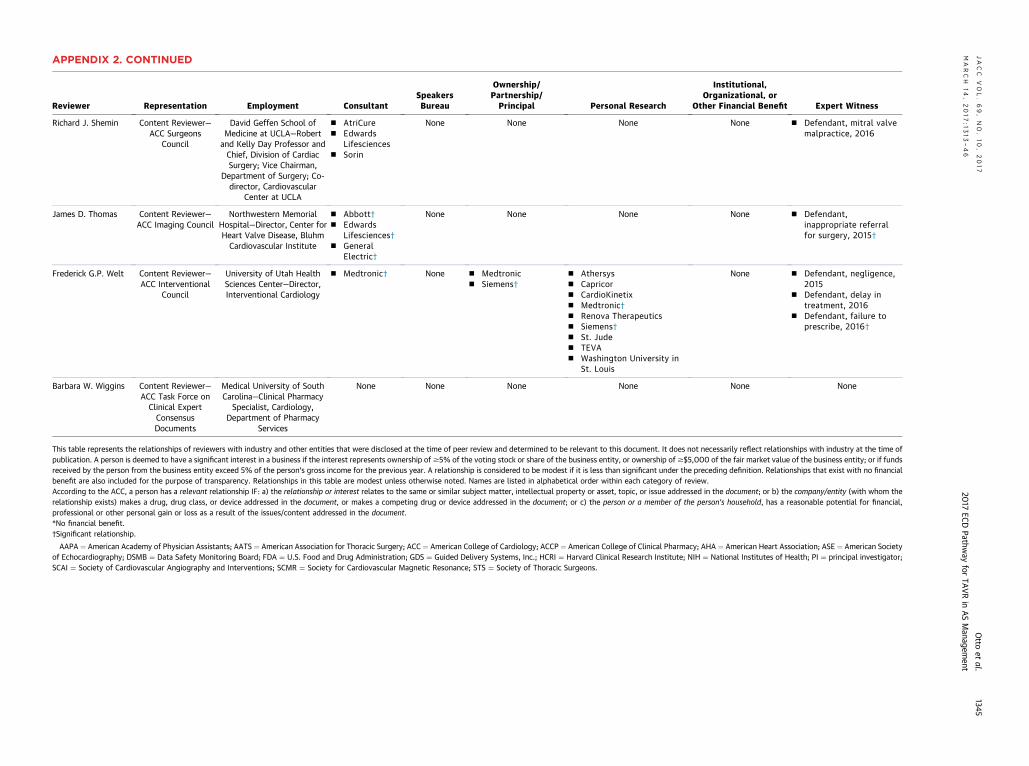
Reviewer Representation Employment ConsultantSpeakersBureau
Ownership/Partnership/Principal Personal Research
Institutional,Organizational, or
Other Financial Benefit Expert Witness
Richard J. Shemin Content Reviewer—ACC Surgeons
Council
David Geffen School ofMedicine at UCLA—Robert
and Kelly Day Professor andChief, Division of CardiacSurgery; Vice Chairman,
Department of Surgery; Co-director, Cardiovascular
Center at UCLA
n AtriCuren Edwards
Lifesciencesn Sorin
None None None None n Defendant, mitral valvemalpractice, 2016
James D. Thomas Content Reviewer—ACC Imaging Council
Northwestern MemorialHospital—Director, Center forHeart Valve Disease, BluhmCardiovascular Institute
n Abbott†n Edwards
Lifesciences†n General
Electric†
None None None None n Defendant,inappropriate referralfor surgery, 2015†
Frederick G.P. Welt Content Reviewer—ACC Interventional
Council
University of Utah HealthSciences Center—Director,Interventional Cardiology
n Medtronic† None n Medtronicn Siemens†
n Athersysn Capricorn CardioKinetixn Medtronic†n Renova Therapeuticsn Siemens†n St. Juden TEVAn Washington University in
St. Louis
None n Defendant, negligence,2015
n Defendant, delay intreatment, 2016
n Defendant, failure toprescribe, 2016†
Barbara W. Wiggins Content Reviewer—ACC Task Force onClinical ExpertConsensusDocuments
Medical University of SouthCarolina—Clinical Pharmacy
Specialist, Cardiology,Department of Pharmacy
Services
None None None None None None
This table represents the relationships of reviewers with industry and other entities that were disclosed at the time of peer review and determined to be relevant to this document. It does not necessarily reflect relationships with industry at the time ofpublication. A person is deemed to have a significant interest in a business if the interest represents ownership of$5% of the voting stock or share of the business entity, or ownership of$$5,000 of the fair market value of the business entity; or if fundsreceived by the person from the business entity exceed 5% of the person’s gross income for the previous year. A relationship is considered to be modest if it is less than significant under the preceding definition. Relationships that exist with no financialbenefit are also included for the purpose of transparency. Relationships in this table are modest unless otherwise noted. Names are listed in alphabetical order within each category of review.According to the ACC, a person has a relevant relationship IF: a) the relationship or interest relates to the same or similar subject matter, intellectual property or asset, topic, or issue addressed in the document; or b) the company/entity (with whom therelationship exists) makes a drug, drug class, or device addressed in the document, or makes a competing drug or device addressed in the document; or c) the person or a member of the person’s household, has a reasonable potential for financial,professional or other personal gain or loss as a result of the issues/content addressed in the document.*No financial benefit.†Significant relationship.
AAPA ¼ American Academy of Physician Assistants; AATS ¼ American Association for Thoracic Surgery; ACC ¼ American College of Cardiology; ACCP ¼ American College of Clinical Pharmacy; AHA ¼ American Heart Association; ASE ¼ American Societyof Echocardiography; DSMB ¼ Data Safety Monitoring Board; FDA ¼ U.S. Food and Drug Administration; GDS ¼ Guided Delivery Systems, Inc.; HCRI ¼ Harvard Clinical Research Institute; NIH ¼ National Institutes of Health; PI ¼ principal investigator;SCAI ¼ Society of Cardiovascular Angiography and Interventions; SCMR ¼ Society for Cardiovascular Magnetic Resonance; STS ¼ Society of Thoracic Surgeons.
APPENDIX 2. CONTINUEDJACC
VOL.69,NO.10,2017
Otto
etal.
MARCH
14,2017:1
313–46
2017
ECDPathw
ayfor
TAVRin
ASManagem
ent
1345

Otto et al. J A C C V O L . 6 9 , N O . 1 0 , 2 0 1 7
2017 ECD Pathway for TAVR in AS Management M A R C H 1 4 , 2 0 1 7 : 1 3 1 3 – 4 6
1346
APPENDIX 3. ABBREVIATIONS
AC
AF
AH
AR
AS
AV
CM
CT
EC
EF
LV
MD
MR
MV
SA
ST
TA
TE
TT
C ¼ American College of Cardiology
¼ atrial fibrillation
A ¼ American Heart Association
¼ aortic regurgitation
¼ aortic stenosis
R ¼ aortic valve replacement
R ¼ cardiac magnetic resonance
¼ computed tomography
G ¼ electrocardiogram
¼ ejection fraction
¼ left ventricular
CT ¼ multidetector computed tomography
¼ mitral regurgitation
¼ mitral valve
VR ¼ surgical aortic valve replacement
S ¼ Society of Thoracic Surgeons
VR ¼ transcatheter aortic valve replacement
E ¼ transesophageal echocardiography
E ¼ transthoracic echocardiography