2012 william allan award: adventures in cytogenetics1 · human genome project (hgp) got underway,...
TRANSCRIPT
ASHG AWARDS AND ADDRESSES
2012 William Allan Award: Adventures in Cytogenetics1
Uta Francke2,3,*
Ladies and gentlemen, members and guests, colleagues
and friends,
I am deeply honored to be chosen for this award; thank
you, Madam President, members of the Awards
Committee, and Tayfun Ozcelik for your kind introduc-
tion. I accept this award on behalf of a large group of scien-
tists, numerous postdocs, graduate and undergraduate
students, research associates, and technicians. I am grateful
to all of them, some of whom are here, and to the many
collaborators who contributed to our success.
You heard about the wide range of research activities of
my laboratory, so for this address, I had to select what to
cover. The microscope in the Allan Award medal inspired
me to focus on my adventures in cytogenetics.
Let’s start from the beginning. In the gymnasium, the
German equivalent to high school plus junior college, I
majored in mathematics and physics and studied Latin
for the language requirement, so I couldn’t speak much
English when I came to this country. In medical school
in Germany, I thought biochemistry was the most inter-
esting subject. I remember that one day the professor
1This article is based on the address given by the author at the meeting of
in San Francisco, CA, USA. The audio of the original address can be found at2Departments of Genetics and Pediatrics, Stanford University School of Medic
*Correspondence: [email protected]
http://dx.doi.org/10.1016/j.ajhg.2013.01.010. �2013 by The American Societ
The Ame
walked in, all excited, and told us that mRNA had
been discovered and that now we understood how the
genetic information is transmitted from the nucleus to
the cytoplasm.
Therefore, after my postgraduate medical training,
which ended with a pediatrics residency at Children’s
Hospital Los Angeles in California, I wanted to subspe-
cialize in endocrinology, a field that offered some under-
standing of the role of molecules and pathways in disease
processes, and rational approaches to treatment. However,
coming from a foreign medical graduate, my application
for a pediatric endocrinology fellowship was not even
considered.
Meanwhile, at University of California, Los Angeles
(UCLA), a new fellowship program in pediatric genetics
had been established under Stanley Wright, and they
gladly accepted me. So, I got into genetics at the beginning
of a new era, and I have not regretted it for a moment. This
step allowedme to embark on an unbelievable journey and
to participate in the development of our field for the past
40 years. It has been a constant learning experience.
Chromosome Banding and Identification and High-
Resolution Ideograms
When I started my fellowship at UCLA, human chromo-
somes were uniformly stained and, with a few exceptions,
could not be individually identified. Then one day, Stan
Wright announced the big news thatCaspersson in Sweden
(1988 Allan Award recipient) had told him that with the
fluorescent dye quinacrine they could reliably distinguish
chromosomes 17 and 18. This was exciting, and I had to
try it. I dug up a microscope with a movable mirror—it
looked like theone in theAllanAwardmedal—andafluores-
cent lamp and spent many hours in the dark room identi-
fying each human chromosome,1 as well as the chromo-
somes involved in translocations in cases that the
Genetics Division had previously collected.2
When mouse geneticist Muriel Nesbitt joined the
faculty, we collaborated on constructing the first quina-
crine-stained mouse karyotype. The bands were fuzzy
and needed to be documented further by densitometry
profiles and interpreted in ideograms.3 Identifying the
mouse chromosomes was a momentous advance for
mouse genetics because linkage groups had already been
the American Society of Human Genetics (ASHG) on November 9, 2012,
the ASHG website.
ine, Stanford, CA 94305, USA; 323andMe, Mountain View, CA 94043, USA
y of Human Genetics. All rights reserved.
rican Journal of Human Genetics 92, 325–337, March 7, 2013 325

associated with abnormal yet unidentified chromosomes.
By identifying the mouse chromosomes involved in trans-
locations, we were able to assign entire linkage groups to
their respective physical locations.4–6 O.J. Miller and
colleagues were also working on mouse chromosome iden-
tification, but we were unaware of it at the time.7 So, I got
an early taste for gene mapping and mouse genetics, areas
which I continued to pursue.
As Giemsa (G)-banding was introduced and our chromo-
somes were banded at higher resolution, Muriel and I con-
structed banding ideograms for each mouse chromosome
while taking into account the relative staining intensity
of each band. We used calipers for band measurements,
and I drew the chromosomes with shapes as I saw them
in the microscope. We divided each mouse chromosome
into major regions, designated by capital letters, and
then subdivided the regions into numerals that could be
further subdivided by decimals.8 That numbering system
was carried over to the current standard mouse chromo-
some nomenclature.9
Meanwhile, the International Committee for Human
Cytogenetic Nomenclature (ISCN) had devised a system
for human chromosome bands with ideograms that were
based on impressions, not measurements.10 Subsequently,
cell-synchronization methods enabled the study of longer
prometaphase chromosomes, resulting in high-resolution
banding (HRB). In my lab, then at the University of Cali-
fornia, San Diego (UCSD), we decided to do band measure-
ments and design accurate chromosome representations
that also included various intensities of staining, just as
Muriel and I had done for the mouse chromosomes.11
In 1981, ISCN incorporated HRB information by subdi-
viding their original bands into arbitrary subbands. The re-
sulting HRB-ISCN ideograms had little resemblance to the
looks of actual high-resolutionG-banded chromosomes, so
I decided to take our measurements and adapt them to the
ISCN numbering system. The rules that white bands had to
separate dark (or gray) bands necessitated compromises.12
Our contribution was included in the official ISCN
report.13
To continue the ideogram story, in the early 1990s the
Human Genome Project (HGP) got underway, and people
coming in with molecular biology and computer science
backgrounds were talking about the genome as a one-
dimensional string of four letters, an informational entity,
whose function they would be able to unravel given
enough computing power. I felt that they were missing
the point because the single molecule of DNA that runs
from one end of the chromatid to the other is packaged
into metaphase chromosomes in distinctly uneven
patterns, e.g., dark bands condense earlier than light bands
in the continuous process of prophase and prometaphase.
The location of the DNA sequences that were being assem-
bled by the HGP with respect to chromosome bands must
have functional implications. As Holger Hoehn had
already pointed out in 1975, trisomies for chromosomes
with relatively more dark-staining G-bands are compatible
326 The American Journal of Human Genetics 92, 325–337, March 7
with development to term and live births, which contrasts
with trisomies for equally sized chromosomes that contain
more lightly staining chromatin and are only found in
spontaneous abortions.14
To call attention to the contribution that chromosome-
banding informationcouldmake to theHGP, Ihadour ideo-
grams professionally redrawn and published them together
with the underlying measurements in an article pointing
out their features and potential uses (Figure 1).15 The paper
is not available online, and today’s graduate students are
not likely to venture into the stacks of the library, but the
ideograms survive. You can find them in numerous illustra-
tions, textbooks, databases,16 and commercial products—
and nobody remembers where they came from. Although
this makes me proud, I also know their imperfections. No
cytogeneticist with access to better-quality chromosomes
has done what we did: caliper measurements of prometa-
phase chromosome bands at distinct stages of condensa-
tion. Would there be any reason to repeat the work now
with higher-quality preparations?
In 2009, a Science paper entitled ‘‘Comprehensive
mapping of long-range interactions reveals folding princi-
ples of the human genome’’ reported on genome-wide
chromatin-conformation analyses assessing the physical
proximity of DNA segments.17 Using a ‘‘Hi C’’ method,
the authors probed the three-dimensional architecture of
whole genomes by coupling proximity-based ligation
with massively parallel DNA sequencing. Their principal-
component analyses revealed that the genome is compart-
mentalized into closed and open compartments and that
chromatin packaging is an integral element. The compart-
ments correspond to gene density, specific histone-methyl-
ation patterns, and DNase I sensitivity profiles.17 As soon
as I looked at Figure 3G in this paper, I realized that I had
seen the pattern before. It lined up with our ideogram,
leading to the surprising insight that the chromatin orga-
nization of the genome in interphase appears to be carried
through to metaphase and is reflected in the banding
patterns (Figure 2). The closed and open chromatin regions
correspond to dark and light prometaphase G-bands,
respectively, and gray bands are an unresolved mixture of
both types. I was thrilled, and told my undergraduate
molecular genetics class the next morning. However, the
lineup did not work perfectly for all chromosomes. Last
year, I finally met Dr. Lieberman-Aiden and told him about
my hypothesis, and he is now actively testing it.
Mapping Genes onto Chromosomal Regions
One reason for generating chromosome-banding maps is
to have a template onto which genes can be placed. Fortu-
nate circumstances enabled me to enter the field of phys-
ical mapping of mammalian chromosomes at its begin-
ning. In 1971, after my year of genetics fellowship at
UCLA, my first husband, Bertold Francke, and I moved to
San Diego, and I started to work in Bill Nyhan’s biochem-
ical genetics laboratory at UCSD. There, I studied the X-
inactivation mosaicism in women who were carriers for
, 2013
Figure 1. High-Resolution Human Chromosome Ideograms Based on Trypsin-G-Banded Prometaphase Lymphocyte ChromosomesBand widths are based on measurements, and relative staining intensities are represented by black, white, and three shades of gray. Theband numbering system is consistent with ISCN nomenclature.13 This figure was modified from Figure 1 in Francke.15
Lesch-Nyhan syndrome, an X-linked recessive disorder
caused by mutations in the X-linked gene HGPRT, and
developed a carrier-detection test by using electrophoresis
of hair-root lysates.18 When Jerry Schneider, a faculty
member in the department, returned from a sabbatical in
Ephrussi’s lab in Paris and brought back the somatic cell
hybridization (SCH) technology,19 I had an idea for a doable
project: I proposed to regionally map genes on chromo-
somes by fusing human cells carrying a balanced reciprocal
translocation to a mouse or Chinese hamster cell line.
Initially, human chromosomes are randomly lost from
such interspecies hybrid cells, but by repeated subcloning,
one canobtain cell lineswith a stable subset ofhumanchro-
mosomes. Being able to identify human and rodent chro-
mosomes by their banding patterns and to separate human
and rodent isozymes by gel electrophoresis of hybrid cell
extracts, I could assign species-specific cellular phenotypes
to defined chromosome regions. To get support for this
The Ame
work, I applied to the National Institutes of Health for an
R01grant. The study sectionvoted in favor and even recom-
mended to increase the budget so I could buy the best
microscope on the market. As funding was contingent
upon UCSD’s giving me a faculty position, I became an
assistant professor in residence in pediatrics.
Inmy first somatic cell hybridization experiment, I fused
mouse cells with a reciprocal X-autosome translocation to
a Chinese hamster cell line. By studying the hybrid cell
clones, we were able to map genes within the X chromo-
some and chromosome 16 in the mouse.20 Subsequently,
we produced many human translocation-derived hybrid
cell lines and constructed hybrid panels that allowed us
to map any human gene or cellular phenotype to a chro-
mosome and, in many cases, to a chromosomal region.
Physical mapping by SCH had been promoted mostly by
Frank Ruddle (1983 Allan Award recipient) at Yale,21 and
he arranged for me to be invited to the second Human
rican Journal of Human Genetics 92, 325–337, March 7, 2013 327
Figure 2. Interaction Map, ReflectingSpatial Proximity in the InterphaseNucleus, of Chromosome 14 at a Resolu-tion of 100 kbThe principal component (eigenvector)correlates with the distribution of genesand with features of open chromatin.This interphase-chromatin organizationis carried over to metaphase and is re-flected in the chromosome-bandingpatterns. The closed and open regionscorrespond to dark and light prometa-phase G-bands, respectively. This figurewas modified with permission fromFigure 3G of Lieberman-Aiden et al.17
Gene Mapping Conference in Rotterdam in 1974 to
present our initial work.22,23 At these biannual, or later
annual, conferences supported by the March of Dimes
Birth Defects Foundation, researchers from all over the
world came together to share their latest physical and
genetic-mapping data, which were then integrated and
compiled by chromosome-specific committees. The up-
dated mapping reports were published in Cytogenetics and
Cell Genetics. In the mid 1990s, when data acquisition
accelerated and the meetings became too large, they were
split up into separate international chromosome-specific
workshops. What many young people today do not realize
is that when the HGP started with a focus on large-insert
clone tiling paths and DNA sequencing technology,
a rather dense physical map was already available to facil-
itate the assembly of the first human reference genome.
Initially, the phenotypes we could map in somatic cell
hybrids were limited to expressed proteins for which the
human-specific forms could be distinguished from the
rodent forms, e.g., metabolic enzymes,22–24 cell-surface
antigens, such as human leukocyte antigen,25 polypeptide
spots on two-dimensional protein gels,26 or yet-unidenti-
fied factors responsible for virus replication in cultured
human cells.27
In the early 1980s, with the advent of molecular probes,
we were able to map hybridizing restriction fragments on
Southern blots made with DNA from interspecies somatic-
cell-hybrid clones. The first human cDNAs that became
available were for hemoglobins, and we mapped the
b-globin gene cluster to the short arm of chromosome 11
in collaboration with Alec Jeffreys (1992 Allan Award recip-
ient).28 Restriction-fragment-length polymorphisms
(RFLPs) were found to represent a powerful new set of
genetic markers, and we collaborated with Ray White and
David Botstein (1989 Allan Award recipients) to map the
first highly polymorphic human RFLP locus (D14S1) to
a regionof chromosome1429 andtodiscover thefirstmolec-
328 The American Journal of Human Genetics 92, 325–337, March 7, 2013
ular-defined locus (DXYS1) present on
both the X and Y chromosomes.30
In addition to mapping human
genes, we also studied SCH between
Chinese hamster and mouse cells
that had retained reduced sets of mouse chromosomes
and mapped the murine orthologs to mouse chromo-
somes, initially in collaboration with Peter Lalley and
JohnMinna.31–33 As human cDNA probes cross-hybridized
to rodent restriction fragments and vice versa, we used
mouse-hamster hybrid panels, in addition to the human-
rodent panels, to map any newly cloned gene simulta-
neously in mice and humans and thus contributed to the
recognition and delineation of conserved syntenic chro-
mosomal regions.34
Withmolecular probes in hand, we also employed in situ
hybridization of labeled probes directly to banded chromo-
somes. This independent approach enabled us to confirm
andfurther refine the regionalmappositions. Initially, small
cDNA probes were tritium labeled, autoradiographic grains
were counted, and the counts were statistically analyzed.34
In the late1980s, radioactively labeled cDNAprobeswere re-
placed with fluorescently labeled cosmid probes for which
hybridization signals were detected in a fluorescent micro-
scope. This technology—known as fluorescence in situ
hybridization—was pioneered by Peter Lichter in David
Ward’s lab at Yale while I was in the same department,35
and we adapted it to our mapping experiments.36
With our reliable gene-mapping tools, we had the great
fortune to collaborate with the premier gene-cloning labs
and mapped HRAS with Robert Weinberg;37 NGFB, IGF1,
IGF2, INSR, EGFR, TGFB, PDFRA, and other genes with
Axel Ullrich, Rick Derynck, and other scientists at Genen-
tech;38–42 genes for the low-density-lipoprotein receptor
with Mike Brown and Joe Goldstein (1985 Allan Award
recipients);43 genes for adrenergic receptors with Brian Ko-
bilka and Robert Lefkowitz;44 the gene encoding synapto-
physin with Thomas Sudhof;45 and many more.
Finding Disease-Associated Genes by Mapping
While actively contributing to the human and mouse
genetic maps, our underlying motivation in gene mapping
Figure 3. Xp21 Map of MolecularlyDefined Intraband Deletions and DerivedLocalization of Disease Loci(A) G-band map of Xp.(B) Location of some DNA probes.(C) Extent of deletions based on DNAsegments missing from individual cases.(D) Brackets indicate derived localizationsof disease loci and of XK, mutations inwhich cause McLeod syndrome.This figure is the precursor to Figure 3 inFrancke et al.57
was to find genes associated with human disease. I never
liked the metaphor ‘‘gene hunting’’ because the genes that
when mutated cause human disease are not running
away—they just sit there in hiding while waiting to be
discovered. If we could map a cloned gene to a location
that coincided with a previously mapped disease locus or
mouse mutant phenotype, we might be able to directly
identify the disease-causing gene. Over the years, we tested
many positional candidate genes in DNA samples from
affected individuals or mutant mouse strains and usually
had negative results. However, we did get lucky one time:
when the gene-encoding peripheral myelin protein 22
(PMP-22) was cloned in Eric Shooter’s lab at Stanford, we
mapped it near the gene for the Trembler phenotype in
mice, and a Pmp22 mutation was detected in these mice.46
Trembler had already been considered as a mouse model
of Charcot-Marie-Tooth disease, and we mapped PMP22
to the small chromosomal duplication known to be associ-
ated with Charcot-Marie-Tooth disease type 1A.47
Locating Disease-Associated Genes by Cytogenetic
Analysis
Direct cytogenetic analysis can also lead to mapping and
discovery of disease-associated genes. In this approach,
genes can be localized, or even identified, if the disease
phenotype is associated with a chromosomal aberration
that is visible in the microscope, e.g., a tiny deletion might
remove the relevant locus, or a translocation might
disrupt it.
This approach started with a little girl who we found to
have sporadic bilateral retinoblastoma and an interstitial
deletion of 13q.48 Comparing this case with scattered
literature reports allowed me to predict the location of
RB in band 13q14,49 and this information assisted in
the cloning of the gene.50 In a similar fashion, aniridia
The American Journal of Human
and Wilms tumor were both known
to segregate as autosomal-dominant
traits. In collaboration with Vincent
Riccardi, our discovery of a 11p13
microdeletion in individuals affected
by both conditions placed both loci
into this region51,52 and facilitated
the cloning of the responsible
genes.53
In 1984, we received a blood sample and lymphoblastoid
cell line from an adopted boy (BB) who was apparently
affected by four different X-linked diseases and who had
been studied by several specialists in Seattle but for
whom no genetic abnormality had been identified. On
high-resolution chromosome banding, we discovered
a tiny interstitial deletion of subband Xp21.2, but we
needed a molecular probe to confirm that. We obtained
Xp probes from collaborators, and there were few available
at the time, but none were missing in BB. If the missing
Xp21.2 material had been inserted somewhere else, we
would not be able to see that microscopically. Therefore,
we made somatic cell hybrids with a Chinese hamster
cell line and isolated BB’s X chromosome away from all
his other chromosomes. Ultimately, one probe, called
754, from Peter Pearson’s lab in the Netherlands, was
deleted from his X chromosome and from his total DNA,
proving that he indeed had a deletion.54 We interpreted
this finding as indicating that genes in the deletion might
contribute to the expression of Duchenne muscular
dystrophy (DMD), chronic granulomatous disease (CGD),
retinitis pigmentosa, and McLeod syndrome, all mani-
fested by BB.
Louis Kunkel (2004 Allan Award recipient) used the cells
from BB to isolate DNA fragments from the deletion,55 and
Brigitte Royer-Pokora and colleagues subsequently used
them to clone the gene for X-linked CGD.56 This was the
first human disease-associated gene cloned on the basis
of its location with no prior knowledge of its function
(‘‘positional cloning’’). Subsequently, we identified addi-
tional cases with overlapping Xp21 deletions and various
phenotypes that allowed the mapping and ordering of
disease genes in this chromosome band (Figure 3).57–59
To directly identify the DMD gene, we studied a female
diagnosed with DMD and a reciprocal X-autosome
Genetics 92, 325–337, March 7, 2013 329
translocation. X-inactivation studies revealed the preferen-
tial inactivation of the normal X chromosome, and we
separated the translocation chromosomes in SCH.
Hypothesizing that the translocation had disrupted the
DMD gene, we figured that cloning the translocation
breakpoint might lead us directly to the gene. During
a sabbatical at the European Molecular Biology Laboratory
in Heidelberg with Hans Lehrach, I made a cosmid library
from this individual. Joe Giacalone, a graduate student in
my lab at Yale, isolated breakpoint-bridging cosmids and
sequenced across the translocation breakpoints.60
However, by that time, others had already succeeded in
finding the DMD gene.
Finding Disease-Associated Genes by Positional
Cloning
Moving to Stanford in 1989 as a Howard Hughes Medical
Institute investigator in the new Beckman Center for
Molecular and Genetic Medicine allowed me to expand
work on a number of disease-specific projects. In the early
1990s, as the human linkage map became enriched by
highly polymorphic RFLPs and microsatellite markers,
more genes for Mendelian diseases were mapped and
became candidates for positional cloning. One early
success story involves the isolation, in my lab, of the
gene associated with the X-linked immunodeficiency Wis-
kott-Aldrich syndrome (WAS). Jonathan Derry, a postdoc-
toral fellow, constructed a yeast artificial chromosome
(YAC) and cosmid contig covering the region of
Xp11.22–p11.23, to which the WAS locus had been as-
signed. Hans D. Ochs, a pediatric immunologist (who
had also referred case BB) in Seattle provided us with lym-
phoblastoid cells from several of his WAS cases. Jonathan
isolated cDNA fragments complementary to the YACs
and tested them on blots of cases’ RNA. Lack of expression
in two of them led to the gene,61 andmutations confirmed
it.61,62 The gene was expressed exclusively in hematopoi-
etic tissues and encoded a previously unknown protein
that we called ‘‘Wiskott-Aldrich syndrome protein’’
(WASP). Its function in modulating the actin cytoskeleton
only became clear after collaboration with Arie Abo’s lab.63
I had long been fascinated by Roberts syndrome, a rare
autosomal-recessive condition characterized by limb and
other malformations and associated with distinct chromo-
somal features: premature separation of centromeres and
repulsion of heterochromatin regions.64 The unique chal-
lenge herewas to find the gene associatedwith aMendelian
disorder that can be diagnosed by cytogenetic analysis, and
we approached it by looking for genes that could comple-
ment the cellular and/or chromosomal phenotype. First,
while at Yale University, we showed that the characteristic
chromosomal features are corrected in interspecies somatic
cell hybrids.65 David van den Berg, a graduate student in
my lab at Stanford, then focused on cellular abnormalities
in order to devise a selective system for complementa-
tion.66 However, this extensive effort failed to lead to the
discovery of the gene, which was later identified by Vega
330 The American Journal of Human Genetics 92, 325–337, March 7
and colleagues via homozygosity mapping in inbred fami-
lies as ESCO2, one of two human orthologs of a yeast gene
required for sister chromatid cohesion.67 Birgitt Schule,
a postdoc in the lab, documented ESCO2 mutations not
only in Roberts syndrome but also in individuals diag-
nosed with SC phocomelia, thus confirming that the two
clinically defined conditions are allelic.68
Since the mid 1980s, I had been intrigued by Rett
syndrome, a sporadic neurological disorder limited to
females and characterized by postnatal onset and develop-
mental regression, loss of hand use, truncal ataxia, apraxia,
seizures, and acquired microcephaly.69 Among many
possible genetic hypotheses, de novo mutations in an X
chromosome gene seemed to be the most plausible. We
collected samples from families with girls affected by Rett
syndrome, established lymphoblastoid cell lines, and
tested their DNA for mutations in neuronal genes that
we and others had mapped to the X chromosome, e.g.,
SYN1, SYP, GLUD2, GDI1, and GRPR, but we had no
success. To narrow down the X chromosome region that
could contain a gene associated with Rett synrome, we
turned to exclusion mapping by studying rare familial
cases.70,71 In collaboration with Huda Zoghbi, we
continued candidate-gene testing in the unexcluded
Xq28 region, which led to the discovery of mutations in
MECP2 in Huda’s lab.72,73 The gene encodes MeCP2,
a methyl-CpG-binding protein that binds to chromatin-
modification factors and that was thought to function as
a global transcriptional repressor.74,75 I had considered
this gene an unlikely candidate for Rett syndrome because
chimeric mice harboring Mecp2-knockout cells had been
reported to die in utero,76 whereas Rett-syndrome-affected
girls who are mutation heterozygotes and X-inactivation
mosaics survive and are born normal.
Discovery of MECP2 as the gene mutated in Rett
syndrome generated a wave of scientific work in many
laboratories with the goal of unraveling the reasons for
the frequent recurrent de novo mutations and the patho-
physiological mechanism of the postnatal neurological
damage. Under the hypothesis of transcriptional dysregu-
lation, we attempted to identify downstream targets by
transcriptome-expression-array studies of single-cell-
derived fibroblast clones from Rett-syndrome-affected girls
with identified MECP2 mutations77 and from dissected
brain regions of mouse mutants.78
To understand how MECP2 itself is regulated and to
define the ‘‘MECP2 functional expression module,’’ Jin-
glan Liu, a postdoc in the lab, identified cis-acting control
elements and employed chromosome conformation
capture to generate an interaction map of enhancers and
repressors with the MECP2 promoter.79 Despite the enor-
mous amount of work in many neuroscience laboratories
around the world, the complete pathophysiology of Rett
syndrome is not yet understood beyond the recognition
that the defect resides at the synapse.
In the interest of focusing on adventures in cytogenetics,
I will skip over projects on genotype-phenotype
, 2013
characterization of Marfan syndrome and related connec-
tive-tissue disorders; these were a collaboration with the
Stanford Center for Marfan Syndrome and the experi-
mental pathology lab of my late husband, Heinz Furth-
mayr.
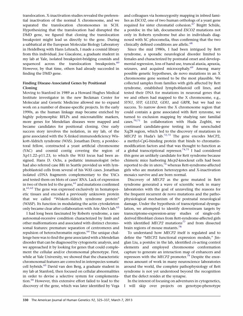
Dissecting Microdeletion Syndromes
The next challenge in cytogenetics was the dissection of
clinically and cytogenetically defined microdeletion
syndromes. First, we needed to find all genes in the deletion
and then determine which of the many deleted genes are
responsible for which of the phenotypes by considering
issues of haploinsufficiency and penetrance, gene-gene
interactions, genomic imprinting, and potential effects of
structural chromosome rearrangements on the expression
of neighboring genes. The deletion syndromes we focused
on, Williams-Beuren syndrome (WBS) and Prader-Willi
syndrome (PWS), are caused by nonallelic homologous
recombination (NAHR) between flanking repeats, and
therefore the same genes are deleted in most cases.
WBS with multisystem manifestations and unique neu-
robehavioral features is caused by a barely visible
7q11.23 deletion that contains elastin (ELN).80 Before the
first draft of the human genome sequence became avail-
able, my lab developed a contiguous cosmid map across
the 1.5MbWBS deletion and flanking regions by screening
cosmid libraries and sequencing the ends.81 Along the way,
we discovered several protein-coding genes within the
deletion.82–88 To assess the functional consequences of
gene loss, we knocked out one of them, Fzd9 (encoding
a Wnt receptor), in mice, but heterozygous-knockout
mice did not show distinct phenotypes.89
Because cases with partial deletions of theWBS region are
very rare, we decided to create Wbs partial-deletion mouse
models by chromosome engineering. Mouse models would
be useful for dissecting the molecular mechanisms under-
lying theunique featuresofWBSforphysical andbehavioral
studies, for providing access to brain tissue, and for enabling
control of the genetic background. The human 7q11.23
region is conserved in mouse chromosome band 5G2. Or-
thologous genes are in the same order, but the region is in-
verted with respect to the centromere, and there are no
flanking repeats in the mouse. Rather than removing the
entire orthologous Wbs deletion region, we generated
mice with two complementary half deletions (PD and DD)
(Figure 4). Interbreeding half-deletion heterozygotes
produces litters comprising four different genotypes that
can be compared in studies of phenotypes. Double hetero-
zygotes (D/P) represent a model for the human WBS. The
successful completion of this project required a huge effort
by many skilled and devoted people over many years. The
mice were studied extensively and found to reproduce
some human WBS features that could be assigned to one
or the other half of the deletion.90 Increased sociability
and acoustic startle response are associated with PD, and
cognitive defects are associated with DD. Skulls are short-
ened and brains are smaller in DD mice, whereas in PD
The Ame
mice, the lateral ventriclevolumesare reducedandneuronal
cell density is increased in the somatosensory cortex.Motor
skills aremost impaired inD/P. Gene-transcript levels in the
brain are generally consistent with gene dosage. Together,
these partial-deletion mice replicate crucial aspects of the
human disorder and serve for the identification of genes
and gene networks contributing to the neural substrates of
complex behaviors. The regions can now be dissected
further by genetic complementation studies.90
In the case of PWS, the deletion involves an imprinted
region, i.e., deleted genes are on the paternally inherited
chromosome 15, and the maternal copies are silent. The
PWS project started with our assignment of the first
protein-coding gene, SNRPN (small nuclear ribonucleopro-
tein polypeptide N), to the deletion region in 1992.91 We
then systematically searched for expressed sequences in
the region and found various expressed noncoding DNA
segments92 and a cluster of small C/D box nucleolar
RNAs (snoRNAs) located in introns; we called these
PWCR1.93 Whereas the ~100 bp sequences of this snoRNA
cluster were moderately conserved between humans and
mice, the exons of the noncoding-RNA host genes were
not conserved at all, suggesting a functional role for these
intronic sequences.
To delineate the minimal deletion region responsible for
the PWS phenotype, we studied reciprocal translocation
cases by precise molecular mapping of breakpoints.94
Taking our data together with others in the literature led
us to propose that a small region between SNRPN and
UBE3A and containing the PWCR1 snoRNA cluster—
discovered in our lab93 and independently by Cavaille
et al. (who named it HBII-85)95 and now called
SNORD116—was responsible for the PWS phenotype.96
To test this hypothesis, Feng Ding in the lab took on the
arduous task of making a mouse model in which the
Snord116 cluster was deleted by chromosome engineering
in embryonic stem cells (Figure 5). In contrast to previous
PWS mouse models that have a high rate of neonatal
mortality,98 the Snord116-deletionmice were viable. Exten-
sive metabolic, dietary, and behavioral studies of mice
carrying the deletion on the paternally derived chromo-
some revealed some PWS-like phenotypes, such as growth
delay and hyperphagia, but lacked others, e.g., hypotonia
and obesity.97 Then, surprisingly, Art Beaudet’s lab and
others found that human cases with deletions of these
snoRNAs met criteria for a PWS diagnosis, as discovered
by array comparative genomic hybridization.99,100 How
the lack of SNORD116 snoRNAs produces the phenotype
is a fascinating question. The answer involves discovering
the normal function of SNORD116 snoRNAs. We know
that they do not modify rRNA as most known C/D box
snoRNAs do, but are they involved in other aspects of
RNA processing such as mRNA turnover, alternative
splicing, or RNA editing? Transcriptome expression arrays
of dissected hypothalamic tissue at postnatal days 5 and
13 revealed similar expression profiles in mice with dele-
tion and normal genotypes at both developmental
rican Journal of Human Genetics 92, 325–337, March 7, 2013 331
Figure 4. Generating Mouse Models for the WBS DeletionThe gene order across the WBS deletion region on 7q11.23 is conserved in mouse chromosome band 5G2 except for an inversion withrespect to the centromere (CEN) and telomere (TEL). For the generation of partial-deletion mice, targeting constructs containing loxPsites (arrowheads) and partialHprt sequences were inserted by homologous recombination in embryonic stem cells.Cre-mediated recom-bination between the loxP sites at Gtf2i and Limk1 and between the loxP sites at Limk1 and Trim50 generated the proximal deletion (PD)and the distal deletion (DD), respectively. This figure was modified from Figures 1A and 1B in Li et al.90
stages.101 When I closed my lab in 2008, Feng Ding took
a faculty position in China and continues to study these
mice. We also deposited them at The Jackson Laboratory
to be available for any researcher who wants to tackle these
questions.
Predicting the Future and Personal Genomics
In 1999, when ASHG last met in San Francisco, I served as
president. In my presidential address, entitled ‘‘Human
Genetics in the Information Age,’’ I strongly advocated
for open-access scientific publishing online. I did not
submit my presidential address for publication in The
American Journal of Human Genetics because the editor
and board of directors at the time had not agreed to accom-
modate an open-access model for The Journal (the presi-
dent has little power!), but a copy of my speech is available
on the ASHG website.
Meanwhile, open-access publishing has been growing at
an accelerating pace. Last year, about 17% of all scientific
articles were published in open access-journals (biomedi-
cine was the leading field), and the ‘‘online only with
a fee’’ category is rising the fastest.102 I believe that the
332 The American Journal of Human Genetics 92, 325–337, March 7
next year will see further acceleration in the move to this
publishing model hand in hand with the rapid expansion
of massive open online courses.
In my 1999 presidential address, I also predicted that
medical genetics services would be provided online: ‘‘There
is a huge need to provide online genetic information
tailored to consumers that is accurate, up-to-date, and
accessible. There should be career opportunities for site
developers and database curators,’’ and ‘‘I have no doubt
that interactive web-based systems will be developed that
can provide accurate, timely, and individualized genetic
information.’’
In 2006, when Anne Wojcicki and Linda Avers started
the personal genomics company 23andMe and invited
me to serve as consultant for the editorial team, I accepted
because I knew that this was the direction I wanted to go
in;103 in 2010, I joined the company as a part-time
employee. Working with the company’s research team
opened new opportunities for different kinds of research.
With a rapidly growing database of more than 180,000
people genotyped (approximately one million SNPs) and
a huge collection of self-reported phenotypes, 23andMe
, 2013
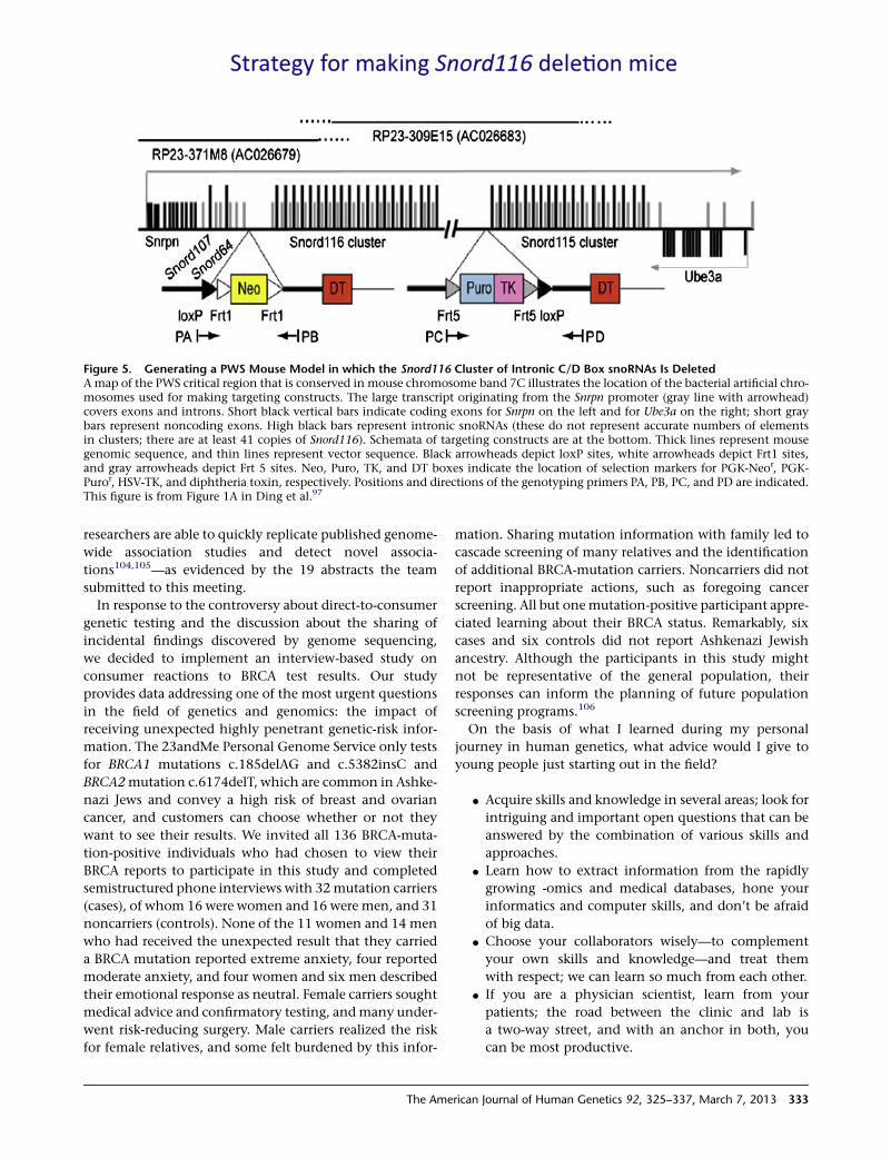
Figure 5. Generating a PWS Mouse Model in which the Snord116 Cluster of Intronic C/D Box snoRNAs Is DeletedAmap of the PWS critical region that is conserved in mouse chromosome band 7C illustrates the location of the bacterial artificial chro-mosomes used for making targeting constructs. The large transcript originating from the Snrpn promoter (gray line with arrowhead)covers exons and introns. Short black vertical bars indicate coding exons for Snrpn on the left and for Ube3a on the right; short graybars represent noncoding exons. High black bars represent intronic snoRNAs (these do not represent accurate numbers of elementsin clusters; there are at least 41 copies of Snord116). Schemata of targeting constructs are at the bottom. Thick lines represent mousegenomic sequence, and thin lines represent vector sequence. Black arrowheads depict loxP sites, white arrowheads depict Frt1 sites,and gray arrowheads depict Frt 5 sites. Neo, Puro, TK, and DT boxes indicate the location of selection markers for PGK-Neor, PGK-Puror, HSV-TK, and diphtheria toxin, respectively. Positions and directions of the genotyping primers PA, PB, PC, and PD are indicated.This figure is from Figure 1A in Ding et al.97
researchers are able to quickly replicate published genome-
wide association studies and detect novel associa-
tions104,105—as evidenced by the 19 abstracts the team
submitted to this meeting.
In response to the controversy about direct-to-consumer
genetic testing and the discussion about the sharing of
incidental findings discovered by genome sequencing,
we decided to implement an interview-based study on
consumer reactions to BRCA test results. Our study
provides data addressing one of the most urgent questions
in the field of genetics and genomics: the impact of
receiving unexpected highly penetrant genetic-risk infor-
mation. The 23andMe Personal Genome Service only tests
for BRCA1 mutations c.185delAG and c.5382insC and
BRCA2mutation c.6174delT, which are common in Ashke-
nazi Jews and convey a high risk of breast and ovarian
cancer, and customers can choose whether or not they
want to see their results. We invited all 136 BRCA-muta-
tion-positive individuals who had chosen to view their
BRCA reports to participate in this study and completed
semistructured phone interviews with 32mutation carriers
(cases), of whom 16 were women and 16 were men, and 31
noncarriers (controls). None of the 11 women and 14 men
who had received the unexpected result that they carried
a BRCA mutation reported extreme anxiety, four reported
moderate anxiety, and four women and six men described
their emotional response as neutral. Female carriers sought
medical advice and confirmatory testing, andmany under-
went risk-reducing surgery. Male carriers realized the risk
for female relatives, and some felt burdened by this infor-
The Ame
mation. Sharing mutation information with family led to
cascade screening of many relatives and the identification
of additional BRCA-mutation carriers. Noncarriers did not
report inappropriate actions, such as foregoing cancer
screening. All but onemutation-positive participant appre-
ciated learning about their BRCA status. Remarkably, six
cases and six controls did not report Ashkenazi Jewish
ancestry. Although the participants in this study might
not be representative of the general population, their
responses can inform the planning of future population
screening programs.106
On the basis of what I learned during my personal
journey in human genetics, what advice would I give to
young people just starting out in the field?
d Acquire skills and knowledge in several areas; look for
intriguing and important open questions that can be
answered by the combination of various skills and
approaches.
d Learn how to extract information from the rapidly
growing -omics and medical databases, hone your
informatics and computer skills, and don’t be afraid
of big data.
d Choose your collaborators wisely—to complement
your own skills and knowledge—and treat them
with respect; we can learn so much from each other.
d If you are a physician scientist, learn from your
patients; the road between the clinic and lab is
a two-way street, and with an anchor in both, you
can be most productive.
rican Journal of Human Genetics 92, 325–337, March 7, 2013 333
Acknowledgments
I wish to thank my mentors and collaborators who worked with
me in the lab and in the clinic, and I apologize to those who
were not named and whose projects were not mentioned. For
the projects included in this review, I am grateful for research
support from the National Institutes of Health, the Howard
Hughes Medical Institute, the March of Dimes Birth Defects Foun-
dation, the International Rett Syndrome Association, the Rett
Syndrome Research Foundation, the Foundation for Prader-Willi
Research, and 23andMe, where I am currently employed as senior
medical director.
Web Resources
The URL for data presented herein is as follows:
American Society of Human Genetics, www.ashg.org
References
1. Caspersson, T., Lomakka, G., and Zech, L. (1972). The 24
fluorescence patterns of the human metaphase chromo-
somes - distinguishing characters and variability. Hereditas
67, 89–102.
2. Francke, U. (1972). Quinacrine mustard fluorescence of
human chromosomes: Characterization of unusual translo-
cations. Am. J. Hum. Genet. 24, 189–213.
3. Francke, U., and Nesbitt, M. (1971). Identification of the
mouse chromosomes by quinacrine mustard staining. Cyto-
genetics 10, 356–366.
4. Nesbitt, M., and Francke, U. (1971). Linkage groups II and XII
of themouse: Cytological localization by fluorochrome stain-
ing. Science 174, 60–62.
5. Francke, U., and Nesbitt, M. (1971). Cattanach’s transloca-
tion: Cytological characterization by quinacrine mustard
staining. Proc. Natl. Acad. Sci. USA 68, 2918–2920.
6. Nesbitt, M., and Francke, U. (1971). Analysis of the T(3?)6 Ca
and T(14;17)264Ca translocations in the mouse by quina-
crine mustard staining. Genetics 69, 517–522.
7. Miller, O.J., Miller, D.A., Kouri, R.E., Allderdice, P.W., Dev,
V.G., Grewal, M.S., and Hutton, J.J. (1971). Identification of
the mouse karyotype by quinacrine fluorescence, and tenta-
tive assignment of seven linkage groups. Proc. Natl. Acad. Sci.
USA 68, 1530–1533.
8. Nesbitt, M.N., and Francke, U. (1973). A system of nomencla-
ture for band patterns of mouse chromosomes. Chromosoma
41, 145–158.
9. International Committee on Standardized Genetic Nomen-
clature for Mice (2005). http://www.informatics.jax.org/
mgihome/nomen/anomalies.shtml#general.
10. An international system for human cytogenetic nomencla-
ture (1978) ISCN (1978). Report of the Standing Commitee
on Human Cytogenetic Nomenclature. Cytogenet. Cell
Genet. 21, 309–409.
11. Francke, U., and Oliver, N. (1978). Quantitative analysis of
high-resolution trypsin-giemsa bands on human prometa-
phase chromosomes. Hum. Genet. 45, 137–165.
12. Francke, U. (1981). High-resolution ideograms of trypsin-
Giemsa banded human chromosomes. Cytogenet. Cell
Genet. 31, 24–32.
13. An international system for human cytogenetic nomencla-
ture—High-resolution banding (1981). ISCN (1981). Report
334 The American Journal of Human Genetics 92, 325–337, March 7
of the Standing Commitee on Human Cytogenetic Nomen-
clature. Cytogenet. Cell Genet. 31, 5–23.
14. Hoehn, H. (1975). Functional implications of differential
chromosome banding. Am. J. Hum. Genet. 27, 676–685.
15. Francke, U. (1994). Digitized and differentially shaded
human chromosome ideograms for genomic applications.
Cytogenet. Cell Genet. 65, 206–218.
16. Hindorff, L.A., Sethupathy, P., Junkins, H.A., Ramos, E.M.,
Mehta, J.P., Collins, F.S., and Manolio, T.A. (2009). Potential
etiologic and functional implications of genome-wide associ-
ation loci for human diseases and traits. Proc. Natl. Acad. Sci.
USA 106, 9362–9367.
17. Lieberman-Aiden, E., van Berkum, N.L., Williams, L., Ima-
kaev, M., Ragoczy, T., Telling, A., Amit, I., Lajoie, B.R., Sabo,
P.J., Dorschner, M.O., et al. (2009). Comprehensive mapping
of long-range interactions reveals folding principles of the
human genome. Science 326, 289–293.
18. Francke, U., Bakay, B., and Nyhan, W.L. (1973). Detection of
heterozygous carriers of the Lesch-Nyhan syndrome by elec-
trophoresis of hair root lysates. J. Pediatr. 82, 472–478.
19. Schneider, J.A., Francke, U., Hammond, D.S., Pellett, O.L.,
and Becker, F.L.A. (1973). Properties of cystinotic fibroblast-
D98 cell hybrids studied by somatic cell hybridization.
Nature 244, 289–292.
20. Francke, U., and Taggart, R.T. (1980). Comparative gene
mapping: Order of loci on the X chromosome is different
in mice and humans. Proc. Natl. Acad. Sci. USA 77, 3595–
3599.
21. Ruddle, F.H. (1972). Linkage analysis using somatic cell
hybrids. Adv. Hum. Genet. 30, 173–235.
22. Francke, U., and Busby, N. (1975). Assignments of the human
genes for lactate dehydrogenase-A and thymidine kinase to
specific chromosomal regions. Cytogenet. Cell Genet. 14,
313–319.
23. Francke, U. (1975). Regional localization of the human genes
for malate dehydrogenase-1 and isocitrate dehydrogenase-1
on chromosome 2 by interspecific hybridization using
human cells with the balanced reciprocal translocation
t(1;2) (q32;q13). Cytogenet. Cell Genet. 14, 308–312.
24. Francke, U., Busby, N., Shaw, D., Hansen, S., and Brown,
M.G. (1976). Intrachromosomal gene mapping in man:
assignment of nucleoside phosphorylase to region 14cen
leads to 14q21 by interspecific hybridization of cells with
a t(X;14) (p22;q21) translocation. Somatic Cell Genet. 2,
27–40.
25. Francke, U., and Pellegrino, M.A. (1977). Assignment of the
major histocompatibility complex to a region of the short
arm of human chromosome 6. Proc. Natl. Acad. Sci. USA
74, 1147–1151.
26. Taggart, R.T., and Francke, U. (1982). Mapping of polypep-
tide genes by two-dimensional gel electrophoresis of hybrid
cell extracts. Cytogenet. Cell Genet. 32, 99–110.
27. Francke, U., and Francke, B. (1981). Requirement of the
human chromosome 11 long arm for replication of herpes
simplex virus type 1 in nonpermissive Chinese hamster x
human diploid fibroblast hybrids. Somatic Cell Genet. 7,
171–191.
28. Jeffreys, A.J., Craig, I.W., and Francke, U. (1979). Localisation
of the G g-, A g-, d- and b-globin genes on the short arm of
human chromosome 11. Nature 281, 606–608.
29. de Martinville, B., Wyman, A.R., White, R., and Francke, U.
(1982). Assignment of first random restriction fragment
, 2013

length polymorphism (RFLP) locus ((D14S1) to a region of
human chromosome 14. Am. J. Hum. Genet. 34, 216–226.
30. Page, D., de Martinville, B., Barker, D., Wyman, A., White, R.,
Francke, U., and Botstein, D. (1982). Single-copy sequence
hybridizes to polymorphic and homologous loci on human
X and Y chromosomes. Proc. Natl. Acad. Sci. USA 79,
5352–5356.
31. Francke, U., Lalley, P.A., Moss, W., Ivy, J., and Minna, J.D.
(1977). Gene mapping in Mus musculus by interspecific cell
hybridization: assignment of the genes for tripeptidase-1 to
chromosome 10, dipeptidase-2 to chromosome 18, acid
phosphatase-1 to chromosome 12, and adenylate kinase-1
to chromosome 2. Cytogenet. Cell Genet. 19, 57–84.
32. Lalley, P.A., Minna, J.D., and Francke, U. (1978). Conserva-
tion of autosomal gene synteny groups in mouse and man.
Nature 274, 160–163.
33. Francke, U., Tetri, P., Taggart, R.T., and Oliver, N. (1981).
Conserved autosomal syntenic group on mouse (MMU)
chromosome 15 and human (HSA) chromosome 22: assign-
ment of a gene for arylsulfatase A to MMU 15 and regional
mapping of DIA1, ARSA, and ACO2 on HSA 22. Cytogenet.
Cell Genet. 31, 58–69.
34. Yang-Feng, T.L., DeGennaro, L.J., and Francke, U. (1986).
Genes for synapsin I, a neuronal phosphoprotein, map to
conserved regions of human and murine X chromosomes.
Proc. Natl. Acad. Sci. USA 83, 8679–8683.
35. Lichter, P., Tang, C.J., Call, K., Hermanson, G., Evans, G.A.,
Housman, D., and Ward, D.C. (1990). High-resolution
mapping of human chromosome 11 by in situ hybridization
with cosmid clones. Science 247, 64–69.
36. Milatovich, A., Travis, A., Grosschedl, R., and Francke, U.
(1991). Gene for lymphoid enhancer-binding factor 1
(LEF1) mapped to human chromosome 4 (q23-q25) and
mouse chromosome 3 near Egf. Genomics 11, 1040–1048.
37. deMartinville, B., Giacalone, J., Shih, C., Weinberg, R.A., and
Francke, U. (1983). Oncogene from human EJ bladder carci-
noma is located on the short arm of chromosome 11. Science
219, 498–501.
38. Francke, U., de Martinville, B., Coussens, L., and Ullrich, A.
(1983). The human gene for the beta subunit of nerve growth
factor is located on the proximal short arm of chromosome 1.
Science 222, 1248–1251.
39. Brissenden, J.E., Ullrich, A., and Francke, U. (1984). Human
chromosomal mapping of genes for insulin-like growth
factors I and II and epidermal growth factor. Nature 310,
781–784.
40. Yang-Feng, T.L., Francke, U., and Ullrich, A. (1985). Gene for
human insulin receptor: Localization to site on chromosome
19 involved in pre-B-cell leukemia. Science 228, 728–731.
41. Coussens, L., Yang-Feng, T.L., Liao, Y.-C., Chen, E., Gray, A.,
McGrath, J., Seeburg, P.H., Libermann, T.A., Schlessinger, J.,
Francke, U., et al. (1985). Tyrosine kinase receptor with
extensive homology to EGF receptor shares chromosomal
location with neu oncogene. Science 230, 1132–1139.
42. Fujii, D., Brissenden, J.E., Derynck, R., and Francke, U.
(1986). Transforming growth factor b gene maps to human
chromosome 19 long arm and to mouse chromosome 7.
Somat. Cell Mol. Genet. 12, 281–288.
43. Francke, U., Brown, M.S., and Goldstein, J.L. (1984). Assign-
ment of the human gene for the low density lipoprotein
receptor to chromosome 19: Synteny of a receptor, a ligand,
The Ame
and a genetic disease. Proc. Natl. Acad. Sci. USA 81, 2826–
2830.
44. Kobilka, B.K., Matsui, H., Kobilka, T.S., Yang-Feng, T.L.,
Francke, U., Caron, M.G., Lefkowitz, R.J., and Regan, J.W.
(1987). Cloning, sequencing, and expression of the gene
coding for the human platelet a 2-adrenergic receptor.
Science 238, 650–656.
45. Ozcelik, T., Lafreniere, R.G., Archer, B.T. 3rd, Johnston, P.A.,
Willard, H.F., Francke, U., and Sudhof, T.C. (1990). Synapto-
physin: Structure of the human gene and assignment to the
X chromosome in man and mouse. Am. J. Hum. Genet. 47,
551–561.
46. Suter, U., Welcher, A.A., Ozcelik, T., Snipes, G.J., Kosaras, B.,
Francke, U., Billings-Gagliardi, S., Sidman, R.L., and Shooter,
E.M. (1992). Trembler mouse carries a point mutation in
a myelin gene. Nature 356, 241–244.
47. Patel, P.I., Roa, B.B., Welcher, A.A., Schoener-Scott, R., Trask,
B.J., Pentao, L., Snipes, G.J., Garcia, C.A., Francke, U.,
Shooter, E.M., et al. (1992). The gene for the peripheral
myelin protein PMP-22 is a candidate for Charcot-Marie-
Tooth disease type 1A. Nat. Genet. 1, 159–165.
48. Francke, U., and Kung, F. (1976). Sporadic bilateral retino-
blastoma and 13q- chromosomal deletion. Med. Pediatr.
Oncol. 2, 379–385.
49. Francke, U. (1976). Retinoblastoma and chromosome 13. Cy-
togenet. Cell Genet. 16, 131–134.
50. Friend, S.H., Bernards, R., Rogelj, S., Weinberg, R.A., Rapa-
port, J.M., Albert, D.M., and Dryja, T.P. (1986). A human
DNA segment with properties of the gene that predisposes
to retinoblastoma and osteosarcoma. Nature 323, 643–646.
51. Riccardi, V.M., Sujansky, E., Smith, A.C., and Francke, U.
(1978). Chromosomal imbalance in the Aniridia-Wilms’
tumor association: 11p interstitial deletion. Pediatrics 61,
604–610.
52. Francke, U., Holmes, L.B., Atkins, L., and Riccardi, V.M.
(1979). Aniridia-Wilms’ tumor association: Evidence for
specific deletion of 11p13. Cytogenet. Cell Genet. 24,
185–192.
53. Francke, U. (1990). Molecular genetics. A gene for Wilms
tumour? Nature 343, 692–694.
54. Francke, U., Ochs, H.D., de Martinville, B., Giacalone, J.,
Lindgren, V., Disteche, C., Pagon, R.A., Hofker, M.H., van
Ommen, G.-J.B., Pearson, P.L., et al. (1985). Minor Xp21
chromosome deletion in a male associated with expression
of Duchenne muscular dystrophy, chronic granulomatous
disease, retinitis pigmentosa, and McLeod syndrome. Am. J.
Hum. Genet. 37, 250–267.
55. Kunkel, L.M., Monaco, A.P., Middlesworth, W., Ochs, H.D.,
and Latt, S.A. (1985). Specific cloning of DNA fragments
absent from the DNA of a male patient with an X chromo-
some deletion. Proc. Natl. Acad. Sci. USA 82, 4778–4782.
56. Royer-Pokora, B., Kunkel, L.M., Monaco, A.P., Goff, S.C.,
Newburger, P.E., Baehner, R.L., Cole, F.S., Curnutte, J.T.,
and Orkin, S.H. (1986). Cloning the gene for an inherited
human disorder—chronic granulomatous disease—on the
basis of its chromosomal location. Nature 322, 32–38.
57. Francke, U., Harper, J.F., Darras, B.T., Cowan, J.M., McCabe,
E.R.B., Kohlschutter, A., Seltzer, W.K., Saito, F., Goto, J., Har-
pey, J.-P., et al. (1987). Congenital adrenal hypoplasia, myop-
athy, and glycerol kinase deficiency: Molecular genetic
evidence for deletions. Am. J. Hum. Genet. 40, 212–227.
rican Journal of Human Genetics 92, 325–337, March 7, 2013 335
58. Francke, U. (1984). Random X inactivation resulting in
mosaic nullisomy of region Xp21.1——p21.3 associated
with heterozygosity for ornithine transcarbamylase defi-
ciency and for chronic granulomatous disease. Cytogenet.
Cell Genet. 38, 298–307.
59. Lindgren, V., de Martinville, B., Horwich, A.L., Rosenberg,
L.E., and Francke, U. (1984). Human ornithine transcarba-
mylase locus mapped to band Xp21.1 near the Duchenne
muscular dystrophy locus. Science 226, 698–700.
60. Giacalone, J.P., and Francke, U. (1992). Common sequence
motifs at the rearrangement sites of a constitutional X/auto-
some translocation and associated deletion. Am. J. Hum.
Genet. 50, 725–741.
61. Derry, J.M.J., Ochs, H.D., and Francke, U. (1994). Isolation of
a novel gene mutated in Wiskott-Aldrich syndrome. Cell 78,
635–644.
62. Derry, J.M.J., Kerns, J.A., Weinberg, K.I., Ochs, H.D., Volpini,
V., Estivill, X., Walker, A.P., and Francke, U. (1995). WASP
gene mutations in Wiskott-Aldrich syndrome and X-linked
thrombocytopenia. Hum. Mol. Genet. 4, 1127–1135.
63. Symons, M., Derry, J.M.J., Karlak, B., Jiang, S., Lemahieu, V.,
Mccormick, F., Francke, U., and Abo, A. (1996). Wiskott-
Aldrich syndrome protein, a novel effector for the GTPase
CDC42Hs, is implicated in actin polymerization. Cell 84,
723–734.
64. Van Den Berg, D.J., and Francke, U. (1993). Roberts
syndrome: A review of 100 cases and a new rating system
for severity. Am. J. Med. Genet. 47, 1104–1123.
65. Krassikoff, N.E., Cowan, J.M., Parry, D.M., and Francke, U.
(1986). Chromatid repulsion associated with Roberts/SC
phocomelia syndrome is reduced in malignant cells and
not expressed in interspecies somatic-cell hybrids. Am. J.
Hum. Genet. 39, 618–630.
66. Van den Berg, D.J., and Francke, U. (1993). Sensitivity of Rob-
erts syndrome cells to gamma radiation, mitomycin C, and
protein synthesis inhibitors. Somat. Cell Mol. Genet. 19,
377–392.
67. Vega, H., Waisfisz, Q., Gordillo, M., Sakai, N., Yanagihara, I.,
Yamada, M., van Gosliga, D., Kayserili, H., Xu, C., Ozono, K.,
et al. (2005). Roberts syndrome is caused by mutations in
ESCO2, a human homolog of yeast ECO1 that is essential
for the establishment of sister chromatid cohesion. Nat.
Genet. 37, 468–470.
68. Schule, B., Oviedo, A., Johnston, K., Pai, S., and Francke, U.
(2005). Inactivating mutations in ESCO2 cause SC phocome-
lia and Roberts syndrome: No phenotype-genotype correla-
tion. Am. J. Hum. Genet. 77, 1117–1128.
69. Hagberg, B., Aicardi, J., Dias, K., and Ramos, O. (1983). A
progressive syndrome of autism, dementia, ataxia, and loss
of purposeful hand use in girls: Rett’s syndrome: Report of
35 cases. Ann. Neurol. 14, 471–479.
70. Schanen, N.C., Dahle, E.J., Capozzoli, F., Holm, V.A., Zoghbi,
H.Y., and Francke, U. (1997). A new Rett syndrome family
consistent with X-linked inheritance expands the X chromo-
some exclusion map. Am. J. Hum. Genet. 61, 634–641.
71. Schanen, C., and Francke, U. (1998). A severely affected male
born into a Rett syndrome kindred supports X-linked inher-
itance and allows extension of the exclusion map. Am. J.
Hum. Genet. 63, 267–269.
72. Amir, R.E., Van den Veyver, I.B., Wan, M., Tran, C.Q.,
Francke, U., and Zoghbi, H.Y. (1999). Rett syndrome is caused
336 The American Journal of Human Genetics 92, 325–337, March 7
by mutations in X-linked MECP2, encoding methyl-CpG-
binding protein 2. Nat. Genet. 23, 185–188.
73. Wan, M., Lee, S.S.J., Zhang, X., Houwink-Manville, I., Song,
H.-R., Amir, R.E., Budden, S., Naidu, S., Pereira, J.L.P., Lo,
I.F.M., et al. (1999). Rett syndrome and beyond: recurrent
spontaneous and familial MECP2 mutations at CpG hot-
spots. Am. J. Hum. Genet. 65, 1520–1529.
74. Lewis, J.D., Meehan, R.R., Henzel, W.J., Maurer-Fogy, I., Jep-
pesen, P., Klein, F., and Bird, A. (1992). Purification,
sequence, and cellular localization of a novel chromosomal
protein that binds to methylated DNA. Cell 69, 905–914.
75. Jones, P.L., Veenstra, G.J., Wade, P.A., Vermaak, D., Kass, S.U.,
Landsberger, N., Strouboulis, J., and Wolffe, A.P. (1998).
Methylated DNA and MeCP2 recruit histone deacetylase to
repress transcription. Nat. Genet. 19, 187–191.
76. Tate, P., Skarnes, W., and Bird, A. (1996). The methyl-CpG
binding protein MeCP2 is essential for embryonic develop-
ment in the mouse. Nat. Genet. 12, 205–208.
77. Traynor, J., Agarwal, P., Lazzeroni, L., and Francke, U. (2002).
Gene expression patterns vary in clonal cell cultures from
Rett syndrome females with eight different MECP2 muta-
tions. BMC Med. Genet. 3, 12.
78. Jordan, C., Li, H.H., Kwan, H.C., and Francke, U. (2007).
Cerebellar gene expression profiles of mouse models for
Rett syndrome reveal novel MeCP2 targets. BMC Med.
Genet. 8, 36.
79. Liu, J., and Francke, U. (2006). Identification of cis-regulatory
elements forMECP2 expression. Hum.Mol. Genet. 15, 1769–
1782.
80. Ewart, A.K., Morris, C.A., Atkinson, D., Jin, W., Sternes, K.,
Spallone, P., Stock, A.D., Leppert, M., and Keating, M.T.
(1993). Hemizygosity at the elastin locus in a developmental
disorder, Williams syndrome. Nat. Genet. 5, 11–16.
81. Peoples, R., Franke, Y., Wang, Y.-K., Perez-Jurado, L.A., Pa-
perna, T., Cisco, M., and Francke, U. (2000). A physical
map, including a BAC/PAC clone contig, of the Williams-
Beuren syndrome—deletion region at 7q11.23. Am. J.
Hum. Genet. 66, 47–68.
82. Peoples, R., Perez-Jurado, L., Wang, Y.-K., Kaplan, P., and
Francke, U. (1996). The gene for replication factor C subunit
2 (RFC2) is within the 7q11.23 Williams syndrome deletion.
Am. J. Hum. Genet. 58, 1370–1373.
83. Wang, Y.-K., Samos, C.H., Peoples, R., Perez-Jurado, L.A.,
Nusse, R., and Francke, U. (1997). A novel human homo-
logue of the Drosophila frizzled wnt receptor gene binds
wingless protein and is in the Williams syndrome deletion
at 7q11.23. Hum. Mol. Genet. 6, 465–472.
84. Peoples, R.J., Cisco, M.J., Kaplan, P., and Francke, U. (1998).
Identification of the WBSCR9 gene, encoding a novel tran-
scriptional regulator, in the Williams-Beuren syndrome dele-
tion at 7q11.23. Cytogenet. Cell Genet. 82, 238–246.
85. Perez Jurado, L.A., Wang, Y.-K., Peoples, R., Coloma, A.,
Cruces, J., and Francke, U. (1998). A duplicated gene in the
breakpoint regions of the 7q11.23 Williams-Beuren
syndrome deletion encodes the initiator binding protein
TFII-I and BAP-135, a phosphorylation target of BTK. Hum.
Mol. Genet. 7, 325–334.
86. Paperna, T., Peoples, R., Wang, Y.-K., Kaplan, P., and Francke,
U. (1998). Genes for the CPE receptor (CPETR1) and the
human homolog of RVP1 (CPETR2) are localized within
the Williams-Beuren syndrome deletion. Genomics 54,
453–459.
, 2013

87. Franke, Y., Peoples, R.J., and Francke, U. (1999). Identifica-
tion of GTF2IRD1, a putative transcription factor within
the Williams-Beuren syndrome deletion at 7q11.23. Cytoge-
net. Cell Genet. 86, 296–304.
88. Perez Jurado, L.A., Wang, Y.-K., Francke, U., and Cruces, J.
(1999). TBL2, a novel transducin family member in the
WBS deletion: Characterization of the complete sequence,
genomic structure, transcriptional variants and the mouse
ortholog. Cytogenet. Cell Genet. 86, 277–284.
89. Ranheim, E.A., Kwan, H.C., Reya, T., Wang, Y.K., Weissman,
I.L., and Francke, U. (2005). Frizzled 9 knock-out mice have
abnormal B-cell development. Blood 105, 2487–2494.
90. Li, H.H., Roy, M., Kuscuoglu, U., Spencer, C.M., Halm, B.,
Harrison, K.C., Bayle, J.H., Splendore, A., Ding, F., Meltzer,
L.A., et al. (2009). Induced chromosome deletions cause hy-
persociability and other features of Williams-Beuren
syndrome in mice. EMBO Mol Med 1, 50–65.
91. Ozcelik, T., Leff, S., Robinson, W., Donlon, T., Lalande, M.,
Sanjines, E., Schinzel, A., and Francke, U. (1992). Small
nuclear ribonucleoprotein polypeptide N (SNRPN), an ex-
pressed gene in the Prader-Willi syndrome critical region.
Nat. Genet. 2, 265–269.
92. Wevrick, R., Kerns, J.A., and Francke, U. (1994). Identifica-
tion of a novel paternally expressed gene in the Prader-Willi
syndrome region. Hum. Mol. Genet. 3, 1877–1882.
93. de los Santos, T., Schweizer, J., Rees, C.A., and Francke, U.
(2000). Small evolutionarily conserved RNA, resembling C/
D box small nucleolar RNA, is transcribed from PWCR1,
a novel imprinted gene in the Prader-Willi deletion region,
which Is highly expressed in brain. Am. J. Hum. Genet. 67,
1067–1082.
94. Schule, B., Albalwi, M., Northrop, E., Francis, D.I., Rowell,
M., Slater, H.R., Gardner, R.J., and Francke, U. (2005). Molec-
ular breakpoint cloning and gene expression studies of
a novel translocation t(4;15)(q27;q11.2) associated with
Prader-Willi syndrome. BMC Med. Genet. 6, 18.
95. Cavaille, J., Buiting, K., Kiefmann, M., Lalande, M., Brannan,
C.I., Horsthemke, B., Bachellerie, J.P., Brosius, J., and Hut-
tenhofer, A. (2000). Identification of brain-specific and im-
printed small nucleolar RNA genes exhibiting an unusual
genomic organization. Proc. Natl. Acad. Sci. USA 97,
14311–14316.
96. Gallagher, R.C., Pils, B., Albalwi, M., and Francke, U. (2002).
Evidence for the role of PWCR1/HBII-85 C/D box small
The Ame
nucleolar RNAs in Prader-Willi syndrome. Am. J. Hum.
Genet. 71, 669–678.
97. Ding, F., Li, H.-H., Zhang, S., Solomon, N.M., Camper, S.A.,
Cohen, P., and Francke, U. (2008). SnoRNA Snord116
(Pwcr1/MBII-85) deletion causes growth deficiency and
hyperphagia in mice. PLoS One 3, e1709.
98. Yang, T., Adamson, T.E., Resnick, J.L., Leff, S., Wevrick, R.,
Francke, U., Jenkins, N.A., Copeland, N.G., and Brannan,
C.I. (1998). A mouse model for Prader-Willi syndrome
imprinting-centre mutations. Nat. Genet. 19, 25–31.
99. Sahoo, T., del Gaudio, D., German, J.R., Shinawi, M., Peters,
S.U., Person, R.E., Garnica, A., Cheung, S.W., and Beaudet,
A.L. (2008). Prader-Willi phenotype caused by paternal defi-
ciency for the HBII-85 C/D box small nucleolar RNA cluster.
Nat. Genet. 40, 719–721.
100. de Smith, A.J., Purmann, C., Walters, R.G., Ellis, R.J., Holder,
S.E., Van Haelst, M.M., Brady, A.F., Fairbrother, U.L., Dattani,
M., Keogh, J.M., et al. (2009). A deletion of the HBII-85 class
of small nucleolar RNAs (snoRNAs) is associated with hyper-
phagia, obesity and hypogonadism. Hum. Mol. Genet. 18,
3257–3265.
101. Ding, F., Li, H.-H., Li, J., Myers, R.M., and Francke, U. (2010).
Neonatal maternal deprivation response and developmental
changes in gene expression revealed by hypothalamic gene
expression profiling in mice. PLoS ONE 5, e9402.
102. Laakso, M., and Bjork, B.-C. (2012). Anatomy of open access
publishing: A study of longitudinal development and
internal structure. BMC Med. 10, 124.
103. Francke, U. (2010). On the bumpy road towards ‘personal-
ized medicine’. EMBO Mol Med 2, 1–2.
104. Do, C.B., Tung, J.Y., Dorfman, E., Kiefer, A.K., Drabant, E.M.,
Francke, U., Mountain, J.L., Goldman, S.M., Tanner, C.M.,
Langston, J.W., et al. (2011). Web-based genome-wide associ-
ation study identifies two novel loci and a substantial genetic
component for Parkinson’s disease. PLoS Genet. 7,
e1002141.
105. Eriksson, N., Benton, G.M., Do, C.B., Kiefer, A.K., Mountain,
J.L., Hinds, D.A., Francke, U., and Tung, J.Y. (2012). Genetic
variants associated with breast size also influence breast
cancer risk. BMC Med. Genet. 13, 53.
106. Francke, U., Dijamco, C., Kiefer, A.K., Eriksson, N., Moiseff,
B., Tung, J.Y., and Mountain, J.L. (2013). Dealing with the
unexpected: Consumer responses to direct access BRCA
mutation testing. PeerJ 1, e8. http://dx.doi.org/10.7717/
peerj.8.
rican Journal of Human Genetics 92, 325–337, March 7, 2013 337